pharmacokinetics and safety of oral glyburide in dogs … and safety of oral glyburide in dogs with...
TRANSCRIPT

Submitted 13 November 2017Accepted 29 January 2018Published 26 February 2018
Corresponding authorNick Jeffery, [email protected]
Academic editorMinjun Chen
Additional Information andDeclarations can be found onpage 12
DOI 10.7717/peerj.4387
Copyright2018 Jeffery et al.
Distributed underCreative Commons CC-BY 4.0
OPEN ACCESS
Pharmacokinetics and safety of oralglyburide in dogs with acute spinal cordinjuryNick Jeffery1, C. Elizabeth Boudreau1, Megan Konarik2, Travis Mays2 andVirginia Fajt3
1Department of Small Animal Clinical Sciences, Texas A&M University, College Station, TX,United States of America
2Veterinary Medical Diagnostic Laboratory, Texas A&M University, College Station, TX,United States of America
3Department of Veterinary Physiology & Pharmacology, Texas A&M University, College Station, TX,United States of America
ABSTRACTBackground. Glyburide (also known as glibenclamide) is effective in reducing theseverity of tissue destruction and improving functional outcome after experimentalspinal cord injury in rodents and so has promise as a therapy in humans. There aremany important differences between spinal cord injury in experimental animals andin human clinical cases, making it difficult to introduce new therapies into clinicalpractice. Spinal cord injury is also common in pet dogs and requires new effectivetherapies, meaning that they can act as a translational model for the human conditionwhile also deriving direct benefits from such research. In this study we investigated thepharmacokinetics and safety of glyburide in dogs with clinical spinal cord injury.Methods. We recruited dogs that had incurred an acute thoracolumbar spinal cordinjury within the previous 72 h. These had become acutely non-ambulatory on thepelvic limbs and were admitted to our veterinary hospitals to undergo anesthesia,cross sectional diagnostic imaging, and surgical decompression. Oral glyburide wasgiven to each dog at a dose of 75 mcg/kg. In five dogs, we measured blood glucoseconcentrations for 10 h after a single oral dose. In six dogs, wemeasured serumglyburideand glucose concentrations for 24 h and estimated pharmacokinetic parameters toestimate a suitable dose for use in a subsequent clinical trial in similarly affected dogs.Results. No detrimental effects of glyburide administration were detected in anyparticipating dog. Peak serum concentrations of glyburide were attained at a meanof 13 h after dosing, and mean apparent elimination half-life was approximately 7 h.Observed mean maximum plasma concentration was 31 ng/mL. At the glyburidedose administered there was no observable association between glyburide and glucoseconcentrations in blood.Discussion. Our data suggest that glyburide can be safely administered to dogs thatare undergoing anesthesia, imaging and surgery for treatment of their acute spinalcord injury and can attain clinically-relevant serum concentrations without developinghazardous hypoglycemia. Serumglyburide concentrations achieved in this study suggestthat a loading dose of 150 mcg/kg followed by repeat doses of 75 mcg/kg at 8-hourlyintervals would lead to serum glyburide concentrations of 25–50 ng/mL within anacceptably short enough period after oral administration to be appropriate for a clinicaltrial in canine spinal cord injury.
How to cite this article Jeffery et al. (2018), Pharmacokinetics and safety of oral glyburide in dogs with acute spinal cord injury. PeerJ6:e4387; DOI 10.7717/peerj.4387

Subjects Veterinary Medicine, Pharmacology, Translational MedicineKeywords Drug therapy, Functional recovery, Safety
INTRODUCTIONDespite extensive research on the mechanisms mediating the transition from traumaticspinal cord injury to tissue destruction, an unequivocally-accepted medical interventionfor reduction in tissue destruction has not been introduced into clinical medicine. Thisfailure is especially surprising bearing in mind the large number of interventions that havebeen effective in experimental rodent models (Kwon et al., 2011; Tetzlaff et al., 2011). Forthat reason, it appears that, rather than a lack of discovery of putative interventions, theroadblock to introduction of new therapies lies in translating new therapies from laboratoryto clinic.
There is a ‘large translational gap’ between rodent experiments and human clinicalpractice meaning that testing in additional models is required to bolster confidence intaking forward specific interventions to human clinical trials (Kwon et al., 2015). There isa role for many models to achieve this aim: for instance, laboratory models of spinal cordinjury in primates are important in investigation of strategies to improve hand use (Salegioet al., 2016) and pig models are useful for making measurements of intradural pressurefollowing experimental spinal cord injury (Streijger et al., 2017). However, an importantdifference between laboratory models and human spinal cord injury is the heterogeneity ofclinical patients; notably they vary in genetics, age, comorbidities and time delay betweeninjury and access to therapy. Such heterogeneity introduces several sources of variationinto outcome measures and implies that apparent effectiveness of an intervention in thelaboratory may not be apparent when applied in clinical patients. It is difficult to model thisdisparity using laboratory animals but spinal-injured veterinary patients mimic both lesionand treatment of human spinal cord injury. Pet dogs show considerable heterogeneity(similar to human patients), commonly incur spinal cord injury in the course of theireveryday lives and undergo similar diagnostic and therapeutic interventions as humanpatients, with similar limitations in their recovery rates (Moore et al., 2017). Therefore,there are many benefits to investigating putative therapies for spinal cord injury in canineveterinary patients. In addition, from a veterinary perspective, it is also necessary to identifynew therapies for the dogs that incur such injuries.
Glyburide (also known as glibenclamide) has been widely studied in treatment of manyCNS lesions in the laboratory, including stroke (Simard et al., 2006), traumatic braininjury (Simard et al., 2009) and spinal cord injury (Simard et al., 2007; Simard et al., 2012;Popovich et al., 2012). Glyburide’s mechanism of action in CNS injury is via blockade ofthe Sur1-Trpm4 channel that is highly upregulated after CNS injury, particularly in bloodvessels (Simard et al., 2007; Gerzanich et al., 2009). The Sur1-Trpm4 channel allows ingressof cations into affected cells, resulting in cytotoxic edema and cell lysis (Simard, Kahle& Gerzanich, 2010). Glyburide binds to Sur1 at very low concentrations (10–100 nM;Aguilar-Bryan et al., 1990), especially at the low pH found in ischemic tissue (Simard et al.,2008) and its effects compare well with other drugs that might aid in preserving damaged
Jeffery et al. (2018), PeerJ, DOI 10.7717/peerj.4387 2/15

spinal cord after insult when tested in rodent models (Hosier et al., 2015). Furthermore, it isan attractive candidate for translation because its pharmacological effects are well-known, adose range to achieve therapeutic levels has been calculated for human clinical trials (Shethet al., 2016a), the drug is in widespread use (in humans) for treatment of type II diabetes(Rendell, 2004) and it is widely available as an inexpensive oral preparation. Pancreatic betacells also express Sur1 receptors (Panten, Schwanstecher & Schwanstecher, 1996) meaningthat the drug effects on spinal cord endothelial (and other) cells are mediated via the samemechanism as that on blood glucose. Therefore, the main safety concern with use of thisdrug for spinal cord injury is hypoglycemia.
The safety of oral glyburide in healthy dogs has been evaluated previously (Guan et al.,2014; Liu et al., 2014; http://products.sanofi.ca/en/diabeta.pdf). In this study we wished toextend these assessments into a cohort of dogs that had spinal cord injuries for which theywere undergoing investigation and surgery because these other interventions and the lesionitself may alter blood drug concentrations or risk of adverse effects. For instance, drugabsorption can be compromised by the reduction in gut motility associated with anesthesia(Torjman et al., 2005) or spinal cord injury itself (Cruz-Antonio et al., 2006; Cruz-Antonioet al., 2012). Specifically, our objectives were to evaluate the effects on blood glucose ofglyburide and to assess drug disposition, in order to make recommendations for doseregimens for a subsequent clinical trial in dogs with similar injuries.
MATERIALS & METHODSWe recruited pet dogs with acute, naturally-occurring spinal cord injury. Dogs affectedin this way routinely undergo general anesthesia, cross-sectional imaging, decompressivespinal surgery, and they receive intravenous fluid infusions, any of which might affectdrug disposition or the effects of glyburide on blood glucose. Animals were treated in thisstudy as they would be clinically, for instance, intravenous fluid was given according toeach individual’s requirements as determined by the attending anesthetist, because wewere interested in mimicking future clinical application of glyburide. There were severalphases to this study: (i) testing whether oral glyburide caused hypoglycemia in the targetpatient population; (ii) determining concentrations of glyburide over the 24-hour periodfollowing a single oral dose of glyburide administered just before induction of anesthesiafor imaging and surgery; and, (iii) estimating a dose regimen that would achieve a plasmaconcentration of glyburide between 25 and 50 ng/mL for at least 72 h (Simard et al., 2008;Sheth et al., 2016a; Sheth et al., 2016b).
AnimalsDogs included in this study presented for treatment of acute spinal cord injury to the smallanimal hospital of the Colleges of Veterinary Medicine at Iowa State University or TexasA&M University, and study protocols were approved by the relevant Institutional AnimalCare and Use Committees (Iowa State, log number: 1-16-8148-K; Texas A&M, IACUC2016-0324 CA, reference number 044949). For inclusion, dogs had to have become acutelynon-ambulatory with suspected intervertebral disc herniation occurring less than 72 hpreviously and be ready to undergo general anesthesia and surgery (i.e., fasted); those that
Jeffery et al. (2018), PeerJ, DOI 10.7717/peerj.4387 3/15

were thought likely to have lesions other than intervertebral disc herniation, or that werenot likely to undergo imaging and surgery, were not invited to take part. Dogs in eitherof these categories that were initially included (i.e., given the drug) were subsequentlyexcluded from further analysis. Dogs diagnosed with hyperadrenocorticism or diabetesmellitus were excluded.
MaterialsFor glyburide dosing we obtained commercially available generic glyburide tablets of 1.5mg(Teva) and 1.25 mg (Heritage) each. Blood glucose was measured using a commercially-available glucometer and test strips designed for diabetic monitoring in dogs (AlphaTrak2;Abbott Laboratories, Abbott Park, IL, USA); the performance of this testingmethod againstreference standards has been previously published (Cohen et al., 2009).
For measurement of glyburide concentration in blood samples, control normal canineplasma was obtained from Equitech-Bio, Inc. (#SCAPE35-0100; Kerrville, TX, USA).Glyburide (USP Reference, #1295505) and Glipizide (G117) reference materials wereobtained from Sigma (St. Louis, MO, USA). PBS Buffer, pH 7.0, was made using 8 g NaCl,0.2 g KCl, 1.44 g Na2HPO4, and 0.24 g KH2PO4 dissolved in 1 L reverse-osmosis deionized(RO-DI) water, pH adjusted to 7.0 with a 6NHCl solution, sourced in-house. All chemicalsand reagents were ACS grade and obtained from VWR Scientific (Randor, PA, USA).
MethodsSafety studyFive dogs that were paraplegic and underwent imaging and surgery under generalanesthesia were included; in this part of the study there were no dog weight restrictionsapplied. After receiving informed owner consent, each dog received 75 mcg/kg glyburideorally; the dose was chosen based on previous safety studies in dogs (http://products.sanofi.ca/en/diabeta.pdf) and the dosages given in similar human clinical trials (https://www.clinicaltrials.gov/ct2/show/NCT02524379?term=glyburide+spinal+cord&rank=1;Sheth et al., 2014). Partial tablets were used as appropriate for the weight of the dog.At hourly intervals from 0 to 4 h, and 2-hourly intervals from 4 to 10 h after drugadministration, a single drop of blood was withdrawn by needle puncture of a peripheralvein and tested for blood glucose concentration using a blood glucose test strip(AlphaTrak2). If a value of less than 50 mg/dL was recorded then that individual receivedintravenous glucose as required to normalize the glucose to within the laboratory referenceinterval (76–119 mg/dL).
Anesthesia, surgery and ICU technicians were asked to record any observed adverseeffects that occurred at any time (with direction to pay particular attention to signs ofataxia, disorientation, or other potential signs of hypoglycemia), and notes were made ofany need for supplementary glucose administration. (It should be noted that blood glucoseis not routinely monitored in dogs undergoing anesthesia and surgery.)
Pharmacokinetic studySix dogs weighing more than 5 kg and undergoing imaging and surgery for treatment ofacute intervertebral disc herniation resulting in non-ambulatory paraparesis or paraplegia
Jeffery et al. (2018), PeerJ, DOI 10.7717/peerj.4387 4/15
were included in this part of the study. As before, after obtaining informed consent, eachdog was given 75 mcg/kg glyburide orally as a tablet or broken tablet. Blood samples (1 mL)were obtained from a peripheral vein via a preplaced long catheter to measure glyburideconcentration and evaluate blood glucose concentrations at 1, 2, 3, 4, 5, 6, 8, 10, 12, 14, 16,20, and 24 h after drug administration. Before each blood sample was obtained an aliquotof 5 mL blood was withdrawn through the catheter into a heparinized syringe, retained, andthen injected back into the dog after the test sample had been obtained. Each 1mL bloodsample was tested for blood glucose concentration using a glucose test strip (AlphaTrak2).In this study (because of different IACUC recommendations at different institutions, whichalso altered our dog weight inclusion criteria), if a value of less than 60mg/dL was recorded,then intravenous glucose was administered as required to normalize the glucose to withinthe laboratory reference interval (76–119 mg/dL). The remainder of the sample was placedinto a heparinized plastic blood collection tube. This second aliquot was centrifuged at2,500 G for 15 min and then the plasma was decanted into microcentrifuge tubes andfrozen at −80 ◦C until analyzed.
When all plasma samples had been collected from all six dogs, glyburide concentrationwas measured by liquid chromatography/tandem mass spectrometry (LC/MC/MC). Using300µL negative canine plasma, the calibration curve was spiked accordingly, with calibratorconcentrations of 1, 5, 10, 20, 50, 100, 250, and 500 ng/mL. Sample aliquots of 300 µL wereused, and 50 µL glipizide were added to all samples as the internal standard. Each samplewas diluted with 1,700 µL PBS Buffer, pH 7.0, making a final volume of 2 mL. Sampleswere vortexed and allowed to rest at room temperature for approximately 5 min beforesolid phase extraction (SPE).
Samples were extracted by SPE using the SPEWare CEREX48 Processor (SPEWare Corp.,Baldwin Park, CA, USA). Water Wettable Polymer (WWP) SPE cartridges (SPEWare #12-170418) were used, conditioned with 1 mL methanol, 1 mL RO-DI water, and 1 mLPBS Buffer, pH 7.0. Samples were added to cartridges and allowed to filter through at1–2 mL/min. Cartridges were washed with 1 mL RO-DI water and dried at full pressure(80 psi) for approximately 10 min. Samples were eluted with 1 mL methanol and dried to aresidue under nitrogen at 40 ◦C. They were reconstituted in 100 µL of a 95:5 reagent gradewater:acetonitrile solution before analysis by LC/MS/MS.
Samples were analyzed using a Thermo TSQ Endura LC/MS/MS (Thermo Instruments,San Jose, CA, USA) system. The analytes were separated using an Agilent Eclipse Plus C182.1×50 mm, 1.8 µm column (#959757-902; Agilent Technologies, Santa Clara, CA, USA).Themobile phases were composed of 0.1% formic acid in water (Mobile Phase A) and 0.1%Formic acid in acetonitrile (Mobile Phase B). The gradient began at 25%B and increased to95%B to 3.0 min with a flow of 250 µL/min, then increased flow to 500 µL/min for 0.7 min.The LC/MS/MS used a HESI ion source in positive ion mode and a mass resolution of 0.7on both quadrupoles. Table S1 summarizes the mass spectrometer conditions.
Pharmacokinetic calculationsNon-compartment and compartmental analyses were performed to provide estimates ofvarious pharmacokinetic parameters. Non-compartmental analysis makes no assumption
Jeffery et al. (2018), PeerJ, DOI 10.7717/peerj.4387 5/15

about the underlyingmodel of disposition behavior, so it is useful as a descriptive approach.However, compartmental models can be more useful for making predictions and forexplaining observed drug disposition characteristics. Therefore, in this study, we performedboth to provide both approaches to estimating pharmacokinetic parameters.
Non-compartmental analysis was performed to estimate various pharmacokineticparameters of glyburide for each individual animal. The following parameters wereestimated: time of observed peak plasma drug concentration (Tmax), observed peak drugconcentration (Cmax), apparent elimination half-life (t 1/2, calculated as ln(2)/ λz , λz beingthe first-order rate constant associated with the terminal portion of the time-concentrationcurve as estimated by linear regression of time versus log concentration) area under theplasma concentration-time curve calculated to the last measured concentration (AUC0-last,calculated by the linear trapezoidal rule), and that from time zero extrapolated to infinity(AUC0-inf, calculated by adding the last observed concentration divided by λz to theAUC0-last), area under the moment curve from time zero to last observed concentration(AUMC0-last), area under the moment curve from time zero extrapolated to infinity(AUMC0-inf), mean resident time estimated using time zero to last observed concentrations(MRT0-last, calculated as AUMC0-last /AUC0-last), and mean residence time estimated usingtime zero to infinity (MRT0-inf, calculated as AUMC0-inf /AUC0-inf).
Compartmental modeling was also attempted to estimate the pharmacokineticparameters in plasma for each individual animal. One- and two-compartment modelswere attempted, and Akaike’s Information Criterion and visual assessment of observedversus predicted values were used to select the best fit. The following parameters wereestimated for each animal: observed time of peak plasma drug concentration (Tmax),observed peak drug concentration (Cmax), apparent absorption and elimination rate (K ),apparent absorption and elimination half-life (t 1/2), area under the time-concentrationcurve (AUC), time from drug administration to onset of absorption (T lag), volume ofdistribution corrected for bioavailability (V /F), and clearance corrected for bioavailability(Cl/F). All analyses were performed in industry-standard pharmacokinetic software(WinNonLin 7.0.0.2535; Pharsight, Princeton, NJ, USA).
Finally, the available data were used to estimate the drug dosage regimen needed toachieve blood concentrations of glyburide between 25 and 50 ng/mL for at least 72 h. Westipulated that, for convenience of subsequent clinical use of this drug, dosing could notbe more frequent than every 8 h. Nonparametric superpositioning in Phoenix WinNonLinwas used to create a graph of the mean estimated concentrations of glyburide for 72 h of adosing regimen consisting of a loading dose of 150 mcg/kg, followed by 75 mcg/kg every8 h in Dogs 2–5 (the observed data from these dogs had reasonable terminal slopes fromwhich to calculate the elimination rate).
RESULTSSafety studyFive dogs were included: 5 year-old Bichon Frise mixed breed (10.2 kg), 9 year-oldDachshund (8.1 kg), 11 year-old Cardigan Corgi (14 kg), 6 year-old Dachshund (6.8 kg),
Jeffery et al. (2018), PeerJ, DOI 10.7717/peerj.4387 6/15
0 2 4 6 8 1 0
0
2 0
4 0
6 0
8 0
1 0 0
1 2 0
1 4 0
1 6 0
T im e (h r )
Blo
od
glu
co
se
(m
g/d
L)
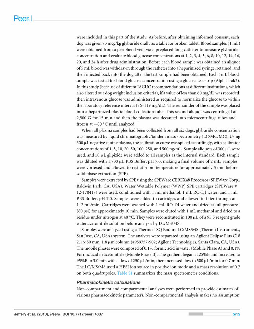
Figure 1 Relationship between blood glucose concentration and time after oral administration of 75mcg/kg glyburide at time 0; bars indicate standard error of the mean (s.e.m.).
Full-size DOI: 10.7717/peerj.4387/fig-1
6 year-old Dachshund (4.2 kg). An initially-enrolled Dobermann was withdrawn from thestudy at 3 h because a diagnosis of spinal neoplasia was made.
No adverse effects were noted in any dog receiving glyburide. Blood glucoseconcentrations of less than 50 mg/dL were not observed in any dog, although this exactvalue was reached in one dog 2 h after receiving the 75 mcg/kg oral dose of glyburide.
Overall, blood glucose concentrations appeared largely unaffected by administration ofthis dose of glyburide to dogs in this study and remained more-or-less constant throughoutthe 10-hour period of observation (Fig. 1). Blood concentrations for each dog are shownin Table S2.
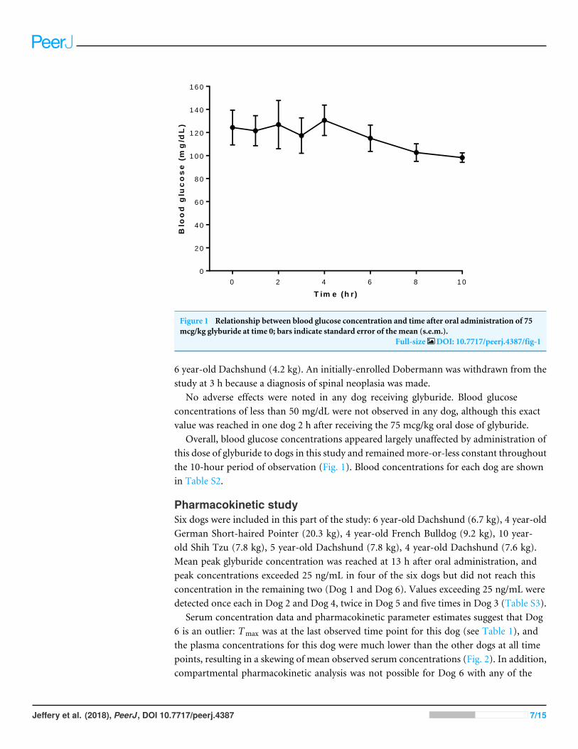
Pharmacokinetic studySix dogs were included in this part of the study: 6 year-old Dachshund (6.7 kg), 4 year-oldGerman Short-haired Pointer (20.3 kg), 4 year-old French Bulldog (9.2 kg), 10 year-old Shih Tzu (7.8 kg), 5 year-old Dachshund (7.8 kg), 4 year-old Dachshund (7.6 kg).Mean peak glyburide concentration was reached at 13 h after oral administration, andpeak concentrations exceeded 25 ng/mL in four of the six dogs but did not reach thisconcentration in the remaining two (Dog 1 and Dog 6). Values exceeding 25 ng/mL weredetected once each in Dog 2 and Dog 4, twice in Dog 5 and five times in Dog 3 (Table S3).
Serum concentration data and pharmacokinetic parameter estimates suggest that Dog6 is an outlier: Tmax was at the last observed time point for this dog (see Table 1), andthe plasma concentrations for this dog were much lower than the other dogs at all timepoints, resulting in a skewing of mean observed serum concentrations (Fig. 2). In addition,compartmental pharmacokinetic analysis was not possible for Dog 6 with any of the
Jeffery et al. (2018), PeerJ, DOI 10.7717/peerj.4387 7/15
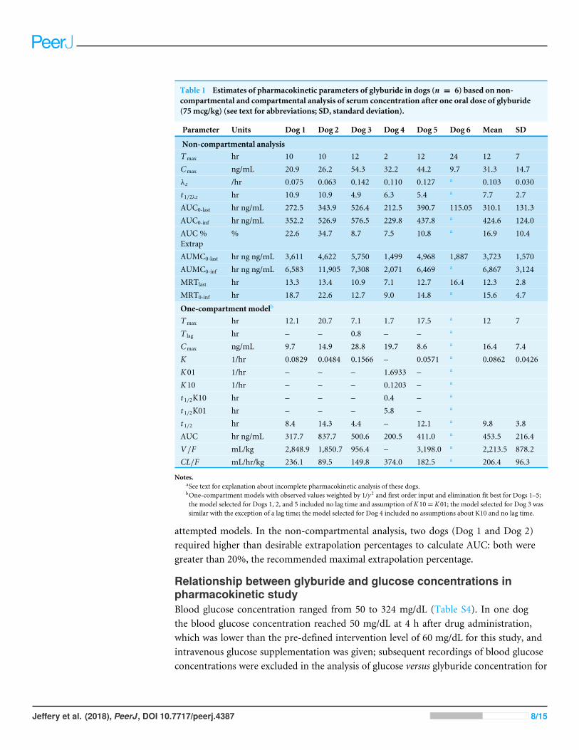
Table 1 Estimates of pharmacokinetic parameters of glyburide in dogs (n = 6) based on non-compartmental and compartmental analysis of serum concentration after one oral dose of glyburide(75 mcg/kg) (see text for abbreviations; SD, standard deviation).
Parameter Units Dog 1 Dog 2 Dog 3 Dog 4 Dog 5 Dog 6 Mean SD
Non-compartmental analysisTmax hr 10 10 12 2 12 24 12 7Cmax ng/mL 20.9 26.2 54.3 32.2 44.2 9.7 31.3 14.7λz /hr 0.075 0.063 0.142 0.110 0.127 a 0.103 0.030t 1/2λz hr 10.9 10.9 4.9 6.3 5.4 a 7.7 2.7AUC0-last hr ng/mL 272.5 343.9 526.4 212.5 390.7 115.05 310.1 131.3AUC0-inf hr ng/mL 352.2 526.9 576.5 229.8 437.8 a 424.6 124.0AUC %Extrap
% 22.6 34.7 8.7 7.5 10.8 a 16.9 10.4
AUMC0-last hr ng ng/mL 3,611 4,622 5,750 1,499 4,968 1,887 3,723 1,570AUMC0-inf hr ng ng/mL 6,583 11,905 7,308 2,071 6,469 a 6,867 3,124MRTlast hr 13.3 13.4 10.9 7.1 12.7 16.4 12.3 2.8MRT0-inf hr 18.7 22.6 12.7 9.0 14.8 a 15.6 4.7
One-compartment modelb
Tmax hr 12.1 20.7 7.1 1.7 17.5 a 12 7T lag hr – – 0.8 – – a
Cmax ng/mL 9.7 14.9 28.8 19.7 8.6 a 16.4 7.4K 1/hr 0.0829 0.0484 0.1566 – 0.0571 a 0.0862 0.0426K01 1/hr – – – 1.6933 – a
K10 1/hr – – – 0.1203 – a
t 1/2K10 hr – – – 0.4 – a
t 1/2K01 hr – – – 5.8 – a
t 1/2 hr 8.4 14.3 4.4 – 12.1 a 9.8 3.8AUC hr ng/mL 317.7 837.7 500.6 200.5 411.0 a 453.5 216.4V /F mL/kg 2,848.9 1,850.7 956.4 – 3,198.0 a 2,213.5 878.2CL/F mL/hr/kg 236.1 89.5 149.8 374.0 182.5 a 206.4 96.3
Notes.aSee text for explanation about incomplete pharmacokinetic analysis of these dogs.bOne-compartment models with observed values weighted by 1/y2 and first order input and elimination fit best for Dogs 1–5;the model selected for Dogs 1, 2, and 5 included no lag time and assumption of K10= K01; the model selected for Dog 3 wassimilar with the exception of a lag time; the model selected for Dog 4 included no assumptions about K10 and no lag time.
attempted models. In the non-compartmental analysis, two dogs (Dog 1 and Dog 2)required higher than desirable extrapolation percentages to calculate AUC: both weregreater than 20%, the recommended maximal extrapolation percentage.
Relationship between glyburide and glucose concentrations inpharmacokinetic studyBlood glucose concentration ranged from 50 to 324 mg/dL (Table S4). In one dogthe blood glucose concentration reached 50 mg/dL at 4 h after drug administration,which was lower than the pre-defined intervention level of 60 mg/dL for this study, andintravenous glucose supplementation was given; subsequent recordings of blood glucoseconcentrations were excluded in the analysis of glucose versus glyburide concentration for
Jeffery et al. (2018), PeerJ, DOI 10.7717/peerj.4387 8/15

0 2 4 6 8 1 0 1 2 1 4 1 6 1 8 2 0 2 2 2 4
0
1 0
2 0
3 0
4 0
T im e (h rs )
Pla
sm
a g
lyb
urid
e (
ng
/mL
)
Figure 2 Relationship between serum glyburide concentration and time after oral administration of75 mcg/kg glyburide at time 0; bars indicate s.e.m.Dashed horizontal line indicates desired minimumtherapeutic concentration (see text).
Full-size DOI: 10.7717/peerj.4387/fig-2
this dog. Linear regression analysis and plot (Fig. 3) of the relationship between drug andglucose concentration in our whole study population suggested there was not a significantassociation between these variables at this glyburide dosage (R2
= 0.008; P = 0.464).Superpositioning of observed concentrations in Dogs 2–5 using a loading dose of 150
mcg/kg glyburide followed by 8-hourly dosing at 75 mcg/kg produced an average plasmalevel of above 25 ng/mL, but below 50 ng/mL, for most of a simulated 72-hour treatmentperiod (Fig. 4).
DISCUSSIONWe show here that our conservatively-selected glyburide dosage of 75 mcg/kg achievedthe target blood level of between 25 and 50 ng/mL in only four of six dogs in ourpharmacokinetic study, and the periods for which this was achieved were brief. On the otherhand, this dose did not cause unsafe reductions in blood glucose concentrations in any ofthe dogs. In two dogs there was a reduction in blood glucose to <60 mg/dL but neitherof these animals exhibited any ill-effects, although they were anesthetized at the time. Itseems likely that these periods of hypoglycemia were not the result of glyburide itself sincethere appeared to be no relationship between glyburide and glucose blood concentrationsin these dogs at this dosage, suggesting that glyburide can be safely administered at this,or higher, doses, as previously reported (http://products.sanofi.ca/en/diabeta.pdf). All 11dogs recruited to this study uneventfully recovered the ability to walk after surgery.
In a clinical trial evaluating glyburide for acute spinal cord injury, because it is a rapidlyevolving condition, there is a need to achieve effective blood concentrations as rapidly
Jeffery et al. (2018), PeerJ, DOI 10.7717/peerj.4387 9/15

0 2 0 4 0 6 0
0
5 0
1 0 0
1 5 0
2 0 0
2 0 0
2 5 0
3 0 0
3 5 0
4 0 0
P la s m a g ly b u r id e (n g /m L )
Blo
od
glu
co
se
(m
g/d
L)
Figure 3 Relationship between blood glucose and serum glyburide concentrations. The line indicateslinear regression line of best fit (dotted lines indicate 95% confidence intervals) implying that these vari-ables are not significantly associated (for glyburide concentrations in this range); R2
= 0.008; P = 0.464.Full-size DOI: 10.7717/peerj.4387/fig-3
as possible and maintain these for at least 72 h (in line with other clinical uses of thisdrug; Sheth et al., 2016a; Sheth et al., 2016b). We estimate that a safe and effective doseregimen would be a loading dose of 150 mcg/kg, followed by 75 ng/kg every 8 h. Byextrapolation from the data we have collected in this study such a regimen is expectedto lead to therapeutic concentrations (>25 ng/mL; see Simard et al., 2008) 2–3 h afterdosing and for up to for 72 h. The loading dose is proposed as a means to ensure thatthe drug attains a therapeutic concentration as soon as possible after oral dosing. Theassumption in treatment of spinal cord injury is that rapid treatment is essential foreffective neuroprotection. Although intravenous preparations (see Sheth et al., 2014) maybe preferable for this reason, in veterinary medicine much of the delay between onset ofinjury and initiation of treatment is caused by delay in owner recognition of the condition,travel to a specialist clinic, triage and obtaining owner consent for treatment, whichcannot easily be eliminated. Fortunately, although the stimulus for the secondary injurymechanisms of spinal cord injury is the moment of impact, it can take hours to days for thetissue-destructive mechanisms to become fully up-regulated (Crowe et al., 1997). Indeed,Sur1, the target of glyburide, becomes increasingly widely expressed during the first 24 hafter experimental spinal cord injury (Simard et al., 2007), suggesting that, although earlieradministration is likely to be superior, attaining appropriate plasma drug concentrationswithin 2–3 h after examination at a veterinary clinic may still be efficacious.
Jeffery et al. (2018), PeerJ, DOI 10.7717/peerj.4387 10/15

0
10
20
30
40
50
60
70
0 8 16 24 32 40 48 56 64 72
Pla
sm
a g
lyb
urid
e c
on
cen
trati
on
(n
g/
mL)
Time after first dose (hrs)
Figure 4 Predicted glyburide concentrations based on superpositioning of observed concentrations inindividual dogs. Shaded area represents one standard deviation from mean.
Full-size DOI: 10.7717/peerj.4387/fig-4
An important aspect of this current study is that we used a sample of dogs similarto those that will be targeted by this therapy in a clinical trial. Although such cases willproduce more heterogeneous pharmacokinetic data than normal, conscious, laboratoryanimals, it is important to assess drug disposition and adverse effects on blood glucose thatcould possibly occur in this category of veterinary patient. Specifically, dogs undergoinganesthesia for imaging and surgery are routinely administered intravenous fluids that byexpanding the circulating volume may affect drug concentration and, in addition, mayreceive other drugs including antibiotics, analgesics and gaseous anesthetic agents, all ofwhich may alter glyburide concentration in blood or its effects on blood glucose. Ourstudy shows that despite these factors the dose of 75 mcg/kg can achieve sufficiently highconcentrations to affect function of the Sur1 channel (see Simard et al., 2008) and has nodetectable adverse effects. Our data also imply that the dose will need to be scaled-upto achieve rapid and maintained plasma concentrations adequate to achieve the desiredclinical benefit.
CONCLUSIONSThis study shows that glyburide given orally at 75 mcg/kg only just achieves bloodconcentrations of the drug appropriate for targeting function of the Sur1 channel incells in the injured spinal cord but does not appear to adversely affect blood glucoseconcentrations. To achieve rapid and appropriate concentrations of glyburide we suggest
Jeffery et al. (2018), PeerJ, DOI 10.7717/peerj.4387 11/15

using an initial loading dose of 150 mcg/kg followed by repeat dosing at 75 mcg/kgevery 8 h.
ACKNOWLEDGEMENTSWe thank the ‘Dogs Helping Dogs’ laboratory at Texas A&M University for access to theirequipment for blood sample processing.
ADDITIONAL INFORMATION AND DECLARATIONS
FundingThe authors received no funding for this work.
Competing InterestsThe authors declare there are no competing interests.
Author Contributions• Nick Jeffery, Megan Konarik, Travis Mays and Virginia Fajt conceived and designedthe experiments, performed the experiments, analyzed the data, contributedreagents/materials/analysis tools, prepared figures and/or tables, authored or revieweddrafts of the paper.• C. Elizabeth Boudreau conceived and designed the experiments, authored or revieweddrafts of the paper.
Animal EthicsThe following information was supplied relating to ethical approvals (i.e., approving bodyand any reference numbers):
Dogs included in this study presented for treatment of acute spinal cord injury to thesmall animal hospital of the Colleges of Veterinary Medicine at Iowa State University orTexas A&M University, and study protocols were approved by the relevant InstitutionalAnimal Care and Use Committees (Iowa State, log number: 1-16-8148-K; Texas A&M,IACUC 2016-0324 CA, reference number 044949).
Data AvailabilityThe following information was supplied regarding data availability:
All raw data is supplied as Tables in the Supplementary Information.
Supplemental InformationSupplemental information for this article can be found online at http://dx.doi.org/10.7717/peerj.4387#supplemental-information.
REFERENCESAguilar-Bryan L, Nelson DA, Vu QA, HumphreyMB, Boyd 3rd AE. 1990. Photoaffinity
labeling and partial purification of the beta cell sulfonylurea receptor using a novel,biologically active glyburide analog. Journal of Biological Chemistry 265:8218–8224.
Jeffery et al. (2018), PeerJ, DOI 10.7717/peerj.4387 12/15

Cohen TA, Nelson RW, Kass PH, Christopher MM, Feldman EC. 2009. Evaluationof six portable blood glucose meters for measuring blood glucose concentrationin dogs. Journal of the American Veterinary Medical Association 235:276–280DOI 10.2460/javma.235.3.276.
CroweMJ, Bresnahan JC, Shuman SL, Masters JN, Beattie MS. 1997. Apoptosis anddelayed degeneration after spinal cord injury in rats and monkeys. Nature Medicine3:73–76 DOI 10.1038/nm0197-73.
Cruz-Antonio L, Arauz J, Franco-Bourland RE, Guízar-Sahagún G, Castañeda-Hernández G. 2012. Contrasting effects of cord injury on intravenous and oralpharmacokinetics of diclofenac: a drug with intermediate hepatic extraction. SpinalCord 50:632–635 DOI 10.1038/sc.2012.20.
Cruz-Antonio L, Flores-Murrieta FJ, García-Löpez P, Guízar-Sahagún G, Castañeda-Hernández G. 2006. Understanding drug disposition alterations induced byacute spinal cord injury: role of injury level and route of administration for agentssubmitted to extensive liver metabolism. Journal of Neurotrauma 23:75–85DOI 10.1089/neu.2006.23.75.
Gerzanich V,Woo SK, Vennekens R, Tsymbalyuk O, Ivanova S, Ivanov A, Geng Z,Chen Z, Nilius B, Flockerzi V, Freichel M, Simard JM. 2009. De novo expressionof Trpm4 initiates secondary hemorrhage in spinal cord injury. Nature Medicine15:185–191 DOI 10.1038/nm.1899.
Guan J, Han J, Zhang D, Chu C, Liu H, Sun J, He Z, Zhang T. 2014. Increased dissolu-tion rate and oral bioavailability of hydrophobic drug glyburide tablets produced us-ing supercritical CO2 silica dispersion technology. European Journal of Pharmaceuticsand Biopharmaceutics 86:376–382 DOI 10.1016/j.ejpb.2013.10.008.
Hosier H, Peterson D, Tsymbalyuk O, Keledjian K, Smith BR, Ivanova S, GerzanichV, Popovich PG, Simard JM. 2015. A direct comparison of three clinically relevanttreatments in a rat model of cervical spinal cord injury. Journal of Neurotrauma32:1633–1644 DOI 10.1089/neu.2015.3892.
Kwon BK, Streijger F, Hill CE, Anderson AJ, BaconM, Beattie MS, Blesch A, BradburyEJ, Brown A, Bresnahan JC, Case CC, Colburn RW, David S, Fawcett JW, FergusonAR, Fischer I, Floyd CL, Gensel JC, Houle JD, Jakeman LB, Jeffery ND, JonesLA, Kleitman N, Kocsis J, Lu P, Magnuson DS, Marsala M, Moore SW,MotheAJ, OudegaM, Plant GW, Rabchevsky AS, Schwab JM, Silver J, Steward O, XuXM, Guest JD, Tetzlaff W. 2015. Large animal and primate models of spinal cordinjury for the testing of novel therapies. Experimental Neurology 269:154–168DOI 10.1016/j.expneurol.2015.04.008.
Kwon BK, Okon E, Hillyer J, Mann C, Baptiste D,Weaver LC, Fehlings MG, TezlaffW. 2011. A systematic review of non-invasive pharmacologic neuroprotectivetreatments for acute spinal cord injury. Journal of Neurotrauma 28:1545–1588DOI 10.1089/neu.2009.1149.
Liu H, Shang K, LiuW, Leng D, Li R, Kong Y, Zhang T. 2014. Improved oral bioavail-ability of glyburide by a self-nanoemulsifying drug delivery system. Journal ofMicroencapsulation 31:277–283 DOI 10.3109/02652048.2013.843598.
Jeffery et al. (2018), PeerJ, DOI 10.7717/peerj.4387 13/15

Moore SA, Granger N, Olby NJ, Spitzbarth I, Jeffery ND, Tipold A, Nout-Lomas YS,Da Costa RC, Stein VM, Noble-Haeusslein LJ, Blight AR, Grossman RG, BassoDM, Levine JM. 2017. Targeting translational successes through CANSORT-SCI:using pet dogs to identify effective treatments for spinal cord injury. Journal ofNeurotrauma 34:2007–2018 DOI 10.1089/neu.2016.4745.
Panten U, Schwanstecher M, Schwanstecher C. 1996. Sulfonylurea receptors andmechanism of sulfonylurea action. Experimental and Clinical Endocrinology andDiabetes 104:1–9 DOI 10.1055/s-0029-1211414.
Popovich PG, Lemeshow S, Gensel JC, Tovar CA. 2012. Independent evaluationof the effects of glibenclamide on reducing progressive hemorrhagic necro-sis after cervical spinal cord injury. Experimental Neurology 233:615–622DOI 10.1016/j.expneurol.2010.11.016.
Rendell M. 2004. The role of sulphonylureas in the management of type 2 diabetesmellitus. Drugs 64:1339–1358 DOI 10.2165/00003495-200464120-00006.
Salegio EA, Bresnahan JC, Sparrey CJ, CamisaW, Fischer J, Leasure J, Buckley J, Nout-Lomas YS, Rosenzweig ES, Moseanko R, Strand S, Hawbecker S, LemoyMJ, HaefeliJ, Ma X, Nielson JL, Edgerton VR, Ferguson AR, Tuszynski MH, Beattie MS. 2016.A unilateral cervical spinal cord contusion injury model in non-human primates(Macaca mulatta). Journal of Neurotrauma 33:439–459 DOI 10.1089/neu.2015.3956.
Sheth KN, Elm JJ, Beslow LA, Sze GK, KimberlyWT. 2016a. Glyburide advantage inmalignant edema and stroke (GAMES-RP) trial: rationale and design. NeurocriticalCare 24:132–139 DOI 10.1007/s12028-015-0189-7.
Sheth KN, Elm JJ, Molyneaux BJ, Hinson H, Beslow LA, Sze GK, Ostwaldt AC, DelZoppo GJ, Simard JM, Jacobson S, KimberlyWT. 2016b. Safety and efficacy ofintravenous glyburide on brain swelling after large hemispheric infarction (GAMES-RP): a randomised, double-blind, placebo-controlled phase 2 trial. Lancet Neurology15:1160–1169 DOI 10.1016/S1474-4422(16)30196-X.
Sheth KN, KimberlyWT, Elm JJ, Kent TA, Mandava P, Yoo AJ, Thomalla G, CampbellB, Donnan GA, Davis SM, Albers GW, Jacobson S, Simard JM, Stern BJ. 2014.Pilot study of intravenous glyburide in patients with a large ischemic stroke. Stroke45:281–283 DOI 10.1161/STROKEAHA.113.003352.
Simard JM, ChenM, Tarasov KV, Bhatta S, Ivanova S, Melnitchenko L, TsymbalyukN,West GA, Gerzanich V. 2006. Newly expressed SUR1-regulated NC(Ca-ATP)channel mediates cerebral edema after ischemic stroke. Nature Medicine 12:433–440DOI 10.1038/nm1390.
Simard JM, Kahle KT, Gerzanich V. 2010.Molecular mechanisms of microvascular fail-ure in central nervous system injury-synergistic roles of NKCC1 and SUR1/TRPM4.Journal of Neurosurgery 113:622–629 DOI 10.3171/2009.11.JNS081052.
Simard JM, KilbourneM, Tsymbalyuk O, Tosun C, Caridi J, Ivanova S, Keledjian K,Bochicchio G, Gerzanich V. 2009. Key role of sulfonylurea receptor 1 in progressivesecondary hemorrhage after brain contusion. Journal of Neurotrauma 26:2257–2267DOI 10.1089/neu.2009.1021.
Jeffery et al. (2018), PeerJ, DOI 10.7717/peerj.4387 14/15

Simard JM, Tsymbalyuk O, Ivanov A, Ivanova S, Bhatta S, Geng Z,Woo SK, GerzanichV. 2007. Endothelial sulfonylurea receptor 1-regulated NC(Ca-ATP) channelsmediate progressive hemorrhagic necrosis following spinal cord injury. Journal ofClinical Investigation 117:2105–2113 DOI 10.1172/JCI32041.
Simard JM, Tsymbalyuk O, Keledjian K, Ivanov A, Ivanova S, Gerzanich V. 2012.Comparative effects of glibenclamide and riluzole in a rat model of severe cervicalspinal cord injury. Experimental Neurology 233:566–574DOI 10.1016/j.expneurol.2011.11.044.
Simard JM,Woo SK, Bhatta S, Gerzanich V. 2008. Drugs acting on SUR1 totreat CNS ischemia and trauma. Current Opinion in Pharmacology 8:42–49DOI 10.1016/j.coph.2007.10.004.
Streijger F, So K, Manouchehri N, Tigchelaar S, Lee JHT, Okon EB, Shortt K, Kim SE,McInnes K, Cripton P, Kwon BK. 2017. Changes in pressure, hemodynamics, andmetabolism within the spinal cord during the first 7 days after injury using a porcinemodel. Journal of Neurotrauma 34:3336–3350 DOI 10.1089/neu.2017.5034.
Tetzlaff W, Okon EB, Karimi-Abdolrezaee S, Hill CE, Sparling JS, Plemel JR, PlunetWT, Tsai EC, Baptiste D, Smithson LJ, Kawaja MD, Fehlings MG, Kwon BK. 2011.A systematic review of cellular transplantation therapies for spinal cord injury.Journal of Neurotrauma 28:1611–1682 DOI 10.1089/neu.2009.1177.
TorjmanMC, Joseph JI, Munsick C, Morishita M, Grunwald Z. 2005. Effects ofisoflurane on gastrointestinal motility after brief exposure in rats. InternationalJournal of Pharmacology 294:65–71 DOI 10.1016/j.ijpharm.2004.12.028.
Jeffery et al. (2018), PeerJ, DOI 10.7717/peerj.4387 15/15