pharm ch 21
DESCRIPTION
PharmacologyTRANSCRIPT
Pharm Ch. 21: Pharm of Vascular Tone
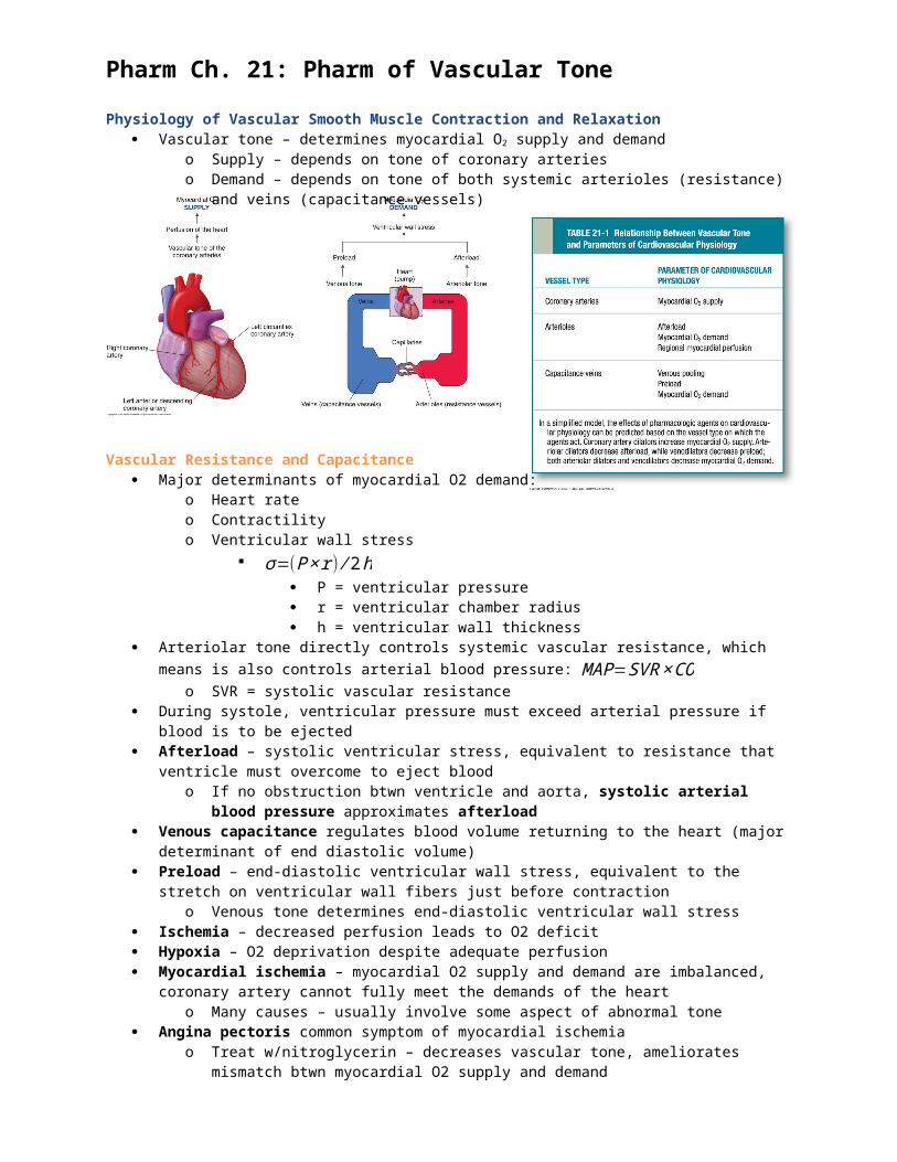
Physiology of Vascular Smooth Muscle Contraction and Relaxation Vascular tone – determines myocardial O2 supply and demand
o Supply – depends on tone of coronary arterieso Demand – depends on tone of both systemic arterioles (resistance) and veins (capacitance
vessels)
Vascular Resistance and Capacitance Major determinants of myocardial O2 demand:
o Heart rateo Contractilityo Ventricular wall stress
σ=(P×r) ⁄ 2h P = ventricular pressure r = ventricular chamber radius h = ventricular wall thickness
Arteriolar tone directly controls systemic vascular resistance, which means is also controls arterial blood pressure: MAP=SVR×CO
o SVR = systolic vascular resistance During systole, ventricular pressure must exceed arterial pressure if blood is to be ejected Afterload – systolic ventricular stress, equivalent to resistance that ventricle must overcome to eject
bloodo If no obstruction btwn ventricle and aorta, systolic arterial blood pressure approximates
afterload Venous capacitance regulates blood volume returning to the heart (major determinant of end diastolic
volume) Preload – end-diastolic ventricular wall stress, equivalent to the stretch on ventricular wall fibers just
before contraction o Venous tone determines end-diastolic ventricular wall stress
Ischemia – decreased perfusion leads to O2 deficit Hypoxia – O2 deprivation despite adequate perfusion Myocardial ischemia – myocardial O2 supply and demand are imbalanced, coronary artery cannot fully
meet the demands of the heart o Many causes – usually involve some aspect of abnormal tone
Angina pectoris common symptom of myocardial ischemia o Treat w/nitroglycerin – decreases vascular tone, ameliorates mismatch btwn myocardial O2
supply and demand Vascular Smooth Muscle Contraction and Relaxation
Regulators of vascular tone act by influencing the actin-myosin contractile apparatus of vascular smooth muscle cells
Actin-myosin contractile app regulated by intracellular [Ca2+] Stimulation of vascular smooth muscle cells increases Ca2+ by 2 mechanisms
o Ca2+ can enter cell via voltage-gated Ca2+ selective channels in sarcolemma o Intracellular Ca2+ released from sarcoplasmic reticulum

Vasoconstriction initiated by voltage-gated L-type Ca2+ channels in sarcolemma during plasma membrane depolarization
Regulation of Vascular ToneVascular EndotheliumNitric Oxide
Endothelin-derived growth factor released from endothelial cells is NO Things that promote endothelial cell-synthesis and release of NO: ACh, shear stress, histamine,
bradykinin, sphingosine, 1-phosphate, serotonin, substance P, ATP Endothelial isoform of nitric oxide synthase is responsible for endothelial-cell NO synthesis (vascular
tone, platelet aggregation) NO affects vasodilation by activating guanylyl cyclase AND Ca2+-dependent K+ channels in vascular smooth
muscle cells o K+ channels activated via guanylyl cyclase independent mechanism hyperpolarization
vasodilation Endothelin
21-amino acid vasoconstrictor peptide Most potent endogenous vasoconstrictor “Functional mirror image” of NO In addition to vasoconstriction, also inotropic (strength of
contractions) and chronotropic (heart rate) actions on heart
Also plays role in lungs, kidney, brain 3 isoforms – ET-1, ET-2, ET-3
o ET-1 – cardiovascular contractions, [ET-1] in vascular wall 100x > circulation (ET-1 secreted primarily on basal side of endothelial cell)
Endothelin precursors proteolytically processed:o Preproendothelin cleaved big endothelin
cleaved by endothelin-converting enzyme endothelin
o 2 endothelial receptor subtypes: both are G-protein coupled receptors, effect phospholipase C modulated pathways
ETA - binds ET-1 on vascular smooth muscle cells (mediate vasoconstriction)
ETB – binds ET-1 on endothelial (vasodilation via release of prostacyclin and NO) and vascular smooth muscle receptor cells (mediate vasoconstriction)

Autonomic Nervous System Activation of α1-adrenergic receptor on vascular smooth muscle vasoconstriction Activation of β2-adrenergic receptors on vascular smooth muscle vasodilation Effect of norepinephrine at α1-adrenergic receptors greater than effect at β2-adrenergic receptors,
especially in organs that receive decreased blood flow during “fight or flight” (aka skin and viscera)o Net effect of norepinephrine at these vascular beds vasoconstrictive
Blood vessels are not innervated by parasympathetics Neurohormonal Mechanisms
Act on vascular smooth muscle cells, endothelial cells, neurons to regulate vascular toneo Circulating catecholamines – α1 and β2 adrenergic receptor o Angiotensin II – stimulates AT1 to vasoconstrict arterioles and increase intravascular volumeo Aldosterone – mineralcorticoid receptor, increases intravascular volumeo Natriuretic peptides – promote renal natriuresis, vasodilate via guanylyl cyclase receptors o ADH – stimulates arteriolar V1 receptors to constrict arterioles and activates V2receptors to
increase intravascular volume Local Mechanisms
Autoregulation – homeostatic mechanism where vascular smooth muscle cells respond to increases/decreases in perfusion pressure by vasoconstriction/vasodilation, goal to preserve blood at constant level
Vascular tone also governed by metabolites – H+, CO2, O2, adenosine, lactate, K+ - produced in surrounding tissue
Local mechanisms of vascular tone regulation predominate in vascular beds of essential organs and can be adjusted quickly to local metabolic demands
Pharmacologic Classes and Agents
Organic Nitrates, Inhaled NO, and Sodium NitroprussideMOA:
Organic nitrates are reduced to release NO, can react directly with guanylyl cyclase
NO is an endogenous signaling molecule that can cause vascular smooth muscle relaxation
NO can dilate both arteries and veins, venous dilation predominates at therapeutic doses
o NO dilation increases capacitance decrease in venous return to right heart decreased right ventricular and left ventricular end-diastolic pressure and volume

o Decreased in preload reduces myocardial demand of O2
W/o reflex tachycardia, arterial vasodilation decrease in systemic vascular resistance decrease in systolic wall stress (afterload) decrease myocardial O2 demand
Coronary circulation – NTG dilates predominantly the large epicardial arteries
o This preferential dilation prevents coronary steal phenomenon (dipyramidole) that produces intense dilation of the coronary resistance vessels
Stiffened, calcific atherosclerotic coronary arteries may remain non compliant, even in the face of coronary artery vasodilators
Administration of nitrates in doses sufficient to vasodilate the large epicardial arteries can be dangerous, could induce excessive peripheral arteriolar vasodilation and refractory hypotension
o Excessive decreases in MAP dizziness, lightheadedness, overt syncope, myocardial ischemia Coronary perfusion pressure depends pressure gradient btwn aorta and endocardium during diastole,
marked decrease in diastolic aortic pressure insufficient O2supply to the heart Systemic hypotension can lead to reflex tachycardia, which also decreases myocardial O2supply by
shortening diastole and thus myocardial perfusion timeo Reflex tachycardia – typically happens when baroreceptors in aortic arch and carotid sinuses
sense a decrease in BP Reflex tachycardia is rare in pts with overt HF! So, nitrates can be used to decrease pulmonary congestion
in pts with HF (by effecting venodilation and decreasing end-diastolic pressure) w/o eliciting significant reflex tachycardia
Most commonly used organic nitrates: NTG, isosorbide dinitrate, isosorbide 5-mononitrate Inhaled nitric oxide gas
o Used to selectively dilate pulmonary vasculatureo NO is rapidly inactivated by binding to Hb in the blood, NO gas has very little effect on systemic
BPo Used to treat primary pulmonary hypertension in the newborn
Sodium Nitroprussideo Effects vasodilation by release of NOo Appears to liberate NO through a nonenzymatic process
not targeted to specific types of vessels drug dilates both arteries and veins
o Used in intravenously for powerful hemodynamic control in hypertensive emergencies and severe HF
o Rapid onset, short duration of action must be infused w/continuous BP monitoring and careful titration of drug dose to drug effect
o Spontaneously decomposes to liberate NO and cyanideo Excessive cyanide accumulation acid-base
disturbances, cardiac arrhythmias, death o Thiocyanate toxicity can also occur in pts with impaired
renal function disorientation, psychosis, muscle spasms, seizures
Pharmacokinetics Pharmacokinetics of the different nitrate preparations and formulations is the basis for preferential use of
specific agents and doses in certain setting o Ex: rapid onset of sublingual nitrate preps good for rapid relief of acute angina attackso Ex: longer-acting nitrates more valuable for angina prophylaxis in long-term management of
coronary artery disease Orally administered NTG and isosorbide dinitrate have low bioavailability bc organic nitrate reductases in
the liver rapidly metabolize these drugs. To circumvent first-pass effect and to attain therapeutic blood levels within minutes, NTG or isosorbide dinitrate administered sublingually

Intravenous NTG indicated when continuous titration of drug is necessary (ex: treatment of unstable angina or acute HF)
NTG has a short half life (5 minutes), then denitrated into biologically active glyceryl dinitrate metabolites that have longer half-lives
o Equivalent doses of isosorbide dinitrate can be more effective than NTG (isosorbide dinitrate has a longer half life)
o Partially denitrated metabolites (isosorbide 5-mononitrate) have even longer half lives (4 hrs) Isosorbide 5-mononitrate – prolonged therapeutic effects, well absorbed from GI, not susceptible to first
pass metabolism in the liver, 100% bioavailability, significantly more effective than equivalent amounts of isosorbide dinitrate
Pharmacologic Tolerance Nitrates can be offset by compensatory sympathetic NS responses (ex: increases in reflex vascular tone)
and compensatory renal responses (ex: increased salt and water excretion); physiologic tolerance Pharmacologic tolerance – limits efficacy of vasodilators, can limit tolerance by including daily “nitrate-
free intervals” – most effective way! 2 hypotheses to pharm tolerance:
o Classic (sulfydryl) hypothesis – tolerance results from intracellular depletion of sulfhydryl-containing groups (glutathione and/or other forms of cysteine) involved in the formation of NO from organic nitrates; tolerance could be reversed by administration of thiol-containing compounds like N-acetylcysteine
o Free-radical (superoxide) hypothesis – cellular rolerance results from the formation of peroxynitrite, highly reactive metabolite of NO that inhibits guanylyl cyclase; tolerance could be attenuated/reversed by agents that inhibit free-radical formation
Effects of Nitrates in Addition to Vasodilation Relaxation of other types of smooth muscle – esophageal, bronchial, biliary, intestinal, GU Ability of NTG to relieve the angina-like chest pain of esophageal spasm can occasionally result in
misdiagnosis of CAD NO also acts as an antiplatelet agents via increases in platelet cGMP (inhibits platelet aggregation) Antiplatelet aggregating effect and vasodilator effect decreases likelihood of coronary artery
thrombosis Nitrate-induced platelet inhibition – rest angina/unstable angina
Contraindications Hypotension Elevated intracranial pressure – NO mediated vasodilation of cerebral arteries could further elevate
intracranial pressure Don’t use in pts with angina pain associated with hypertrophic obstructive cardiomyopathy bc obstruction
could be worsened by nitrate-mediated preload reduction Use w/caution in pts with diastolic HF who depend on elevated ventricular preload for optimal cardiac
output Don’t use in pts taking sildenafil or other PDE type V inhibitors!!!
Phosphodiesterase Inhibitors Prevent hydrolysis of cyclic nucleotides (cAMP, cGMP) to their monophosphate forms Sildenafil – prototype PDE inhibitor that is highly selective for cGMP Phosphodiesterase type V (PDE5),
expressed mainly in the smooth muscle cells of the corpus cavernosum but also in the retina and vascular smooth muscle cells
Tadalafilb (“tada!!”) – longer time to onset of action and more prolonged half life than the other PDE5 inhibitors
Inhibition of of the cGMP PDE in corpus cavernosum smooth muscle potentiates the effects of endogenous NO-cGMP signaling erection
High doses of sildenafil efficacious in treatment of pulmonary HTN Adverse effects – from drug-induced vasodilation in the systemic vasculature; headache and flushing Presence of excess NO inhibition of cGMP degradation can markedly amplify the vasodilatory effect,
excesive vasodilation can lead to severe refractory hypotension All three PDE5 inhibitors are contraindicated in pts taking oral nitrate vasodilators Other adverse effects: transient/permanent vision loss (nonarteritic ischemic optic neuropathy) Patients taking vasodilatory antihypertensive medications together with a PDE5 inhibitor should be
regarded as at risk for potentially dangerous hypotension Ca2+ Channel Blockers
Ca2+ channel blockers act on both vascular smooth muscle and the myocardium
Ca2+ channel blockers are predominantly arteriolar dilators (organic nitrates – mainly venodilators)
Commonly used in HTN, cardiac arrhythmias, forms of angina MOA
Ca2+ flux through L-type channel – important determinant of vascular tone and cardiac contractility
Ca2_ channel blockers in current use ALL act by inhibiting Ca2+ entry through L-type channels
Smooth muscle cells – decreased Ca2+ entry keep intracellular [Ca2+] low, reducing Ca2+-CaM-mediated activation of myosin light chain kinase, and smooth muscle cell contraction
Ca2+ channel blockers appear to have the greatest effect on vascular smooth muscle
o Arteriolar smooth muscle is more responsive than venous smooth muscle
Vasodilation of resistance arterioles reduces systemic vascular resistance and lowers arterial blood pressure, thereby decreasing ventricular systolic wall stress and myocardial O2 demand
Drug-induced dilation of coronary arteries may also increase O2 supply In cardiac myocytes, reduced Ca2+ influx through L-type channels decreases in myocardial contractility,
SA-node pacemaker rate, and AV-node conduction velocity Skeletal muscle is NOT significantly effected by Ca2+ channel blockers because Ca2+ used in skeletal
muscle contraction released from sarcoplasmic reticulum
Chemical Classes 3 – dihydropyridines, benzothiazapines, phenylalkylamines; all 3 classes block L-type channel! Differences due to different binding sites on Ca2+ channel
o Nifedipine – binds N siteo Diltiazem – binds D siteo Verapamil – binds v siteo D and V sites overlap, N site is in a different regiono Nifedipine and diltiazem bind synergistically, nifedipine and verapamil reciprocally inhibit each
other’s binding Nifedipine and amlodipine – representative members of dihydropyridine calss; cause significantly more
arterial vasodilation, little effect on cardiac tissue Compared to diltiazem and verapamil, they dihydropyridines cause less depression of myocardial
contractility and AV-node conduction velocity Amlodipine (3rd generation dihydropyridine) differs from nifedipine (1st generation) – amlodipine has a
pKaof 8.7m predominantly (+) at physiologic pH, positive charge allows it to bind to cell membranes (negatively charged) with high affinity and contributes to drug’s late peak plasma concentration
Clevidipine – recently approved, available only in intravenous form, administered as a continuous infusion for management of hypertensive urgency and emergency
compared to dihydropiridines, nondihydropiridine Ca2+ blockers diltiazem and verapamil have a lower ratio of vascular-to-cardiac selectivity

in the heart, both diltiazem and verapamil act as negative inotropes, verapamil has a greater suppressive effect on cardiac contractility than diltiazem
Diltiazem and verapamil also slow rate of Ca2+ channel recovery so cardiac conduction is significantly decreased by these drugs
Pharmacokinetics Oral dosage forms typically Nifedipine and verapamil are excreted by the kidney Diltiazem excreted by the liver Bioavailability of oral formulations nifedipine, diltiazem, and verapamil is lowered by significant first-pass
metabolism in the gut and liver Oral nifedipine – rapid onset of action (<20min) and can cause brisk, precipitous fall in BP, causing drug
induced hypotension which activates severe reflexive tachycardia which can worsen the myocardial ischemia by increasing the O2 demand and decreasing the O2 supply; short half life – administer frequently, ~ every 4 hrs
Amlodipine – higher bioavailability permits it to be effective at lower doses, late peak plasma concentration causes significantly less reflexive tachycardia, slow hepatic degradation long plasma half life, which enables once daily dosing
Toxicities and Contraindications Toxicities are mainly mechanism-based: flushing, constipation from excessive smooth muscle relaxation In excess, negative inotropic effects of dilitiazem and verapamil can lead to bradycardia, atrioventricular
block, and HF Patients taking β-blockers are advised not to use dilitiazem or verapamil concomitantly bc of increased
likelihood of excessive cardiac depression Some studies have shown Ca2+ channel blockers increase mortality in pts with HF, so Ca2+ channel
blockers are contraindicated in the management of HF Some studies show short-acting nifedipine assicoated with increased risk of myocardial ischemia and
infaction by virtue of this drug’s tendency to disturb myocardial O2 supply:demand balanceK+ Channel Openers
Cause direct arterial vasodilation by opening ATP-modulated K+ channels in the plasma membrane of vascular smooth muscle cells
Mechanism is entirely different from other vasodilators, so these drugs can be used to treat hypertension refractory to other antihypertensive drugs
Opening K+ channels hyperpolarizes the membrane, is enough K+ channels are open at the same time, normal excitatory stimuli are not able to depolarize the membrane. W/o depolarization, Ca2+ channels do not open, and Ca2+ influx and smooth muscle contraction are inhibited
K+ ATP channel opener drugs: minoxidil, cromakain, pinacidil, nicorandil – act primarily on arterial smooth muscle cells, and therefore decrease arterial blood pressure
Adverse effects: headache and flushing When arterial vasodilators (Ca2+ channel blockers and K+ ATP channel openers) are used as
monotherapy, decrease in arterial pressure often elicits reflex sympathetic discharge tachycardia increased cardiac work; use of β-blockers in combination with arterial vasodilators helps block effects of reflex sympathetic activity and preserves therapeutic utility of the arterial vasodilators
Endothelin Receptor Antagonists Bosentan – competitive antagonist for ETA and ETB receptors, used in treatment of pulmonary
hypertension, pts taking this drug have significanlt yimporved 6 minute walk test Major adverse effect: elevation in serum transaminase levels, monitor liver funciton monthly Ambrisentan – endothelin receptor antagonist with specificity for ETA, pts taking this drug have
significantly improved 6 minute walk test, may have less hepatotoxicity than bosentan Other Drugs that Modulate Vascular ToneHydralazine
Orally administered arteriolar vasodilator sometimes use in treatment of HTN and in combination with isosorbide dinitrate in treatment of HF
MOA unclear Appears to prevent development of nitrate tolerance Combination pill of hydralazine and isosorbide nitrate found to decrease M&M in black Americans with
advanced HF May be a treatment option for pts with HTN and HF who have contraindications to other vasodilators
(ACE inhibitors)

Low bioavailability bc of first-pass metabolism in the liver, rate of metabolism depends on whether pt is a slow/fast acetylator
Slow acetylators – slower rate of hepatic degradation, higher bioavailability and higher plasma concentrations
Rare adverse effect: reversible lupus erythematosus-like syndrome, slow acetylators α1-Adrenergic Antagonists
Epinephrine and norepinephrine stimulate α1-adrenergic receptors on vascular smooth muscle vasoconstriction
α1-adrenergic receptor is a G protein coupled receptor, activates phospholipase C to generate inositol triphosphate and diacylglycerol
α1-adrenergic receptor antagonists, prazosin, block α1-adrenergic receptors in arterioles and venules vasodilation; effect greater in arterioles
α1-adrenergic antagonists cause significant reduction in arteriol pressure, useful in treatment of HTN initiation of therapy with α1-adrenergic antagonists may cause orthostatic hypotension; like other arterial
vasodilators, α1-adrenergic antagonists can also cause retention of salt and water β-adrenergic blockers and diuretics may be used together with α1-adrenergic antagonists to mitigate
compensatory responses some α1-adrenergic antagonists, terazosin, used principally to inhibit contraction of nonvascular smooth
muscle (e.g. prostatic smooth muscle) β-Adrenergic Antagonists
Activation of β2-adrenergic receptors on vascular smooth muscle cells vasodilation Increased intracellular cAMP induced by β2 receptor stimulation may cause smooth muscle relaxation by
accelerating inactivation of myosin light chain kinase and by increasing the extrusion of Ca2+ from the cell Activation of β2-adrenergic receptors on endothelial cells vasodilation through activation of endothelial
NO synthase Β-adrenergic antagonists are of major clinical importance in treating HTN, angina, cardiac arrhythmias
o At cardiac β1-adrenergic receptors, β-adrenergic antagonists have negative inotropic and chronotropic affects on the heart, these actions reduce CO
Antagonism of β2-adrenergic receptors on vascular smooth muscle cells can lead to unopposed vasoconstriction mediated by α1-adrenergic receptors, and to an increase in systemic vascular resistance
Net effect is a decrease in blood pressure, despite initial increases in systemic vascular resistance with β-adrenergic antagonists; hypotensive effect from negative inotropic effect (leads to decrease in CO), inhibition of renin secretion, and CNS effects of the β-blockers
Renin-Angiotensin System Blockers Inhibition of the renin-angiotensin system vasorelaxation Hypotensive effect of ACE inhibitors may be cause in part by decreased catabolism of bradykinin
(vasorelaxant) released in response to inflammatory stimuli Antagonists at the AT1 receptor, selectively inhibits angiotensin II-mediated vasoconstriction at target
organ, have a more direct effect ACE inhibitors at AT1 receptor antagonists considered “balanced” vasodilators bc they affect both arterial
and venous tone