pg degree synopsis format part a general information
TRANSCRIPT

PG DEGREE SYNOPSIS FORMAT
PART A – GENERAL
INFORMATION
1 Title of the Dissertation COMPARISON OF SHEAR BOND
STRENGTH AND ADHESIVE
REMNANT INDEX OF METAL AND
CERAMIC BRACKETS USING
THREE DIFFERENT ORTHODONTIC
ADHESIVES- AN IN-VITRO STUDY
2 Name of the Candidate with mobile number and email ID: DR. PRIYA.B
Phone no : + 91 8220289849
Email :
3 Name of the Institute : SRI DHARMASTHALA
MANJUNATHESHWARA DENTAL
COLLEGE AND HOSPITALS,
SATTUR,DHARWAD-580009,
KARNATAKA, INDIA.
4 University Registration Number: 20DPG020
5 Name of the programme studying: MASTER OF DENTAL SURGERY
(MDS)-ORTHODONTICS AND
DENTOFACIAL ORTHOPAEDICS.
6 University Program Code:

7 Year of Admission: 2020
8 Month and year of appearing for final examination 2023
9 Month and year of submitting Dissertation DECEMBER 2022
10 Name (s), Designation (s) & Addresses of the guide and co-
guide (s) with mobile numbers and email ID
DR. ROOPAK D NAIK ,
MDS-ORTHODONTICS,
ADDITIONAL PROFESSOR,
DEPARTMENT OF ORTHODONTICS
AND DENTOFACIAL
ORTHOPAEDICS,
SDM COLLEGE OF DENTAL
SCIENCE,DHARWAD.
MOBILE NUMBER: +91 9449028869
EMAIL: [email protected]
11 A. State whether the study is intradepartmental or
interdepartmental:
INTRADEPARTMENTAL STUDY
B. If the study is interdepartmental:
i. Mention the names of collaborating departments
ii. Mention whether consent has been obtained from
them (Copy to be Attached)
NA
12 Total funds required for the study (in rupees): 20,000 – 25,000 /-
13 Source of funding SELF FUNDING
PART B – TECHNICAL DETAILS
1 Title of the dissertation COMPARISON OF SHEAR BOND STRENGTH
AND ADHESIVE REMNANT INDEX OF
METAL AND CERAMIC BRACKETS USING
THREE DIFFERENT ORTHODONTIC

ADHESIVES- AN IN-VITRO STUDY
2 Introduction Since the advent of the acid-etch technique by
Buonocore and the bonding of orthodontic brackets
by Neumann, various bonding adhesives were
developed. The first and most popular bonding resins
were chemical curing bonding systems. A major
drawback of the self-cure adhesive systems is the
inability to manipulate the setting time of the
composite resin.1-2
Newer self-etching adhesive materials have been
introduced recently in orthodontics to simplify the
bonding process by reducing the bonding steps and
eliminating the need for etching and priming, thus
lessening the risk of contamination and reducing the
bonding time. These self-etching primers combine
the conditioning and priming agents into one acidic
solution and have shown advantages such as reduced
loss of enamel, prevention of saliva contamination
and less chair time.3
Shear bond strength (SBS) is the main factor, which
has to be concerned in the evolution of bonding
materials. The bond strength of the orthodontic
bracket must be able to withstand the forces applied
during the orthodontic treatment. Reynolds et al in
his study stated that 5.9–7.8 MPa resistances are

sufficient to withstand masticatory forces. Bishara et
al. compared bond strengths of an acidic primer and
composite resin with a conventional adhesive system
and found mean bond strengths of 10.4 and 11.8
Mpa. The SBS of self-etching primers can vary
widely, ranging from 2.8 to 16.6 MPa.4
An ideal orthodontic adhesive should have adequate
bond strength while maintaining unblemished
enamel after debonding. Therefore, researchers have
been working hard to achieve the best quality and
gentlest procedures for bonding orthodontic
brackets. A gentler etch pattern has been obtained
with self-etching primers, and scanning electron
microscope (SEM) studies have shown that these
conditioners yield shorter resin tags.5
Over the years, ARI scores have been one of the
most frequently evaluated aspects in studies on
orthodontic adhesives. Because the adhesive remnant
score system is qualitative and subjective, many
attempts have been made to modify the original
system, or to develop new quantitative methods that
can be used to more accurately assess the adhesive
remnant. Many Studies have debated whether the
differences in ARI scores reflect a difference in bond
strength between the enamel and the adhesive for the

different adhesive systems.
As of now , the present available literature lacks
information regarding these aspects. Hence, there is
a need for evaluation of SBS and ARI of orthodontic
brackets bonded with various orthodontic adhesives.
The purpose of this study will be,
1. To compare and evaluate the SBS of metal and
ceramic orthodontic brackets bonded with three
different orthodontic adhesives .
2. To compare and evaluate the ARI of metal and
ceramic orthodontic brackets bonded with three
different orthodontic adhesives.
A. Problem statement Since the advent of adhesives, many studies have
been performed in providing better information
regarding the adhesive system .
But it is still a questionable debate where many
literature lacks proper information due to lack of
standardised instrumentations and machines.
This study will be using standard instrument and
machine to provide accurate values of bond
strength of adhesives used in orthodontic brackets.
B. Rationale There are many factors that can cause bond failure
of orthodontic brackets .The true effectiveness and
performance of any particular bracket-bonding

system in in vitro studies become questionable when
different studies are compared. However, if studies
are performed under standardized testing conditions,
they may generate more reliable information that
may be useful in future studies.Therefore, an ideal
orthodontic adhesive should have adequate bond
strength while maintaining unblemished enamel after
debonding.
Therefore, this study will be based on testing three
commercially available adhesives bonded on metal
and ceramic brackets , whose aim will be to compare
and analyse (in-vitro) the shear bond strength and
determine which adhesive shows more bond strength
to which bracket (metal / ceramic). To evaluate
enamel surface conditions after debonding through
adhesive remnant index .
C. Novelty Though various studies has been performed in bond
strength , there is lack of formation nor accuracy in
value of bond strength,where it is still a questionable
debate.
Therefore, this study will be using standardised
instrument and machine to provide accurate value of
bond strength of adhesives used in orthodontic
brackets.

D. Expected outcome and application Comparing the shear bond strength of three
commercially used adhesives ( Transbond xt ,
Ormco ,GC solare universal bond)
Expected outcome:
1. Transbond xt adhesive shows significantly
higher shear bond strength compared to other
two adhesives used on ceramic brackets than
metal brackets.
2. Transbond xt shows lesser enamel fracture
compared to other adhesives used.
3 Research question(s) Which adhesive shows more bond strength to the
bracket and leaves more adhesive remnant on tooth
surface?
4 Research hypothesis (es), if any Null hypothesis :
There is no significant difference in metal
and ceramic brackets bonded with orthodontic
adhesives like Transbond XT, Ormco, GC
universal solare
Alternate hypothesis:
There is significant difference in metal and
ceramic brackets bonded with orthodontic
adhesives like Transbond xt, Ormco, GC
universal solare.
5 Objectives of the
Study: A. Primary
objective(s)
B. Secondary objective(s)
A. Compare and Evaluate the shear bond strength
(SBS)of metallic and ceramic brackets using three
different adhesives.
B. To Evaluate the Adhesive remnant index (ARI)

on tooth surface after debonding procedure.
C. To derive a clinical implications,which will be
helpful for a clinician in choosing a appropriate and
advantageous bonding agent for day today’s clinical
practice.
6 Review of literature A study was performed on a simple method of
increasing the adhesion of acrylic filling materials to
enamel surface. Phosphoric acid and a
phosphomolybdateoxalic acid treatment have been
employed to alter enamel surfaces chemically. The
phosphoric acid treatment seems to give better
results and is simpler to use. The use of this type of
treatment for sealing acrylics in pits and fissures as a
method of caries prevention is suggested.1
A study was performed by direct bonding of
orthodontic attachments to the tooth surface (in
vivo), using an epoxy resin after etching with 40 %
phosphoric acid for 60 seconds, considered the
curing time of 15 minutes for this epoxy resin to be
too long. Further work was carried out with modified
acrylic resins reducing this curing time to
approximately 5 minutes .The bonding procedures
were standardized and were intended to parallel
closely techniques employed by the orthodontist
under clinical conditions. A modified shear test was
used to determine the joint strengths. The testing
instrument employed was a Chatillon Model DTC

Universal Tester.” And significant SBS was
obtained.2
A study was performed on Forty-eight noncarious
human canine teeth and brackets were bonded to
these teeth with the use of the acid-etch technique
and a composite resin. A = stainless steel brackets
and chemically cured resin; B = ceramic brackets
and chemically cured resin; C = ceramic brackets
and light-cured resin; D = stainless steel brackets
and light-cured resin (via transillumination). After
curing, the teeth were stored for 1 week in distilled
water at 37 degrees C. The Instron machine was used
to test the shear bond strengths of the brackets to the
teeth. The brackets were individually tested to failure
of the bond, which was recorded along with the site
of fracture and concluded that light cured resins has
more bond strength than chemically cured resins.3
A study used a 10 % dilution of the polyacrylic acid
supplied with the zinc polyacrylate cement for 60
seconds. Most commonly used is 50 % phosphoric
acid (sometimes buffered with 7 % zinc oxide) for
60 seconds. The surface is then liberally washed
with water and dried thoroughly and stated that SBS
of 5.9–7.8 MPa resistances are sufficient to
withstand masticatory forces. The SBS of self-

etching primers can vary widely, ranging from 2.8 to
16.6 Mpa.4
A study on epoxy resins as restorative materials,
reported a tensile and shear test with several ground
and treated tooth surfaces. Adhesion was increased
as a result of the coefficient of thermal expansion of
the resin mixture and the tooth being nearly equal
and the epoxy resin having minimal shrinkage
during cure.5
A study was performed that the least enamel loss
occurs when a SEP is used for conditioning and the
enamel is cleaned up with a slow-speed tungsten
carbide bur. Compared the shear bond strength
(SBS) of orthodontic brackets bonded with 4 self-
etching adhesives concluded that the least enamel
loss occurs when a self-etching primer is used for
conditioning and the enamel is cleaned up with a
slow-speed tungsten carbide bur.6
A study compared the shear bond strength and the
quantity of adhesive remaining on the tooth after the
debonding of brackets bonded with two light-cured
orthodontic resin adhesive systems (Transbond XT
and Light-Bond) and a dual-cured resin cement
(RelyX Unicem). Seventy-five premolars were

divided into three groups. In each group, brackets
were bonded with one of the adhesives according to
the manufacturer's instructions. Shear bond strength
was measured using a universal test machine at a
crosshead speed of one mm/min, and adhesive
remnant was quantified using image analysis
equipment the resin cement produced significantly
lower bond strength than the two orthodontic resin
adhesive systems. It was also observed that the bond
strength produced by Light-Bond was significantly
greater than that of Transbond XT. 7
In an in-vitro study a total of 100 orthodontically
extracted premolars with sound crown structure were
divided into 4 equal groups of different primers.
Bonding on the buccal surface of all teeth was done
after acid etching with upper premolar brackets
using different primers followed by light curing.
Shear bond strength was evaluated with or without
salivary contamination with both adhesives. A shear
force for deboning the bracket was done with
universal testing machine concluded Transbond Plus
hydrophilic resin had good shear bond strength
under both dry and contamination condition
compared to hydrophobic Transbond XT resin
material.8

A study performed to evaluate the shear bond
strength of stainless steel orthodontic brackets
directly bonded to extracted human premolar teeth.T
he brackets were placed on the buccal and lingual
surfaces of each tooth, and the specimens were
stored in distilled water (24 hours) at 37 degree C
and thermocycled. Teeth were mounted on acrylic
block frames, and brackets were debonded using an
Instron machine. Shear bond strength values at
fracture (Nw) were recorded. the SBS of Rely-a-
Bond was 12.26 Mpa. The findings of this study are
consistent with the study of Toledano et al., who
evaluated the SBS of different self-cure and light-
cure composite and found that the SBS of self-cure
composite was 13.71 MPa.9,10
A study compared the bond strengths and evaluated
the debonding site using the adhesive remnant index
(ARI) provided by a conventional acid-etch
conditioner and a new self-etching adhesive system,
Xeno IV (Dentsply Caulk). Mean SBS of Xeno V
with Xeno ortho in the present study was 13.51 MPa.
They revealed no significant difference in SBS of
Transbond XT group with Xeno IV group; however,
Transbond XT group attained higher SBS in
comparison with the Xeno IV group.11

A study compared the shear bond strength and
determined the area of residual adhesive on teeth
after the debonding of brackets bonded with two
types of orthodontic adhesives.Transbond XT
showed higher ARI scores of 2 and 3, indicating that
all or more than half of the adhesive remained on
tooth surfaces. Therefore, the greatest percentage of
mixed failures (85%) found in this group. Similarly,
Rely-a-Bond also showed ARI scores of 2 and 3
(65%), whereas, less residual adhesive was found in
Transbond Plus with Transbond XT and Xeno V and
Xeno Ortho, and there were less ARI scores of 2 or 3
in these groups. This could be clinically
advantageous, since, when brackets fail at the
enamel-adhesive interface, less adhesive remains,
and tooth cleanup is likely to be easier and faster.12,13
7 Methodology
A. Study design COMPARATIVE IN-VITRO STUDY –
RANDOMISED TRIAL
B. Study participants (human, animals or both) EXTRACTED HUMAN TEETH
i. Inclusion criteria
ii. Exclusion criteria
iii. Withdrawal criteria, if any (trial-related
therapy, follow-up and documentation are
terminated prematurely as it is indicated to
INCLUSION CRITERIA:
1) Extracted human premolars for the purpose of
orthodontic treatment will be selected only if
buccal enamel is intact .
EXCLUSION CRITERIA:
1) Teeth having surface cracks during extraction

ensure safety of the participants)
iv. Rescue criteria, if applicable (starting
symptomatic therapy either to control
symptoms of disease or to overcome lack
of
adequate efficacy of the study drug or
placebo)
procedure.
2) Teeth treated with chemical agents and
restorations
3) Teeth with dental caries
4) Teeth undergone attrition
5) Teeth previously not bonded or undergone any
orthodontic treatment
v. Number of groups to be
studied, identify groups
with definition
66 EXTRACTED HUMAN PREMOLARS
GROUP I- 33 premolars bonded with orthodontic metallic
brackets
SUBGROUP I A (11 Teeth) - Transbond xt adhesive +primer
SUBGROUP I B (11 Teeth) - Ormco adhesive +primer
SUBGROUP I C (11 Teeth) - GC solare univeral
adhesive+primer
GROUP II- 33 premolars bonded with orthodontic ceramic
brackets.
SUBGROUP II A (11 Teeth) - Transbond xt adhesive +primer
SUBGROUP II B (11 Teeth) - Ormco adhesive +primer
SUBGROUP II C (11 Teeth) - GC solare universal
adhesive+primer

C. Sampling
a. Sampling population
b. Sample size
calculation
c. Sampling technique
Sample size – 66 Extracted premolars (33 per group- 11 per
sub group).
B. SAMPLE SIZE CALCULATION:
N = 2 S2( Z1 + Z2)2
(M1 - M2)2
M1 Mean test intervention 11*
M2 Mean control intervention 12.62*
S1 Standard deviation of M1 1.85*
S2 Standard deviation of M2 2.47*
S Pooled SD 2.18
AH One sided=1, Two sided =2 2
1-α Set level of confidence. Usual values 0.95; 0.99 0.99
1-β Set level of power of test. Usual values 0.8, 0.9 0.95
Z1 Z value associated with alpha ** 2.57
Z2 Z value associated with beta 1.64
N Minimum sample size 66
The level of significance is set at 5% (P Value = <0.05%)
D. Randomization details (for
interventional studies)-
Intervention details with
standardization techniques
(drugs / devices / invasive
procedures / noninvasive
procedures / others)
Non invasive procedure where the study will be performed in-
vitro on extracted human premolar teeth.
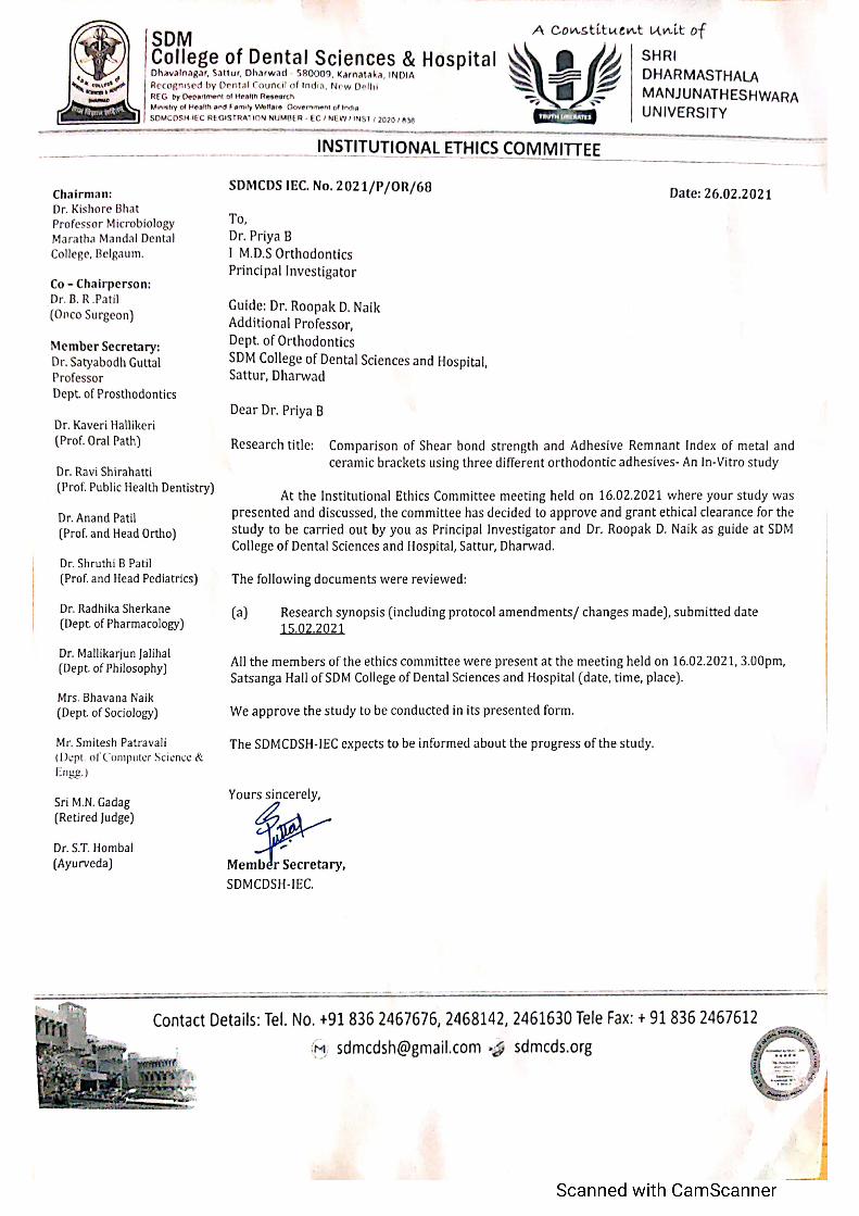
E. Ethical Clearance from the
Institution’s Ethics
Committee Obtained? (Copy

to be Attached)
F. Study procedure Evaluate :
1. Shear bond strength - Universal testing machine
2. Adhesive remnant index - Stereomicroscope
G. Data collection methods including
settings and periodicity
Samples of extracted premolars will be collected from patients
undergoing extraction in Department of Oral Surgery for
orthodontic treatment in SDM college of dental science.
H. List of statistical tests to be used
for data analysis
1. Statistical Package For Social Sciences (SPSS) version 20. (IBM
SPASS statistics [IBM corp. released 2011] will be used to perform
the statistical analysis.
2. Inferential statistics like
Chi-square test will be applied for qualitative variables.
Independent sample t-test / Mann Whitney test (Based on
data distribution) will be applied to compare the Shear
Bond strength between the groups.
ANOVA / Kruskal-Wallis test will be applied to compare
the shear bond strength among the sub groups with post hoc
Bonferroni / Mann Whitney test to compare the SBS
between the sub groups
I. If it’s a Clinical Trial: Clinical
Trials Registry of India or
equivalent registration number to
be mentioned
NA
8 List risks and benefits of the
study
NA
9 Relevant references for the 1. Buonocore MG. A simple method of increasing the

project
(Minimum 10, Maximum 20) (in
Vancouver style)
adhesion of acrylic filling materials to enamel surfaces. J
Dent Res. 1955;34:849–53.
2. 2. Newman GV. Epoxy adhesives for orthodontic
attachments: Progress report. Am J Orthod. 1965;51:901–
12.
3. 3. Joseph VP, Rossouw E. The shear bond strengths of
stainless steel and ceramic brackets used with chemically
and light-activated composite resins. Am J Orthod
Dentofacial Orthop. 1990;97:121–5.
4. Reynolds IR. A review of direct orthodontic bonding. Br J
Orthod. 1975;2:171–8.
5. Bowen RL. Use of epoxy resins in restorative materials. J
Dent Res. 1956;35:360–9.
6. Hosein I, Sherriff M, Ireland AJ. Enamel loss during
bonding, debonding, and cleanup with use of a self-etching
primer. Am J Orthod Dentofacial Orthop. 2004;126:717–
24.
7. Vicente A, Bravo LA, Romero M, Ortiz AJ, Canteras M. A
comparison of the shear bond strength of a resin cement and
two orthodontic resin adhesive systems. Angle
Orthod. 2005;75:109–13.
8. Scougall Vilchis RJ, Yamamoto S, Kitai N, Yamamoto K.
Shear bond strength of orthodontic brackets bonded with
different self-etching adhesives. Am J Orthod Dentofacial
Orthop. 2009;136:425–30.
9. Toledano M, Osorio R, Osorio E, Romeo A, de la Higuera
B, García-Godoy F. Bond strength of orthodontic brackets

uslny ñ i t’§ggCflf )i;;ht »^a self-curi ng «
Orthod. 2003:73:5Mfi3
10. Pitlion MM. dos Santos RL, Ruellas AT, $ znt Anns ZY-
of orthodontic bonding system? Ear J
Ortliod. 2010:32:567-10.
11. llslaiii ian L, Borzabadi-Parahanl A. Moiisavi N, Ghaseini
A. A comparative study of shear bond strength between
metal and ceramic brackets and artificially aged composite
restorations using different surface treatments. Eur I
Orthod. 2012;34:61 W7.
12. Al Shamsi A, Cunningham JL, Lamey PJ, Lynch E. Shear
bond strength and residual adhesive afler orthodontic
bracket debonding. Angle Orthod. 2006;76:694-95
13. Shark JA, Reddy RK, Bhagyalakshini K, Shah MJ,
Madhavi O, Raiiiesli SV. In vitro Evaltlation of Shear Bond
Strength ot”Orthodontic Brackets Bonded xx'ith Dit4”erent
Adhesives Conteinp Clin Dent. 2018 Apr-Jun;9(2):28W92.
10 Conflict of interest for any other NA
investigator(s) (if yes, please
explain in brief)
11 Declarations/Remarks by the
Guide
Scanned with CamScanner

Signature of the head of department.
DR. ANAND K PATIL
FIEAD OF TFIE DEP›\RTMEIT
DEPA RTMENT OF ORTHODONTICS
DII. ftOOPA K D NAIK
/\I3DI4’JONA L Ph OF ESSOR.
DEPAR"fMENT OF OR FFl ODONT iCS