persistent elimination of erbb-2/her2-overexpressing ... · receptor degradation in lysosomes. it...
TRANSCRIPT
Persistent elimination of ErbB-2/HER2-overexpressingtumors using combinations of monoclonal antibodies:Relevance of receptor endocytosisTsipi Ben-Kasusa,1, Bilha Schechtera,1, Sara Lavib, Yosef Yardenb, and Michael Selaa,2
aDepartments of Immunology and bBiological Regulation, The Weizmann Institute of Science, Rehovot 76100, Israel
Contributed by Michael Sela, December 1, 2008 (sent for review August 21, 2008)
Monoclonal antibodies (mAbs) to ErbB-2/HER2 or to its sibling, theepidermal growth factor receptor (EGFR), prolong survival ofcancer patients, especially when combined with cytotoxic thera-pies. However, low effectiveness of therapeutic mAbs and theevolution of patient resistance call for improvements. Here we testin animals pairs of anti-ErbB-2 mAbs and report that pairs com-prising an antibody reactive with the dimerization site of ErbB-2and an antibody recognizing another distinct epitope better inhibitErbB-2-overexpressing tumors than other pairs or the respectiveindividual mAbs. Because the superiority of antibody combinationsextends to tumor cell cultures, we assume that nonimmunologicalmechanisms contribute to mAb synergy. One potential mechanism,namely the ability of mAb combinations to instigate ErbB-2 endo-cytosis, is demonstrated. Translation of these lessons to clinicalapplications may enhance patient response and delay acquisitionof resistance.
cancer � growth factor � immunotherapy � signal transduction �tyrosine kinase
ErbB-2/HER2 is a member of the epidermal growth factorreceptor (EGFR) family. When transactivated, ErbB-2/
HER2 stimulates several downstream signaling cascades, includ-ing the mitogen-activated protein kinase cascade (1). Thisligand-less receptor is moderately expressed in normal adulttissues, where it regulates cell growth and differentiation. Bycontrast, amplification of the corresponding gene and conse-quent overexpression of the HER2/ErbB-2 protein have beenreported in 20–30% of tumors of the breast (2–4) and ovary (4).In general, erbB-2 gene amplification associates with enhancedmetastatic potential and poor prognosis. Because ErbB-2 isexpressed at relatively low levels in normal tissues, it makes anattractive target for immunotherapy. This was originally dem-onstrated in animals by Greene et al. (5), who targeted Neu, therodent form of ErbB-2, and later developed this into a widelyused clinical strategy (6). The molecular mechanisms underlyingthe growth-inhibitory effects of anti–ErbB-2 monoclonal anti-bodies (mAbs) may involve indirect tumor cell cytotoxicitythrough immunological mechanisms, including antibody-dependent cellular cytotoxicity (ADCC), complement-dependent cytotoxicity (CDC), increased cancer cell apoptosis,as well as direct interference with signaling cascades (6).
Clinical studies established that Trastuzumab (Herceptin), ahumanized mAb directed against ErbB-2, is active againstErbB-2-overexpressing metastatic breast cancer, leading to itsapproval for clinical use (7). The objective response rates toTrastuzumab monotherapy is relatively low (�15%) and shortlived (median duration, 9 months) (8). On the other hand, mAbsseem to display a synergistic effect when combined with che-motherapy, probably because of interruption of ErbB-2-drivensurvival pathway (9). Yet another strategy, relevant to pancreaticcancer, combines antibodies to EGFR and to ErbB-2 (10). Thepresent study explores an alternative strategy to enhance thetherapeutic activity of anti-receptor antibodies, namely combin-ing two or more epitope-distinct antibodies. This strategy was
previously demonstrated by Drebin et al. (11) and by Kasprzyket al. (12). It was later proposed that the superior activity of mAbcombinations is attributable to a combination of various factors,including improved effector cell recruitment as ADCC and CDC(13, 14). We have previously demonstrated that combininganti-EGFR mAbs that engage distinct epitopes proved moreeffective in down-regulating the receptor in vitro than eachantibody alone (15) because of the generation at the cell surfaceof very large receptor-antibody complexes or lattices, whichcollapse into the cytoplasm and eventually undergo degradationin lysosomes. Here we demonstrate that pairs of mAbs specificto distinct epitopes of ErbB-2, of which one epitope is involvedin dimerization, are highly effective anti-tumor agents in vivoand are capable of inhibiting tumor cell growth in vitro. We alsoshow that a noninhibitory mAb, which obviously does not inducea cellular response, contributes to the synergistic effect. Thisimplies that direct effects of the mAbs occur in addition toeffector mechanisms.
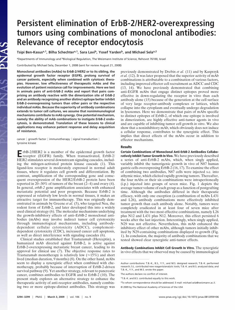
ResultsCertain Combinations of Monoclonal Anti-ErbB-2 Antibodies Collabo-ratively Inhibit Tumor Growth In Vivo. We have previously describeda series of anti-ErbB-2 mAbs, which, when singly applied,variably inhibit the tumorigenic growth in vivo of N87 humangastric cells overexpresing ErbB-2 (16, 17). To examine the effectof combining two antibodies, N87 cells were injected s.c. intoathymic mice, which elicited rapidly growing tumors. Thereafter,the four mAbs or their six combinations were intraperitoneally(i.p.) injected into groups of seven mice. Fig. 1 depicts theaverage tumor volume of each group as a function of postgraftingtime. Although the antibodies differed in their therapeuticefficacy, with only one exception (a combination of mAbs L431and L26), antibody combinations more effectively inhibitedtumor growth than each antibody alone. Notably, tumors werecompletely eradicated in at least four of seven mice aftertreatment with the two most effective combinations, namely L26plus N12 and L431 plus N12. Moreover, this effect persisted 6weeks after the last injection. Interestingly, when singly applied,N29 was not effective. Nevertheless, this mAb enhanced theinhibitory effect of other mAbs, although tumors initially inhib-ited by N29-containing combinations displayed re-growth (Fig.1). In conclusion, the majority of antibody combinations that wetested showed clear synergistic anti-tumor effects.
Antibody Combinations Inhibit Cell Growth In Vitro. The synergisticin vivo effects that we observed may be caused by immunological
Author contributions: T.B.-K., B.S., Y.Y., and M.S. designed research; T.B.-K. performedresearch; S.L. contributed new reagents/analytic tools; T.B.-K. and B.S. analyzed data; andT.B.-K., Y.Y., and M.S. wrote the paper.
The authors declare no conflict of interest.
1T.B.-K. and B.S. contributed equally to this work.
2To whom correspondence should be addressed. E-mail: [email protected].
© 2009 by The National Academy of Sciences of the USA
3294–3299 � PNAS � March 3, 2009 � vol. 106 � no. 9 www.pnas.org�cgi�doi�10.1073�pnas.0812059106
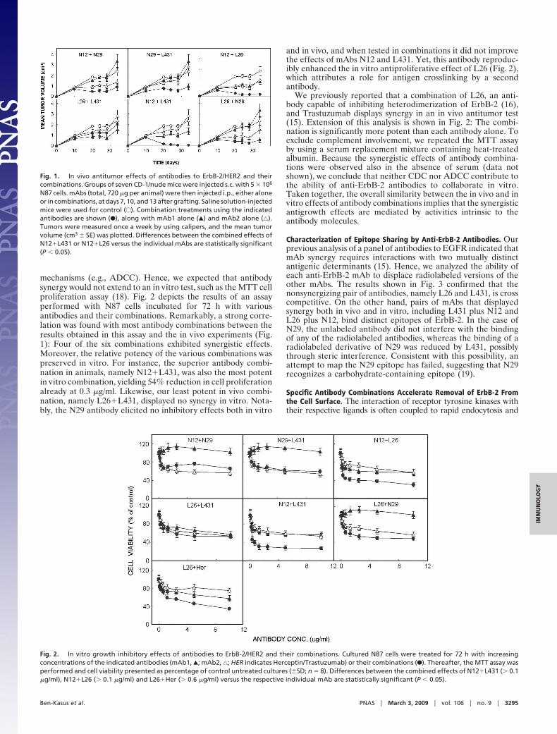
mechanisms (e.g., ADCC). Hence, we expected that antibodysynergy would not extend to an in vitro test, such as the MTT cellproliferation assay (18). Fig. 2 depicts the results of an assayperformed with N87 cells incubated for 72 h with variousantibodies and their combinations. Remarkably, a strong corre-lation was found with most antibody combinations between theresults obtained in this assay and the in vivo experiments (Fig.1): Four of the six combinations exhibited synergistic effects.Moreover, the relative potency of the various combinations waspreserved in vitro. For instance, the superior antibody combi-nation in animals, namely N12�L431, was also the most potentin vitro combination, yielding 54% reduction in cell proliferationalready at 0.3 �g/ml. Likewise, our least potent in vivo combi-nation, namely L26�L431, displayed no synergy in vitro. Nota-bly, the N29 antibody elicited no inhibitory effects both in vitro
and in vivo, and when tested in combinations it did not improvethe effects of mAbs N12 and L431. Yet, this antibody reproduc-ibly enhanced the in vitro antiproliferative effect of L26 (Fig. 2),which attributes a role for antigen crosslinking by a secondantibody.
We previously reported that a combination of L26, an anti-body capable of inhibiting heterodimerization of ErbB-2 (16),and Trastuzumab displays synergy in an in vivo antitumor test(15). Extension of this analysis is shown in Fig. 2: The combi-nation is significantly more potent than each antibody alone. Toexclude complement involvement, we repeated the MTT assayby using a serum replacement mixture containing heat-treatedalbumin. Because the synergistic effects of antibody combina-tions were observed also in the absence of serum (data notshown), we conclude that neither CDC nor ADCC contribute tothe ability of anti-ErbB-2 antibodies to collaborate in vitro.Taken together, the overall similarity between the in vivo and invitro effects of antibody combinations implies that the synergisticantigrowth effects are mediated by activities intrinsic to theantibody molecules.
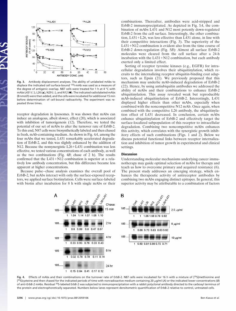
Characterization of Epitope Sharing by Anti-ErbB-2 Antibodies. Ourprevious analysis of a panel of antibodies to EGFR indicated thatmAb synergy requires interactions with two mutually distinctantigenic determinants (15). Hence, we analyzed the ability ofeach anti-ErbB-2 mAb to displace radiolabeled versions of theother mAbs. The results shown in Fig. 3 confirmed that thenonsynergizing pair of antibodies, namely L26 and L431, is crosscompetitive. On the other hand, pairs of mAbs that displayedsynergy both in vivo and in vitro, including L431 plus N12 andL26 plus N12, bind distinct epitopes of ErbB-2. In the case ofN29, the unlabeled antibody did not interfere with the bindingof any of the radiolabeled antibodies, whereas the binding of aradiolabeled derivative of N29 was reduced by L431, possiblythrough steric interference. Consistent with this possibility, anattempt to map the N29 epitope has failed, suggesting that N29recognizes a carbohydrate-containing epitope (19).
Specific Antibody Combinations Accelerate Removal of ErbB-2 Fromthe Cell Surface. The interaction of receptor tyrosine kinases withtheir respective ligands is often coupled to rapid endocytosis and
Fig. 1. In vivo antitumor effects of antibodies to ErbB-2/HER2 and theircombinations. Groups of seven CD-1/nude mice were injected s.c. with 5 � 106
N87 cells. mAbs (total, 720 �g per animal) were then injected i.p., either aloneor in combinations, at days 7, 10, and 13 after grafting. Saline solution-injectedmice were used for control (E). Combination treatments using the indicatedantibodies are shown (F), along with mAb1 alone (Œ) and mAb2 alone (‚).Tumors were measured once a week by using calipers, and the mean tumorvolume (cm3 � SE) was plotted. Differences between the combined effects ofN12�L431 or N12�L26 versus the individual mAbs are statistically significant(P � 0.05).
Fig. 2. In vitro growth inhibitory effects of antibodies to ErbB-2/HER2 and their combinations. Cultured N87 cells were treated for 72 h with increasingconcentrations of the indicated antibodies (mAb1, Œ; mAb2, ‚; HER indicates Herceptin/Trastuzumab) or their combinations (F). Thereafter, the MTT assay wasperformed and cell viability presented as percentage of control untreated cultures (�SD; n � 8). Differences between the combined effects of N12�L431 (� 0.1�g/ml), N12�L26 (� 0.1 �g/ml) and L26�Her (� 0.6 �g/ml) versus the respective individual mAb are statistically significant (P � 0.05).
Ben-Kasus et al. PNAS � March 3, 2009 � vol. 106 � no. 9 � 3295
IMM
UN
OLO
GY
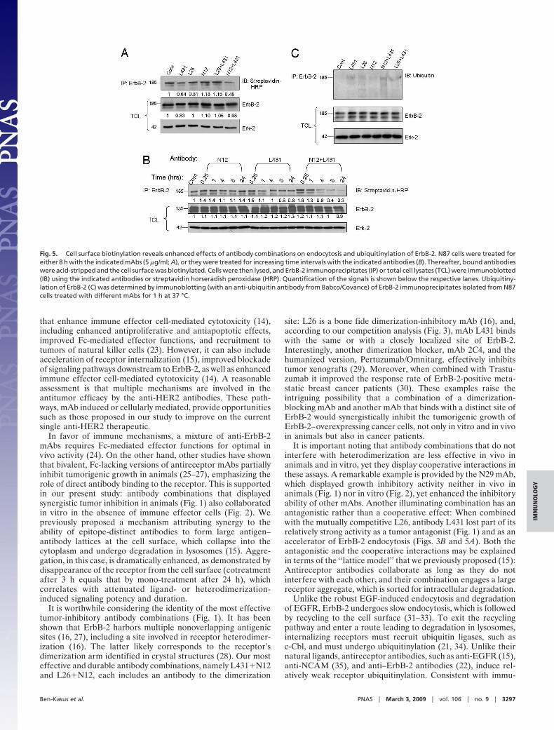
receptor degradation in lysosomes. It was shown that mAbs caninduce an analogous, albeit slower, effect (20), which is associatedwith inhibition of tumorigenesis (12). Therefore, we tested thepotential of our set of mAbs to alter the turnover rate of ErbB-2.To this end, N87 cells were biosynthetically labeled and then chasedin fresh, mAb-containing medium. As shown in Fig. 4A, among thefour mAbs that we tested, L431 remarkably accelerated degrada-tion of ErbB-2, and this was slightly enhanced by the addition ofN12. Because the nonsynergistic L26�L431 combination was lesseffective, we tested various concentrations of each antibody, as wellas the two combinations (Fig. 4B; chase of 2 h). The resultsconfirmed that the L431�N12 combination is superior at a rela-tively low antibody concentration, but this difference became lessapparent at higher concentrations.
Because pulse–chase analysis examines the overall pool ofErbB-2, but mAbs interact with only the surface-exposed recep-tors, we applied surface biotinylation. Cells were surface-labeledwith biotin after incubation for 8 h with single mAbs or their
combinations. Thereafter, antibodies were acid-stripped andErbB-2 immunoprecipitated. As depicted in Fig. 5A, the com-bination of mAbs L431 and N12 most potently down-regulatedErbB-2 from the cell surface. Interestingly, the other combina-tion, L431�L26, was less effective than L431 alone, in line withtheir competitive interactions (Fig. 3). The superiority of theL431�N12 combination is evident also from the time course ofErbB-2 down-regulation (Fig. 5B): Almost all surface ErbB-2molecules were cleared from the cell surface after a 24-hincubation with the L431�N12 combination, but each antibodyexerted only a limited effect.
Sorting of receptor tyrosine kinases (e.g., EGFR) for intra-cellular degradation involves their ubiquitinylation, which re-cruits to the internalizing receptor ubiquitin-binding coat adap-tors, such as Epsin (21). We previously proposed that thismechanism may underlie mAb-induced degradation of ErbB-2(22). Hence, by using antiubiquitin antibodies we addressed theability of mAbs and their combinations to enhance ErbB-2ubiquitinylation. This assay revealed weak but reproduciblemAb-induced ubiquitinylation of ErbB-2. Interestingly, L431displayed higher effects than other mAbs, especially whencombined with the noncompetitive N12 mAb. Once again, whencombined with the competitive L26 antibody, the ubiquitinyla-tion effect of L431 decreased. In conclusion, certain mAbsenhance ubiquitinylation of ErbB-2 and effectively target thesurface-localized subpopulation of this receptor to intracellulardegradation. Combining two noncompetitive mAbs enhancesthis activity, which correlates with the synergistic growth inhib-itory effects of such combinations (Figs. 1 and 2). Below wediscuss potential functional links between receptor internaliza-tion and inhibition of tumor growth in experimental and clinicalsettings.
DiscussionUnderstanding molecular mechanisms underlying cancer immu-notherapy may guide optimal selection of mAbs for therapy andteach us how to overcome primary and acquired resistance (6).The present study addresses an emerging strategy, which en-hances the therapeutic activity of antireceptor antibodies bycombining two mAbs engaging distinct epitopes. In general, thissuperior activity may be attributable to a combination of factors
Fig. 3. Antibody displacement analyses. The ability of unlabeled mAbs todisplace the indicated cell surface-bound 125I-mAb was used as a measure ofthe degree of antigenic overlap. N87 cells were treated for 1 h at 4 °C withmAbs L431 (E), L26 (Œ), N29 (‚), and N12 (F). The indicated radiolabeled mAbs(8 nmol/l) were then added, and the cells were incubated for additional 15 minbefore determination of cell-bound radioactivity. The experiment was re-peated three times.
Fig. 4. Effects of mAbs and their combinations on the turnover rate of ErbB-2. N87 cells were incubated for 16 h with a mixture of [35S]methionine and[35S]cysteine and then chased for the indicated periods of time with nonradioactive medium containing 20 �g/ml (A) or the indicated lower concentrations (B)of anti-ErbB-2 mAbs. Residual 35S-labeled ErbB-2 was subjected to immunoprecipitation with a rabbit polyclonal antibody directed to the carboxyl terminus ofthe protein and electrophoretically separated. Numbers below lanes represent densitometric quantification of ErbB-2 relative to control, untreated cells.
3296 � www.pnas.org�cgi�doi�10.1073�pnas.0812059106 Ben-Kasus et al.
that enhance immune effector cell-mediated cytotoxicity (14),including enhanced antiproliferative and antiapoptotic effects,improved Fc-mediated effector functions, and recruitment totumors of natural killer cells (23). However, it can also includeacceleration of receptor internalization (15), improved blockadeof signaling pathways downstream to ErbB-2, as well as enhancedimmune effector cell-mediated cytotoxicity (14). A reasonableassessment is that multiple mechanisms are involved in theantitumor efficacy by the anti-HER2 antibodies. These path-ways, mAb induced or cellularly mediated, provide opportunitiessuch as those proposed in our study to improve on the currentsingle anti-HER2 therapeutic.
In favor of immune mechanisms, a mixture of anti-ErbB-2mAbs requires Fc-mediated effector functions for optimal invivo activity (24). On the other hand, other studies have shownthat bivalent, Fc-lacking versions of antireceptor mAbs partiallyinhibit tumorigenic growth in animals (25–27), emphasizing therole of direct antibody binding to the receptor. This is supportedin our present study: antibody combinations that displayedsynergistic tumor inhibition in animals (Fig. 1) also collaboratedin vitro in the absence of immune effector cells (Fig. 2). Wepreviously proposed a mechanism attributing synergy to theability of epitope-distinct antibodies to form large antigen–antibody lattices at the cell surface, which collapse into thecytoplasm and undergo degradation in lysosomes (15). Aggre-gation, in this case, is dramatically enhanced, as demonstrated bydisappearance of the receptor from the cell surface (cotreatmentafter 3 h equals that by mono-treatment after 24 h), whichcorrelates with attenuated ligand- or heterodimerization-induced signaling potency and duration.
It is worthwhile considering the identity of the most effectivetumor-inhibitory antibody combinations (Fig. 1). It has beenshown that ErbB-2 harbors multiple nonoverlapping antigenicsites (16, 27), including a site involved in receptor heterodimer-ization (16). The latter likely corresponds to the receptor’sdimerization arm identified in crystal structures (28). Our mosteffective and durable antibody combinations, namely L431�N12and L26�N12, each includes an antibody to the dimerization
site: L26 is a bone fide dimerization-inhibitory mAb (16), and,according to our competition analysis (Fig. 3), mAb L431 bindswith the same or with a closely localized site of ErbB-2.Interestingly, another dimerization blocker, mAb 2C4, and thehumanized version, Pertuzumab/Omnitarg, effectively inhibitstumor xenografts (29). Moreover, when combined with Trastu-zumab it improved the response rate of ErbB-2-positive meta-static breast cancer patients (30). These examples raise theintriguing possibility that a combination of a dimerization-blocking mAb and another mAb that binds with a distinct site ofErbB-2 would synergistically inhibit the tumorigenic growth ofErbB-2–overexpressing cancer cells, not only in vitro and in vivoin animals but also in cancer patients.
It is important noting that antibody combinations that do notinterfere with heterodimerization are less effective in vivo inanimals and in vitro, yet they display cooperative interactions inthese assays. A remarkable example is provided by the N29 mAb,which displayed growth inhibitory activity neither in vivo inanimals (Fig. 1) nor in vitro (Fig. 2), yet enhanced the inhibitoryability of other mAbs. Another illuminating combination has anantagonistic rather than a cooperative effect: When combinedwith the mutually competitive L26, antibody L431 lost part of itsrelatively strong activity as a tumor antagonist (Fig. 1) and as anaccelerator of ErbB-2 endocytosis (Figs. 3B and 5A). Both theantagonistic and the cooperative interactions may be explainedin terms of the ‘‘lattice model’’ that we previously proposed (15):Antireceptor antibodies collaborate as long as they do notinterfere with each other, and their combination engages a largereceptor aggregate, which is sorted for intracellular degradation.
Unlike the robust EGF-induced endocytosis and degradationof EGFR, ErbB-2 undergoes slow endocytosis, which is followedby recycling to the cell surface (31–33). To exit the recyclingpathway and enter a route leading to degradation in lysosomes,internalizing receptors must recruit ubiquitin ligases, such asc-Cbl, and must undergo ubiquitinylation (21, 34). Unlike theirnatural ligands, antireceptor antibodies, such as anti-EGFR (15),anti-NCAM (35), and anti–ErbB-2 antibodies (22), induce rel-atively weak receptor ubiquitinylation. Consistent with immu-
Fig. 5. Cell surface biotinylation reveals enhanced effects of antibody combinations on endocytosis and ubiquitinylation of ErbB-2. N87 cells were treated foreither 8 h with the indicated mAbs (5 �g/ml; A), or they were treated for increasing time intervals with the indicated antibodies (B). Thereafter, bound antibodieswere acid-stripped and the cell surface was biotinylated. Cells were then lysed, and ErbB-2 immunoprecipitates (IP) or total cell lysates (TCL) were immunoblotted(IB) using the indicated antibodies or streptavidin horseradish peroxidase (HRP). Quantification of the signals is shown below the respective lanes. Ubiquitiny-lation of ErbB-2 (C) was determined by immunoblotting (with an anti-ubiquitin antibody from Babco/Covance) of ErbB-2 immunoprecipitates isolated from N87cells treated with different mAbs for 1 h at 37 °C.
Ben-Kasus et al. PNAS � March 3, 2009 � vol. 106 � no. 9 � 3297
IMM
UN
OLO
GY
notherapeutic relevance of antibody-induced receptor degrada-tion (Fig. 4A), removal from the cell surface (Fig. 5 A and B),and ubiquitinylation (Fig. 5C), in these assays the synergisticcombination L431�N12 is more active than the nonsynergisticpair (L431�L26).
In summary, by using combinations of anti-ErbB-2 antibodies,our study provides evidence supporting the possibility thatmAb-induced internalization and degradation of surface recep-tors contribute to immunotherapy, at least in an animal model.We predict that certain combinations of mAbs directed toErbB-2 or to other receptor tyrosine kinases will enhancetherapeutic efficacy and synergy with chemotherapy. This maybe especially important for breast cancer patients who eventuallydevelop secondary resistance to antibodies like Trastuzumab(36). Future studies will address the molecular mechanismsunderlying the endocytic superiority of antibody combinations.
Materials and MethodsMaterials, Antibodies, and Cells. Unless indicated, materials were purchasedfrom Sigma (St. Louis), cells from the American Type Culture Collection (Ma-nassas, VA), and antibodies from Santa Cruz Biotechnology (Santa Cruz, CA).Radiochemicals were purchased from Amersham (Buckinghamshire, UnitedKingdom). Trastuzumab was provided by Genentech (South San Francisco,CA). The previously described mAbs to ErbB-2 (16, 17) were purified on proteinG plus agarose.
Surface Biotinylation Assay. Cells were incubated at 37 °C with mAbs, trans-ferred to ice, and the mAbs removed by using a low pH solution (0.15 mol/lacetic acid containing 0.15 mol/l NaCl; 4 min). The cells were washed andincubated for 60 min at 4 °C with N-hydroxysuccinimide (NHS)–biotin (0.5mg/ml; Calbiochem, San Diego). Coupling of biotin was blocked with 15mmol/l glycine (15). Thereafter, cells were solubilized by the addition of lysisbuffer consisting of 50 mmol/l HEPES (pH 7.5), 150 mmol/l NaCl, 10% glycerol,1% Triton X-100, 1 mmol/l EDTA, 1 mmol/l EGTA, 1 mmol/l phenylmethylsul-fonyl fluoride, 50 mmol/l sodium fluoride, 0.5 mmol/l Na3VO4, 5 �g/ml leu-peptin and 5 �g/ml aprotonin, and a rabbit antibody to ErbB-2 used forimmunoprecipitation.
Radiolabeling of Antibodies. MAbs (100 �g in 0.2 ml phosphate-buffered salinesolution) were radiolabeled by using Na125I (5 �l; 0.5 mCi [18.5 MBq]) andchloramine-T (10 mg/10 �l). The reaction mixture was chromatographed onSephadex G-25 yielding radiolabeled mAbs of 1–2 mCi/mg protein. MAb N29was radiolabeled by using the [125I]-Bolton-Hunter reagent (PerkinElmer Sci-ences, Boston).
Antibody Competition Assay. N87 cells (250,000 cells/well) grown in 24-wellplates were treated for 1 h at 4 °C with increasing concentrations of unlabeledmAbs. Radiolabeled mAbs (8 nmol/l) were then added, and the cells incubatedfor an additional 15 min at 4 °C. After washing, the cells were solubilized in 0.5N NaOH solution before determination of radioactivity.
Inhibition of N87 Tumor Cell Growth in Culture. Antibodies were added to N87cells (10,000 cells/well) grown in 96-well plates. Incubation at 37 °C proceededfor 72 h and cell viability determined by using the MTT [3-(4,5-dimethylthia-zol-2-yl)-2,5-diphenyltetrazolium bromide] reagent (18).
Determination of the Effect of Antibodies on Receptor Turnover. N87 cells werelabeled by incubation (16 h at 37 °C) in methionine- and cysteine-free mediumsupplemented with 10% dialyzed fetal calf serum and Promix, a mixture of[35S]methionine and [35S]cysteine (50 �Ci/ml). Thereafter, cells were chased forvarious periods of time by incubation in fresh medium in the absence orpresence of the antibodies. The cells were then washed, and lysates weresubjected to immunoprecipitation.
Tumorigenic Cell Growth in Animals. Female CD/nude mice were injected s.c.with N87 cells (5 � 106 per mouse). The mAbs were injected i.p. at days 7, 10,and 13 after grafting. Groups of seven mice were used, with each mousereceiving 0.72 mg of purified mAb. Tumor parameters were measured once aweek.
Statistical Analysis. Student’s t test (two-tailed) was used to test differencesbetween the effects of antibody combinations and single treatment. Values ofP � 0.05 were considered statistically significant.
ACKNOWLEDGMENTS. This work was supported by grants from the Mark RichFoundation (the Linda de Picciotto Pancreas Cancer Research Program), theDr. Miriam and Sheldon G. Adelson Medical Research Foundation, the Evelynand Harold Igdaloff Foundation, and the National Cancer Institute GrantCA72981.
1. Citri A, Yarden Y (2006) EGF-ERBB signalling: Towards the systems level. Nat Rev MolCell Biol 7:505–16.
2. King CR, et al. (1989) Heterogeneous expression of erbB-2 messenger RNA in humanbreast cancer. Cancer Res 49:4185–4191.
3. Slamon DJ, et al. (1987) Human breast cancer: Correlation of relapse and survival withamplification of the HER-2/neu oncogene. Science 235:177–182.
4. Slamon DJ, et al. (1989) Studies of the HER-2/neu proto-oncogene in human breast andovarian cancer. Science 244:707–712.
5. Drebin JA, Link VC, Weinberg RA, Greene MI (1986) Inhibition of tumor growth by amonoclonal antibody reactive with an oncogene-encoded tumor antigen. Proc NatlAcad Sci USA 83:9129–9133.
6. Ben-Kasus T, Schechter B, Sela M, Yarden Y (2007) Cancer therapeutic antibodies comeof age: Targeting minimal residual disease. Mol Oncol 1:42–54.
7. Carter P, et al. (1992) Humanization of an anti-p185HER2 antibody for human cancertherapy. Proc Natl Acad Sci USA 89:4285–4289.
8. Cobleigh MA, et al. (1999) Multinational study of the efficacy and safety of humanizedanti-HER2 monoclonal antibody in women who have HER2-overexpressing metastaticbreast cancer that has progressed after chemotherapy for metastatic disease. J ClinOncol 17:2639–2648.
9. Arteaga CL, et al. (1994) p185c-erbB-2 signal enhances cisplatin-induced cytotoxicity inhuman breast carcinoma cells: Association between an oncogenic receptor tyrosinekinase and drug-induced DNA repair. Cancer Res 54:3758–3765.
10. Larbouret C, et al. (2007) In vivo therapeutic synergism of anti-epidermal growth factorreceptor and anti-HER2 monoclonal antibodies against pancreatic carcinomas. ClinCancer Res 13:3356–3362.
11. Drebin JA, Link VC, Greene MI (1988) Monoclonal antibodies specific for the neuoncogene product directly mediate anti-tumor effects in vivo. Oncogene 2:387–394.
12. Kasprzyk PG, Song SU, Di Fiore PP, King CR (1992) Therapy of an animal model ofhuman gastric cancer using a combination of anti-erbB-2 monoclonal antibodies.Cancer Res 52:2771–2776.
13. Nahta R, Hung MC, Esteva FJ (2004) The HER-2-targeting antibodies trastuzumab andpertuzumab synergistically inhibit the survival of breast cancer cells. Cancer Res64:2343–2346.
14. Spiridon CI, et al. (2002) Targeting multiple Her-2 epitopes with monoclonal antibodiesresults in improved antigrowth activity of a human breast cancer cell line in vitro andin vivo. Clin Cancer Res 8:1720–1730.
15. Friedman LM, et al. (2005) Synergistic down-regulation of receptor tyrosine kinases bycombinations of mAbs: Implications for cancer immunotherapy. Proc Natl Acad Sci USA102:1915–1920.
16. Klapper LN, et al. (1997) A subclass of tumor-inhibitory monoclonal antibodies toErbB-2/HER2 blocks crosstalk with growth factor receptors. Oncogene 14:2099 –2109.
17. Stancovski I, et al. (1991) Mechanistic aspects of the opposing effects of monoclonalantibodies to the ERBB2 receptor on tumor growth. Proc Natl Acad Sci USA 88:8691–8695.
18. Mosmann T (1983) Rapid colorimetric assay for cellular growth and survival: Applica-tion to proliferation and cytotoxicity assays. J Immunol Methods 65:55–63.
19. Yip YL, Smith G, Koch J, Dubel S, Ward RL (2001) Identification of epitope regionsrecognized by tumor inhibitory and stimulatory anti-ErbB-2 monoclonal antibodies:Implications for vaccine design. J Immunol 166:5271–5278.
20. Yarden Y (1990) Agonistic antibodies stimulate the kinase encoded by the neu pro-tooncogene in living cells but the oncogenic mutant is constitutively active. Proc NatlAcad Sci USA 87:2569–2573.
21. Marmor MD, Yarden Y (2004) Role of protein ubiquitylation in regulating endocytosisof receptor tyrosine kinases. Oncogene 23:2057–2070.
22. Klapper LN, Waterman H, Sela M, Yarden Y (2000) Tumor-inhibitory antibodies toHER-2/ErbB-2 may act by recruiting c-Cbl and enhancing ubiquitination of HER-2.Cancer Res 60:3384–3388.
23. Clynes RA, Towers TL, Presta LG, Ravetch JV (2000) Inhibitory Fc receptors modulate invivo cytoxicity against tumor targets. Nat Med 6:443–446.
24. Spiridon CI, Guinn S, Vitetta ES (2004) A comparison of the in vitro and in vivo activitiesof IgG and F(ab�)2 fragments of a mixture of three monoclonal anti-Her-2 antibodies.Clin Cancer Res 10:3542–3551.
25. Fan Z, Lu Y, Wu X, Mendelsohn J (1994) Antibody-induced epidermal growth factorreceptor dimerization mediates inhibition of autocrine proliferation of A431 squa-mous carcinoma cells. J Biol Chem 269:27595–27602.
26. Hurwitz E, Klapper LN, Wilchek M, Yarden Y, Sela M (2000) Inhibition of tumor growthby poly(ethylene glycol) derivatives of anti-ErbB2 antibodies. Cancer Immunol Immu-nother 49:226–234.
27. Xu F, et al. (1993) Antibody-induced growth inhibition is mediated through immuno-chemically and functionally distinct epitopes on the extracellular domain of thec-erbB-2 (HER-2/neu) gene product p185. Int J Cancer 53:401–408.
3298 � www.pnas.org�cgi�doi�10.1073�pnas.0812059106 Ben-Kasus et al.
28. Garrett TP, et al. (2003) The crystal structure of a truncated ErbB2 ectodomain reveals anactive conformation, poised to interact with other ErbB receptors. Mol Cell 11:495–505.
29. Agus DB, et al. (2002) Targeting ligand-activated ErbB2 signaling inhibits breast andprostate tumor growth. Cancer Cell 2:127–137.
30. Baselga J, et al. (2007) Objective response rate in a phase II multicenter trial ofpertuzumab (P), a HER2 dimerization inhibiting monoclonal antibody, in combinationwith trastuzumab (T) in patients (pts) with HER2-positive metastatic breast cancer(MBC) which has progressed during treatment with T. J Clin Oncol (2007 ASCO AnnualMeeting Proceedings Part I) 25:1004 (abstr).
31. Baulida J, Kraus MH, Alimandi M, Di Fiore PP, Carpenter G (1996) All ErbB receptorsother than the epidermal growth factor receptor are endocytosis impaired. J Biol Chem271:5251–5257.
32. Pinkas-Kramarski R, et al. (1996) Diversification of Neu differentiation factor andepidermal growth factor signaling by combinatorial receptor interactions. EMBO J15:2452–2467.
33. Worthylake R, Opresko LK, Wiley HS (1999) ErbB-2 amplification inhibits down-regulation and induces constitutive activation of both ErbB-2 and epidermal growthfactor receptors. J Biol Chem 274:8865–8874.
34. Katzmann DJ, Odorizzi G, Emr SD (2002) Receptor down-regulation and multivesicular-body sorting. Nat Rev Mol Cell Biol 3:893–905.
35. Diestel S, Schaefer D, Cremer H, Schmitz B (2007) NCAM is ubiquitylated, endocytosedand recycled in neurons. J Cell Sci 120:4035–4049.
36. Hynes NE, Lane HA (2005) ERBB receptors and cancer: The complexity of targetedinhibitors. Nat Rev Cancer 5:341–354.
Ben-Kasus et al. PNAS � March 3, 2009 � vol. 106 � no. 9 � 3299
IMM
UN
OLO
GY