persistence of bronchial dysplasia is ...cancerpreventionresearch.aacrjournals.org/content/...1...
TRANSCRIPT
1
PERSISTENCE OF BRONCHIAL DYSPLASIA IS ASSOCIATED WITH DEVELOPMENT OF INVASIVE SQUAMOUS CELL CARCINOMA
Daniel T. Merrick1, Dexiang Gao2, York E. Miller3,4, Robert L. Keith3,4, Anna E. Baron5, William Feser5, Timothy C. Kennedy4, Patrick J. Blatchford2, Sarah Braudrick5, Fred R. Hirsch1, Lynn Heasley6, Paul A. Bunn Jr.7, and Wilbur A. Franklin1
1Department of Pathology, University of Colorado Anschutz Medical Campus 2Department of Pediatrics, University of Colorado Anschutz Medical Campus 3Department of Medicine/Division of Pulmonary Medicine, Denver Veterans Affairs Medical Center 4Department of Medicine/Division of Pulmonary Medicine, University of Colorado Anschutz Medical Campus 5Department of Biostatistics and Informatics, Colorado School of Public Health 6Department of Craniofacial Biology, University of Colorado Anschutz Medical Campus 7Department of Medicine /Division of Medical Oncology, University of Colorado Anschutz Medical Campus
RUNNING TITLE: Persistence of bronchial dysplasia and lung cancer risk
KEY WORDS: Bronchial dysplasia, Cancer risk, Biomarkers, Chemoprevention, Squamous cell carcinoma of the lung GRANT SUPPORT: Lung Specialized Programs of Research Excellence P50 CA058187 and Cancer Center Support Grant P30 CA046934 (all authors received P30 CA046934) CORRESPONDING AUTHOR:
Daniel Thomas Merrick, M.D. University of Colorado Denver, Anschutz Medical Campus Department of Pathology Mail Stop 8104 12801 E 17th Avenue, room 5114 Aurora, CO 80045
CONFLICT OF INTEREST: Dr. Merrick has no conflicts of interest to report
MANUSCRIPT DETAILS: Abstract – 250 words; Text – 5,004 words; 3 figures; 3 tables, 36 refs
Cancer Research. on June 15, 2018. © 2015 American Association forcancerpreventionresearch.aacrjournals.org Downloaded from
Author manuscripts have been peer reviewed and accepted for publication but have not yet been edited. Author Manuscript Published OnlineFirst on November 5, 2015; DOI: 10.1158/1940-6207.CAPR-15-0305
2
ABSTRACT
Background: Bronchial dysplasia (BD), a presumed precursor of pulmonary squamous cell carcinoma
(SCC), rarely progresses to invasive cancer. A high risk cohort at the University of Colorado provided an
opportunity to directly sample airway epithelium at mapped sites on successive bronchoscopies. We
have hypothesized that persistent dysplastic lesions showing a similar or higher level of dysplasia on
follow-up biopsy, are associated with increased risk for the development of SCC.
Methods and Material: Endoscopic biopsies from 188 high risk subjects were histologically classified
according to the current WHO classification for BD utilizing a numeric histology score ranging from 1-8
representing normal bronchial mucosa through invasive lung cancer. Differences in follow-up histology
scores were compared between sites classified by clinical, histologic and immunohistochemical
variables.
Results: Subjects with a higher frequency of sites that persist or progress to high grade dysplasia
(>37.5% persist/progress, N=35 versus <37.5% persist/progress, N=114) show a significant association
with development of incident invasive SCC (adjusted hazard ratio: 7.84; 95% confidence interval: 1.56,
39.39), and those with incident lung SCC have adjusted mean follow-up histology scores 1.55 units
higher than in subjects without lung cancer. Current smoking, elevated Ki-67 growth fraction, histologic
features of angiogenic squamous dysplasia (ASD) and higher histology score in baseline biopsies are
significantly associated with increased follow up histology scores.
Conclusions: These results show that persistent BD is associated with the development of invasive SCC.
Furthermore, increased expression of Ki-67, the presence of angiogenic change and degree of baseline
atypia are associated with persistence of BD.
Cancer Research. on June 15, 2018. © 2015 American Association forcancerpreventionresearch.aacrjournals.org Downloaded from
Author manuscripts have been peer reviewed and accepted for publication but have not yet been edited. Author Manuscript Published OnlineFirst on November 5, 2015; DOI: 10.1158/1940-6207.CAPR-15-0305

3
INTRODUCTION
Bronchial dysplasia (BD) (1) is a presumed precursor lesion of squamous cell carcinoma (SCC) of the lung,
and elimination of these lesions has been proposed as a way to prevent invasive SCC of the lung (2). A
scoring system for BD that stratifies these lesions by severity of squamous atypia has provided a
framework for using histology scores to assess the effect of chemoprevention agents on bronchial
mucosa (3-6). Understanding the behavior of BD could provide means to assess risk for the
development of lung cancer and elucidate mechanisms underlying progression.
Molecular alterations that parallel those seen in invasive cancer for site specific loss of heterozygosity
(LOH), and mRNA, miRNA and immunohistochemical marker expression levels that become more
prominent in higher grades of BD has provided data to support a relationship between BD and invasive
squamous cell carcinoma (SCC) of the lung (7-10). Additionally, chromosomal aneusomy, gene copy
number gains, increased PI3K pathway activation and alterations in telomere length have been shown to
be more common in BD from patients with known lung cancer as compared to those without (11-14). A
relationship between increased expression of multiple tumor related markers or specific LOH in BD and
subsequent development of carcinoma-in-situ (CIS) or SCC has also been shown (15,16). In a few
selected cases, an increase in SOX2 amplification has been described as sites of BD progressed in atypia
and ultimately developed invasive SCC (17). Progression of atypia or development of cancer has also
been related to baseline histologic atypia and other clinical features, but has frequently produced
contradictory findings. While some studies have correlated baseline histology with smoking status,
more frequent progression of atypia or development of cancer (18-23), others have failed to detect
significant relationships with these parameters (15,18,24). Important issues compromising
interpretation of these data include use of CIS as a malignant outcome, small numbers of cases per
individual report, frequent pre-selection of cases with some cohorts being composed entirely of patients
with previous lung or head and neck cancer, confounding of outcome by use of therapeutic intervention,
Cancer Research. on June 15, 2018. © 2015 American Association forcancerpreventionresearch.aacrjournals.org Downloaded from
Author manuscripts have been peer reviewed and accepted for publication but have not yet been edited. Author Manuscript Published OnlineFirst on November 5, 2015; DOI: 10.1158/1940-6207.CAPR-15-0305

4
and variable, often short periods of time to follow up assessment of lesions. The limitations associated
with these factors were also noted in a recent meta-analysis, in which findings suggested that higher
degrees of atypia in BD are associated with more frequent progression or persistence than are lesions of
lower grade (25). These findings were not related to development of lung cancer.
Prediction of outcome in BD is of paramount importance to the establishment of reliable, informative
screening programs and effective prevention measures. Surveillance techniques such as
autofluorescence bronchoscopy have improved sensitivity for detection of BD (26-28) thus providing an
opportunity to accurately follow these lesions over time. We have examined the relationship between
differences in follow-up histology scores and a variety of parameters and found that more atypical
outcomes are associated with subsequent incidence of SCC, increased baseline histologic atypia,
smoking status, features of angiogenic squamous dysplasia (ASD) and Ki67 expression levels. We have
identified a pattern of persistent BD that defines a subset of subjects with aggressive airway disease and
increased risk for development of invasive SCC that may benefit from close follow-up and potential
preventive measures.
MATERIALS AND METHODS
Patients at high risk for the development of lung cancer were recruited to bronchoscopy protocols
established through the Colorado SPORE in Lung Cancer program. High risk subjects included those with
tobacco smoking histories of greater than 20 pack years and those with a personal history of lung cancer
or prior cancer of the upper aerodigestive tract. In subjects without a history of cancer, sputum
screening was performed and bronchoscopy was offered to individuals with moderate or worse
cytologic atypia. Informed consent for enrollment in the protocols and collection of associated clinical
data were approved by the Colorado Multiple Institutional Review Board (CoMIRB). Histologic, clinical
and other data from subjects for which there was any bronchial site that was sampled on at least two
occasions were assembled for this study. Specimens that were collected less than three months after
Cancer Research. on June 15, 2018. © 2015 American Association forcancerpreventionresearch.aacrjournals.org Downloaded from
Author manuscripts have been peer reviewed and accepted for publication but have not yet been edited. Author Manuscript Published OnlineFirst on November 5, 2015; DOI: 10.1158/1940-6207.CAPR-15-0305
5
the original baseline biopsy at a specific site were excluded. During the timeframe of specimen
collection (1993-2010), two Colorado Lung SPORE sponsored chemoprevention trials were conducted in
which all-trans retinoic acid or the prostacyclin analog, Iloprost, were employed as potential
chemopreventive agents (3,4). Iloprost, but not all-trans retinoic acid, was shown to be associated with
a significant reduction in histology score on follow up biopsy among former smokers as compared with
former smokers on the placebo arm of the study. Therefore, all biopsies from treatment arm subjects
collected after the Iloprost trial enrollment bronchoscopy were excluded.
Biopsy site classification
Biopsies were assigned a numeric histology score (see figure 1) ranging from 1 to 8 for normal (score
=1), basal cell hyperplasia (score = 2), squamous metaplasia without atypia (score = 3), mild dysplasia
(score = 4), moderate dysplasia (score = 5), severe dysplasia (score = 6), carcinoma-in-situ (score = 7) and
invasive carcinoma (score = 8) with each group being defined by the histologic features described in the
WHO classification (1). Baseline histologic score was defined as the diagnosis for the first biopsy at a
given site. Follow-up histologic scores were classified into three groups: those with biopsies collected
between 3 months to two years, 2-4 years and >4 years after baseline biopsies. If more than one biopsy
had been collected during the timeframe of one of these groups, the biopsy with the highest diagnosis
was used. Most analyses employed grouping of histology scores into non-dysplastic (scores 1-2), low
grade dysplasia (LGD, scores 3-4) and high grade dysplasia (HGD, scores 5-7) histologic groups. In
analyses comparing groups defined by a pre-assigned persistent/progressive or regressive classification,
persistent/progressive dysplasia (referred to as “persistent” BD throughout manuscript) was defined as
any baseline LGD that showed LGD or higher histologic score on follow-up and any baseline HGD that
showed HGD or higher histologic score on follow-up unless otherwise noted. Biopsies of histology score
3 or greater were also characterized as angiogenic squamous dysplasias if they showed projections of
Cancer Research. on June 15, 2018. © 2015 American Association forcancerpreventionresearch.aacrjournals.org Downloaded from
Author manuscripts have been peer reviewed and accepted for publication but have not yet been edited. Author Manuscript Published OnlineFirst on November 5, 2015; DOI: 10.1158/1940-6207.CAPR-15-0305
6
vascular structures into overlying epithelium as described in previous publications (29) and shown in
figures 1E and 1F.
Cancer status was based on subject-level tissue diagnoses with incident carcinoma defined as those
cases in which a diagnosis of invasive carcinoma was made 6 months or longer after the date of the
baseline bronchoscopy. All other subjects with known lung cancer diagnoses were considered prevalent
(or prior) cancer cases. Twenty three of the 41 cases with associated lung carcinomas demonstrated
incident carcinomas (in one subject, two synchronous, incident carcinomas occurred in contralateral
lung lobes). Fourteen of these were SCC, 4 adenocarcinoma and 6 not otherwise specified (NOS).
Tumor diagnoses and sites were established by bronchoscopic biopsy in 8 cases (all SCC), cytologic
sampling in 3 (2 SCC, 1 adenocarcinoma) or resection specimens in 7 (4 SCCs, 2 adenocarcinomas and 1
NOS), whereas in 6 cases the method of diagnosis/site were unknown (1 adenocarcinoma and 5 NOS).
Among the incident cases, 6 represented second or higher primaries (4 SCC, 1 adenocarcinoma, 1 NOS).
Among the 18 cases associated only with prevalent lung carcinomas, 12 were SCC, 5 adenocarcinomas
and 1 NOS. Throughout the manuscript, analyses of cancer associated cases indicate whether this group
includes prevalent and incident cancers or incident or prevalent cancers alone. Each baseline biopsy
was associated with clinical data that were indexed at the time of the baseline biopsy including subject
age, gender, smoking status (current, former, never) and pack year smoking history. A subject was
considered a former smoker if they had quit at least twelve months before the baseline biopsy was
collected.
Immunohistochemistry
All immunohistochemical (IHC) stains had been performed previously in studies assessing the
relationship between bronchial histology and marker expression levels (3,4,9). None of the markers,
except for a subset of Ki67, had been previously correlated with follow-up histologic scores of biopsy
sites. The Ki67 scores represent an expanded set of data, and this analysis of the relationship between
Cancer Research. on June 15, 2018. © 2015 American Association forcancerpreventionresearch.aacrjournals.org Downloaded from
Author manuscripts have been peer reviewed and accepted for publication but have not yet been edited. Author Manuscript Published OnlineFirst on November 5, 2015; DOI: 10.1158/1940-6207.CAPR-15-0305

7
expression of this parameter and lesion outcome has not been previously published. IHC analyses
utilized marker levels from the first biopsy at a site stained with the marker. Follow-up histology scores
were based on biopsies collected subsequent to the biopsy on which the IHC was performed. IHC
scoring has been previously described (3,4,9). Briefly, EGFR and HER2 immunostains were classified as
normal if the staining was confined to the basal layer of the bronchial epithelium and overexpressed if
the staining was seen to extend into the upper half of the epithelium. Ki67, MCM2 and p53 were scored
as percent of positive epithelial cells with a goal of counting 400 cells per biopsy from the area that
established the diagnosis for that site.
Statistical analysis
Descriptive statistics were used to summarize demographic and clinical characteristics of the data.
Mean, standard deviation, median and range were used for continuous variables and frequency and
percentage were used for categorical variables. The follow-up time was grouped into three periods: 3
months-2 years, 2-4 years, and greater than 4 years. The dysplasia status was categorized into
persistent/progressive or regressive as described in the earlier section. The chi-square test was used to
assess the association between grouped baseline histologic score distribution (non-dysplastic, LGD, and
HGD) and demographic or clinical characteristics. Multivariable linear mixed effects models were used
to evaluate the association between the follow-up histology score, primarily at the window of 3 months-
2 years, and predictor variables including dysplasia status, cancer status, tobacco use, smoking status,
and biomarker expression, while adjusting for baseline histology score and gender , etc. Correlation
among biopsies at different sites from the same patient was accounted for by including a random
patient effect. Kaplan-Meier survival curves were obtained for developing SCC based on categorized
persistent dysplasia. Cox proportional hazards regression models were used to estimate hazard ratios
for developing cancer for persistent dysplasia while adjusting for worst baseline diagnosis score and
smoking status only due to a limited number of available events. Persistent dysplasia was modeled as a
Cancer Research. on June 15, 2018. © 2015 American Association forcancerpreventionresearch.aacrjournals.org Downloaded from
Author manuscripts have been peer reviewed and accepted for publication but have not yet been edited. Author Manuscript Published OnlineFirst on November 5, 2015; DOI: 10.1158/1940-6207.CAPR-15-0305
8
continuous variable and also as a categorical variable based on quartile cutoff values of its distribution
initially and then combining categories if similar survival functions were observed based on the log(-
log(survival curves)). The more appropriate functional form between the continuous and categorical
was then selected based on the smaller AIC (Akaike information criterion). A bootstrap sampling
approach was used to obtain robust hazard ratio estimates and their associated 95% confidence limits.
Overall predictive accuracy of the Cox model was assessed using ROC curves following the approach of
Heagerty and Zheng (Heagerty and Zheng 2005 survival model predictive accuracy and ROC curves). SAS
9.2 (SAS Inc. Cary, NC, USA) and R (3.12) were used for analysis. P-value <=0.05 was considered
statistically significant throughout the paper.
RESULTS
Study subject and biopsy site characteristics
3042 biopsies representing 1170 biopsy sites from 188 subjects were included in the analysis (see
supplemental table S1). 402 sites were sampled on three or more occasions. Subjects were more
frequently male than female (72.3% male), and ages ranged from 39 to 83 (mean and median 61) years.
There were fewer current (79) than former (104) smokers and five subjects were never smokers. 148
biopsy sites were from 41 subjects with diagnoses of lung cancer, and 75 of these sites were from 23
subjects with incident carcinomas. 202 biopsies with histologic features of angiogenic squamous
dysplasia (ASD) were associated with follow-up biopsies. Table 1 describes the relationship between
baseline histology score and a variety of clinical characteristics. Strong, significant correlation between
higher baseline histology score and carcinoma status, cancer subtype, tobacco use, tobacco pack years,
gender and age were noted. As described below, several of these variables were also associated with
differences in follow-up histology scores although age and gender were never or only infrequently
associated with differences in follow-up histology scores, respectively. This lead to the inclusion of all of
these parameters except age in multivariable analyses of differences in follow-up histology score
Cancer Research. on June 15, 2018. © 2015 American Association forcancerpreventionresearch.aacrjournals.org Downloaded from
Author manuscripts have been peer reviewed and accepted for publication but have not yet been edited. Author Manuscript Published OnlineFirst on November 5, 2015; DOI: 10.1158/1940-6207.CAPR-15-0305

9
presented below. Male gender was adjusted for in those analyses when sample size or number of
events was adequate to allow for this.
Baseline histology is directly correlated with persistence on follow-up biopsy
Each site was evaluated for follow-up histology score at three, two-year interval follow-up time periods.
A direct comparison of the frequency of follow-up histology scores at each unique baseline histologic
diagnosis indicates two distinct groups of lesions at three months to two years of follow-up: those that
show dysplasia (histology score > 3) and those that are non-dysplastic (histology score < 3) on repeat
biopsy (figure 2A). These follow-up data show that the proportion of follow-up biopsies with dysplastic
morphology increases with increasing baseline histology score. To investigate this relationship, sites
with LGD or HGD were compared to those with non-dysplastic histology at baseline and found to show
significantly higher frequencies of persistence (follow-up histology score of 3 or greater) in the three
months to two years follow-up period with crude risk ratios (RR) of 2.68 (95% CI = 1.66 – 4.34) and 5.22
(95% CI = 3.39 – 8.04) respectively (figure 2B). Persistence was more frequent in the HGD versus the
LGD baseline groups in this time interval (crude RR 1.95, 95% CI = 1.21 – 3.14). Significantly higher
frequencies of persistence for baseline LGD and HGD compared to non-dysplastic sites were also seen at
2-4 years of follow-up (LGD: crude RR 2.19, 95% CI = 1.15 – 4.18; HGD: crude RR 2.13, 95% CI = 1.14 –
3.95). Although a similar trend was seen in the analysis of sites with follow-up biopsies collected greater
than four years post-baseline biopsy (figure 2B), these differences in frequency were not statistically
significant possibly due to much smaller number of sites followed for this length of time (see
supplemental table S1). Multivariable linear mixed effects model analyses showed that adjusted follow-
up histology scores in the three months to two year post-baseline group were on average 0.68 (95% CI, )
units higher in HGD compared to LGD baseline sites and 0.78 (95% CI, ) units higher in LGD compared to
non-dysplastic baseline sites following adjustment for tobacco status (current/former/never), pack year
tobacco exposure and cancer status (supplemental table S2). Similar, statistically significant, but
Cancer Research. on June 15, 2018. © 2015 American Association forcancerpreventionresearch.aacrjournals.org Downloaded from
Author manuscripts have been peer reviewed and accepted for publication but have not yet been edited. Author Manuscript Published OnlineFirst on November 5, 2015; DOI: 10.1158/1940-6207.CAPR-15-0305

10
progressively smaller differences were seen in the 2-4 year and > 4 year follow-up groups with the
exception that the mean follow-up histology scores were nearly identical in the baseline LGD and HGD
groups for the 2-4 year follow-up period (supplemental table S2).
Relationship between development of lung cancer and persistence of bronchial dysplasia
Per subject classifications of airway disease were undertaken to determine if persistence of BD could act
as an indicator of risk for lung cancer in patients undergoing bronchoscopic evaluation. Two subgroups
of cases with baseline BD (LGD or higher) were assessed. These subgroups were composed of cases with
one or more BDs at baseline or cases with multiple (2 or more) BDs at baseline. For each of these
subgroups two subject level definitions of persistent BD were employed. In one set, persistent BD was
defined as presence of LGD or HGD in follow-up biopsies of sites with BD at baseline, and in the second,
presence of HGD only on follow-up biopsy of dysplastic baseline sites was considered to represent
persistent BD. Chi-square analyses revealed a statistically significant correlation with SCC for the
persistent group defined by multiple dysplastic sites persisting as or progressing to HGD but not for the
three other alternatively defined persistent BD groups (figure 3A). The same relationship was found
when considering persistence or progression to dysplasia for sites of any baseline histology
(supplemental figure 1). Cox proportional hazards regression analysis was used to assess the association
between the time to diagnosis of incident SCC and percent of sites with HGD on follow-up biopsy. The
model fit criteria indicated that the data fit either a continuous or dichotomized model equally well.
When considered as a continuous variable with correction for tobacco status and baseline diagnosis,
percent of persistent sites showed a hazard ratio of 1.34 (bootstrap 95% CI 1.03, 1.97; p=0.017;
(incidence of SCC, n=9)) for every 10% increase in percent of persistent sites. Dichotomization was
based on a cutoff (>37.5% of dysplastic sites showing persistence vs less) selected using AIC as described
in the statistical methods section that corresponded to the 75th percentile of the percent of persistent
dysplastic sites. A hazard ratio of 7.84 (95% CI: 1.56, 39.39; p=0.003) was obtained for the group in
Cancer Research. on June 15, 2018. © 2015 American Association forcancerpreventionresearch.aacrjournals.org Downloaded from
Author manuscripts have been peer reviewed and accepted for publication but have not yet been edited. Author Manuscript Published OnlineFirst on November 5, 2015; DOI: 10.1158/1940-6207.CAPR-15-0305

11
which 37.5% or more sites showed persistence as or progression to HGD compared to the group with
less frequent persistence (figure 3B). However, this relationship was not significant and showed a much
wider confidence interval using a bootstrap sampling approach (HR=6.63; CI 0.44, >107) indicating that
larger numbers of case events are needed to define a reliable cutoff for risk and a more precise estimate
of the hazard ratio. Mean baseline histologic score, but not highest baseline histology score, also
showed a correlation with SCC in univariable analyses (supplemental figure 2). When mean baseline
histology score was treated as a continuous variable, a 2.4 fold (95% CI, 1.2, 4.6) increase in the hazard
for developing SCC with each unit increase in histology score was seen. However, a statistically
significant cutoff incorporating time to diagnosis for risk of invasive SCC based on mean baseline
histology score could not be identified.
Comparison of follow-up histology scores in SCC, adenocarcinoma and non-cancer associated cases
A determination of the degree of difference in follow-up histology score for sites from SCC associated
cases as compared to those from adenocarcinoma associated cases or cases in which there had been no
documented lung cancer was undertaken to characterize the features of persistence that are associated
with increased risk for progression to invasive SCC. When considering follow-up histology scores from
all prevalent and incident cancer cases together, multivariable linear mixed effects model analyses
showed a significant mean increase of 0.82 units (95% CI: 0.32, 1.32) in follow-up histology scores from
SCC associated sites compared to the non-cancer associated sites (table 2A). Comparisons of sites from
adenocarcinoma associated cases with non-cancer or SCC associated sites did not show significant
differences in follow-up histology scores. Compared to non-cancer associated cases, sites from cases
with prior SCC but not adenocarcinoma associated cases again showed significantly increased follow-up
histology score with a mean difference of 0.73 (95% CI = 0.17 – 1.29) (table 2B). The highest increase in
adjusted mean follow-up histology score was seen in the group of biopsies from cases with incident SCC.
Incident SCC associated sites showed adjusted mean follow-up histology scores that were 1.55 (CI = 0.80
Cancer Research. on June 15, 2018. © 2015 American Association forcancerpreventionresearch.aacrjournals.org Downloaded from
Author manuscripts have been peer reviewed and accepted for publication but have not yet been edited. Author Manuscript Published OnlineFirst on November 5, 2015; DOI: 10.1158/1940-6207.CAPR-15-0305

12
– 2.30) higher than those from non-cancer sites and the differences were statistically significant
regardless of whether sites from cases in which the incident SCC represented a second primary were
included or excluded (table 2C). Additionally, incident and prevalent SCC associated sites showed
significantly higher frequencies of progression to or persistence as HGD in follow-up biopsies as
compared to non-cancer associated sites with the highest frequency seen in sites from patients with
incident SCC (supplemental figure 3).
Relationship between tobacco history and follow-up histology
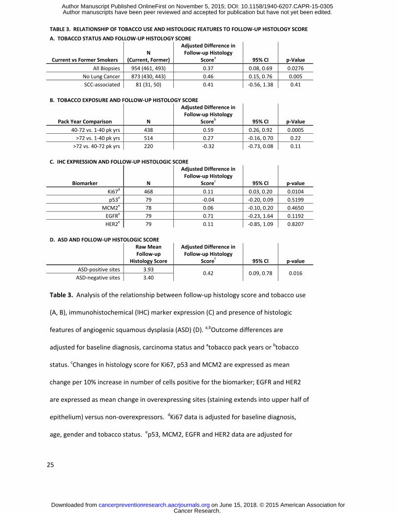
Tobacco history showed a significant relationship with follow-up histology scores. In multivariable
analyses, bronchial sites from current smokers showed a mean 0.37 (95% CI = 0.08 – 0.69) unit increase
in follow-up histology score when compared to former smokers (table 3A). In addition, an interaction
was noted between smoking status and lung cancer status. The effect of tobacco use was also
evaluated by comparisons of subjects divided into three groups characterized by increasing pack year
smoking histories (table 3B). Follow-up histology scores at biopsy sites from subjects with 40-72 pack
year smoking histories were significantly higher than those from subjects with less than 40 pack years.
Interestingly, there was no difference in follow-up histology scores when comparing sites from subjects
with more than 72 pack years to those with less than 40 or 40-72 pack year histories.
Relationship between histologic features and follow-up histology
Immunohistochemical expression of several biomarkers was evaluated to determine if altered
expression was associated with an effect on follow-up histology. In univariable analyses, HER2, p53 and
MCM2 did not show associations with altered follow-up histology score, but EGFR and Ki67
overexpression were associated with increased follow-up histology scores. We had previously
demonstrated that both of these parameters showed a direct correlation with baseline histology score,
and therefore adjusted models were employed (9). Ki67 expression retained a significant relationship
showing a 0.11 (95% CI = 0.03 – 0.20) unit increase in follow-up histology score per 10% increase in
Cancer Research. on June 15, 2018. © 2015 American Association forcancerpreventionresearch.aacrjournals.org Downloaded from
Author manuscripts have been peer reviewed and accepted for publication but have not yet been edited. Author Manuscript Published OnlineFirst on November 5, 2015; DOI: 10.1158/1940-6207.CAPR-15-0305
13
baseline Ki67 positivity (table 3C). Analysis of the relationship between ASD and follow-up histology
score was restricted to biopsies with a baseline diagnosis of squamous metaplasia without atypia or
higher (histology score of 3 – 7) because the morphologic features of ASD are generally not seen in non-
dysplastic lesions. Of note, it was found that there was a significantly higher proportion of high grade
dysplasia at baseline in the ASD versus dysplasia without ASD groups (71.9% vs. 53.0% respectively, p <
0.001). Nonetheless, after adjustment for a number of clinical parameters including baseline diagnosis,
the presence of histologic features of ASD was also associated with a significant increase in follow-up
histology score as compared to sites without features of ASD (table 3D).
DISCUSSION
The potential to use lesion specific changes over time as an indication of risk for the development of
invasive SCC of the lung was explored in this study. The findings demonstrated that an increased
frequency of sites that show persistence as or progression to HGD was associated with a significant 7.84
(CI = 1.56 – 39.39) fold increase in risk for development of invasive SCC, and indicated that subjects with
multiple dysplastic sites that persist or progress to HGD represent a subset of patients with aggressive
airway disease. The demonstration that subjects with multiple sites of persistent disease show the
strongest association with development of SCC emphasizes the importance of performing a thorough
evaluation of the airway, and adds support to the role of field carcinogenesis in the development of
invasive lung cancer. Increased risk for invasive SCC has previously been associated with multiple sites
of abnormal appearing mucosa by autofluorescence bronchoscopy (AFB) (30). Additionally, the data
demonstrate that higher histology scores in follow-up biopsies imply important differences in potential
for progression. For instance, cases with dysplastic sites that persisted or progressed to HGD showed a
significant increase in risk for development of SCC whereas those in which persistence as low or high
Cancer Research. on June 15, 2018. © 2015 American Association forcancerpreventionresearch.aacrjournals.org Downloaded from
Author manuscripts have been peer reviewed and accepted for publication but have not yet been edited. Author Manuscript Published OnlineFirst on November 5, 2015; DOI: 10.1158/1940-6207.CAPR-15-0305

14
grade BD did not, despite the fact that the latter definition of persistence allowed for inclusion of several
more cases in the overall evaluation. This finding is similar to those of Alaa, et. al., who demonstrated
that the development of new severely dysplastic lesions, regardless of the baseline histology, was more
common in subjects that developed invasive cancer or CIS (31). Our data showed similar findings,
demonstrating a significant relationship between development of SCC and presence of multiple
persistent HGDs when sites of any baseline histology score were included (see supplemental figure 2).
However, as discussed below, we and others have observed that CIS often regresses (20). Therefore,
establishment of a relationship between persistence of BD and risk for invasive SCC is an important
extension of these previous findings.
The demonstration of a relationship between HGD in follow-up biopsies and risk for invasive SCC may
also have implications for the management of patients at risk for aggressive airway disease. The tumors
that develop in association with persistent BD are more centrally located and are less likely to be
associated with identifiable radiographic abnormalities in the early course of disease. Thus, screening
for BD and identification of patients at high risk for progression to invasive SCC will likely require a
different modality from high resolution CT to be effective. Our findings suggest that multiply sampling
the airways may be important and that the employment of a bronchoscopic technique that increases
the sensitivity for detection of BD, such as AFB, may be advisable (27, 30). Furthermore, in patients with
features of aggressive airway disease, close follow-up would likely be indicated and consideration of
potential benefit from preventive therapy might be suggested. With respect to invasive cancer, our
study showed that subjects with persistent BD developed invasive cancer both at sites that were
associated with and remote from those with baseline dysplastic change. Eight subjects developed
incident SCC at sites that were previously biopsied and five at sites that had not been previously
biopsied (including one patient that developed synchronous, incident SCCs in two different contralateral
lung lobes). Six of the previously biopsied sites showed dysplasia in baseline biopsies, of which two had
demonstrated persistence prior to development of SCC (SCC was diagnosed in the second biopsy for the
Cancer Research. on June 15, 2018. © 2015 American Association forcancerpreventionresearch.aacrjournals.org Downloaded from
Author manuscripts have been peer reviewed and accepted for publication but have not yet been edited. Author Manuscript Published OnlineFirst on November 5, 2015; DOI: 10.1158/1940-6207.CAPR-15-0305

15
other four). Thus, two SCCs developed at non-dysplastic sites and six others developed at sites that had
not been previously sampled suggesting they did not appear abnormal on AFB. This may indicate that
chemopreventive rather than local therapy will be necessary to significantly reduce the incidence of SCC
in this setting. Finally, the findings also support the use of reduced bronchial histology scores as an
informative endpoint in trials evaluating efficacy of potential preventive agents. While we have shown
that the frequency of persistent BD is associated with subsequent development of SCC, a potential
drawback associated with our analysis is the inclusion of some incident SCC cases in which this tumor
represents a second lung primary. Information regarding therapies that patients with prior lung cancer
may have received was not available. It is possible that such treatments could influence the course of
bronchial dysplasia in the group of patients with prior carcinoma. However, our finding that primary
incident SCC is also associated with increased follow-up histology scores further supports a relationship
between persistence or progression of BD and risk for the development of invasive SCC.
The association of higher grades of dysplasia at baseline with increased histologic scores on follow-up
corroborates findings from the prospective study of Bota, et. al. (21) and the meta-analysis of follow-up
data from four different chemoprevention trials performed in the British Columbia Lung Health Study
that included over 700 subjects (2). Although different classifications of outcome were used in the latter
study, their finding of a 4-5 fold higher rate of progression in sites with baseline diagnoses of moderate
or severe dysplasia as compared to those with lower diagnoses is consistent with the findings in our
analysis. Strengths of our study that may more firmly establish some of these relationships include
fewer numbers of sites coming from subjects with prior lung or head and neck cancers (8.8%), inclusion
of sites with lengthy follow-up (48.2% with > 2 years), and confinement of our study group to subjects
from the non-treatment protocols or the placebo arm of prevention trials with positive findings.
Previously carcinoma-in-situ (CIS) has been reported to progress to invasive frequently with the majority
progressing to cancer in some reports (19). In our cohort, nine subjects had 23 sites that showed CIS
and had follow-up biopsies. Although histologically normal at the baseline biopsy, one of these sites
Cancer Research. on June 15, 2018. © 2015 American Association forcancerpreventionresearch.aacrjournals.org Downloaded from
Author manuscripts have been peer reviewed and accepted for publication but have not yet been edited. Author Manuscript Published OnlineFirst on November 5, 2015; DOI: 10.1158/1940-6207.CAPR-15-0305

16
developed CIS and progressed to invasive SCC seven months later. Additionally, two other subjects
developed incident SCCs, but not at their sites of CIS. While16 of 22 (72.7%) CIS sites persisted as HGD,
including all of those in cases with associated SCC, six sites in two subjects regressed to non-dysplastic
histology including five that were followed over a course of 35 months and were re-biopsied 1-3 times.
Taking the biopsy with the highest diagnosis prior to development of SCC, one site with CIS at baseline
(1/22, 4.54%), five sites with baseline moderate or severe dysplasia (5/282, 1.77%), one with baseline
LGD (1/204, 0.49%) and four with non-dysplastic baseline diagnoses progressed to SCC (4/667, 0.60%).
Although the overall number of CIS lesions is small in this cohort, the findings support the aggressive
nature that other publications have found to be associated with these lesions, but also suggests that the
rate of progression is not high and document regression of CIS.
Our data show that tobacco use has an impact on the course of BD with current tobacco users having
higher follow-up histology scores than former tobacco users. These findings compliment the findings of
Clement, et al. in which duration of smoking history was found to correlate with increased incidence of
bronchial dysplasia (32). This information could be useful clinically for physicians counseling their
patients to quit tobacco use as a measure to prevent the development of lung cancer.
Angiogenic squamous dysplasias were also shown to be associated with an increased level of atypia on
follow-up biopsy. Angiogenesis is well established as a prognostic factor in invasive carcinoma, and we
have previously shown that expression of vascular endothelial growth factor increases with higher
grades of BD (33). Furthermore, our recent analysis of vandetanib, the VEGFR2 inhibitor with multi-
target inhibitory capacity, showed preventive activity of this agent in a mouse model of lung
carcinogenesis (34). These findings also correlate with previous work that has demonstrated more
frequent ASD in subjects with lung cancer than in those without (35). Angiogenic changes could support
an increased level of epithelial cell proliferation that may be important in promoting BD persistence and
progression. Given that poor vascular integrity has been associated with VEGF dominant
Cancer Research. on June 15, 2018. © 2015 American Association forcancerpreventionresearch.aacrjournals.org Downloaded from
Author manuscripts have been peer reviewed and accepted for publication but have not yet been edited. Author Manuscript Published OnlineFirst on November 5, 2015; DOI: 10.1158/1940-6207.CAPR-15-0305

17
neoangiogenesis (36), it is also possible that ASD lesions are associated with an altered
microenvironment that promotes progression. Additionally, our IHC analyses suggest that higher levels
of expression of Ki67 could also serve as biomarkers of increased risk in BD.
The results of this study suggest that an important subset of aggressive airway disease is represented by
cases that show the presence of multiple dysplastic lesions that persist or progress to HGD, and
demonstrate that in patients with this presentation there is increased risk for invasive SCC. Further
characterization of these persistent lesions should allow for the development of more precise predictive
markers. Furthermore, obtaining an understanding of the biologic characteristics that drive these BD
with a high risk for progression to invasive lung cancer will help identify effective targets for prevention.
Cancer Research. on June 15, 2018. © 2015 American Association forcancerpreventionresearch.aacrjournals.org Downloaded from
Author manuscripts have been peer reviewed and accepted for publication but have not yet been edited. Author Manuscript Published OnlineFirst on November 5, 2015; DOI: 10.1158/1940-6207.CAPR-15-0305

18
REFERENCES
1. Travis WD, Brambilla E, Burke AP, Marx A, Nicholson AG. WHO Classification of Tumours of the
Lung, Pleara, Thymus and Heart, 4th Edition. Lyon: IARC; 2015. p. 59-63.
2. Ishizumi T, McWilliams A, Macaulay C, Gazdar A and Lam S. Natural history of bronchial
preinvasive lesions. Cancer Metastasis Rev 2010;29:5-14.
3. Keith RL, Blatchford PJ, Kittelson J, Minna JD, Kelly K, Massion PP, et. al. Oral iloprost improves
endobronchial dysplasia in former smokers. Cancer Prev Res (Phila) 2011;4:793-80.
4. Kelly K, Kittelson J, Franklin WA, Kennedy TC, Klein CE, Keith RL, et.al. A randomized phase II
chemoprevention trial of 13-CIS retinoic acid with or without alpha tocopherol or observation in
subjects at high risk for lung cancer. Cancer Prev Res (Phila) 2009;5:440-9.
5. Lam, S., leRiche, J. C., McWilliams, A., MacAulay, C., Dyachkova, Y., Szabo, E., et al. A randomized
phase IIb trial of pulmicort turbuhaler (budesonide) in persons with dysplasia of the bronchial
epithelium. Clinical Cancer Research 2004;10:6502–6511.
6. Lam, S., McWilliams, A., Leriche, J., MacAulay, C., Wattenburg, L., & Szabo, E. A phase I study of
myo-inositol for lung cancer chemoprevention. Cancer Epidemiology Biomarkers & Prevention
2006;15:1526–1531.
7. Wistuba, I. I., Behrens, C., Milchgrub, S., Bryant, D., Hung, J., Minna, J. D., et al. Sequential
molecular abnormalities are involved in the multistage development of squamous cell lung
carcinoma. Oncogene 1999;18:643–650.
8. Wistuba, I. I., Behrens, C., Virmani, Ak, Mele, G., Milchgrub, S., Girard, L., et al. High resolution
chromosome 3p allelotyping of human lung cancer and bronchial epithelium reveals multiple,
discontinuous sites of 3pallele loss and three regions of frequent breakpoints. Cancer Research
2000;60:1949–1960.
Cancer Research. on June 15, 2018. © 2015 American Association forcancerpreventionresearch.aacrjournals.org Downloaded from
Author manuscripts have been peer reviewed and accepted for publication but have not yet been edited. Author Manuscript Published OnlineFirst on November 5, 2015; DOI: 10.1158/1940-6207.CAPR-15-0305
19
9. Merrick DT, Kittelson J, Winterhalder R, Kotantoulas G, Ingeberg S, Keith RL, et. al. Analysis of c-
ErbB1/epidermal growth factor receptor and c-ErbB2/HER-2 expression in bronchial dysplasia:
evaluation of potential targets for chemoprevention of lung cancer. Clin Cancer Res
2006;12:2281-8.
10. Mascaux C, Laes JF, Anthoine G, Haller A, Ninane V, Burny A, et. al. Evolution of microRNA
expression during human bronchial squamous carcinogenesis. Eur Respir J 2009;33:352-9.
11. Jonsson, S., Varella-Garcia, M., Miller, Y., Wolf, H. J., Byers, T., Braudrick, S., et al. Chromosomal
aneusomy in bronchial high grade lesions is associated with invasive lung cancer. American
Journal of Respiratory and Critical Care Medicine 2008;177:342–347.
12. Massion, P., Zou, Y., Uner, H., Kiatsimkul, P.,Wolf, H. J., Baron, A. E., et al. Recurrent genomic
gains in preinvasive lesions as a biomarker of risk for lung cancer. PLoS ONE 2009;4:e5611.
13. Gustafson, A.M., Soldi, R., Anderlind, C., Scholand, M.B., Qian, J., Zhang, X., et. al. Airway PI3K
pathway activation is an early and reversible event in lung cancer development. Sci Transl Med
2010;2:1-11.
14. Lantuejol, S., Raynaud, C., Salameire, D., Gazzeri, S., Moro-Sibilot, D., Soria, J-C., et. al. Telomere
maintenance and DNA damage response during lung carcinogenesis. Cllin Cancer Res
2010;16:2979-2988.
15. Jeanmart, Lantuejoul, S., Fievet, F., Moro, D., Sturm, N., Brambilla, C., et al. Value of
immunohistochemical markers in preinvasive bronchial lesions in risk assessment of lung cancer.
Clin Cancer Res 2003;9:2195–2203.
Cancer Research. on June 15, 2018. © 2015 American Association forcancerpreventionresearch.aacrjournals.org Downloaded from
Author manuscripts have been peer reviewed and accepted for publication but have not yet been edited. Author Manuscript Published OnlineFirst on November 5, 2015; DOI: 10.1158/1940-6207.CAPR-15-0305

20
16. Salaun, M., Sesboue, R., Moreno-Swire, S., Metayer, J., Bota, S., Bourguignon, J., et al. Molecular
predictive factors for progression of high-grade preinvasive bronchial lesions. American Journal
of Respiratory and Critical Care Medicine 2008;177:880–886.
17. McCaughan F, Pole JCM, Bankier AT, Konfortov BA, Carroll B, Falzon M,et. al. Progressive 3q
Amplification Consistently Targets SOX2 in Preinvasive Squamous Lung Cancer. Am J Resp Crit
Care Med 2010;182:83-91.
18. Breuer, R. H., Pasic, A., Smit, E. F., van Vliet, E., Vonk Noordegraaf, A., Risse, E. J., et al. The
natural course of preneoplastic lesions in bronchial epithelium. Clin Cancer Res 2005;11:537–
543.
19. Venmans, B., van Boxem, A., Smit, E., Postmus, P., & Sutedja, T. Outcome of bronchial carcinoma
in-situ. Chest 2000;117:1572–1576.
20. Moro-Sibilot, D., Fievet, F., Jeanmart, M., Lantuejoul, S., Arbib, F., Laverribre, M. H., et al. Clinical
prognostic indicators of high-grade pre-invasive bronchial lesions. Eur Respirology Journal
2004;24:24–29.
21. Bota, S., Auliac, J. B., Paris, C., Metayer, J., Sesboue, R., Nouvet, G., et al. Follow-up of bronchial
precancerous lesions and carcinoma in situ using fluorescence endoscopy. American Journal of
Respiratory and Critical Care Medicine 2001;164:1688–1693.
22. Salaun, M., Bota, S., & Thiberville, L. Long-term followup of severe dysplasia and carcinoma in-
situ of the bronchus. J Thorac Oncol 2009;4:1187–1188.
23. Hoshino, H., Shibuya, K., Chiyo, M., Iyoda, A., Yoshida, S., Sekine, Y., et al. Biological features of
bronchial squamous dysplasia followed up by autofluorescence bronchoscopy. Lung Cancer
2004;46:187–196.
Cancer Research. on June 15, 2018. © 2015 American Association forcancerpreventionresearch.aacrjournals.org Downloaded from
Author manuscripts have been peer reviewed and accepted for publication but have not yet been edited. Author Manuscript Published OnlineFirst on November 5, 2015; DOI: 10.1158/1940-6207.CAPR-15-0305

21
24. Pasic, A., van Vliet, E., Breuer, R. H., Risse, E. J., Snijders, P. J., Postmus, P. E., et al. Smoking
behavior does not influence the natural course of pre-invasive lesions in bronchial mucosa. Lung
Cancer 2004;45:153–154.
25. Banerjee, A. K. Preinvasive lesions of the bronchus. J Thorac Oncol 2009;4:545–551.
26. Lam, S., Kennedy, T., Unger, M., Miller, Y. E., Gelmont, D., Rusch, V., et al. Localization of
bronchial intraepithelial neoplastic lesions by fluorescence bronchoscopy. Chest 1998:113:696–
702.
27. Hirsch, F. R., Prindiville, S. A., Miller, Y. E., Franklin, W. A., Dempsey, E. C., Murphy, J. R., et al.
Fluorescence versus white-light bronchoscopy for detection of preneoplastic lesions: A
randomized study. J Natl Cancer Inst 2001;93:1385–1391.
28. Edell, E., Lam, S., Pass, H., Miller, Y. E., Sutedja, T., Kennedy, T., et al. Detection and localization
of intraepithelial neoplasia and invasive carcinoma using fluorescence-reflectance
bronchoscopy: An international, multi-center clinical trial. J Thorac Oncol 2009;4:49–54.
29. Keith RL, Miller YE, Gemmill RM, Drabkin HA, Dempsey EC, Kennedy TC, et. al. Angiogenic
squamous dysplasia in bronchi of individuals at high risk for lung cancer. Clin Cancer Res
2000;6:1616-25.
30. Pasic A, Vonk-Noordegraaf A, Risse EK, Postmus PE, Sutedja TG. Multiple suspicious lesions
detected by autofluorescence bronchoscopy predict malignant development in the bronchial
mucosa in high risk patients. Lung Cancer 2003;41:295-301.
31. Alaa MRM, Shibuya K, Fujiwara T, Wada H, Hoshino H, Yoshida S, et. al. Risk of lung cancer in
patients with preinvasive bronchial lesions followed by autofluorescence bronchoscopy and
chest computed tomography. Lung Cancer 2011;72: 303–308.
Cancer Research. on June 15, 2018. © 2015 American Association forcancerpreventionresearch.aacrjournals.org Downloaded from
Author manuscripts have been peer reviewed and accepted for publication but have not yet been edited. Author Manuscript Published OnlineFirst on November 5, 2015; DOI: 10.1158/1940-6207.CAPR-15-0305

22
32. Clément-Duchênea C, Alla F, Gauchotte G, Marie B,Carnin C, Menard O, et al. Is there a
relationship between the presence of lung mucosa preinvasive lesions and lung cancer
incidence? Influence of tobacco consumption. Lung Cancer 2014;84:134–138.
33. Merrick DT, Haney J, Petrunich S, Sugita M, Miller YE, Keith RL, et. al. Overexpression of vascular
endothelial growth factor and its receptors in bronchial dysplasia demonstrated by quantitative
RT-PCR analysis. Lung Cancer 2005;48:31-45.
34. Karoor V, Le M, Merrick D, Dempsey EC, Miller YE.Cancer Vascular endothelial growth factor
receptor 2-targeted chemoprevention of murine lung tumors. Prev Res (Phila) 2010;3:1141-7.
35. Karimi S, Mohammadi F, Khodadad K, Sadr M, Seyfollahi L, and Masjedi MR. Relationship
between angiogenic squamous dysplasia and bronchogenic carcinoma in patients undergoing
white light bronchoscopy. Can Respir J 2012;19:201-208.
36. Greenberg JI, and Cheresh DA. VEGF as an inhibitor of tumor vessel maturation: implications for
cancer therapy. Expert Opin Biol Ther 2009;9:1347-1356.
Cancer Research. on June 15, 2018. © 2015 American Association forcancerpreventionresearch.aacrjournals.org Downloaded from
Author manuscripts have been peer reviewed and accepted for publication but have not yet been edited. Author Manuscript Published OnlineFirst on November 5, 2015; DOI: 10.1158/1940-6207.CAPR-15-0305

23
TABLE 1. HISTOLOGIC AND CLINICAL CHARACTERISTICS OF BASELINE BIOPSIES
Non-Dys (%) LGD (%) HGD (%) p-val LUNG CANCER STATUSa
Cancer negative 598 (59.0) 179 (17.7) 237 (23.4) <0.001 Prevalent lung cancer 34 (46.6) 8 (11.0) 31 (42.5)
Incident lung cancer 33 (42.9) 14 (18.2) 30 (40.0)
LUNG CANCER SUBTYPEb Cancer negative 598 (58.7) 179 (17.8) 237 (23.5)
<0.001 SCC positive 34 (37.6) 19 (16.8) 53 (45.5) Adenocarcinoma positive 16 (85.0) 1 (5.0) 2 (10.0)
TOBACCO STATUS Current smoker 231 (42.8) 138 (25.6) 171 (31.7)
<0.001 Former smoker 423 (68.9) 66 (10.8) 125 (20.4) Never smoker 13 (81.3) 0 (0.0) 3 (18.8)
TOBACCO - PACK YEARS Never smoker 13 (81.3) 0 (0.0) 3 (18.8)
<0.001 0-40 pack years 273 (64.8) 73 (17.3) 75 (17.8)
40-72 pack years 238 (48.7) 107 (21.9) 144 (29.4) >72 pack years 143 (58.6) 24 (9.8) 77 (31.6)
GENDER Male 478 (54.9) 162 (18.6) 231 (26.5)
0.0369 Female 189 (63.2) 42 (14.1) 68 (22.7)
AGE <55 y.o. 189 (59.1) 66 (20.6) 65 (20.3)
0.0079 55-65 y.o. 259 (52.6) 88 (17.9) 145 (29.5) >65 y.o. 219 (61.2) 50 (14.0) 89 (24.8)
Table 1. Correlation between baseline histology and clinical
variables. aCancer status unknown for 6 sites; bCancer subtype
unknown for 31 sites.
Cancer Research. on June 15, 2018. © 2015 American Association forcancerpreventionresearch.aacrjournals.org Downloaded from
Author manuscripts have been peer reviewed and accepted for publication but have not yet been edited. Author Manuscript Published OnlineFirst on November 5, 2015; DOI: 10.1158/1940-6207.CAPR-15-0305

24
TABLE 2. CANCER STATUS AND FOLLOW-UP HISTOLOGY SCORE A. FOLLOW-UP HISTOLOGY SCORE IN SUBJECTS WITH INCIDENT OR PREVALENT LUNG SCC OR ADENOCARCINOMA VS SUBJECTS WITHOUT ASSOCIATED LUNG CANCER
Comparison Na Difference in
Follow-up HSb Confidence
Interval p-value SCC vs. No CA 90, 1022 0.82 0.32, 1.32 0.001
Adenocarcinoma vs. No CA 19, 1022 0.10 -0.96, 1.16 0.855 SCC vs. Adenocarcinoma 90, 19 0.72 -0.44, 1.88 0.221
B. FOLLOW-UP HISTOLOGY SCORES FROM SUBJECTS WITH PREVALENT LUNG SCC OR ADENOCARCINOMA VS SUBJECTS WITHOUT ASSOCIATED LUNG CANCER
Prevalent Lung Cancer vs. No Cancer Na Difference in
Follow-up HSb Confidence
Interval p-value Prevalent SCC vs. No CA 65, 1022 0.73 0.17, 1.29 0.011
Prevalent Adenocarcinoma vs. No CA 16, 1022 0.01 -1.04, 1.05 0.999
C. FOLLOW-UP HISTOLOGY SCORES FROM SUBJECTS WITH INCIDENT LUNG SCC VS SUBJECTS WITHOUT ASSOCIATED LUNG CANCER
SCC vs. No Lung Cancer Na Difference in
Follow-up HSb Confidence
Interval p-value Incident SCC, All vs. No CA 61, 1022 1.55 0.80, 2.30 <0.001
Incident SCC, No prior CA vs. No CA 30, 1022 0.99 0.08, 1.90 0.034
Table 2. Differences in follow-up histology scores (HS) as compared to cases
without known lung cancer (No CA) for all prevalent or incident lung cancer
cases (A), prevalent lung cancer cases alone (B) or incident SCC cases alone (C).
aNumber of biopsies (N) for each of the comparator groups respectively. bData
for all analyses are adjusted for baseline diagnosis and tobacco parameters in
the multivariable linear mixed effects model.
Cancer Research. on June 15, 2018. © 2015 American Association forcancerpreventionresearch.aacrjournals.org Downloaded from
Author manuscripts have been peer reviewed and accepted for publication but have not yet been edited. Author Manuscript Published OnlineFirst on November 5, 2015; DOI: 10.1158/1940-6207.CAPR-15-0305
25
TABLE 3. RELATIONSHIP OF TOBACCO USE AND HISTOLOGIC FEATURES TO FOLLOW-UP HISTOLOGY SCORE A. TOBACCO STATUS AND FOLLOW-UP HISTOLOGY SCORE
Current vs Former Smokers N
(Current, Former)
Adjusted Difference in Follow-up Histology
Scorea 95% CI p-Value All Biopsies 954 (461, 493) 0.37 0.08, 0.69 0.0276
No Lung Cancer 873 (430, 443) 0.46 0.15, 0.76 0.005 SCC-associated 81 (31, 50) 0.41 -0.56, 1.38 0.41
B. TOBACCO EXPOSURE AND FOLLOW-UP HISTOLOGY SCORE
Pack Year Comparison N
Adjusted Difference in Follow-up Histology
Scoreb 95% CI p-Value 40-72 vs. 1-40 pk yrs 438 0.59 0.26, 0.92 0.0005
>72 vs. 1-40 pk yrs 514 0.27 -0.16, 0.70 0.22 >72 vs. 40-72 pk yrs 220 -0.32 -0.73, 0.08 0.11
C. IHC EXPRESSION AND FOLLOW-UP HISTOLOGIC SCORE
Biomarker N
Adjusted Difference in Follow-up Histology
Scorec 95% CI p-value Ki67d 468 0.11 0.03, 0.20 0.0104 p53e 79 -0.04 -0.20, 0.09 0.5199
MCM2e 78 0.06 -0.10, 0.20 0.4650 EGFRe 79 0.71 -0.23, 1.64 0.1192 HER2e 79 0.11 -0.85, 1.09 0.8207
D. ASD AND FOLLOW-UP HISTOLOGIC SCORE
Raw Mean Follow-up
Histology Score
Adjusted Difference in Follow-up Histology
Scoref 95% CI p-value ASD-positive sites 3.93 0.42 0.09, 0.78 0.016
ASD-negative sites 3.40 Table 3. Analysis of the relationship between follow-up histology score and tobacco use
(A, B), immunohistochemical (IHC) marker expression (C) and presence of histologic
features of angiogenic squamous dysplasia (ASD) (D). a,bOutcome differences are
adjusted for baseline diagnosis, carcinoma status and atobacco pack years or btobacco
status. cChanges in histology score for Ki67, p53 and MCM2 are expressed as mean
change per 10% increase in number of cells positive for the biomarker; EGFR and HER2
are expressed as mean change in overexpressing sites (staining extends into upper half of
epithelium) versus non-overexpressors. dKi67 data is adjusted for baseline diagnosis,
age, gender and tobacco status. ep53, MCM2, EGFR and HER2 data are adjusted for
Cancer Research. on June 15, 2018. © 2015 American Association forcancerpreventionresearch.aacrjournals.org Downloaded from
Author manuscripts have been peer reviewed and accepted for publication but have not yet been edited. Author Manuscript Published OnlineFirst on November 5, 2015; DOI: 10.1158/1940-6207.CAPR-15-0305

26
baseline diagnosis only. fThe calculated difference in histology score is adjusted for
baseline diagnosis, tobacco status and pack year history in the multivariable linear mixed
effects models.
Cancer Research. on June 15, 2018. © 2015 American Association forcancerpreventionresearch.aacrjournals.org Downloaded from
Author manuscripts have been peer reviewed and accepted for publication but have not yet been edited. Author Manuscript Published OnlineFirst on November 5, 2015; DOI: 10.1158/1940-6207.CAPR-15-0305
27
FIGURE LEGENDS
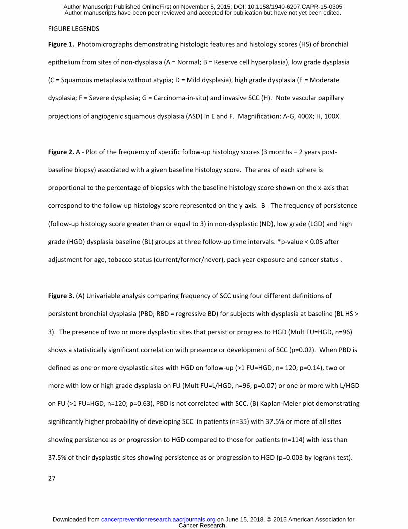
Figure 1. Photomicrographs demonstrating histologic features and histology scores (HS) of bronchial
epithelium from sites of non-dysplasia (A = Normal; B = Reserve cell hyperplasia), low grade dysplasia
(C = Squamous metaplasia without atypia; D = Mild dysplasia), high grade dysplasia (E = Moderate
dysplasia; F = Severe dysplasia; G = Carcinoma-in-situ) and invasive SCC (H). Note vascular papillary
projections of angiogenic squamous dysplasia (ASD) in E and F. Magnification: A-G, 400X; H, 100X.
Figure 2. A - Plot of the frequency of specific follow-up histology scores (3 months – 2 years post-
baseline biopsy) associated with a given baseline histology score. The area of each sphere is
proportional to the percentage of biopsies with the baseline histology score shown on the x-axis that
correspond to the follow-up histology score represented on the y-axis. B - The frequency of persistence
(follow-up histology score greater than or equal to 3) in non-dysplastic (ND), low grade (LGD) and high
grade (HGD) dysplasia baseline (BL) groups at three follow-up time intervals. *p-value < 0.05 after
adjustment for age, tobacco status (current/former/never), pack year exposure and cancer status .
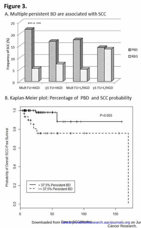
Figure 3. (A) Univariable analysis comparing frequency of SCC using four different definitions of
persistent bronchial dysplasia (PBD; RBD = regressive BD) for subjects with dysplasia at baseline (BL HS >
3). The presence of two or more dysplastic sites that persist or progress to HGD (Mult FU=HGD, n=96)
shows a statistically significant correlation with presence or development of SCC (p=0.02). When PBD is
defined as one or more dysplastic sites with HGD on follow-up (>1 FU=HGD, n= 120; p=0.14), two or
more with low or high grade dysplasia on FU (Mult FU=L/HGD, n=96; p=0.07) or one or more with L/HGD
on FU (>1 FU=HGD, n=120; p=0.63), PBD is not correlated with SCC. (B) Kaplan-Meier plot demonstrating
significantly higher probability of developing SCC in patients (n=35) with 37.5% or more of all sites
showing persistence as or progression to HGD compared to those for patients (n=114) with less than
37.5% of their dysplastic sites showing persistence as or progression to HGD (p=0.003 by logrank test).
Cancer Research. on June 15, 2018. © 2015 American Association forcancerpreventionresearch.aacrjournals.org Downloaded from
Author manuscripts have been peer reviewed and accepted for publication but have not yet been edited. Author Manuscript Published OnlineFirst on November 5, 2015; DOI: 10.1158/1940-6207.CAPR-15-0305

28
Cancer Research. on June 15, 2018. © 2015 American Association forcancerpreventionresearch.aacrjournals.org Downloaded from
Author manuscripts have been peer reviewed and accepted for publication but have not yet been edited. Author Manuscript Published OnlineFirst on November 5, 2015; DOI: 10.1158/1940-6207.CAPR-15-0305

Published OnlineFirst November 5, 2015.Cancer Prev Res Daniel T. Merrick, Dexiang Gao, York E. Miller, et al. CARCINOMAWITH DEVELOPMENT OF INVASIVE SQUAMOUS CELL PERSISTENCE OF BRONCHIAL DYSPLASIA IS ASSOCIATED
Updated version
10.1158/1940-6207.CAPR-15-0305doi:
Access the most recent version of this article at:
Material
Supplementary
05.DC1
http://cancerpreventionresearch.aacrjournals.org/content/suppl/2016/01/09/1940-6207.CAPR-15-03Access the most recent supplemental material at:
Manuscript
Authoredited. Author manuscripts have been peer reviewed and accepted for publication but have not yet been
E-mail alerts related to this article or journal.Sign up to receive free email-alerts
Subscriptions
Reprints and
To order reprints of this article or to subscribe to the journal, contact the AACR Publications
Permissions
Rightslink site. Click on "Request Permissions" which will take you to the Copyright Clearance Center's (CCC)
.05http://cancerpreventionresearch.aacrjournals.org/content/early/2015/11/05/1940-6207.CAPR-15-03To request permission to re-use all or part of this article, use this link
Cancer Research. on June 15, 2018. © 2015 American Association forcancerpreventionresearch.aacrjournals.org Downloaded from
Author manuscripts have been peer reviewed and accepted for publication but have not yet been edited. Author Manuscript Published OnlineFirst on November 5, 2015; DOI: 10.1158/1940-6207.CAPR-15-0305