peripheral neuroepithelioma with ganglion cells: report of two cases and review of the literature
TRANSCRIPT

CASE REPORTS
Peripheral Neuroepithelioma withGanglion Cells: Report of Two Casesand Review of the Literature
SUSAN WILLIAMS,1 DAVID M. PARHAM,2 AND JESSE J. JENKINS III3*1Department of Pathology, University of Tennessee, 899 Madison Avenue, Memphis, TN 38163, USA2Department of Pathology, University of Arkansas for Medical Sciences, 800 Marshall Street,Little Rock, AR 72202, USA3Department of Pathology and Laboratory Medicine, St. Jude Children’s Research Hospital, 332 North LauderdaleStreet, and Department of Pathology, University of Tennessee, 899 Madison Avenue, Memphis, TN 38105-2794,USA
Received August 27, 1997; accepted March 27, 1998.
ABSTRACTPeripheral neuroepitheliomas, also known as peripheralprimitive neuroectodermal tumors, are by definitionprimitive embryonal lesions generally composed of poorlydifferentiated neuroectodermal elements. We have exam-ined two cases that paradoxically contain extensive fociof ganglionic differentiation similar to that of ganglioneu-roblastoma, in addition to primitive elements. One tu-mor arose from the chest wall of an 8-year-old male andthe other from the abdominal wall of a 15-year-old male.Differentiation into a mature ganglionic phenotype wasconfirmed by immunohistochemistry in one case. Rareperipheral neuroepitheliomas have a capacity for matu-ration that is not generally appreciated. These lesionsshould not be confused with ganglioneuroblastomas,which are genotypically unrelated neoplasms.
Key words: peripheral neuroepithelioma, ganglion cells,neuroblastoma, ganglioneuroblastoma, primitive neuro-ectodermal tumor
INTRODUCTIONPeripheral neuroepithelioma (PN), or peripheralprimitive neuroectodermal tumor, is an embryonal
neoplasm that is closely related to Ewing’s sar-coma, as both a pathological and clinical entity.This relatedness is apparent from their commonreciprocal translocation, the t(11;22)(q24;q12),which fuses the Fli-1 and EWS genes [1,2], fromphenotypic features, such as expression of parasym-pathetic neuroenzymes [3] and the MIC2 antigen[4–6]; and from their tendency to occur in the softtissues and skeletal system of adolescents andyoung adults and to metastasize to the lung [7,8].In contrast, neuroblastomas are characterized bychromosome 1p deletions [9,10] and N-myc ampli-fication and/or overexpression [11,12], and by occur-rence in the adrenal glands and sympathetic chainof infants and young children, with metastasisprimarily to bone marrow and lymph nodes andparadoxical absence of pulmonary spread [13].
Like Ewing’s sarcomas, PN are generally primi-tive neoplasms with only rudimentary neural differ-entiation. However, recent observations indicatethat there is a wider range of phenotypic potentialthan was initially realized, as lesions with epithe-lial, glial, schwannian, and myogenic features have*Corresponding author
Pediatric and Developmental Pathology 2, 42–49, 1999 Pediatric and Developmental Pathology
r1999 Society for Pediatric Pathology
been described [14–16]. We have examined two PNthat appear to further extend the range of differen-tiation shown by these lesions. These tumors arosein the soft tissues of older children, were similar totypical PN, but were characterized by extensiveareas of ganglionic differentiation, similar to thatseen in neuroblastic tumors. Because PN and neu-roblastomas are such fundamentally different neo-plasms with radically dissimilar therapies, it isimportant to recognize that rare PN have a capacityfor similar neuronal maturation.
MATERIALS AND METHODSBoth cases were retrieved from the files of St. JudeChildren’s Research Hospital. For case 1, ninehematoxylin and eosin–stained slides were avail-able for review. For case 2, two paraffin blocks offormalin-fixed tissues were available and werestained with hematoxylin and eosin. Clinical chartswere reviewed for each patient.
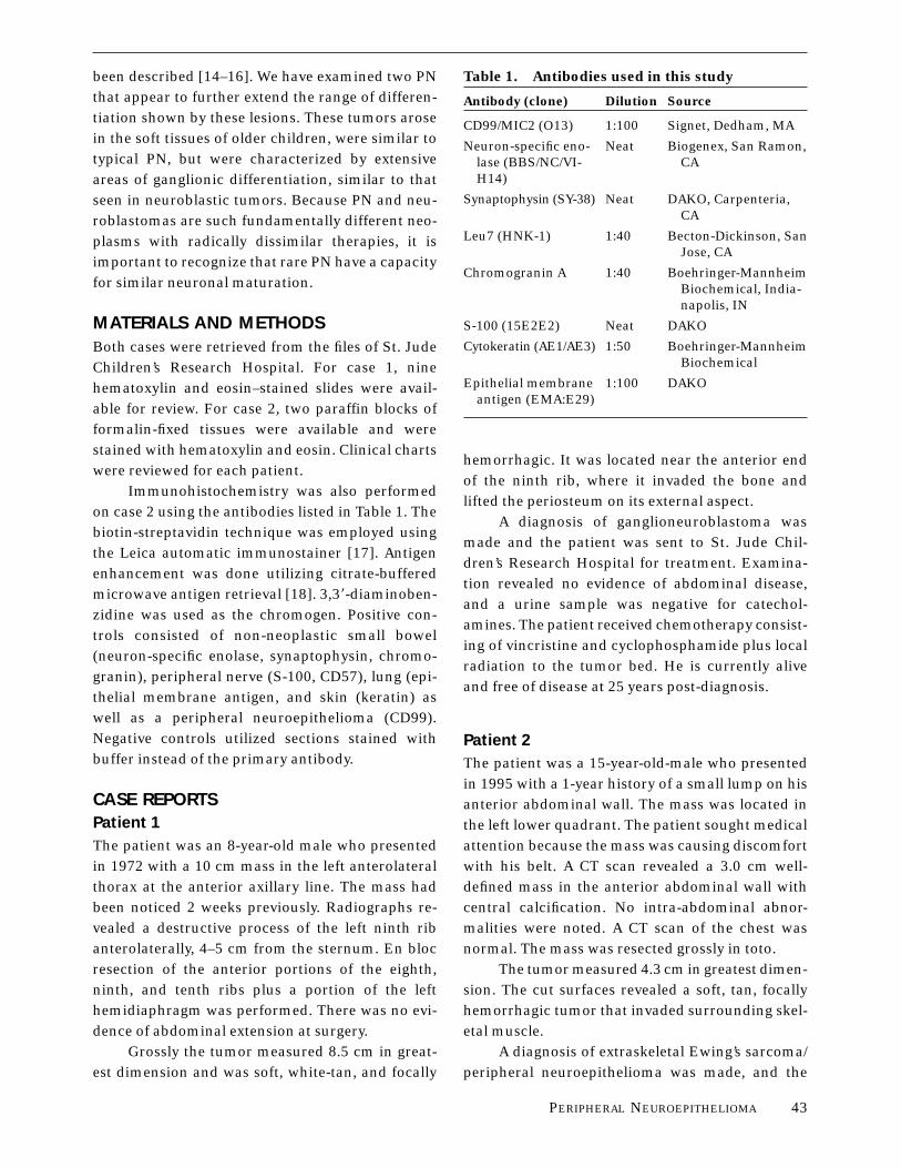
Immunohistochemistry was also performedon case 2 using the antibodies listed in Table 1. Thebiotin-streptavidin technique was employed usingthe Leica automatic immunostainer [17]. Antigenenhancement was done utilizing citrate-bufferedmicrowave antigen retrieval [18]. 3,38-diaminoben-zidine was used as the chromogen. Positive con-trols consisted of non-neoplastic small bowel(neuron-specific enolase, synaptophysin, chromo-granin), peripheral nerve (S-100, CD57), lung (epi-thelial membrane antigen, and skin (keratin) aswell as a peripheral neuroepithelioma (CD99).Negative controls utilized sections stained withbuffer instead of the primary antibody.
CASE REPORTSPatient 1The patient was an 8-year-old male who presentedin 1972 with a 10 cm mass in the left anterolateralthorax at the anterior axillary line. The mass hadbeen noticed 2 weeks previously. Radiographs re-vealed a destructive process of the left ninth ribanterolaterally, 4–5 cm from the sternum. En blocresection of the anterior portions of the eighth,ninth, and tenth ribs plus a portion of the lefthemidiaphragm was performed. There was no evi-dence of abdominal extension at surgery.
Grossly the tumor measured 8.5 cm in great-est dimension and was soft, white-tan, and focally
hemorrhagic. It was located near the anterior endof the ninth rib, where it invaded the bone andlifted the periosteum on its external aspect.
A diagnosis of ganglioneuroblastoma wasmade and the patient was sent to St. Jude Chil-dren’s Research Hospital for treatment. Examina-tion revealed no evidence of abdominal disease,and a urine sample was negative for catechol-amines. The patient received chemotherapy consist-ing of vincristine and cyclophosphamide plus localradiation to the tumor bed. He is currently aliveand free of disease at 25 years post-diagnosis.
Patient 2The patient was a 15-year-old-male who presentedin 1995 with a 1-year history of a small lump on hisanterior abdominal wall. The mass was located inthe left lower quadrant. The patient sought medicalattention because the mass was causing discomfortwith his belt. A CT scan revealed a 3.0 cm well-defined mass in the anterior abdominal wall withcentral calcification. No intra-abdominal abnor-malities were noted. A CT scan of the chest wasnormal. The mass was resected grossly in toto.
The tumor measured 4.3 cm in greatest dimen-sion. The cut surfaces revealed a soft, tan, focallyhemorrhagic tumor that invaded surrounding skel-etal muscle.
A diagnosis of extraskeletal Ewing’s sarcoma/peripheral neuroepithelioma was made, and the
Table 1. Antibodies used in this study
Antibody (clone) Dilution Source
CD99/MIC2 (O13) 1:100 Signet, Dedham, MA
Neuron-specific eno-lase (BBS/NC/VI-H14)
Neat Biogenex, San Ramon,CA
Synaptophysin (SY-38) Neat DAKO, Carpenteria,CA
Leu7 (HNK-1) 1:40 Becton-Dickinson, SanJose, CA
Chromogranin A 1:40 Boehringer-MannheimBiochemical, India-napolis, IN
S-100 (15E2E2) Neat DAKO
Cytokeratin (AE1/AE3) 1:50 Boehringer-MannheimBiochemical
Epithelial membraneantigen (EMA:E29)
1:100 DAKO
PERIPHERAL NEUROEPITHELIOMA 43

patient was sent to St. Jude Children’s ResearchHospital for treatment. He was placed on theEwing’s sarcoma protocol, which includes combina-tion chemotherapy (dactinomycin, vincristine, ifos-famide, VP-16, cyclophosphamide, and adriamy-cin) and local radiation therapy to the tumor bed.The patient is alive and free of disease at 24 monthspost-diagnosis.
RESULTSThe microscopic features of these two cases weresimilar. On low power examination, both caseswere characterized by large islands and anastomos-ing trabeculae of small hyperchromatic tumor cells(Fig. 1). While the tumor islands and trabeculae incase 1 were fairly well demarcated, the tumor nestsin case 2 were less well defined with single-cell and‘‘Indian-file’’ type infiltration of the stroma andskeletal muscle at the edges of the nests. The cellsin these nests in both cases were small with scantcytoplasm and round to polygonal nuclei with
granular chromatin and occasional small nucleoli.Rosettes were not present. Small foci of necrosiswere common.
The most striking feature on high magnifica-tion were several large foci in which the tumor cellnests were separated by large sheets of ganglioncells embedded in fibrillar neuropil matrix (Fig.2a,b). There was a range of ganglionic maturationfrom cells with vesicular nuclei, small nucleoli, andscant cytoplasm (Fig. 2b) to cells with prominentnucleoli and abundant well-defined cytoplasm(Fig. 2a). Multinucleation was common. Nissl sub-stance was not apparent even in the most maturecells. The ganglion cells were intimately admixedwith the primitive round cells in the periphery ofthe tumor islands (Fig. 2a).
The stroma in areas separate from the gan-glion cells was largely acellular and composed ofcollagen fibers and a few elongated fibroblasts.Occasional myxoid areas were present in case 1,whereas case 2 had several areas of stromal calcifi-cation. Stroma composed of S-100-positive schwanncells was not a feature of either tumor.
The pattern of growth in both tumors wasinvasive with both single-cell infiltration and largepushing borders. In case 1 the rib was involved,with almost complete replacement of the marrowby tumor. Although a portion of the left hemidia-phragm was suspicious for involvement at surgery,histologic examination revealed no evidence oftumor. The surgical margins in case 1 were free oftumor. Case 2 had invasion of surrounding skeletalmuscle and adipose tissue with microscopicallypositive margins.
A mature ganglionic phenotype was con-firmed in case 2 by immunohistochemistry. Theganglion cell cytoplasm including the fibrillarstroma was strongly positive for synaptophysin,chromogranin, and neuron-specific enolase (NSE),whereas the primitive round cells were only weaklypositive for synaptophysin and NSE and werenegative for chromogranin (Fig. 3a). In contrast,CD99 (MIC2) antigen expression was strongly posi-tive in the small round-cell population and wasnegative in the ganglion cells (Fig. 3b). Addition-ally, weak Leu7 immunoreactivity was noted in thesmall round cells, whereas the ganglion cells werenegative.
Figure 1. Typical appearance of the islands of a smallhyperchromatic tumor cell.
44 S. WILLIAMS ET AL.

Immunostaining for epithelial membrane an-tigen, S-100, and cytokeratin was negative in bothcell populations as well as the stroma.
DISCUSSIONPeripheral neuroepitheliomas are generally primi-tive neoplasms with only rudimentary neural differ-entiation. A wider range of phenotypic expressionhas been observed using immunohistochemicalstains for epithelial, glial, schwannian, and myo-genic antigens [14–16]. The two PN presented herefurther extend the range of differentiation exhib-ited by these lesions, showing the ability of thesetumors to undergo differentiation to a matureganglionic phenotype both histologically and immu-nohistochemically.
The two tumors presented here represent theeighth and ninth reported cases in the literaturewith this differentiation pattern. Shuangshoti firstreported this phenomenon in a case of a soft tissue
mass in the posterolateral neck in a 4-year-old girl[15]. The mass had been present for two years. Nofollow-up was given. Schmidt et al., in a series of 17PN, described two cases. Although the specific sitesand ages of the patients were not given, most of thetumors in their series arose in the chest wall ofolder children and adolescents [19]. Hasegawa etal. reported a case that arose in the soft tissue ofthe back in a 40-year-old male [20]. The patienthad a metastasis to the lung and died 4 monthsafter diagnosis. Most recently, Ushigome et al.reported three cases of PN with ganglion cells [21].Two cases arose in bone. One case involved therib in a 13-year-old female and one involved thetibia of a 15-year-old male. Their third case arose inthe soft tissue of the abdomen in a 37-year-oldfemale. The patient with the tibial lesion subse-quently developed pulmonary metastases and died2 years after diagnosis. The other two patients werealive with no evidence of disease with follow-up
Figure 2. Sheets of ganglion cells separate the nests ofsmall primitive neuroepithelial cells. a: Mature ganglioncells with abundant well-defined cytoplasm and promi-
nent nucleoli in case 1. b: Immature ganglion cells withscant cytoplasm and small central nucleoli in case 2.
PERIPHERAL NEUROEPITHELIOMA 45

periods of 8 years (rib lesion) and 5 months (ab-dominal lesion).
These cases are very similar to the two casesreported here. The tumors arose in bone or softtissue in older children. Primary neuroblastomacould be ruled out in all cases because these tumorswere located in areas distant from the adrenalgland or sympathetic paraganglia. Metastatic neu-roblastoma was also ruled out by physical examina-tion, radiographic studies, and urinary catechol-amine screening.
There have been two other cases reported inwhich ganglion cells were described in ‘‘peripheralneuroepithelioma’’ [22] and ‘‘malignant peripheralneuroectodermal tumor’’ [23]. However, these tu-mors were histologically most consistent with neu-roblastoma and sacrococcygeal teratoma, respec-tively.
We wish to draw attention to this importantphenotypic variant of peripheral neuroepithe-
lioma, which has heretofore only been mentionedin passing in various case reports. Recognition andproper classification of PN with ganglion cell differ-entiation is of utmost concern with regard totreatment. Neuroblastoma and ganglioneuroblas-toma arise from the adrenal glands and extra-adrenal paraganglia. Approximately 50% are diag-nosed before 2 years of age and over two-thirds arediagnosed during the first 5 years of life [13]. Theyhave a high incidence of metastases to the bonemarrow, lymph nodes, and skin [13] but rarely tothe lungs. The majority of patients have elevatedserum and urinary catecholamines [13]. N-mycamplification [24–26] and DNA ploidy [27,28] areimportant prognostic indicators and have a signifi-cant impact on the intensity of therapy. Neuroblas-tomas are also characterized by deletion of thedistal short arm of chromosome 1 in 70% of cases[9] as well as loss of heterozygosity at one or moreloci on chromosome 1 [10] and 14q [29]. Treatment
Figure 3. a: Immunostaining for synaptophysin. Thereis strong diffuse positivity in the ganglion cells whereasthe primitive neuroepithelial cells are only weakly posi-
tive. b: Immunostaining for CD99/MIC2. There is strongdiffuse positivity in the small round-cell population andcomplete absence of staining in the ganglion cells.
46 S. WILLIAMS ET AL.

of children with stage 1 or 2 neuroblastoma withlow-risk features involves surgical resection only,whereas chemotherapy is added for those childrenwith high-risk features or stage 3 disease [30,31].Therapy of patients with disseminated neuroblas-toma utilizes either high-dose chemotherapy plussurgical resection or surgery, myeloablative therapy,and bone marrow autotransplantation [32,33].
Peripheral neuroepithelioma was originallydescribed by Stout as a tumor arising from theulnar nerve in an adult [34]. The current experiencewith PN, however, is typically that of a neoplasmarising in non-neural soft tissues in children andadolescents. The primary sites include the chestwall, paraspinal region, abdominal wall, and ex-tremities [7,35–38]. These tumors have a highincidence of metastasis to the lungs, bones, liver,and brain but rarely involve lymph nodes or bonemarrow [8,36,39]. Whereas DNA ploidy is an impor-tant prognostic indicator in neuroblastoma, DNAploidy analysis does not appear to contribute to theprognostic assessment of PN [40]. Cytogeneticanalyses of PN and Ewing’s sarcoma reveal arecurrent t(11;22)(q24;q12) translocation associ-ated with up to 95% of these tumors [1,2]. HBA-71,a monoclonal antibody specific for the MIC2 geneproduct (CD99), and anti-b2-microglobulin are posi-tive in 84% and 76%, respectively, of PN whereasneuroblastomas are negative for these markers[6,41]. Treatment of PN involves an initial aggres-sive surgical approach followed by an intense che-motherapeutic and radiotherapeutic regimen, re-gardless of stage [7,8,36,39,42]. Myeloablativetherapy with autologous bone marrow transplanta-tion has not been found to significantly affectoutcome in patients with metastastic PN [36]. Ourfirst patient was initially diagnosed with ganglioneu-roblastoma and was treated as such. The treatmentconsisted of aggressive surgical resection, combina-tion chemotherapy (vincristine and cyclophospha-mide), and local radiation therapy. This therapeuticregimen most closely resembles the modern treat-ment protocol for PN and probably accounts forthe successful outcome in this patient.
The differential diagnosis would include meta-static or extra-abdominal neuroblastoma and, per-haps, ectomesenchymoma, rhabdomyosarcoma,and lymphoblastic lymphoma. The possibility ofmetastatic or extra-abdominal neuroblastoma is
unlikely because of the unusual histology, with its
striking mixture of mature elements and com-
pletely undifferentiated elements, including ab-
sence of rosettes; the unusual sites, which are not
related to sympathetic chain ganglia; the CD99
(MIC2) positivity in the second case and negative
catecholamine studies in the first case. A primary
consideration in our distinction of these two tu-
mors from neuroblastomas is their location; if
classical neuroblastoma is defined as ‘‘arising from
neural crest precursors in the sympathetic nervous
system, adrenal medulla, or sympathetic ganglia’’
[43], then these tumors are not neuroblastomas in
the classical sense. The chest wall is a common
location, however, for PN, and the soft tissue of the
abdominal wall is an acceptable primary site [7].
Radiologic studies and the favorable outcome seen
in these patients would tend to rule out an occult
primary elsewhere. Catecholamine excretion corre-
lates with tumor differentiation in neuroblastomas
and would have been expected for these particular
lesions, which show marked terminal ganglionic
differentiation had they been neuroblastomas. CD99
(MIC2) continues to be negative in all neuroblasto-
mas tested to date, in spite of the nonspecificity
that has been demonstrated in other tumors [6].
Ectomesenchymoma and rhabdomyosarcoma
are unlikely possibilities in the absence of rhabdo-
myoblastic differentiation. If this were present, the
tumors would have to be classified as the former
diagnosis and not the latter, in view of the gangli-
onic differentiation. Nor would lymphoblastic lym-
phoma be associated with ganglionic differentia-
tion, and the undifferentiated cells would be
arranged in a noncohesive pattern more typical of
hematopoietic tumors.
Tissue was not available for ancillary studies
such as t(11;22) or N-myc amplification. However,
approximately 5% to 30% of cases of Ewing sar-
coma/PN fail to demonstrate a t(11;22) [1, 44–47],
so that even if studies were done, one could not
absolutely exclude these diagnoses from negative
results. In view of the dismal prognosis of tumors
possessing N-myc amplification, it would be surpris-
ing for either of these patients to show such a
favorable clinical behavior, as virtually all affectedchildren have a rapidly fatal and progressive dis-ease [24–26].
PERIPHERAL NEUROEPITHELIOMA 47

In summary, peripheral neuroepithelioma canexhibit a spectrum of differentiation both immuno-histochemically and histologically with a range ofmaturation not previously appreciated. We wish tobring attention to the ability of PN to show differen-tiation to a mature ganglionic phenotype. Sincetreatment regimens for PN and neuroblastomadiffer dramatically, it is of utmost importance whenassessing soft tissue lesions in children and adoles-cents to be aware of this phenomenon. Immunohis-tochemistry and/or genetic analysis may assist inmaking this distinction.
ACKNOWLEDGMENTS
This study was supported by Cancer Center Sup-port (CORE) Grant P30 CA 21765 from the Na-tional Cancer Institute and by the American Leba-nese-Syrian Associated Charities (ALSAC).
REFERENCES1. Delattre O, Zucman J, Melot T, et al. The Ewing family of
tumors—a subgroup of small-round-cell tumors defined byspecific chimeric transcripts. N Engl J Med 1994;331:294–299.
2. Whang-Pheng J, Triche TJ, Knutsen T, Miser J, DouglassEC, Israel MA. Chromosome translocation in peripheralneuroepithelioma. N Engl J Med 1984;311:584–585.
3. Tsokos M, Pfeifer A, Ross RA, Mark GE, Mims S, Triche TJ.Peripheral neuroepithelioma vs. neuroblastoma in vitro:Morphology, enzyme analysis and oncogene expression[Abstract]. Lab Invest 1987;56:80A.
4. Ambros IM, Ambros PF, Strehl S, Kovar H, Gadner H,Salzer-Kuntschik M. MIC2 is a specific marker for Ewing’ssarcoma and peripheral primitive neuroectodermal tu-mors. Evidence for a common histogenesis of Ewing’ssarcoma and peripheral primitive neuroectodermal tu-mors from MIC2 expression and specific chromosomeaberration. Cancer 1991;67:1886–1893.
5. Fellinger EJ, Garin-Chesa P, Triche TJ, Huvos AG, RettigWJ. Immunohistochemical analysis of Ewing’s sarcomacell surface antigen p30/32MIC2. Am J Pathol 1991;139:317–325.
6. Stevenson AJ, Chatten J, Bertoni F, Miettinen M. CD99(p30/32MIC2) neuroectodermal/Ewing’s sarcoma antigen asan immunohistochemical marker. Review of more than600 tumors and the literature experience. Appl Immunohis-tochem 1994;2:231–240.
7. Marina NM, Etcubanas E, Parham DM, Bowman LC,Green A. Peripheral primitive neuroectodermal tumor(peripheral neuroepithelioma) in children. A review of theSt. Jude experience and controversies in diagnosis andmanagement. Cancer 1989;64:1952–1960.
8. Miser JS, Kinsella TJ, Triche TJ, et al. Treatment ofperipheral neuroepithelioma in children and young adults.J Clin Oncol 1987;5:1752–1758.
9. Brodeur GM, Green AA, Hayes FA, Williams KJ, WilliamsDL, Tsiatis AA. Cytogenetic features of human neuroblasto-mas and cell lines. Cancer Res 1981;41:4678–4686.
10. Fong C-T, Dracopoli NC, White PS, et al. Loss of heterozy-gosity for the short arm of chromosome 1 in human
neuroblastomas: correlation with N-myc amplification.Proc Natl Acad Sci USA 1989;86:3753–3757.
11. Kohl NE, Kanda N, Schreck RR, et al. Transposition andamplification of oncogene-related sequences in humanneuroblastomas. Cell 1983;35:359–367.
12. Schwab M, Alitalo K, Klempnauer KH, et al. AmplifiedDNA with limited homology to myc cellular oncogene isshared by human neuroblastoma cell lines and a neuroblas-toma tumor. Nature 1983;305:245–248.
13. Lopez-Ibor B, Schwartz AD. Neuroblastoma. Pediatr ClinNorth Am 1985;32:775–778.
14. Hachitanda Y, Tsuneyoshi M, Enjoji M, Nakagawara A,Ikeda K. Congenital primitive neuroectodermal tumorwith epithelial and glial differentiation. An ultrastructuraland immunohistochemical study. Arch Pathol Lab Med1990;114:101–105.
15. Shuangshoti S. Primitive neuroectodermal (neuroepithe-lial) tumor of soft tissue of the neck in a child: demonstra-tion of neuronal and neuroglial differentiation. Histopathol-ogy 1986;10:651–658.
16. Sorensen PHB, Shimada H, Liu XF, Lim JF, Thomas G,Triche TJ. Biphenotypic sarcomas with myogenic andneural differentiation express the Ewing’s sarcoma EWS/FLI1 fusion gene. Cancer Res 1995;55:1385–1392.
17. Hsu S-M, Raine L, Fanger H. A comparative study of theperoxidase-antiperoxidase method and an avidin-biotincomplex method for studying polypeptide hormones withradioimmunoassay antibodies. Am J Clin Pathol 1981;75:734–738.
18. Cattoretti G, Suurmeiger AJH. Antigen unmasking informalin-fixed paraffin-embedded tissues using micro-waves: a review. Adv Anatom Pathol 1995;2:2–9.
19. Schmidt D, Harms D, Burdach S. Malignant peripheralneuroectodermal tumours of childhood and adolescence.Virchows Arch A Pathol Anat Histopathol 1985;406:351–365.
20. Hasegawa T, Hirose T, Kudo E, Hizawa K, Yamawaki S,Ishii S. Atypical primitive neuroectodermal tumors. Com-parative light and electron microscopic and immunohisto-chemical studies on peripheral neuroepitheliomas andEwing’s sarcomas. Acta Pathol Jpn 1991;41:444–454.
21. Ushigome S, Shimoda T, Nikaido T, et al. Primitive neuro-ectodermal tumors of bone and soft tissue with referenceto histologic differentiation in primary or metastatic foci.Acta Pathol Jpn 1992;42:483–493.
22. Bolen JW, Thorning D. Peripheral neuroepithelioma: alight and electron microscopic study. Cancer 1980;46:2456–2462.
23. Seemayer TA, Thelmo WL, Bolande RP, Wiglesworth FW.Peripheral neuroectodermal tumors. Perspect PediatrPathol 1975;2:151–171.
24. Look AT, Hayes FA, Shuster JJ, et al. Clinical relevance oftumor cell ploidy and N-myc gene amplification in child-hood neuroblastoma: a Pediatric Oncology Group study. JClin Oncol 1991;9:581–591.
25. Seeger RC, Brodeur GM, Sather H, et al. Association ofmultiple copies of the N-myc oncogene with rapid progres-sion of neuroblastomas. N Engl J Med 1985;313:1111–1116.
26. Brodeur GM, Azar C, Brother M, et al. Neuroblastoma:effect of genetic factors on prognosis and treatment.Cancer 1992;70 Suppl:1685–1694.
27. Bowman LC, Castleberry RP, Altshuler G, et al. Therapybased on DNA index (DI) for infants with unresectable anddisseminated neuroblastoma (NB): preliminary results ofthe Pediatric Oncology Group ‘‘Better Risk’’ study [Ab-stract]. Med Pediatr Oncol 1990;18:364.
28. Look AT, Hayes FA, Nitschke R, McWilliams NB, Green AA.Cellular DNA content as a predictor of response to chemo-
48 S. WILLIAMS ET AL.

therapy in infants with unresectable neuroblastoma. NEngl J Med 1984;311:231–235.
29. Suzuki T, Yokota J, Mugishima H, et al. Frequent loss ofheterozygosity on chromosome 14q in neuroblastoma.Cancer Res 1989;49:1095–1098.
30. De Bernardi B, Conte M, Mancini A, et al. Localizedresectable neuroblastoma: results of the second study ofthe Italian Cooperative Group for neuroblastoma. J ClinOncol 1995;13:884–893.
31. Garaventa A, De Bernardi B, Pianca C, et al. Localized butunresectable neuroblastoma: treatment and outcome of145 cases. Italian Cooperative Group for Neuroblastoma. JClin Oncol 1993;11:1770–1779.
32. Kushner BH, LaQuaglia MP, Bonilla MA, et al. Highlyeffective induction therapy for stage 4 neuroblastoma inchildren over 1 year of age. J Clin Oncol 1994;12:2607–2613.
33. Mugishima H, Iwata M, Okabe I, et al. Autologous bonemarrow transplantation in children with advanced neuro-blastoma. Cancer 1994;74:972–977.
34. Stout AP. A tumor of the ulnar nerve. Proc NY Pathol Soc1918;18:2–12.
35. Askin FB, Rosai J, Sibley RK, et al. Malignant small celltumor of the thoracopulmonary region in childhood: adistinctive clinicopathologic entity of uncertain histogen-esis. Cancer 1979;43:2438–2451.
36. Kushner BH, Hajdu SI, Gulati SC, Erlandson RA, ExelbyPR, Lieberman PH. Extracranial primitive neuroectoder-mal tumors. The Memorial Sloan Kettering Cancer Centerexperience. Cancer 1991;67:1825–1829.
37. Malone M. Soft tissue tumours in childhood. Histopathol-ogy 1993;23:203–216.
38. Shimada H, Newton WA, Soule EH, Qualman SJ, AoyamaC, Maurer HM. Pathologic features of extraoseus Ewing’ssarcoma: a report from the Intergroup Rhabdomyosar-coma Study. Hum Pathol 1988;19:442–452.
39. Jurgens H, Bier V, Harms D, et al. Malignant peripheralneuroectodermal tumors. A retrospective analysis of 42patients. Cancer 1988;61:349–357.
40. Swanson PE, Jaszcz W, Nakhleh RE, Kelly DR, Dehner LP.Peripheral primitive neuroectodermal tumors. A flow cyto-metric analysis with immunohistochemical and ultrastruc-tural observations. Arch Pathol Lab Med 1992;116:1202–1208.
41. Pappo AS, Douglass EC, Meyer WH, Marina N, ParhamDM. Use of HBA-71 and anti-b2-microglobulin to distin-guish peripheral neuroepithelioma from neuroblastoma.Hum Pathol 1993;24:880–885.
42. Shamberger RC, Tarbell NJ, Perez-Atayde AR, Grier HE.Malignant small round cell tumor (Ewing’s-PNET) of thechest wall in children. J Pediatr Surg 1994;29:179–185.
43. Kelly DR, Joshi VV. Neuroblastoma and related tumors. In:Parham DM, ed. Pediatric Neoplasia: Morphology andBiology. Philadelphia: Lippnicott-Raven, 1996;105–152.
44. Sorensen PH, Liu XF, Delattre O, et al. Reverse transcrip-tase PCR amplification of EWS/FLI-1 fusion transcripts asa diagnostic test for peripheral primitive neuroectodermaltumors of childhood. Diagn Mol Pathol 1993;2:147–157.
45. Ladanyi M, Lewis R, Garin-Chesa P, et al. EWS rearrange-ment in Ewing’s sarcoma and peripheral neuroectodermaltumor. Molecular detection and correlation with cytoge-netic analysis and MIC2 expression. Diagn Mol Pathol1993;2:141–146.
46. Downing JR, Head DR, Parham DM, et al. Detection of the(11;22)(q24;q12) translocation of Ewing’s sarcoma andperipheral neuroectodermal tumor by reverse transcrip-tion polymerase chain reaction. Am J Pathol 1993;143:1294–1300.
47. Douglass EC, Valentine M, Green AA, Hayes FA, ThompsonEI. t(11;22) and other chromosomal rearrangements inEwing’s sarcoma. J Natl Cancer Inst 1986;77:1211–1215.
PERIPHERAL NEUROEPITHELIOMA 49