peripheral nerve injuries by : - dr.sanjeev. structure of a nerve it has an outer covering which...
TRANSCRIPT
Peripheral nerve injuries
By : - Dr .Sanjeev
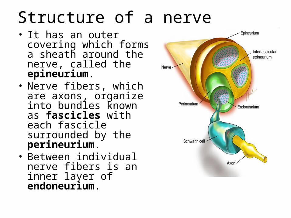
Structure of a nerve• It has an outer covering
which forms a sheath around the nerve, called the epineurium.
• Nerve fibers, which are axons, organize into bundles known as fascicles with each fascicle surrounded by the perineurium.
• Between individual nerve fibers is an inner layer of endoneurium.

Peripheral nerve injury
Dermotome :
• is an area of skin supplied by a single spinal root
Myotome :
• Represents a muscle unit supplied by a single spinal root

Seddon's classification Neurapraxia -- temporary paralysis of a nerve
caused by lack of blood flow or by pressure on the affected nerve with no loss of structural continuity.
Axonotmesis – • neural tube intact, but axons are disrupted. • nerves are likely to recover.
Neurotmesis – • the neural tube is severed. • Injuries are likely permanent without repair.
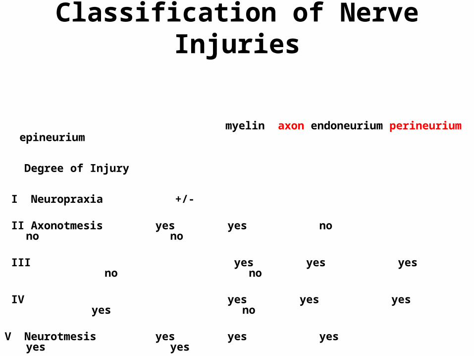
Classification of Nerve Injuries
myelin axon endoneurium perineurium epineurium
Degree of Injury
I Neuropraxia +/- II Axonotmesis yes yes no no no III yes yes yes no no IV yes yes yes yes no
V Neurotmesis yes yes yes yes yes

Sunderland`s classification• Grade I
– Same as Seddon's neuropraxia.
• Grade II– Same as Seddon's axonotmesis.
• Grade III– Neurotmesis with preservation of the perineurium.
• Grade IV– Neurotmesis with preservation of the epineurium.
Everything else is disrupted. – Nerve grossly appear edematous. – Nerve grafting is required.
• Grade V– Complete transection of the nerve trunk.
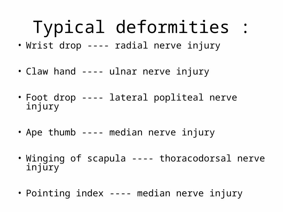
Typical deformities :• Wrist drop ---- radial nerve injury
• Claw hand ---- ulnar nerve injury
• Foot drop ---- lateral popliteal nerve injury
• Ape thumb ---- median nerve injury
• Winging of scapula ---- thoracodorsal nerve injury
• Pointing index ---- median nerve injury
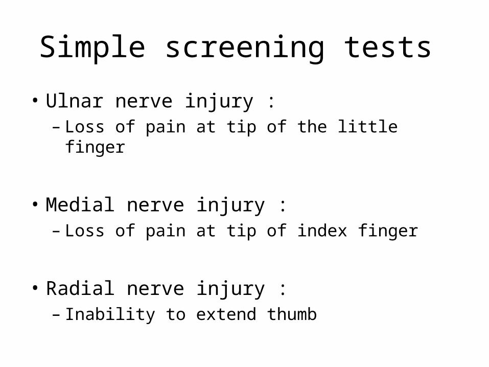
Simple screening tests
• Ulnar nerve injury : – Loss of pain at tip of the little finger
• Medial nerve injury :– Loss of pain at tip of index finger
• Radial nerve injury :– Inability to extend thumb
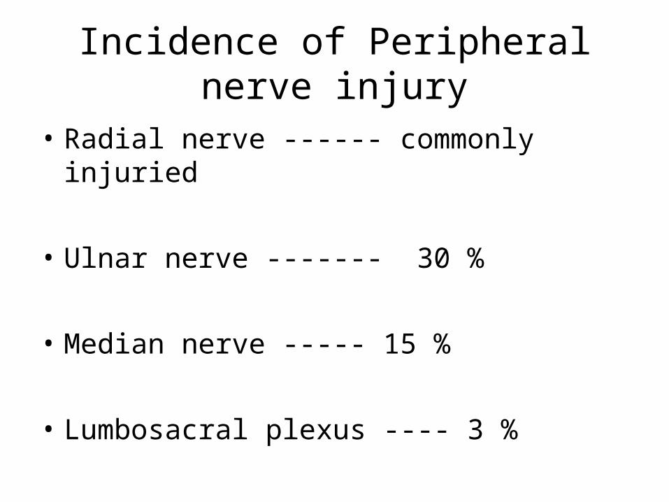
Incidence of Peripheral nerve injury
• Radial nerve ------ commonly injuried
• Ulnar nerve ------- 30 %
• Median nerve ----- 15 %
• Lumbosacral plexus ---- 3 %

Ulnar nerve injury Causes : General causes : metabolic diseases , collagen
diseases , malignancies , endogenous or exogenous toxins , chemical or mechanical trauma , etc.
Local causes : Causes in the axilla :
– Crutch pressure– Aneurysm of the axillary vessels
Causes in the arm :– # shaft of humerus– Gunshot and penetrating injuries

Cont ..
Causes at the elbow :– Compression by the accessory muscles– # lateral epicondyle of humerus– Repeated occupational strains – Recurrent subluxation of the nerve– Compression by the osteophytes as in rheumatoid
and osteoarthritis
Causes in the forearm :– # both bones forearm– Incised wounds , gunshot wounds and penetrating
injuries of the forearm
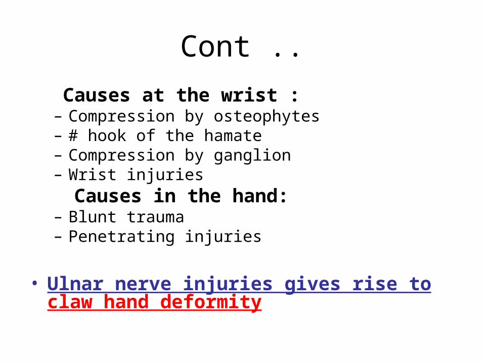
Cont ..
Causes at the wrist :– Compression by osteophytes– # hook of the hamate– Compression by ganglion– Wrist injuries
Causes in the hand:– Blunt trauma – Penetrating injuries
• Ulnar nerve injuries gives rise to claw hand deformity

Claw hand deformity
• It is a deformity with hyperextension of the MCP joints and flexion of the IP joints of the fingers
( loss of flexon at MCP and extension at IP joints )
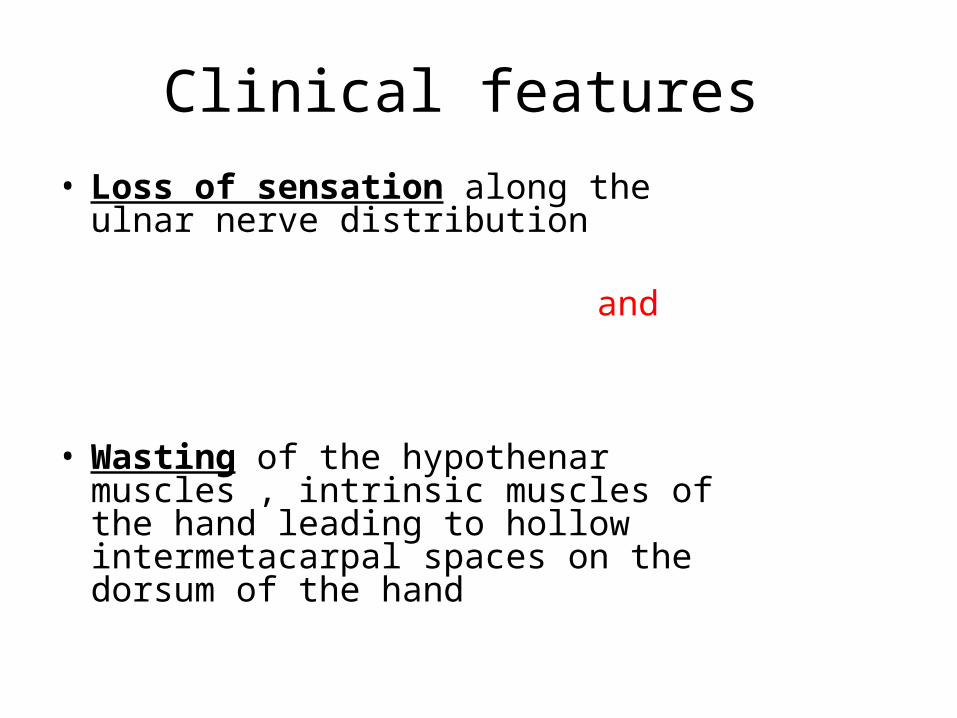
Clinical features
• Loss of sensation along the ulnar nerve distribution
and
• Wasting of the hypothenar muscles , intrinsic muscles of the hand leading to hollow intermetacarpal spaces on the dorsum of the hand

.

Levels of the lesion High : above the level of elbow , entire nerve
function is lost Low : Below the elbow at the junction of the middle
and lower third of forearm : Spared : - function of FDP and FUC Lost :
– Motor : HTM ,Its , Lum ,PB– Sensory : dorsal aspect of hand and one and half
fingers
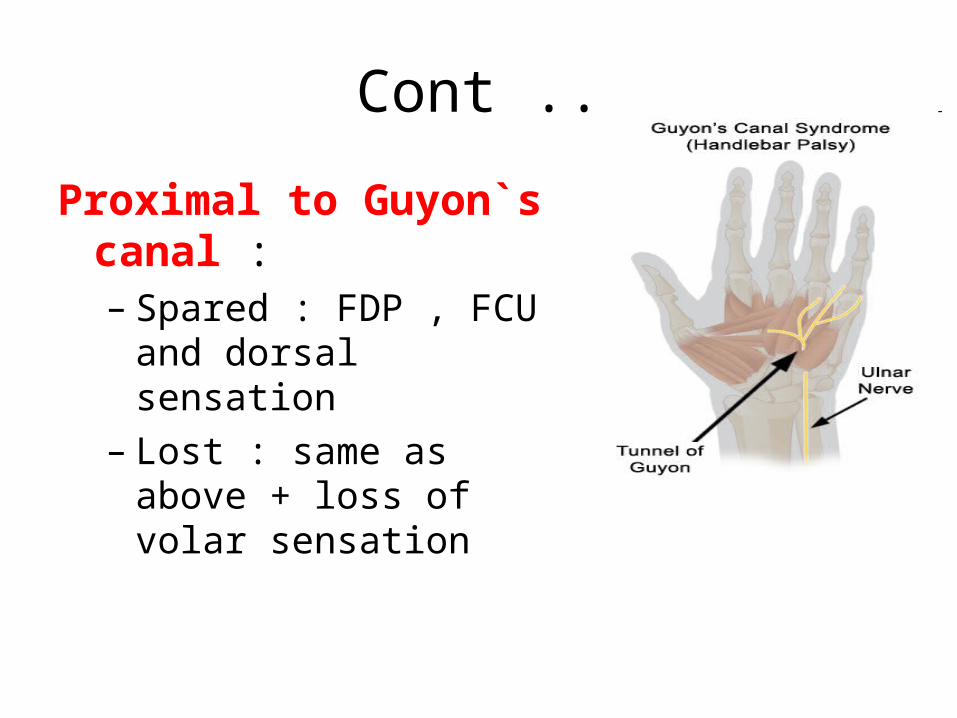
Cont ..
Proximal to Guyon`s canal : – Spared : FDP , FCU and
dorsal sensation– Lost : same as above +
loss of volar sensation
Cont ..
Distal to Guyon`s canal : -• Spared : FDP , FCU , HTM , PB, dorsal and
volar sensation• Lost : interossei and lumbricals
– FCU – flexor carpi ulnaris– FDP – flexor digitorum profundus– HTM – hypothenar muscles– PB – palmaris brevis– Lum – lumbricals – Its - interossei
Clinical tests :
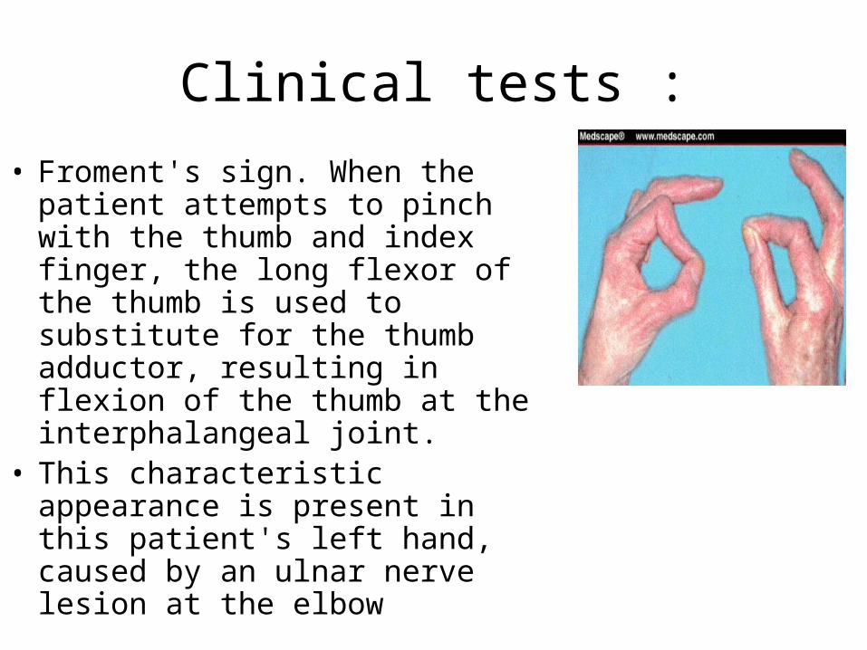
• Froment's sign. When the patient attempts to pinch with the thumb and index finger, the long flexor of the thumb is used to substitute for the thumb adductor, resulting in flexion of the thumb at the interphalangeal joint.
• This characteristic appearance is present in this patient's left hand, caused by an ulnar nerve lesion at the elbow
Card test • Inability to hold a card or paper in between
fingers due to loss of adduction by the palmar interossei
Pen test • Unable to touch the pen due to the loss of
action of abductor pollicic brevis
Egawa test ( median nerve injury )• With palm flat on the table the patient is asked to
move the middle finger sideways( test for the dorsal interossei of middle finger )
• In total clawing median nerve is also injuried
Pointing index or oschner`s clasp test :• When both the hands are clapsed together , index
and middle fingers , fail to flex due to the loss of action of long finger flexors of the index and middle fingers which are supplied by the median nerve .
Treatment of ulnar nerve injury• Unless there is a lot of muscle
wasting, (nonsurgical treatment ) Prevention • Avoid frequent use of the arm with
the elbow bent • If you use a computer frequently,
make sure that your chair is not too low. Do not rest the elbow on the armrest.
• Avoid putting pressure on the inside of the arm (do not drive with the arm resting on the open window ).
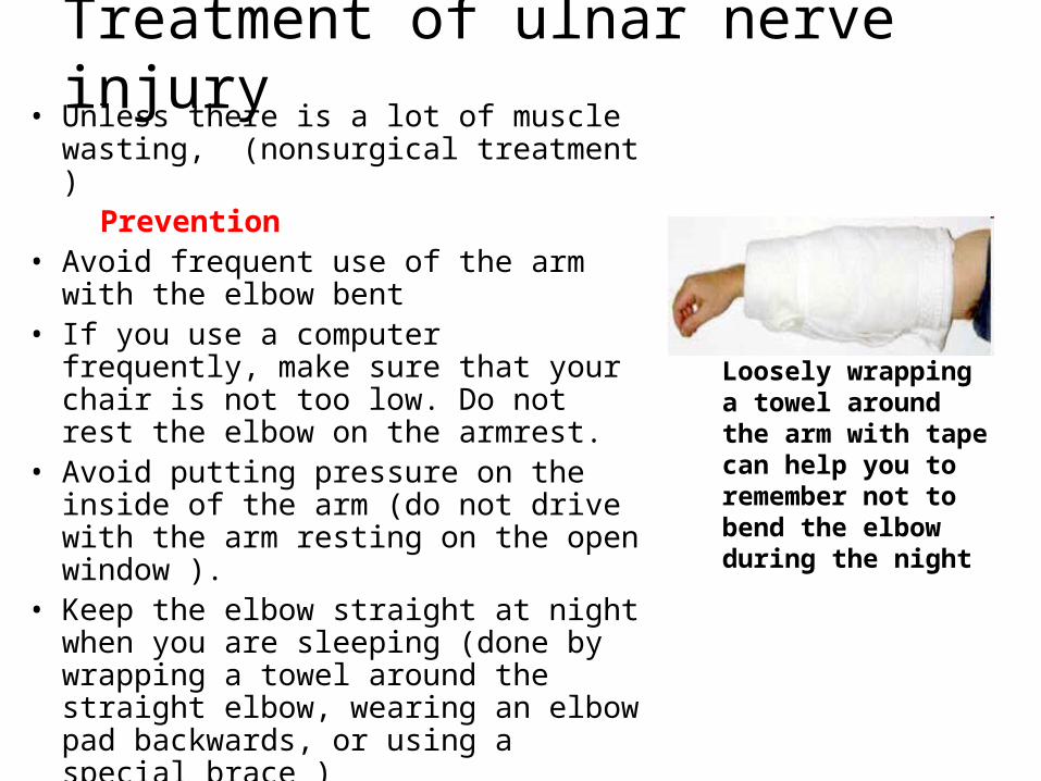
• Keep the elbow straight at night when you are sleeping (done by wrapping a towel around the straight elbow, wearing an elbow pad backwards, or using a special brace )
Loosely wrapping a towel around the arm with tape can help you to remember not to bend the elbow during the night
Nonsurgical Treatment
• If symptoms have only just started,
• Anti – inflammatory drugs, ibuprofen,( to reduce swelling around the nerve ).
• Steroid (cortisone) injections around the ulnar nerve are not generally used because there is a risk of damage to the nerve.
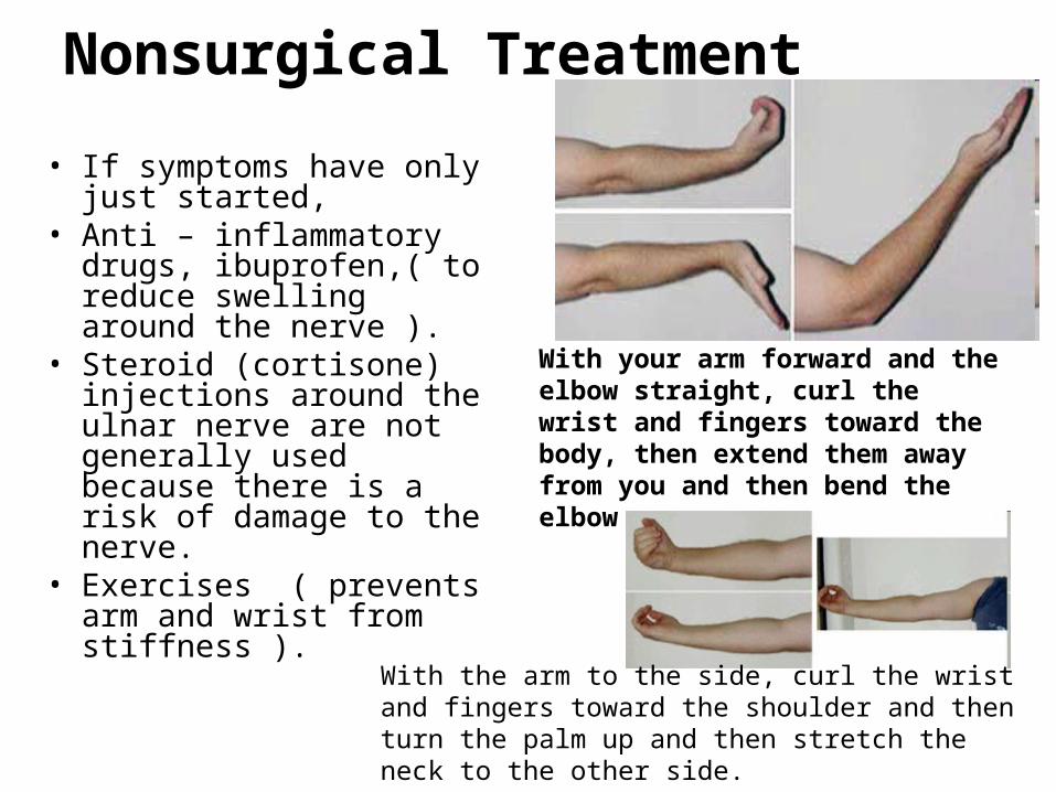
• Exercises ( prevents arm and wrist from stiffness ).
With your arm forward and the elbow straight, curl the wrist and fingers toward the body, then extend them away from you and then bend the elbow
With the arm to the side, curl the wrist and fingers toward the shoulder and then turn the palm up and then stretch the neck to the other side.
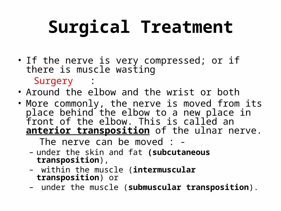
Surgical Treatment
• If the nerve is very compressed; or if there is muscle wasting
Surgery : • Around the elbow and the wrist or both • More commonly, the nerve is moved from its
place behind the elbow to a new place in front of the elbow. This is called an anterior transposition of the ulnar nerve.
The nerve can be moved : - – under the skin and fat (subcutaneous
transposition),– within the muscle (intermuscular transposition) or– under the muscle (submuscular transposition).
.
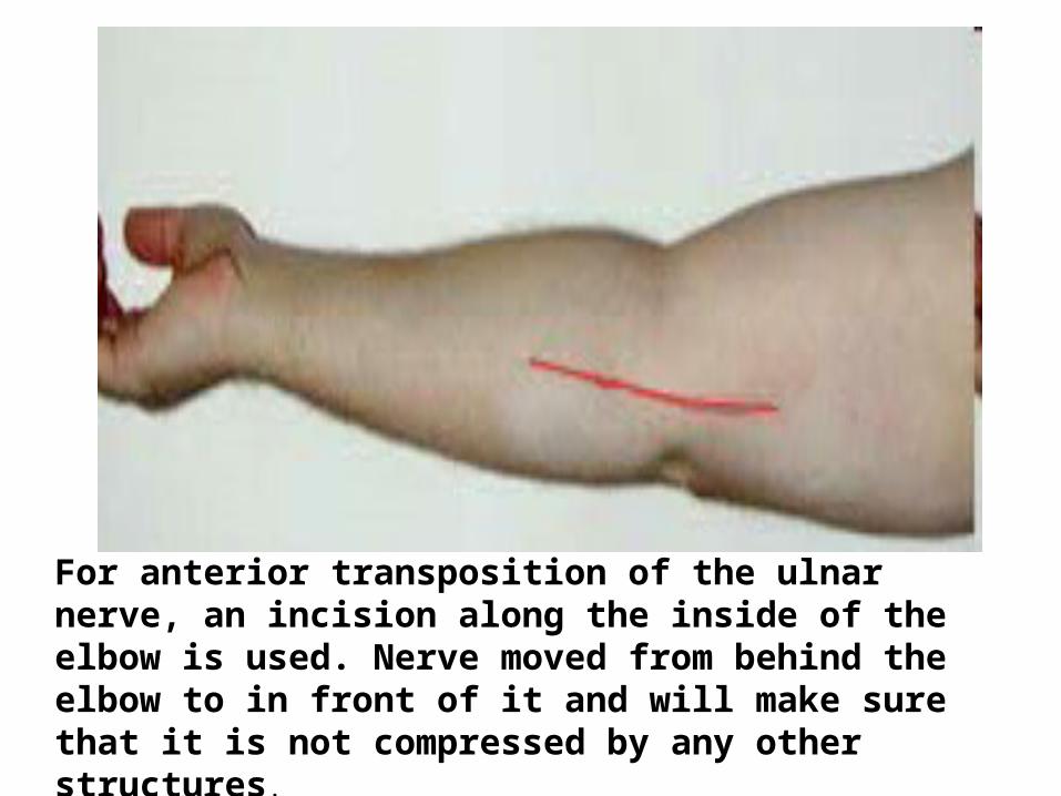
For anterior transposition of the ulnar nerve, an incision along the inside of the elbow is used. Nerve moved from behind the elbow to in front of it and will make sure that it is not compressed by any other structures.
.
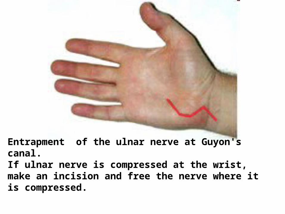
Entrapment of the ulnar nerve at Guyon's canal. If ulnar nerve is compressed at the wrist, make an incision and free the nerve where it is compressed.
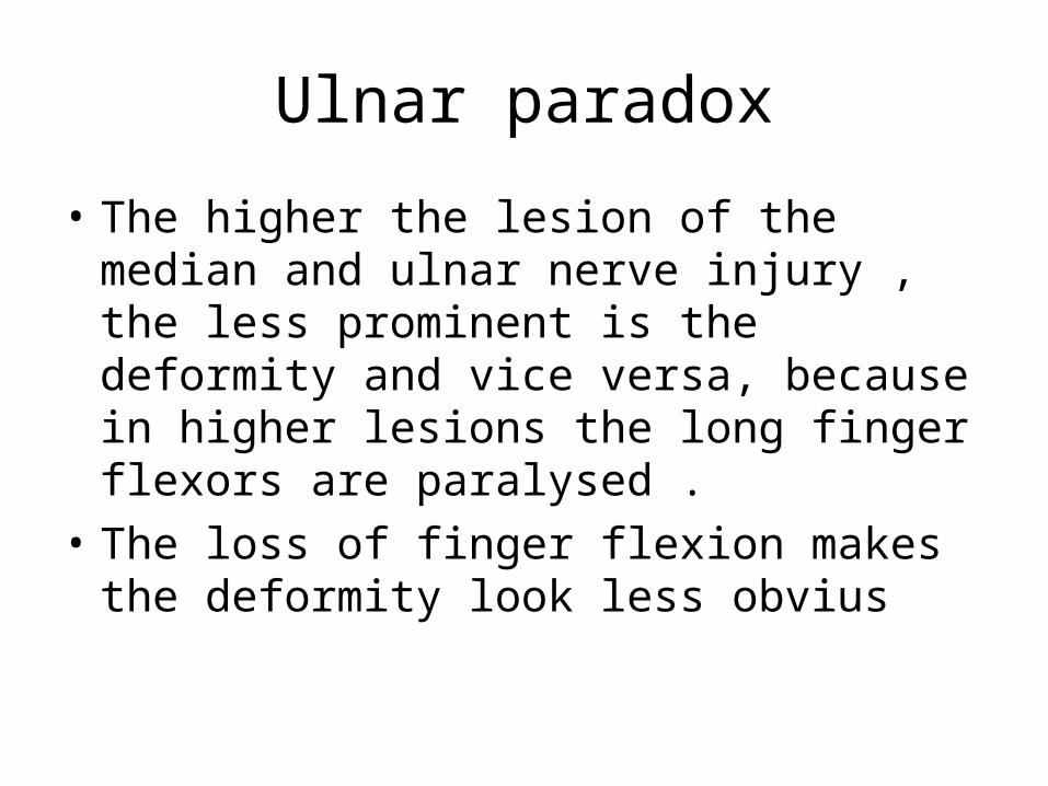
Ulnar paradox
• The higher the lesion of the median and ulnar nerve injury , the less prominent is the deformity and vice versa, because in higher lesions the long finger flexors are paralysed .
• The loss of finger flexion makes the deformity look less obvius
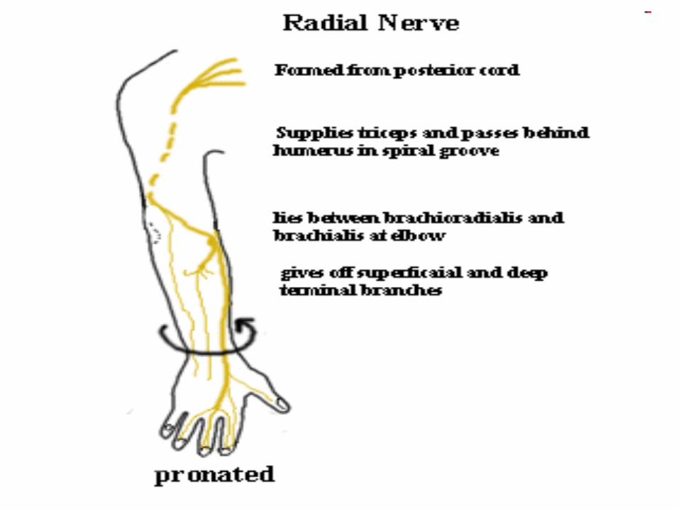
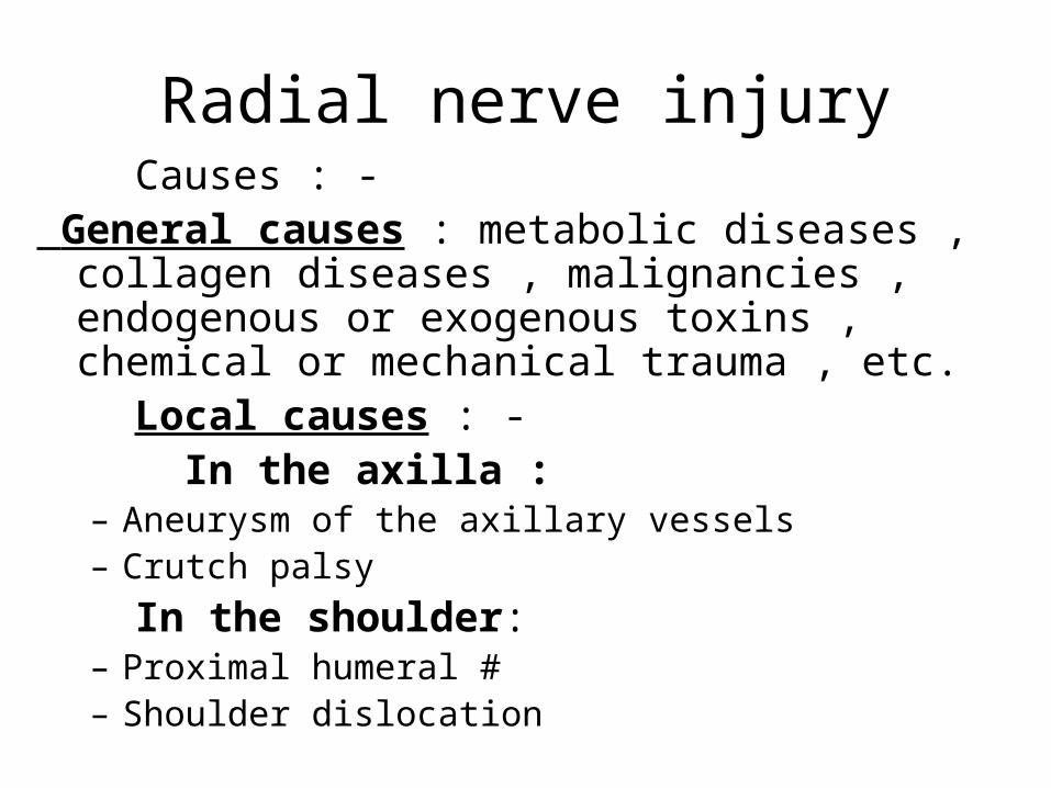
Radial nerve injury Causes : - General causes : metabolic diseases , collagen
diseases , malignancies , endogenous or exogenous toxins , chemical or mechanical trauma , etc.
Local causes : - In the axilla :
– Aneurysm of the axillary vessels– Crutch palsy
In the shoulder:– Proximal humeral #– Shoulder dislocation
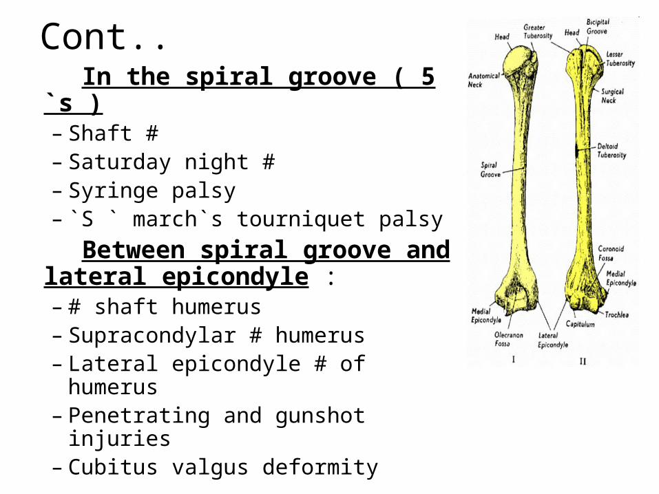
Cont.. In the spiral groove ( 5 `s )
– Shaft #– Saturday night #– Syringe palsy– `S ` march`s tourniquet palsy
Between spiral groove and lateral epicondyle : – # shaft humerus– Supracondylar # humerus– Lateral epicondyle # of humerus– Penetrating and gunshot injuries– Cubitus valgus deformity
Cont …
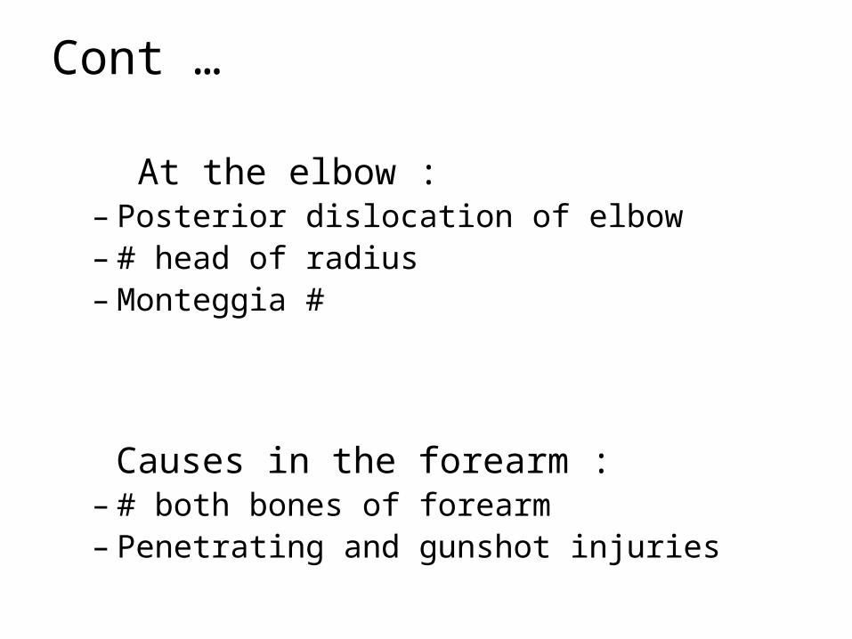
At the elbow :– Posterior dislocation of elbow– # head of radius– Monteggia #
Causes in the forearm :– # both bones of forearm– Penetrating and gunshot injuries
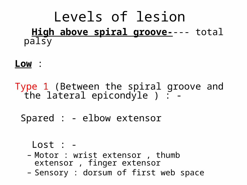
Levels of lesion High above spiral groove---- total palsy Low : Type 1 (Between the spiral groove and the lateral
epicondyle ) : - Spared : - elbow extensor
Lost : -– Motor : wrist extensor , thumb extensor , finger
extensor– Sensory : dorsum of first web space

Cont .. Low
• Type 2 ( below the elbow ) : Spared :
– Elbow extensor– Wrist extensor
Lost :
– Motor : thumb extensor , finger extensor– Sensory :– First web space
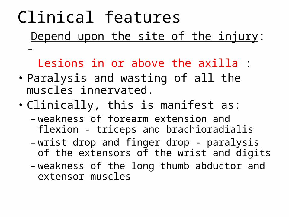
Clinical features Depend upon the site of the injury: - Lesions in or above the axilla : • Paralysis and wasting of all the muscles
innervated. • Clinically, this is manifest as:
– weakness of forearm extension and flexion - triceps and brachioradialis
– wrist drop and finger drop - paralysis of the extensors of the wrist and digits
– weakness of the long thumb abductor and extensor muscles
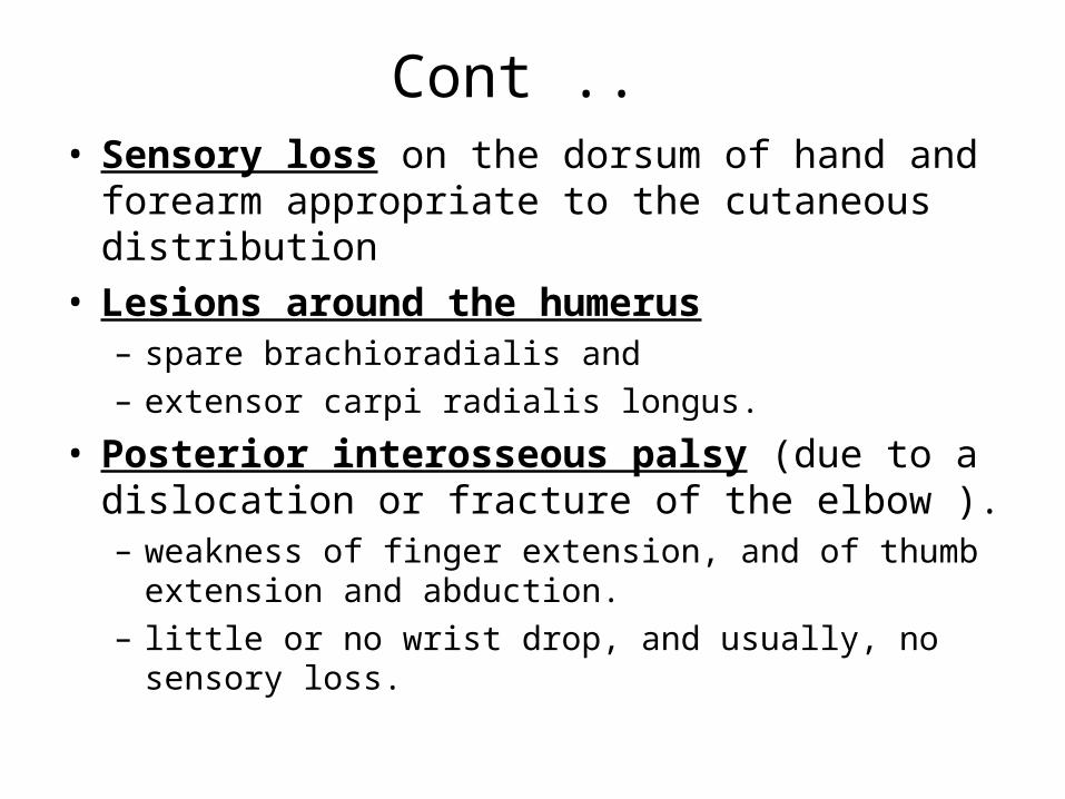
Cont .. • Sensory loss on the dorsum of hand and
forearm appropriate to the cutaneous distribution• Lesions around the humerus
– spare brachioradialis and – extensor carpi radialis longus.
• Posterior interosseous palsy (due to a dislocation or fracture of the elbow ). – weakness of finger extension, and of thumb extension
and abduction. – little or no wrist drop, and usually, no sensory loss.

Fig : - Wrist drop
• .
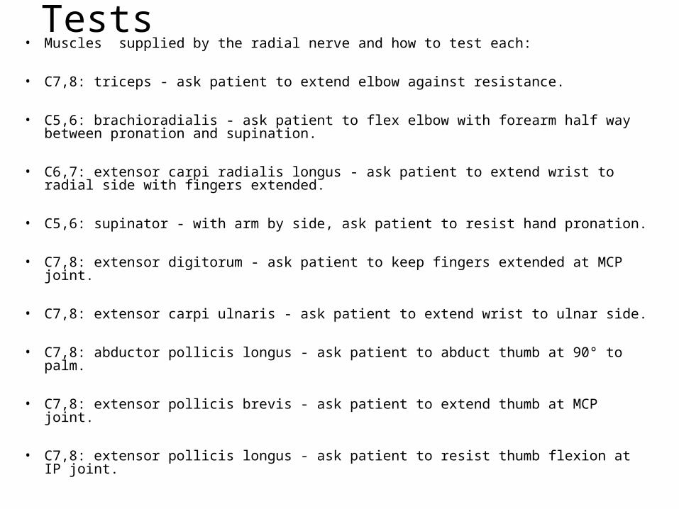
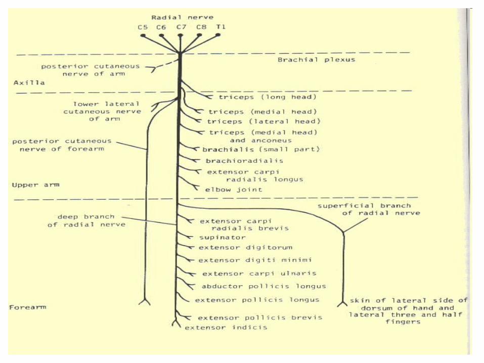
Tests • Muscles supplied by the radial nerve and how to test each:
• C7,8: triceps - ask patient to extend elbow against resistance.
• C5,6: brachioradialis - ask patient to flex elbow with forearm half way between pronation and supination.
• C6,7: extensor carpi radialis longus - ask patient to extend wrist to radial side with fingers extended.
• C5,6: supinator - with arm by side, ask patient to resist hand pronation.
• C7,8: extensor digitorum - ask patient to keep fingers extended at MCP joint.
• C7,8: extensor carpi ulnaris - ask patient to extend wrist to ulnar side.
• C7,8: abductor pollicis longus - ask patient to abduct thumb at 90° to palm.
• C7,8: extensor pollicis brevis - ask patient to extend thumb at MCP joint.
• C7,8: extensor pollicis longus - ask patient to resist thumb flexion at IP joint.
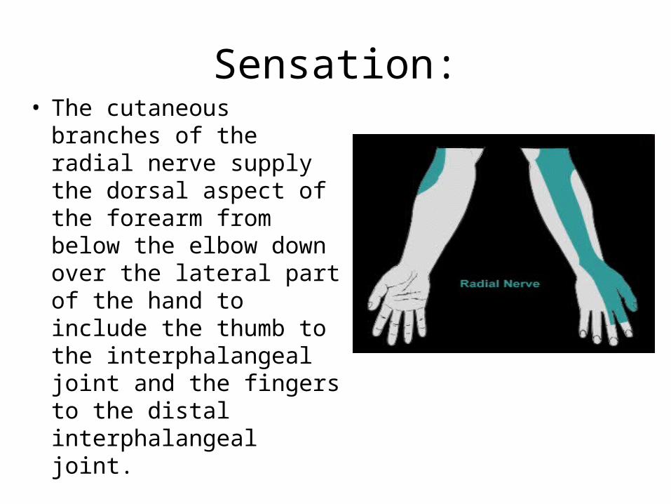
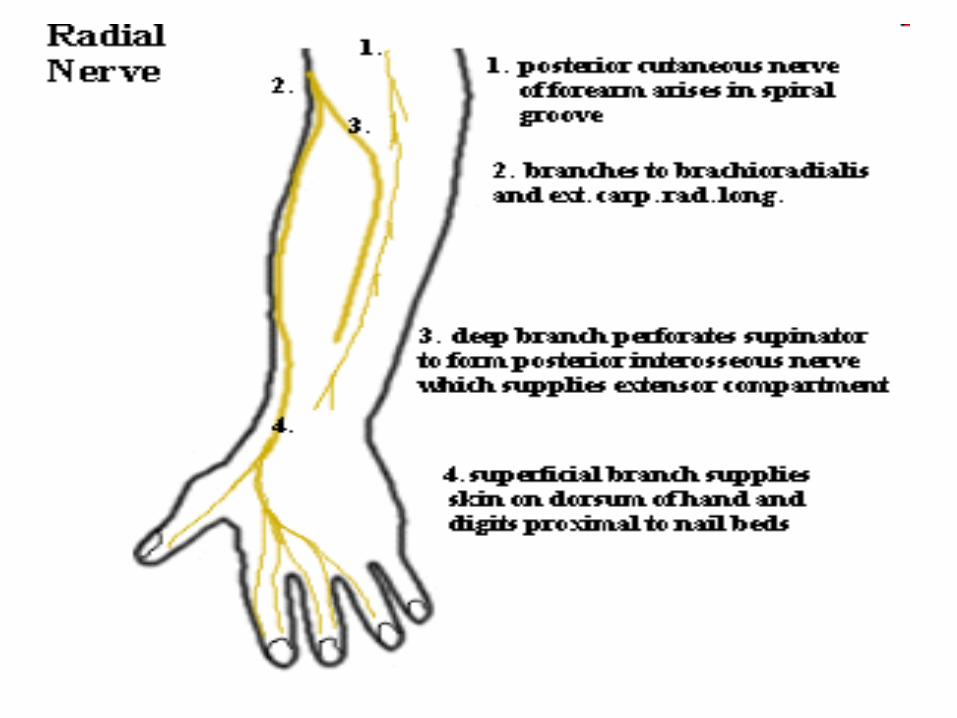
Sensation:• The cutaneous
branches of the radial nerve supply the dorsal aspect of the forearm from below the elbow down over the lateral part of the hand to include the thumb to the interphalangeal joint and the fingers to the distal interphalangeal joint.
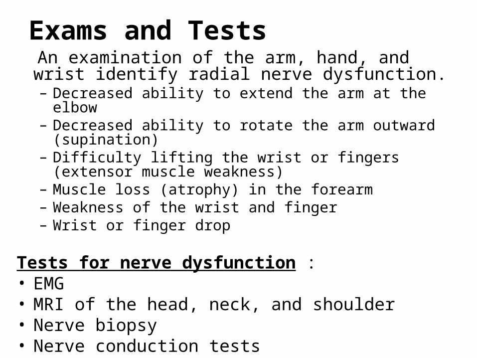
Exams and Tests An examination of the arm, hand, and wrist identify
radial nerve dysfunction.– Decreased ability to extend the arm at the elbow – Decreased ability to rotate the arm outward (supination) – Difficulty lifting the wrist or fingers (extensor muscle
weakness) – Muscle loss (atrophy) in the forearm – Weakness of the wrist and finger – Wrist or finger drop
Tests for nerve dysfunction :• EMG • MRI of the head, neck, and shoulder • Nerve biopsy • Nerve conduction tests
Treatment
Closed fracture
CONTROL OF SYMPTOMS• Analgesics ( to control pain neuralgia) • Phenytoin, carbamazepine, or tricyclic antidepressants (amitriptyline) to
reduce stabbing pain • Steroids (prednisone) to reduce swelling Other treatments include:• Braces, splints, • Physical therapy to help maintain muscle strength • Occupational therapy, or job counseling• Surgery : - • Failure of conservative by 12 to 18 months
Surgery ( open # )Clean wound : Primary repair , splint , physiotherapy
Contaminated wound :Delayed primary repair and secondary repair
Late cases :
– Tendon transfers– Arthrodesis

Splints
Complications
• Mild to severe deformity of the hand
• Partial or complete loss of feeling in the hand
• Partial or complete loss of wrist or hand movement
• Recurrent injury to the hand
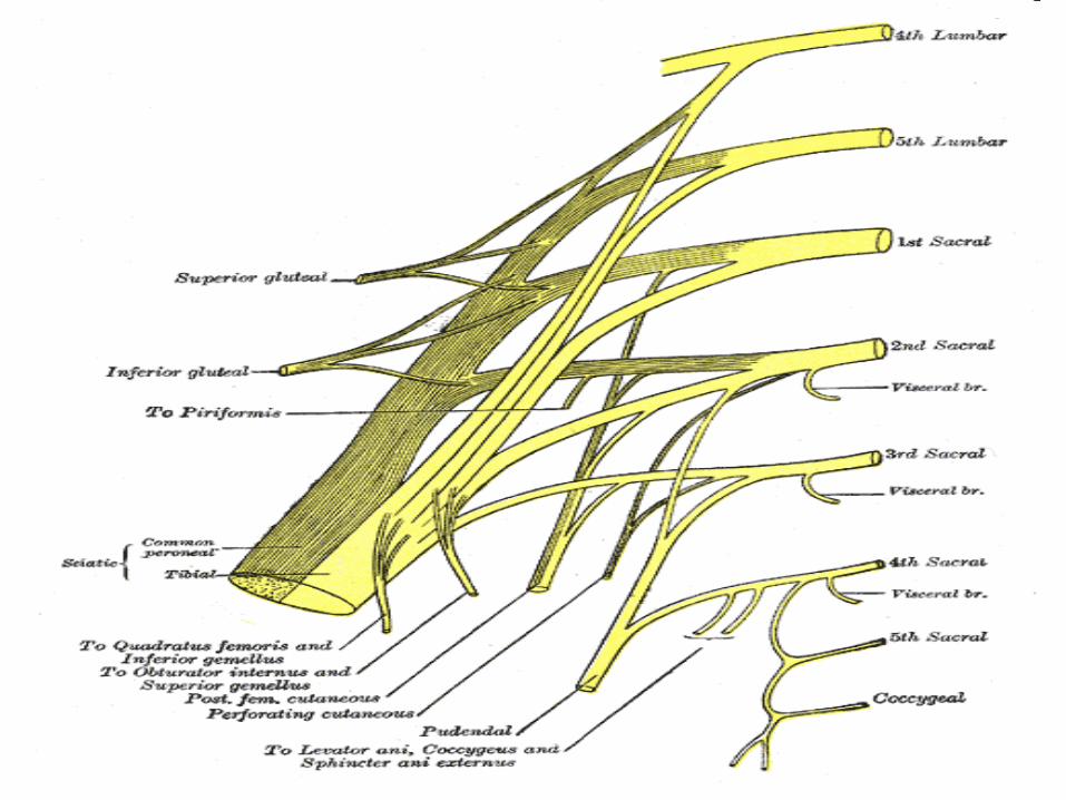
Sciatic nerve injury
• Thickest nerve in the body
• Leprosy is the commonest cause
• High stepping gait is the characterisic
• Conservative treatment is indicated up to one year
Foot drop
Causes • General causes : metabolic diseases , collagen
diseases , malignancies , endogenous or exogenous toxins , chemical or mechanical trauma , etc.
Local : At the spine :
– Spina bifida– Tumors – Disc prolapse
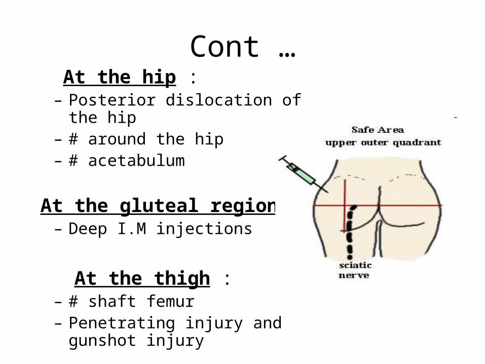
Cont … At the hip :
– Posterior dislocation of the hip– # around the hip– # acetabulum
At the gluteal region :– Deep I.M injections
At the thigh : – # shaft femur– Penetrating injury and gunshot
injury
Cont …
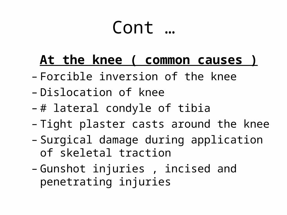
At the knee ( common causes )– Forcible inversion of the knee– Dislocation of knee– # lateral condyle of tibia– Tight plaster casts around the knee– Surgical damage during application of skeletal
traction– Gunshot injuries , incised and penetrating
injuries
Levels of lesion High lesion ( above knee ) :• Both tibial and common peroneal nerve are
paralysed
Low lesion ( below knee ) Type 1 ( anterior tibial nerve injury )
– Lost : tibialis anterior , extensor hallucis longus , extensor digitorium longus
– Sensation : over first web space is lost Type 2 ( musculocutaneous nerve injury ):
– Spared : all the above muscles innervated by anterior tibial nerve
– Lost : peroneous longus and brevis– Sensation : over outer leg and foot

Clinical features
Foot drop :
Complete ( sciatic or lateral popliteal nerve injury )
Incomplete ( superficial or deep peroneal nerve )– High lesions ------total foot drop – Low lesions ------ incomplete foot drop
Low lesions
Type 1 : – Dorsiflexion and inversion is not possible – Front of the leg is wasted– Sensation over the dorsal web space is lost
Type 2 :
– Cannot evert but can dorsiflex and invert the foot – Wasting of the outer half of the leg – Sensation lost over outer leg and foot
• Gait : - high stepping gait is characteristic .

Treatment • Braces or splints. • Physical therapy. • Nerve stimulation :
– In some cases, a small, battery-operated electrical stimulator is strapped to the leg just below the knee.
– In other cases, the stimulator is implanted in the leg.
• Surgery. – Tendon transfer ( for mobile foot drop )– Tendon – Achilles lengthening ( in fixed )
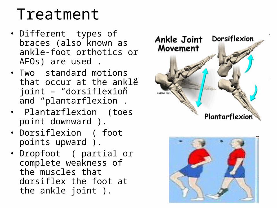
Treatment • Different types of braces
(also known as ankle-foot orthotics or AFOs) are used .
• Two standard motions that occur at the ankle joint – “dorsiflexion” and “plantarflexion”.
• Plantarflexion (toes point downward ).
• Dorsiflexion ( foot points upward ).
• Dropfoot ( partial or complete weakness of the muscles that dorsiflex the foot at the ankle joint ).
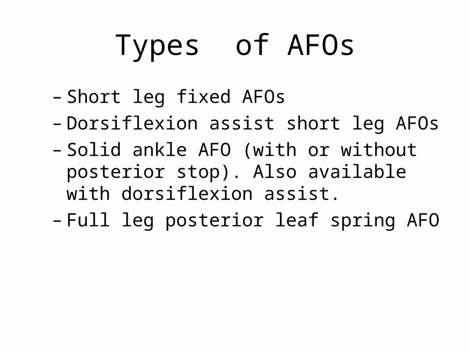
Types of AFOs
– Short leg fixed AFOs – Dorsiflexion assist short leg AFOs – Solid ankle AFO (with or without posterior
stop). Also available with dorsiflexion assist. – Full leg posterior leaf spring AFO
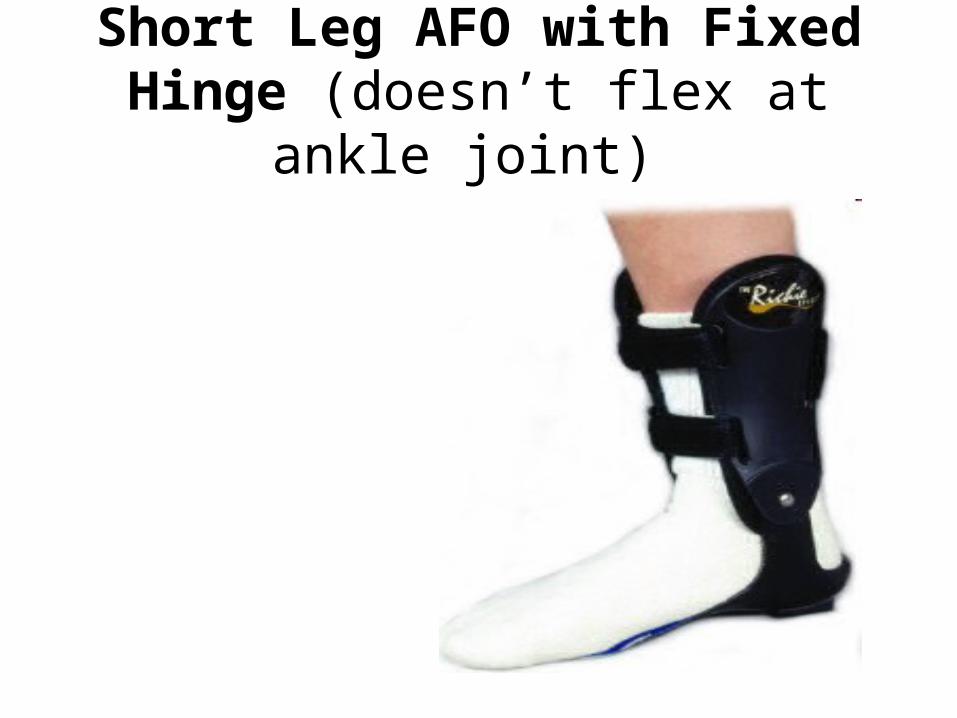
Short Leg AFO with Fixed Hinge (doesn’t flex at ankle joint)

Dorsiflexion Assist AFO (dorsiflex the ankle) :
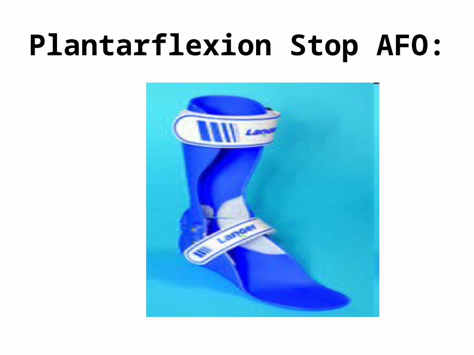
Plantarflexion Stop AFO:

Solid AFO:(stops plantarflexion and also stops
or limits dorsiflexion).
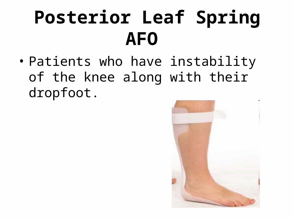
Posterior Leaf Spring AFO
• Patients who have instability of the knee along with their dropfoot.

Brachical plexus injuries
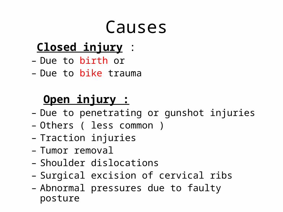
Causes Closed injury :
– Due to birth or– Due to bike trauma
Open injury : – Due to penetrating or gunshot injuries– Others ( less common )– Traction injuries– Tumor removal– Shoulder dislocations– Surgical excision of cervical ribs– Abnormal pressures due to faulty posture
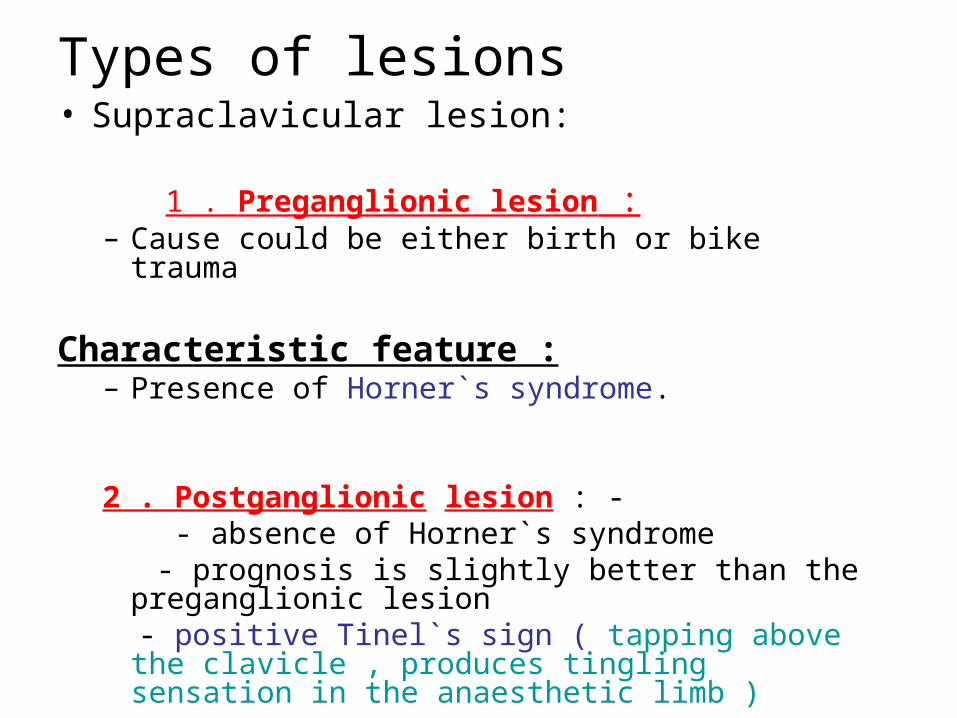
Types of lesions• Supraclavicular lesion: 1 . Preganglionic lesion :
– Cause could be either birth or bike trauma Characteristic feature :
– Presence of Horner`s syndrome.
2 . Postganglionic lesion : - - absence of Horner`s syndrome - prognosis is slightly better than the preganglionic
lesion - positive Tinel`s sign ( tapping above the clavicle ,
produces tingling sensation in the anaesthetic limb )
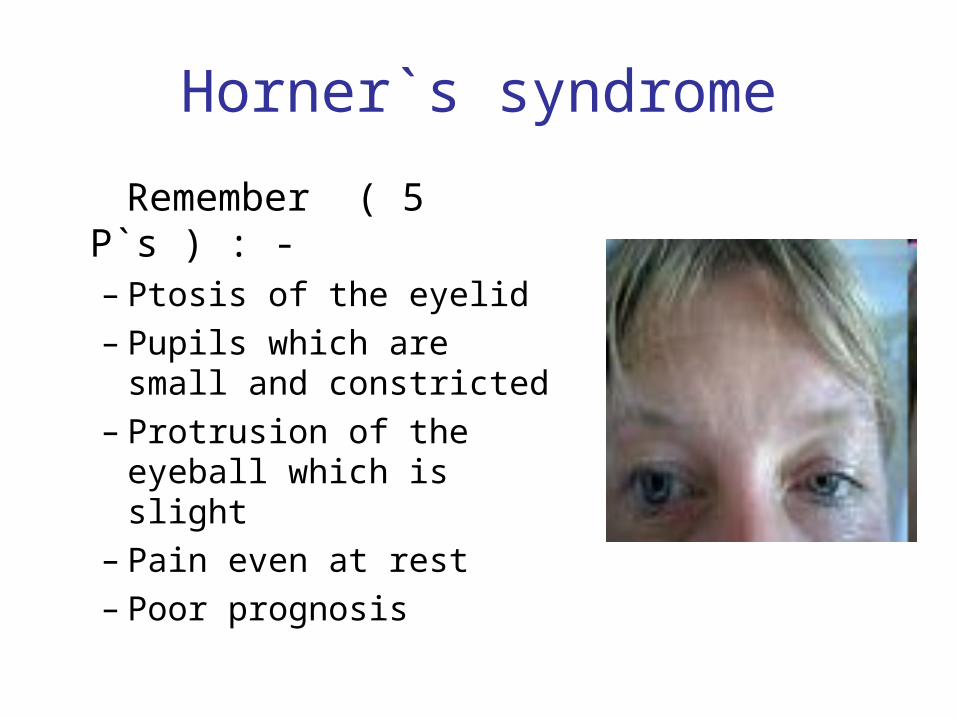
Horner`s syndrome
Remember ( 5 P`s ) : -– Ptosis of the eyelid– Pupils which are small
and constricted– Protrusion of the eyeball
which is slight– Pain even at rest– Poor prognosis
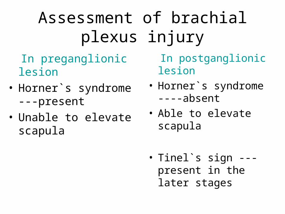
Assessment of brachial plexus injury
In preganglionic lesion• Horner`s syndrome ---
present• Unable to elevate
scapula
In postganglionic lesion• Horner`s syndrome ----
absent • Able to elevate scapula
• Tinel`s sign --- present in the later stages

Investigation • X – ray ( to rule out # )
• CT scan ( study cross – section anatomy )
• MRI ( study the soft tissue damages )
• Electromyogram (EMG or electromyography)
• Nerve conduction study
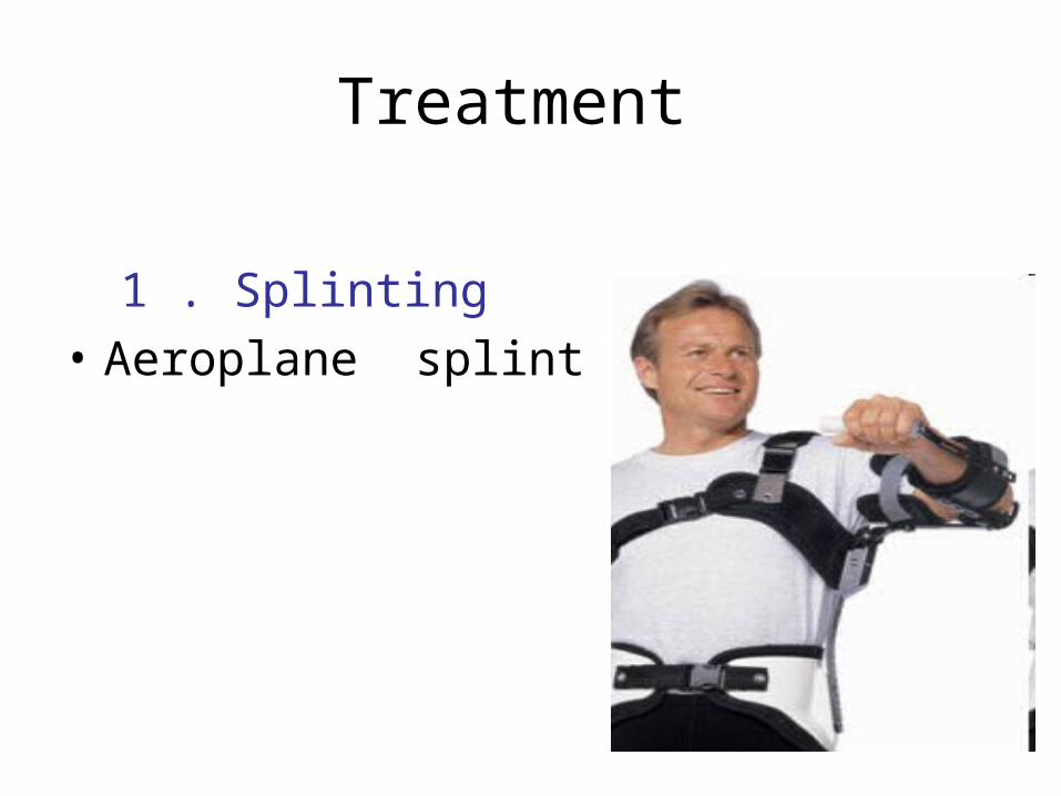
Treatment
1 . Splinting
• Aeroplane splint
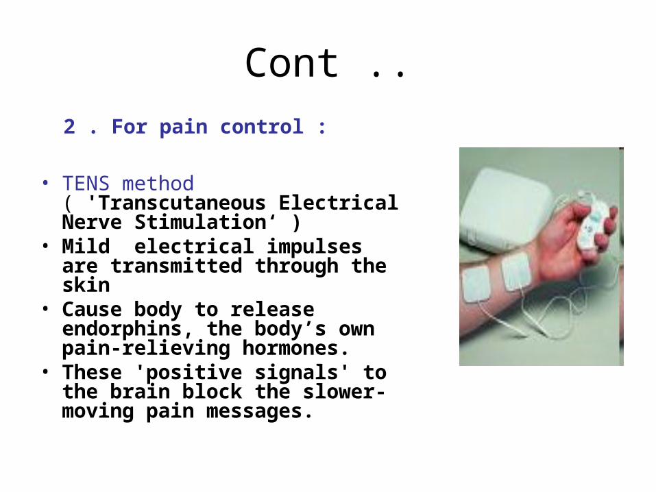
Cont ..
2 . For pain control :
• TENS method ( 'Transcutaneous Electrical Nerve Stimulation‘ )
• Mild electrical impulses are transmitted through the skin
• Cause body to release endorphins, the body’s own pain-relieving hormones.
• These 'positive signals' to the brain block the slower-moving pain messages.

Surgical measures
• Types of surgery
Nerve graft : - • the damaged part
of the brachial plexus is removed and replaced with sections of nerves cut from other parts of body
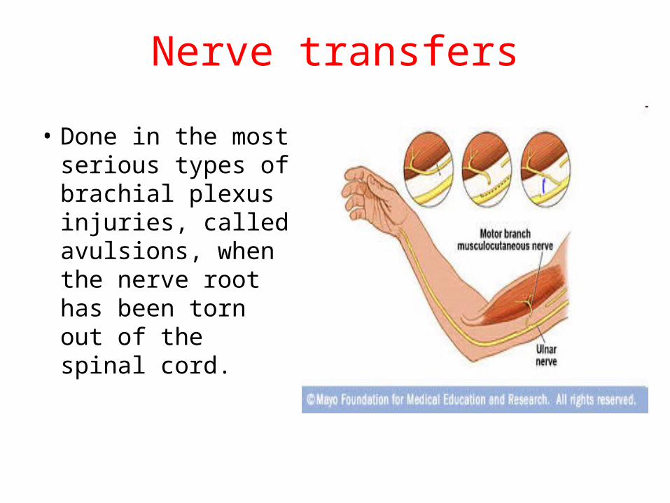
Nerve transfers
• Done in the most serious types of brachial plexus injuries, called avulsions, when the nerve root has been torn out of the spinal cord.
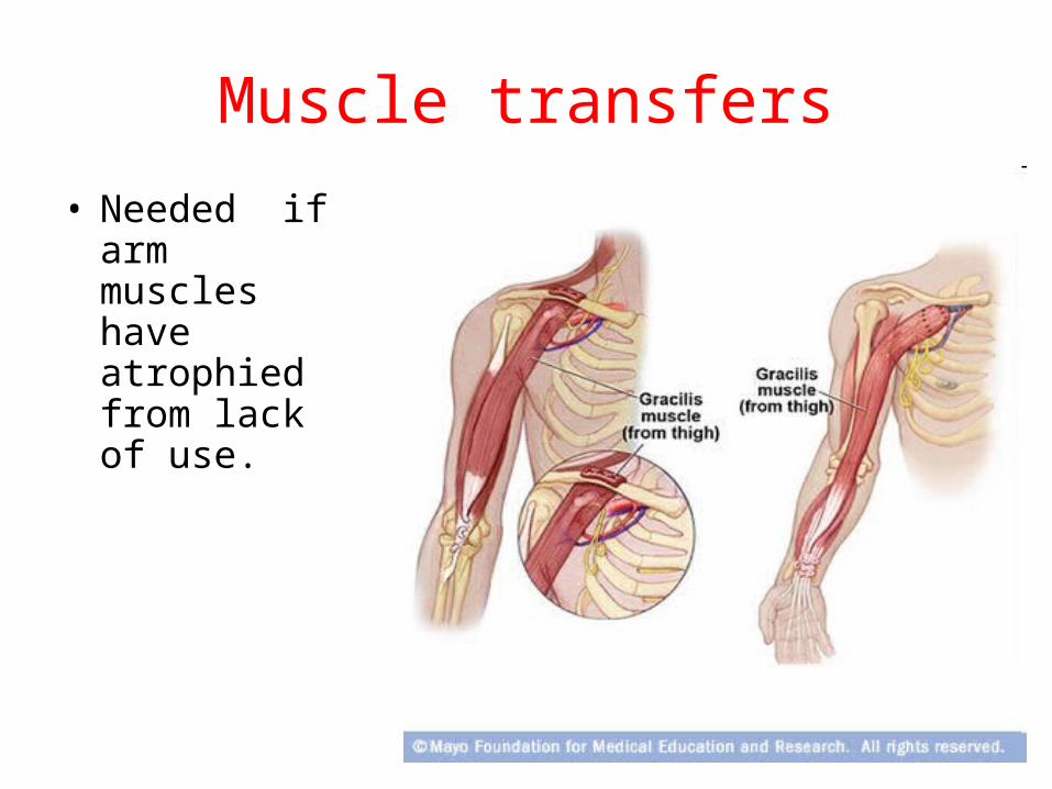
Muscle transfers
• Needed if arm muscles have atrophied from lack of use.
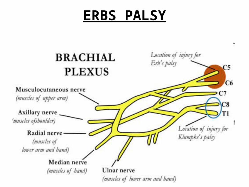
ERBS PALSY

Erb's palsy
• paralysis of the muscles in a baby's arm, caused by injury of the nerves in the shoulder at birth (during delivery).
• The baby lies with one arm and hand twisted backward and does not move the arm as much as the other.
• If the full range of motion of the arm is not kept through regular exercise, contractures will develop .
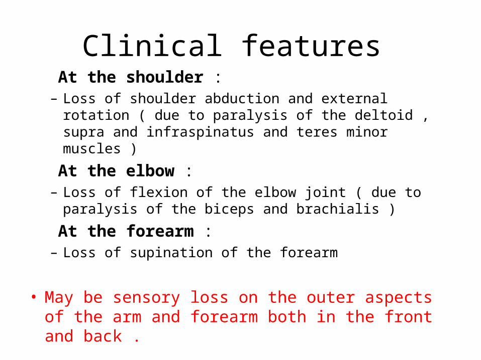
Clinical features At the shoulder :
– Loss of shoulder abduction and external rotation ( due to paralysis of the deltoid , supra and infraspinatus and teres minor muscles )
At the elbow : – Loss of flexion of the elbow joint ( due to paralysis of
the biceps and brachialis )
At the forearm :– Loss of supination of the forearm
• May be sensory loss on the outer aspects of the arm and forearm both in the front and back .

Policeman or Waiter`s tip
• Shoulder --- internally rotated
• Elbow ----- extension
• Forearm --- pronated
• Wrist ------ flexion

Treatment 1 . Splinting
– Aeroplane splint
2 . For pain control : – TENS method
• Types of surgery - Nerve graft . - Nerve transfers . - Muscle transfers . - release of soft tissue contractures .
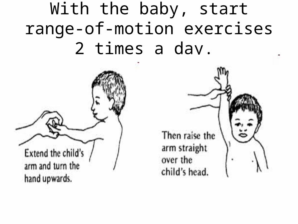
With the baby, start range-of-motion exercises 2 times a day.
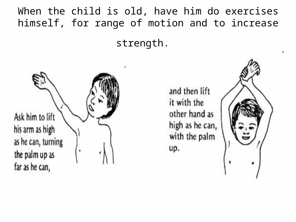
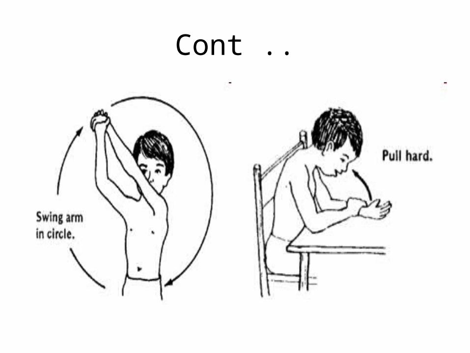
When the child is old, have him do exercises himself, for
range of motion and to increase strength.
Cont ..

Cont ..
