peripheral intravenous initiation - fraser health · the clinical setting until proficiency is...
TRANSCRIPT
Picture provided courtesy of BD®
Peripheral Intravenous
Initiation
Written by Patty Hignell RN, BSN, MN, ENC(C)
Vascular Access Clinical Practice Committee
Fraser Health Authority Printshop #256024
8th Edition June 2018
Self-Learning Module
2
The 8th Edition (current) of this document replaces all previous
versions.
The supplement “Pediatric Considerations for Peripheral IV Initiation
(2012)” is also available on request.
This publication has been created for the express use of Fraser Health and
its Partners. Permission to reprint or electronically reproduce any
document or graphic in whole or in part for any reason is expressly
prohibited, unless prior written consent is obtained from the respective
copyright holder(s).
Please send any requests for use of this material to:
Fraser Health Authority
Clinical Quality & Patient Safety
Central City Tower, 5th Floor
13450 – 102nd Avenue,
Surrey, British Columbia
Canada V3T 0H1
TABLE OF CONTENTS
INTRODUCTION ................................................................................................................................ 4
INSTRUCTIONS FOR USE ............................................................................................................. 4
COMPETENCY AND SCOPE OF PRACTICE ............................................................................ 5
OUTCOMES ........................................................................................................................................... 6
ANATOMY AND PHYSIOLOGY .................................................................................................... 7
FLUID AND ELECTROLYTE BALANCE ................................................................................... 12
INFECTION CONTROL .................................................................................................................. 16
SKIN CARE ......................................................................................................................................... 19
PIV INSERTION KEYPOINTS .................................................................................................... 20
EQUIPMENT ....................................................................................................................................... 22
TECHNIQUES IN VENIPUNCTURE .......................................................................................... 24
TROUBLESHOOTING IV INSERTION .................................................................................... 31
IV FLOW RATE MAINTENANCE ............................................................................................... 32
TROUBLESHOOTING IV INFUSION ...................................................................................... 33
PERIPHERAL IV DAILY ASSESSMENT ................................................................................. 34
DISCONTINUING IV THERAPY................................................................................................ 35
DOCUMENTATION .......................................................................................................................... 36
COMPLICATIONS OF IV THERAPY ........................................................................................ 37
Phlebitis and Thrombophlebitis ............................................................................................... 37
Infiltration and Extravasation .................................................................................................. 39
Hematoma ....................................................................................................................................... 43
Infection ........................................................................................................................................... 44
Catheter Embolism ....................................................................................................................... 45
Pulmonary Embolism ................................................................................................................... 46
Air Embolism................................................................................................................................... 47
Pulmonary Edema ......................................................................................................................... 48
Speed Shock ................................................................................................................................... 49
REVIEW QUESTIONS .................................................................................................................... 50
ANSWERS TO REVIEW QUESTIONS ..................................................................................... 58
REFERENCES ..................................................................................................................................... 59
APPENDIX A: EDUCATION AND COMPETENCY GUIDELINES .................................. 61
APPENDIX B: CLINICAL COMPETENCY VALIDATION ................................................. 63
4
INTRODUCTION
Welcome to the “Initiation of Intravenous Therapy” Self Learning Module!
Infusion therapy has evolved from an extreme measure used only as a last resort with the
most critically ill, to a highly scientific, specialized form of treatment used for greater than
90% of hospitalized clients. Performing venipuncture is one of the more challenging clinical
skills you will need to master. The Infusion Therapy Practitioner is a Healthcare Practitioner
(HCP) who, through study, supervised practice and validation of competency, gains the
acquired knowledge and skills necessary for the practice of infusion therapy.
Nurses provided with specialized training in peripheral vascular access, along with
supportive organizational structures and processes, results in improved client outcomes and
decreased complications. Although we recognize that HCPs other than Nursing may have
intravenous (IV) insertion and therapy included in their scope of practice, this Self-Learning
Module has been written based primarily on the scope of practice of a Registered Nurse
(RN), Registered Psychiatric Nurses (RPN), and Licensed Practical Nurse (LPN).
Completing this Self Learning Module does not imply that you are competent in IV initiation
and therapy. Competency assessment is multi-faceted (see pg.61). All HCPs must practice
within their own level of competence. When aspects of care or skill are beyond the HCP’s
level of competence, it is the HCP’s responsibility to seek education and/or supports needed
for that care setting (College of Registered Nurses of British Columbia, 2015).
This Self Learning Module does not cover or imply the ability to administer
medications by the intravenous route.
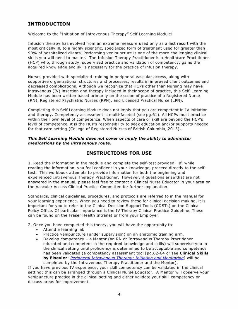
INSTRUCTIONS FOR USE
1. Read the information in the module and complete the self-test provided. If, while
reading the information, you feel confident in your knowledge, proceed directly to the self-
test. This workbook attempts to provide information for both the beginning and
experienced Intravenous Therapy Practitioner. However, if questions arise that are not
answered in the manual, please feel free to contact a Clinical Nurse Educator in your area or
the Vascular Access Clinical Practice Committee for further explanation.
Standards, clinical guidelines, procedures, and protocols are referred to in the manual for
your learning experience. When you need to review these for clinical decision making, it is
important for you to refer to the Clinical Decision Support Tools (CDSTs) on the Clinical
Policy Office. Of particular importance is the IV Therapy Clinical Practice Guidel ine. These
can be found on the Fraser Health Intranet or from your Employer.
2. Once you have completed this theory, you will have the opportunity to:
Attend a learning lab
Practice venipuncture (under supervision) on an anatomic training arm.
Develop competency – a Mentor (an RN or Intravenous Therapy Practitioner
educated and competent in the required knowledge and skills) will supervise you in
the clinical setting until proficiency is determined to be acceptable and competency
has been validated (a competency assessment tool [pg.62-64 or see Clinical Skills
by Elsevier: Peripheral Intravenous Therapy: Initiation and Monitoring] will be
completed by the Intravenous Therapy Practitioner and the Mentor).
If you have previous IV experience, your skill competency can be validated in the clinical
setting; this can be arranged through a Clinical Nurse Educator. A Mentor will observe your
venipuncture practice in the clinical setting and either validate your skill competency or
discuss areas for improvement.
5
COMPETENCY AND SCOPE OF PRACTICE
COMPETENCY is defined as the knowledge, skill, attitude and judgment required to provide
safe, compassionate and ethical care, and includes consideration of the context in which the
care is provided. Competence should initially be validated at the time of employment, after
orientation to the organization, on an ongoing periodic basis, when scope of practice
changes, and with the introduction of new equipment and technology. The frequency of
ongoing competence validation shall be determined by the setting and associated risk.
(Infusion Nurses Society, 2016) All healthcare practitioners are legally responsible to be aware
of, understand, and comply with their scope of practice and understand their level of
individual competence before performing skills related to IV Therapy.
A Registered Nurse (RN) and the Registered Psychiatric Nurse (RPN) may perform the
assessments and skills related to insertion, care, and maintenance of PIVs. The Registered
Nurse and Registered Psychiatric Nurse providing infusion therapy shall be proficient in its
clinical aspects, shall have validated competency in clinical judgment and practice, and shall
practice in accordance with the Health Professions Act, the Scope of Practice for Registered
Nurses or Registered Psychiatric Nurses in accordance with the College of Registered Nurses
of British Columbia or the College of Registered Psychiatric Nurses of British Columbia, and
the Fraser Health’s Scope of Practice policy. (Canadian Nurses Association, 2008) (College
of Registered Nurses of British Columbia, 2015) (College of Registered Psychiatric Nurses of
British Columbia, 2016) (Fraser Health Authority, Scope of Practice, 2010) (Government of
British Columbia, 2015a) (Government of British Columbia, 2015b) (Infusion Nurses
Society, 2016)
LPNs may, with additional educational preparation, administer medication via the IV route to
patients with stable and predictable states of health and change IV bags infusing via
peripheral access (not central or midline).
This does not include:
a) Administration of medication via the IV Direct route
b) Starting transfusions of blood or blood products
c) Starting or monitoring parenteral nutrition
d) Administration of radiopaque dyes via parenteral instillation
e) Accessing central venous catheters (CVCs)
Please contact FHA Professional Practice for further information and for help clarifying your
Scope of Practice. (College of Licensed Practical Nurses of British Columbia, 2017)
Other Intravenous Therapy Practitioners, including Medical Imaging Technologists, and
Nuclear Medicine Technologists may be authorized to initiate, monitor, and remove a PIV
with an order from regulated members of health professions authorized by the employing
agency (e.g. Physician or nurse practitioner). Clinical Competency Validation of these skills
may be required prior to practicing these skills.
Student Nurses and Employed Student Nurses (ESN) who have completed education within
their Nursing Program and under direct supervision of their Clinical Instructor or Preceptor
may insert PIVs.
Each Intravenous Therapy Practitioner needs to check with their registering body,
regulatory body (i.e. College), and their employer for their specific scope of
practice standards, limits, and conditions. (See also Education and Competency
Guidelines – Appendix A pg. 61)

6
An Infusion Therapy Practitioner’s scope of practice includes:
o Specific knowledge and understanding of the vascular system and its
relationship with other body systems and intravenous treatment modalities
o Skills necessary for the administration of infusion therapies
o Knowledge of psychosocial aspects, including recognition of a sensitivity to
the patient’s wholeness, uniqueness, and significant social relationships, along
with knowledge of community and economic resources
o Interdisciplinary communication, collaboration and participation in the clinical
decision making process.
OUTCOMES
Upon completion of this module the learner will be able to:
Locate Clinical Decision Support Tools that contain standards,
policies, and clinical practice guidelines related to IV Therapy
(e.g. INS Guidelines, Clinical Policy Office, HPA regulation, etc.)
Locate relevant learning material
Describe and identify the anatomy and physiology of the venous
system
Describe precautions to use to prevent the spread of infection
and avoid self-contamination
Select appropriate insertion site for prescribed therapy (and understand why site
selection will vary)
Identify equipment used for venipuncture including IV cannula, IV Start Pack,
needleless connector (IV cap), IV extension set, and securement dressing/device
Select appropriate cannula for prescribed therapy
Identify equipment used for the delivery of intravenous therapy, including IV tubing
and electronic infusion pump or flow control device
Perform venipuncture on a training arm, secure, and dress the site
Identify approaches to take to prevent, detect, and minimize complications
Document appropriate information in the patient’s permanent health record
Describe the procedure for discontinuing the IV

7
ANATOMY AND PHYSIOLOGY
The systemic circulation consists of the arterial and the venous systems.
The venous system channels blood from the capillary bed back to the vena cava and the right atrium
of the heart. The blood travels to the right ventricle of the heart where it is pumped to the lungs,
via the pulmonary artery, for oxygenation. The lungs oxygenate the blood and it flows via the left
atrium to the left ventricle, which pumps the blood to the aorta and all parts of the body.
Figure 1- Image courtesy of sciencekids.co.uk Figure 2 – Image courtesy Natalia Design Co.©
http://www.sciencekids.co.nz/pictures/humanbody/heartdiagram.html
Arteries are a high pressure system and when they are close to the surface of the skin a pulse can
be palpated (e.g. radial or brachial pulse). The muscle layer in arteries is stronger and they will not
collapse like veins. Arteries are also deeper than veins and are surrounded by nerve endings,
making arterial puncture painful.
The Venous System consists of superficial and deep veins. The superficial or cutaneous veins are
those used for venipuncture. Superficial veins and deep veins unite freely in the lower extremities.
For example, the small saphenous vein which drains the dorsum of the foot ascends the back of the
leg and empties directly into the deep popliteal vein. Because thrombosis of the superficial veins of
the lower extremities can easily extend to the deep veins, it is important to avoid the use of these
veins. Superficial veins are typically bluish in colour. The pressure within veins is low and therefore
a pulse will not be palpated in a vein.
Blood in the venous system is moved back to the heart by valves and the action of muscular
contraction. Damage to the valves results in stasis of blood and varicosities. Initiation of an IV
below a varicosity will result in reduced flow and decreased absorption of added medications and
should be avoided.

8
Knowledge of vein wall anatomy and physiology is necessary in understanding the potential
complications of IV therapy. The vein wall consists of three layers and each has very specific
characteristics and considerations involved in the introduction of IV catheters and the administration
of IV fluid.
Tunica Intima (inner layer):
This is a smooth, elastic, endothelial lining which also forms the valves (arteries have no valves).
Valves may interfere with the withdrawal of blood, as they close the lumen of the vein when suction
is applied. Slight readjustment of the IV needle will solve the problem.
Complications including phlebitis and/or thrombus may arise from damage to this layer. Injury to
the lining can result from:
MECHANICAL DAMAGE - Inserting a vascular access device causes mechanical disruption
to the skin, the body’s first line of defense against infection. Movement of the cannula in the
site also causes mechanical injury to the vein, as does using a catheter too large for the site.
CHEMICAL DAMAGE - Chemical phlebitis can be related to the tonicity of the fluid, the
number and dosage of medications, the pH of the medications, or wet skin. In particular, 5%
dextrose in water is isotonic in the bag but hypotonic in the body after the dextrose is infused
and metabolized. Hypertonic fluids pull fluids from the endothelium, causing the cells to
shrink and making them vulnerable to infiltrations and phlebitis. Chemical irritation also
happens when the skin is not allowed to dry after cleaning and before insertion of the
catheter.
BACTERIAL INTRODUCTION - When the skin is breached, patients are at risk for bacterial
infection. Hand hygiene and cleansing the insertion site thoroughly before insertion and
applying a dressing after are the major steps to prevent bacterial infection. The number of
PIV restarts increases the risk of contamination and may also influence bacterial phlebitis. (Dychter, Gold, Carson, & Haller, 2012) (Groll, Davies, MacDonald, Nelson, & Virani, 2010) (Hadaway, Short Peripheral Catheters and Infection, 2012) (Infusion Nurses Society, 2016) (Tripathi, Kaushik, &
Singh, 2008) (Washington & Barrett, 2012)
Tunica Media (middle layer):
The middle layer of the vein wall consists of muscle and elastic tissue. This layer is thick and
comprises the bulk of the vein. This layer is stronger in arteries than veins, to prevent collapse of
the artery. Stimulation or irritation of the tissue may produce spasms in the vein or artery, which
impedes blood flow and causes pain. The application of heat promotes vasodilation and reduces
pain. If venospasm occurs, apply heat above the IV site to help reduce spasm.
Tunica Adventicia (outer layer):
This consists of areolar connective tissue, which supports the vessel. It is thicker in arteries than in
veins because of the greater blood pressure exerted on arteries.
Figure 3 – Vein Wall Anatomy
Image courtesy of 3M®
Tunica Intima
Tunica Media
Tunica Adventicia
9
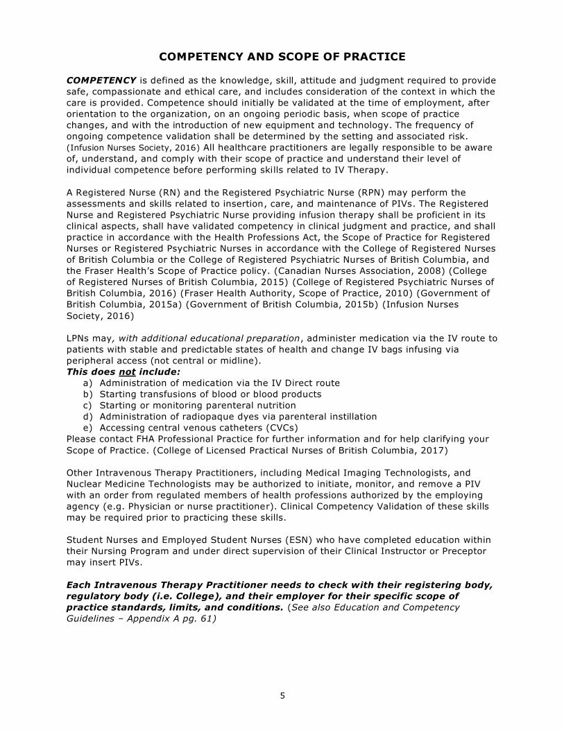
Valves:
Valves are structures within the lumen of veins that are formed by the lining of the Tunica Intima.
They are a system of semi-circular flaps that are arranged in pairs and function to help keep blood
flowing towards the heart by opening and closing like a “trapdoor”. Valves present as bumps along
the course of primarily large veins and also occur at areas where two veins join (bifurcations).
Assess veins for the presence of valves prior to venipuncture and avoid inserting a catheter near a
valve. (BD Medical, 2015)
Digital Veins:
The dorsal digital veins flow along the lateral portions of the fingers. If large enough they may
accommodate a small gauge needle, however they are used as a last resort.
Metacarpal Veins:
The metacarpal veins are formed by the union of the digital veins. They are usually visible, lie flat
on the hand, are easy to feel, and are easily accessible. The hand provides a flat surface for
stabilization and as this vein is in the extremity it allows successive venipunctures to be performed
above the site. These veins may therefore be the first choice for venipuncture and can often
accommodate 20 to 24 gauge catheters. When using, however, the distance from the insertion site
to the prospective catheter tip must be considered to avoid tip positioning in the wrist area. It is
preferred that the wrist not be immobilized. One must consider the impact that limited ability to use
the hand will present to patients requiring hands to support position changes, use crutches, walkers,
and home infusion therapies. Don’t use this site for vesicants, irritants, or medications known to
cause phlebitis.
Cephalic Vein:
The cephalic vein flows upward along the radial aspect of the forearm. Its size readily
accommodates a large needle (often up to 16 gauge), while its position provides easy access and
natural splinting. This vein can be accessed from the wrist to the upper arm (using the most distal
region of the vein first). These veins tend to “roll” so “anchoring” the vein during venipuncture
essential. The large size is an excellent choice for infusing irritants. However, because the fadial
nerve is close to this vein, perform venipuncture 10 to 13 cm above the wrist.
Accessory Cephalic Vein:
The accessory cephalic vein ascends the arm and joins the cephalic vein below the elbow. Its large
size accommodates a large needle (usually up to an 18 gauge). Be cautious not to place the IV
catheter tip in the bend of the arm.
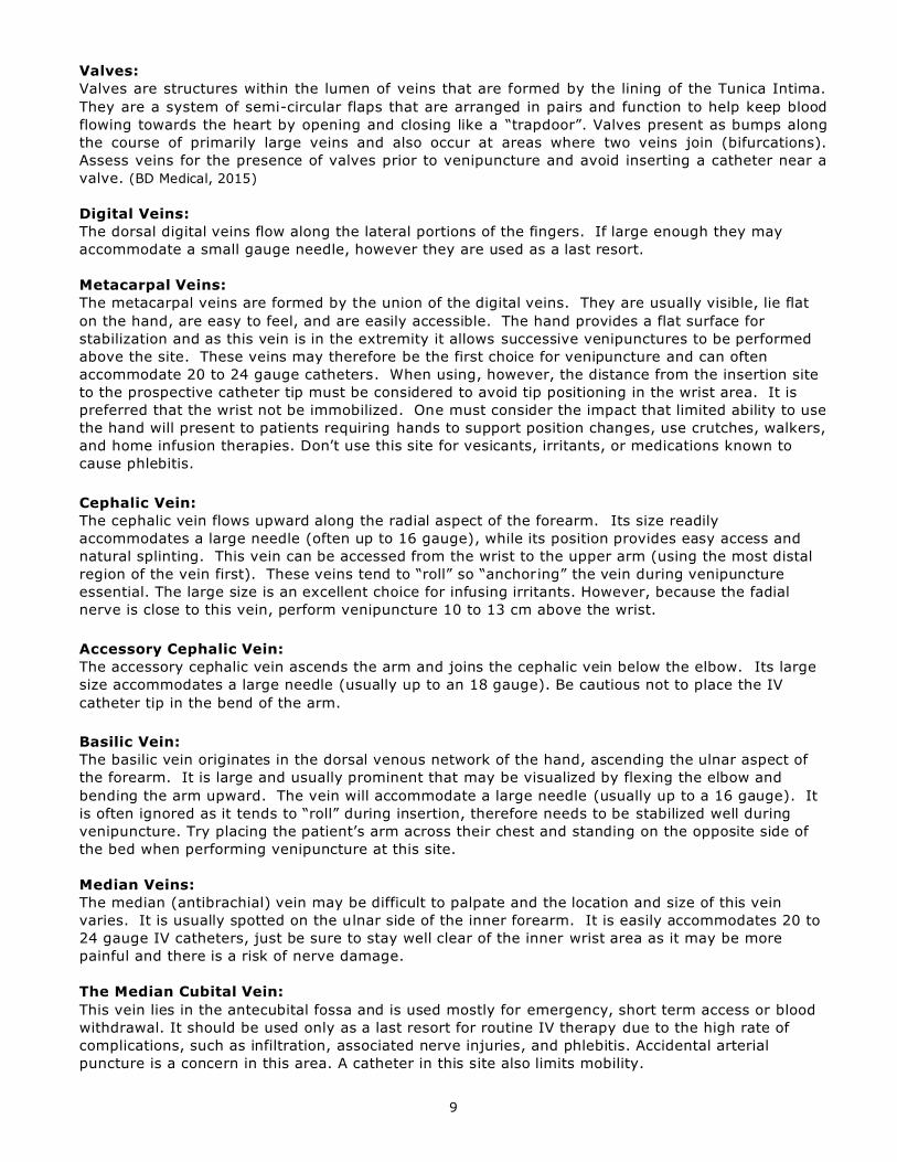
Basilic Vein:
The basilic vein originates in the dorsal venous network of the hand, ascending the ulnar aspect of
the forearm. It is large and usually prominent that may be visualized by flexing the elbow and
bending the arm upward. The vein will accommodate a large needle (usually up to a 16 gauge). It
is often ignored as it tends to “roll” during insertion, therefore needs to be stabilized well during
venipuncture. Try placing the patient’s arm across their chest and standing on the opposite side of
the bed when performing venipuncture at this site.
Median Veins:
The median (antibrachial) vein may be difficult to palpate and the location and size of this vein
varies. It is usually spotted on the ulnar side of the inner forearm. It is easily accommodates 20 to
24 gauge IV catheters, just be sure to stay well clear of the inner wrist area as it may be more
painful and there is a risk of nerve damage.
The Median Cubital Vein:
This vein lies in the antecubital fossa and is used mostly for emergency, short term access or blood
withdrawal. It should be used only as a last resort for routine IV therapy due to the high rate of
complications, such as infiltration, associated nerve injuries, and phlebitis. Accidental arterial
puncture is a concern in this area. A catheter in this site also limits mobility.
10
Figure 3 - Image courtesy of Teleflex®
11
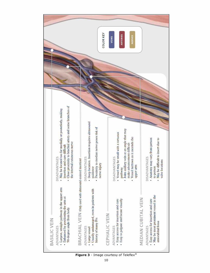
Figure 4 - Superficial veins of the upper limb
From Dorland’s Illustrated Medical Dictionary, 30th ed., Plate 53, p. 2015, © 2003, used with permission from Elsevier. (Infusion Nurses Society, 2016)

12
FLUID AND ELECTROLYTE BALANCE
The concepts discussed in this section will alert the IV nurse to the potential dangers of electrolyte
therapy and changes in the patient’s condition which might alter the therapy. Knowledge of fluid
and electrolytes in the body will contribute to safe and successful therapy.
INTRACELLULAR FLUID – 40% of Body Weight
EXTRACELLULAR FLUID (ECF)
– 20% of Body Weight
Interstitial Fluid –
80% of ECF
Plasma
– 20%
of ECF
TOTAL BODY WATER – 60% OF Body Weight
Figure 5 – Total Body Water
Total body fluid is about 60% of the body weight. The body fluid content in infancy is 70-80% of
the total body weight. Aging reduces the total body fluid to about 52% after age 60 years. The
proportion in newborn infants is approximately three-fifths intracellular and two-fifths extracellular,
but changes to the adult ratio by the time the child is 30 months old.
Body Fluid Balance
When the volume or composition of body fluid is in the compartments deviates even a small
amount, the cells and vital organs of the body suffer.
The intravascular compartment is the most accessible. Fluid is filtered from it to the kidney, lungs,
skin; fluid can enter it from the GI tract and directly from IV fluids.
The interstitial space is next in accessibility, acting as a sort of storage area. The body can store
extra fluid here (over time) or fluid can be borrowed from this space.
The intracellular space is the least accessible space and is protected by the cell membranes. Gains
or losses of hypertonic or hypotonic solutions (to be discussed later) will affect this compartment,
causing the cell to gain or lose fluid. Cells function best in a constant environment.
Composition of Body Fluids
Body fluid contains two types of solutes (dissolved particles): non-electrolytes such as glucose,
creatinine and urea, and electrolytes (see Figure 6 and Table 1).
Figure 6 - The Concentrations of Different Elements in Key Bodily Fluids
Source: (OpenStax College, 2013) http://cnx.org/content/m46411/1.3/

13
Table 1: Electrolytes
ELECTROLYTE LOCATION FUNCTION/SIGNS OF IMBALANCE
Potassium (K+)
HIGH RISK IV
MEDICATION
Intracellular essential for normal function of muscle tissue, especially heart muscle tissue
a low K+ will cause generalized decrease in
muscular activity, apathy, postural hypotension
excess K+ causes heart irregularities, ECG changes
and tingling or numbness in extremities
Magnesium (Mg++) Intracellular enzyme action important for the metabolism of proteins and carbohydrates
necessary to maintain osmotic pressure and
neuromuscular stability (like calcium)
Sodium (Na+) Extracellular essential for regulating water distribution in the
body (water follows sodium)
deficiency will cause weakness, dehydration and
weight loss excess sodium can cause oliguria, dry mucous
membranes and convulsions
Chloride (Cl-) Extracellular tends to follow sodium
deficit leads to potassium defect and vice versa
Bicarbonate (HCO3-) Extracellular is the most important buffer in the body and helps to maintain acid base balance
excess bicarbonate causes alkalosis
deficiencies result in acidosis
Normal range 7.35 - 7.45
Calcium (Ca+) Extracellular essential for blood clotting and required for muscular contraction (e.g. heart muscle) and important for
bone development
deficit causes muscular irritability, cramps and
convulsions
All of these electrolytes are available to be given intravenously to provide, maintain, or correct fluid
and electrolyte balances or to treat other associated co-morbidities. Their use must be approached
with caution, particularly when administered as a dedicated treatment or therapy, and follow the
standards set out in the Parenteral Drug Therapy Manual (PDTM). It is not within the Scope of
Practice of many Intravenous Therapy Practitioners to administer IV medications. Please contact FHA
Professional Practice for further information and for help clarifying your Scope of Practice.
Movement of Body Fluids and Electrolytes
Body fluid compartments are separated by a semi-permeable membrane that allows both body fluids
and solutes to move back and forth. Movement of water and electrolytes between compartments
occurs in four ways:
Diffusion is the random movement of molecules and ions from an area of higher concentration
to an area of lower concentration.
Osmosis is the movement of water across a semi-permeable membrane in response to osmotic
pressure. Osmotic pressure is regulated by electrolytes and non-electrolyte particles in the
fluid. If the extracellular fluid contained a large number of particles and the intracellular fluid
contained a smaller number of particles, the water would pass from the cell into the extracellular
space, until the particle ratio was equal. In this case the cell might be deprived of needed water.
Active transport is a mechanism used to move molecules across a semi-permeable membrane
against a concentration gradient. This process requires cellular energy. An example is the
14
sodium pump, which uses energy to keep the sodium in the extracellular space and the
potassium in the intracellular space. Otherwise they would equalize over time.
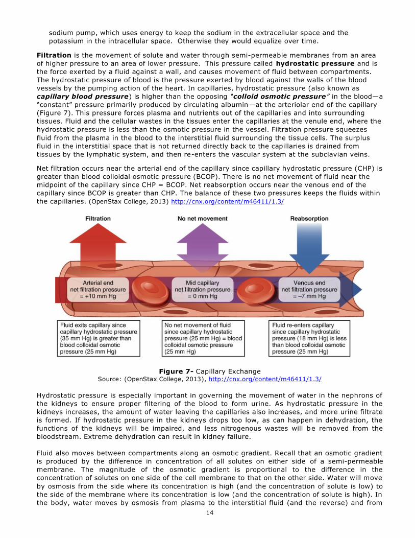
Filtration is the movement of solute and water through semi-permeable membranes from an area
of higher pressure to an area of lower pressure. This pressure called hydrostatic pressure and is
the force exerted by a fluid against a wall, and causes movement of fluid between compartments.
The hydrostatic pressure of blood is the pressure exerted by blood against the walls of the blood
vessels by the pumping action of the heart. In capillaries, hydrostatic pressure (also known as
capillary blood pressure) is higher than the opposing “colloid osmotic pressure” in the blood—a
“constant” pressure primarily produced by circulating albumin—at the arteriolar end of the capillary
(Figure 7). This pressure forces plasma and nutrients out of the capillaries and into surrounding
tissues. Fluid and the cellular wastes in the tissues enter the capillaries at the venule end, where the
hydrostatic pressure is less than the osmotic pressure in the vessel. Filtration pressure squeezes
fluid from the plasma in the blood to the interstitial fluid surrounding the tissue cells. The surplus
fluid in the interstitial space that is not returned directly back to the capillaries is drained from
tissues by the lymphatic system, and then re-enters the vascular system at the subclavian veins.
Net filtration occurs near the arterial end of the capillary since capillary hydrostatic pressure (CHP) is
greater than blood colloidal osmotic pressure (BCOP). There is no net movement of fluid near the
midpoint of the capillary since CHP = BCOP. Net reabsorption occurs near the venous end of the
capillary since BCOP is greater than CHP. The balance of these two pressures keeps the fluids within
the capillaries. (OpenStax College, 2013) http://cnx.org/content/m46411/1.3/
Figure 7- Capillary Exchange Source: (OpenStax College, 2013), http://cnx.org/content/m46411/1.3/
Hydrostatic pressure is especially important in governing the movement of water in the nephrons of
the kidneys to ensure proper filtering of the blood to form urine. As hydrostatic pressure in the
kidneys increases, the amount of water leaving the capillaries also increases, and more urine filtrate
is formed. If hydrostatic pressure in the kidneys drops too low, as can happen in dehydration, the
functions of the kidneys will be impaired, and less nitrogenous wastes will be removed from the
bloodstream. Extreme dehydration can result in kidney failure.
Fluid also moves between compartments along an osmotic gradient. Recall that an osmotic gradient
is produced by the difference in concentration of all solutes on either side of a semi-permeable
membrane. The magnitude of the osmotic gradient is proportional to the difference in the
concentration of solutes on one side of the cell membrane to that on the other side. Water will move
by osmosis from the side where its concentration is high (and the concentration of solute is low) to
the side of the membrane where its concentration is low (and the concentration of solute is high). In
the body, water moves by osmosis from plasma to the interstitial fluid (and the reverse) and from

15
the interstitial fluid to the intracellular fluid (and the reverse). In the body, water moves constantly
into and out of fluid compartments as conditions change in different parts of the body.
For example, if you are sweating, you will lose water through your skin. Sweating depletes your
tissues of water and increases the solute concentration in those tissues. As this happens, water
diffuses from your blood into sweat glands and surrounding skin tissues that have become
dehydrated because of the osmotic gradient. Additionally, as water leaves the blood, it is replaced
by the water in other tissues throughout your body that are not dehydrated. If this continues,
dehydration spreads throughout the body. When a dehydrated person drinks water and rehydrates,
the water is redistributed by the same gradient, but in the opposite direction, replenishing water in
all of the tissues. (Open Stax College, 2013)
Because cells of the vital organs require precise and constant source of fluids and electrolytes and
correct pH, IV therapy is important to replace losses caused by GI suction, burns, NPO, diuresis or
diaphoresis.
Acid-Base Balance
The topic of Acid-Base balance is complex and cannot be covered in this module in its entirety.
Therefore we will only touch upon it briefly as it applies to IV infusions.
The acidity or alkalinity of body fluid depends upon the hydrogen ion concentration expressed as the
pH. The extracellular fluid pH is 7.35 - 7.45, which is the optimum for cells to function.
Acidosis is a decrease in pH
Alkalosis is an increase in pH
Both extracellular and intracellular fluids contain systems to buffer or maintain the proper acid-base
balance. The carbonic acid-sodium bicarbonate system is the most important buffer system in
the extracellular compartment. Other organs in the body also help to maintain fluid, electrolytes
and acid-base balance. Healthy kidneys, skin and lungs are the main regulating organs, by
selectively retaining or secreting electrolytes and fluid according to the body’s needs.
The buffer systems functioning in blood plasma include plasma proteins (e.g. hemoglobin),
phosphate, and bicarbonate and carbonic acid buffers. The kidneys help control acid-base balance
by excreting hydrogen ions and generating bicarbonate that helps maintain blood plasma pH within
a normal range. Protein buffer systems work predominantly inside cells. (OpenStax College, 2013)
The pH of a medication or fluid may play a part in contributing to chemical phlebitis.
Medications/fluids with an osmolarity of greater than 900, a pH of less than 5 or greater than 9,
vesicants, and known irritants need to be given via a CVC. (Infusion Nurses Society, 2016)

16
INFECTION CONTROL
Infection Control Principles
Phlebitis and Catheter Related Bloodstream Infections (CR-BSI) are a preventable nosocomial
infections and adverse events. (Stevens & Schulman, 2012) These infections increase hospital length of
stay and facility costs.
Infective organisms may access the vascular access device surface by either:
i. Invasion of the percutaneous tract
ii. Contamination of the catheter hub
iii. Seeding from a remote source of localized infection
(Association for Professionals in Infection Control and Epidemiology, 2009)
Increased CR-BSI rates are associated with a PIV inserted in the antecubital fossa, a PIV placed in
the Emergency Department, or a PIV placed outside of hospital (e.g. EHS). (Trinh, et al., 2011)
Airborne bacteria increase in number when the activity in the area increases. They interfere with
aseptic technique and may also find their way into unprotected IV solutions, which hang during
intermittent infusion.
The skin is the main source of bacteria responsible for IV infections. Resident bacteria adhering to
the skin include: Staphylococcus albus, Staphylococcus epidermidus
In hospitalized patients the following may also be present: Staphylococcus aureus, Klebsiella,
Enterobacter, Serratia. (Most hospital-acquired infections are now of the gram negative type)
Blood may also harbour microorganisms such as:
Hepatitis B and HIV; dangerous to the health care worker. Adhering to the Standard Precautions
(Universal Precautions), including the use of recommended gloves for blood and body fluids, is
essential. Needles or stylets should not be recapped, but should be disposed of in rigid, tamper-
proof containers
Other bacteria from a distant site of infection may seed the cannulated area. Assessment is
necessary to determine early signs of a low grade infection
Cannula contamination can occur (see Figure 8):
From skin during insertion. Carefully follow site preparation
At the hub by health care worker, breaking system during tubing changes. Perform Hand
Hygiene and maintain strict aseptic technique during tubing and needleless connector changes.
At the tip if a thrombus occurs and is seeded by a distant local infection
During manufacturing
Figure 8
Image courtesy of 3M©
17
Solution contamination can occur:
During admixture of drugs; use of a filtered needle is recommended
When accessing needleless connectors; should be cleansed for 30 sec with 70% alcohol and
allowed to dry completely as per IV Therapy Clinical Practice Guideline.
By improper protection of tubing of intermittent infusions; use single-use intermittent IV tubing
By allowing IV bag to hang for prolonged periods (> 24 hrs)
On the shelf or during handling if small punctures occur to the bag.
If using expired solutions
More frequently in nutrient-rich solutions such as TPN and blood. Use laminar hood to prepare
TPN solutions and follow protocol re: tubing changes. Follow Clinical Practice Guidelines for
Parenteral Nutrition and/or Blood and Blood Product Administration.
General Measures to Reduce IV-Related Infections
Use of strict aseptic technique
Tourniquets and all insertion equipment are to be single patient use (i.e. IV Start Pack)
Careful skin preparation
Careful site management
Examine equipment for integrity and expiry date
Use of filter needle for IV medications
Correct storage and handling of blood products
Schedule for change of IV tubing and solutions (see Table 2)
Ongoing assessment to find signs of infection early
Use of safety engineered IV catheters
Infection Control Guidelines/ Policies
All staff will follow the latest Infection Control Guidelines for Principles of Infection Prevention
and control, Routine Practices (including hand hygiene, application of personal protective
equipment, and sharps handling and disposal) and Additional Precautions, and blood and body
fluid spills clean-up.
All IV insertion sites will be cleansed with a vigorous fraction scrub using 2% Chlorhexidine with
70% alcohol solution, prior to insertion. Cleanse an area larger than the intended dressing and
allow solution to air-dry completely prior to applying dressing.
The site must dry completely prior to the catheter insertion to allow CHG to have maximum
antimicrobial effect.
All needleless connectors will be cleansed with 70% alcohol for 30 seconds and allowed to dry
completely before accessing.
It is strongly recommended that all IV insertion sites will be covered with a transparent semi-
permeable membrane dressing.
A securement dressing/device must be used to stabilize the IV catheter. Some transparent IV
dressings (Tegaderm IV Advanced®) are rated as a securement device. If a “stand-alone”
securement device is used, (e.g. Statlock®) placed under a sterile dressing it must be changed at
least every 7 days.
The use of “IV baskets/trays” is strongly discouraged as the potential for cross-contamination
between patients is greatly increased. Single patient use IV start packs should be used whenever
possible. A minimum of IV supplies should be taken to the patient’s bedside. Unused supplies
that have been in contact with the patient or their bedding can be wiped down with a dis infectant
wipe provided there is no blood or body fluid contamination. If they are contaminated they
should be discarded before leaving the patient bedside/room.
Photo courtesy of BD®
18
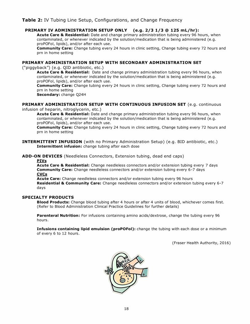
Table 2: IV Tubing Line Setup, Configurations, and Change Frequency
PRIMARY IV ADMINISTRATION SETUP ONLY (e.g. 2/3 1/3 @ 125 mL/hr): Acute Care & Residential: Date and change primary administration tubing every 96 hours, when
contaminated, or whenever indicated by the solution/medication that is being administered (e.g. proPOFol, lipids), and/or after each use.
Community Care: Change tubing every 24 hours in clinic setting, Change tubing every 72 hours and
prn in home setting
PRIMARY ADMINISTRATION SETUP WITH SECONDARY ADMINISTRATION SET
(“piggyback”) (e.g. QID antibiotic, etc.) Acute Care & Residential: Date and change primary administration tubing every 96 hours, when
contaminated, or whenever indicated by the solution/medication that is being administered (e.g.
proPOFol, lipids), and/or after each use. Community Care: Change tubing every 24 hours in clinic setting, Change tubing every 72 hours and
prn in home setting
Secondary: change Q24H
PRIMARY ADMINISTRATION SETUP WITH CONTINUOUS INFUSION SET (e.g. continuous
infusion of heparin, nitroglycerin, etc.) Acute Care & Residential: Date and change primary administration tubing every 96 hours, when
contaminated, or whenever indicated by the solution/medication that is being administered (e.g. proPOFol, lipids), and/or after each use.
Community Care: Change tubing every 24 hours in clinic setting, Change tubing every 72 hours and
prn in home setting
INTERMITTENT INFUSION (with no Primary Administration Setup) (e.g. BID antibiotic, etc.) Intermittent infusion: change tubing after each dose
ADD-ON DEVICES (Needleless Connectors, Extension tubing, dead end caps) PIVs
Acute Care & Residential: Change needleless connectors and/or extension tubing every 7 days Community Care: Change needleless connectors and/or extension tubing every 6-7 days
CVCs
Acute Care: Change needleless connectors and/or extension tubing every 96 hours
Residential & Community Care: Change needleless connectors and/or extension tubing every 6-7
days
SPECIALTY PRODUCTS Blood Products: Change blood tubing after 4 hours or after 4 units of blood, whichever comes first. (Refer to Blood Administration Clinical Practice Guidelines for further details)
Parenteral Nutrition: For infusions containing amino acids/dextrose, change the tubing every 96
hours.
Infusions containing lipid emulsion (proPOFol): change the tubing with each dose or a minimum
of every 6 to 12 hours.
(Fraser Health Authority, 2016)

19
SKIN CARE
All interventions related to IV Therapy, have the potential to affect skin integrity; from the cleansing
solution, to the friction scrub, to the securement device, to the application of the dressing. Catheter
insertion also creates an unavoidable full-thickness wound at the entry site. Cleansing solutions and
a friction scrub disrupt the skin and create shearing forces on the skin. Antimicrobial preparation of
the skin is necessary, but disrupts the natural flora of the skin by design.
Despite their accepted safety, alcohol, chlorhexidine gluconate (CHG), and povidone-iodine have all
been shown to cause contact reactions in otherwise healthy individuals.
Securement devices and dressings can contribute to shearing forces on the skin. Unfortunately,
despite the prevalence of IV Therapy, little research has been done on the effects on the skin.
When erythema is present after application of cleansing solutions, it is generally presumed to be an
allergic reaction. However, two forms of contact dermatitis have been reported in the literature.
Allowing the cleansing solution to dry completely before dressing application and the use of new
dressing products made for sensitive skin will help to alleviate this is a large sector of the
population.
Adhesive trauma is also present, particularly skin stripping and tension blisters. Correct application
and removal techniques for adhesive products are vital to avoid injury to the skin. Ensure you follow
all of the manufacturer’s directions for use.
(Thayer, 2012)
To prevent damage to the skin, a sterile barrier product (see Figure 9) shall be
applied to the skin after the cleansing solution has dried completely.
Use of a sterile clear acrylic absorbent dressing product (see Figure 10)
underneath the TSM dressing has been shown to be effective in managing skin
tears without compromising the efficacy of a TSM dressing with securement
properties. See also Clinical Skills by Elsevier for application procedure. In cases of
more severe or complex skin damage, complete a Wound Assessment and consult
with your Wound Care Clinician as required.
Figure 10 Image courtesy of 3M©
Figure 9 Image courtesy of 3M©
20
PIV INSERTION KEY POINTS
Approach to the Patient
The approach of the nurse is important in the patient’s ability to accept the therapy. Safety both
physical and psychological is important to the patient. Although routine to the nurse, many
procedures in hospital are frightening to the patient. Exaggerated fear triggers the “stress
response” with a cascade of undesirable physiological events, including fluid retention and increased
work of the heart. Avoid using words that might add to the patient’s apprehension, such as “needle”
or “stick”. You might say “I’m going to put this soft plastic catheter in your arm to deliver your
medication” (Hadaway, 2005).
Check patient’s chart for IV order and pertinent history and allergies (e.g. to tape or cleansing
solution)
Identify the patient by identiband and by asking his/her name and birthdate (at least 2
identifiers)
Address the patient by name
The patient’s level of comfort should be assessed and pain controlled if possible, and positioning
should be adjusted as needed for access to the desired insertion site.
By calm explanation of the therapy and it’s expected benefits, the patient’s misinterpretations
and fears may be alleviated
Involve the patient in site selection (if possible)
Draw bedside curtain and ensure privacy (as needed)
Key points to Site Selection
Many factors should be considered when choosing a vein for venipuncture:
- Patient’s age, body size, condition and level of physical activity
- Patient’s condition and medical history
- Vein condition, size and location
- Type and duration of prescribed therapy. If prolonged therapy is anticipated, preservation of
veins is essential. Select most distal and appropriate vein first. If medication/solution has high
potential for vein irritation, select the largest and most appropriate vessel to accommodate the
infusion. Perform venipuncture proximal to a previously cannulated site, injured vein, b ruised area
or site of a recent complication (infiltration, phlebitis, infection) or where impaired circulation is
suspected.
- Patient activity
- Your skill at venipuncture
- Surgery to be done, position of limb during surgery, or if orthopedic surgery, avoid hands (needed
for crutch walking)

21
Points to Ponder on Insertion
Do not routinely re-site PIVs. The decision to replace the PIV should be based on whether the
PIV is still clinically indicated, functioning, the integrity of the insertion site, and whether it is a
source of sepsis. (Infusion Nurses Society, 2012) (Infusion Nurses Society, 2016)
No more than 2 attempts at insertion should be made by any one IV practitioner whenever
feasible. Efforts should be made to have subsequent insertion attempts made by a more
experienced IV Therapy practitioner. If attempts by 2 IV practitioners are unsuccessful, consult
with Physician re: early referral for Extended-Dwell PIV or CVC. Patients with difficult vascular
access require a careful assessment of their vascular access device needs and collaboration with
the healthcare team to discuss appropriate options. Multiple unsuccessful attempts limit future
vascular access, cause the patient unnecessary pain, and increase their risk of developing
phlebitis. (Infusion Nurses Society, 2016) (Washington & Barrett, 2012)
Use strategies to "Save the Vein" when selecting a PIV site with Renal patients, including using
the dorsum of the hand of the non-access limb for PIVs. See details in BC Renal Agency Chronic
Kidney Disease: Vein Preservation Vascular Access Guideline. (BC Renal Agency, 2012)
Avoid using areas of flexion, areas of pain on palpation, compromised veins, areas near valves,
areas where there are planned surgical procedures, or the extremity on the side of breast
surgery with axillary node dissection, after radiation therapy to that side, with the presence of
lymphedema, or the affected side after a stroke. (Infusion Nurses Society, 2016)
Use of the veins in the antecubital fossa should be used for emergent access only due to
associated high rates of phlebitis and nerve injuries. Consider the use of alternate veins in the
lower arm and hand before using the antecubital fossa or the inner aspect of the wrist. (Infusion
Nurses Society, 2016)(Masoorli, 2007)
Use of the veins in the inner aspect of the wrist should be avoided due to high rates of phlebitis
and nerve injuries. Venipuncture should be initiated at least 2 in and/or 5 cm above the crease
of the wrist in an adult patient and 1 in above the crease of the wrist of a baby. (Masoorli, 2007)
The use of veins in the lower extremities does not require an Order. These veins should not be
used routinely in the adult population and should be limited to emergent access. Any patient who
requires an IV in a lower limb due to access problem with an upper limb should be considered for
the insertion of a central venous catheter (CVC). This route should be considered with caution in
patients with peripheral vascular disease (e.g. diabetes).
Consider application of transdermal topical anesthetic cream, spray, or patch to intended
insertion site as needed and when available. Remove anesthetic cream/ patch after
manufacturer recommended application time. Remove any residual cream with a gauze pad. The
use of transdermal topical anesthetic cream, spray, or patch to intended insertion site by an RN
does not require an Order.
Obese patients are considered difficult vascular-access patients. IV site assessment should be
conducted during the initial patient assessment or preoperative examination. Traditional
methods of vascular access placement have low success rates in the obese population. Consider
referral to a PICC RN or MRP for insertion using real-time ultrasonography in conjunction with
longer peripheral IV cannulas to improve outcomes for obese patients. (Houston, 2013)
External Jugular Peripheral Intravenous Catheters (EJ PIVs) (Infusion Nurses Society, 2008)
o Are used for emergent access or for individual situations when other veins cannot be
accessed.
o May only be inserted by a Physician in FHA.
o Contraindicated for use with power injectors or contrast media.
o Dwell times must be limited to prevent formation of fibrin sheaths. Plan immediately
to place an alternative vascular access device.
o Patients with an EJPIV must have alternate access established before transfer to a
non-Critical Care unit or staff on the receiving unit must have training and be
assessed as competent in their use.

22
Evaluating the Selected Vein
Carefully examine both extremities using observation and palpation before selecting the most
appropriate vein. By using the same fingers (not thumbs) consistently, palpation skills will become
more sensitive. To palpate a vein, place one or two fingertips (not thumbs) over it and press lightly.
Release pressure to assess the vein’s elasticity and rebound filling. To acquire a highly developed
sense of touch, palpate before every cannulation – even if the vein looks easy to cannulate
(Hadaway, 2005).
EQUIPMENT
The Cannula
The selected cannulation device should be the smallest gauge and shortest length to accommodate
the prescribed therapy. This allows better blood flow around the catheter, reducing the risk of
phlebitis and promoting proper hemodilution of the fluid (Hadaway, 2005)
IV catheters are available in a range of sizes:
Table 3
Catheter Gauge Size Use this size gauge for:
16 – 18 Trauma patients/Rapid Infusions
High Viscosity Fluids
20 Pre-Operative Patients
Blood Transfusions
22 General Infusions
Blood Transfusions
Children and Elderly
(Not suitable for rapid infusions)
24 Fragile-Veined Patients
Children
Figure 11
Image courtesy of BD®
The IV Solution
The Physician’s Order should be checked for type, amount and rate of solution
The colour, clarity and expiry date of the solution
The integrity of the container and the administration set should be inspected
The IV administration set should be primed
The fluid should be suspended approximately 3 feet above the pump on an IV pole.

23
Electronic Infusion Devices
To prevent or closely control fluid volumes an electronic or flow control infusion device should be
used.
Examples may include a Large Volume IV pump, CADD pump (often used for epidural and
subcutaneous infusions), Patient Controlled Analgesia (PCA) pump, or syringe pump.
Ensure you have completed the Infusion Pump Learning Program at least every 2 years to
maintain your competency and meet Accreditation Canada standards. (Accreditation Canada, 2016).
IV Infusion pumps shall be used for administration of all IV fluids and medications, including
blood and blood products. Appropriate tubing type and/or filter size should be considered when
selecting the correct tubing for the intended purpose. See medication specific information in the
PDTM and transfusion specific recommendations from FH Transfusion Medicine and FH Blood
Administration Guidelines.
Use of gravity flow-based IV fluid administration is limited to circumstances where an IV Pump
cannot be used or with specified uses in specific care areas. These may include IV rates greater
than 999 mL/hr when a Rapid Infuser is not used, during Medical Imaging procedures that do
not allow for the use of a pump or where other infusion devices are used (e.g. Magnetic
Resonance Imaging, CT Scans using pressure injection of contrast media, Nuclear Medicine
injection of radioactive isotopes), in Community home-based settings with intermittent infusions,
and in areas where the primary route is IV Direct only making the use of a pump unworkable
(e.g. Procedural/Conscious sedation, endoscopy procedures). In general, gravity flow-based
medication administration is not recommended and should be avoided due to the risks of
medication error and patient safety. Exception: In certain circumstances administration of
chemotherapeutic agents may be required to be infused via gravity. For further information,
refer to the BC Cancer Agency Guidelines at www.bccancer.bc.ca. (BC Cancer Agency, 2014)
Unique or different infusion devices should be used for administration of fluids and/or
medications via different routes (e.g. IV, epidural, subcutaneous, gastrointestinal). Using
physically separate infusion devices for different routes prevents medication errors by making
the routes and associated infusion devices separate and easily distinguishable. When using
multiple infusion devices with the same patient, ensure you trace the line from the pump to the
patient. Label each line at the connection nearest the patient with the fluid and/or medication
and the route it is being administered.
(Institute for Safe Medication Practices, 2003) (Institute for Safe Medication Practices, 2009) (Koczmara,
Hyland, & Cheng, 2007) (Mattox, 2012) (National Patient Safety Agency, 2007) (Reason, 2000)
Figure 12
Image courtesy of Carefusion®

24
TECHNIQUES IN VENIPUNCTURE
GATHER EQUIPMENT
Non-sterile Gloves
IV Catheter
Start Pack Kit/ Insertion supplies:
- Chlorhexidine 2% with Alcohol 70% swab
- Single use tourniquet
- Transparent securement dressing
- Tape
- Sterile 2x2 gauze
IV Catheter Extension set with needleless connector
Pre-filled 5 mL NS syringe
Stand-alone securement device (optional and primarily only used for some Pediatric and
NICU patients)
IV solution as required (prepared with appropriate primed tubing suspended on a pole)
Electronic Infusion Device or Flow Rate Control Device as required
Figure 13 - IV Start Pack Image courtesy of P. Hignell©
VENOUS DISTENTION
Perform hand hygiene and put on non-sterile gloves. Apply a single-use disposable tourniquet tightly
enough to distend the vein, while still allowing an arterial pulse. Latex-free tourniquets are
preferred as they can be a source of exposure to those with a latex allergy. The tourniquet is
applied to the mid-forearm for use of hand veins, and to the upper arm for veins in the forearm.
Apply the tourniquet flat, to avoid pulling hair or pinching skin. Venous distension may take longer
in elderly or dehydrated patients.
Figure 14
Image courtesy of J. Switzer©
25
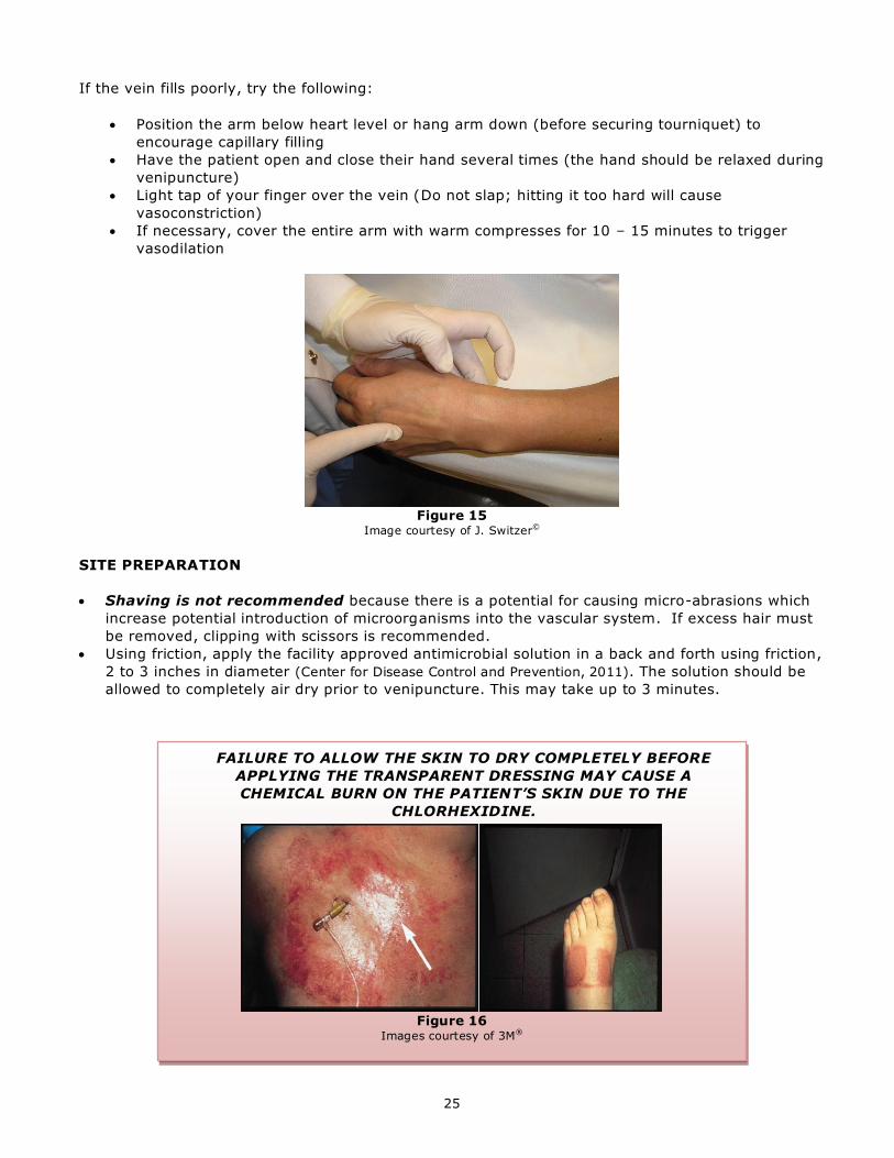
If the vein fills poorly, try the following:
Position the arm below heart level or hang arm down (before securing tourniquet) to
encourage capillary filling
Have the patient open and close their hand several times (the hand should be relaxed during
venipuncture)
Light tap of your finger over the vein (Do not slap; hitting it too hard will cause
vasoconstriction)
If necessary, cover the entire arm with warm compresses for 10 – 15 minutes to trigger
vasodilation
Figure 15
Image courtesy of J. Switzer©
SITE PREPARATION
Shaving is not recommended because there is a potential for causing micro-abrasions which
increase potential introduction of microorganisms into the vascular system. If excess hair must
be removed, clipping with scissors is recommended.
Using friction, apply the facility approved antimicrobial solution in a back and forth using friction,
2 to 3 inches in diameter (Center for Disease Control and Prevention, 2011). The solution should be
allowed to completely air dry prior to venipuncture. This may take up to 3 minutes.
FAILURE TO ALLOW THE SKIN TO DRY COMPLETELY BEFORE
APPLYING THE TRANSPARENT DRESSING MAY CAUSE A
CHEMICAL BURN ON THE PATIENT’S SKIN DUE TO THE
CHLORHEXIDINE.
Figure 16
Images courtesy of 3M®

26
STABILIZING THE VEIN
Stabilizing or “anchoring” the vein prevents movement of the vein during insertion and minimizes
the pain associated with venipuncture. Superficial veins have a tendency to roll because they lie in
loose, superficial connective tissue. To prevent rolling, maintain vein in a taut, distended, stable
position.
Hand Vein - Grasp the patient’s hand with your non-dominant hand. Place your fingers under his
palm and fingers, with your thumb on top of his fingers below the knuckles. Pull his hand downward
to flex his wrist, creating an arch (Hadaway, 2005). Use your thumb to stretch the skin down over
the knuckles to stabilize the vein.
Forearm Vein - Encircle the patient’s arm with your non-dominant hand and use your thumb to pull
downward on the skin below the venipuncture site. If the skin is particularly loose, the vein may
need to be held taut downward below the vein and to the side of the intended site.
Maintain a firm grip of the vein throughout venipuncture.
Figure 17
Image courtesy of J. Switzer©
METHODS OF VENIPUNCTURE
Direct Method - performed by holding the skin taut and entering the skin directly over the vein at a
5 – 15 degree angle. This technique is useful for large veins. If inserted too far it may penetrate
the back wall of the vein.
Indirect Method - the skin is entered beside the vein, and the catheter is redirected to enter the
side of the vein. This motion reduces the risk of piercing the back wall.

27
INSERTING THE CANNULA
Before performing venipuncture, remove the cover from the IV catheter and examine the tip for
smoothness. If any barbs are evident, discard the catheter. Rotate the catheter 360 degrees to
release the catheter from the stylet as they are heat sealed during the manufacturing process.
Once you have anchored the vein, press the vein lightly to check for rebound elasticity and to get a
sense of its depth and resilience. Palpate the portion where the cannula tip will rest, not the point
where you intend to insert the cannula. Note: If you touch the insertion site after cleansing,
you will need to re-clean the site and let it dry completely before proceeding. DO NOT
remove fingertips from gloves for PIV insertion.
Figure 18
Image courtesy of BD®
Figure 19
Image courtesy of BD®
While holding the skin taut (and keeping the vein immobilized) with your non-dominant hand,
grasp the cannula (bevel facing up to reduce the risk of piercing the vein’s back wall). Your
fingers should be placed so that you can see blood backflow in the flash chamber or extension
tubing. Some catheters are designed to provide early flashback of blood between the needle and
the catheter.
Figure 20
Image courtesy of BD®
28
Encourage the patient to relax (breathe slowly in and out as you insert the cannula). Talk to the
patient through the procedure to educate them and decrease their anxiety.
Insert catheter at a 5 to 15 degree angle (depending on depth of the vein), about 1 cm below
the point where the vein is visible.
Don’t always expect to feel a “popping” or “giving-way” sensation (not usual on thin walled, low
volume vessels). Look for blood backflow to tell you that you have entered the vein lumen.
When you see continuous backflow (and you are confident the stylet tip is in the vein), lower
your angle (almost to skin level) and advance slightly (approximately 1/8 inch) to ensure the
cannula tip is also in the lumen of the vein. Continue to hold the stylet hub with your dominant
hand.
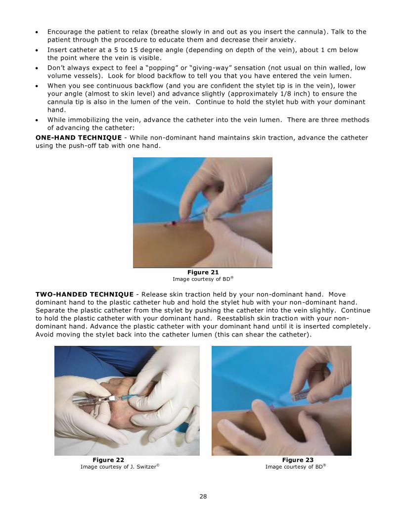
While immobilizing the vein, advance the catheter into the vein lumen. There are three methods
of advancing the catheter:
ONE-HAND TECHNIQUE - While non-dominant hand maintains skin traction, advance the catheter
using the push-off tab with one hand.
Figure 21
Image courtesy of BD®
TWO-HANDED TECHNIQUE - Release skin traction held by your non-dominant hand. Move
dominant hand to the plastic catheter hub and hold the stylet hub with your non-dominant hand.
Separate the plastic catheter from the stylet by pushing the catheter into the vein slightly. Continue
to hold the plastic catheter with your dominant hand. Reestablish skin traction with your non-
dominant hand. Advance the plastic catheter with your dominant hand until it is inserted completely.
Avoid moving the stylet back into the catheter lumen (this can shear the catheter).
Figure 22 Figure 23
Image courtesy of J. Switzer© Image courtesy of BD®

29
“FLOATING” THE CANNULA INTO THE VEIN – Connect the primed administration set to the
catheter hub (when the catheter is only partly inserted into the vein). Flush catheter with IV
solution while advancing the catheter.
Figure 24 Figure 25
Image courtesy of J. Switzer© Image courtesy of J. Switzer©
Once the cannula is totally advanced into the vein, apply digital pressure beyond the cannula tip
and release the tourniquet.
Figure 26 Image courtesy of BD®
If using a cathalon with Blood Control® technology, the tourniquet may be released and the
safety mechanism activated without applying digital pressure, due to the valve in the cathalon
hub. The blood control valve will only function when initially connected to the IV tubing and will
not function as a blood control mechanism afterwards.
Stabilize the hub and activate the safety mechanism. Dispose of the shielded needle in a sharps
container.
Connect the pre-primed extension set with needleless connector with/without continuous IV
tubing. Flush extension tubing with needleless connector with 2-3mL of NS using a pre-filled 3-5
mL NS syringe. Clamp extension set for safety in case of accidental removal of needleless
connector. The frequency the PIV is flushed is every 12 – 24 hrs, depending on setting (e.g.
acute vs community).
Figure 27 – Prefilled 5 mL NS syringe
Image courtesy of BD®
Apply dressing (see next page)
If the IV is continuous, loop the administration set tight (without kinking tubing) and secure with
tape. Set appropriate IV rate.
30
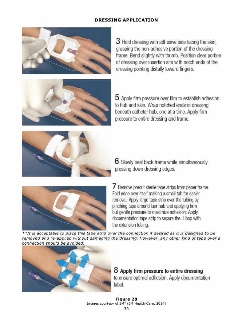
DRESSING APPLICATION
**It is acceptable to place this tape strip over the connection if desired as it is designed to be
removed and re-applied without damaging the dressing. However, any other kind of tape over a
connection should be avoided.
Figure 28 Images courtesy of 3M® (3M Health Care, 2014)
31
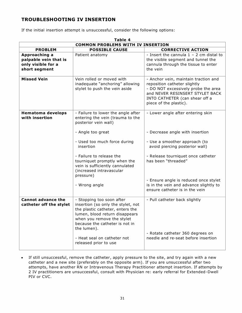
TROUBLESHOOTING IV INSERTION
If the initial insertion attempt is unsuccessful, consider the following options:
Table 4
COMMON PROBLEMS WITH IV INSERTION
PROBLEM POSSIBLE CAUSE CORRECTIVE ACTION
Approaching a
palpable vein that is
only visible for a
short segment
Patient anatomy - Insert the cannula 1 – 2 cm distal to
the visible segment and tunnel the
cannula through the tissue to enter
the vein
Missed Vein Vein rolled or moved with
inadequate “anchoring” allowing
stylet to push the vein aside
- Anchor vein, maintain traction and
reposition catheter slightly
- DO NOT excessively probe the area
and NEVER RESINSERT STYLET BACK
INTO CATHETER (can shear off a
piece of the plastic).
Hematoma develops
with insertion
- Failure to lower the angle after
entering the vein (trauma to the
posterior vein wall)
- Angle too great
- Used too much force during
insertion
- Failure to release the
tourniquet promptly when the
vein is sufficiently cannulated
(increased intravascular
pressure)
- Wrong angle
- Lower angle after entering skin
- Decrease angle with insertion
- Use a smoother approach (to
avoid piercing posterior wall)
- Release tourniquet once catheter
has been “threaded”
- Ensure angle is reduced once stylet
is in the vein and advance slightly to
ensure catheter is in the vein
Cannot advance the
catheter off the stylet
- Stopping too soon after
insertion (so only the stylet, not
the plastic catheter, enters the
lumen, blood return disappears
when you remove the stylet
because the catheter is not in
the lumen).
- Heat seal on catheter not
released prior to use
- Pull catheter back slightly
- Rotate catheter 360 degrees on
needle and re-seat before insertion
If still unsuccessful, remove the catheter, apply pressure to the site, and try again with a new
catheter and a new site (preferably on the opposite arm). If you are unsuccessful after two
attempts, have another RN or Intravenous Therapy Practitioner attempt insertion. If attempts by
2 IV practitioners are unsuccessful, consult with Physician re: early referral for Extended-Dwell
PIV or CVC.
32
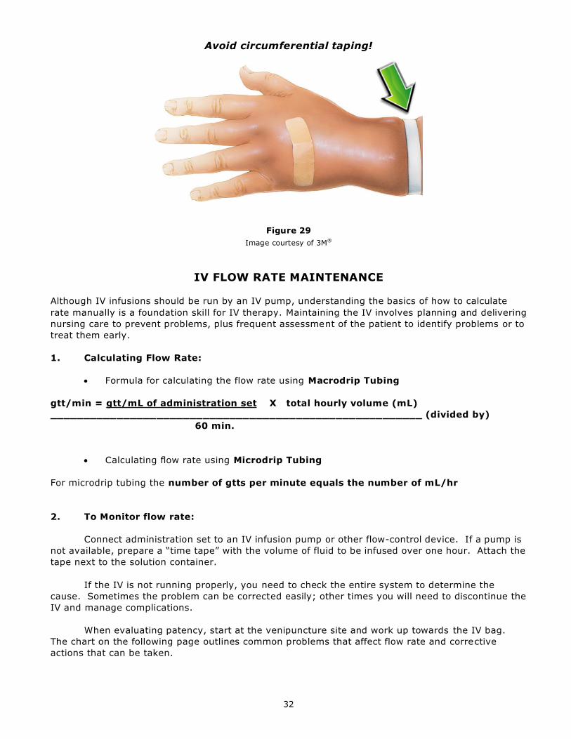
Avoid circumferential taping!
Figure 29
Image courtesy of 3M®
IV FLOW RATE MAINTENANCE
Although IV infusions should be run by an IV pump, understanding the basics of how to calculate
rate manually is a foundation skill for IV therapy. Maintaining the IV involves planning and delivering
nursing care to prevent problems, plus frequent assessment of the patient to identify problems or to
treat them early.
1. Calculating Flow Rate:
Formula for calculating the flow rate using Macrodrip Tubing
gtt/min = gtt/mL of administration set X total hourly volume (mL)
________________________________________________________ (divided by)
60 min.
Calculating flow rate using Microdrip Tubing
For microdrip tubing the number of gtts per minute equals the number of mL/hr
2. To Monitor flow rate:
Connect administration set to an IV infusion pump or other flow-control device. If a pump is
not available, prepare a “time tape” with the volume of fluid to be infused over one hour. Attach the
tape next to the solution container.
If the IV is not running properly, you need to check the entire system to determine the
cause. Sometimes the problem can be corrected easily; other times you will need to discontinue the
IV and manage complications.
When evaluating patency, start at the venipuncture site and work up towards the IV bag.
The chart on the following page outlines common problems that affect flow rate and corrective
actions that can be taken.
33
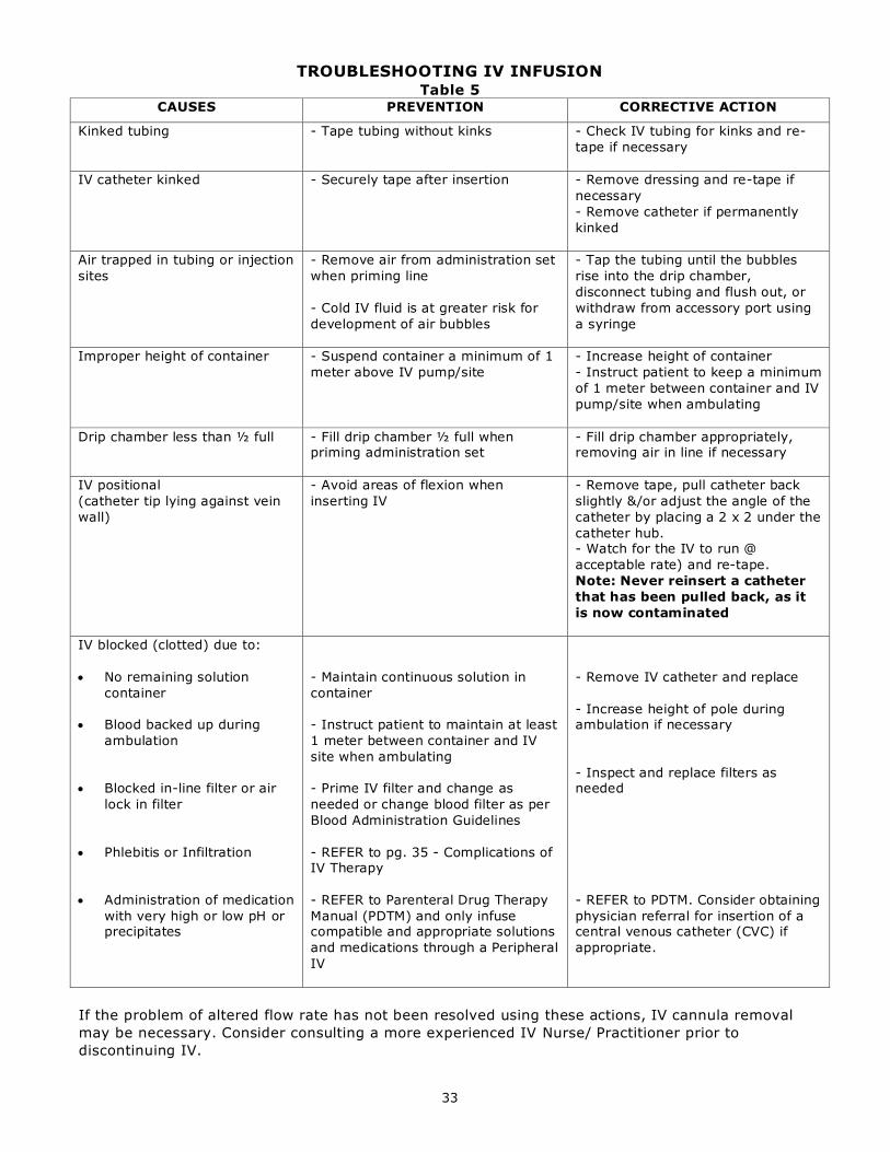
TROUBLESHOOTING IV INFUSION Table 5
CAUSES PREVENTION CORRECTIVE ACTION
Kinked tubing - Tape tubing without kinks - Check IV tubing for kinks and re-
tape if necessary
IV catheter kinked - Securely tape after insertion - Remove dressing and re-tape if
necessary
- Remove catheter if permanently
kinked
Air trapped in tubing or injection
sites
- Remove air from administration set
when priming line
- Cold IV fluid is at greater risk for
development of air bubbles
- Tap the tubing until the bubbles
rise into the drip chamber,
disconnect tubing and flush out, or
withdraw from accessory port using
a syringe
Improper height of container - Suspend container a minimum of 1
meter above IV pump/site
- Increase height of container
- Instruct patient to keep a minimum
of 1 meter between container and IV
pump/site when ambulating
Drip chamber less than ½ full - Fill drip chamber ½ full when priming administration set
- Fill drip chamber appropriately, removing air in line if necessary
IV positional
(catheter tip lying against vein
wall)
- Avoid areas of flexion when
inserting IV
- Remove tape, pull catheter back
slightly &/or adjust the angle of the
catheter by placing a 2 x 2 under the
catheter hub. - Watch for the IV to run @
acceptable rate) and re-tape.
Note: Never reinsert a catheter
that has been pulled back, as it
is now contaminated
IV blocked (clotted) due to:
No remaining solution
container
Blood backed up during
ambulation
Blocked in-line filter or air
lock in filter
Phlebitis or Infiltration
Administration of medication
with very high or low pH or precipitates
- Maintain continuous solution in
container
- Instruct patient to maintain at least
1 meter between container and IV
site when ambulating
- Prime IV filter and change as
needed or change blood filter as per
Blood Administration Guidelines
- REFER to pg. 35 - Complications of IV Therapy
- REFER to Parenteral Drug Therapy
Manual (PDTM) and only infuse compatible and appropriate solutions
and medications through a Peripheral
IV
- Remove IV catheter and replace
- Increase height of pole during ambulation if necessary
- Inspect and replace filters as needed
- REFER to PDTM. Consider obtaining
physician referral for insertion of a central venous catheter (CVC) if
appropriate.
If the problem of altered flow rate has not been resolved using these actions, IV cannula removal
may be necessary. Consider consulting a more experienced IV Nurse/ Practitioner prior to
discontinuing IV.

34
PERIPHERAL IV DAILY ASSESSMENT
PIV sites should be routinely assessed for redness, tenderness, swelling, drainage, and/or the
presence of paresthesias, numbness, or tingling at the specified frequency listed below. Assessment
should minimally include visual assessment, palpation, and subjective information from the patient.
Use the Phlebitis Scale and Infiltration Scales as warranted by the assessment and whenever the PIV
is discontinued. If there is tenderness at the site, the dressing may be removed to more carefully
visualize the site:
1) At least every 4 hours:
a. Patients who are receiving non-irritant/ non-vesicant infusions and who are alert and
oriented and who are able to notify the nurse of any signs of problems such as pain,
swelling, or redness at the site.
2) At least every 1 to 2 hours:
a. Critically ill patients
b. Adult patients who have cognitive/ sensory deficits or who are receiving sedative-type
medications and are unable to notify the nurse of any symptoms
c. PIVs placed in a high-risk location (e.g. external jugular, area of flexion)
3) At least every hour:
a. Neonatal patients
b. Pediatric patients
4) More frequently: every 5 to 10 minutes:
a. Patients receiving intermittent infusions of vesicants
(NOTE: The nurse should advocate for central vascular access administration of vesicant
medications whenever possible. The peripheral infusion of vesicant agents should be
limited to less than 30 to 60 minutes. In addition to visual assessment of the site, a
blood return should be verified every 5 to 10 minutes during the infusion.)
b. Patients receiving infusions of vasoconstrictor agents.
(NOTE: The nurse should advocate for central vascular access administration of
vasoconstrictor agents whenever possible as these agents can cause severe tissue
necrosis with extravasation.)
5) With every Home/ Outpatient visit - For patients receiving peripheral infusions at home as
overseen by Community or Outpatient nurses, patient and family education should include:
• What to look for: redness, tenderness, swelling, or site drainage
• To check the site at least every 4 hours during waking hours
• Ways to protect the site during sleep and activities
• How to stop the infusion if signs/symptoms occur
• To promptly report to the nurse
• The organization’s 24-hour contact telephone numbers
6) For all patients who have a locked PIV for intermittent infusions, the site should be assessed with
every catheter access/infusion or at a minimum of twice per day.
7) Temperature should be checked at a frequency according to Physician’s Orders, unit standards,
and more often based on nursing clinical judgment. The possibility of Catheter-Related
Bloodstream Infection should be considered when there is fever in any patient with a PIV even in
the absence of site redness, tenderness, swelling, or drainage.
(Infusion Nurses Society, 2012)

35
DISCONTINUING IV THERAPY
An order from a regulated member of a health professions authorized by the employing agency is
required to discontinue IV therapy (e.g. continuing order of IV fluids or parenteral medication).
However, upon discharge of the patient from the healthcare facility, and when they will not be
returning for outpatient IV therapy, the IV may be discontinued without an order.
IV catheters do not need to be re-sited at a pre-determined interval. They should be discontinued
and re-sited upon suspected contamination or complications (e.g. interstitial, phlebitis, etc - see
Complications of IV Therapy starting on pg. 38).
Table 6 - Discontinuing a Peripheral IV
1. Verify physician’s or authorized prescriber’s written order
Figure 30 – Dressing Removal
Images courtesy of 3M®
2. Verify patient’s identification, using at least 2 independent
identifiers, not including the patient’s room number
3. Explain procedure to patient
4. Position patient as condition allows. Recumbent or Semi-
Fowler’s position is preferred.
5. Perform hand hygiene and don gloves
6. Select and assemble equipment
7. Discontinue administration of all infusates in the IV line to
be discontinued.
8. Gently remove all adhesive materials, including dressing. Lifts tapes toward the catheter-skin junction by stabilizing skin
surrounding venipuncture site. (see Figure
9. Assess site for any complication, such as infiltration or
phlebitis. Send swab for Culture if infection suspected.
10. Place sterile gauze above site and withdraw catheter,
using a slow, steady motion. Keep the hub parallel to the skin.
11. Apply pressure with sterile gauze to the insertion site for 1-2 minutes or until hemostasis is achieved.
12. Assess integrity of the catheter that was removed.
Compare catheter to original insertion length to ensure entire
catheter is removed. If catheter is not removed intact, notify
physician immediately.
13. Once hemostasis is achieved, apply a sterile dressing to
exit wound as needed.
14. Discard expended equipment in appropriate receptacles
Note: Do not discard catheter if it was not intact on removal.
15. Remove gloves, disposes in appropriate receptacle, and
perform hand hygiene.
16. Instructs patient as to:
a. Recommended activity level b. Removal of dressing
c. Recognition and reporting of post-catheter removal
complication(s)
17. Documents removal procedure in patient’s permanent health record on PIV Insertion and Maintenance Record.
a. Site assessment pre- and post-catheter removal
b. Time IV was discontinued c. Catheter condition and length
c. Achievement of hemostasis and dressing materials
d. Ancillary procedures such as culture of exit wound and catheter
e. Patient response and education
f. Name and title of clinician

36
DOCUMENTATION
Always document in black ink.
Documentation must include the number of insertion attempts, insertion vein location, extremity
accessed, and removal. The preferred form for documentation is the PIV Insertion and
Maintenance Record (see Figure 31).
Figure 31
PIV Insertion and Maintenance Record:
Date and time of insertion – start, re-site or removal
If not original site (re-site) - describe condition of the previous IV site (phlebitis,
infiltration, infection, etc.)
Length and gauge of catheter inserted
Location of site
# of attempts, type of dressing applied and patient’s response to the procedure
Daily assessments, cap and tubing changes, patency assessment
NS flushes
IV Flow Sheet/ Fluid Balance Record:
NS flush
Amount and type of IV solution infusing
Kardex:
Date IV extension tubing with intermittent IV cap is due (every 96 hrs)
Date next tubing change is due (every 96 hrs)
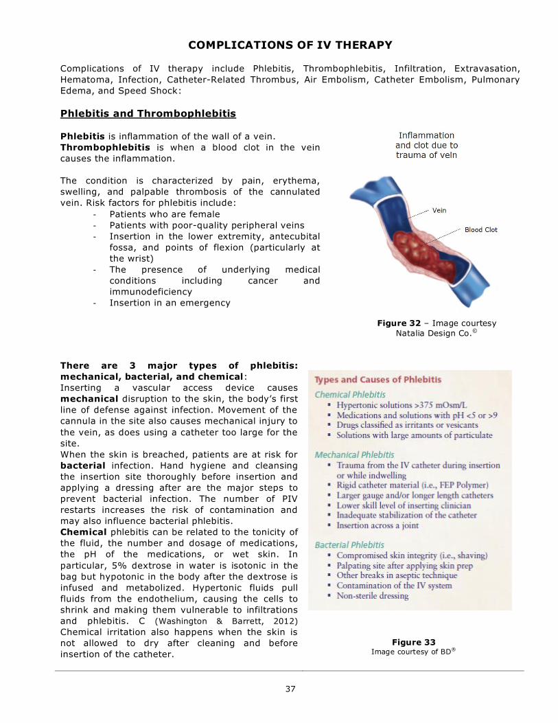
37
COMPLICATIONS OF IV THERAPY
Complications of IV therapy include Phlebitis, Thrombophlebitis, Infiltration, Extravasation,
Hematoma, Infection, Catheter-Related Thrombus, Air Embolism, Catheter Embolism, Pulmonary
Edema, and Speed Shock:
Phlebitis and Thrombophlebitis
Phlebitis is inflammation of the wall of a vein.
Thrombophlebitis is when a blood clot in the vein
causes the inflammation.
The condition is characterized by pain, erythema,
swelling, and palpable thrombosis of the cannulated
vein. Risk factors for phlebitis include:
- Patients who are female
- Patients with poor-quality peripheral veins
- Insertion in the lower extremity, antecubital
fossa, and points of flexion (particularly at
the wrist)
- The presence of underlying medical
conditions including cancer and
immunodeficiency
- Insertion in an emergency
There are 3 major types of phlebitis:
mechanical, bacterial, and chemical:
Inserting a vascular access device causes
mechanical disruption to the skin, the body’s first
line of defense against infection. Movement of the
cannula in the site also causes mechanical injury to
the vein, as does using a catheter too large for the
site.
When the skin is breached, patients are at risk for
bacterial infection. Hand hygiene and cleansing
the insertion site thoroughly before insertion and
applying a dressing after are the major steps to
prevent bacterial infection. The number of PIV
restarts increases the risk of contamination and
may also influence bacterial phlebitis.
Chemical phlebitis can be related to the tonicity of
the fluid, the number and dosage of medications,
the pH of the medications, or wet skin. In
particular, 5% dextrose in water is isotonic in the
bag but hypotonic in the body after the dextrose is
infused and metabolized. Hypertonic fluids pull
fluids from the endothelium, causing the cells to
shrink and making them vulnerable to infiltrations
and phlebitis. C (Washington & Barrett, 2012)
Chemical irritation also happens when the skin is
not allowed to dry after cleaning and before
insertion of the catheter.
Figure 33
Image courtesy of BD®
Figure 32 – Image courtesy
Natalia Design Co.©
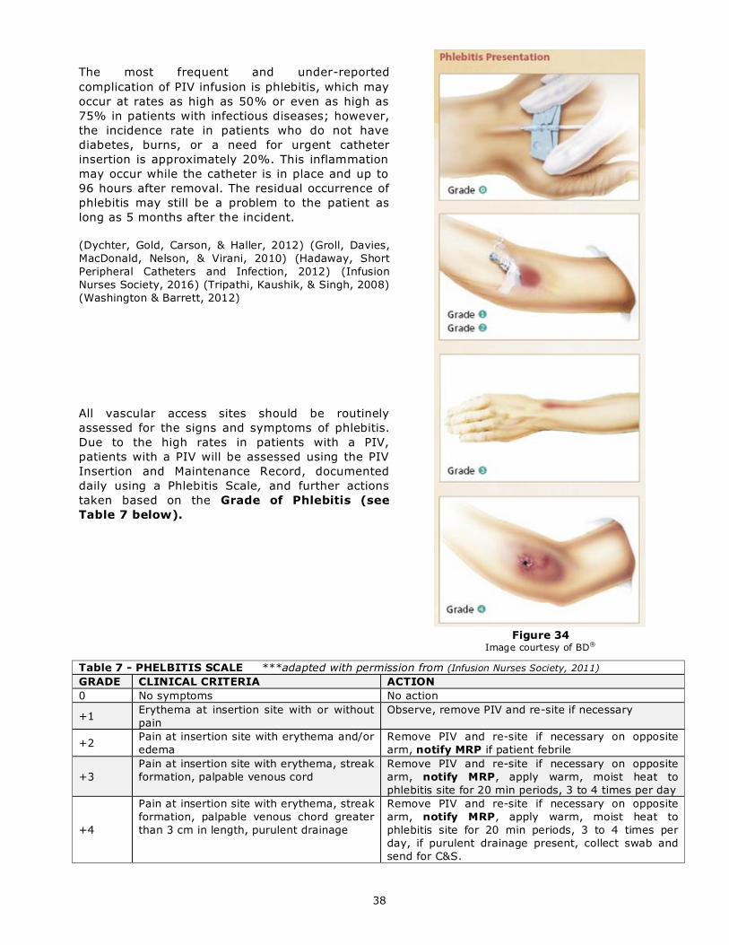
38
The most frequent and under-reported
complication of PIV infusion is phlebitis, which may
occur at rates as high as 50% or even as high as
75% in patients with infectious diseases; however,
the incidence rate in patients who do not have
diabetes, burns, or a need for urgent catheter
insertion is approximately 20%. This inflammation
may occur while the catheter is in place and up to
96 hours after removal. The residual occurrence of
phlebitis may still be a problem to the patient as
long as 5 months after the incident.
(Dychter, Gold, Carson, & Haller, 2012) (Groll, Davies,
MacDonald, Nelson, & Virani, 2010) (Hadaway, Short
Peripheral Catheters and Infection, 2012) (Infusion
Nurses Society, 2016) (Tripathi, Kaushik, & Singh, 2008)
(Washington & Barrett, 2012)
All vascular access sites should be routinely
assessed for the signs and symptoms of phlebitis.
Due to the high rates in patients with a PIV,
patients with a PIV will be assessed using the PIV
Insertion and Maintenance Record, documented
daily using a Phlebitis Scale, and further actions
taken based on the Grade of Phlebitis (see
Table 7 below).
Figure 34
Image courtesy of BD®
Table 7 - PHELBITIS SCALE ***adapted with permission from (Infusion Nurses Society, 2011)
GRADE CLINICAL CRITERIA ACTION
0 No symptoms No action
+1 Erythema at insertion site with or without
pain
Observe, remove PIV and re-site if necessary
+2 Pain at insertion site with erythema and/or
edema
Remove PIV and re-site if necessary on opposite
arm, notify MRP if patient febrile
+3
Pain at insertion site with erythema, streak
formation, palpable venous cord
Remove PIV and re-site if necessary on opposite
arm, notify MRP, apply warm, moist heat to
phlebitis site for 20 min periods, 3 to 4 times per day
+4
Pain at insertion site with erythema, streak
formation, palpable venous chord greater
than 3 cm in length, purulent drainage
Remove PIV and re-site if necessary on opposite
arm, notify MRP, apply warm, moist heat to
phlebitis site for 20 min periods, 3 to 4 times per
day, if purulent drainage present, collect swab and
send for C&S.

39
Infiltration and Extravasation Infiltration is defined as the inadvertent leakage of a non-vesicant solution or medication into surrounding
tissue.
Extravasation is the leakage of a drug or fluid from a vein into the surrounding tissue during intravenous
administration (NHS Cheshire and Merseyside Strategic Clinical Networks, 2016). These injuries range from less
significant erythematous reactions to skin sloughing and necrosis. Whilst extravasation is possible with any intravenous injection it is considered to be especially problematic with compounds known to have irritant or
vesicant properties. The onset of symptoms may occur immediately or several days to weeks after
administration. If left undiagnosed or inappropriately treated, necrosis and functional loss of tissue and limb
concerned may ensue. (WOSCAN Cancer Nursing and Pharmacy Group, 2012)
Extravasation is associated with cytotoxic chemotherapy agents such as doxorubicin, paclitaxel, and vinca
alkaloids, as well as a number of non-cytotoxic drugs, including phenytoin, sodium bicarbonate (>5%),
calcium chloride and gluconate, amphotericin B, acyclovir, ganciclovir, digoxin, diazepam, potassium (>40 mmol/L), dextrose 50%, cefotaxime, IV contrast media, and mannitol, that can also cause tissue necrosis.
The incidence of infiltration and extravasation is hard to determine because of limited reporting; however,
extravasation injury from cancer chemotherapy is reported to be 11% in children and 22% in adults. One
study found that, of all the complications associated with PIVs, 33.7% occurred as a result of inf iltration. Rates of extravasation with cancer chemotherapy infused through implanted ports range from 0.3% to 6%. (Dychter, Gold, Carson, & Haller, 2012) (Hadaway, 2007)
***See also Extravasation Drug and Antidote Table on the FH Pulse.
1. Vesicant - A drug which has corrosive properties and has the potential to cause tissue destruction if
extravasated. Varying degrees of pain, edema, erythema, blistering and necrosis may occur. Vesicants are further divided into two groups. When extravasated, non-DNA binding agents (vinblastine, vinorelbine,
vincristine) are inactivated or quickly metabolized and follow the normal healing process whereas DNA
binding agents (epirubicin, mitomycin, doxorubicin, daunorubicin, idarubicin) remain in the tissues
resulting in long-term injury.
2. Exfoliant - A drug capable of causing inflammation and shedding of skin but less likely to cause tissue death.
3. Irritant - This has the potential to cause pain, aching, tightness and phlebitis with or without
inflammation, rarely progressing to tissue breakdown.
4. Inflammitant - Drug with the potential to cause mild to moderate inflammation and flare in local tissues. 5. Neutral Drugs – Drugs that cause very little or no tissue damage when extravasation occurs. (BC Cancer Agency, 2016) (NHS Royal Cornwall Hospitals, 2014)(WOSCAN Cancer Nursing and Pharmacy Group, 2012) All vascular access sites should be routinely assessed for the signs and symptoms of infiltration and
extravasation. Due to the high rates in patients with a PIV, patients with a PIV will be assessed using the PIV
Insertion and Maintenance Record, documented daily using an Infiltration Scale (see Table 8 below), and
further actions taken based on the Grade of infiltration (see table below). Children and infants are vulnerable
populations requiring special attention and the use of appropriate assessment tools. Pediatric assessment has been incorporated into this tool.
Measures to prevent infiltration and extravasation include:
- Selection of an appropriate site for catheter insertion - Avoid areas of joint flexion such as wrist and antecubital fossa
- Selection of an appropriate size catheter - Use the smallest size catheter needed for the prescribed
therapy
- Use of appropriate fluids depending on vascular access type – Medications/fluids with an osmolarity of
greater than 900 mOsm/L, a pH of less than 5 or greater than 9, and vesicants need to be given via a CVC.
- Stabilization of the catheter – Always use a securement device/dressing. Avoid circumferential taping of
the limb.
- Use of proper administration techniques - The patency of the catheter and vein should be assessed frequently so that infiltration or extravasation can be prevented. Before administering each dose of
medication, the nurse should visually inspect and palpate the site, checking for vein cording, edema, skin
temperature, and tenderness or discomfort. Check for a positive blood return. Flushing the catheter with
normal saline while palpating the site also makes detection of swelling at the PIV catheter tip easier. Each
drug should be diluted in a separate syringe. When dilution is done either in a syringe or by slow injection through an infusing, compatible IV fluid, according to the PDTM, the injection can be stopped if the
patient complains of any problems. (Hadaway, 2007)
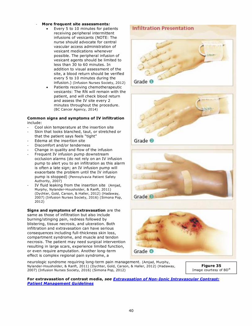
40
Figure 35 Image courtesy of BD®
- More frequent site assessments:
Every 5 to 10 minutes for patients
receiving peripheral intermittent infusions of vesicants (NOTE: The
nurse should advocate for central
vascular access administration of
vesicant medications whenever
possible. The peripheral infusion of vesicant agents should be limited to
less than 30 to 60 minutes. In
addition to visual assessment of the
site, a blood return should be verified every 5 to 10 minutes during the
infusion.) (Infusion Nurses Society, 2012)
Patients receiving chemotherapeutic
vesicants: The RN will remain with the
patient, and will check blood return and assess the IV site every 2
minutes throughout the procedure. (BC Cancer Agency, 2014)
Common signs and symptoms of IV infiltration
include:
- Cool skin temperature at the insertion site - Skin that looks blanched, taut, or stretched or
that the patient says feels “tight”
- Edema at the insertion site
- Discomfort and/or tenderness
- Change in quality and flow of the infusion - Frequent IV infusion pump downstream
occlusion alarms (do not rely on an IV infusion
pump to alert you to an infiltration as this alarm
is often a late sign; an IV infusion pump will exacerbate the problem until the IV infusion
pump is stopped) (Pennsylvavia Patient Safety
Authority, 2007) - IV fluid leaking from the insertion site (Amjad,
Murphy, Nylander-Housholder, & Ranft, 2011)
(Dychter, Gold, Carson, & Haller, 2012) (Hadaway,
2007) (Infusion Nurses Society, 2016) (Simona Pop,
2012)
Signs and symptoms of extravasation are the
same as those of infiltration but also include burning/stinging pain, redness followed by
blistering, tissue necrosis, and ulceration. Both
infiltration and extravasation can have serious
consequences including full-thickness skin loss, compartment syndrome, and muscle and tendon
necrosis. The patient may need surgical intervention
resulting in large scars, experience limited function,
or even require amputation. Another long-term
effect is complex regional pain syndrome, a
neurologic syndrome requiring long-term pain management. (Amjad, Murphy,
Nylander-Housholder, & Ranft, 2011) (Dychter, Gold, Carson, & Haller, 2012) (Hadaway,
2007) (Infusion Nurses Society, 2016) (Simona Pop, 2012)
For extravasation of contrast media, see Extravasation of Non-Ionic Intravascular Contrast: Patient Management Guidelines

41
Table 9 - Distinguishing Extravasation from other conditions:
Characteristic
Flare reaction
Vessel irritation
Venous
shock
Extravasation
Immediate
Symptoms
Itchy blotches or hives;
pain and burning
uncommon
Aching and
tightness
Muscular
wall of the
blood vessel
in spasm
Severe pain or burning at the PIV
site and/ or anywhere along the
vein that lasts minutes or hours
and eventually subsides; usually
occurs while the drug is being
given.
Colouration
Raised red streak,
blotches or “hive-like”
erythema along the
vessel; diffuse or
irregular pattern
Erythema or dark
discolouration
along vessel
Erythema around area of needle
or around the venipuncture site
Timing
Transient and usually
resolves with or without
treatment within 30 min,
sometimes within 1 – 2
hours, and rarely takes
up to 24 hrs.
Usually appears
within minutes
after injection.
Colouration may
only appear later
in the process.
Usually
appears
right after
injection
Symptoms start to appear right
after injection, symptoms endure
Swelling
Unlikely
Unlikely
Occurs often; does not dissipate
for several days
Blood return
Usually, but not always
intact
Usually, but not
always intact
Often absent Usually absent or sluggish
(BC Cancer Agency, 2016) (European Oncology Nursing Society, 2007)
FLARE: painless local reaction along the vein or near the intact injection site characterised by:
- Immediate, red blotches or streaks (histamine release phenomenon), or local wheals; edema may
sometimes occur
- With or without pruritus or irritation
- Symptoms usually subside with or without treatment 30 min after the infusion is stopped, although they
may last for 1-2 hours and rarely more than 24 hours
Table 8 - INFILTRATION SCALE ***adapted from (Amjad, Murphy, Nylander-Housholder, & Ranft, 2011)(Infusion Nurses Society, 2011, p. 113) (Simona Pop, 2012)
GRADE CLINICAL CRITERIA ACTION 0 No symptoms No action
+1
Skin blanched, edema less than 3 cm in any direction or
1%-10% of the extremity above or below the insertion site,
cool to touch, with or without pain
Discontinue infusion, remove PIV and re-site if
necessary on opposite arm
+2
Skin blanched, edema 3-15 cm in any direction or up to ¼
of the extremity above or below the insertion site or 10%-
25% of the extremity above or below the insertion site,
cool to touch, with or without pain
Discontinue infusion, remove PIV and re-site if
necessary on opposite arm, apply warm, moist
heat to infiltrations site for 20 min periods, 3
to 4 times per day to alleviate discomfort and
help absorb infiltration.
+3
Skin blanched, translucent, gross edema greater than 15
cm in any direction or ¼ - ½ of the extremity above or
below the insertion site or 25%-50% of the extremity
above or below the insertion site, cool to touch, mild to
moderate pain, possible numbness
Discontinue infusion; determine type,
concentration, and volume of solution infused.
Notify MRP. Discontinue infusion, remove PIV
and re-site if necessary on opposite arm,
apply warm, moist heat to infiltrations site for
20 min periods, 3 to 4 times per day to
alleviate discomfort and help absorb
infiltration.
+4
Skin blanched, translucent; skin tight, leaking; skin
discoloured, bruised, swollen; gross edema greater than 15
cm in any direction or greater than ½ of the extremity
above or below the insertion site or greater than 50% of
the extremity above or below the insertion site; deep
pitting edema; circulatory impairment; moderate to severe
pain; infiltration of any amount of blood product, irritant, or
vesicant.
***EXTRAVASATION IS ALWAYS GRADED AS +4
Discontinue infusion; determine type,
concentration, and volume of solution infused.
Notify MRP. Follow instruction in Error!
Reference source not found. for treatment
(see also “Potential Hazards of Administration”
in PDTM drug monograph).
Consider referral to Plastic Surgery
**COMPLETE PSLS

42
VESSEL IRRITATION: Aching and tightness occurs along the vein. Seen with drugs such as vinorelbine and
dacarbazine. Applying warmth to dilate the vein can relieve this. Blood return is usually intact although
erythema or redness may be present.
VENOUS SHOCK: Rapid administration or the administration of very cold drugs can cause the muscle wall of
the vein to go into spasm. Blood return may be lost. Heat can help to relax and dilate the vein.
NOTE: For oncologic agents for cancer treatment, you may defer to the BCCA Systemic Therapy Guidelines for drug categories and administration procedures.
Immediate Treatment
If extravasation is suspected, it is important to act quickly to prevent tissue necrosis. A Plastic Surgery consult
is recommended for further advice to discuss removal of the device and immediate and future treatment as this may differ with each event.
1. Stop the infusion or injection immediately – DO NOT remove the PIV at this point.
2. Seek assistance if needed. 3. Disconnect the infusion (not the PIV catheter).
4. Leave the PIV catheter in place and try to aspirate as much of the drug as possible from the
cannula using a 10 mL syringe. Avoid applying direct manual pressure to suspected extravasation
site.
5. Remove the PIV. Elevate and apply gentle pressure to site till hemostasis is achieved. Exception: With extravasation of Mechlorethamine the antidote Sodium Thiosulfate is to be given through
the existing IV line immediately after the extravasation and preferably though the same needle to
insure injection into the same tissue plane as the extravasation.
6. Mark the affected area and take digital images of the site (with patient consent). 7. Obtain extravasation kit or appropriate antidote and notify the MRP, and PCC or Charge Nurse.
8. Decide on how the extravasation should be treated. See Error! Reference source not found..
9. Apply warm towel or ice pack wrapped in towel/cold compresses to the extravasation site for 1
hour as per Extravasation Drug and Antidote Recommendation Table. Care must be taken to avoid
tissue injury from excessive heat or cold. 10. Administer pain relief as needed.
11. For vesicants, consult Plastic Surgery ASAP.
12. Document the injury with a digital image if possible. See Fraser Health Media Consent Form.
13. Provide patient education and complete all documentation, including PSLS. See Also FH Policy: Disclosure of Unintended Medical Outcomes
(BC Cancer Agency, 2016) (NHS Cheshire and Merseyside Strategic Clinical Networks, 2016) (NHS Royal
Cornwall Hospitals, 2014) (NHS Wales, 2008) (Reynolds, MacLaren, Mueller, & Fish, 2014)
Extravasation kits should be stocked on or available to all inpatient and outpatient Oncology Units. For access to the medications on other sites/units, contact the Oncology Unit or Site Pharmacy. They may include
the following: Pain Ease® spray
25 gauge needles
Dimethylsulfoxide (DMSO) 99% topical solution 3 mL syringes
Hyaluronidase 1500 units /mL injection (HYALASE®) ampoule**
Black indelible ink marker
Hydrocortisone 1% cream Phentolamine 10 mg
Ice pack - in freezer
Sodium thiosulfate 25% injection, 10 mL vial***
Sterile gauze dressings and tape
10 mL syringe (for preparing sodium thiosulfate)*** ** Available through Health Canada Special Access Program
*** Only if intravenous mechlorethamine is available for use (BC Cancer Agency, 2016) (Reynolds, MacLaren, Mueller, & Fish, 2014)

43
Hematoma
Hematoma – A localized mass of blood located outside the vessel, usually creating a hard, painful
lump.
Prevention:
Apply firm pressure to insertion site with sterile gauze after unsuccessful attempt start
Do not reapply tourniquet to the same limb after an unsuccessful start
Be aware of patients taking anti-coagulants
Signs and Symptoms:
Swelling at the site
Discomfort at site
Raised area of ecchymosis
Occasionally bleeding at the site
Interventions
Discontinue IV and re-site IV in opposite limb if possible
Elevate the affected limb and apply direct pressure to the site with sterile gauze
Apply cold compress
Figure 36
Image courtesy of BD®
Figure 37
Image courtesy of P. Hignell®

44
Infection
Infection – Catheter Related Bloodstream Infections (CR-BSI) are a preventable nosocomial
infection and adverse event. (Stevens & Schulman, 2012) These infections increase hospital length
of stay and facility costs. Studies of CR-BSIs that control for the underlying severity of illness
suggest that mortality attributable to these infections is between 4-20%. (Safer Healthcare Now,
2012)
Infective organisms may access the vascular access device surface by either:
- Invasion of the percutaneous tract
- Contamination of the catheter hub
- Seeding from a remote source of localized infection (Association for Professionals in Infection Control and Epidemiology, 2009)
Increased CR-BSI rates are associated with a PIV inserted in the antecubital fossa, a PIV placed in
the Emergency Department, or a PIV placed outside of hospital (e.g. EHS). (Trinh, et al., 2011)
The nurse and/or IV Therapy Practitioner shall assess patients for suspected infusion related and
CR-BSIs and document signs and symptoms, interventions implemented and patient response to
treatment in the patient’s permanent health record and the Patient Safety Learning System (PSLS). (Infusion Nurses Society, 2016)
Prevention:
Use aseptic technique when caring for IV
Thoroughly inspect medication and solution containers prior to use
Inspect access site and equipment regularly
Change administration set and solution according to IV Therapy Guideline (see Table 3 pg. 18)
Utilize single-use intermittent medication tubing
Signs and symptoms of CR-BSI include:
- Erythema, edema, induration, or drainage at the vascular device insertion site
- Elevated body temperature
Diagnosis: Purulent drainage from the catheter-skin junction of a PIV should be collected using
the Levine Technique (cleanse site with normal saline prior to collecting swab for culture and twirl
the end of the cotton-tipped applicator on a 1-cm2 area of the wound bed with enough pressure to
cause minimal bleeding). (Spear, 2014)
Treatment: PIVs with purulent drainage present should be removed and re-sited based on patient
need and condition. Send the catheter to Microbiology for culture if purulent drainage is noted on
removal. When seen in conjunction with elevated body temperature, consult physician for order for
Blood Cultures and treatment plan.
(Center for Disease Control and Prevention, 2011) (Pennsylvavia Patient Safety Authority, 2007) (Royer,
2010) (Safer Healthcare Now, 2012)

45
Catheter Embolism
Catheter Embolism – Catheter embolism is the result of catheter damage or rupture, resulting in
the breaking off of a portion of the PIV into the bloodstream. (Canadian Vascular Access Association,
2013)
Prevention:
- Never use vascular access devices for power-injection that are not rated for this
purpose.
- The size of the flush syringe should be in accordance with the type of vascular access
device and its intended use.
- The most frequent cause for catheter embolism is re-insertion of the stylet into a PIV
catheter
- Catheter dysfunction, such as the inability to aspirate blood or fluid with localized pain
and/or subcutaneous swelling, may be a precursor to catheter embolism. Leaking at the
catheter insertion may indicate catheter rupture. PIVs which exhibit these signs should
be carefully discontinued and re-sited (note any damage to the PIV tip upon removal).
- Catheter embolism should be suspected when the patient exhibits symptoms such as
palpitations, arrhythmias, dyspnea, cough, or thoracic pain not associated with patient’s
diagnosis or comorbidities.
- Upon removal, inspect all catheters for damage and possible fragmentation. (Infusion Nurses Society, 2016)
When vascular access device removal is difficult or if damage to the catheter is seen:
1. Carefully assess the patient for signs and symptoms of catheter fragmentation
and;
2. Save catheter and report via PSLS and BCCSS Product Concern Form.
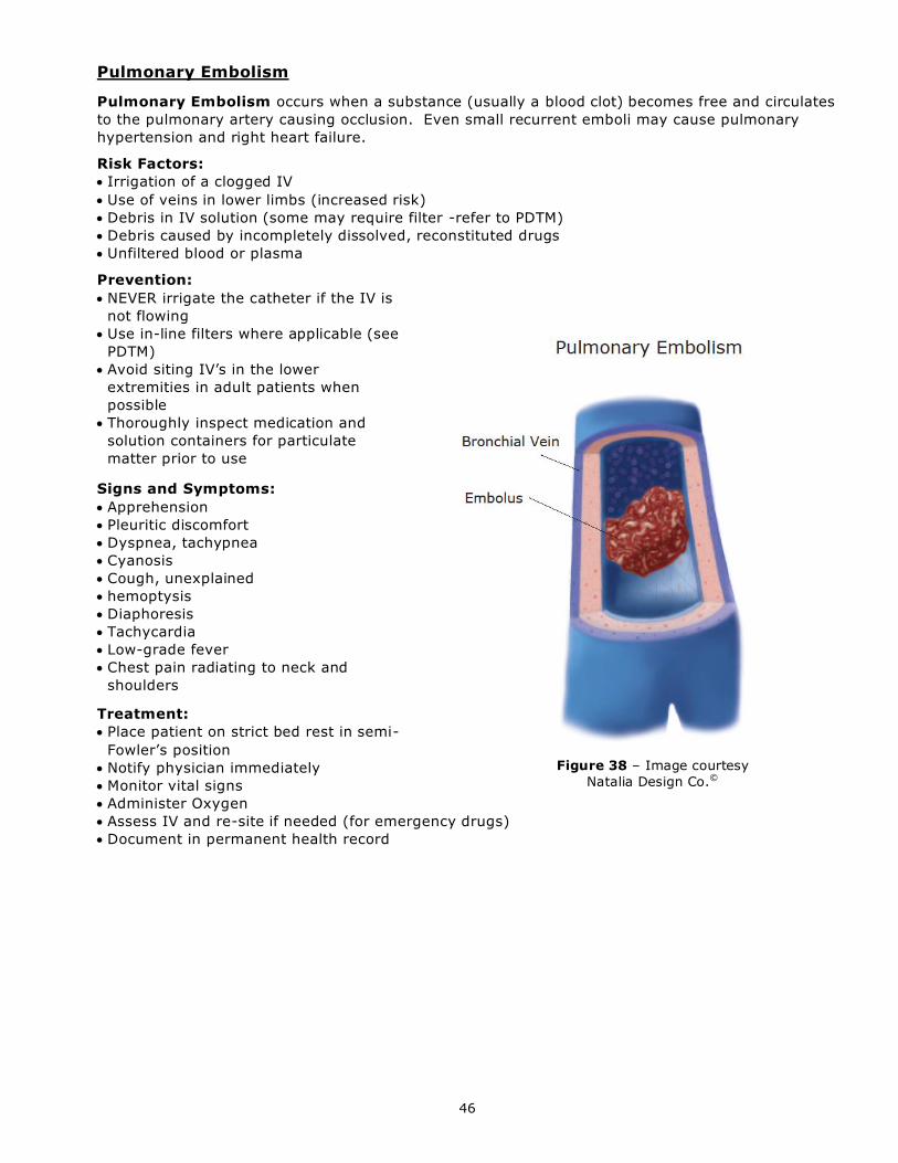
46
Pulmonary Embolism
Pulmonary Embolism occurs when a substance (usually a blood clot) becomes free and circulates
to the pulmonary artery causing occlusion. Even small recurrent emboli may cause pulmonary
hypertension and right heart failure.
Risk Factors:
Irrigation of a clogged IV
Use of veins in lower limbs (increased risk)
Debris in IV solution (some may require filter -refer to PDTM)
Debris caused by incompletely dissolved, reconstituted drugs
Unfiltered blood or plasma
Prevention:
NEVER irrigate the catheter if the IV is
not flowing
Use in-line filters where applicable (see
PDTM)
Avoid siting IV’s in the lower
extremities in adult patients when
possible
Thoroughly inspect medication and
solution containers for particulate
matter prior to use
Signs and Symptoms:
Apprehension
Pleuritic discomfort
Dyspnea, tachypnea
Cyanosis
Cough, unexplained
hemoptysis
Diaphoresis
Tachycardia
Low-grade fever
Chest pain radiating to neck and
shoulders
Treatment:
Place patient on strict bed rest in semi-
Fowler’s position
Notify physician immediately
Monitor vital signs
Administer Oxygen
Assess IV and re-site if needed (for emergency drugs)
Document in permanent health record
Figure 38 – Image courtesy
Natalia Design Co.©
47
Air Embolism
Air Embolism is the presence of air in the vascular system. (Infusion Nurses Society, 2016)
A venous air embolism occurs when air is introduced into the venous system and travels to the
right ventricle and/or pulmonary circulation. An arterial air embolism results from air entry into the
arterial system and can produce ischemia of and organ with poor collateral circulation . (Broadhurst,
2013) Air embolism is reported to occur more frequently during catheter removal than during
insertion. (Truscott, 2013)
The minimum amount of air that is lethal to humans is not known, however, the risks from smaller
amounts increase with a device closer to the central vasculature (CVC) or with pediatric and
neonatal patients, particularly in presence of a patent foramen ovale. (Cook, 2013)
The introduction of microbubbles into the vascular system is also related to the development of an
air embolism and may go unnoticed. Cardiopulmonary bypass, hemodialysis, mechanical heart
valves, major surgeries, warming of cold infusates, and high IV infusion flow rates during trauma
resuscitation can produce large amounts of microbubbles. (Broadhurst, 2013) (Cook, 2013)
Factors:
- Failure to occlude the needle hub and/or catheter during insertion or removal
- An improperly primed IV administration set
- An incorrect technique when administering drugs via the IV route
- Inadvertent infusions of air
- An accidental disconnection at the catheter hub, connector, or IV administration set
- A stopcock placed in the wrong position
- A ruptured catheter
- Poor technique during needleless connector changes
- IV fluid infusions that are completed or rapid infusion through an air-filled drip chamber
with an IV administration set that is unclamped
Prevention:
- Minimize PIV manipulations
- Remove all air from syringes, IV administration sets, needleless connectors, stopcocks,
and all other devices added to a PIV
- Trace all IV lines from the catheter hub to the IV fluid container to prevent
misconnections
- Remove air bubbles detected during an IV infusion. Infrequent tiny pinhead bubbles,
even though not desirable, can be left but should be monitored.
- Never use scissors near a PIV to prevent accidental severing of the catheter.
- Carefully fill and prime IV administration sets and filters and ensure vented tubing in
clamped off before the container is completely empty.
- Use IV infusion pumps with air sensing technology for IV fluid and medication infusions.
- Instruct patients and caregivers to not disconnect and reconnect any IV administration
sets or connectors from the catheter hub to properly prime tubing, to check connections
frequently, how to prevent displacement/disconnection, and actions to take in case of
PIV displacement or damage. (Broadhurst, 2013) (Cook, 2013) (Infusion Nurses Society, 2016)
Signs and Symptoms:
- Sudden onset of dyspnea
- Continued coughing
- Breathlessness
- Agitation or irritability, often expressed as a feeling of impending doom
- Shoulder and chest pain
- Lightheadedness
- Hypotension
- Jugular venous distention
- Tachyarrhythmias
- Wheezing
- Tachypnea
- Altered mental status

48
- Symptoms that emulate stroke including altered speech, changes in facial appearance,
numbness and paralysis (Cook, 2013) (Infusion Nurses Society, 2016)
Diagnosis:
Diagnosis may not be straightforward but prompt diagnosis and treatment will decrease potential
mortality and morbidity. Radiological techniques including transthoracic echocardiography and
precordial ultrasonography have been used to detect air embolisms. (Cook, 2013)
Treatment:
Treatment should begin immediately even if an air embolism is only suspected to prevent further
air from entering the vasculature:
- Close, fold, or clamp the existing catheter to occlude entry of passive air.
- Occlude the puncture site of a catheter that had been removed.
- Place the patient in Trendelenberg left lateral decubitus position (left side, head flat, feet
up, right side uppermost) if not contraindicated by other conditions such as increased
intracranial pressure or respiratory illness. The goal of this positioning is to trap the air
in the lower portion of the right ventricle and prevent it from travelling to the pulmonary
arteries.
- Administer oxygen at 100%
- If possible, attempt to aspirate air from catheter.
- Monitor vital signs and start CPR as needed.
- Notify physician. (Broadhurst, 2013) (Cook, 2013) (Infusion Nurses Society, 2016) (Wilkins & Unverdorben, 2012)
Pulmonary Edema Pulmonary Edema is an increase in venous pressure with increased pressure in the right
ventricle, pulmonary artery and subsequent fluid in the alveoli.
Risk Factors:
Rapid or excessive fluid administration, especially in patients with impaired renal or cardiac
function, or elderly or very young patients
Prevention:
Maintain prescribed flow rate with regular patient assessment
Use volumetric infusion pump for patients whenever possible to avoid accidental fluid overload
Signs and Symptoms:
EARLY Signs:
o Restlessness
o Slow increase in pulse rate
o Headache
o Shortness of breath
o Non-productive cough
o Skin flushing
LATE Signs:
o Hypertension
o Severe dyspnea with coarse crackles
o Engorged neck veins (JVD)
o Pitting edema
o Pink, frothy sputum
o Puffy eyelids
o Shock, respiratory or cardiac arrest
Treatment:
Place patient in high fowlers position
Slow the IV to keep vein open
Notify the physician
Administer medications as requested by physician
Monitor vital signs
Administer high-flow Oxygen
Document in permanent health care record
Submit report via Patient Safety Learning System (PSLS) when due to accidental fluid overload

49
Speed Shock Speed Shock is a sudden adverse physiologic reaction to IV medications or drugs that are
administered too quickly.
Risk Factors: Caused by rapid infusion of drugs or solution causing toxic proportions to reach the
heart and brain.
Prevention:
Monitor gravity-flow administration sets closely to ensure correct prescribed flow rate
Use volumetric infusion pump for medications as directed in the PDTM
Follow recommended infusion rate for medication as per PDTM
Signs and Symptoms:
EARLY Signs:
o Dizziness
o Facial flushing
o Headache
o Irregular Heart Rate
o Chest pain/ tightness
o Sudden onset of symptoms associated
with particular medication being
administered
LATE Signs:
o Syncope
o Shock
o Cardiac arrest
Treatment:
Stop the infusion
Maintain IV access for emergency
treatment
Notify physician immediately or Code Team if
applicable
Monitor vital signs
50
REVIEW QUESTIONS
1. A “Competency Assessed IV Therapy Practitioner” will:
A. Have successfully completed a theoretical and practical experience
B. Have had at least two years of experience in IV therapy
C. Be automatically certified if certified at another health agency
D. Will receive more money because of this skill
2. IV related standards, procedures and protocols may be found in which of the following
manuals?
A. INS Infusion Standards of Practice
B. FHA Clinical Policy Office
C. Site-based policy manual
D. Parenteral Drug Therapy Manual and Infection Control Manual
1) A. B, C
2) A, B, D
3) A, C, D
4) All of the above
3. Which of the following statements are true of the arterial circulatory system?
A. Arteries carry blood to the tissues
B. Blood is under high pressure in the arterial system
C. Arteries have valves to help blood flow
D. Arteries have a palpable pulse
1) A, B, C
2) A, B, D
3) A, C, D
4) All of the above
4. Which of the following statements are true of the venous circulatory system?
A. The venous system is a low pressure system
B. Veins have a weak pulse
C. Superficial and deep veins unite in the lower extremities
D. Damage to valves may result in varicosities
1) A, B, C
2) A, B, D
3) A, C, D
4) B, C, D
5. Arteries and veins have:
A. One layer
B. Two layers
C. Three layers
D. Four layers

51
6. Name the veins and arteries on the following diagram:
Figure 4 - Superficial veins of the upper limb
From Dorland’s Illustrated Medical Dictionary, 30th ed., Plate 53, p. 2015, © 2003,
used with permission from Elsevier. (Infusion Nurses Society, 2016)
7. The PIV catheter most frequently used today is:
A. PICC
B. Cutdown
C. Needle-safe
D. Internal jugular
8. The gauge of needle/cannula most commonly used for routine IV therapy is:
A. #18g
B. #20g
C. #22g
D. #24g
9. Prime factors to be considered when choosing a vein for infusion include:
A. Location
B. Condition
C. Purpose
D. Duration
1) A, B, C
2) A, B, D
3) A, C, D
4) All of the above

52
10. Which of the following statements are true about a needless connector (saline lock)?
A. The needleless connector lock requires short extension tubing to be added
B. The site should be assessed and changed as required
C. The needleless connector is flushed every 12-24 hours (and/or after medication) with
2-3 mL of normal saline
D. After removal check site for bleeding, edema, signs of infection
1) A, B, C
2) A, B, D
3) B, C, D
4) All of the above
11. Which of the following are nursing actions to be done before starting the PIV?
A. Check patient’s chart for allergies and doctor’s order
B. Identify the patient by identity band and by asking their name
C. Involve the patient in site selection
D. Ensure privacy
1) A & B
2) A, B, D
3) B, C, D
4) All of the above
12. Venous distention may be achieved by which of the following?
A. Tourniquet to mid-forearm for use of the hand veins
B. Tourniquet tight enough to occlude an arterial pulse
C. Position the arm below heart level
D. Application of warm packs 10-15 minutes prior to IV start
1) A, B, C
2) A, B, D
3) A, C, D
4) B, C, D
13. Stabilizing the vein minimizes the pain associated with venipuncture:
A. True
B. False
14. Factors affecting flow rate of the IV are:
A. Phlebitis
B. Height of the IV fluid container
C. Positional IV
D. Air trapped in the IV tubing
1) A, B, C
2) A, B, D
3) B. C. D
4) All of the above

53
15. Signs of infiltration include:
A. Sluggish flow rate
B. Warm, reddened skin at the site
C. Swelling of the limb
D. Slow backflow
1) A, B, C
2) A, B, D
3) A, C, D
4) B, C, D
16. Phlebitis is caused by injury or irritation from which of the following?
A. Long term therapy
B. Acidic or alkaline substances
C. Trauma during insertion
D. Poor aseptic technique
1) A, B, C
2) A, B, D
3) B, C, D
4) All of the above
17. Signs of Catheter-Related Bloodstream Infection (CR-BSI) include:
A. Fever
B. Increase in pulse rate
C. Hypotension
D. Sudden onset of chills
1) A, B, C
2) A, B, D
3) B, C, D
4) All of the above
18. To prevent Catheter-Related Bloodstream Infection (CR-BSI) which of the following are
recommended?
A. Examine solutions carefully
B. Cut the fingertips off gloves of IV Therapy Practitioner prior to insertion
C. Mix solutions at the bedside
D. Mix solutions in Pharmacy in a laminar flow hood
1) A & C
2) A & D
3) C & D
4) B & D
19. Symptoms of Catheter Embolus include which of the following?
A. Dyspnea
B. Chest pain
C. Diaphoresis
D. Weak rapid pulse
1) A, B, C
2) A, B, D
3) B, C, D
4) All of the above
54
20. If an Air Embolus is suspected, the appropriate intervention is to:
A. Turn patient on left side, head down
B. Turn patient on right side, head down
C. Turn patient on left side, head raised
D. Turn patient on right side, head raised
21. Early signs of Pulmonary Edema include:
A. Restlessness
B. Headache
C. Shortness of breath
D. Fever
1) A, B, C
2) A, B, D
3) A, C, D
4) B, C, D
22. Factors predisposing the patient to Catheter-Related Bloodstream Infection (CR-BSI)
including all the following except:
A. Age less than one year or more than 60 years
B. State of immune system
C. Increased red blood cell count
D. Presence of distant infection
23. The most common source of bacteria responsible for IV infection comes from:
A. Airborne sources
B. The patient’s skin
C. The cannula
D. Healthcare worker’s hands
24. Which of the following measures help to reduce IV related infections?
A. Handwashing prior to venipuncture
B. Use of a laminar hood for IV admixtures
C. Protection of intermittent infusion IV tubing
D. Sterile dressing using aseptic technique
1) A, B, C
2) A, B, D
3) B, C, D
4) All of the above
25. What percentage of the adult’s weight is body fluid?
A. 5%
B. 15%
C. 40%
D. 60%
26. What percentage of the adult’s weight is contained in intracellular fluid?
A. 5%
B. 15%
C. 20%
D. 40%

55
27. How often should needleless connectors and extension sets be changed in acute care?
A. Every 6-7 days
B. Every 24 hours
C. Every shift
D. Every 96 hours
28. Body water and electrolytes move in which of the following ways?
A. Colloidal
B. Diffusion
C. Active transport
D. Osmosis
1) A, B, C
2) A, B, D
3) B, C, D
4) All of the above
29. Which electrolyte is responsible for regulating water distribution in the body?
A. Potassium
B. Calcium
C. Sodium
D. Chloride
30. Which electrolyte is essential for the normal function of the heart muscle?
A. Potassium
B. Zinc
C. Sodium
D. Chloride
31. Which statements are true about acid-base balance?
A. Kidneys, lungs and skin are the main regulating organs
B. The normal pH is 7.35-7.45
C. Acidosis is a decrease in pH
D. Alkalosis is an increase in pH
1) A, B, C
2) A, B, D
3) B, C, D
4) All of the above
32. Hypotonic is a term which means that a solution has:
A. A higher osmotic pressure than the blood
B. A lower osmotic pressure than the blood
C. The same osmotic pressure as blood
D. Ability to carry O2 to the cells
33. Which of the following statements are true of a hypertonic solution?
A. Draws water out of the cells and interstitial space and into the intravascular space
B. Has a higher osmotic pressure than the blood
C. Has a lower osmotic pressure than the blood
D. Dextrose 10% and 50% in water are examples of a hypertonic solution
1) A, B, C
2) A, B, D
3) B, C, D

56
34. 3% and 5% saline have been called dangerous solutions because:
A. They move water out of the cell and interstitial space and into the intravascular space
B. They cause imbalance in the intracellular water and sodium
C. They are isotonic
D. They contain a lot of potassium
35. Which of the following statements are true regarding Infusion Pumps?
A. Only appropriate vendor-specific IV tubing should be used with IV Infusion pumps
B. Different Infusion devices should be used when therapies are administered via
different routes (e.g. IV, epidural, subcutaneous)
C. IV Infusion pumps do not have a safety clamp (anti-free flow clamp) which is
activated by opening the door
D. An IV Infusion Pump should be used for most IV fluid and medication administration
1) A, B, C
2) A, B, D
3) A, C, D
4) B, C, D
36. Which of the following statements are true about the immediate treatment for extravasation?
A. Stop the infusion immediately
B. Leave the PIV catheter in place and try to aspirate as much of the drug as possible
C. Apply direct manual pressure to suspected extravasation site
D. Obtain extravasation kit or appropriate antidote
1) A, B, C
2) A, B, D
3) B, C, D
4) All of the above
37. Which of the following should be charted on the patient’s permanent health record?
A. Length and gauge of the IV catheter
B. Site of venipuncture
C. Number of missed insertion attempts
D. Patient’s response to the procedure
1) A, B, C
2) A, B, D
3) B, C, D
4) All of the above
38. Which of the following statements are true about PIV Assessment?
A. All PIV sites should be assessed routinely
B. Neonatal and pediatric patients should have their PIV sites assessed every 4 hours
C. PIV sites should be assessed every 5-10 min when administering vesicants
D. PIV sites should be assessed with every Home or Outpatient visit
1) A, B, C
2) A, B, D
3) B, C, D
4) A, C, D
57
39. All patients with a PIV should be assessed daily using the Phlebitis Scale and the Infiltration
Scale.
A. True
B. False
40. The most frequent cause for catheter embolism is re-insertion of the stylet into a PIV catheter.
A. True
B. False
58
ANSWERS TO REVIEW QUESTIONS
1. A 21. 1
2. 2 22. C
3. 2 23. B
4. 3 24. 2
5. C 25. D
6. See pg. 12 26. D
7. C 27. A
8. B 28. 3
9. 4 29. C
10. 4 30. A
11. 4 31. 4
12. 3 32. B
13. A 33. 2
14. 4 34. B
15. 3 35. 2
16. 4 36. 2
17. 4 37. 4
18. 2 38. 4
19. 4 39. 1
20. A 40. 1
59
REFERENCES
3M Health Care. (2014). 3M™ Tegaderm™ I.V. Advanced Securement Dressing; 1683 Application Technique.
Ontario, Canada: Skin & Wound Care Division, 3M Canada.
Accreditation Canada. (2016, September). Required Organizational Practices, 2, 34. Ottawa, ON. Amjad, I., Murphy, T., Nylander-Housholder, L., & Ranft, A. (2011). A New Approach to Management of
Intravenous Infiltration in Pediatric Patients: Pathophysiology, Classification, and Treatment. Journal of
Infusion Nursing, 34(4), 242-249.
Association for Professionals in Infection Control and Epidemiology. (2009). An APIC Guide. Guide to the
Elimination of Catheter-Related Bloodstream Infection. Washington, DC. BC Cancer Agency. (2014, November). CHEMOTHERAPEUTIC DRUGS, ADMINISTRATION OF:. Nursing Practice
Reference. Vancouver, BC, Canada. Retrieved September 21, 2015, from
http://www.bccancer.bc.ca/nursing-site/Documents/C252ChemotherapeuticAgentsAdministrationOf.pdf
BC Renal Agency. (2012). Chronic Kidney Disease: Vein Preservation. Vascular Access Guideline. Retrieved March 27, 2014, from http://www.bcrenalagency.ca/sites/default/files/documents/files/Vein-
Preservation-Renal-Patients-Update-Aug-17-2012.pdf
BD Medical. (2015). Layers of the Vein. Sandy, Utah.
Broadhurst, D. (2013). Death by Air: How Much is Too Much? Vascular Access, 7(1), 16-25.
Campisi, C., Biffi, R., & Pittiruti, M. (2007). Catheter-Related Central Venous Thrombosis: The Development of a Nationwide Consensus Paper in Italy. Journal of the American Vascular Association, 12(1), 38-46.
Canadian Nurses Association. (2008). Code of Ethics for Registered Nurses. Retrieved from Canadian Nurses
Association.
Canadian Vascular Access Association. (2013). Occlusion Management Guideline for Central Venous Access Devices (CVADs). Vascular Access, 7(S1).
Center for Disease Control and Prevention. (2011). Guidelines for the Prevention of Intravascular Catheter-
Related Infections. Atlanta: Center for Disease Control and Prevention.
College of Licensed Practical Nurses of British Columbia. (2015). Scope of Practice for Licensed Practical Nurses.
Vancouver: College of Licensed Practical Nurses of British Columbia. College of Licensed Practical Nurses of British Columbia. (2017, June 16). Scope of Practice for Licensed
Practical Nurses. BC: CLPNBC.
College of Registered Nurses of British Columbia. (2010). Scope of Practice for Registered Nurses: Standards,
limits and conditions. Retrieved September 6, 2013 College of Registered Nurses of British Columbia. (2015). Scope of Practice for Registered Nurses: Standards,
limits and conditions. Retrieved September 6, 2013
College of Registered Psychiatric Nurses of British Columbia. (2016, March 18). Scope of Practice for Psychiatric
Nurses: Standards, Limits, and Conditions.
College of Registered Psychiatric Nurses of British Columbia. (2016, March 18). Scope of Practice for Psychiatric Nurses: Standards, Limits, and Conditions. Retrieved March 7, 2016, from College of Registered
Psychiatric Nurses of British Columbia: http://www.crpnbc.ca/wp-content/uploads/2014/04/Scope.pdf
Cook, L. S. (2013). Infusion-Related Air Embolism. Journal of Infusion Nursing, 36(1), 26-36.
Dychter, S. S., Gold, D. A., Carson, D., & Haller, M. (2012). Intravenous Therapy: A Review of Complications and Economic Considerations of Peripheral Access. Journal of Infusion Nursing, 35(2), 84-91.
Fraser Health Authority. (2010). Scope of Practice. Fraser Health Authority.
Fraser Health Authority. (2011, November). Parenteral Drug Administration Guidelines. Parenteral Drug
Therapy Manual. Surrey, BC. Fraser Health Authority. (2012). Pediatric Considerations for Peripheral Intravenous Initiation. Fraser Health
Authority.
Fraser Health Authority. (2016). Clinical Practice Guideline: Intravenous Therapy. Surrey, BC.
Government of British Columbia. (2015a). Health Professions Act [Nurses (Registered) and Nurse Practitioners
Regulation, B.C. Regulation of the Minister of Health Services. Victoria: Government of British Columbia.
Government of British Columbia. (2015b). Health Professions Act Nurses (Registered Psychiatric) Regulation,
B.C. Victoria: Province of British Columbia.
Government of British Columbia. (2017). Health Professions Act [Nurses (Registered) and Nurse Practitioners Regulation, B.C. Regulation of the Minister of Health Services. Victoria: Government of British
Columbia.
Groll, D., Davies, B., MacDonald, J., Nelson, S., & Virani, T. (2010). Evaluation of the Psychometric Properties
of the Phlebitis and Infiltration Scales for the Assessment of Complications of Peripheral Vascular Access
Devices. Journal of Infusion Nursing, 33(6), 385-390. Hadaway, L. (2005). On the Road to Successful IV Starts. Nursing, 35, 1-14.
Hadaway, L. (2007). Infiltration and Extravasation: Preventing a complication of IV catheterization. American
Journal of Nursing, 107(8), 64-72.

60
Hadaway, L. (2012). Short Peripheral Catheters and Infection. Journal of Infusion Nursing, 35, 4, 230-240. Houston, P. (2013). Obtaining Vascular Access in the Obese Patient Poplulation. Journal of Infusion Nursing,
36(1), 52-56.
Infusion Nurses Society. (2008). INS Position Paper: The Role of the Registered Nurse in the Insertion of
External Jugular Peripherally Inserted Central Catheters (EJ PICC) and External Jugular Peripheral
Intravenous Catheters (EJ PIV). Infusion Nurses Society. Infusion Nurses Society. (2011). Infusion Nursing Standards of Practice. Journal of Infusion Nursing.
Infusion Nurses Society. (2012). INS Position Paper: Recommendations for Frequency of Assessment of the
Short Peripheral Catheter Site. Journal of Infusion Nursing, 35(5), 290-292.
Infusion Nurses Society. (2016). Infusion Therapy Standards of Practice. Journal of Infusion Nursing, 39(1S). Infusion Nurses Society. (2016). Infusion Therapy Standards of Practice. Journal of Infusion Nursing, 39(1S).
Institute for Safe Medication Practices. (2003, January). Reports of Epidural Infusion Errors. ISMP Canada
Safety Bulletin, 3, 1.
Institute for Safe Medication Practices. (2009). Proceedings from the ISMP Summit on the Use of Smart
Infusion Pumps: Guidelines for Safe Implementation and Use. Horsham, PA: ISMP. Koczmara, c., Hyland, s., & Cheng, R. (2007). ISMP Canada: Epidural medications given intravenously may
result in death. Canadian Association of Critical Care Nurses, 18(3), 34-36.
Masoorli, S. (2007). Nerve Injuries Related to Vascular Access Insertion and Assessment. Journal of Infusion
Nursing, 30(6), 346-350. Mattox, E. A. (2012). Strategies for Improving Patient Safety: Linking Task Type to Error Type. Critical Care
Nurse, 32(1), 52-78.
National Patient Safety Agency. (2007, March). Safer practice with epidural injections and infusions. Patient
safety alert.
Open Stax College. (2013, July 3). Acid-Base Balance. Retrieved July 25, 2016, from http://cnx.org/contents/OWD-ifaA@4/Acid-Base-Balance
OpenStax College. (2013, June 20). Body Fluids and Fluid Compartments. OpenStax-CNX. Retrieved July 25,
2016, from http://cnx.org/content/m46411/1.3/
Pennsylvavia Patient Safety Authority. (2007). IV infiltration: Be alarmed even when your pump isn't. Patient Safety Advisory, 4, 1-4.
Reason, J. (2000). Human error: models and management. British Medical Journal, 320, 768-770.
Royer, T. (2010). Implementing a Better Bundle to Achieve and Sustain a Zero Central Line-Associated
Bloodstream Infection Rate. Journal of Infusion Nursing, 33(6), 398-406. Safer Healthcare Now. (2012). Central Line-Associated Bloodstream Infection (CLABSI). Retrieved from Safer
Healthcare Now: http://www.saferhealthcarenow.ca/EN/Interventions/CLI/Pages/default.aspx
Simona Pop, R. (2012). A Pediatric Peripheral Intravenous Infiltration Assessment Tool. Journal of Infusion
Nursing, 35(4), 243-248.
Spear, M. (2014). When and how to culture a chronic wound. Wound Care Advisor, 3(1), 23-25. Stevens, T. P., & Schulman, J. (2012). Evidence-based approach to preventing central line-associtaed
bloodstream infection in the NICU. Acta Paediatrica, Supp 464, 11-16.
Thayer, D. (2012). Skin Damage Associated with Intravenous Therapy. Journal of Infusion Nursing, 35(6), 390-
401. Trinh, T. T., Chan, P. A., Edwards, O., Hollenbeck, B., Huang, B., Burdick, N., . . . Mermel, L. A. (2011).
Peripheral Venous Catheter-Related Staphyloccoccus Aureus Bacteremia. Infection Control and Hospital
Epidemiology, 32(6), 579-583.
Tripathi, S., Kaushik, V., & Singh, V. (2008). Peripheral IVs: Factors Affecting Complications and Patency—A
Randomized Controlled Trial. Journal of Infusion Nursing, 31(3), 182-188. Truscott, W. (2013). Preventing Central Line Complications - Common and Not-Yet-Common Approaches: Part
1. Vascular Access, 7(2), 22-26.
Washington, G. T., & Barrett, R. (2012). Peripheral Phlebitis: A Point Prevalence Study. Journal of Infusion
Nursing, 35(4), 252-258. Wilkins, R. G., & Unverdorben, M. (2012). Accidental Intravenous Infusion of Air. Journal of Infusion Nursing,
35(6), 404-408.
Yacopetti, N. (2008). Central Venous Catheter-Related Thrombosis: A Systematic Review. Journal of Infusion
Nursing, 31(4), 241-248.

61
APPENDIX A: EDUCATION AND COMPETENCY GUIDELINES
1) PIV INSERTION EDUCATION AND COMPETENCY – ADULTS
***NOTE: Does not apply to the insertion of Extended-Dwell PIVs, where insertion is limited
to a competent PICC Nurse***
RN/ RPN/ LPN INTRAVENOUS THERAPY PRACTITIONER
Initial Training:
IV Insertion Skills Session
Self-Learning Module
Emphasis on initiation, maintenance, and
becoming clinical experts
Initial Training:
IV Insertion Skills Session
Self-Learning Module
Emphasis on initiation and maintenance
Training done by:
Clinical Nurse Educator or expert RN (may be
provided during Nursing Orientation)
Demonstration of competency to:
Novice
Minimum 3 successful starts with expert RN -
expert RN would be defined as a person who
can mentor, teach, observe.
Experienced
Demonstration x1 with expert RN
Training done by:
Clinical Nurse Educator or expert Intravenous
Therapy Practitioner in their area
Demonstration of competency to:
Novice
Minimum 3 successful starts with expert Intravenous
Therapy Practitioner - expert Intravenous Therapy
Practitioner would be defined as a person who can
mentor, teach, observe.
Experienced
Demonstration x1 with expert IV Therapy Practitioner Note: Use appropriate competency guidelines listed above, dependent upon skill requirements for clinical area and scope of
practice.
Peripheral IV Skill
RNs/ RPNs
LPNs
IV Therapy
Practitioners
ESNs
PIV initiation Yes Yes, adults pts only Yes (depending on
care area)
Yes, only ESNs who have
completed education within
their Nursing Program and
under direct supervision of
their Clinical Nurse
Educator or Mentor RN may
insert PIVs.
Extended-Dwell PIV
initiation
Advanced
Competency Assessed
PICC RN
No No No
PIV and needleless
connector
discontinuation
Yes Yes Yes Yes
IV Maintenance
Adjusts rates,
changes bags &
tubing of all
medicated IV
solutions appropriate
for practice area and
scope
Adjusts rates, changes bags
& tubing of all medicated IV
solutions (with additional
education) appropriate for
practice area and scope for
patients with stable and
predictable states of health
Adjusts rates,
changes bags &
tubing of NaCl, D5W,
2/31/3 & RL with NO
added medications
Including KCL
Adjusts rates, changes bags
& tubing of all medicated IV
solutions appropriate for
practice area and scope.
Maintenance of
Needleless
Connectors
Yes Yes Yes (extra training
may be required) Yes
Evaluates response to
IV therapy and takes
actions to prevent
complications related
to IV therapy
Yes Yes Yes Yes

62
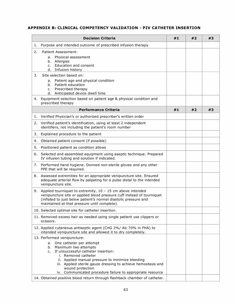
63
APPENDIX B: CLINICAL COMPETENCY VALIDATION - PIV CATHETER INSERTION
Decision Criteria #1 #2 #3
1. Purpose and intended outcome of prescribed infusion therapy
2. Patient Assessment:
a. Physical assessment
b. Allergies c. Education and consent
d. Infusion history
3. Site selection based on:
a. Patient age and physical condition
b. Patient education
c. Prescribed therapy
d. Anticipated device dwell time
4. Equipment selection based on patient age & physical condition and
prescribed therapy
Performance Criteria #1 #2 #3
1. Verified Physician’s or authorized prescriber’s written order
2. Verified patient’s identification, using at least 2 independent
identifiers, not including the patient’s room number
3. Explained procedure to the patient
4. Obtained patient consent (if possible)
5. Positioned patient as condition allows
6. Selected and assembled equipment using aseptic technique. Prepared
IV infusion tubing and solution if indicated.
7. Performed hand hygiene. Donned non-sterile gloves and any other
PPE that will be required.
8. Assessed extremities for an appropriate venipuncture site. Ensured
adequate arterial flow by palpating for a pulse distal to the intended
venipuncture site.
9. Applied tourniquet to extremity, 10 – 15 cm above intended
venipuncture site or applied blood pressure cuff instead of tourniquet
(inflated to just below patient’s normal diastolic pressure and
maintained at that pressure until complete).
10. Selected optimal site for catheter insertion.
11. Removed excess hair as needed using single patient use clippers or
scissors.
12. Applied cutaneous antiseptic agent (CHG 2%/ Alc 70% in FHA) to
intended venipuncture site and allowed it to dry completely.
13. Performed venipuncture:
a. One catheter per attempt
b. Maximum two attempts c. If unsuccessful catheter insertion:
i. Removed catheter
ii. Applied manual pressure to minimize bleeding
iii. Applied sterile gauze dressing to achieve hemostasis and
wound protection iv. Communicated procedure failure to appropriate resource
14. Obtained positive blood return through flashback chamber of catheter.
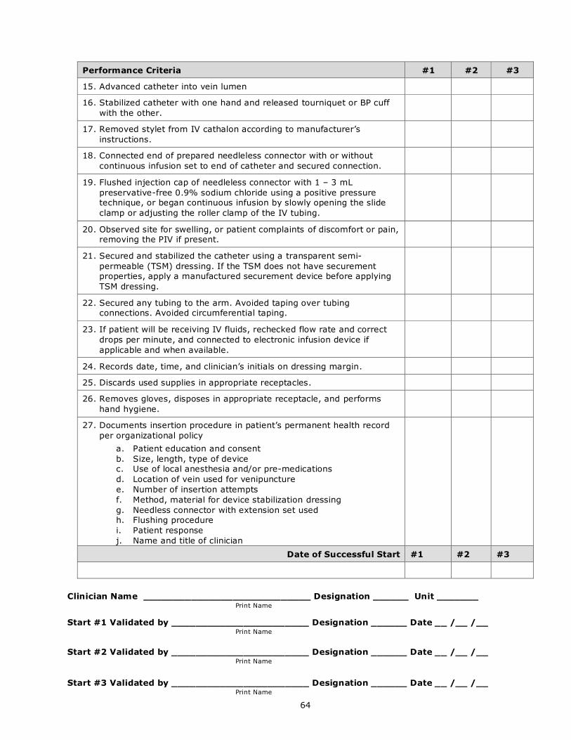
64
Performance Criteria #1 #2 #3
15. Advanced catheter into vein lumen
16. Stabilized catheter with one hand and released tourniquet or BP cuff
with the other.
17. Removed stylet from IV cathalon according to manufacturer’s
instructions.
18. Connected end of prepared needleless connector with or without
continuous infusion set to end of catheter and secured connection.
19. Flushed injection cap of needleless connector with 1 – 3 mL
preservative-free 0.9% sodium chloride using a positive pressure technique, or began continuous infusion by slowly opening the slide
clamp or adjusting the roller clamp of the IV tubing.
20. Observed site for swelling, or patient complaints of discomfort or pain, removing the PIV if present.
21. Secured and stabilized the catheter using a transparent semi-
permeable (TSM) dressing. If the TSM does not have securement properties, apply a manufactured securement device before applying
TSM dressing.
22. Secured any tubing to the arm. Avoided taping over tubing connections. Avoided circumferential taping.
23. If patient will be receiving IV fluids, rechecked flow rate and correct
drops per minute, and connected to electronic infusion device if
applicable and when available.
24. Records date, time, and clinician’s initials on dressing margin.
25. Discards used supplies in appropriate receptacles.
26. Removes gloves, disposes in appropriate receptacle, and performs
hand hygiene.
27. Documents insertion procedure in patient’s permanent health record
per organizational policy
a. Patient education and consent
b. Size, length, type of device c. Use of local anesthesia and/or pre-medications
d. Location of vein used for venipuncture
e. Number of insertion attempts
f. Method, material for device stabilization dressing
g. Needless connector with extension set used h. Flushing procedure
i. Patient response
j. Name and title of clinician
Date of Successful Start #1 #2 #3
Clinician Name ____________________________ Designation ______ Unit _______
Print Name
Start #1 Validated by _______________________ Designation ______ Date __ /__ /__ Print Name
Start #2 Validated by _______________________ Designation ______ Date __ /__ /__
Print Name Start #3 Validated by _______________________ Designation ______ Date __ /__ /__
Print Name

65

66
Fraser Health
©2018