perfusion monitoring during cardiopulmonary bypass: which ......• 10 minutes after initiation of...
TRANSCRIPT

Universitätsklinik für Anästhesiologie und Schmerztherapie
Perfusion Monitoring during Cardiopulmonary Bypass: Which Parameter is Best?
Catherine S. Reid, MD, MSE
Universitätsklinik für Anästhesiologie und Schmerztherapie
Inselspital

CONFLICTS OF INTEREST
• None to declare

IF YOU HAD ONLY ONE MONITOR AVAILABLE, WHICH WOULD YOU
CHOOSE?
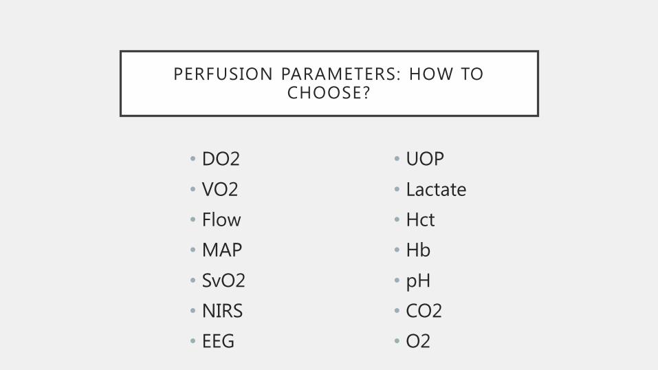
PERFUSION PARAMETERS: HOW TO CHOOSE?
• DO2
• VO2
• Flow
• MAP
• SvO2
• NIRS
• EEG
• UOP
• Lactate
• Hct
• Hb
• pH
• CO2
• O2

NEAR-INFRARED SPECTROSCOPY: WHAT IS IT?
HOW EFFECTIVE IS IT?HOW CAN I USE IT?

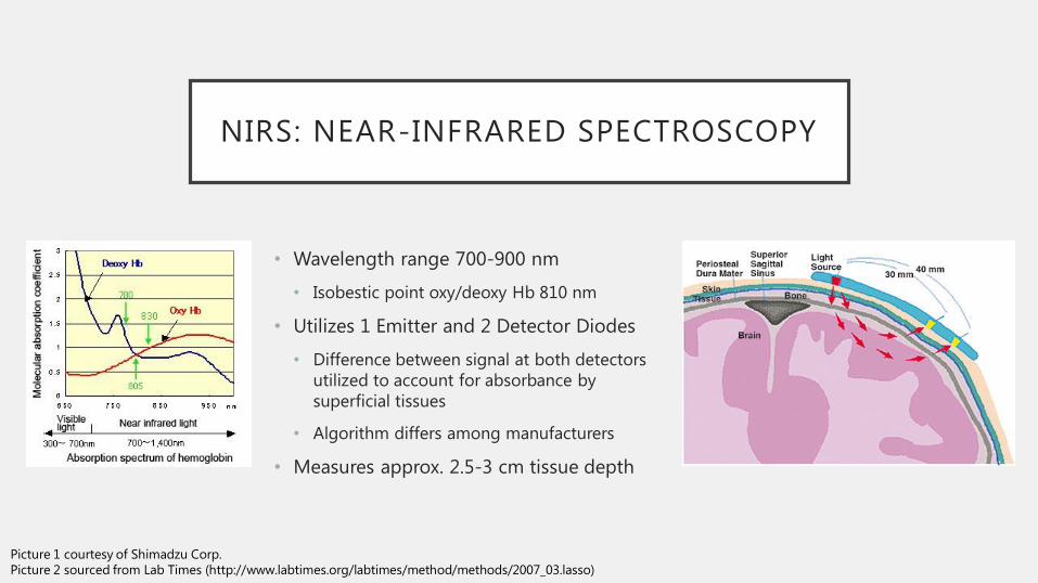
NIRS: NEAR-INFRARED SPECTROSCOPY
** Photos sourced from
manufacturers’ website
NIRS: NEAR-INFRARED SPECTROSCOPY
• Wavelength range 700-900 nm
• Isobestic point oxy/deoxy Hb 810 nm
• Utilizes 1 Emitter and 2 Detector Diodes
• Difference between signal at both detectors
utilized to account for absorbance by
superficial tissues
• Algorithm differs among manufacturers
• Measures approx. 2.5-3 cm tissue depth
Picture 1 courtesy of Shimadzu Corp.
Picture 2 sourced from Lab Times (http://www.labtimes.org/labtimes/method/methods/2007_03.lasso)
NIRS: NEAR-INFRARED SPECTROSCOPY
• Non-pulsatile, predominantly wave reflectance (not transmission, like SpO2)
• Principally venous blood – thus measurement of cerebral venous saturation
• Assumption usually 70% venous/30% arterial blood
• Kim, et al. demonstrated good correlation of rScO2 to jugular venous bulb saturation under conditions of hyper/hypocarbia and hypoxia (healthy volunteers)
• Manufacturer-Dependent Algorith
M. Kim, et al. Estimation of Jugular Venous O2 Saturation from Cerebral Oximetry. J Clin Monit Comput. 2000;16(3):191-9.

CASE
• 70-y/o female for emergent ascending aorta replacement graft +/- AVR
due to a Type A aortic dissection.
• Patient arrives hemodynamically stable, GCS 15, with no neurologic
deficits.
• Anesthetic induction, initial intraoperative course unremarkable.
• Aortic cannula located in the right subclavian artery, single venous
cannula in the right atrium.
• Baseline rScO2 65% bilaterally

CASE
• 10 Minutes after initiation of CPB, abrupt fall in rScO2 to <20 bilaterally
• Possible etiologies?
• Embolic phenomena
• Cannula malposition (perfusion of false lumen?)
• O2 delivery
• Extension of dissection
• Ultrasound of bilateral carotid arteries demonstrated dissection flap, with likely perfusion of false lumen
• Circulatory arrest initiated (core temperature 30 C) and anterograde perfusion catheters placed in the right brachiocephalic and left common carotid artery. rScO2 remained < 20 bilaterally.
• Doppler US revealed no flow, despite placement under direct sight.
• Problem with pump flow to anterograde catheters; after reinitation of flow, rScO2immediately increased to within 20% of baseline.

AT-RISK POPULATION
• Risk of debilitating neurological injury after cardiac surgery: 1.6-5%1,2
• Patients requiring CABG often have concomitant cerebrovascular disease
• Embolic phenomena well-described
• Hypertension: shift of cerebral autoregulation curve to the right
• Increased susceptibility to watershed infarcts
• Multiple comorbidities (diabetes, hyperlipidemia, tobacco use, sleep apnea)
Photo from Peterson EC, et al. Regulation of cerebral blood flow. Int J Vasc Med
(2011)
Photo reproduced from www. radiologyassistant.nl
1Salazar JD et al. Stroke after cardiac surgery: short- and long-term outcomes. Ann
Thorac Surg. 2001;72:1195–201.2Tarakji KG, et. al. Temporal onset, risk factors and outcomes associated with
stroke after coronary artery bypass grafting. JAMA. 2011;305:381–90.

NIRS AND OUTCOMES:SHOW ME THE EVIDENCE
• CASE REPORTS: multiple starting late 90s – early 00s
• OBSERVATIONAL STUDIES: multiple, most retrospective
• 2011 Heringlake, et al. (Anesthesiology)
• 1178 patients undergoing CABG, valve repair, aortic arch repair, combined procedures
• Morbidity and mortality increased in patients with low baseline rScO2
• 2004 Yao, et al. (Journal of Cardiothoracic and Vascular Anesthesia)
• 101 patients undergoing cardiac surgery with CPB.
• Patients with nadir rScO2<35% higher risk of postop MMSE and ASEM impairment
• Similar outcome in patients with rScO2<40% for >10 minutes
• No intervention for low rScO2

NIRS AND OUTCOMES:SHOW ME THE EVIDENCE
• RANDOMIZED, CONTROLLED TRIALS
• 2007 Murkin, et al. (Anesthesia & Analgesia)
• 200 patients undergoing CABG with CPB
• Blinded control rSO2 vs. rSO2 with treatment
protocol for >25% decrease from baseline
• Control: higher number of major morbidity or
mortality (death, >48h ventilation, stroke, MI, re-
op), longer ICU stay, longer hospital stay
• 2009 Slater, et al. (Annals of Thoracic Surgery)
• 265 patients undergoing CABG with CPB.
• Larger battery of neuropsychologic tests
• Patients with rSO2 desaturation score > 3000%s
higher risk of early postop cognitive decline
• Patients with rSO2 desaturation score > 3000%s 3x
risk of prolonged hospital stay
• Poor adherence to intervention protocol

NIRS AND OUTCOMES:SHOW ME THE EVIDENCE
• FEASIBILITY STUDIES
• 2016 Deschamp, et al. (Anesthesiology)
• 201 patients at 8 Canadian hospitals undergoing high-risk cardiac surgery
• 57% (control) – 70% (intervention) of patients exhibited rSO2 desaturations (>10%)
• Successful reversal of 97% of desaturations using treatment algorithm
• No difference in adverse outcomes at 30 days between groups
• 2016 Subramanian, et al. (Anesthesia & Analgesia)
• 235 patients at 8 US hospitals undergoing CABG or valvular surgery
• 50-75% of patients exhibited rSO2 desaturations (>20% ); 10% were not identified by clinicians
• In the identified events, treatment algorithm reversed desaturation in the majority of cases

APPLICATIONS: ADULT CARDIAC SURGERY
• Detects otherwise clinically silent episodes of ischemia or reduced perfusion
• Known cerebrovascular disease (h/o TIA, CVI)
• Known carotid stenosis
• Poorly-controlled hypertension
• Other risk factors: diabetes, tobacco use, sleep apnea????
• Aortic cannula malposition
• Detects venous congestion
• Deep hypothermic circulatory arrest: how effective is anterograde perfusion?
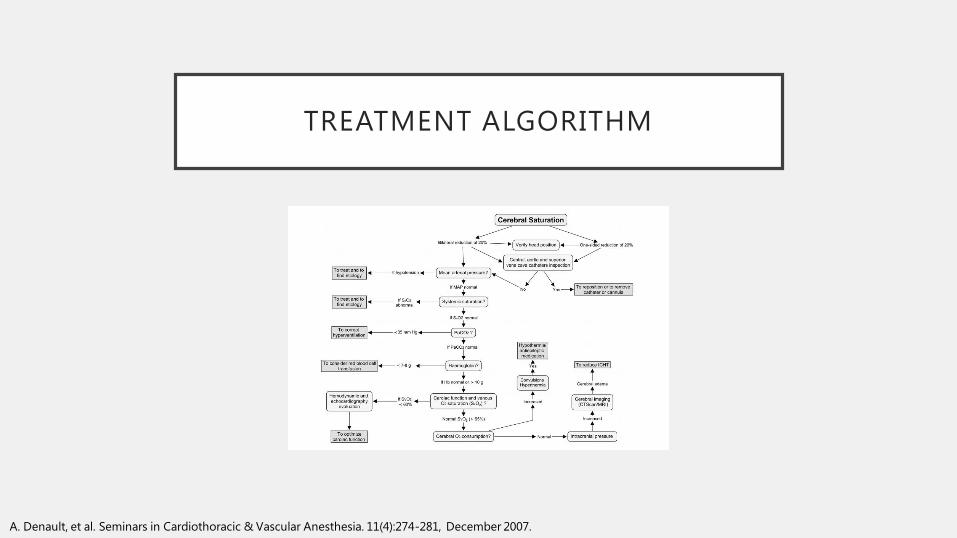
TREATMENT ALGORITHM
A. Denault, et al. Seminars in Cardiothoracic & Vascular Anesthesia. 11(4):274-281, December 2007.

APPLICATIONS: PEDIATRIC
• Cerebral Oximetry
• Complex physiology and repairs in congenital heart surgery, often with right-to-left shunt
• Cerebral circulation especially sensitive to changes in PCO2
• Aortic cannula size and position
• Regional organ oximetry
• Possible due to superficial location of kidney, liver, splanchnic circulation in neonatal and pediatric population
• Important in complex corrective surgeries with possible regional perfusion differences
• May help with early detection of low-output syndrome after cardiac surgery
• Pediatric Intensive Care

APPLICATIONS: NON-CARDIAC SURGERY
• Trauma
• Cardiopulmonary resuscitation: measure of adequacy of resuscitation
• Hypothermic Injuries: ECMO
• ICU: ECMO, LVAD, Post-cardiac surgery

PITFALLS
• Cortical measurement
• Trend measurement, not absolute
• Differences in patient anatomy (thickness of skull, subcutaneous edema, scalp or subdural/epidural hematoma)
• Distribution of arterial and venous blood in the sample volume
• Can appear normal in non-metabolizing or dead tissue due to sequestration of venous capillary blood (Schwartz, et al )
• Frontal lobe measurement
• Ischemic vs. Hemorrhagic Phenomena
Schwarz G, Litscher G, Kleinert R, Jobstmann R. Cerebral oximetry in dead subjects. J Neurosurg Anesthesiol 1996; 8: 189 – 93

TAKE-HOME MESSAGES: NIRS
• End-organ monitor!
• Can help detect otherwise clinically silent episodes of brain
hypoperfusion or ischemia
• Studies demonstrate trend in reduction of poor neurological outcomes,
mortality, major morbidity, reduction in ICU/hospital stay
• Strongly consider use in patients with higher-risk procedures, risk
factors for cerebral hypoperfusion
• Noninvasive and easy to use: ask yourself, “Why Not?”

IF YOU HAD ONLY ONE MONITOR AVAILABLE, WHICH WOULD YOU
CHOOSE?

Universitätsklinik für Anästhesiologie und Schmerztherapie
Questions?????
Thank you for your attention!