performance and results annual report · annual report 1 fraunhofer institute ... leader of the...
TRANSCRIPT

Performance and ResultsAnnual Report


Annual Report 1
Fraunhofer Institute for Biomedical Engineering IBMT
Annual Report

2 Annual Report
In superficial hindsight the results andsuccesses at an institute like the IBMT,with a history of two decades, appearas evenly dispersed, almost continuousachievements. A closer look over theyears reveals that most of the successescan be traced back to usually unspec-tacular decisions made some time ago.However, in retrospect it is often possi-ble to determine exactly which discus-sion and sometimes even which state-ment was seminal for a very fruitfuldevelopment that over the yearsproved scientifically and economicallyprofitable for the institute. This posesthe question of whether such decisionscan be reached in a more targetedmanner, a question I would like toreturn to at the end of this editorial.
One of these sustainable decisions wastaking on research projects with afocus on cell biology in the traditionalfield of medical and technical ultra-sound. In fact, the department for“Ultrasound”, led by Dr. Robert Lemorsince May 2006, has now grown to asize of nearly forty personnel and isthus one of the largest ultrasounddevelopment units in Europe. Thedepartment has extended its researchand development area from medicalimaging far into cell biology fields suchas high resolution ultrasonic micros-copy and technical applications such as underwater sonar. This developmentessentially stems from strategic deci-sions and investments made some four to five years ago. Last year, thedepartment developed an ultrasonicmicroscope that is not only combinedwith a laser scanning microscope butalso allows time-lapse recordings ofcell cultures over several days at a resolution of about one micrometer,and moreover, is easy to use. An advantage of ultrasonic microscopy isthe high-resolution visualization ofmechanical characteristics of cells invitro even in total darkness and on optically non-transparent surfaces. In
comparison to the rapid developmentof laser scanning microscopy over thelast decades the possibilities of ultra-sonic microscopy are not nearlyexhausted yet.
Another decision made some twoyears ago has considerably influencedand enriched the scientific and eco-nomic profile, as well as the scope ofthe IBMT in the period covered by thisreport: It was the decision to join pro-grams in developing countries, espe-cially those concerning the fightagainst HIV and AIDS. What can anInstitute of the Fraunhofer-Gesellschaftachieve in this wide field of globalactivities? Much, as it turns out, sincethe IBMT possesses a portfolio of tech-nologies, device development strate-gies and experience in setting up andcoordinating networks, which areurgently needed in public health pro-grams to combat epidemics such asAIDS, tuberculosis, hepatitis and malar-ia. The Bill & Melinda Gates Founda-tion, whose founding capital wasrecently increased by US$ 31 billion byWarren Buffett, put out a call world-wide at the beginning of 2005 for sub-mission of projects to combat AIDS,investigate HIV and develop vaccines.On the basis of its established cryo-technology, the IBMT proposed theinstallation of a “Central HIV Cryo-Depository” linked to a global networkfor the use of these samples in HIVresearch, in close cooperation with theWorld Health Organization (WHO) andseven scientific partners in Europe andthe USA – and was successful. Out of abroad field of competitors this researchinitiative was granted a US$ 7.5 millionbudget, and since August 2006 iscoordinated by the Fraunhofer IBMT in
Editorial
Institute director Prof. Dr. Günter Rolf Fuhr.

Annual Report 3
St. Ingbert/Sulzbach. It is the first pro-ject of the Gates Foundation to beheaded by German scientists. Dr.Hagen von Briesen, virologist andleader of the project at the IBMT, hastaken on the challenging task of glob-ally coordinating virus registration anddeposition. The Federal State of Saar-land and the Fraunhofer-Gesellschaftare supporting the project withresearch funding, so that the large-scale project has access to a sum ofover ten million over its three-yearduration. You can find further detailsabout this in the present annualreport.
However, not only a consolidated pro-ject portfolio, but also knowledgetransfer brings scientists and customerstogether. In 2006, the first internation-al workshop on laser medicine tookplace in St. Ingbert/Sulzbach. The topic“Advanced Multiphoton and Fluores-cence Lifetime Imaging Techniques”attracted scientists and developersfrom the most diverse applicationareas of femtosecond lasers toexchange experiences. Althoughplanned as a test, this initiative alsoturned out to be a success. Prof. Dr.Karsten König, head of the depart-ment of “Microsystems and LaserMedicine” summarizes the results onpage 30. A second workshop followedin 2007, building upon the experiencegained from the first one.
A central, and currently the largestresearch project at the IBMT is “Cell-PROM”, an integrated EU project with-in the 6th Framework Program. Afterhaving started in 2004, 27 Europeanpartners assembled in May 2006 andApril 2007 for an “assessment meet-ing” important for the continuation ofthe project. The approach to encour-aging cell differentiation by artificialbiological surfaces and developingnovel technologies for the in vitro culti-vation of stem cells has paid off. Theresults and progress in the projectheaded by physicist Daniel Schmittwere evaluated extremely positively onevery aspect in the assessment report.Particular praise was given to the inno-vative device concept with magneticmicrocarriers upon which cells grow,and the professional project manage-ment with a number of tools meetingspecial requirements, such as “WhitePapers” to follow-up the current stateof the art in this multidisciplinaryresearch area, “Key Experiments” toverify unproven hypotheses and ahighly illustrated work plan up-datedmonthly. Over the next years, twoautomated differentiation devices foranimal and human stem cells will beproduced – prototypes for a new generation of cell handling systemsthat will create the basis for novelapproaches in regenerative medicineand tissue engineering.
The development of gentle cell hand-ling systems for stem cell research andapplications is another example of anearly decision with profound conse-quences. In 2003, the IBMT was thefirst Fraunhofer Institute to initiate abroad development of stem cellmanipulation and characterization sys-tems. This initially comprised the pro-fessional cultivation of adult andembryonic stem cells and, what is verylikely to be fundamental for their laterclinical use, low temperature storagewith high survival rates. In cooperation
with the group of Dr. Charli Kruse atthe University of Lübeck and thedepartments of “Cryobiophysics andCryotechnology” and “CellularBiotechnology and Biochips” led byProf. Dr. Heiko Zimmermann and Dr.Claus Duschl, respectively, complemen-tary groups were set up at the IBMTlocations in Lübeck, St. Ingbert andBerlin. The development of novel,more gentle and higher-yield isolationprocesses allowed the harvesting andestablishment of an extensive stem cellcollection, which apart from mouse,rat and human stem cells, also con-tains exotic and so far uncharacterizedstem cell isolates from fish, birds andmammals. The demand and orders forstem cell samples from academia andindustry are validating the concept.Currently, nine stem cell projects withexternal funding and further pilot pro-jects financed by the Institute’s ownfunds are being pursued at the IBMT.
An additional novelty has to be report-ed: In summer 2006 the IBMT was thefirst Fraunhofer Institute to be grantedapproval for importing human embry-onic stem cells within the context oftwo EU projects. One project aims atimproving the cryopreservation effi-ciency of human and adult stem cells;the other project focuses on develop-ing novel non-invasive measuringmethods for monitoring osteogenicdifferentiation. Both projects efficientlyreflect the strategy of the IBMT, i.e.developing both instruments andprocesses.

4 Annual Report
Within the context of DFG-fundedresearch in polar regions, with theobjective of establishing an algae col-lection of low temperature-tolerant,so-called psychrophilic unicellularorganisms from both hemispheres, thefirst Antarctic exploration was carriedout between January and March 2006,following eight previous expeditions tothe Arctic. Dr. Thomas Leya led thisexpedition to King George Island incooperation with the University ofInnsbruck, and transferred more than70 Antarctic algae strains in stable lab-oratory cultures. The “CCCryo” collec-tion now comprises more than 300cryopreserved and living strains and istherefore one of the largest collectionsof extremophilic algae worldwide.Apart from questions concerning tax-onomy and low temperature tolerance,the algae collection is of economical
importance due to the secondarymetabolites such as asthaxanthin, fattyacids and cold-adapted enzymes andmembrane transporters. As a result ofan Arctic expedition in 2000/2001, astretch of land in the Raudfjorden onSpitsbergen was named by the Norwe-gian Geographic Naming Committeefollowing the suggestion of IBMT sci-entists (see page 27).
Finally, I would like to mention thenew IBMT building in Potsdam-Golm(Brandenburg) whose architecture fitsharmoniously into the collection ofexisting institutes. Constructed asplanned in about two years, andequipped with state-of-the-art fittings,
this building has been available to thedepartments of “Molecular Bioanalyt-ics and Bioelectronics” and “CellularBiotechnology and Biochips” sinceOctober 2006. Locations at the Hum-boldt University in Berlin and the Ger-man Institute for Nutrition Research inNuthetal were vacated and the roomsreturned to the host institutions withmodern laboratory equipment. We aregrateful for their hospitality and lookback on fertile cooperation, which wewill continue in the future throughjoint projects.
To a great extent the functionalaspects of the new building weredefined by the personnel and include anumber of special features that webelieve could be exemplary for othernew institute buildings in the biotech-nology sector. The IBMT is now wellequipped, with its parent institute inSt. Ingbert and its branch in Potsdam-
Current stem cell research projects at the Fraunhofer IBMT
Adult stem cells:
1. EU IP CellPROM Mesenchymal stem cellsPancreatic stem cellsHematopoetic stem cells
2. BMBF project Mesenchymal stem cells3. Project Schleswig-Holstein Pancreatic stem cells4. Cryoprojects (industry) All adult stem cells5. EU CCS Breast cancer stem cells
Embryonic stem cells:
6. Project Saarland Mouse embryonic stem cells7. BMBF EU CCS Cardiomyocytes and chicken embryonic stem cells
Human embryonic stem cells:
8. EU STREP 2005 NIH cell lines, isolated before 1.1.20029. EU CryoP 2006
Annual Report 5
Golm, and more than able to effective-ly meet all the requirements of aFraunhofer Institute in the field of lifesciences.
Altogether, these were very successfulyears for the IBMT. Analyzing the rea-sons for success leads back to the ini-tial question: Can one catalyze suchdevelopments in a more targeted man-ner? The answer is: yes and no. Yes,because every successful projectenriches our experience and gives con-fidence for tackling new tasks. As aconsequence of such accumulatedexperience, predictions become morereliable. The expertise of the Instituteand interactions between the researchgroups are enhanced. The atmosphereof a scientific institution particularlydepends on its tradition and history,i.e. projects pursued in the past. Onthe other hand, the answer is also“no”, because, as in business, a suc-cessful project in science cannot berepeated by merely copying a previous-ly successful strategy. The interrelationsare too complex. Some luck in solvinga task and a fortuitous internationalsituation play just as important a roleas the good idea itself. It is still risky tobelieve that a particular researchapproach will be successful. Initiating anumber of well-prepared projects inparallel and carefully analyzing theirdevelopment is more a recipe for suc-cess. Which project will eventually pre-vail remains uncertain. Precisely thisaspect is what makes research anddevelopment work at a FraunhoferInstitute so interesting and satisfyingfor all, from the student to the direc-tor.
I wish to thank all our customers andpartners for their confidence in thework of our Institute. We appreciateyour visits and your commissions. As atechnology developer and traditionalinstitute for medical engineering, theIBMT aims to solve your problems withall its workgroups and departmentsheaded by renowned staff. Discoverfor yourself our range of competentservices in this annual report and findyour best contact partner. My personalthanks are directed to the staff at allIBMT locations. You have not onlyworked successfully on all projectsdespite difficult relocations and addi-tional problems, but were even able toexpand the Institute’s scientific profile.Therefore, well-prepared we look intothe future, which will certainly be one of medical technology as well as molecular and cellular biotechnology.
Prof. Dr. Günter R. Fuhr(Director of the IBMT)

6 Annual Report
Editorial 2
The Institute in Profile 8Objectives 10Short portrait 11Organization and contact partners 12Central work topics 14New institute building in Potsdam-Golm 16Competencies and applications 19Advisory committee 19Scientific events and awards 20Nanobiotechnology as a future research area 35
Contract Research and Services 38Institute-specific offers for contract research 39Contracts and patent agreements 41Customers 41Product catalogue 42Contact and further information 43
The Institute in Numbers 44Personnel development 45Operative budget 45Contract research with industry 45
The Fraunhofer-Gesellschaft at a Glance 46Summary of overall competence 47Research areas 48Target groups 48Services offered 48Advantages of contract research 48
Selected Research Results and Applications 49
Microsystems/Laser Medicine 50Non-invasive, high resolution imaging with magnetic resonance tomography in combination with multiphoton tomography
Ultrasound 58Portfolio of the Department
Telematics/Tele-Medicine 68SmartHEALTH – smart, integrated, biodiagnostic systems for cancer diagnosis
Medical Engineering & Neuroprosthetics 74Highly flexible, textile-integrable electrode material to record ECGs within a 24/7 monitoring scheme
Contents

Annual Report 7
Cryobiophysics & Cryotechnology 80Development of tumor models for the cryobanks of the personalized medicine
Biohybrid Systems 86Technology platform for accelerated HIV vaccine development
Computer-aided Simulations 92Setup of the CellPROM application laboratory
Cell Differentiation & Cell Technology 98Human pancreatic stem cells differentiating into cardiac muscle cells
Cellular Biotechnology & Biochips 104Lab-on-Chip – Gentle handling of valuable cells
Molecular Bioanalytics & Bioelectronics 114NUCAN – Nucleic Acid Based Nanostructures
Biomedical Competence Centers 126Technology consulting by experts
Facts and Statistics 132Names, Dates, Events 133
National/international guests: scientists, research fellows and guest lecturers 133Exhibition and event list 133
Scientific publications 134Diplomas, masters, bachelors and PhD theses 134Publications and talks 2006 135Patents 148
Imprint

8 Annual Report
The Institute in Profile
– Objectives– Short portrait – Organization and contact partners– Central work topics– New institute building in Potsdam-Golm– Competencies and applications– Advisory committee– Scientific events and awards– Nanobiotechnology as a future research area
Parent institute in St. Ingbert.

Sulzbach
Nuthetal (until October 2006)
New building Potsdam-Golm (since October 11, 2006)
Annual Report 9
St. Ingbert
Humboldt University Berlin (until October 2006)
Shenzhen, ChinaLübeck
Eurocryo Saar, microsystem-based cell bank in the Saarland
Objectives
10 Annual Report
The Fraunhofer Institute for BiomedicalEngineering (IBMT) is one of the fivelife science institutes of the Fraun-hofer-Gesellschaft and primarily focus-es on technology development. Sinceits foundation in 1987, the FraunhoferIBMT is a partner with industry forsolving tasks in the areas of biomedicaland medical engineering, laser medi-cine, biotechnology, health telematics,environment technology, laboratorydevelopment, cryotechnology, materialtesting technology, home, air condi-tioning and security technologies, aswell as industrial process automationand in-line/on-line process surveillance,particularly for the food, chemical andpharmaceutical industries. The institutesupports “living” technology transferin medicine and biotechnology and invarious branches of production indus-try and knowledge-intensive services.Core competencies are: non- and mini-mal-invasiveness, miniaturization, link-ing technical microsystems to biologi-cal microsystems (biohybrid systems,molecular bioanalytics, neuroprosthet-ics), molecular and cellular biotechnol-ogy, nano(bio)technology, cryo(bio)-technology, biocompatibility, ultra-sound technology, sensor manufactur-ing processes, magnetic resonance,telemetric data and energy transfer,multilocal sensors linked by communi-cation technology and telematic systems. Central application areas aremedical diagnostics, therapy and therapy monitoring as well as analo-gous issues in the industrial sector. Important new focus areas are meth-ods and technologies for industrialimplementation of innovations frommolecular and cellular biotechnologyand cryotechnology for storing viablesamples at low temperatures, as wellas isolating, cultivating and differenti-ating stem cells for regenerative medi-cine. The Fraunhofer IBMT has beenworking in stem cell research for threeyears and was the only institute of theFraunhofer-Gesellschaft to be granted
approval (No. 18 and 19) by the RobertKoch Institute to import human embry-onic stem cells. Technology transferfrom basic research is achieved alongthe innovation chain of scientific andtechnical consulting, feasibility studies,prototype development, field tests andmanufacturing processes. If needed,IBMT spin-offs take on system manu-facturing as contract services, ensuringas rapid as possible implementationand maturation of our customers’wishes into marketable products. Addi-tional operation areas are advising ven-ture capital (VC) companies, preparingstudies and reports and assisting start-up companies. The IBMT is located infour regions (Saarland, Brandenburg,Schleswig-Holstein, Shenzhen [China]),and there it contributes to the overly-ing tasks of re-directing regional struc-tures towards a global orientation andto creating new regional employmentpotential.
The Institute in Profile
Founding director of the Fraunhofer IBMT,Prof. Dr. Klaus Gersonde (1987-2001).

Annual Report 11
By founding the precursor of the Insti-tute for Biomedical Engineering in1987, the Fraunhofer-Gesellschaft’sgoal was to promote natural sciencesand engineering research, moderntechnology and technology transferwithin clinical research in the Saarlandin cooperation with the University Hos-pital in Homburg/Saar. The originalinstitute is located at St. Ingbert (Saar-land) and since April 1, 2001, directedby Prof. Dr. Günter Rolf Fuhr, who wassimultaneously appointed professor ofBiotechnology and Medical Engineer-ing at the Medical Faculty of the Uni-versity of Saarland. His predecessor,Prof. Dr. Klaus Gersonde, was appoint-ed in 1987 to the newly establishedchair of Medical Engineering at theUniversity of Saarland, meaning that atthe same time as being co-director ofthe Frauhofer Institute for Non-destructive Test Procedures (IZFP), hetook over leadership of the precursorof the IBMT, i.e. the main departmentof Medical Engineering in St. Ingbert.On the basis of its continuous develop-ment, this department was convertedinto an autonomous Fraunhofer Insti-tute for Biomedical Engineering in1992. Consequently, pursuing the triedand tested technology transfer strate-gy, the IBMT branch in Sulzbach/Saarwas founded in 1994, where theworkgroup for sensor manufacturingstarted working.
The Institute is financed by contractresearch and development commis-sioned by public and private (industrial)customers and the close connectionbetween medical engineering, biotech-nology and microsystems technologyensures it an outstanding position inEurope. Since 1997, the IBMT housesthe European Center of Competence
for Biomedical Microdevices (MEDICS)located in Sulzbach/Saar. On October1, 1998, the IBMT presence in Chinastarted with its branch in Shenzhen/Guandong (FTeCS) going into opera-tion. It was headed by Prof. Dr. Nai-Teng Yu (the Hong Kong Universityof Science and Technology, HKUST)and promotes contacts to regionalgovernments and industry in China asa further part of the IBMT network. In2000, activities in China were comple-mented by the Fraunhofer IBMT Tech-nology Center in Xiamen (FTeCX).
On April 1, 2001, when the previousdirector retired, leadership of theFraunhofer IBMT passed on to Profes-sor Fuhr. A biophysicist, Günter RolfFuhr joined the Fraunhofer-Gesellschaftand the University of Saarland fromthe Humboldt University Berlin (Chairof Membrane Physiology since 1993,and in parallel representing the Chairof Experimental Biophysics since 2000).Like his predecessor, he is a member ofthe Medical Faculty and at the sametime elected member of the Faculty ofPhysics and Mechatronics, as well asmember of the Center for Bioinformat-ics and elected member of the Hum-boldt University Berlin. Professor Fuhrreceived his PhD in 1981 on the sub-ject of photo-morphogenesis in higherplants, and in 1985 he qualified as aprofessor in biophysics. In 1999, hefounded a Center for Biophysics andBioinformatics at the Humboldt Univer-sity Berlin, where he was the first executive director until his departureon April 1, 2001.
The IBMT is a member of the associa-tion of 80 Fraunhofer establishments,of which 58 are Fraunhofer Institutes.In 2006, the IBMT comprised a staff of134 scientific and 60 technical andadministrative personnel as well as 29student assistants and 57 project stu-dents. The IBMT is connected to theUniversity of Potsdam through Prof. Dr.
Frank Bier, head of the department forMolecular Bioanalytics & Bioelectronics(Chair of Applied Bioelectronics andBiochip Technology). A chair of Bio-medical Engineering links the IBMT tothe “Hochschule für Technik undWirtschaft” (HTW) (College for Engi-neering, Industry and Commerce) ofSaarland. The Chair of Microsensorswith Assembly and Packaging Technol-ogy, held by Prof. Dr. Karsten König,links the IBMT to the Faculty of Physicsand Mechatronics of the University ofSaarland. Moreover, the IBMT hosted10 guest scientists and a junior profes-sorship in conjunction with the Univer-sity of Saarland.
The institute is subdivided into eightdepartments corresponding to itsresearch areas: Microsystems/LaserMedicine, Ultrasound, Biohybrid Sys-tems, Cryobiophysics & Cryotechnolo-gy, Telematics/Tele-Medicine, MolecularBioanalytics & Bioelectronics, CellularBiotechnology & Biochips and theFraunhofer IBMT Technology CenterShenzhen (China). The departmentsare run as separate profit and cost cen-ters. Besides the departments, thereare independent workgroups that areon their way to developing intoautonomous departments. Since Sep-tember 2001, the IBMT is the foundingmember of the Fraunhofer Life Sci-ences Association.
Short portrait

12 Annual Report
Director Prof. Dr. Günter R. Fuhr +49 (0) 6894/980-100 [email protected]
Deputy Director Branch Potsdam-Golm Prof. Dr. Frank F. Bier +49 (0) 331/58187-200 [email protected]
(since 2007)
Head of Administration Bärbel Walter +49 (0) 6894/980-104 [email protected]
Marketing/Public Relations Dipl.-Phys. Annette Eva Maurer +49 (0) 6894/980-102 [email protected]
Departments and workgroups:
Microsystems/Laser Medicine Prof. Dr. Karsten König +49 (0) 6894/980-150 [email protected]
Miniaturized Systems Dr. Thomas Velten +49 (0) 6894/980-301 [email protected]
Magnetic Resonance Priv.-Doz. Dr. Frank Volke +49 (0) 6894/980-405 [email protected]
Laser Medicine Dr. Iris Riemann +49 (0) 6894/980-190 [email protected]
Ultrasound Dr. Robert Lemor +49 (0) 6894/980-225 [email protected]
Active Materials Dr. Frank Tiefensee +49 (0) 6894/980-270 [email protected]
Piezosystems & Manufacturing Technology Dipl.-Ing. Christian Degel +49 (0) 6894/980-221 [email protected]
Ultrasound Systems Development Dipl.-Ing. Peter Weber +49 (0) 6894/980-227 [email protected]
Biomedical Ultrasound Research Dr. Robert Lemor +49 (0) 6894/980-225 [email protected]
Telematics/Tele-Medicine
Medical Networks Dipl.-Phys. Bertram Bresser +49 (0) 6894/980-206 [email protected]
Home Care Dipl.-Inform. Stephan Kiefer +49 (0) 6894/980-156 [email protected]
Medical Engineering & Neuroprosthetics Prof. Dr. Klaus-Peter Hoffmann +49 (0) 6894/980-401 [email protected]
Neuromonitoring Prof. Dr. Klaus-Peter Hoffmann +49 (0) 6894/980-401 [email protected]
Neuroprosthetics Dr. Klaus Peter Koch +49 (0) 6894/980-404 [email protected]
Cryobiophysics & Cryotechnology Prof. Dr. Heiko Zimmermann +49 (0) 6894/980-257 [email protected]
Cryoequipment & Cryorobotics Dipl.-Phys. Uwe Schön +49 (0) 6897/9071-30 [email protected]
BMBF Junior Research Group
Cryonanobiotechnology Prof. Dr. Heiko Zimmermann +49 (0) 6894/980-257 [email protected]
Cryoresearch and Demonstration Bank Dr. Frank Obergrießer +49 (0) 6897/9071-90 [email protected]
Biohybrid Systems
Cell-based Sensors & Biomonitoring Dr. Hagen Thielecke +49 (0) 6894/980-162 [email protected]
Molecular Cell & Tissue Engineering Priv.-Doz. Dr. Hagen von Briesen +49 (0) 6894/980-286 [email protected]
IP-CellPROM Coordination Dipl.-Phys. Daniel Schmitt +49 (0) 6894/980-120 [email protected]
Computer-aided Simulations Dipl.-Phys. Daniel Schmitt +49 (0) 6894/980-120 [email protected]
Cell Differentiation & Cell Technology Priv.-Doz. Dr. Charli Kruse +49 (0) 451/2903-210 [email protected]
Cellular Biotechnology & Biochips Priv.-Doz. Dr. Claus Duschl +49 (0) 331/58187-300 [email protected]
Lab-on-Chip Technology Dr. Magnus Sebastian Jäger +49 (0) 331/58187-305 [email protected]
Cell Assay Development Dr. Andreas Lankenau +49 (0) 331/58187-303 [email protected]
Extremophile Research Dr. Thomas Leya +49 (0) 331/58187-304 [email protected]
Organization and contact partners
Annual Report 13
Molecular Bioanalytics & Bioelectronics Prof. Dr. Frank F. Bier +49 (0) 331/58187-200 [email protected]
Biosensors Dr. Nenad Gajovic-Eichelmann +49 (0) 331/58187-204 [email protected]
Nanobiotechnology Dr. Markus von Nickisch-Rosenegk +49 (0) 331/58187-206 [email protected]
Microarray & Biochip Technology Dr. Eva Ehrentreich-Förster +49 (0) 331/58187-203 [email protected]
Biomedical Competence Centers
(MEDICS, MOTIV, CC-NanoChem, Nano2Life) Dipl.-Ing. Andreas Schneider +49 (0) 6897/9071-42 [email protected]
Links to universities and colleges:
Chair of Biotechnology and Medical EngineeringClinical Medicine (Medical Faculty)Elected member of the Natural and Technical Science Faculties II and IIIUniversity of Saarland andElected member of the Faculty of Mathematics and Natural Sciences I of the Humboldt University BerlinProf. Dr. Günter R. Fuhr
Chair of Microsensors with Assembly and Packaging TechnologyFaculty of Physics and Mechatronics(Faculty of Science and Technology II)University of SaarlandProf. Dr. Karsten König
Junior Professorship of Cryobiophysics and Cellular BioinformaticsFaculty of Chemistry, Pharmacy, Biological Substances and Materials Sciences(Faculty of Science and Technology III)University of SaarlandProf. Dr. Heiko Zimmermann
Chair of Applied Bioelectronics and Biochip TechnologyInstitute for Biochemistry and BiologyFaculty of Mathematics and Natural SciencesUniversity PotsdamProf. Dr. Frank F. Bier
Chair (Master’s program) of Biomedical EngineeringElectrical EngineeringCollege for Engineering, Industry and Commerce of Saarland (HTW)Prof. Dr. Klaus-Peter Hoffmann

14 Annual Report
The Fraunhofer IBMT sees itself as atechnology developer and focuses ontechnological issues such as linkingtechnical microsystems to biologicalcomponents such as cells and tissues;molecular and cellular biotechnologywith medical objectives; nano(bio)tech-nology; biocompatibility testing; cryo-biotechnology; biochip development;laser medicine; microsystems technolo-gy (microsensor, microacting and signalprocessing); ultrasound; sensor manu-facturing technology; multi-local sen-sor connected by communication tech-nology; health telematics; telemetricdata and energy transfer; magneticresonance; imaging and spectroscopy.The required basic knowledge is gath-ered in a project-related manner andthen converted through contractdevelopment into products and pro-duction lines in cooperation withindustry. The scope of its activitiesranges from basic technological analy-ses to the development of componentsand systems and construction ofdemonstration units for industrialoperations. Not only medical technolo-gy and biotechnology companies, butalso other areas such as the polymerand ceramic industry, manufacturers ofsemiconductors, environmental tech-nology, hydraulics industry, food indus-try, house and air conditioning tech-nologies, process and process surveil-lance technologies, production andautomation technologies and materialtesting technologies find competentadvice and problem-specific solutionsat the IBMT. Successful improvementsand innovations are based on feasibili-ty studies, prototype development, thecreation of small-scale production linesand permanent sensor manufacturinglines. With an area covering more than3 800 square meters, the neighboringindustry park Sulzbach-Neuweiler is athriving development site for flexiblemanufacturing setups of sensors andcryoequipment, enabling small andmedium-sized companies to produceultrasound and microsensors at com-petitive costs. Regional and nationwide
customers are supported by the IBMTto become competitive on the Euro-pean market.
Another important future field of inter-est has developed since 1994 by rein-forced activities in the area of medicaltelematics. Novel approaches to indi-vidual healthcare of patients by tele-medical services are being realized, forexample in the two promising telemat-ics projects “Stroke Aftercare Saar”(“Home Care” area) and “PatientAccompanying Documentation –PaDok” (physician-physician and physi-cian-hospital networking).
In the context of ongoing globalizationof the IBMT’s activities, of special noteis the successful founding of the Chi-nese IBMT branch, the FraunhoferIBMT Technology Center in Shen-zhen/Guandong (FTeCS) in 1999. Aftera short interruption, research effortswill be resumed on a larger scale in2007. The long-term IBMT collaboratorDr. Jianbo Gao will take on coordina-tion of activities in China. The mainfocus of the FeTCS R&D services is sup-porting the automation and operationof surveillance technologies of manydifferent industrial sectors by imple-menting microsystems, microsensors,microactors and signal processing rou-tines. Initial customers come fromindustries involved in medical tech-nologies, processing polymers andrefining chemicals. Apart from thesespecific tasks, the FTeCS is a contactpoint for R&D customers who want tocapitalize on the expertise of thewhole Fraunhofer-Gesellschaft. FTeCStherefore represents the Fraunhofer-Gesellschaft in China. Another essen-
Central work topics
Dr. Jianbo Gao.

Annual Report 15
as an IBMT member institute in Pots-dam-Golm to accommodate these pre-viously decentralized workgroups. Thefirst spade of earth was dug on August30, 2004, the topping-out ceremonytook place on June 22, 2005, and staffmoved in and started work mid-Octo-ber 2006. The R&D scopes of bothdepartments complement each otheralmost ideally to form a competencecluster for biochip systems andnanobiotechnology.
Together with the State President ofSaarland, Peter Müller, the Fraunhofer-Gesellschaft under the presidency ofProfessor Hans-Jörg Bullinger openedthe cryoresearch bank EuroCryo Saar inSulzbach/Saar on September 9, 2003.Including the Center for Cryobiotech-nology and Cryobiophysics, thisbecame the second technology plat-form to be implemented by the IBMTthat specifically addresses futuredemands from biotechnology andmedicine. The European cryoresearchbank is intended to support and storevaluable and unique cell collections(bioresources) from many differentbranches of life sciences and to devel-op and demonstrate modern automat-ed technologies. The storage of viablecell suspensions not only allows propa-gation at any later point in time, butalso retrospective sample analyses. Thismeans that decades from now, genes,macromolecules, diseases, pathogensand contaminations can be identifiedfor which neither knowledge normethods exist today. Setting up a cellbank thus represents the most exten-sive and complete documentation ofbiological sample properties. Cryostor-age tanks with a net volume of up to
1 400 liters each will be installed in anarea of more than 1 200 squaremeters. Apart from fulfilling itsresearch tasks, the cryoresearch bank isalso meant to be a demonstrationbank for new technologies, particularlyfor industrial users and public bodies.
In 2004 the external Fraunhofer IBMTworkgroup “Cell Differentiation & CellTechnology” was founded at the Uni-versity of Lübeck, which primarilyfocuses on the medical use of adultstem cells. Via this cooperation withthe University of Lübeck, the IBMTembarked on stem cell research withthe aim of supporting regenerativemedicine and tissue engineering. Theworkgroup is headed by Associate Pro-fessor Dr. Charli Kruse and moved intonew rooms in the multi-functional cen-ter at the University of Lübeck campuson November 8, 2004. Over the lasttwo years the workgroup succeeded inestablishing a considerable number ofstem cell isolates and cell clones, whichrepresent research resources belongingto the IBMT. In September 2006 therented laboratory area was expandeddue to the excellent results.
tial task is supporting German compa-nies in China with installing and opti-mizing sensor production processesand production lines and introducingbiotechnology.
The Fraunhofer IBMT Technology Cen-ter Hialeah (FTeCH), continuouslydeveloped since its foundation in1996, left the IBMT in 2004 to becomean autonomous institution under thepatronage of the City of Hialeah. ThisIBMT spin-off on the American conti-nent is the successful conclusion tomany years of shaping an internationalprofile. During the course of the year2006, IBMT’s long-term experience inthe USA resulted in acquiring a large-scale project funded by the Bill &Melinda Gates Foundation.
In November 1998, the workgroupMolecular Bioanalytics was founded asa new IBMT branch in Potsdam-Rehbrücke. Playing a major role in thechoice of this location was its vicinityto the Institute for Biochemistry of theUniversity of Potsdam, where biosen-sors have been successfully developedup to market standards for years, andalso the fast-growing biotechnologymarket in the Berlin-Brandenburgregion. The goal of the new work-group was the development of on-siteanalysis systems for cost-effective diag-nostics and therapy monitoring andenvironment surveillance, e.g. point-of-care analyses for immediate medicaldiagnostics, sampling of contaminatedsoil or systematic monitoring duringthe manufacture of biotechnologicalproducts. In the year 2000, this work-group evolved into the department ofMolecular Bioanalytics & Bioelectronicsand was integrated, together with thenewly adopted (in 2001) workgroup ofMedical Biotechnology & Biochips atthe Center of Biophysics & Bioinfor-matics at the Humboldt UniversityBerlin, into the Fraunhofer-Gesellschaftworkgroup Medical Biotechnology(AMBT). In the year of the presentreport, a new building was completed

dark room laboratories, utility roomsand the library. The length of the par-allel corridors running east to west isaccentuated by the lights that wereinstalled lengthwise. This design is dis-sected by the many possible ways topass across the corridors. This is notonly achieved by transecting corridors,but mostly indirectly, e.g. by severaldirectly connected laboratories that areaccessible from both the north andsouth corridors. The glass insets in thedoors and a number of glass walls notonly allow a visitor to observe work inthe laboratory without disturbing thepersonnel, but also let in light even tothe central laboratories (e.g. cell cul-ture cluster and production room)making the rooms appear light andspacious.
The library, the foyer with the free-hanging staircase connecting all threefloors and the technical area form ver-tical axes across the building.
The laboratories, designed by thearchitect Mr. Hammes, include the lat-est highly functional fittings and arefurnished with the most up to datetechnology and equipment. The plan-ning took into account working proce-dures where several laboratories cancombine into a cluster, e.g. the cell cul-ture laboratory, the open-plan labora-tories on the ground floor and thecryolaboratories on the second floor.The state-of-the-art research buildingmeets the challenges of molecularmedicine and biotechnology. The insti-tute focuses on research and develop-ment in molecular diagnostics, lab-on-chip device development as well asnanobiotechnology and preliminarysteps towards regenerative medicine.Examples of developed applications aresystems for the gentle handling of cellsand their targeted manipulation onsurfaces that can be used withinregenerative medicine for checkups,early diagnosis and optimization oftherapies.
16 Annual Report
After a construction period of twoyears, the new building for the Fraun-hofer IBMT Branch in Potsdam-Golmwas officially handed over on October11, 2006, by R. Bartl and B. Wagner(Fraunhofer-Gesellschaft, ConstructionDepartment) in a short ceremony dur-ing the last on-site construction con-sultation. The three-storey buildingwith its characteristic façade nowaccommodates the departments ofMolecular Bioanalytics & Bioelectronicsand Cellular Biotechnology & Biochipsin an area of nearly 4 000 m2.
The meandering shape of the façadegives the square building a dynamicand open appearance. Only at a sec-ond glance does one realize that thewindows are at different heights,which gives an individual character toeach room. The main entry is on thenorth side. A path through the pinetree grove leading towards the bridgecreates a second connection to theFraunhofer IAP. In addition to the 12pine trees on the north side, an oakwas planted in front of the main entryand a row of sweet gum trees on thesouth side. The L-shaped pond at thenorth-west corner takes on the shapeof the terrace floored with oak boards.Materials and colors are borrowedfrom the natural surroundings of Golmand Potsdam, especially from thebuildings of Schinkel and Persius.
The architects hammeskrause(Stuttgart) continued the strictly rec-tangular shape of the building in theinterior with a tripartite structure oflaboratories on the south side andoffices on the north side. The centralarea also houses laboratories, including
Building handed over to the IBMT in Golm
New institute building in Potsdam-Golm
View of the new building from the south-east with the container docking station (by the black wall).
Mr. Wagner (Fraunhofer-Gesellschaft, Department C3)hands over the building to Professor Bier.

Annual Report 17
Eight docking sites for special contain-er laboratories are located on the eastside of the building. Through flexiblecustomers contracts, containment level3 or GMP laboratories can be connect-ed here for complete media supply and monitoring by the in-house tech-nology. This offers the advantage ofrapid expansion of laboratory capacity,avoiding reconstruction costs and inaddition being able to hand back theirown laboratory to the customer aftersuccessful installation and test produc-tion.
Twenty years of laboratory experienceof the Fraunhofer IBMT went into thedesign, arrangement and the technicalcontrol systems of the building. Morethan in any other institute of theFraunhofer-Gesellschaft, emphasis wasput on flexibility, economical efficiencyand at the same time a pleasantatmosphere. The institute building canbe seen as an example for futurebiotechnology institutions, despite thefact that construction costs per squaremeter were considerably lower thanfor university buildings.
The philosophy of the institute is toavoid long distances between officeand laboratory bench and to promotepersonal exchange between the scien-tists. The library, equipped with wood-
View from the north. North-west side.
Cell culture laboratory 1.S 048/49. Genetic engineering laboratory 1.S 042.
Technical area.Foyer staircase.

18 Annual Report
en fittings, can be entered from twofloors and not only serves as the maingathering point for scientists, but canalso be used for small-audience talksor seminars.
The library can also be used by person-nel from the neighboring institutes ofthe science park Golm, the FraunhoferIAP, the Max Planck Institutes and theinstitutes of the Potsdam University.The close vicinity to the institutes,which will be reached even morerapidly after completion of the railwayline tunnel to the University of Pots-dam in autumn 2007, promotes scien-tific exchange and cooperationbetween projects. In addition, theadjacent GO:IN Technology Center fin-ished at almost the same time and,affording space for companies andspin-offs, will provide yet more oppor-tunities for cooperation.
The new address is:
Fraunhofer Institute for BiomedicalEngineering (IBMT)Branch Potsdam-GolmAm Mühlenberg 1314476 PotsdamGermany
ContactDr. Stephanie SchwarzTelephone: +49 (0) 331/58187-101Fax: +49 (0) 331/[email protected]
The new building in Potsdam-Golmunites the IBMT departments in Pots-dam-Nuthetal (Molecular Bioanalytics& Bioelectronics) and in Berlin (CellularBiotechnology & Biochips at the Hum-boldt University Berlin) after six yearsof working separately.
The main scientific focus of the newdaughter institute is molecular and cel-lular biotechnology, and in particular:biosensors and bioanalytics, biochiptechnology (development of on-siteanalysis systems for cost-effective diag-nosis and therapy control and for moni-toring the environment, as well as thedevelopment of production technolo-gies for manufacturing biochips andDNA chip development), nanobiotech-nology with surface-based animal andhuman cell cultures, cell conservationtechniques and cell sorting, cell manip-ulation in suspension, lab-on-chip forcustomer-specific cell characterizationand separation tasks, microfluidics sim-ulation, development of dynamic, chip-based immunoassays, special micro-scope developments, prototype pro-
duction of microstructures usingExcimer laser and the cultivation ofcryophilic freshwater microalgae (snowalgae) in a culture collectionCCCryo/extremozyme research.
Architects and PlannersArchitect and construction monitoring:hammeskrause, Stuttgart; weight-bearing construction planning: Weiske + Partner, Stuttgart; landscaping: Büro Eurich, Wendlingen;ground analysis: IngenieurbüroMaschke, Michendorf; surveying: BüroMissewitz-Kaden, Teltow; structuralengineering/statics: Dr.-Ing. Zauft,Potsdam; fire protection inspection:Technische Prüfgesellschaft Lehmann,Berlin.
Executing companiesShell construction: Bateg IngenieurbauGmbH, Berlin; façade: Hupfeld &Schlöffel Metallbau, Berkatal; metalwork/locksmith: MebatecStahlbau GmbH, Neuruppin; roof insulation: BDG BedachungenGmbH, Teltow; flooring and surfaces:SFT Saale Fußbodentechnik, Ammel-städt; interior decoration/painting:Hornstein GmbH, Neustrelitz; elevator:A.S. Aufzug und Service, Magdeburg.
Facts and numbersTotal area (together with IAP): 43 922 m2, of which ca. 22 000 m2
make up the IBMT branch Potsdam-GolmTotal number of employees after mov-ing in: 142Total area: 4 095 m2 (office and utilityrooms: 1 400 m2; work and laboratoryrooms: 2 700 m2).
Multi-functional laboratory O.S 029. Media station in laboratory O.S 033. Library with gallery.

Annual Report 19
The scientific insights and practicalresults from many years of experiencein the areas microsystems/laser medi-cine, ultrasound and magnetic reso-nance, as well as more recent experi-ences in the fields of sensor manufac-turing, (nano)biotechnology, biosys-tems, cryotechnology, biochip tech-nology and medical telematics, guar-antee high quality performance inresearch and development and flexiblecustomer and problem-oriented defini-tion of tasks. Numerous talks, publica-tions and patents document the quali-fication of the personnel and the mod-ern state-of-the-art level of the instal-lations and equipment in all the IBMTinstitute’s departments.
In 2002, the IBMT began to restructureits patent policy and now offers morethan 150 patents for licensing via thecompetence center in Sulzbach.Income from patents exceeded thecosts by about four-fold in 2006.
The advisory committee comprisesexcellent physicians and scientists aswell as decision-makers from industry,commerce, politics, federal stateauthorities and academia. It advisesthe Institute directors and the execu-tive board and assesses the Institute’sperformance annually.
Members of the advisory committeeare:
Dr. Christian Ege, State Secretary, Ministry for Economy and Employmentof Saarland, Saarbrücken
Prof. Dr. Emmeran Gams, Director of the Hospital for Thorax andCardiovascular Surgery at the HeinrichHeine University, Düsseldorf
Dr. Karsten Henco, CEO, U3 Pharma, Martinsried
Prof. Dr. Hartmut Juhl, Managing Director, Indivumed GmbH,Hamburg
Prof. Dr. Michael Menger, Director, Department for SurgicalResearch, Faculty of Medicine, University of Saarland, Homburg/Saar
Dipl.-Ing. (diploma in engineering)Otmar Peter Schön, Chairman of the Board of Members,Hydac Technology GmbH,Sulzbach/Saar
Dr.-Ing. (PhD in engineering) Harald Stallforth, Member of the Board of Management,Research & Development, Aesculap AG & Co. KG, Tuttlingen
Dr. Ekkehard Warmuth, Head of the Department for BiologicalResearch and Technology, Federal Min-istry of Education and Research, Berlin
Prof. Dr. Volker Linneweber, President of the University of Saarland,Saarbrücken
Competencies and applications Advisory committee
Imaging systems(sonography, NMR)
Monitoring systems(volume flow, vital parameters)
Process monitoring(airborne ultrasound, fluid control)
Plate wave sensors(biosensors, mass sensitive sensors)
Tactile sensors, endosystems(e.g. endosensors)
NMR probe head development(high frequency systems)
Material characterization(polymers, pharmaceuticals, cosmetics)
Bio-interfaces(wetware, neuronal interfaces, micro-implants)
Cryobiotechnology
Biochip technologies
Regenerative medicine
Laser medicine technology
Min
iatu
riza
tio
n/m
icro
stru
ctu
rin
g(o
f al
tern
ativ
e m
ater
ials
)
Thic
k fi
lm/t
hin
film
sen
sors
(hyb
rid
s)
Ult
raso
un
d s
enso
rs a
nd
sys
tem
s (1
-D/2
-Dar
ray
tech
no
log
y/h
ard
war
e/so
ftw
are)
Med
ical
tel
emat
ics
(sen
sors
, co
mm
un
icat
ion
and
info
rmat
ion
tec
hn
olo
gy)
Mag
net
ic r
eso
nan
ce (
mic
rosc
op
y,sp
ectr
osc
op
y, im
agin
g)
Mu
ltilo
cal s
enso
rs a
nd
tel
eco
mm
un
icat
ion
In-l
ine
pro
cess
co
ntr
ol
Bio
syst
ems/
bio
com
pat
ibili
ty(c
ell a
nd
an
imal
mo
del
s)
Ove
rall
syst
ems
(hea
lth
, en
viro
nm
ent)
Sen
sor
man
ufa
ctu
rin
g(d
evel
op
men
t, s
ervi
ce)
Nan
ob
iote
chn
olo
gy
In v
itro
cel
l an
d t
issu
e cu
ltu
re
Imm
un
olo
gy
and
HIV
rep
osi
tory
Competency matrix. Rapidly developing. Central IBMT areas.

20 Annual Report
The Minister for Economic Affairs andLabor of Saarland, Dr. Hanspeter Georgi,the State Secretary for the Ministry ofEducation, Culture and Science ofSaarland, Dr. Susanne Reichrath, repre-sentatives of the BMBF (Federal Min-istry of Education and Research) andguests from the networks NanoBioNete.V., OptoNet e.V., BioRegio Jena e.V.,and the companies GrinTech GmbH,JenLab GmbH and Carl Zeiss Jena AGparticipated in the opening of theFraunhofer IBMT new laboratory wingin St. Ingbert. This coincided with thestart of the BioChance Plus project“Multiphoton Endoscope”, supportedby the Federal Ministry of Educationand Research, on January 10, 2006.This joint project is led by ProfessorKönig. In addition to developing med-ical technology, the funding of approx.1 million € will be used to establishlinks between the nanobiotechnologynetwork in the region Saarland/Rhein-land-Pfalz and the optical networks inThüringen. Thus nanobiotechnologyopens the way for Saarland to enternew attractive areas of medical fem-tosecond laser applications.
Medical technology is the major end-user of such applications and has alsodeveloped into a starting point forfuture advanced technologies. In addi-tion to classical device engineering andbasic medical research, new potentialapplication areas for automation tech-nologies are, for example, in pharma-ceutical research, imaging, bloodanalysis, molecular diagnostics, andrapid prototyping for the production ofdentures and prosthetics. Moreover,modern information and communica-tion technologies also play a role instimulating innovation in the medical
Inauguration of the new ground floor laboratories at the Fraunhofer IBMT in St. Ingbertin parallel to the start of the BMBF project “Multiphoton Endoscope”
Scientific events and awards
Symbolic inauguration of the laboratories, from left to right: Prof. Dr. Karsten König, Head of the departmentMicrosystems/Laser Medicine, Dr. Susanne Reichrath, State Secretary for the Ministry of Education, Culture and Sci-ence of Saarland, Dr. Hanspeter Georgi, Minister for Economic Affairs and Labor of Saarland, Georg Jung, Mayor ofthe town of St. Ingbert, Prof. Dr. Günter Fuhr, Director of the Fraunhofer IBMT.
Glimpse into one of the new laser laboratories.

Annual Report 21
sector. Important impulses for medicaltechnology also come from laser andnanotechnology, information technolo-gy and increasingly cognitive sciences.Since small and medium size enterpris-es typically cannot pursue basicresearch and can only undertakeapplied research to a certain extent,funded academic research (at universi-ties or extramural) represents animportant innovation potential formedical technology.
The development of the IBMT in St.Ingbert reflects the current rate ofgrowth in medical technology. To beefficiently prepared for future chal-lenges, new laboratories were put intooperation six months ago by scientistsfrom the three departments ofMicrosystems/Laser Medicine, Cryobio-physics & Cryotechnology and MedicalEngineering & Neuroprosthetics.
The Fraunhofer IBMT attracts a signifi-cant amount of industrial and publicfunding to Saarland. As the newBioChance Plus project clearly demon-strates, this creates new high-qualityjobs with good future prospects fortechnical engineers and academic sci-entists.
For two years now the FraunhoferIBMT has been developing new tech-nologies and systems for lasermicroscopy, laser nanobiotechnologyand laser nanomedicine in Saarland.These research activities gained out-standing recognition in 2005 with the
International Pascal Rol Award, theInternational Award for Skin Pharma-cology and the newly created Fraun-hofer Award “Technology for People”.The research results open up perspec-tives for a new generation of novellaser biodevices. Currently, the mostmodern femtosecond laser tomogra-phy system for the detection of patho-logical skin changes is located at theFraunhofer IBMT. In the context of theplanned network project “MultiphotonEndoscope” the spectrum of laserdiagnostics should be extended by alsoenabling innovative high-resolutionoptical imaging of the inner body.
Together with the Fraunhofer IBMT, inthe context of the BMBF-fundedBioChance Plus, two young innovativespin-off companies from the Fraun-hofer Venture Group and the networkOptonet e.V. and BioRegio Jena e.V.from Thüringen will develop a multi-photon laser endoscope over the nextthree years. The BioChance Plus pro-gram promotes the startup andgrowth of young biotechnology com-panies and at the same time createsroom for new developments, networksand basic medical research.
The project aims to develop a novellaser endoscope, which for the firsttime will conduct near infrared fem-tosecond laser impulses via amicrostructured fiber into the body.Localization of pathological changeswill be traced due to the autofluores-cence of the tissues. This research willbe carried out together with partnersof the networks NanoBioNet e.V. fromthe region Saarland/Rheinland Pfalz aswell as OptoNet e.V. and Bioinstru-ments Jena e.V. from Thüringen.
During the inauguration event the newlaser laboratories at the FraunhoferIBMT were handed over to the scien-tists and presented to the public. Cur-rently, three femtosecond laser scan-ning microscopes for novel cancerdiagnostics and the development of anano-scalpel are in operation at theIBMT.
22 Annual Report
Today, the medical industry is as animportant driving force for technologi-cal innovation. On average, companiesin this sector realize more than halftheir turnover with products that haveexisted for less than two years. Thisfact alone proves the exceptionallyhigh innovation rate in medical engi-neering.
Companies operating in other areascan capitalize on this innovation capac-ity, since observing medical technologyapproaches can provide inspiring noveltechnical ideas for their own product
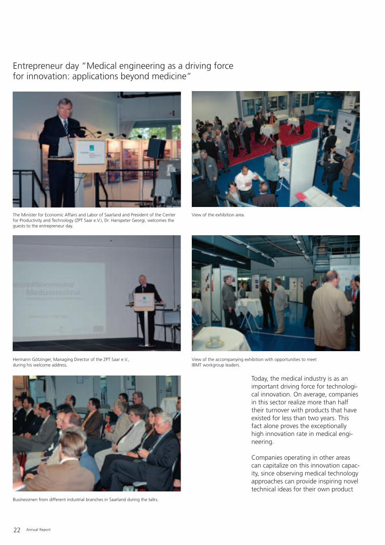
Entrepreneur day “Medical engineering as a driving forcefor innovation: applications beyond medicine”
The Minister for Economic Affairs and Labor of Saarland and President of the Centerfor Productivity and Technology (ZPT Saar e.V.), Dr. Hanspeter Georgi, welcomes theguests to the entrepreneur day.
View of the exhibition area.
Hermann Götzinger, Managing Director of the ZPT Saar e.V., during his welcome address.
View of the accompanying exhibition with opportunities to meet IBMT workgroup leaders.
Businessmen from different industrial branches in Saarland during the talks.

Annual Report 23
development. There are many exam-ples for applications spanning differentsectors. Methods for measuring bloodflow are now used in gas meters, andan ultrasound application for woundcleaning has been adopted for gentlecleaning of construction parts. Elec-tronic components that operate at lowtemperatures are not only used for cryo-banks but also in the space industry.
To open up this innovation potential tointerested companies, the Center forProductivity and Technology Saar e.V.(ZPT) and the Fraunhofer IBMT orga-nized the forum “Medical engineeringas a driving force for innovation: appli-cations beyond medicine” on May 11,2006, in Sulzbach/Saar.
Addressing the audience of approx. 50entrepreneurs from Saarland and polit-ical representatives, the morning talksoutlined those developments from thefields of classical medical engineering,device engineering, information tech-nology, biomedicine and biotechnolo-gy that can be transferred to applica-tions in other areas. Mr. Steck, CEO ofProsensys GmbH (an IBMT spin-off),presented further examples of thisknow-how transfer from the perspec-tive of a Saarland company. In theafternoon session, the guests had theopportunity to discuss ideas and con-crete questions with department andworkgroup leaders of the FraunhoferIBMT.
On May 13, 2006, the FraunhoferIBMT’s Berlin department of “CellularBiotechnology & Biochips” participatedin the sixth “Long Night of Sciences”in Berlin. Almost 9 000 interested peo-ple took advantage of the more than 1 600 demonstrations from science,technology and research. The program,particularly designed for children andyoung people, included numerousexperiments, guided tours and presen-tations – ranging from a glimpse intonanoworlds to a paper chase usingGPS and a football match betweenrobot dogs.
Over 300 visitors made their way tothe IBMT until late in the night, mar-veling at the trained non-contacting
“cells in the arena of biochips” andlearning which tools can position thecells so precisely. The theme “RedSnow – Green Snow” answered ques-tions about the microscopically small,cryophilic Arctic algae, e.g. why greensnow algae appear red (“blood-redsnow”) and what enables them to sur-vive in Polar regions.
Another “Long Night of Sciences” fol-lowed on November 29, 2006, at thehospital campus of the University ofSaarland in Homburg. The IBMT alsoactively participated in this event. Pro-fessor Fuhr held a talk on the topic“Frozen, but living world – deep-frozen cells for regenerative medi-cine.”
The Long Night of Sciences 2006
Dr. Thomas Leya, leader of the workgroup on extremophilic organisms at the Fraunhofer IBMT, gives an introduction to snow algae research: algae cultures intubes with tilted agar.
Ulrike Bley (student at the Humboldt University Berlin and student assistant at theFraunhofer IBMT) explains her poster on the search for enzymes active at low temperatures using molecular biological methods.

24 Annual Report
The question of how leaves at the topsof trees over 100 meters high (e.g.Douglas firs and Sequoias) are suppliedwith water has been controversiallydiscussed among scientists for morethan two hundred years, because treesdo not possess pumps as we under-stand them technically. The textbookexplanation is that water evaporationthrough the stomata of the leaves cre-ates a suction (pulling) tension in thevessel system of the trunk and branch-es (xylem), which pulls water upagainst gravity from the roots to the
treetop. Upon closer inspection thisprocess turns out to be more complex,since in fact transport is a coupled sys-tem similar to a paternoster lift. Whilewater is transported from the roots tothe treetop, the energy-rich metabolicproducts of the leaves (assimilates) alsodissolved in water are reciprocallychanneled through a separate vesselsystem (phloem) downwards to supplythe roots and keep them alive. There-fore, not all the water should evapo-rate, since some of the available wateris needed for growth and some for thephloem transport just described. More-over, the transport system of trees isnot absolutely watertight, i.e. evapora-tion through the leaves cannot becompletely prevented in times ofdrought even with all the stomataclosed. In spring, the situationbecomes even more complicated fordeciduous trees, since unlike the ever-green conifers they do not yet haveleaves to trigger the evaporationprocess. Apparently, considerably low-er root pressure, another coupledosmotic phenomenon, presses waterinto the trunk under these conditions.This can be impressively demonstratedon the stump surface of felled trees inspring, which exude a watery carbohy-drate mixture. In addition, the xylem ofmost trees is not continuously filledwith water – an essential requirementfor a purely suction mechanism. Asearly as the end of the 19th century,Julius von Sachs knew that during thesummer months the xylem of treescontain more air than water, in otherwords that there is no continuouswater column in trees. Even more dis-turbing is that a continuous water col-umn exposed to tensile stress becomesunstable if it is higher than 10 meters.This is due to the weight of the watercolumn and the pressure the water
Tree researchers from Würzburg use old and infirm oak tree from the formerSchmelz forest on the IBMT grounds
Oak tree on the grounds of the Fraunhofer IBMT, Ensheimer Strasse 50, in St. Ingbert.

Annual Report 25
column exerts, which must be com-pensated for by the tensile stress. Aheight of 10 meters must generate atensile stress of 1 atm, i.e. there mustbe a vacuum in the xylem. At 20meters the tensile stress would have toreach 2 atm, meaning the pressure inthe xylem must have a negative value.Supplying a 100 meter-high tree wouldrequire negative pressures in the orderof -20 to -30 atm in the xylem of thetree (if in addition to the weight of thewater column, one also takes intoaccount flow resistance). Water undernegative pressure is comparable tosuper-heated water, i.e. water prevent-ed from boiling. In both cases anyminor vibrations lead to an explosiveevaporation of water (so-called cavita-tion) and thus to a breakdown of thetransport system. Such embolismsactually do occur.
Nowadays, we know from investiga-tions of different kinds of trees thatdepending on the species, location andseason, different forces are involved insupplying leaves with water. In particu-lar, it seems that water is transportedto the treetops in phases, cushioned byair, analogous to lifting ships in canallocks. Capillary and osmotic forces aswell as transpiration-related tensilestress play important roles in thismechanism. The importance of osmot-ic forces was already realized by Wil-helm Friedrich Philipp Pfeffer morethan 100 years ago. Osmotic and capil-lary forces are particularly importantfor filling the xylem of deciduous treesin spring before the leaves appear.Osmotic processes in the roots gener-ate positive pressure, which pushes thewater plus dissolved nutrientsupwards. At the same time, photosyn-thesis and enzymatic processes inbranches and twigs supply the xylemwith osmotically active sugars, whichpull the water up into the highest tipsof the tree so that the leaves can startto bud. The xylem and phloem are
coupled transport systems. As early asaround 1900, Haberlandt postulatedthat the water in the xylem is partlydriven upwards by the flow pressure inthe phloem. Today, we know that thispaternoster lift or cable railway mecha-nism for transporting water in thexylem against gravity is also a criticalcomponent of the water transport sys-tem.
Although today much is known aboutthe water transport in trees, many par-ticular aspects are still not understood,since it involves a very complex inter-play of different forces all stronglyinfluenced by the tree species, climateand the environment. However,detailed understanding of how theseforces act is important, for examplewhen trying to recultivate salty soil oruse rapidly growing trees for “energyfarming”, i.e. for energy and fuel pro-duction. New insights can be expectedif one could visualize the water distri-bution, water flow and air cushions inthe xylem of trees. However, this is atechnically demanding task and only afew research groups worldwide areinvestigating this. In principal, thewater transport in both vessel systems(phloem and xylem) can be visualizedwith contrast media, which can betraced in the laboratory with modernimaging procedures in wood blocks,trunk segments and branches afterfelling the tree. But the injection ofcontrast media into the vessels in theupper parts of a tree requires experi-enced tree climbers, and there are onlya very few qualified people in Ger-many. These techniques have beentested for smaller and less precioustrees and have delivered valuableresults. There is rarely an opportunityto apply these techniques to large, andespecially old trees such as oaks.
The landscape remodeling on the IBMTgrounds in the Ensheimer Strasse in St.Ingbert provided this opportunity. Anapproximately 200-year-old oak withsome signs of infirmity but partially stillin a suitable state for research had tobe felled and offered the chance toinvestigate water transport. In thecourse of obtaining permission toremove the tree, which was originallypart of the forest of Schmelz, theFraunhofer IBMT contacted the bio-physicist Professor Ulrich Zimmermannin Würzburg. He at once agreed to
Working with the oak tree in preparation and duringthe scientific experiments.

26 Annual Report
take the opportunity to examine thetree and use the excellent technicalfacilities of the Fraunhofer Institute inthe direct vicinity. Contrast medium forNMR imaging and labeling was inject-ed into the tree on May 22, 2006. Thecontrast medium used was Gadolini-um, a chemical element with theatomic number 64, and a paramagnet-ic substance that amplifies nuclear res-onance signals. As with humans inmedicine, trees or at least major partsof them can be imaged by nuclearmagnetic spin tomography. On thenext day, after the contrast medium
had been distributed throughout thetree, it was cut down piece by pieceand subjected (among other tests) tohigh resolution nuclear magnetic reso-nance imaging in the IBMT laborato-ries. This revealed the distribution ofthe water and contrast medium, whichwill yield valuable insights into themode of water transport in this treespecies.
Obviously, the time between cuttingoff sections and the analysis plays animportant role, so it was lucky that thistree, which had to be felled, stood onthe grounds of an institute with suchexcellent medical technology facilities.In parallel, how intact the tree was andwhat age-related diseases it had couldbe investigated and documented.Hence, the removal of the oak tree,which incidentally will be replaced bytwo new trees, had a particular value:first as part of a forest, then contribut-ing to the city landscape of St. Ingbert,the oak tree finally served an impor-tant role in science.
Treatment of the wood samples and cross-section viewof a labeled branch.

Annual Report 27
In June 2006, the Norwegian PolarInstitute announced that it had accept-ed the proposal made by the expedi-tion members of the DFG project“CCCryo Resource” to name a slopeon Spitsbergen (Svalbard, Norway)after the algae found there. The areawill be called Raudalgeura (red algaescree field) in the future and be record-ed on the relevant maps.
The Fraunhofer IBMT has launchedexpeditions to Spitsbergen for a num-ber of years to collect and investigateextremophilic microalgae, so-calledsnow algae, that have adaptedextremely well to low temperatures.Different cell stages of these algaecause the snow to turn red or green.The collection of cryophilic algae(CCCryo Culture Collection ofCryophilic Algae) set up in 1999 at theinstitute division for Biomedical Engi-neering (AMBT) in Berlin represents aunique bioresource for research onextremophilic organisms in Germanyand Europe.
The idea of naming the location wasdeveloped during the expedition KOL07/2000 to the Northwest of Spitsber-gen (Svalbard). Members of the expe-dition were:
Dr. Günter R. Fuhr, Fraunhofer IBMT(leader of the expedition)Dr. Thomas Leya, Fraunhofer IBMTDr. Hau U. Ling, Australian colleague,formerly Australian Antarctic Division,Hobart, AustraliaHans Lund, Danish captain of the“Arctica”Dr. Torsten Müller, Evotec AOI AG, atthe time of the expedition still at theHumboldt University Berlin
Scientists of the Fraunhofer IBMT name area on Svalbard
Raudalgeura on the south coast of Hamiltonbukta (Spitsbergen, Svalbard – 79°47’ N- 11°52’ E)
View from the north towards the southern coast of Hamiltonbukta with the Raudalgeura area (centre). Snowfields on the slopes stained red by the long-term stages of algae arealready visible from a distance.
Long-term stages of a snow alga stained red by differ-ent carotinoids. These cell stages of normally chloro-phyll-stained green algae macroscopically cause thephenomenon known since the Middle Ages as “bloodsnow” or “red snow”, now acknowledged in the namegiven to the area (Raudalgeura means “scree field withred-colored algae”).
Correspondence dating between 2000 and 2006 withthe Norsk Polarinstitutt concerning naming the area.
A red snowfield at the foot of Raudalgeura on Hamil-tonbukta reaches down to sea level.

28 Annual Report
The Working Group “Technology inMedicine” was founded in 1977 and isan autonomous network of highereducation institutions, which offerstudy courses in this field, plus repre-sentatives from industry and stateauthorities who are committed to theeducation and training of new youngscientists. The group particularly focus-es on education and training in theareas of biomedical technology, med-ical technology, hospital operationaltechnologies and medical informationtechnology. Among other things, thegroup defines its tasks as promoting:
– education by direct, further andremote studies and in internationalcourses,
– certification of study courses andquality control in education andtraining,
– national and international studentand teaching staff exchange,
– further development of study sub-jects and the profile of graduates,
– recognition of the subjects andrespective professions
– public relations for these fields.
This year the 29th annual meeting ofthe Working Group “Technology inMedicine” was organized by ProfessorKlaus-Peter Hoffmann in cooperationwith Professor Wolfgang Langguth ofthe University for Technology, Industryand Commerce of Saarland (HTW) andtook place on June 15 and 16, 2006,for the first time in Saarland. The rea-son the members of the group wantedto come to Saarbrücken and St. Ing-bert was because they wished to learnmore about the study program “Bio-medical Technology” as well asresearch at the IBMT. This study pro-gram was very successfully launched inthe winter semester of 2005 at thedepartment of Electrical Engineering atthe University for Technology, Industryand Commerce of Saarland (HTW)
29th Annual Meeting of the Working Group “Technology in Medicine” from June 15-16, 2006 in St. Ingbert and Saarbrücken
Members of the Working Group “Technology in Medicine” during a tour through the Fraunhofer IBMT (from left toright, 1st row: Prof. Dr. Klaus-Peter Hoffmann, Fraunhofer IBMT and HTW of Saarland, Prof. Dr. Werner Trampisch,Technical University of Gießen; 2nd row: Prof. Dr. Jürgen Dräger, Technical University of Stralsund, Prof. Dr. RainerDammer, University of Bremerhaven, Prof. Dr. Hans-Dieter Reidenbach, Technical University of Köln; 3rd row: StephanKlein, Technical University of Lübeck, Prof. Dr. Leonore Heiland, Technical University of Zwickau, Vera Damman, Tech-nical University of Gießen; center: Prof. Dr. Wolfgang Cornetz, Rector of the University for Technology and Industryand Commerce of Saarland).

Annual Report 29
with a Master’s degree, followed lastsemester with a Bachelors degree. Cur-rently, a total of 116 students are reg-istered in this program at the HTW.
The study course is an integral compo-nent of the trilaterial initiative of Saar-land to create a biotechnology plat-form. In close cooperation betweenthe Ministry of Economic Affairs andLabor and the Ministry of Education,Culture and Research, the University ofSaarland, the HTW and the Fraun-hofer-Gesellschaft, the study programis intended to make an essential contri-bution to the structural change fromcoal and steel industries to biologicaland information technologies, includ-ing biomedical technology. The quali-fied personnel trained in these courseswill be a crucial requirement for con-tinuing the successful trend of spin-offs and establishing companies inthese sectors.
The members of the group wereimpressed by this unique study pro-gram featuring:
– Broad basic knowledge with project-oriented knowledge transfer in theBachelors program involving thecooperation partners and regionalcompanies.
– Highly specialized training in theMaster’s program with a strongemphasis on research-oriented com-ponents and courses in English whenspecializing in “Neural Engineering”,particularly run by the IBMT and theUniversity Clinics of Saarland, and anapplication-orientated componentfor specializing in medical physics.
Further topics on the agenda were thecareer prospects for graduates, certifi-cation of the degree courses, the Inter-net presentation of the Working Groupand the short reports from individualhigher education institutions. It be-came apparent that the sustained posi-tive development of the medical tech-nology sector in Germany continues to open up enormous opportunities inthe labor market for graduates ofthese study programs.
In connection with these discussionsthe members of the Working Groupwere impressed by the excellence ofresearch at the Fraunhofer IBMT.Klaus-Peter Hoffmann, founding pro-fessor of this study course and head ofthe IBMT department of Medical Engi-neering & Neuroprosthetics, led aguided tour through his laboratories atthe IBMT. Particularly impressive werethe implantable microelectrodes,which could serve as a biotechnologi-cal interface between neuroprosthesesand the peripheral nervous system.Examples of joint developments withinternational project partners are theretina implant, the hand prosthesiswith a sense of touch and a stimulatorfor bladder control. But the depart-ment also focuses on the monitoringand telemetric transmission of vitalparameters during the process ofactive aging in cooperation with theUniversity for Technology, Industry andCommerce of Saarland and the inter-disciplinary network “Products and ser-vices for all generations”. For example,it collaborates in a project for long-term monitoring of cardiovascularparameters. The goal is to supportpatients with a high risk of severe car-diovascular diseases in order toincrease their autonomy in their every-day life and protect them againstemergencies. This involves developingnovel intelligent sensors.
30 Annual Report
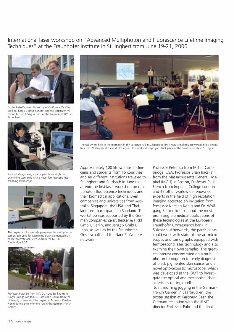
Approximately 100 life scientists, clini-cians and students from 16 countriesand 40 different institutions traveled toSt. Ingbert and Sulzbach in June toattend the first laser workshop on mul-tiphoton fluorescence techniques andtheir biomedical applications. Evencompanies and universities from Aus-tralia, Singapore, the USA and Thai-land sent participants to Saarland. Theworkshop was supported by the Ger-man companies Zeiss, Becker & HicklGmbH, Berlin, and JenLab GmbH,Jena, as well as by the Fraunhofer-Gesellschaft and the NanoBioNet e.V.network.
Professor Peter So from MIT in Cam-bridge, USA, Professor Brian Bacskaifrom the Massachussetts General Hos-pital (MGH) in Boston, Professor PaulFrench from Imperial College Londonand 13 other worldwide renownedexperts in the field of high resolutionimaging accepted an invitation fromProfessor Karsten König and Dr. Wolf-gang Becker to talk about the mostpromising biomedical applications ofthese technologies at the EuropeanFraunhofer Cryoresearch bank inSulzbach. Afterwards, the participantscould work with state-of-the-art micro-scopes and tomographs equipped withfemtosecond laser technology and alsoexamine their own samples. The great-est interest concentrated on a multi-photon tomograph for early diagnosisof black pigmented skin cancer and anovel opto-acoustic microscope, whichwas developed at the IBMT to investi-gate the optical and mechanical char-acteristics of single cells.Joint morning jogging in the German-French Garden in Saarbrücken, theposter session at Karlsberg Beer, theCrémant reception with the IBMTdirector Professor Fuhr and the final
International laser workshop on “Advanced Multiphoton and Fluorescence Lifetime ImagingTechniques” at the Fraunhofer Institute in St. Ingbert from June 19-21, 2006
Dr. Michelle Digman, University of California, Dr. KlausSuhling, King’s College London and the organizer Pro-fessor Karsten König in front of the Fraunhofer IBMT inSt. Ingbert.
The talks were held in the mornings in the Eurocryo hall in Sulzbach before it was completely converted into a deposi-tory for HIV samples at the end of the year. The workstation projects took place at the Fraunhofer site in St. Ingbert.
Aisada Uchugonova, a participant from Kirghizia, examining stem cells with a novel femtosecond laserscanning microscope.
The organizer of a workshop explains the multiphotontomograph used for examining black pigmented skincancer to Professor Peter So from the MIT in Cambridge, USA.
Professor Peter So from MIT, Dr. Klaus Suhling fromKing’s College London, Dr. Christoph Biskup from theUniversity of Jena and the organizer Professor KarstenKönig during their morning run in the German-FrenchGarden.

Annual Report 31
dinner in the Archipenko, combinedwith guided tours of the Saarlandmuseum also all contributed to theextraordinary success of this first inter-national workshop. One of the partici-pating companies was so impressed bythe research and development know-how of the Fraunhofer IBMT that onlya few days after the workshop theyplaced their first orders with thedepartment of Microsystems/LaserMedicine headed by Professor König.Further companies and universitieshave signaled their interest withrespect to cooperating in joint projectsin the future. It is intended to organizethis international event of the highestscientific level in St. Ingbert every year.
On September 25, 2006, the Presi-dents of the Federal and State AuditOffices of Germany, Switzerland andAustria visited the cryoresearch bankof the Fraunhofer IBMT in Sulzbach
after their annual conference in Saar-brücken and took advantage of theopportunity to find out about cryore-search activities at the IBMT.
Visit of the Presidents of the Federal and State Audit Offices
The presidents of the Federal and State Audit Offices visiting the cryohall, led by Manfred Plaetrich, President of theState Audit Office of Saarland (foreground).

32 Annual Report
The development of ultrasound appli-cations for medicine in recent yearshas been striving to include higher andhigher frequencies. While establishedultrasound technology works at a fre-quency range of up to 15 MHz, cur-rent developments can reach as highas 2 GHz. Increasing the frequencyrange by a factor of 100 means animprovement in the spatial resolutionby approximately the same factor. Inbasic biological research as well as inapplication areas such as ophthalmolo-gy, dermatology and investigatingblood vessel linings, the use of highfrequency ultrasound promises signifi-cantly improved diagnosis by visualiz-ing smaller and smaller anatomicalstructures. Nanotechnology can con-tribute significantly to this progresswith novel nano-treated materials.
To comprehensively pursue this objec-tive, which will be of great scientificand economical importance, tworesearch institutes in Saarland withcomplementary research and technolo-gy skills launched a research alliance:the Institute for New Materials of theLeibniz Association and the FraunhoferIBMT.
The department “Technology of Non-Metal Inorganic Materials” of the Leib-niz Institute for New Materials (INM)has the know-how for developing andproducing materials with entirely novelcharacteristics using solid/gel technolo-gies.
Since its foundation in 1987, theFraunhofer IBMT department of Ultra-sound in St. Ingbert has developed intoEurope’s largest R&D institution in thisfield with a staff of almost 40. TheChair of Medical Biotechnology &Medical Engineering and the IBMTworkgroup “Biomedical Ultrasound”both possess the necessary experienceand resources to support and rapidlyrealize the application of novel techni-cal ultrasound developments based onhigh technology.
Taken together, these comprise exactlythe requirements necessary for devel-oping the desired medical applications.
The project is supported by funds fromthe Ministry of Education, Culture andResearch of Saarland. Minister JürgenSchreier handed over the fundingagreement in person during a visit tothe IBMT in St. Ingbert on September29, 2006.
Nanotechnology for the application of high resolution, high frequency ultrasound in medicine – Fraunhofer IBMT receives funding from the Ministry of Education, Culture and Research
Handing over of the funding agreement (from left to right: Dr. Robert Lemor, head of the department for Ultrasoundof the Fraunhofer IBMT, Minister Jürgen Schreier, Dr. Frank Tiefensee, project leader at the Fraunhofer IBMT, Prof. Dr. Günter Fuhr, director of the Fraunhofer IBMT, Dr. Wolfgang Bach, head of the department for Science andResearch, Universities and Teacher Training at the Ministry of Education, Culture and Research of Saarland).

Annual Report 33
The SaarLB (State Bank Saar) sponsorsarts, culture and science. As part of thescience initiative, the SaarLB ResearchAward, endowed with 25 000 €, wasfirst created in 1999 together with theMinistry of Education, Culture andResearch of Saarland. The award paystribute to scientific work leading toresults and information that can beapplied to economically strengtheningthe Saarland region. An independentjury evaluates the submitted work.
On October 20, 2006, Prof. Dr. HeikoZimmermann, head of the departmentof Cryobiophysics & Cryotechnology atthe Fraunhofer IBMT and junior profes-sor at the University of Saarland, washonored with the SaarLB 2005Research Award. He received the prizein recognition for his work in develop-ing methods for cryopreservation andlinking microcapsules for the treatmentof diabetes mellitus. Diabetes is cur-rently treated by injection of molecules
not produced by the body, whichtherefore have a short half-life. Themethods developed by Heiko Zimmer-mann allow one to surround micro-encapsulated islets of Langerhans cellsof allogeneic or animal origin with bio-compatible alginate capsules ofdefined characteristics, so that thebody does not mount an immuneresponse against the foreign, cryopre-served cells. Another result of the workis a significantly higher survival rate ofthe cells after cryostorage. Cryopreser-vation is indispensable for later use ofsuch transplants. The junior professor’sconsiderable contribution to theseprocesses was the reason why the jurydecided to grant him the award.
Presentation of the 7th SaarLB Research Award toProf. Dr. Heiko Zimmermann
Presentation of the SaarLB Research Award 2005 (from left to right: Prof. Dr. Hans-Jörg Bullinger, President of theFraunhofer-Gesellschaft, Prof. Dr. Günter Fuhr, Director of the Fraunhofer IBMT, Prof. Dr. Heiko Zimmermann, award-winner, Dr. Max Häring, chair of the SaarLB board of directors). Photograph: Wolfgang Klauke.
34 Annual Report
On November 14, 2006 the innovationcompetition prizes to promote medicaltechnology were awarded to selectedprojects during the opening of theMedica 2006 exhibition. Dr. KlausPeter Koch from the Fraunhofer IBMT,as the coordinator, received the prizegranted to the project “ContinuousIntraoperative Nerve Monitoring as aMicrotechnological Navigation Tool”from the Federal Minister of Educationand Research, Dr. Annette Schavan.
Intelligent and autonomous nerve cellmonitoring can protect nerves duringsurgery against damage due to theeffects of tension, pressure or temper-ature: electrodes constantly registerchanges in conductivity of the nerves.The signals are recorded immediatelyand processed by computer. Thisshould considerably reduce the risk ofinjury. Dr. Klaus Peter Koch’s groupwas one of the winners of the innova-tion competition 2006 and will receiveapproximately 1.5 million € in fundingfrom the Federal Ministry of Educationand Research (BMBF).
Damage to nerves is a frequent andpotentially life-threatening conse-quence of surgery. Nerve fibers aresimilar in structure and color to con-nective tissue and small blood vessels,so the risk of confusion and resultingdamage is high. This can have fatalconsequences: Nervous dysfunctionsafter surgery can lead to severe handi-caps that can affect the ability to con-tinue in any employment or cause awithdrawal from social life. Surgeryinvolving the thyroid gland, for exam-ple, carries the risk of injuries to thevocal chord nerves, which can lead tochronic hoarseness, loss of vocal abili-ties or even to life-threatening breath-ing difficulties. Nerve injuries fromsurgery in the lower abdomen, e.g.
rectal operations, can disturb normalbladder and sexual functions. Theseare only a few of many other examplesof the post-operative consequences ofnerve injuries. To minimize these risksand better control potential complica-tions, the nerves should be monitoredduring an operation. This would allowpressure and tension forces as well asthe effects of temperature to bedetected and avoided before any injuryoccurs.
Electrodes are the best security systemfor nerve cells. They are highly sensitiveat registering everything that couldthreaten the nerves and immediatelytrigger an alarm – both visually andacoustically. The flexible electrodesdeveloped during the project are madefrom a biocompatible material. Theyshould be easy to use and not disturbor obstruct the surgical procedure. Fur-thermore, it is important that the sig-nal transmission is relatively indepen-dent from errors in placing the elec-trodes correctly or displacement of theelectrodes during the operation. Allthis is guaranteed by combining theelectrodes with intelligent software,which automatically searches for theoptimal stimulation and conductivitypoint. Dr. Klaus Peter Koch from theFraunhofer IBMT and his team expectthat continuous monitoring duringsurgery will reduce surgery-relatednerve injuries by at least 50%. Thiswould also be of national economicimportance, since for thyroid glandsurgery alone, follow-up costs due tonerve injuries amount to some 70 mil-lion € per year in Germany.
Contact
General Coordinator:Dr.-Ing. Klaus Peter KochFraunhofer Institute for Biomedical EngineeringProf. Dr. Günter R. FuhrDirector of the InstituteEnsheimer Strasse 4866386 St. IngbertGermanyTelephone: +49 (0) 6894/980-404Fax: +49 (0) 6894/[email protected]
Innovation competition Medical Technology 2006 of the Federal Ministry of Educationand Research – Bodyguards for the nerves, nerve monitoring lowers injury risk

Annual Report 35
CellPROM – Integrated EU researchprogram
The 6th Research Framework Programof the European Union is a new fund-ing scheme designed to stimulate thedevelopment and application ofpromising future technologies in theEuropean research area, in addition tothe proven funding instruments suchas STREP and CRAFT projects. Exam-ples of this new funding scheme arethe Integrated Projects, where numer-ous partners from European researchareas collaborate on an innovativelarge-scale project.
With a funding volume of 27 million €,CellPROM is the largest Integrated Project in the area of nanobiotechnolo-gy and links 27 academic and industri-al partners from 12 countries for aperiod of four years. It is headed byProf. Dr. Günter Fuhr and coordinatedby the Fraunhofer IBMT. The abbrevia-tion CellPROM stands for “Cell PRO-graMming by nanoscaled devices”.The project’s objective is surface-sup-ported cell differentiation on a largetechnological scale. For this, artificial
macromolecular landscapes based onthe model of cell surfaces will bedeveloped and tested by nanotechno-logical procedures. In addition to solu-ble signaling molecules such as differ-entiation and growth factors, theseartificial landscapes support the differ-entiation of cells via multiple surfacecontacts in a way that resembles thenormal biological situation: thesemacromolecular landscapes(NanoScapes) imitate functions per-formed by cell surface contacts tomatrix elements and adjacent cells inthe tissue and body. This approach ismeant to fill a technological gap andto create a new generation of novelmodules for in vitro cell imprinting.Mastering these processes on anindustrial scale is a prerequisite foropening up important applicationareas in the fields of biotechnology,medicine or pharmacy and for acceler-ating the technology development.The CellPROM project will enter new
Nanobiotechnology as a future research area
A human fibroblast on a nanostructured silicon surface photographed with a scanning electron microscope (Courtesy of Dr. Alisa Katsen).

36 Annual Report
interdisciplinary territory and con-tribute to the development ofnanoscopic tools for cell handling inbiotechnology and regenerative medi-cine. Upon completion, the four-yearproject should provide functional mod-ules that can demonstrate the techni-cal solutions and biological processes,forming the basis for scaling-up pro-duction and conceiving ideas for fur-ther applications, altogether consider-ably strengthening Europe’s position inthe promising future market ofnanobiotechnology.
The Integrated Project CellPROM start-ed in March 2004, with the kick-offmeeting held on March 25 and 26.Work meetings are held quarterly withall partners, supplemented by regularmeetings with 8 work package leadersand their representatives. On March 14/ 15, 2006, the second annual assess-ment meeting took place in St. Ing-bert. Establishment of an operational
and effective management structure,evaluation and preparation of possibledevice concepts for different modules,first biological cell experiments andintegration of technological prototypesdominated the second project year,and all aspects were evaluated verypositively in the March 2006 assess-ment.
Two different concepts for handlingcells were developed and presented:one concept is based on magneticallymanipulated targets and allows manip-ulation of adherent cells by miniatur-ized surface carrier substrates (see Fig-ure 2).
The second approach, based on a fluidlab-on-chip concept, involves manip-ulating cells without any direct contact(see Figure 1). Both concepts will bepursued in parallel during the nextphase of the project and the corre-sponding modules will be developedand evaluated.
Partners of the project are companiessuch as Evotec Technologies (Ger-many), Leister Process Technologies(Switzerland), GeSIM (Germany), Sys-melec (Switzerland), Eurogentec (Bel-gium), Silex (Sweden), Surface ImagingSystems, AMO, Eurice and tp21 (Ger-many) as well as institutes such as theRoyal Institute of Technology (Sweden),the Institute of Experimental Biologyand Technology (Portugal), the PasteurInstitute (France), the Institute forSpectrochemistry and Applied Spec-troscopy, the Institute for New Materi-als, the Georg Speyer House and theMax Planck Institute for Biophysical
Figures 1 and 2: Simulations to illustrate the modules currently being realized. Left: Manipulation of cells without any surface contact via miniaturized high frequency fields. Right:Magnetic manipulation of “nanoscapes” on microcarriers in a complex channel system.

Chemistry (Germany) and the universi-ties of Lausanne (Switzerland),Barcelona (Spain), Saarbrücken (Ger-many), Vienna (Austria), Kaiserslautern(Germany), Pavia (Italy), Ljubljana(Slovenia), Tel Aviv (Israel) and Vilnius(Lithuania): in other words, a coopera-tion almost unique of its kind.
Coordinating such a large-scale projectis a challenge not only in terms of sci-entific themes, but also with respect tomanagement and requires noveladministration tools. The FraunhoferIBMT is supported in this task by itsintegration into the Fraunhofer LifeSciences Alliance and the administra-tion department of the Fraunhofer-Gesellschaft.
ContactDipl.-Phys. Daniel SchmittTelephone: +49 (0) 6894/[email protected]
Figure 3: Magnetically manipulated microcarriers for cell culture in the circular holes(width of one target is 1 mm).
Figure 4: Cell programming in the CellPROM project. Differentiation of human adultstem cells into adipocytes on microstructured surfaces (Photograph: Claudia Brose).
Annual Report 37
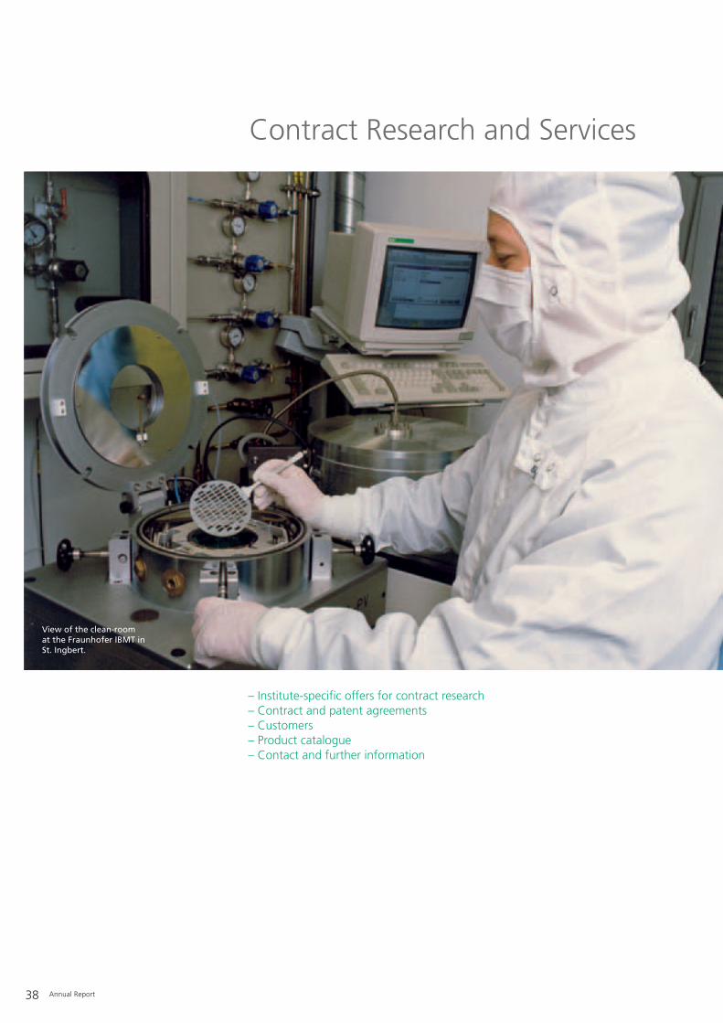
38 Annual Report
View of the clean-roomat the Fraunhofer IBMT inSt. Ingbert.
– Institute-specific offers for contract research– Contract and patent agreements– Customers– Product catalogue– Contact and further information
Contract Research and Services

Annual Report 39
Procedure:R&D projects are performed in success-oriented phases, starting with an initialtechnical market survey, the feasibilitystudy deduced from this, then proto-type development, field tests (clinicalstudies), the development of cost-opti-mized production processes and therequired technology. Companies canbe suggested for outsourcing sensorand microsystem production.
Relevance to practice:Projects are pursued at the FraunhoferIBMT in close coordination with therespective customer in order to estab-lish the maximum relevance to prac-tice. Close contact with the customeris a specific feature and an importantrequirement to meet the marketdemands when starting out from basicresearch.
Flexibility:The concrete form, the orientation andthe extent of the project work followthe requirements and ideas of the cus-tomer.
Synergy:Integration into the research strategyof the Fraunhofer-Gesellschaft with its58 institutes, and the Life SciencesAlliance of five Fraunhofer Institutes(IBMT, IGB, IME, ITEM, IZI) establishedin 2001, creates synergistic effects.Expertise from diverse research fieldscan be utilized in collaborations, allow-ing competent approaches even tomultidisciplinary topics. Cooperationcontracts offer IBMT customers com-plete product pipelines.
Quality:Punctual delivery and reliability arecharacteristic for the work of theFraunhofer IBMT. The creation of a“duty list” in cooperation with the cus-tomer guarantees that work on theproject adheres to the agreed upontheme and projected time-frame.
Prices:Commissioned research and develop-ment are carried out on a customer-paid basis. As an institute of the Fraunhofer-Gesellschaft, the IBMT is acharitable (non-profit) institution andmainly finances necessary application-orientated and preliminary researchthrough public contractor participa-tion.
R&D results:The customer receives all the resultsafter successful completion of the R&Dproject.
Confidentiality:If the customer wishes, enquiries arehandled with absolute confidentiality.
Institute-specific offers for contract research

40 Annual Report
Phase model:Work on projects at the FraunhoferIBMT is performed in stages: the startof a project involves scientific andtechnical consultation. This serves toreveal possible problems and assessesthe risks of the project based on exist-ing know-how, as well as applying lit-erature, patent and market research.The second phase comprises a feasibili-ty study, which specifically defines theproject and evaluates the necessaryefforts. Development of a laboratoryprototype then demonstrates the prac-tical operation. This stage leads to thedevelopment of a field prototype end-ing up with its extensive testing. Sub-sequently, re-design, technology opti-mization, small-scale production andtechnology transfer are all essentialelements in preparing for large-scaleproduction. As an auxiliary service, theFraunhofer IBMT offers support withmarketing and quality control, which
serves to get production startedsmoothly and eliminates risks involvedin manufacturing. The client can subdi-vide their commission according tothese phases and decide at the end ofeach individual stage whether it isworthwhile embarking on the nextstage. These criteria make it easier forthe customer as well as the IBMT toplace or accept a commission and theyproduce transparent and calculableproject times and costs.
Feasibilitystudy
Prototypedevelopment Field tests
Productiontechnology Market
- existing know- how- literature research- patent inquiries- expert reports- market analyses
- studies- simulations- experiments
- special modules- mechanical components- electronics- joining technologies- special software- definition of test criteria
- test under field conditions- evaluation- reliability analysis- optimization- test support
- product develop- ment- optimization of the production technology- small-scale production- technology transfer
Scientific andtechnical advice
App l i ca t ion -o r i en ta ted bas i c re sea rch
Number of projects Goal
- quality control
Risk-minimized product development.

Annual Report 41
Drawing up a contract:Fair and reliable contract conditions forthe customer are of primary impor-tance. Here, the scientists and engi-neers are supported by an experiencedcontract department of the Fraun-hofer-Gesellschaft.
Beneficial interest:The customer possesses exclusiverights to the patents generated in thecourse of the project. If the customerwishes, individual agreements can besettled. The IBMT is represented bymore than five well-known patentattorney agencies.
Coordination:The Fraunhofer IBMT is experienced inthe coordination of complex multi-partner programs and generic leadprojects. In this context, the IBMTtakes on administration and coordina-tion tasks and ensures excellent com-munication between the project part-ners to minimize “frictional loss”.
Training:The IBMT also offers the customertraining of personnel if required, tointroduce new methods and technolo-gies. The training can take place on-site at the customer’s company loca-tion.
Quality control:The scientists and development engi-neers of the Fraunhofer IBMT workaccording to the rules of modern pro-ject management. The projects andoperations are thoroughly and con-stantly audited with regard to time andcosts and are oriented towards suc-cessful termination of the project. Eachcommission is monitored by computer-aided project controlling.
Funding possibilities:The Fraunhofer-Gesellschaft helps thecustomer to locate every possiblesource of project funding. With long-standing experience in applying forresearch funding from the EuropeanUnion, the Federal Ministry of Educa-tion and Research (BMBF) or otherfunding sources we proactively supportthe customer in matters of financingresearch projects.
In addition to customers from the bio-medical and medical engineering sec-tor including biotechnology, otherindustry branches (biotechnology, envi-ronmental technology, chemistry, phar-macy, material technology, automobiletechnology, hydraulics, mechanicalengineering, installation engineering,sensor systems) are also clients of theFraunhofer IBMT. Since its foundationthe IBMT has worked together withcompanies of all sizes.
Contracts and patent agreements Customers

Product Market IBMT contact person
PaDok® – Secure communication and medicine, healthcare, Dipl.-Phys. Bertram Bresser
case-based network of records in the health service telematics Tel.: +49 (0) 6894/980-206
TOPCARE – Home care and tele-medicine platform medicine, health care, Dipl.-Inform. Stephan Kiefer
telematics, home care Tel.: +49 (0) 6894/980-156
Flow sensors medicine, food industry, chemistry, Dr. Thomas Velten
environmental testing Tel.: +49 (0) 6894/980-301
NMR sample heads for spectroscopy and micro-imaging medicine, material research, biomedical Priv.-Doz. Dr. Frank Volke
with a coil diameter between 2 and 40 mm, technology, environment, food, Tel.: +49 (0) 6894/980-405
adapted to respective samples chemical and cosmetics industry
State-of-the-art gradient coils for NMR micro-imaging, clinical medicine Priv.-Doz. Dr. Frank Volke
e.g. 200 G/cm gradient systems in x,y,z dimensions Tel.: +49 (0) 6894/980-405
and measurement time lapses starting with 50 microseconds
NMR coils for medical whole body tomographs, clinical medicine Priv.-Doz. Dr. Frank Volke
e.g. lung coils for MRI with clinical equipment Tel.: +49 (0) 6894/980-405
for (polarized) helium and/or xenon
Minimally invasive NMR technology, clinical medicine Priv.-Doz. Dr. Frank Volke
e.g. NMR coils in conjunction with endoscopic operations Tel.: +49 (0) 6894/980-405
Magnetic resonance positioning systems online MRI-aided operations, Priv.-Doz. Dr. Frank Volke
for medical operations clinical medicine Tel.: +49 (0) 6894/980-405
Training courses for NMR spectroscopy and micro-imaging pharmaceutical, life and Priv.-Doz. Dr. Frank Volke
material sciences, industry Tel.: +49 (0) 6894/980-405
Imaging software medicine, material sciences Priv.-Doz. Dr. Frank Volke
Tel.: +49 (0) 6894/980-405
Biochemical structured surface substrates biotechnology, stem cell research Dr. Andreas Lankenau
Tel.: +49 (0) 331/58187-303
Strains from the algae culture collection of detergent, pharmaceutical, Dr. Thomas Leya
psychrophilic microalgae (CCCryo) food and cosmetics industry Tel.: +49 (0) 331/58187-304
Algae raw material from customer-specific cultivation detergent, pharmaceutical, Dr. Thomas Leya
food and cosmetics industry Tel.: +49 (0) 331/58187-304
DNA, RNA, cDNA for downstream processes detergent, pharmaceutical, Dr. Thomas Leya
food and cosmetics industry Tel.: +49 (0) 331/58187-304
Immunosensor analyzer for automatic biotechnology, pharmaceutical industry, Dr. Nenad Gajovic-Eichelmann
competitive immunoassays environmental analytics Tel.: +49 (0) 331/58187-204
42 Annual Report
The Fraunhofer IBMT offers its partners new products, technologies and processes, also for manufacturing, marketing orexploitation of patents and licenses. Please refer to the competence matrix on page 19 and the following product catalogue.
Product catalogue

Annual Report 43
Contact and further information
Please do not hesitate to call us if youhave any questions, or if you wish fur-ther information or a concrete offer.We are happy to send you publicationsand brochures. Please visit our website:
http://www.ibmt.fraunhofer.de.
Fraunhofer Institute for Biomedical Engineering IBMT Ensheimer Strasse 4866386 St. IngbertGermanyTelephone: +49 (0) 6894/980-0Fax: +49 (0) 6894/980-400
Marketing/Public RelationsDipl.-Phys. Annette MaurerTelephone: +49 (0) 6894/[email protected]

44 Annual Report
The Institute in Numbers
– Personnel development– Operative budget– Contract research with industry
Employees of the IBMT during a works outing in 2006 to visit the coalmining training tunnel of the RAG-Deutsche Steinkohle AG (DSK).

Annual Report 45
Personnel development
In 2006, there were 194 scientific,technical and administrative personnel(including professorships), 29 studentassistants and 57 practical students atthe Fraunhofer IBMT. In addition, 10guest scientists were carrying outresearch at the institute for a longertime period.
Operative budget
The estimated operative budget for2006 will amount to 11.4 million €.
Income from industry to cover overallexpenses is predicted to be 2.8 million€ in 2006.
Contract research with industry
Project work predominates theresearch activities at the Institute. Thegoal in 2006 was to reduce the overallnumber of projects in 2000 to 2002 infavor of larger projects. This objectivewas achieved in 2006 with an overallincrease in project scope but limited to355 projects. 145 projects (i.e. approx.41%) were for industrial customers.
Head of AdministrationBärbel WalterTelephone: +49 (0) 6894/[email protected]
20062005
310
1991
93
1992
111
1993
119
1994
125
1995
140
1996
242
1997
299
1998
290
1999
283
2000
379
2001
399
2002
380
2003
322
2004
328
450
400
350
300
250
200
150
100
50
0
Number
355
Personnel development from 1999 to 2006.
Project development from 1991 to 2006.
250
200
150
100
50
0
Number
1999 2000 2001 2002 2003 2004
103
50
6
104
47
6
123
44
9
150
45
4
141
45
6
169
30
14
178
32
12
2005
Total personnelStudent assistantsGuests
194
29
10
2006

46 Annual Report
– Summary of overall competence – Research areas– Target groups– Services offered– Advantages of contract research
Map with all the research establishments ofthe Fraunhofer-Gesellschaft in Germany plusthe locations of the IBMT institutes.
The Fraunhofer-Gesellschaft at a Glance
Fraunhofer & IBMT Project Group “Cell Differentiation & Cell Technology”
IBMT Branch at Potsdam-Golm
IBMT Cryoresearch Bank
Parent Institute St. Ingbert

Annual Report 47
The Fraunhofer-Gesellschaft presentlycomprises 58 institutes that are the-matically organized into eight researchfields. Due to the strong interdiscipli-nary nature of biotechnology, a deci-sive advantage of the Fraunhofer-Gesellschaft, with its institutes andassociations, is that it can cover almostall technology areas from research andindustry. To enable our customers tooptimally utilize these competences,the core areas of the Fraunhofer-Gesellschaft are summarized below.
Summary of overall competence
Research of practical utility lies at theheart of all activities pursued by theFraunhofer-Gesellschaft. Founded in1949, the research organization under-takes applied research that drives eco-nomic development and serves thewider benefit of society. Its services aresolicited by customers and contractualpartners in industry, the service sectorand public administration. The organi-zation also accepts commissions fromGerman federal and Länder ministriesand government departments to par-ticipate in future-oriented researchprojects with the aim of finding inno-vative solutions to issues concerningthe industrial economy and society ingeneral.
Applied research has a knock-on effectthat extends beyond the direct benefitsperceived by the customer: Throughtheir research and development work,the Fraunhofer Institutes help to rein-force the competitive strength of theeconomy in their local region, andthroughout Germany and Europe.They do so by promoting innovation,accelerating technological progress,improving the acceptance of new tech-nologies, and not least by disseminat-ing their knowledge and helping totrain the urgently needed future gen-eration of scientists and engineers.
As an employer, the Fraunhofer-Gesellschaft offers its staff the oppor-tunity to develop the professional andpersonal skills that will allow them totake up positions of responsibility with-in their institute, in other scientificdomains, in industry and in society.Students working at the FraunhoferInstitutes have excellent prospects ofstarting and developing a career inindustry by virtue of the practical train-ing and experience they have acquired.
At present, the Fraunhofer-Gesellschaftmaintains more than 80 research units,including 58 Fraunhofer Institutes, at40 different locations in Germany. Themajority of the 12 500 staff are quali-fied scientists and engineers, whowork with an annual research budgetof 1.2 billion € . Of this sum, morethan 1 billion € is generated throughcontract research. Two thirds of theFraunhofer-Gesellschaft’s contractresearch revenue is derived from con-tracts with industry and from publiclyfinanced research projects. Only onethird is contributed by the Germanfederal and Länder governments in theform of institutional funding, enabling
the institutes to work ahead on solu-tions to problems that will not becomeacutely relevant to industry and societyuntil five or ten years from now.
Affiliated research centers and repre-sentative offices in Europe, the USAand Asia provide contact with theregions of greatest importance to pre-sent and future scientific progress andeconomic development.
The Fraunhofer-Gesellschaft is a recog-nized non-profit organization whichtakes its name from Joseph von Fraun-hofer (1787-1826), the illustriousMunich researcher, inventor and entre-preneur.
Joseph von Fraunhofer (1787-1826).

48 Annual Report
Research areas
Research and development are organized into eight institute groups (clusters) within the Fraunhofer-Gesellschaft:– Material technology/construction
component properties– Production technology/manufactur-
ing technology– Information and communications
technology– Microelectronics/microsystems tech-
nology– Sensor technology and sensor sys-
tems– Process engineering– Energy and construction technology,
environment and health research– Technical and economical
studies/information transfer
To strengthen the biological sciences, aLife Sciences Alliance was establishedin 2001, comprising the four foundinginstitutes (IBMT, IGB, IME, ITEM) andthe IZI, which is currently under devel-opment.
Target groups
The target groups of the Fraunhofer-Gesellschaft are industry and the pub-lic sector.– For customers from industry the
Fraunhofer-Gesellschaft developstechnical and organizational solu-tions up to the point of operationaluse. If system solutions are needed,several Fraunhofer Institutes cooper-ate under the direction and coordi-nation of one commissioned insti-tute.
– Strategic research projects are carriedout upon request from German Fed-eral or State authorities. These areaimed at promoting key technologiesand innovations in areas of particularpublic interest, e.g. environmentalprotection, energy technologies andhealthcare. Within the context of theEuropean Union the Fraunhofer-Gesellschaft participates in technolo-gy programs that serve to increasethe competitiveness of the Europeaneconomy.
Services offered
The Fraunhofer-Gesellschaft offersresearch and development in manyservice areas:– Product optimization, development
of prototypes, optimization ofprocesses and development of newprocesses
– Support in introducing new organi-zation schemes and technologies by– Testing in demonstration centers
with state-of-the-art equipment– On-site training of the personnel
involved– Services after introducing new
processes and products– Technology advice through
– Feasibility studies– Market observation– Trend analyses– Evaluation of economic efficiency– Funding advice, especially for SMEs
– Testing services and granting of testcertificates
– Helping companies start up– Advice on company concepts and
business plans– Generation of economic concepts
Advantages of contract research
The cooperation of all the institutesensures that the customers of theFraunhofer-Gesellschaft can accessnumerous specialists with a broadspectrum of competences. Uniformquality standards and the professionalproject management of the FraunhoferInstitutes guarantee reliable results forthe commissioned research. State-of-the-art laboratory equipment makesthe Fraunhofer-Gesellschaft attractivefor companies of all sizes and from allbranches. In addition to the reliabilityof a strong association, a cooperationalso has economical advantages, sincethe Fraunhofer-Gesellschaft con-tributes valuable starting capital to thepartnership in the form of cost-inten-sive preliminary research.

Annual Report 49
Selected Research Results and Applications
– Microsystems/Laser Medicine– Ultrasound– Telematics/Tele-Medicine– Medical Engineering & Neuroprosthetics– Cryobiophysics & Cryotechnology– Cryoresearch and Cryodemonstration Bank– Biohybrid Systems– Computer-aided Simulations– Cell Differentiation & Cell Technology
Branch Potsdam-Golm– Cellular Biotechnology & Biochips– Molecular Bioanalytics & Bioelectronics
Biomedical Competence Centers
Ultrasound sensors produced by microsystems are agrowing research and application field.

50 Annual Report
Microsystems/Laser Medicine
Services, results and products of the workgroups
– Miniaturized Systems– Magnetic Resonance– Laser Medicine
Project example: Non-invasive, high-resolution imaging with magnetic resonance tomography in combination with multiphoton tomography
Equipment
Line structure on a ripple-structure wafer exposed to an 800 nm femtosecond laser.

Annual Report 51
Photolithography as a core technologyfor chip production, microsensors,microactors and rapid prototyping isbased on UV exposure of photosensi-tive polymers (photoresist) applied tosilicon wafers and a subsequent etch-ing process. From year to year thepacking density on the chips hasincreased due to the ever-decreasingarea of polymer exposed to UV. Since,according to Abbé, the size of theexposed spots correlates with thewavelength, UV excimer lasers areused. Currently, 90 nm technology isused to produce the Pentium 4 proces-sor. Surprisingly, even sub-200 nm andsub-100 nm structures on siliconwafers and photoresist can beachieved with lasers of longer wave-lengths in the near infrared (NIR) spec-tral range. Such sub-wavelengthnanostructuring results from multipho-ton effects, which are induced whenthe light passes through wide aperturelens optics within the focal range. Atthe IBMT, we used ultracompact turn-key NIR femtosecond laser pulses withlow picojoule pulse energy in combina-tion with scanning microscopes toexpose SU-8 photoresist using a two-photon effect. Exact exposure of thephotoresist in all three dimensions,with a precision of 40 nm within thephotoresist, was achieved by varyingthe focus plane with a piezo-controlledlens system. Hence, it was possible togenerate 3-dimensional structuringand bulk structures. The figure (on theleft, p. 56) illustrates an example ofsuch a 3-D two-photon polymerizationof SU-8 photoresist applied to a waferafter exposure to 730 nm femtosecondlaser pulses from an 80 MHz titaniumsapphire laser.
The resulting structure, an “IBMTcube” with an edge length of 60 µm,was generated by initial irradiation of abase in 60 µm coating depth and sub-sequent exposure of the overlyingareas by suitable beam scanning withan x,y-galvoscanner. Interestingly, thedetailed topography of the structuredmicro-cube featured a periodic nano-structure. Analyses by electron andatomic force microscopy revealednanogrooves with an average depth of150 nm. The distance between thesenanogrooves can be modified by vary-ing the scanning parameters. The gen-erated 3-D structure with its periodicnanotopography impressively demon-strates the new technological potentialof ultra-short long-wave laser pulsesfor precise three-dimensional pho-tolithography using multiphoton pho-tochemistry.
If the laser pulse energy is raised to afew nanojoules, even direct nanostruc-turing (laser writing) of silicon waferscan be achieved without the use ofphotochemical processes (photopoly-merization). The figure on the leftshows a ripple-structure in a waferexposed to an 800 nm femtosecondlaser. As yet poorly understood self-organizing processes created a sub-80nm nanostructure. This means that thewidth of the structure is more than anorder of magnitude smaller than thelaser wavelength. Thus, we generatedthe smallest sub-wavelength structuresto date.
Currently, we are investigating the bio-compatibility of such nanostructuresgenerated by femtosecond lasers andtheir potential as tools for manipulat-ing single cells and cell clusters, espe-cially stem cells.
Further application fields for this novelnanostructuring laser technology existin tissue engineering (e.g. the produc-tion of artificial extracellular matrices),the preparation of prostheses, the pro-duction of novel optical 3-D data stor-age media, photonic crystals andnanosystems, as well as rapid proto-typing.
ContactProf. Dr. Karsten KönigTelephone: +49 (0) 6894/[email protected]

Magnetic Resonance
52 Annual Report
Miniaturized systems, if requested withwireless control and data acquisition:– Sensor systems– Actuator systems– Active medical implants
Miniaturized telemetric systems (not only) for medical applications:– Transmitted by induced current– Infrared telemetrics– Radio telemetrics– Production of microcoils
Development of size-optimized sensor,actuator and communication electron-ics:– Microsensors– Mass flow sensors with integrated
conductivity measurement– Sensors to measure the thickness of
films– Tactile sensors (endoscopy, robotics)
Micro-fluidics and biocell handling systems:– Micro-fluidic systems as fluid inter-
face to biosensors and biochips– Multi-jet structure to handle several
cells in parallel– Micro-injection chips for cell injec-
tions (needle + pump on onemicrochip)
Construction and bonding technology:– Packaging of bioanalysis chips– Packaging of micro-implants– Design and production of ultra-thin
(5-10 µm), flexible printed circuitboards with conductor widths ≥ 5µm
– Patented “MicroFlex bonding tech-nology” for flexible printed circuitboards
– Hybrid-integrated layer technologies(thick layer or thin film technology)
Thin layer technology:– Preparation of low-stress silicon
nitride layers (PECVD)– Preparation of water impermeable
parylene layers– Preparation of metallic and dielectric
layers (vapor-coating, sputtering)
Microstructuring:– 3-D rapid prototyping of SU-8 pho-
toresist with femtosecond laser puls-es (resolution: 300 nm)
– Masking via photolithography– Wet chemistry etching– Reactive ion etching (RIE)– Dry etching of parylene and poly-
imide
Replication technologies:– Silicon molding– Rotary hot embossing of (fluidic)
microstructures on large surface,continuous polymer foils
ContactDr. Thomas VeltenTelephone: +49 (0) 6894/[email protected]
Biomedical research (NMR, FT-IR):– Evaluation of active compounds by
NMR spectroscopy and MR imaging– NMR micro-imaging and MRI (mag-
netic resonance tomography)– Formulation of compounds, creams,
gels, etc.– Permeation characteristics of vesicles,
drug carriers and cells– Interactions between membrane-
active pharmaceuticals and model orbio-membranes
– Liposomes as carriers of active com-pounds
– Characterization (in vitro) of cellularcomponents and metabolic processeswith high-resolution solid-state NMRtechnologies
– Molecular characterization of bio-mineralization processes
– Ageing processes in gels, creams,etc.
– Hydration characteristics of biopoly-mers and active agents
– Coating of surfaces (biocompatibility)– In vitro and in vivo studies on the
effects of creams and lotions on theskin
– Analysis of bio-glues– Analysis of biosensors– Cells under extreme conditions (e.g.
cryopreservation, cryoprotection)– Cell-cell and cell-surface interactions
with high-resolution NMR spec-troscopy
– Structure and dynamics of biofilmsunder flow conditions
– Image analysis with the softwareBodyScan® developed in theNMR/MRI workgroup
Material research (NMR, FT-IR, AFM):– Molecular structure and dynamics in
polymers and biopolymers– Diffusion behavior of fluids in poly-
mers
Miniaturized Systems
Services, results and products of the workgroups

Annual Report 53
– NMR micro-imaging of compositematerials
– Swelling capacity of polymers andbiopolymers
– Evaluation of filter material– Evaluation of the protective effect of
waxes
NMR technology:– Non-invasive high-resolution NMR
flow measurements, rapid imagingprocedures for on-line control, flowcharacteristics on different physico-chemical surfaces (biocompatibility)
– Rapid 3-D MR imaging also for solidmaterial
– NMR probes for spectroscopy andmicro-imaging with coil diametersbetween 2 and 40 mm, specificallyadapted to the samples
– State-of-the-art gradient coils forNMR micro-imaging, e.g. 200 G/cmgradient systems in x,y,z dimensionsand measurement time lapses start-ing with 50 microseconds
– NMR coils for medical whole bodyscanners, e.g. lung coils for MRI withclinical equipment for polarized heli-um and/or xenon
– Minimally invasive NMR technology,e.g. NMR coils in conjunction withendoscopic operations
– Magnetic resonance positioning sys-tems for MR-guided surgery
– CAD/CAM for life and material sci-ences
Others:– Consulting and studies (research
institutions, courts, companies,authorities)
– Training courses for NMR spec-troscopy and micro-imaging (industry)
ContactAssistant Professor Dr. Frank VolkeTelephone: +49 (0) 6894/[email protected]
– Multiphoton laser microscopy withfemtosecond lasers in the nearinfrared (NIR) spectrum
– Optical tomography of cells and tis-sues
– Optical melanoma diagnostics anddrug screening
– Nanosurgery within cells and tissues– Microstructuring– Nanostructuring of polymers, metal
foils, silicon– Optical trapping– Stationary and time-correlated fluo-
rescence spectroscopy– Fluorescence lifetime imaging (FLIM)– Raman spectroscopy and imaging– Two-photon fluorescence correlation
spectroscopy– Atomic force microscopy (AFM)– Electron microscopy– Viability test– Cell cloning assay– Reactive oxygen species (ROS) detec-
tion– Imaging of ECM structures (elastin,
collagen)– Detection of pathogenic microorgan-
isms– Detection of accumulated nanoparti-
cles in tissue– Multiphoton acoustic microscopy
ContactDr. Iris RiemannTelephone: +49 (0) 6894/[email protected]
Laser Medicine

54 Annual Report
device. The challenge is to combinedifferent imaging techniques that effi-ciently exploit the information availableat different observable size scales, fromthe nanometer to centimeter range,and allow the detection of specific bio-molecules.For the first time, micro-imaging ofseed embryos using magnetic reso-nance and multiphoton tomographywas performed at the FraunhoferIBMT. In particular, developing seedswere analyzed from fertilization tograin maturity.
Project description
In the context of a project funded bythe German Research Council (DFG), incollaboration with the Institute forGenetics and Research of CulturedPlants (IPK) Gatersleben, barley seedswere analyzed with micro-MRI basedon miniaturized coils and with two-photon autofluorescence imaging. Thismethod does not require any stainingor addition of contrast medium. Bothtechniques allowed non-invasive imag-ing and subsequent three-dimensional
Initial situation
Medicine, cell biology and biotechnol-ogy are increasingly searching for new,rapid and reliable imaging processesand algorithms for evaluating the hugeamount of data being generated. Basicand applied research focuses more andmore on non-invasive observation withhigh temporal and spatial resolution incorrelation with morphological andbiochemical changes and processes inthe studied object. Furthermore, analy-ses of single cells and cell aggregates(tissue engineering) are of particularinterest for understanding tumor gene-sis, contact inhibition during cell-to-cellcontacts and the influence of externalstress factors on cells. Solving suchcomplex tasks requires an innovativeapproach that takes into account bothimaging technologies and handling ofthe studied object. For instance, thesize of MRI/NMR coils can be reducedenormously by microsystems technolo-gy without deterioration of the signal-to-noise ratio. This gives rise to newapplication areas, which among otherthings could lead to a lab-on-chip
reconstruction of the studied object.Changes in internal structures and thedistribution of molecular componentsduring the maturation process weremonitored. MRT resolution in therange of 40 µm could be attainedusing custom-made microcoils. Themicrocoil yielded a very good signal-to-noise ratio due to the homogeneity ofthe magnetic field and an optimal fill-ing factor.
Moreover, using a high aperture objec-tive allowed the detection of specifical-ly fluorescing biomolecules at submi-crometer resolution after multiphotonexcitation. Objectives with lower aper-tures were used for overview pictures.Both detailed and lower resolutionimage information were combinedwith suitable software. At the sametime, the seed embryo was analyzedthree-dimensionally by multiphotontomography, and information fromboth imaging techniques were com-bined.
Tasks
As a partner in the DFG seeds project,the Fraunhofer IBMT is responsible forthe development and analysis ofappropriate imaging technologies, andin the context of a VDI project theFraunhofer IBMT works on furtherminiaturization (MST) of NMR/MRIequipment with the aim of making arobust lab-on-chip NMR/MRI system,which should also find applications inmedical, pharmaceutical and foodindustry areas.
Magnetic Resonance
Project example: Non-invasive, high-resolution imaging with magnetic resonancetomography in combination with multiphoton tomography
Images of a barley embryo (day 14 post fertilization), autofluorescence image, excitation by multiphoton absorptionin the near infrared. Left: overview (scale bar 500 µm); right: cellular details in the area of the primordial cotyledon(scale bar 100 µm).

Annual Report 55
Results
For the first time, µ-MRT was success-fully combined with high-resolutionmultiphoton tomography. The resultswere submitted for publication (Starket al., Multiparametric high-resolutionimaging of barley embryos by multi-photon microscopy and magnetic reso-nance micro-imaging).
The quality of the µ-MRT images ofbarley embryos from the IBMT con-vinced the renowned journal SCIENCEto print a picture of a developingembryo taken 15 days after fertiliza-tion (Volke, F., Manz B., and Weschke,W., SCIENCE, August 4th, 313, page595, 2006).
Furthermore, we succeeded in achiev-ing femtosecond laser induced poly-merization of photo-coatings, whichallows the precise production of minia-turized MRT microcoils. Exposure ofthe photoresist was also achieved withthe multiphoton tomograph. An exam-ple is the laser treatment of SU-8 pho-toresist (see Microsystems/Laser Medi-cine). In principal, very small structurescan be generated with the rapid proto-typing process.
Project funding
The project is funded by the DFG, theIPK Gatersleben and the VDI (BMBF).
ContactAssistant Professor Dr. Frank VolkeTelephone: +49 (0) 6894/980-405Fax: +49 (0) 6894/[email protected]
Dr. Martin StarkTelephone: +49 (0) 6894/980-191Fax: +49 (0) 6894/[email protected]
Laser treatment of SU-8 photoresist, e.g. for miniaturized MRT coils and cell containers.
50 µm

56 Annual Report
Nanopatterns created on silicon wafers with sub-nJpulses.
“IBMT cube” (60 µm) generated with an 80 MHz titanium sapphire laser.
Laser-induced modification of the morphology ofhuman dental pulpa stem cells.

– Complete photolithography withresist processor and double-sidedmask aligner for microstructuring
– Dry etching facilities (reactive ionetching, RIE) for silicon wafers as wellas polymer substrates
– Facilities for anisotropic silicon etch-ing
– Laser for drilling and cutting (e.g. ofsilicon or aluminium oxide ceramics)
– Construction and bonding technolo-gy (die bonder, ball-wedge bonder,wedge-wedge bonder)
– Anodic bonder– Thin film processing facilities (sput-
tering, vapor-coating, PECVD)– Coating facilities for parylene C– Hot embossing facilities– Rotary hot embossing facilities for
large surface foils (reel to reel)– Foil laminator– Laboratory for silicon remolding– Hybrid laboratories– Design technology for mask and cir-
cuit layouts– 3-D laser profilometer– Scanning electron microscope (SEM,
EDX)– Atomic force microscope (SPM, AFM)
– Two 9.4 Tesla high-frequency NMRspectrometers for spectroscopy (flu-ids, gels, solid material) and micro-imaging (resolution down to 6 µm)
– Rapid MR 3-D imaging and non-inva-sive flow measurements
– High-resolution MAS (Magic AngleSpinning) NMR spectroscopy of vis-cous and solid material in combina-tion with multi-dimensional NMR
– Diffusion measurements (self-diffu-sion coefficient) up to 10-14 m2/swith pulsed-field gradient NMR
– CAD and CAM of NMR probes (up to800 MHz) and magnetic field gradi-ent units (up to 500 G/cm) for micro-imaging and custom-made solutionsfor clinical MRT systems
– CAD and CAM of MRI and NMRaccessories, e.g. positioning systems,as well as other applications
– 200 MHz NMR spectrometer withextension for high resolution of solidmaterial (MAS)
– Access to clinical MRI scanners with0.5, 1.5 and 3.0 Tesla
– Access to 600, 750 and 800 MHzwidebore NMR spectroscopy includ-ing Magic Angle Spinning (MAS)
– FT-IT spectrometer with ATR exten-sion for spectroscopy of interfaces
– Medical software (e.g. early diagno-sis of skin cancer)
– RF and magnetic field measurements– Photofinder (TeachScreen)– Device for magnetic field measure-
ments (3-D)
– Femtosecond lasers (titanium sap-phire laser) MaiTai (710 - 990 nm, 80MHz), Chameleon (720 - 930 nm, 90MHz) and Vitesse (800 nm, 80 MHz)
– Ruby laser– Carbon dioxide laser– Modified confocal laser scanning
microscope– Compact scanning microscope for
nanosurgery– Multiphoton laser scanning micro-
scope with spectral imaging module(Zeiss LSM5 10-Meta-NLO)
– Multiphoton imaging system Der-maInspect for in vivo examination ofskin
– Autocorrelator– Pulse picker– Frequency doubler– Beam analysis system (Spiricon)– Module for time-correlated single
photon counting and fluorescencelifetime imaging
– Laser tweezers– Miniaturized cell chambers for long-
term studies (cell cloning assay)– Animal operation room– Cell culture facility
Miniaturized Systems Magnetic Resonance Laser Medicine
Equipment
Annual Report 57

58 Annual Report
Ultrasound
Services, results and products of the workgroups
– Active Materials– Piezosystems & Manufacturing Technology– Ultrasound Systems Development– Biomedical Ultrasound Research
Portfolio of the Department
Equipment
“ADONIS” – Concept for highly sensitive diagnosis of prostate cancer using opto-acoustic molecular imaging. The hybrid imaging combination between optical excitation of a bio-logically functionalized nanoscale contrast medium and acoustic detection allows selective high-resolution imaging with penetration depths of some centimeters. The concept isnot restricted to visualizing prostate disorders but can be applied to the diagnosis and study of many different diseases. The development of this concept is being carried out inconjunction with the EU-funded project “ADONIS” (www.fp6-adonis.net).

Annual Report 59
The usefulness and value of a technol-ogy is determined by the scope andnumber of possible applications. Ageneral rule is: The simpler the basicprinciple, the more successful the tech-nology. The use of mechanical wavesin the high frequency, inaudible range– ultrasound – is a convincing exampleof this principle.
Ultrasound was discovered in the late19th century; however, it took about100 years before the technology ma-tured to its current significance due tothe much later development of elec-tronic real-time signal processing thatit requires. After the first, now well-established application as sonar signals for locating ships and mines and for characterizing industrial constructionmaterials and components, ultrasoundhas been used for more than 50 yearsin medical diagnostics. Indeed, it hasbecome the most frequently used ima-ging technology in this area. Routineexaminations in various medical fields,particularly prenatal diagnostics, firstbecame feasible due to the non-inva-siveness and low costs of this technol-ogy. In addition to these properties,the robustness and ability to scaledown/up this technology means thespectrum of applications is still increas-ing today. By scaling up the frequencyone can visualize and characterize verysmall structures such as single biologi-cal cells in the sub-micrometer range,
analyze tiny sample volumes and gent-ly manipulate samples and particlesdown to the nanometer range. Scalingthe power means ultrasound can beused to modify and accelerate process-es in chemical and biotechnologicalprocessing technologies and, in medi-cine, as a therapy to treat diseases. Atrend in all applications is the use ofincreasingly integrated, higher resolu-tion systems and the combination ofdifferent complementary technologies.For instance, a combination of dielec-trophoretic and ultrasound-basedgeneration of forces can be used forgentle handling of single cells in lab-on-chip systems. The area of molecularimaging is currently pursuing combina-tion systems of optical excitation,acoustic detection and molecular bio-logically activated micro- and nano-scale contrast media.
The department for Ultrasound, with40 personnel, is the largest depart-ment of the Fraunhofer IBMT. With 18years of experience and divided intofour workgroups, it offers overall com-petence for medical, biotechnologicaland technical ultrasound solutions. Thedevelopment services offered rangefrom consulting and feasibility studies,laboratory samples and prototypedevelopment, to certified productdevelopment and evaluation. The com-petences in the workgroups permit in-house development of all system com-ponents, ranging from an ultrasoundtransducer with specifically adaptedmaterial properties, electronic systemcomponents and processes, to manu-facturing sensors.
ContactDr. Robert LemorTelephone: +49 (0) 6894/980-225or +49 (0) 6894/[email protected]
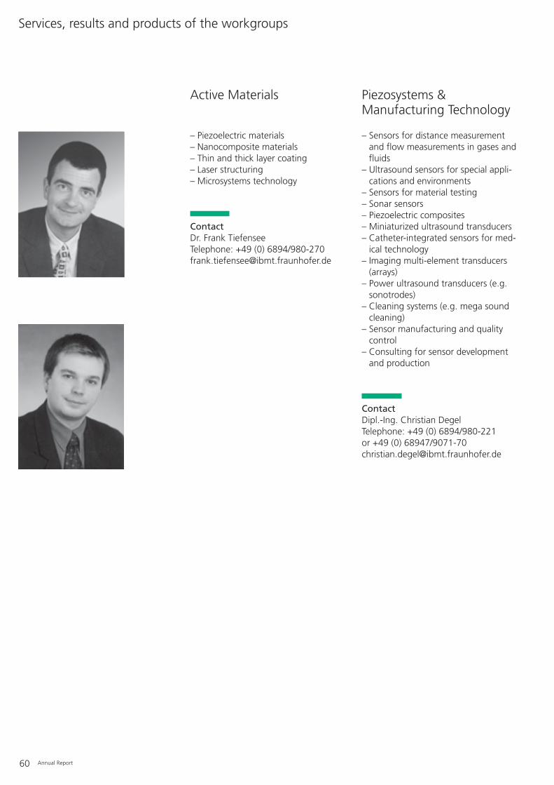
60 Annual Report
– Piezoelectric materials– Nanocomposite materials– Thin and thick layer coating– Laser structuring– Microsystems technology
ContactDr. Frank TiefenseeTelephone: +49 (0) 6894/[email protected]
– Sensors for distance measurementand flow measurements in gases andfluids
– Ultrasound sensors for special appli-cations and environments
– Sensors for material testing– Sonar sensors– Piezoelectric composites– Miniaturized ultrasound transducers– Catheter-integrated sensors for med-
ical technology– Imaging multi-element transducers
(arrays)– Power ultrasound transducers (e.g.
sonotrodes)– Cleaning systems (e.g. mega sound
cleaning)– Sensor manufacturing and quality
control– Consulting for sensor development
and production
ContactDipl.-Ing. Christian DegelTelephone: +49 (0) 6894/980-221 or +49 (0) 68947/[email protected]
Active Materials Piezosystems & Manufacturing Technology
Services, results and products of the workgroups

Annual Report 61
– Analogue & digital circuit develop-ment
– Ultrasound single- and multi-channelsystems
– Embedded systems– Ultrasound phased array systems– Portable systems– High-power sound systems– Flow, distance and level measure-
ment systems
ContactDipl.-Ing. Peter WeberTelephone: +49 (0) 6894/[email protected]
– Application-specific ultrasoundresearch and development
– Material characterization– Signal processing and parameter
extraction– Reconstruction and visualization– Navigation– Therapy monitoring– Microscopy– Manipulation systems– Hybrid imaging systems
ContactDr. Robert LemorTelephone: +49 (0) 6894/[email protected]
Biomedical UltrasoundResearch
Ultrasound Systems Development
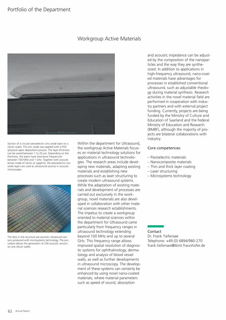
and acoustic impedance can be adjust-ed by the composition of the nanopar-ticles and the way they are synthe-sized. In addition to applications inhigh-frequency ultrasound, nano-coat-ed materials have advantages forprocesses in established conventionalultrasound, such as adjustable rheolo-gy during material synthesis. Researchactivities in the novel material field areperformed in cooperation with indus-try partners and with external projectfunding. Currently, projects are beingfunded by the Ministry of Culture andEducation of Saarland and the FederalMinistry of Education and Research(BMBF), although the majority of pro-jects are bilateral collaborations withindustry.
Core competences:
– Piezoelectric materials– Nanocomposite materials– Thin and thick layer coating– Laser structuring– Microsystems technology
ContactDr. Frank TiefenseeTelephone: +49 (0) 6894/[email protected]
62 Annual Report
Within the department for Ultrasound,the workgroup Active Materials focus-es on material technology solutions forapplications in ultrasound technolo-gies. The research areas include devel-oping new materials, adapting existingmaterials and establishing newprocesses such as laser structuring tocreate modern ultrasound systems.While the adaptation of existing mate-rials and development of processes arecarried out exclusively in the work-group, novel materials are also devel-oped in collaboration with other mate-rial sciences research establishments.The impetus to create a workgrouporiented to material sciences withinthe department for Ultrasound cameparticularly from frequency ranges inultrasound technology extendingbeyond 100 MHz and up to severalGHz. This frequency range allowsimproved spatial resolution of diagnos-tic systems for ophthalmology, derma-tology and analysis of blood vesselwalls, as well as further developmentsin ultrasound microscopy. The develop-ment of these systems can certainly beenhanced by using novel nano-coatedmaterials, where material parameterssuch as speed of sound, absorption
Workgroup Active Materials
Portfolio of the Department
Section of a circular piezoelectric zinc oxide layer on asilicon wafer. The zinc oxide was applied with a PVD(physical vapor deposition) process. The layer thicknesscan be varied between 1 to 20 µm. Depending on thethickness, the layers have resonance frequenciesbetween 100 MHz and 1 GHz. Together with acousticlenses made of silicon or sapphire, the piezoelectric zincoxide layers are used as ultrasound sources in acousticmicroscopes.
The dots in the structure are acoustic ultrasound sen-sors produced with microsystems technology. The pro-cedure allows the generation of 256 acoustic sensorson one silicon wafer.
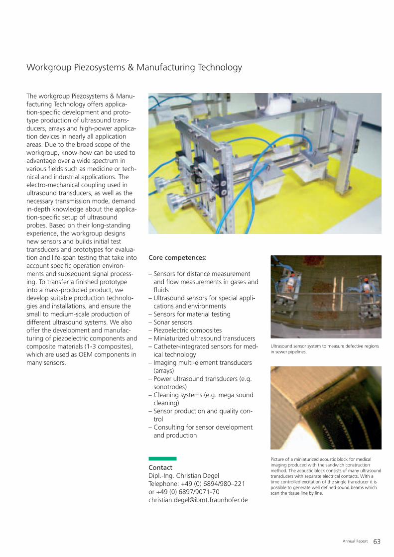
Annual Report 63
The workgroup Piezosystems & Manu-facturing Technology offers applica-tion-specific development and proto-type production of ultrasound trans-ducers, arrays and high-power applica-tion devices in nearly all applicationareas. Due to the broad scope of theworkgroup, know-how can be used toadvantage over a wide spectrum invarious fields such as medicine or tech-nical and industrial applications. Theelectro-mechanical coupling used inultrasound transducers, as well as thenecessary transmission mode, demandin-depth knowledge about the applica-tion-specific setup of ultrasoundprobes. Based on their long-standingexperience, the workgroup designsnew sensors and builds initial testtransducers and prototypes for evalua-tion and life-span testing that take intoaccount specific operation environ-ments and subsequent signal process-ing. To transfer a finished prototypeinto a mass-produced product, wedevelop suitable production technolo-gies and installations, and ensure thesmall to medium-scale production ofdifferent ultrasound systems. We alsooffer the development and manufac-turing of piezoelectric components andcomposite materials (1-3 composites),which are used as OEM components inmany sensors.
Core competences:
– Sensors for distance measurementand flow measurements in gases andfluids
– Ultrasound sensors for special appli-cations and environments
– Sensors for material testing– Sonar sensors– Piezoelectric composites– Miniaturized ultrasound transducers– Catheter-integrated sensors for med-
ical technology– Imaging multi-element transducers
(arrays)– Power ultrasound transducers (e.g.
sonotrodes)– Cleaning systems (e.g. mega sound
cleaning)– Sensor production and quality con-
trol– Consulting for sensor development
and production
ContactDipl.-Ing. Christian Degel Telephone: +49 (0) 6894/980–221 or +49 (0) 6897/[email protected]
Workgroup Piezosystems & Manufacturing Technology
Ultrasound sensor system to measure defective regionsin sewer pipelines.
Picture of a miniaturized acoustic block for medicalimaging produced with the sandwich constructionmethod. The acoustic block consists of many ultrasoundtransducers with separate electrical contacts. With atime controlled excitation of the single transducer it ispossible to generate well defined sound beams whichscan the tissue line by line.

therapy control and monitoring) andtherapy (high-power ultrasound) aswell as for industrial measuring tasks(e.g. flow meters, distance and levelmetering, quality control). In additionto developments in the “classical”medical diagnostic ultrasound area, weoffer developments in the kilohertz,megahertz and gigahertz range. Intelli-gent components (DSP, FPGA) are alsoused for “embedded systems”. Mea-suring tasks including complex signalprocessing can be done without acomputer. The experience of the work-group covers the whole spectrum fromthe concept-phase to the developmentof partial and complete technologicalsystem solutions for applying ultra-sound technology in medical andbiotechnological applications as well astechnical environments.
Core competences:
– Analogue & digital circuit develop-ment
– Ultrasound single- and multi-channelsystems
– Embedded systems– Ultrasound phased array systems– Portable systems– High-power sound systems– Flow, distance and level measure-
ment systems
ContactDipl.-Ing. Peter WeberTelephone: +49 (0) 6894/980–[email protected]
64 Annual Report
Workgroup Ultrasound Systems Development
Flow measurement device “deltaflow” offered by thecompany Systec Controls. The one-channel ultrasoundboard was developed in the workgroup Ultrasound Sys-tems Development. The device is an “embedded sys-tem”. The entire signal processing is performed by aDigital Signal Processor (DSP). The system parameterscan be adapted to various applications like sewageplants, power stations or pipelines by an appropriateDSP programming. The critical time control is done by aProgrammable Logic Device (PLD).
High-frequency ultrasound front-end for blood vessel diagnostics in young children.The ultrasonic probe is connected through a multiplexor board with the multi-channelultrasound system DiPhAS (Digital Phased Array System). The probe is a microsystemcomprising 288 single ultrasound elements. Each of them is connected to an inter-face board by a flex-tape technology cable. The multiplexor distributes the 288 ele-ments to the 64 electronic channels of the DiPhAS. The device is controlled by appli-cation-specific programmed FPGA (field programmable gate array).
The workgroup Ultrasound SystemsDevelopment is specialized on thedevelopment of single and multi-chan-nel ultrasound systems for medical andnon-medical applications. The grouphas a long-term experience and know-how in designing analogue and digitalhardware. The targeted evaluation ofinnovative approaches and a rapidproduct development is supplied byproven and continuously improvedtechnology platforms like the single-channel Transmit Receive Module(TRM) and the multi-channel imagingsystems Digital Phased Array System(DiPhAS). These platforms are the basisfor developing systems for medicaldiagnostics (e.g. imaging, navigation,

Annual Report 65
The workgroup Biomedical UltrasoundResearch focuses on novel applicationsof ultrasound technology in medicaldiagnostics and therapy as well as inbiological research and technology.
In the medical field this includes non-invasive data acquisition for diagnos-tics and the targeted destruction of tis-sue or release of medical drugs duringtherapy. In biology ultrasound can beused for non-destructive characteriza-tion of biological materials and livingorganisms and their targeted manipu-lation as well as an enabling technolo-gy for biotechnological processes. Inaddition to close collaboration with theother workgroups in the Departmentfor Ultrasound to generate and processapplication-specific signals and toreconstruct and visualize ultrasounddata for diagnostics and interventionalimaging (navigation, therapy control),our group pursues intensive researchon novel application areas for ultra-sound technology. High and very highfrequency systems facilitate new
research approaches and more precisediagnostic procedures. In particular,the use of acoustic microscopy in cellbiology research, as well as in systemsfor high resolution imaging of smallanimal models in preclinical research,enables non-invasive and inexpensivemorphological and anatomical investi-gations. In this area our workgroupconcentrates on combining ultrasoundtechnology with other imaging tech-nologies and modules in combinedand hybrid procedures for molecularimaging. Furthermore, the workgroupstudies the effect of ultrasound on bio-logical tissue and related applicationsin biotechnology.
Workgroup Biomedical Ultrasound Research
Figure left: Scan head of the acoustic microscopy system SASAM. Right: Fluorescence-optical and acoustic visualization of adult human stemcells. Due to its non-invasiveness acoustic microscopy is suitable for gentlecharacterization of stem cells and other microscopic objects. Acousticmicroscopy does not need light and can be performed on non-transparentsubstrates.
Core competences:
– Application-specific ultrasoundresearch and development
– Material characterization– Signal processing and parameter
extraction– Reconstruction and visualization– Navigation– Therapy control and monitoring– Microscopy– Manipulation systems– Hybrid imaging systems
ContactDr. Robert Lemor Telephone: +49 (0) 6894/980–[email protected]

66 Annual Report
Left: Prototype of the “SonoPilot®ortho”. Right: 3-D reconstruction of a femur with merged, automatically detected position and size of the femur head. The automatic detectionof anatomical landmarks provides a basis for using imaging in navigation systems.
Annual Report 67
– Photolithography, mask aligner– Sputtering devices, PCD, PECVD,
cleanroom– Sinter ovens– Precision dispensers– Polarizer– Full-parametric 3-D-CAD systems
(Pro/Engineer)– Component preparation: interior
hole diamond circular saw for directcutting of precision components,vacuum mixer for casting, lappingmachine
– Bonding/sensor technology: lateralmoving glue sandwicher, solderingand bonding technology
– Production installation for a low tomoderate number of ultrasound sen-sors
– CNC flat bed sanding machine (Ziersch & Baltrusch)
– Precision lapping and polishingmachines (Wolters)
– CNC universal milling machine(Mikron UM 600), work area: 600 x 500 x 450 mm
– CNC tool milling machine (Korradi UW 10 CNC), work area:500 x 300 x 400 mm
– CNC lathe center (Weiler DZ 32CNC), working diameter 100 mm,length 150 m, power driven tools
– CNC universal lathe (Rael Meka RT 5,cycle-controlled), transverse adjust-ment 200 mm, longitudinal adjust-ment 600 mm, driven tools
– Lathe (Colchester Master VS 3250),working diameter 1-300 mm, length650 mm
– CNC high precision separating andprofile sanding machine (Berney T38-4 CNC), working area: 160 x 220 x 120 mm, NC circulartable 360°, cut width min. ca. 20 µm
– CNC diamond circular saws (DiscoDAD 321)
– CNC micro drilling-milling-sandingmachine (Kern), working area: 220 x160 x 200 mm, movable NC circulartable with five axles
– CNC laser cutting and welding instal-lation (Haas), YAG laser with variablelens optics, cut width 60-200 µm,cutting of ceramics, metals, hollowbodies and metal plates, materialthickness 5 µm - 2 mm
– Conventional drilling-milling lathe(incl. circular sanding facilities)
– Fully automatic band saw, sawingarea: 200 x 200 mm, precision ± 0.1 mm
– Sand jet installation– Screw thread cutting machine– Motorized table scissors– Test station for static and dynamic
pressure tolerance– 5-basin ultrasound cleaning facilities– Plasma cleaning installation– Measuring equipment: pygnometer,
3-D sound field scanner, impedancemeasurement site
– Device for measuring contact angles– Scanning electron microscope– Scanning probe microscope (AFM,
STM, MFM)– Special measurement software for
development areas, impurity mea-surement station
– Laser interferometer measurements– Impedance measurement station– Insertion-loss measurement station– Climate chamber measurement
station– Temperature shock measurement
station– 3-axis measuring microscope incl.
image storage and processing– Cryostat measurements for sensor
characterization and zero-flow mea-surements
– Wave pressure scales– Sound field measurement station– DSP and microcontroller develop-
ment area (microchip, Motorola)– FPGA development area– Computer-aided development area
for electronic boards (ORCAD)– Fitting technology: SMD fine-pitch
fitting
– Electronics for bonding technology:micro-soldering station, surge solder-ing facilities, reflow soldering facili-ties
– SPS development station (Siemens S 6)
– Single and multi-channel ultrasoundsystems
– Phased array and linear array ultra-sound development systems
– Universal ultrasound measurementsfor industrial applications (concrete,steel, synthetic materials)
– 8-channel run-time difference mea-surement system for airborne soundapplications
– Airborne sound sensors (3-D surfacescanner, volume measurements andposition detectors)
– Doppler systems– Flow-through measurement technol-
ogy: laboratory measurement standsfor flow-through (Speckle Tracking,run-time difference; liquid: 7 m/s, DN 50/100/200; gas: variable up to30 m/s, DN 200)
– Zero-flow measurements– Development systems for industrial
imaging (setting, position, OCR, pat-tern matching)
– Ultrasound sensor systems for therapy control (minimally invasivesurgery, laser-induced thermo-therapy)
– Ultrasound navigation system devel-opment platform - SonoPilot®
– Opto-acoustic laboratory– Acoustic microscope systems SASAM– Biological laboratory, cell culture
Ultrasound
Equipment
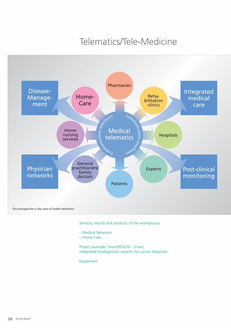
Physiciannetworks
Post-clinicalmonitoring
Disease-Manage-
ment
Integratedmedical
care
Homenursingservices
Pharmacies
Home-Care
Patients
Hospitals
Generalpractitioners/
familydoctors
Experts
Reha-bilitation
clinics
Medicaltelematics
Commun
icat
ion
inte
rfaces Comm
unicationinterfaces
68 Annual Report
The protagonists in the area of health telematics.
Telematics/Tele-Medicine
Services, results and products of the workgroups
– Medical Networks– Home Care
Project example: SmartHEALTH - Smart, integrated biodiagnostic systems for cancer diagnosis
Equipment

Annual Report 69
The term “eHealth” is currently linkedwith the possibility of reducing prob-lematic costs and increasing efficiencyin the health sector by using informa-tion technologies. This expectation isapparent in the efforts to introduce theelectronic health card and the corre-sponding required telematic infrastruc-ture throughout the whole of Ger-many. The potential of combiningtelecommunication and informatics(telematics) was already recognizedand used a decade ago in the Fraun-hofer IBMT when it created a separateresearch unit “Medical Telematics”.The medical telematics section of theIBMT delivers R&D services to industrialand public customers that cover abroad range, from studies to practicallytested, pre-product systems. Duringthe last two years this has led to theformation of the two different, yetoverlapping, activity areas of “MedicalNetworks” and “Home Care”, whichstarting this year are organized as sep-arate workgroups in the department“Telematics/Tele-Medicine”.
After a thorough, sector-wide analysisof the medical and organizationaloperations of healthcare providers, theworkgroup “Medical Networks” devel-oped the communication modelPaDok®. This model is gaining more
and more acceptance and new cus-tomers, which underpins the relevanceof the department’s work. As a com-munication model, PaDok® and thesubsequent product D2D are becom-ing increasingly integrated into every-day processes of the German publichealth system.
As the table (p. 70) listing registrationof physicians for the D2D networkwithin the Associations of CHI Physi-cians (Kassenärztliche Vereinigungen,KV) in Nordrhein and Baden-Württem-berg impressively shows, concomitant-ly with growing numbers of participat-ing doctors, D2D is having the effectof making platforms and informationexchange services more and moreinteresting and useful. Apart from theKV in Nordrhein and Baden-Württem-berg, the KV Bayern will also imple-ment D2D as an eHealth platform,which will again increase the numberof participants. Nearly half of Ger-many, in terms of insured persons andphysicians are now accessible via thecurrently installed D2D platform. Theproportion of D2D-supported primarysystems for outpatient medical carehas now reached far more than 50%.At the moment, hospital systems inparticular are increasing their D2D par-ticipation. They are especially interest-ed in electronic communication viaD2D with the professional association(Berufsgenossenschaft) and the so-called VHITG doctor’s record (Arztbrief)according to the guidelines of the newCDA rel. 2 (Clinical Document Archi-tecture) since D2D is the very first sys-tem adapted to this.
ContactDipl.-Phys. Bertram BresserTelephone: +49 (0) 6894/[email protected]
Dipl.-Inform. Stephan KieferTelephone: +49 (0) 6894/[email protected]

70 Annual Report
Furthermore, our current demographi-cal development urges us to developnovel IT-based concepts for domesticand outpatient care and medical pre-vention. The workgroup “Home Care”is very successfully developing suchadapted concepts and technologies.Comparable models also addressunder-developed rural regions withinsufficient access to basic medicaltreatment. For example, the projectT@lemed, a collaboration of Europeanand Latin American partners coordinat-ed by the IBMT, managed to success-fully establish tele-medical services inColombia based on our TOPCAREhome care and tele-medicine platform.Besides the system development forpersonal healthcare, the Home Caregroup also supports biomedicalresearch, for example by an informa-tion system for setting up an HIVcryosample bank at the IBMT.
The development of applicationstowards implementing new, mostly stillunfamiliar civil techniques such as elec-tronic documenting, electronic signa-tures and electronic representativeroles will ensure a broad range ofactivities for medical telematics at theIBMT in the years to come. The suc-cessful path of medical telematics isalso reflected in this year’s promotionof the workgroup to a department atthe IBMT and will be pursued in thesame way in the future. The IBMTworkgroups combine expertise ininformatics with know-how in settingup networks and knowledge aboutoperators/users. It is obvious thattelematic challenges cannot be solvedby software manufacturers alone;rather a complex understanding of allparticipants and their expectations isneeded.
474 565733
9631149
1302 1385 1450 1529 1577 16531767
213228
283
433
470
533567
593606
635666
685
0
500
1000
1500
2000
2500
Nov Dez Jan Feb Mrz Apr Mai Jun Jul Aug Sep Okt
KVBW KVNo
Number of registered physician in 2005.

Annual Report 71
Products:– PaDok® – Secure communication and
shared case-based patient record inthe public health system
Applied research and development:– Solutions for linking service suppliers
of the public health system– Electronic patient-linked documenta-
tion and shared case-based patientrecord
– Concepts for data protection anddata security in medicine
– Integration of general practice andhospital information systems, home-based stations and medical equip-ment into medical communicationnetworks
– Medical standards (DICOM 3.0, HL7,xDT, ICD10, XML, CDA, etc.)
– Electronic disease management
Service:– Linking-up of service suppliers of the
public health system with the healthtelematics solution PaDok®
– Data safety assessments
ContactDipl.-Phys. Bertram BresserTelephone: +49 (0) 6894/[email protected]
Products:– TOPCARE – a home care and tele-
medicine platform
Applied research and development:– Tele-medicine solutions for home and
portable healthcare for high-risk,elderly and disabled patients
– Tele-medicine solutions for underpro-vided rural regions and epidemiology
– Health-related prevention systems– Gerontological sensors– Smart, linked-up medical equipment
and intelligent environments– Medical standards (HL7, POCT1A,
ICD10, XML, CDISK, etc.)– eLearning environments for primary
care– Semantic integration of biomedical
databases– Integrated IT tools for clinical trials
and epidemiological studies– Information systems for biobanks
Service:– Pilot testing of new home care and
tele-medicine services on the basis ofthe IBMT TOPCARE platform
Contact Home CareDipl.-Inform. Stephan KieferTelephone: +49 (0) 6894/[email protected]
Home CareMedical Networks
Services, results and products of the department

72 Annual Report
Initial situation
In Germany over 400 000 people fall illwith cancer every year, approx. 360 000 of them for the first time.Although today many patients arecured permanently, about 42% of theaffected women and 54% of the malepatients do not survive the next fiveyears. Hence, it is of utmost impor-tance to detect tumors as early as pos-sible with screening procedures toincrease the chance of a permanentcure, or to reduce and control tumorgrowth by suitable long-term therapiesin not completely curable cases, inorder to maintain a patient’s lifetimeand quality of life. Long-term therapyof chronic cancers requires close andindividualized monitoring, if possibleoutside a hospital. Likewise, successfulearly diagnosis needs reliable and pow-erful biodiagnostic systems at the so-called point of care. New molecularand protein-based cancer markers,progress in microsystems technologyand nanobiotechnology linked withinformation and communication tech-nologies create the basis for develop-ing a new generation of intelligent,integrated, biodiagnostic systems forthe diagnosis and monitoring of cancerdiseases. These all fall within the
framework of the EU-funded integrat-ed Research & Development projectSmartHEALTH (project number FP6-2004-IST-NMP-2-016817).
Task
Prevention, early diagnosis, and target-ed and effective therapies form thecornerstones of an efficient health sys-tem. Therefore, new diagnostic cancertests have to deliver precise and reli-able results for therapy decisions andbe optimally integrated into thehealthcare process to avoid unneces-sary treatment and stress for thepatients. SmartHEALTH addresses thesechallenges and is developing a newgeneration of intelligent, biodiagnosticsystems that allow, or support andcomplement optimized disease man-agement and screening programs inhealth services.
Driven by three key applications in can-cer diagnosis (breast, intestinal andcervical cancers) the project is develop-ing intelligent, linked, prototypicaldiagnosis systems for multi-parametriccancer marker analysis for the point ofcare.
The objectives of SmartHEALTHinclude:– Implementation of new sensor sys-
tems for biomarker analysis integrat-ed into future healthcare services toimprove current healthcare concepts.
– Clinical validation of the systems fortarget applications (breast, intestinaland cervical cancers).
– Demonstrating the usefulness of“ambient intelligence” technologiesin medical diagnostic systems andcoupled online services for “perva-sive” healthcare.
– Development of new productiontechnologies to achieve uniform sen-sor solutions that integrate microflu-id components, transducers and bio-logical assays.
Solution
Driven by clinical oncological applica-tions, micro- and nanobiotechnologyand information and communicationtechnologies, the Integrated ProjectSmartHEALTH is developing an open,integrated system platform for novelbiodiagnostic devices to support theindustry in exploiting bioassays andnew application concepts for cancerdiagnosis. The initial system comprisesa disposable microfluidics chip. Thechip is loaded with the biosample andconnected to a desktop reader device,which integrates dynamically into thesurrounding e-health infrastructureand allows wireless communicationwith other instruments.
Subsequently, this system concept willbe cost-optimized and miniaturized,eventually leading to various portableand easily accessible products. The sys-tem platform will allow simultaneousmeasurements and analyses of severalanalytes based on nucleic acids andproteins and can process differenttypes of biological samples. The resultswill be interpreted using artificial neu-
Home Care
Project example: SmartHEALTH – Smart, integrated, biodiagnostic systems for cancer diagnosis
Objectives of the SmartHEALTH project: flexible, intelligent and communicating biodiagnostic equipment for cancerdiagnostics.
Annual Report 73
ronal networks and further analysistools. The adaptive systems will knowtheir user, the patient and the currentcontext. Based on medical standardsthey will maintain wireless communica-tion with patient records at the respec-tive laboratory, hospital or online infor-mation system, strictly complying withdata protection regulations. In addi-tion, they will support public key infra-structures.
The project was initiated in December2005 with 25 European R&D partnersand promises to improve medical can-cer diagnosis by earlier and more pre-cise cancer marker analysis. This willimprove the quality of life of thepatients and strengthen the competi-tiveness of the European industry inthe area of in vitro diagnostics. Thetasks for the IBMT include the integra-tion of microfluidics and sensor com-ponents (workgroup Miniaturized Sys-tems), and particularly pioneering thedevelopment of software that inte-grates the system with technologiessuch as “Ambient Intelligence”, “Ubi-quitous Computing” and the “SemanticWeb” with the goal of making bio-diagnostic devices more intelligent sothey can be easily integrated into thesurrounding IT infrastructure.
Results of the IBMT
The IBMT department Telematics/Tele-Medicine has developed a promisingsoftware architecture, the so-called“Semantic Medical Device Space(SMDS)“, to integrate system intelli-gence into biodiagnostic devices sothat they adapt to changes in theirenvironment, dynamically recognizeand use offered services and/or supplyservices to their surroundings, andauto-adapt to communicate with exist-ing medical information systems. TheSMDS is a pervasive software conceptthat uses semantic web and web ser-vice technologies to equip medicaldevices with intelligence and commu-nication abilities, allowing semantic
interoperability with other informationsystems and devices. To take intoaccount data privacy issues and to con-sider adequate data safety measures inthe SMDS concept, we performed asafety analysis according to the Com-mon Criteria Standard, derived a so-called protection profile for intelligentbiodiagnostic medical devices andincorporated this into the SMDS.
In an initial step, the SMDS conceptwill be integrated into the initial Smart-HEALTH instrument and evaluated byusers. For this, communication stan-dards such as HL7 V2.3 and POCT1Awill be implemented on the basis ofweb service technologies. In addition,the department is developing aSmartHEALTH information system thatis meant to support the clinical valida-tion of cancer markers and offers newonline services for the interpretation ofanalysis results.
Potential
The “Semantic Medical Device Space(SMDS)” concept is an exemplary mod-el for the design of intelligent andinteroperable medical diagnosticdevices. Reusable software compo-nents will allow the transfer to, andadaptation of medical devices of themedical technology industry. The firstdemonstration setups are expected tobe completed in 2007.
ContactDipl.-Inform. Stephan KieferTelephone: +49 (0) 6894/[email protected]
– Hardware platforms, in addition tostandard PCs, particularly HP, SUN,DELL and SGI computers (desktopand server computers)
– Video conferencing systems of differ-ent bandwidth and quality for vari-ous application areas
– Devices for the monitoring of vitalparameters, also online
– Communication systems for continu-ous wireless monitoring of patients
– Software tools for the elaboration ofpresentations, also online
– Software development tools forInternet and database applications(Oracle, SQL servers)
– Software development tools forembedded platforms
Telematics/Tele-Medicine
Equipment

74 Jahresbericht 2006
Structure and application of a thread electrode (tf-LIFE) at a peripheral nerve to detect electricalpotentials and stimulate single nerve fibers.
Medical Engineering & Neuroprosthetics
Services, results and products of the workgroups
– Neuromonitoring– Neuroprosthetics
Project example: Highly flexible, textile-integrable electrode material to record ECGs within a 24/7 monitoring scheme
Equipment
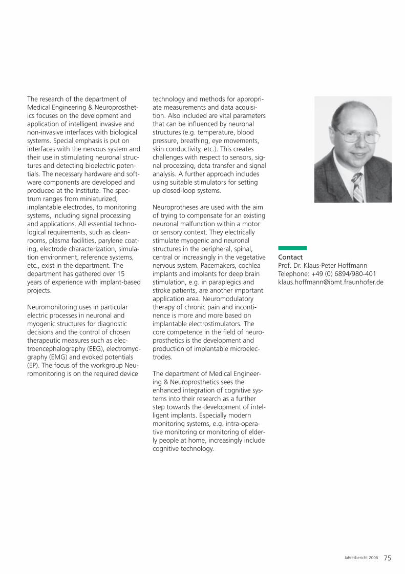
Jahresbericht 2006 75
The research of the department ofMedical Engineering & Neuroprosthet-ics focuses on the development andapplication of intelligent invasive andnon-invasive interfaces with biologicalsystems. Special emphasis is put oninterfaces with the nervous system andtheir use in stimulating neuronal struc-tures and detecting bioelectric poten-tials. The necessary hardware and soft-ware components are developed andproduced at the Institute. The spec-trum ranges from miniaturized,implantable electrodes, to monitoringsystems, including signal processingand applications. All essential techno-logical requirements, such as clean-rooms, plasma facilities, parylene coat-ing, electrode characterization, simula-tion environment, reference systems,etc., exist in the department. Thedepartment has gathered over 15years of experience with implant-basedprojects.
Neuromonitoring uses in particularelectric processes in neuronal andmyogenic structures for diagnosticdecisions and the control of chosentherapeutic measures such as elec-troencephalography (EEG), electromyo-graphy (EMG) and evoked potentials(EP). The focus of the workgroup Neu-romonitoring is on the required device
technology and methods for appropri-ate measurements and data acquisi-tion. Also included are vital parametersthat can be influenced by neuronalstructures (e.g. temperature, bloodpressure, breathing, eye movements,skin conductivity, etc.). This createschallenges with respect to sensors, sig-nal processing, data transfer and signalanalysis. A further approach includesusing suitable stimulators for settingup closed-loop systems.
Neuroprotheses are used with the aimof trying to compensate for an existingneuronal malfunction within a motoror sensory context. They electricallystimulate myogenic and neuronalstructures in the peripheral, spinal,central or increasingly in the vegetativenervous system. Pacemakers, cochleaimplants and implants for deep brainstimulation, e.g. in paraplegics andstroke patients, are another importantapplication area. Neuromodulatorytherapy of chronic pain and inconti-nence is more and more based onimplantable electrostimulators. Thecore competence in the field of neuro-prosthetics is the development andproduction of implantable microelec-trodes.
The department of Medical Engineer-ing & Neuroprosthetics sees theenhanced integration of cognitive sys-tems into their research as a furtherstep towards the development of intel-ligent implants. Especially modernmonitoring systems, e.g. intra-opera-tive monitoring or monitoring of elder-ly people at home, increasingly includecognitive technology.
ContactProf. Dr. Klaus-Peter HoffmannTelephone: +49 (0) 6894/[email protected]

76 Jahresbericht 2006
– Deduction of nerve and muscle signals
– Examination of implant materialsunder physiological conditions andaccelerated aging
– Development of biotelemetrics forthe control of implants
– Development of stimulation patternsfor bladder stimulation
– Development of deduction systemsfor examining intestine mobility
– Microelectrode characterization– Design of cuff electrodes– Design of epimysial electrodes– Development of external electrostim-
ulators– Studies on the functional electrostim-
ulation of peripheral nerves– Parametric characterization of stimu-
lation and deduction systems for prehensile prostheses
– Development of implantable stimula-tors
– Implant technology for differentapplication areas
– Encapsulation methods for microim-plants
– Examination methods for encapsula-tion materials
– Mask design for 2-D and 3-D micro-electrodes
– Production of microelectrodes– Production of microimplants with
integrated electronics– Neuromodulation for selective stimu-
lation of nerves– Development of neuroprostheses– Encapsulation with parylene– Microsystems on polyimide basis
– Retina stimulators– Design of sieve electrodes with track-
ing system– Production of silicon implants for
neuroprosthetics– Development of electrodes for stand-
walk prostheses– Microelectrodes with SU-8 pattern-
ing– Investigating novel organic electrode
materials– Preparation and supervision of clini-
cal trials– Technical assistance for implantations
and experiments– Development and characterization of
surface electrodes– Investigating material characteristics
of surface electrodes– Investigating long-term reactivity of
surface electrodes
Contact NeuromonitoringProf. Dr. Klaus-Peter HoffmannTelephone: +49 (0) 6894/[email protected]
Contact NeuroprostheticsDr. Klaus Peter KochTelephone: +49 (0) 6894/[email protected]
Neuroprosthetics & Neuromonitoring
Services, results and products of the workgroups
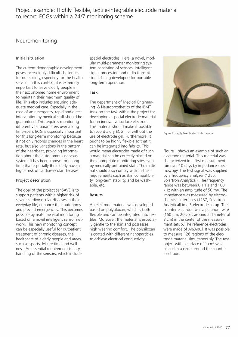
Jahresbericht 2006 77
Initial situation
The current demographic developmentposes increasingly difficult challengesfor our society, especially for the healthservice. In this context, it is extremelyimportant to leave elderly people intheir accustomed home environmentto maintain their maximum quality oflife. This also includes ensuring ade-quate medical care. Especially in thecase of an emergency, rapid and directintervention by medical staff should beguaranteed. This requires monitoringdifferent vital parameters over a longtime-span. ECG is especially importantfor this long-term monitoring becauseit not only records changes in the heartrate, but also variations in the patternof the heartbeat, providing informa-tion about the autonomous nervoussystem. It has been known for a longtime that especially the elderly have ahigher risk of cardiovascular diseases.
Project description
The goal of the project senSAVE is tosupport patients with a higher risk ofsevere cardiovascular diseases in theireveryday life, enhance their autonomyand prevent emergencies. This becomespossible by real-time vital monitoringbased on a novel intelligent sensor net-work. This new monitoring conceptcan be especially useful for outpatienttreatment of chronic diseases, thehealthcare of elderly people and areassuch as sports, leisure time and well-ness. An essential requirement is easyhandling of the sensors, which include
special electrodes. Here, a novel, mod-ular multi-parameter monitoring sys-tem consisting of sensors, intelligentsignal processing and radio transmis-sion is being developed for portablelong-term operation.
Task
The department of Medical Engineer-ing & Neuroprosthetics of the IBMTtook on the task within the project fordeveloping a special electrode materialfor an innovative surface electrode.This material should make it possibleto record a dry ECG, i.e. without theuse of electrode gel. Furthermore, itought to be highly flexible so that itcan be integrated into fabrics. Thiswould mean electrodes made of sucha material can be correctly placed onthe appropriate monitoring sites evenby medically untrained staff. The mate-rial should also comply with furtherrequirements such as skin compatibili-ty, long-term stability, and be wash-able, etc.
Results
An electrode material was developedbased on polysiloxan, which is bothflexible and can be integrated into tex-tiles. Moreover, the material is especial-ly gentle to the skin and possesseshigh wearing comfort. The polysiloxanis coated with different nanoparticlesto achieve electrical conductivity.
Figure 1 shows an example of such anelectrode material. This material wascharacterized in a first measurementrun over 10 days by impedance spec-troscopy. The test signal was suppliedby a frequency analyzer (1255,Solartron Analytical). The frequencyrange was between 0.1 Hz and 100kHz with an amplitude of 50 mV. Theimpedance was measured by electro-chemical interfaces (1287, SolartronAnalytical) in a 3-electrode setup. Thecounter electrode was a platinum wire(150 µm, 20 coils around a diameter of3 cm) in the center of the measure-ment setup. The reference electrodeswere made of Ag/AgCl. It was possibleto measure 128 regions of the elec-trode material simultaneously. The testobject with a surface of 1 cm2 wasplaced in a circle around the counterelectrode.
Neuromonitoring
Project example: Highly flexible, textile-integrable electrode material to record ECGs within a 24/7 monitoring scheme
Figure 1: Highly flexible electrode material.
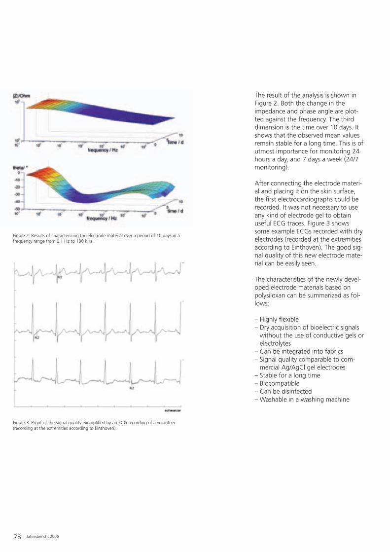
The result of the analysis is shown inFigure 2. Both the change in theimpedance and phase angle are plot-ted against the frequency. The thirddimension is the time over 10 days. Itshows that the observed mean valuesremain stable for a long time. This is ofutmost importance for monitoring 24hours a day, and 7 days a week (24/7monitoring).
After connecting the electrode materi-al and placing it on the skin surface,the first electrocardiographs could berecorded. It was not necessary to useany kind of electrode gel to obtainuseful ECG traces. Figure 3 showssome example ECGs recorded with dryelectrodes (recorded at the extremitiesaccording to Einthoven). The good sig-nal quality of this new electrode mate-rial can be easily seen.
The characteristics of the newly devel-oped electrode materials based onpolysiloxan can be summarized as fol-lows:
– Highly flexible– Dry acquisition of bioelectric signals
without the use of conductive gels orelectrolytes
– Can be integrated into fabrics– Signal quality comparable to com-
mercial Ag/AgCl gel electrodes– Stable for a long time– Biocompatible– Can be disinfected– Washable in a washing machine
78 Jahresbericht 2006
Figure 3: Proof of the signal quality exemplified by an ECG recording of a volunteer(recording at the extremities according to Einthoven).
Figure 2: Results of characterizing the electrode material over a period of 10 days in afrequency range from 0.1 Hz to 100 kHz.
Jahresbericht 2006 79
Project funding
Fraunhofer-GesellschaftMAVO project INMUSENS
ContactProf. Dr. Klaus-Peter HoffmannTel.: +49 (0) 6894/[email protected]
Project consortium
INMUSENS (Intelligent multimodal sen-sors):– Institute for Occupational Economy
and Organization, IAO, Stuttgart– IBMT, St. Ingbert– Institute for Integrated Circuits, IIS,
Erlangen– Institute for Photon Microsystems,
IPMS, Dresden– Institute for Applied Information
Technology, FIT, St. Augustin
– Production of implants– Characterization of electrodes– Laboratory for measurements– Research laboratory for visual
systems– Laboratory for methods of clinical
neurophysiology– Software laboratory– Simulation– Development tools for flexible sub-
strates with integrated electrodes forneuro-implants (CAD: LASI, electro-mechanical simulation: FlexPDE)
– Access to cleanroom facilities forproduction and assembly of neuro-implants with a minimum size ofabout 5 µm (lithography, metal coat-ing, reactive ion etching, polyimideoven, parylene C coating, bonder)
– Laboratory for assembly (gluing, sol-dering, welding) and encapsulation(parylene, silicon) of electrodes, wiresand implants; production of molds
– PC-controlled measurement work-place for characterization of elec-trodes: impedance, transient electricpulses, cyclic voltametry (HP 3245 A,HP 3458 A, EG&G 5302); scanner formeasuring the distribution of theelectric potential in physiologicalmedia; stability under mechanicalstress
– PC-controlled measurement work-place for examination of field distri-butions of microelectrodes
– PC-controlled measurement work-place for electric impedance spec-troscopy (Solartron 1255B/1287)
– PC-controlled measurement work-place for measuring organic semi-conductors
– PC-controlled measurement work-place for characterizing the conduc-tivity of insulation layers for leakagecurrents as low as in the sub-picoam-pere range in physiological media atroom temperature and acceleratedaging (Keithley 617 E electrometer)
– Tools for developing analog and digi-tal circuits and systems for physiolog-ical measurement technology andelectrostimulation, as well as testenvironments for characterizingminiaturized (neuro)implants(OrCAD, Visual C++,LabWindows/CVI, logic analyzerPhilips PM 3585, emulation systemsfor 80C31, PIC and 8051 family, PICand EPROM programmer, digitaloscilloscope HP 54504-400 MHz)
– PC-controlled measurement work-place for examining noise in elec-tronic circuits and systems as well asin electrodes in physiological media(FFT Servo Analyzer Advantest R 924C, Spectrum Analyzer Advantest R 3361 C, multimeter Keithley 199,function generators)
– Facilities for non-invasive measure-ments of prehensile force andmomentum at the lower extremity
– Multi-channel stimulator with arbi-trary pulse forms (constantwattage/voltage) for electrostimula-tion and multi-channel recording sys-tems for electrophysiological mea-surements
– Pneumatic stimulator for studyingsensory nervous signals
Medical Engineering & Neuroprosthetics
Equipment

Cryobiophysics & CryotechnologyCryoresearch and -demonstration Bank
80 Annual Report
Services, results and products of the workgroups
– Cryoequipment & Cryorobotics– BMBF Junior Research Group Cryonanobiotechnology– Cryoresearch and -demonstration Bank
Project example: Development of tumor models for the cryobanks of personalized medicine
Equipment
Cryoelectronics in liquid nitrogen are being tested in thecryoresearch bank in Sulzbach (Photo: Bernd Müller).

Annual Report 81
Until now the young “science ofextremely cold life” (a literal translationof the Greek terms used in the word“cryobiology”) has managed to suc-cessfully conserve cells primarily byempirical methods. One reason for thisis that the causes of damage to cellsduring freezing, storage at the lowtemperatures of liquid nitrogen (usual-ly below -150°C) and thawing arehighly complex. The empirical discov-ery of suitable freezing and thawingrates and tolerable cryoprotectives isjustified if a sufficient proportion of acell population “survives”, as it is usu-ally the case for most cell cultures.However, substituting the empiricalapproach in applied cryobiology by amore systematic procedure is of funda-mental importance for successful appli-cation in those areas where the singlecell and its state are increasingly criti-cal, e.g. in stem cell research, therapywith “programmed” cells, regenerativemedicine and also for the sustainableuse of bioresources by conservation ofviable cell samples. Systematic andknowledge-based optimization of acryopreservation process, i.e. a cycle offreezing, storage and thawing, requiressuitable tools. The development ofsuch tools is the task of the depart-ment of Cryobiophysics & Cryotechnol-ogy with its workgroups Cryoequip-ment & Cryorobotics and the BMBFjunior research group Cryonanobio-technology. For instance, the depart-ment devises miniaturized cryosub-strates made of different materials andvarying in size, optimized freezing andthawing machines, manipulation sys-tems for sterile access to cold samplesthat are suitable for use in industry,and last but not least nanotechnologi-cally optimized surfaces for surface-
based cryopreservation (see figure).These developments form the basis ofa future tool box for systematic cryo-biophysics. Professor Heiko Zimmer-mann and his workgroups were grant-ed approval No. 18 by the Robert KochInstitute (Berlin) to import humanembryonic stem cells (isolated beforethe stipulated date of 01.01.2002).The freezing and thawing protocols forthese cells should be optimized andstandardized and compared to thoseof other cells.
Contact Cryobiophysics & CryotechnologyProf. Dr. Heiko ZimmermannTelephone: +49 (0) 6894/[email protected]
Contact Cryoresearch and -demonstration BankDr. Frank ObergrießerTelephone: +49 (0) 6897/[email protected]

– Storage of biological material forresearch purposes
– Testing of customer-specific cryo-equipment (substrates, heating/cool-ing tables, microscopes, etc.)
– Testing of cryoprocedures– Prototypes of cryobanks– Testing of cryobank concepts– Development and validation of cryo-
databases– Consulting on the setup of customer-
owned cryobanks with specific soft-ware solutions
ContactDr. Frank ObergrießerTelephone: +49 (0) 6897/[email protected]
Cryoresearch & -demonstration Bank
82 Jahresbericht 2006
– Research and development in thearea of low temperature biophysicsand cryobiotechnology
– Development of cryoequipment (substrates, heating/cooling tables,microscopes, etc.)
– Development of freezing proceduresfor single cells, cell groups and tis-sues
– Development of workplaces for mea-suring parameters of low tempera-ture electronics
– Low temperature tolerant and opti-mized digital storage systems
– Database concepts for sample banksat industrial scales
– Research and development in thearea of chip-based, adaptive labora-tory and workflow management(“ChameleonLab” technology)
– Dynamic infrared thermography– Research and development in the
area of microsystem-based cryo-preservation
ContactProf. Dr. Heiko ZimmermannTelephone: +49 (0) 6894/[email protected]
– Development of cryoequipment (substrates, heating/cooling tables,microscopes, etc.)
– Development of automation con-cepts for cryobanks and cryocontain-ers
– Special-purpose production of cryo-infrastructural components (e.g.“intelligent” transport containers,installations for sample security)
– Tool design for cryobiotechnology– Low temperature imaging (special
video solutions), low temperaturesensors
– Research and development in thearea of cryorobotics
– Special-purpose developments forlow temperature measurements andprocess control
– Development and production of lab-oratory containers
ContactDipl.-Phys. Uwe SchönTelephone: +49 (0) 6897/[email protected]
BMBF Junior ResearchGroup “Cryonanobiotech-nology”
– Research on surface-based freezingof cells
– Research in the area of nanostruc-ture-aided cryopreservation
– Development of novel nanostructur-ing methods
– Research on hydrogel micro-encapsu-lation (2-D/3-D) and cell program-ming for cryopreservation
ContactProf. Dr. Heiko ZimmermannTelephone: +49 (0) 6894/[email protected]
Cryobiophysics & Cryotechnology
Cryoequipment & Cryorobotics
Services, results and products of the workgroups

Annual Report 83
Initial situation
The increasing application of cell-basedtherapies in medicine and the storageof samples for later, not yet feasable,analysis is leading to a high-through-put deposition of samples in cryo-banks. High-quality sample banks ofviable cells are particularly necessaryfor the detection of molecular patternsand the ensuing search for personal-ized therapeutic approaches. This iswhy industry and research from areassuch as biotechnology, medicine andpharmacy are storing valuable cells andmicroorganisms in biobanks, e.g. celllines, genetically transformed or modi-fied cells, medical samples (such astumors, blood samples, biopsy sam-ples), as well as primary material forregenerative medicine (e.g. hemato-poietic stem cells from the umbilicalcord, adult and embryonic stem cells)and for reproductive medicine (e.g.sperm, oocytes, embryos, ovary tissue).The differences in cell types and con-figurations (e.g. suspension cultures,multi-cellular systems, tissue regions,adherent cells) with respect to theirbiological and biophysical propertiesrequire specific cryopreservation proto-cols. Especially for multi-cellular sys-tems, i.e. associations of hundreds toabout 10 000 single cells, that engagein specific interactions, for example intumors and tissues, no successful cryo-preservation methods are as yetknown.
Approach towards a solution
A modular construction system consist-ing of cell spheroids and biocompatiblematerials will be developed at theIBMT to help better understand theprocesses during cryopreservation ofmulti-cellular systems and to optimizethe protocols. The goal is to adapt themodel system to primary systems, i.e.actual samples from patients, so that asystematic search for optimal cryo-preservation protocols can be per-formed. This will involve combiningestablished spheroid culture tech-niques with novel biomaterials andprocessing procedures to develop amodel that suits the initial sample. Par-allel experiments with primary materi-al, e.g. human tumors, will be carriedout to determine the agreementbetween the model and the original. Anovel nanoplotter will be used for cryo-preservation, which allows the exactaddition of DMSO (currently the mostcommon cryoprotective) in the picoliter
Computer-generated model of a cover-encapsulated system consisting of a multi-cellular spheroid covered by ahydrogel matrix that contains single cells (Illustration: H. Görgen, IBMT).
Nanoplotter (GeSIM mbH) during loading of a cryosub-strate with a capacity of 30 x 25 µl. The addition of cryoprotectives such as glycerol or DMSO can be precisely controlled in the picoliter range.
Cryobiophysics & Cryotechnology
Project example: Development of tumor models for the cryobanks of personalized medicine

84 Annual Report
range. This application method hasalready significantly improved the cryo-preservation of Islets of Langerhanscells. Miniaturization of the cryovesselsis also believed to have a positiveeffect: in contrast to traditional 1 mltubes, HDPE plates with 30 x 25 µlwells are used. Since DMSO disruptscellular metabolism at physiologicaltemperatures, and must be washedaway as rapidly as possible after thaw-ing, alternative cryoprotectives arebeing investigated following the exam-ple of extremophilic organisms. Tre-halose, starch hydrolysates and starchderivatives are some examples of pro-tein-free alternative cryoprotectives.Besides maintaining viability and func-tionality after thawing, conserving theintegrity of multi-cellular systems is dif-ficult. Hydrogel capsules made of algi-nate offer a possible solution to thisproblem. They are already used for theimmuno-isolated transplantation of
Islets of Langerhans or parathyroidgland tissue, for example.
Results
Despite being the same cell type, opti-mal cryopreservation conditions formulti-cellular systems differ from thosefor the corresponding cell suspension(see Figure 3). These optimal condi-tions can only be determined by exten-sive screening efforts. Model systemsshould help to reduce the use of pri-mary material by first conducting pre-liminary studies with the model systemand then using the best protocols withthe primary system. The cryopreserva-tion of human mammal gland or ovarycarcinomas has already been improvedin this manner.
In tissues, cells are often not directlylinked to each other but adhere to athree-dimensional matrix. The dis-tances between the cells can be simu-lated by incorporating the cells into analginate matrix. It is also possible touse different combinations of variouscell types, cultivation modes (singlecells, spheroids, see Figure 4, left) aswell as the addition of accessory com-ponents such as erythrocytes forenhanced oxygen supply (Figure 4,center and right), HSA for osmotic sta-bilization, collagen fibers or SiO2 beadsfor mechanical consolidation, etc.However, the alginate encapsulationmethod not only possesses potentialfor the model system research, it alsoincreases the success rate of cryo-preservation. Alginate-encapsulatedL929 spheroids have a 5-10% higherviability after thawing, and hemolysisof encapsulated erythrocytes isreduced by up to 90%.
Potential
The Fraunhofer IBMT has the facilitiesto establish model systems for diversetypes of tissue. The different combina-tion possibilities of cell systems (singlecells, multi-cellular systems and mono-layers), capsule characteristics (shape,size, mechanical stability and cell adhe-siveness) and accessory ingredients(erythrocytes, SiO2, collagen) allowspecific adaptation to different primarysystems. This drastically reduces theuse of primary material. Improvedcryopreservation protocols have beenestablished for human tumors.
ContactDipl.-Biol. Friederike EhrhartTelephone: +49 (0) 6894/[email protected]
Figure 3: Screening experiment: viability of L929 cells (murine fibroblasts) in suspension and asmulti-cellular spheroids after cryopreservation in different cryomedia. (F: before freezing; D, C, L,CT, LT, CS, LS: various cryomedia).
Figure 4: Different possibilities for encapsulation (from left to right): L929 spheroid, human erythrocytes, L929 spheroids (green) and human erythrocytes in a capsule.

Annual Report 85
– Low temperature storage systems(down to -196°C) with approval formedical use
– Modified, programmable freezingmachines for applications in biology,material sciences and electronics
– Cell biology laboratory– Modified research microscopes– Inverted cryomicroscope (own devel-
opment, Peltier-based)– Combined reflection/scanning force
microscope for measurements of bio-logical objects in aqueous environ-ments
– Testing equipment (digital/analog) forlow temperature electronics
– Low temperature measuring cham-ber for testing electronics and mate-rials
– Thermography system (temperaturemeasurement range -20°C to 250°C)
– Micropipetting system/automationplatform
– “ChameleonLab”-based laboratorymanagement
– High-speed camera system for micro-drop-based freezing
– Computer-controlled freezingmachines (own developments)
– Cryotank sample handling systems– Sample handling sluice systems– Cold gas devices– Cryotransport containers (own devel-
opments)– 20-channel cryotemperature measur-
ing systems– Cryorobot for sample handling– LN2 gauge ultrasound measuring sys-
tems
BMBF Junior ResearchGroup “Cryonanobiotech-nology”
– Micro-encapsulation installation(crystal gun principle)
– Freezing spin coater for freezing ofultra-thin layers (own development)
– Infrared laser system for precise heat-ing of thin layers (planned)
First sub-sections of the European Cen-ter for Cryobiotechnology are alreadyin operation.
– Low temperature storage systems (-130 to -196°C) with approval formedical use
– Programmable freezing machines– Cell biological laboratory– Cell culture microscope for bright
field, phase contrast, variable reliefcontrast and fluorescence
– High security containers– Ultra-cold freezer with CO2 emer-
gency cooling– File server with RAID system– Testing and development server– Storage tank for 25 000 liters liquid
nitrogen– Sterile workbench– CO2 incubator– Nanoplotter– Emergency power generator 15 kVA– Database server with RAID systems
and LTO tape drive– Oxygen depletion monitoring– Burglar alarm
Cryobiophysics & Cryotechnology
Cryoequipment & Cryorobotics
Cryoresearch & -demonstration Bank
Equipment

86 Annual Report
Biohybrid Systems
Services, results and products of the workgroups
– Cell-based Sensors & Biomonitoring– Molecular Cell & Tissue Engineering
Project example: Technology platform for accelerated HIV vaccine development
Equipment
Interaction between nanoparticles and biological cells.

Annual Report 87
Ever increasing novel therapeuticapproaches for treating many diseasesare emerging from research in biophar-macy, regenerative medicine andnanobiotechnology. However, howquickly new therapeutic approachescan be introduced into broad clinicaluse depends greatly on cell technologyrequirements. There is a growing needto characterize, transfer and processdiminutive biological samples. Suitabletechnologies have been developedduring the last 10 years in the depart-ment of Biohybrid Systems at theFraunhofer IBMT. To achieve highacceptance among customers, thedeveloped technologies for the eco-nomically most promising applicationareas are currently being evaluated.Within the framework of internationaland national research projects thisevaluation is taking place in coopera-tion with research groups and compa-nies that specialize in particular appli-cations. The following current projectsare examples of applications that drawon the developed technologies: Theclinical use of cell therapies requiresstable, safe and well-characterized cellmaterial. The objective of the EU pro-ject OsteoCord is optimizing the isola-tion and expansion of mesenchymalstem cells from blood of the umbilicalcord. The differentiation potential ofthe stem cells is investigated withrespect to their capacity to form bonecells. The EU project CARDIOWORK-BENCH endeavors to optimize thesearch for suitable drugs for treatingcardiovascular diseases and resultingheart disorders. Another EU project,PolExGene, focuses on improving thebiocompatibility of non-viral genetransfer systems for cell therapyapproaches to treat retinal and cardio-vascular diseases. Within the BMBFnetwork project NanoDrug, the
department carries out preclinical testson nanoparticle pharmaceutical sub-stances for tumor therapy. The biologi-cal effects of nanoparticles are also rel-evant for environmental toxicology. Asa partner within the EU project DIPNA,the department is involved in thedevelopment of a platform to elucidatethe toxicological and ecotoxicologicaleffects of nanoparticles. Once again,the technological basis determineshow rapidly novel vaccines can bedeveloped. An international consor-tium, coordinated by the IBMT, wasawarded a grant from the Bill & Melin-da Gates Foundation to develop tech-nologies to accelerate the develop-ment of an HIV vaccine. Dr. Hagen Thielecke obtained approval No. 19from the Robert Koch Institute toimport human embryonic stem cells inthe context of the OsteoCord project.
ContactDr. Hagen ThieleckeTelephone: +49 (0) 6894/[email protected]
Assistant professor Dr. Hagen von BriesenTelephone: +49 (0) 6894/[email protected]

88 Annual Report
– Cell and tissue-based biosensors forfunctional efficacy testing and formedical diagnostics in the areas ofoncology, neurology and cardiology
– Electrochemical microsensors andmethods for the functional, label-free testing of drugs, for in vivomonitoring and for bioprocess tech-nologies
– Bioimpedance spectroscopy (in vitroand in vivo)
– Biointerfaces (e.g. implantable, con-trolled drug release modules)
– Sensor systems medical in vivo diag-nostics
– Sensor systems and processes fortoxicological investigations in theenvironment
– Development of methods for detec-tion and monitoring of neurotoxins(e.g. biological and chemical warfareagents, environmental and food tox-ins)
– Technologies for the gentle charac-terization, manipulation and hand-ling of single cells
– In-line sensors for the food industryand bioprocess control
– Performing theoretical and experi-mental studies in the areas men-tioned above
ContactDr. Hagen ThieleckeTelephone: +49 (0) 6894/[email protected]
Applied research and development:– Cell culture and cell aggregation
models for medical technology anddrug research
– Three-dimensional, organ-specificcell culture technology (tumor, retinaspheroids, in vitro retina, 3-D heartmuscle cell spheroids)
– Models of stem cell differentiation– In vitro cell culture model of the
blood-brain barrier for determiningdrug transport rates
– Development and preclinical testingof nanoparticles for directed drugtransport into different target cells
Biocompatibility tests:– Cytotoxicity of biomaterial and med-
ical equipment according to medicalproduct testing guidelines ISO 10993and EN 30993
Virus safety:– Virus validation of manufacturing
processes for drugs from biologicalsources (e.g. coagulation factors,immunoglobulins, vaccines, mono-clonal antibodies)
– Testing cell lines for virus contamina-tion (cell bank characterization)
– Detection of replication-competentretroviruses and adenoviruses duringgene therapy trials (RCR and RCA)
Validation of microbicides:– Against viruses (enveloped/non-
enveloped)– Against bacteria (Staphylococcus
aureus, Pseudomonas aeruginosa, E. coli)
– Against fungi (Candida albicans,Aspergillus niger)
ContactAssistant Professor Dr. Hagen von BriesenTelephone: +49 (0) 6894/[email protected]
Cell-based Sensors & Biomonitoring
Molecular Cell & Tissue Engineering
Services, results and products of the workgroups
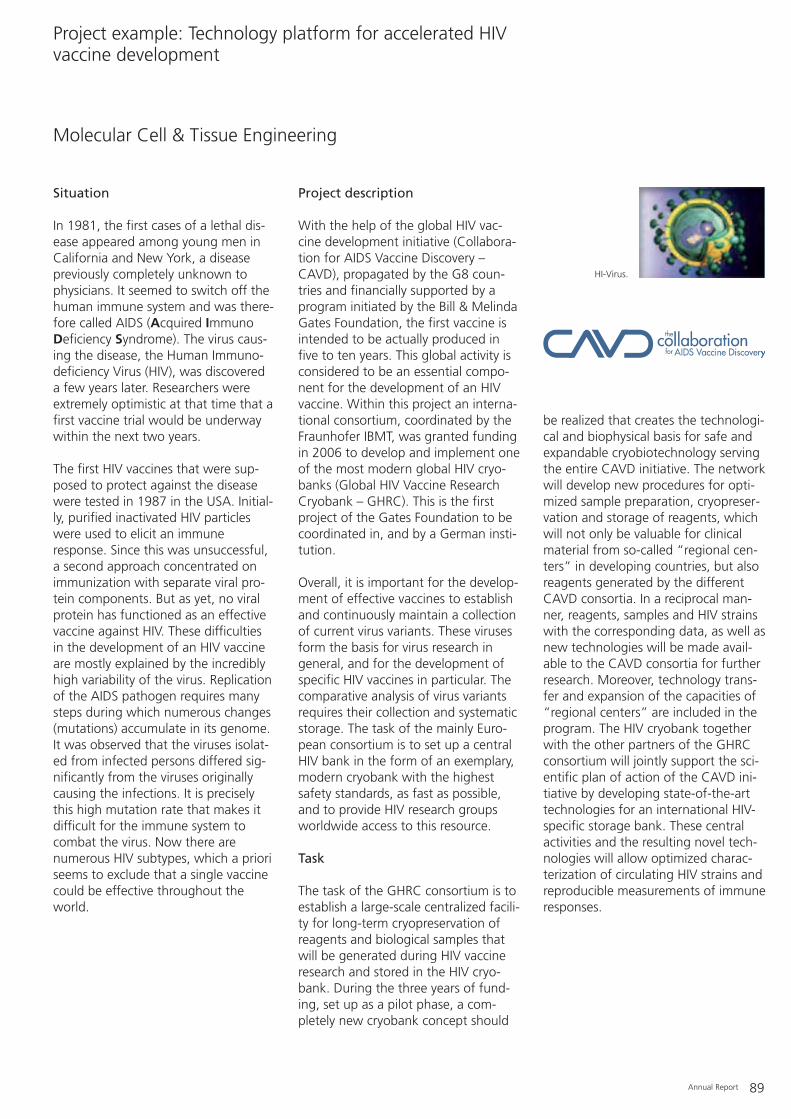
Annual Report 89
Situation
In 1981, the first cases of a lethal dis-ease appeared among young men inCalifornia and New York, a diseasepreviously completely unknown tophysicians. It seemed to switch off thehuman immune system and was there-fore called AIDS (Acquired ImmunoDeficiency Syndrome). The virus caus-ing the disease, the Human Immuno-deficiency Virus (HIV), was discovered a few years later. Researchers wereextremely optimistic at that time that afirst vaccine trial would be underwaywithin the next two years.
The first HIV vaccines that were sup-posed to protect against the diseasewere tested in 1987 in the USA. Initial-ly, purified inactivated HIV particleswere used to elicit an immuneresponse. Since this was unsuccessful,a second approach concentrated onimmunization with separate viral pro-tein components. But as yet, no viralprotein has functioned as an effectivevaccine against HIV. These difficultiesin the development of an HIV vaccineare mostly explained by the incrediblyhigh variability of the virus. Replicationof the AIDS pathogen requires manysteps during which numerous changes(mutations) accumulate in its genome.It was observed that the viruses isolat-ed from infected persons differed sig-nificantly from the viruses originallycausing the infections. It is preciselythis high mutation rate that makes itdifficult for the immune system tocombat the virus. Now there arenumerous HIV subtypes, which a prioriseems to exclude that a single vaccinecould be effective throughout theworld.
Project description
With the help of the global HIV vac-cine development initiative (Collabora-tion for AIDS Vaccine Discovery –CAVD), propagated by the G8 coun-tries and financially supported by aprogram initiated by the Bill & MelindaGates Foundation, the first vaccine isintended to be actually produced infive to ten years. This global activity isconsidered to be an essential compo-nent for the development of an HIVvaccine. Within this project an interna-tional consortium, coordinated by theFraunhofer IBMT, was granted fundingin 2006 to develop and implement oneof the most modern global HIV cryo-banks (Global HIV Vaccine ResearchCryobank – GHRC). This is the firstproject of the Gates Foundation to becoordinated in, and by a German insti-tution.
Overall, it is important for the develop-ment of effective vaccines to establishand continuously maintain a collectionof current virus variants. These virusesform the basis for virus research ingeneral, and for the development ofspecific HIV vaccines in particular. Thecomparative analysis of virus variantsrequires their collection and systematicstorage. The task of the mainly Euro-pean consortium is to set up a centralHIV bank in the form of an exemplary,modern cryobank with the highestsafety standards, as fast as possible,and to provide HIV research groupsworldwide access to this resource.
Task
The task of the GHRC consortium is toestablish a large-scale centralized facili-ty for long-term cryopreservation ofreagents and biological samples thatwill be generated during HIV vaccineresearch and stored in the HIV cryo-bank. During the three years of fund-ing, set up as a pilot phase, a com-pletely new cryobank concept should
be realized that creates the technologi-cal and biophysical basis for safe andexpandable cryobiotechnology servingthe entire CAVD initiative. The networkwill develop new procedures for opti-mized sample preparation, cryopreser-vation and storage of reagents, whichwill not only be valuable for clinicalmaterial from so-called “regional cen-ters” in developing countries, but alsoreagents generated by the differentCAVD consortia. In a reciprocal man-ner, reagents, samples and HIV strainswith the corresponding data, as well asnew technologies will be made avail-able to the CAVD consortia for furtherresearch. Moreover, technology trans-fer and expansion of the capacities of“regional centers” are included in theprogram. The HIV cryobank togetherwith the other partners of the GHRCconsortium will jointly support the sci-entific plan of action of the CAVD ini-tiative by developing state-of-the-arttechnologies for an international HIV-specific storage bank. These centralactivities and the resulting novel tech-nologies will allow optimized charac-terization of circulating HIV strains andreproducible measurements of immuneresponses.
Molecular Cell & Tissue Engineering
Project example: Technology platform for accelerated HIVvaccine development
HI-Virus.

90 Annual Report
The objectives of the GHRC projectin detail are:
– New strategies for sample collection(e.g. HIV isolates from so-called “ear-ly infections”).
– Optimized sample processing andcryopreservation.
– Establishment of a CAVD-specificglobal sample collection.
– Up-scaling of the HIV cryobank.– Progressive technologies for collect-
ing, preparing, preserving and distributing reagents for further net-works.
– Technology transfer to the “regionalcenters” and the other CAVD consortia as well as to developingcountries.
Outlook
The project should establish a uniquevirus bank within three years, whichshould contain the most diversereagents necessary for, and arisingfrom HIV vaccine research, and whichwill be available for extensive virologi-cal and immunological characteriza-tion. This material will be the basis forthe further development of vaccinesand new therapies against HIV. Scien-tists from all over the world will thenbe able to access the samples and theimportant bioinformatics derived fromthe primary biological data.
ContactAssistant Professor Dr. Hagen von BriesenTelephone: +49 (0) 6894/[email protected]
Illustration of the future containment level 3 laboratories in the cryohall for the HIV cryobank project funded by the Bill & Melinda Gates Foundation.

Annual Report 91
– Cell culture laboratories (geneticengineering containment level S1and S2) with sluice area and separatemedia and autoclave rooms for 2 laminar flow class 2 sterile work-benches
– Genetic engineering laboratories(genetic engineering containmentlevel S1 and S2) with 3 laminar flowclass 1 and 2 sterile workbenches
– Phase contrast, differential interfer-ence contrast and fluorescencemicroscopes (reflected and transmit-ted light)
– Imaging system including 3-D videocamera
– Spectral photometer for microtiterplates
– SNOM (optical near field microscope)– Axiphot fluorescence microscope
with photo and digital camera exten-sion
– UV/VIS spectral photometer– Automatic particle counter for cell
concentration and diameter mea-surements (Multisizer II)
– Cryomicrotome– Molecular biology equipment (PCR,
electrophoretic equipment, etc.)– Laboratory for bioelectronics (genetic
engineering containment level S1)– Electrochemical measurements of
impedance with Solatron SI 1260, SI 1281, SI 1287, SI 1294
– Electrophysiological measurementswith data recording system
– Dust stimulator– BX-50-WI research microscope with
micromanipulation unit and incuba-tion hood
– Flow cytometer (BD FACSCalibur system)
– Cell counter (CASY Model TT)
Biohybrid Systems
Equipment

92 Annual Report
Computer-aided Simulations
Services, results and products of the workgroup
– Computer-aided Simulations
Project example: Setup of the CellPROM application laboratory
Equipment
3-D visualization of a T-cell inside a blood vessel (Patrick Müller).

Annual Report 93
Over the years, tight interfacingbetween computer-aided design (CAD)and simulation has become a must. Allmajor software manufacturers offerinterfaces in both directions, in manycases even integration of both tools.Rapid data capture from the construc-tion and direct feedback based onanalyses from a simulation of the com-ponent setup seem to be standard. Is agroup exclusively dedicated to simula-tions still up-to-date?
While testing a mechanical construc-tion component under static loadworks relatively well, it becomes morecomplex for an electromechanical sys-tem under the influence of tempera-ture and time-dependent forces.Thereby, simple determination ofgeometry recedes into the back-ground, and definitions of differentialequations to be solved and of bound-ary conditions become a major part ofthe analysis. Finally, the implementa-tion of CAD software is oriented moretowards later production than conve-nient handling of simulations; theCAD-constructed components aredesigned for transfer into productionand not for straightforward integrationinto finite-element simulations. Typical-ly, the construction details available areof minor importance for the analysis
and thus can be neglected in favor ofreduced model complexity. Suppress-ing these details in the simulation isbased on the experience of the personperforming the analysis, hence, directtransfer of data from the CAD environ-ment to the simulation, at least inthese cases, remains an exception.
For instance, the design of piezoelec-tric ultrasound sensors still begins withthe first step of generating simplifiedsimulation models. This helps withunderstanding and optimizing theprinciple characteristics independentlyof any constructive constraints. Here,small compact models allow many vari-ations and also the expansion of rele-vant and large parameter fields. Onlyin a second step are the CAD dataincorporated directly into the simula-tion software. Despite the normallymuch larger size of the models, this isoften the only possibility for integrat-ing complex construction details intothe simulation with relatively littleeffort.
The workgroup of Computer-aidedSimulations focuses on the design of
complex microfluidic components andon the design and optimization ofpiezoelectric ultrasound sensors. TheCAD environment Solidworks and thefinite-element software Ansys provideour expert staff with highly efficienttools to develop and optimize compo-nents and systems in a project-orien-tated manner.
ContactDipl.-Phys. Daniel SchmittTelephone: +49 (0) 6894/[email protected]

94 Annual Report
– Computer-aided development andtesting of ultrasound transducers
– Computer-aided development ofultrasound arrays
– Sound field calculations– Optimization of ultrasound sensors
and systems– Computer-aided development and
testing of gradient coils– Electromagnetic field calculations– Computer-aided development and
testing of MEMS– Electrical current calculations– Coupled electrical current-acoustics
calculations– Stress analyses and calculations– FEA-based optimization of construc-
tion parts– Simulation of microfluidics compo-
nents and systems– Temperature calculations– 3-D construction– 3-D visualization and animation in
biology, chemistry, physics, medicineand engineering
– Medical imaging and 3-D reconstruc-tion
ContactDipl.-Phys. Daniel SchmittTelephone: +49 (0) 6894/[email protected]
Computer-aided Simulations
Services, results and products of the workgroups

Prototype for pilotexperiments on the
DEP/fluidic deviceconcept.
Liquid nitrogen for storageand cultivation.
Prototype for pilotexperiments on the
magnetic device concept.
Integration unit for modulesand periphery of the
DEP/fluidic [device concept].
Fluidicspreparation
station.
Stamping machine for theproduction of NanoScapesTM.
Integration unit for themagnetic device concept.
Annual Report 95
Situation
The controlled and documented in vit-ro differentiation of cells at the singlecell level imposes very high require-ments on the necessary technical sys-tems. Until now, only a few deviceswere available that fulfilled theserequirements in individual areas.Automation and easy handling seemto be diametrically opposed to thecomplexity and variability of biologicalcell models.
Task
Within the context of the EU-fundedIntegrated Project CellPROM, 26 scien-tific groups and companies are current-ly working on developing new tech-nologies for the controlled differentia-tion of cells. The interactions betweenbiology, nanotechnology, microsystemstechnology and automation imposehigh standards on the environmentand core areas of the device systems.As the coordinator, the FraunhoferIBMT is not only in charge of the scien-tific project management, but is alsoresponsible for the integration of com-ponents, either developed by the IBMTor other partners, and the definition
and implementation of interfaces.Reaching its mid-point in February2006, the project plans to create oper-ational modules and test them in cellbiology experiments within four years.
Results
Combining biology and nanotechnolo-gy demands high standards for sterileand particle-free environments. Fur-thermore, the modular approach callsfor a high degree of flexibility in thedevice components. Therefore, acleanroom microenvironment wasinstalled initially (see Figures 1 and 2).Two workplaces with class 50 clean-
room facilities now allow the complexand flexible setup of systems compris-ing various components and the evalu-ation of two concepts in parallel.
An intricate microfluidic channel sys-tem forms the core of the first system(see Figure 3). Single cells can bemanipulated and moved in it withoutphysical contact by using dielec-trophoresis and ultrasound. This allowscharacterization and differentiation ofsingle cells under defined and constantconditions. Six fluidic and more than30 electrical channels are concentrated
Computer-aided Simulations
Project example: Setup of the CellPROM application laboratory
Class 7 cleanroom facility
Class 5 cleanroom facility
Mag
net
icp
roto
typ
eμ-c
onta
ctsta
mpi
ng
unit
-
Preparation bench
for fluidics
N2
Mag
net
icd
emo
nst
rato
r
DEP
/ fl
uid
icd
emo
nst
rato
r
DEP
/flu
idic
pro
toty
pe
Figure 2: Workplaces and devices in the cleanroom (MagnaLab and NazcaLab).
Figure 1

96 Annual Report
on a small area around the centralchip. They are computer-controlledand can be used to plan and carry outexperiments.
The second concept is also based on acentral fluidic channel system (see Fig-ure 4). Here, however, small magneticcarriers which the cells adhere to dur-ing growth can be moved inside thissystem by external electromagnets.Special areas of the 30 cm wide centralcultivation unit serve as observation orcultivation zones for the cells. The cellsare transported from station to stationeither interactively or according to apre-defined sequence.
In addition to the two central work-places, further single components canbe tested and installed in the clean-room environment.
Potential
During the project’s duration, the Cell-PROM application laboratory is thecentral hub for the integration andtesting of single system components.Already at the mid-point of the projectall partners can test and improve theirmodules in functional prototypes. Theintegration of further modules and theredesign of existing components willincrease the complexity of the systemsduring the second half of the project.The final goal is to create a technicalplatform that allows a multitude ofnovel and interesting experimentsrelating to the controlled differentia-tion of single cells.
Figure 3: Integration of the modules around the NazcaLab chip.
Figure 4: Prototype evaluation for the manipulation of cells on magnetic carriers (MagnaLab).
Annual Report 97
Project collaborators at the IBMT
Leonora PeterHiltrud GörgenMartin BeneckeDipl.-Ing. (FH) Iskan DemirdelenDipl.-Ing. Jürgen MeicheDr. Robert Johann (PhD)
Project funding
European Union, NMP4-CT2004-500039 (CellPROM)
ContactDipl.-Phys. Daniel SchmittTelephone: +49 (0) 6894/[email protected]
Hybrid computer environment withUNIX/Linux and Windows systems andthe following software tools: ANSYSTM
(FEA code), CFDRCTM (FEA code), Flo-tranTM (FEA code), ModulEF (FEA code),FlexPD (FEA code), ProEngineerTM (Stan-dard CAD code), SolidWorksTM (Stan-dard CAD code), AutoCADTM (StandardCAD code), PiezoCadTM (design ofultrasound transducers based on theKLM model), MathematicaTM, SCALP(own development for the calculationof the transient propagation ofacoustic or electromagnetic waves),LabViewTM (signal analysis code), 3D-Studio MAXTM (visualization and anima-tion of complex physical and technicalprocesses), Evoluti (own developmentfor optimization based on geneticalgorithms), AMIRATM (3D imaging andreconstruction), AcapellaTM (micrographimaging).
Computer-aided Simulations
Equipment

98 Annual Report
Project Group LübeckCell Differentiation & Cell Technology
Services, results and products of the workgroup
Cell Differentiation & Cell Technology
Project example: Human pancreatic stem cells differentiating into cardiac muscle cells
Equipment
Nestin-positive pancreatic stem cells (red) are precursors of e.g. glia cell types (green). Furthermore, other cell typeswhose specific proteins were not labeled are present (blue nuclei). The nuclei were counterstained with DAPI (blue).

Annual Report 99
Stem cells are among the most promis-ing approaches for future regenerativemedicine. The idea of isolating stemcells from patients and reprogrammingthem so that they differentiate to cre-ate cell types of dysfunctional organsor tissues has become more feasibleover the last few years. In addition totheir ability to generate specializedcells, stem cells possess a more active(although it varies) proliferation poten-tial. The combination of both thesecharacteristics opens up applicationareas beyond regenerative medicineand cell therapy. One could conceiv-ably create custom-made testing sys-tems for pharmaceutical and cosmeticsindustries with stem cells, employthem for tissue engineering or usethem for the specific production ofhigh-quality biomass.
Stem cells are subdivided according tovarious criteria: according to their ori-gin, into embryonic and adult; andaccording to their ability to differenti-ate into specialized cells, into pluripo-tent, multipotent or unipotent. At pre-sent, embryonic stem cells, which areisolated from an early developmentalstage of the organism, cannot be usedfor applications beyond basic researchdue to ethical objections. Adult stemcells, however, can be isolated withoutany ethical considerations, but theynormally do not possess the same dif-ferentiation and proliferation potentialas embryonic stem cells. Moreover,their precise localization in the organ-ism is often unclear, which frequentlymeans it is not possible to extractthese cells.
Three years ago the workgroup of CellDifferentiation & Cell Technology, aFraunhofer project group at the Uni-versity of Lübeck for two and a halfyears now, published results on theastonishing characteristics of stem cellsisolated from exocrine glands. Sincethen the outstanding reproducibilityand their high differentiation potentialhave been confirmed in all respects,not only by external groups, but con-clusively by the Lübeck group itself andtwo other cell culture groups at theIBMT in St. Ingbert and Potsdam-Golm.Moreover, in 2006 we succeeded indemonstrating their differentiation intocontracting cardiomyoblasts (a celltype of the mesoderm), the cloning ofcells and inducible tissue bodiesderived from these cells, as well asmyogenic, neuronal, adipogenic andchondrogenic transformations. Fur-thermore, for the first time cells couldbe characterized with distinct egg cellcharacteristics (see figure on p. 100).These results have been accepted forpublication, and the patents from theyears 2003/2004 have been granted ina number of countries. Thus, thissource of highly potent stem cells fromexocrine glands will gain an importantposition on the candidate list of adultstem cells for cell therapies. In manyrespects these stem cells resemblethose of embryonic origin. For instance,a now optimized and stable procedureallows reproducible isolation of stemcells from exocrine pancreas (and sali-vary gland tissue) that show a growthrate comparable to embryonic stemcells (doubling time approx. 16 h; culti-vation possible over 150 passages withrat-derived cells). In addition, they differentiate into cell types of all threegerm cell layers (ectodermal, mes-enchymal, endodermal). Furthermore,isolation is highly efficient: 50 millioncells from the initial sample tissue(approx. 1 cm3) yield approx. 100 000proliferating stem cells. This ratio of1:500 is several orders of magnitudebetter than the 1:100 000 ratio of con-ventional mesenchymal stem cells.
ContactAssistant Professor Dr. Charli KruseFraunhofer IBMT Project Group Cell Differentation & Cell TechnologyUniversity of Lübeck MFC / ICLMaria-Goeppert-Strasse 123562 Lübeck GermanyTelephone: +49 (0) 451/2903-210Fax: +49 (0) 451/[email protected]

100 Annual Report
In addition to their excellent availabili-ty, stem cells from exocrine glandsoffer the great advantage of aggregat-ing easily into small three-dimensionalorganoid-bodies. The intensified cell-to-cell contact in these aggregatesleads to new differentiation stimuli andsignificantly expands the spectrum ofpossible applications for these cells.The Fraunhofer IBMT with its externalworkgroup at the University Lübeck isfocusing on the obvious potential ofthese cells. Apart from human stemcells, the workgroup is one of the fewstem cell institutions worldwide to suc-ceed in efficiently isolating stem cellsfrom animals, ranging from fish tomammals, and establishing these cellsin in vitro cultures. In cooperation withthe zoo in Neunkirchen, they areestablishing a comprehensive stem cellcollection of endangered animalspecies. Upon request and payment ofbasic costs the IBMT supplies such cellcultures from fish, spoonbill, crane,chicken, rat, mouse, goat, roe deer,boar, watussi cattle, guanaco, red deer
and long-tailed monkeys for researchpurposes. An industrial use is also pos-sible on a project basis upon agree-ment. Currently, the Fraunhofer IBMTis working on seven large-scale pro-jects centered on stem cells, in addi-tion to two EU projects on comparativeinvestigations with human embryonicstem cells for which the FraunhoferIBMT was granted the importapprovals No. 18 and 19 by the RobertKoch Institute after passing the stipu-lated application routes and an evalua-tion by the central ethics committee.The IBMT is the first, and at presentonly Fraunhofer Institute with suchapproval and probably possesses thebroadest spectrum of available stemcell types.
– Isolation and differentiation ofhuman and animal adult stem cellswith the aim of harnessing them forregenerative medicine and biotech-nology
– Primary cell isolates from differentspecies and different exocrine tissues
– Cloning of cells, establishment of celllines
– Induction of tissue systems from ani-mal and human cell isolates
– Development of stem cell differentia-tion procedures
– Immunological characterization ofcells
– Development of new tools anddevice components for stem cell handling
– Micromanipulation of cells– Courses on cell manipulation– Contract cell cultivation for cus-
tomers
ContactAssistant Professor Dr. Charli KruseFraunhofer IBMT Project Group Cell Differentation & Cell TechnologyUniversity of Lübeck MFC / ICLMaria-Goeppert-Strasse 123562 Lübeck GermanyTelephone: +49 (0) 451/2903-210Fax: +49 (0) 451/[email protected]
Cell Differentiation & Cell Technology
Services, results and products of the workgroup
Figure 1: Clonal pancreatic stem cell aggregates from the rat produce oocyte-like cells. A: Oocyte-like cells in suspen-sion. B-C: Immunocytochemical detection of meiosis-specific proteins. B. Immunostaining of the synaptonemal com-plex protein (SCP3, green). C: Immunostaining of the DMC1 protein (red). Nuclei were stained with DAPI (blue).
A

Annual Report 101
State of the field
The number of tissues in which stemcells and progenitor cells have beenfound has significantly increased dur-ing recent years. Remarkably, the num-ber of stem cell types described to pos-sess pluripotent properties has alsoincreased. These cells can differentiateinto cells of all three germ layers.According to current knowledge thereare at least four origins of these cells:mesenchymal stem cells, stem cells inblood from the umbilical cord, pancre-atic stem cells and testicular stem cells.In addition, several adult stem celltypes exist that can differentiate intocells of at least two germ layers (e.g.stem cells from hair follicles or the liv-er). The glandular stem cells describedand investigated by the IBMT work-group “Cell Differentiation & Cell Tech-nology” at the University Lübeck weremainly isolated in cooperation with theLübeck Surgical Clinic, and various pro-liferating cell populations have nowbeen established. The glandular stemcells are of primary interest for investi-gations carried out here because they(1) are relatively easy to isolate, (2)show a high yield, (3) can be efficientlycryopreserved, (4) can be cultivatedover extended periods, (5) form three-dimensional tissue bodies and (6) dif-ferentiate into cells of all three germlayers (ectoderm, mesenchym, endo-derm) (see Figure 2). In addition to theIBMT workgroup, other renownedresearch groups from all over theworld are investigating these interest-ing stem cells. This means it is impor-tant to maintain and increase the leadin some areas, in particular withrespect to the targeted differentiationinto specific cell types.
Besides the differentiation of stem cellsinto quiescence, other processes ofdedifferentiation and redifferentiationalso seem to be partially responsiblefor the high plasticity of adult cells.Current results indicate that bothprocesses are also relevant in the stemcell isolates from the exocrine pan-creas.
Stem cells and progenitor cells fromexocrine glands
Cells isolated from rats, mice, humansand other vertebrates have been culti-vated in the laboratory for more thanthree and a half years. These sponta-neously form tissue-like aggregationsunder certain conditions, which aredescribed as “organoid tissue bodies”.Fluorescence analyses with highly spe-cific labeled molecules, high-resolutionelectron microscopic sections as well asPCR results indicate that tissue typesemerge that can also be found innerves, muscles, cartilage and skin.
These investigations were performed incollaboration with the Institute forAnatomy at the University Lübeck.
Differentiation into cardiac musclecells
Heart failure is a major cause of deathin industrialized countries, mainly dueto the fact that adult cardiomyocyteshardly regenerate or renew them-selves. This is why injections of stemcells might be an actual alternative forheart regeneration. Currently, suchtreatments are already being carriedout using mesenchymal stem cells,although the results are not alwaysvery clear. Hence, the search for othersuitable stem cells that can producecardiac muscle cells is still in progress,and remarkable progress has been
Cell Differentiation & Cell Technology
Project example: Human pancreatic stem cells differentiating into cardiac muscle cells
Figure 2: Spontaneous differentiation of stem cells/progenitor cells from the exocrine pancreas into cell types of allthree germ layers.
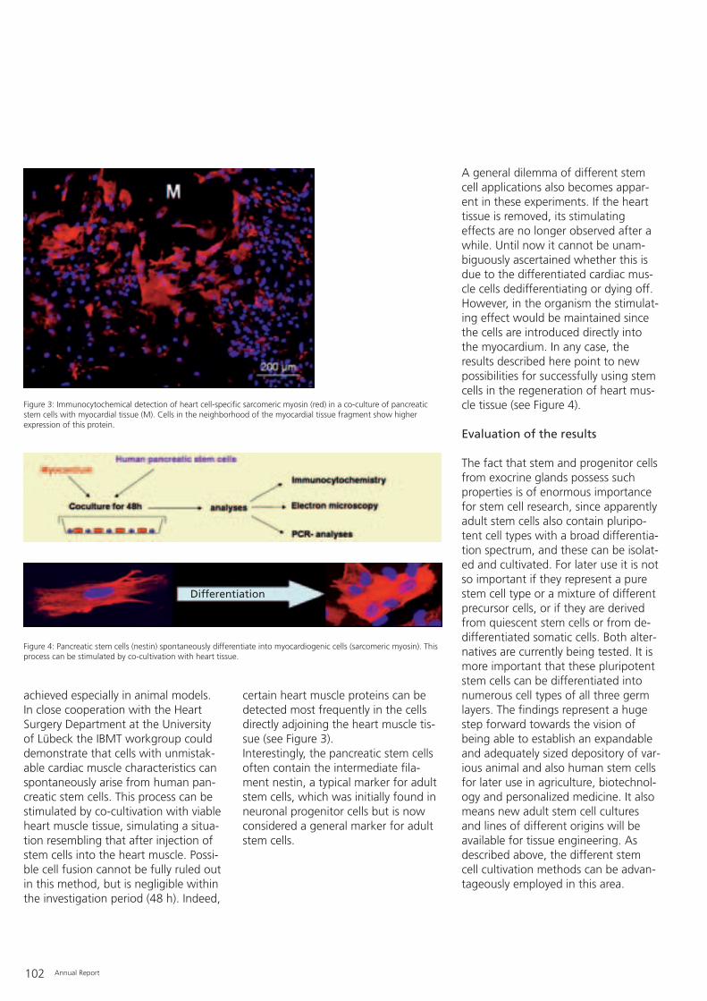
102 Annual Report
achieved especially in animal models.In close cooperation with the HeartSurgery Department at the Universityof Lübeck the IBMT workgroup coulddemonstrate that cells with unmistak-able cardiac muscle characteristics canspontaneously arise from human pan-creatic stem cells. This process can bestimulated by co-cultivation with viableheart muscle tissue, simulating a situa-tion resembling that after injection ofstem cells into the heart muscle. Possi-ble cell fusion cannot be fully ruled outin this method, but is negligible withinthe investigation period (48 h). Indeed,
certain heart muscle proteins can bedetected most frequently in the cellsdirectly adjoining the heart muscle tis-sue (see Figure 3).Interestingly, the pancreatic stem cellsoften contain the intermediate fila-ment nestin, a typical marker for adultstem cells, which was initially found inneuronal progenitor cells but is nowconsidered a general marker for adultstem cells.
A general dilemma of different stemcell applications also becomes appar-ent in these experiments. If the hearttissue is removed, its stimulatingeffects are no longer observed after awhile. Until now it cannot be unam-biguously ascertained whether this isdue to the differentiated cardiac mus-cle cells dedifferentiating or dying off.However, in the organism the stimulat-ing effect would be maintained sincethe cells are introduced directly intothe myocardium. In any case, theresults described here point to newpossibilities for successfully using stemcells in the regeneration of heart mus-cle tissue (see Figure 4).
Evaluation of the results
The fact that stem and progenitor cellsfrom exocrine glands possess suchproperties is of enormous importancefor stem cell research, since apparentlyadult stem cells also contain pluripo-tent cell types with a broad differentia-tion spectrum, and these can be isolat-ed and cultivated. For later use it is notso important if they represent a purestem cell type or a mixture of differentprecursor cells, or if they are derivedfrom quiescent stem cells or from de-differentiated somatic cells. Both alter-natives are currently being tested. It ismore important that these pluripotentstem cells can be differentiated intonumerous cell types of all three germlayers. The findings represent a hugestep forward towards the vision ofbeing able to establish an expandableand adequately sized depository of var-ious animal and also human stem cellsfor later use in agriculture, biotechnol-ogy and personalized medicine. It alsomeans new adult stem cell culturesand lines of different origins will beavailable for tissue engineering. Asdescribed above, the different stemcell cultivation methods can be advan-tageously employed in this area.
Figure 3: Immunocytochemical detection of heart cell-specific sarcomeric myosin (red) in a co-culture of pancreaticstem cells with myocardial tissue (M). Cells in the neighborhood of the myocardial tissue fragment show higherexpression of this protein.
Figure 4: Pancreatic stem cells (nestin) spontaneously differentiate into myocardiogenic cells (sarcomeric myosin). Thisprocess can be stimulated by co-cultivation with heart tissue.
Differentiation

Annual Report 103
– Basic equipment for the isolation andsetup of any in vitro culture
– Differentiation and analysis of adultstem cells
– Microinjection technology– Micro-dissection technology– Equipment for characterization and
isolation of single cells
Cell Differentiation & Cell Technology
Equipment
Potential
As a consequence of the high impactof these results many collaborationswith partners from Germany and otherEuropean countries have arisen. Theseaim to more thoroughly explore thepossibilities of this new stem cellsource and test their use in medicine,biotechnology and agriculture. Thepartners come from basic research,clinical research and industry. The IBMTworkgroup has been integrated intothe EU-funded Integrated Project Cell-PROM due to the high biotechnologi-cal and medical potential of theresults. The project focuses on defined,surface-based induction of cell differ-entiation. Different pilot experimentshave already been performed and pro-duced the first results.
Germany now, as before, has thepotential to occupy a key position instem cell research and the efficientpreservation of stem cells. It is neces-sary to take advantage of this lead andat the same time to extend it by con-certed expansion of basic and appliedresearch.
ContactAssistant Professor Dr. Charli KruseTelephone: +49 (0) 451/[email protected]
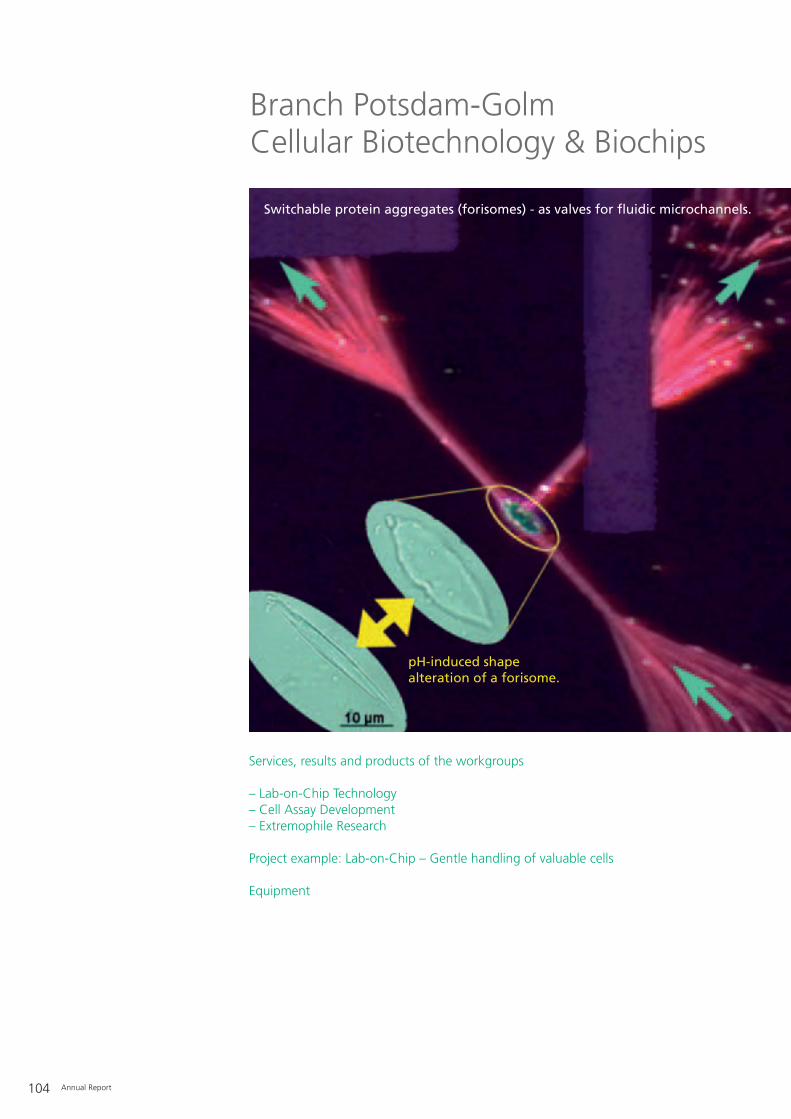
104 Annual Report
Branch Potsdam-GolmCellular Biotechnology & Biochips
Services, results and products of the workgroups
– Lab-on-Chip Technology– Cell Assay Development– Extremophile Research
Project example: Lab-on-Chip – Gentle handling of valuable cells
Equipment
Switchable protein aggregates (forisomes) - as valves for fluidic microchannels.
pH-induced shape alteration of a forisome.
Annual Report 105
In the future, cell-based diagnosticsand therapy approaches will occupykey roles in medicine. However,advances in these fields will only bepossible when the tools and technolo-gies that allow the gentle and repro-ducible manipulation and characteriza-tion of cells become available. Thedepartment of Cellular Biotechnology& Biochips is developing such tools.The basis of this development is theassumption that the cellular stages anddifferentiation patterns must beadjustable with relative precision forsuch intended medical applications.This can only be achieved if one cancontrol the information exchangebetween a cell and its environment.This exchange can be triggered by,both, biochemical and mechanicalprocesses. Our solutions are based ontwo approaches: Single cells can behandled with high precision and stabil-ity in lab-on-chip systems withoutphysical contact using high-frequencyelectromagnetic fields. A clever combi-nation of microelectrodes and fluidicmicrochannels makes it possible to car-ry out important tasks in the chip: Pre-cise positioning to the micrometer for
microscopy, assembly of cells and theformation of cell clusters, sorting ofdifferent cell populations, and separat-ing and processing cells in very smallsamples. The second approach tries tocontrol the attachment and migrationof adherent cells on surfaces in a tar-geted manner through the presenta-tion of various stimuli. Microfluidic sys-tems can generate concentration pro-files of soluble signal molecules withhigh spatial precision, which can beused to analyze the chemotacticresponse of cells. The coating ofswitchable polymers onto nanoscopi-cally structured surfaces allows one toeasily vary the adhesion conditions ofcells. A technology platform is current-ly being established by combining bothapproaches. This will make it possibleto precisely control the developmentand differentiation of stem cells, inparticular via biochemically and topo-graphically defined surface architec-tures.
Within the context of the department’sresearch on extremophilic organisms, asnow algal bank under the acronymCCCryo is installed, which serves as asource of metabolites (enzymes, pig-ments, biopolymers) for biotechnologi-cal applications (e.g. process engineer-ing).
ContactPriv.-Doz. Dr. Claus DuschlBranch Potsdam-GolmAm Mühlenberg 1314476 Potsdam-Golm GermanyTelephone: +49 (0) 331/[email protected]

– Protein analysis with high-resolutionimmunofluorescence microscopy
– Pulse chase technology for investi-gating expression kinetics
– Time-resolved characterization ofmanipulation-induced molecularchanges in the cell using fluores-cence time-lapse and TIRF time-lapsemicroscopy
– Development of microfluidics carriersfor high-resolution microscopy
– Cancer diagnostics based on galvano-taxis and chemotaxis analyses
– Examination of single cells by non-invasive analysis of abandoned celltraces
– Design and fabrication of surfaces– Development of methods for the dif-
ferentiation of stem cells– Non-invasive examination of stem
cell differentiation status– Control of cell trace deposition by
surface modifications– Development of quick assays for
determining chemotactic properties– Correlation of tumor progression
stages with molecular processes dur-ing chemotactic cell movement
– Switchable surfaces for the control ofcell adhesion
– Substrates for cell analysis with phys-iologically compatible surfaces
– Microcontact printing (“µ-CP”) ofbiomolecules for the production ofmicro- and nanostructures for cellmanipulation
106 Annual Report
– Design and development of microflu-idic systems (chips, periphery, detec-tion) for biotechnology and cell biol-ogy
– Design and construction of chip-based microsystems for cell-compati-ble injection of physiological suspen-sions into microfluidic systems, con-tact-free handling of single or lownumbers of biological objects (cells,bacteria, viruses) and defined deposi-tion of previously characterized parti-cles for further cultivation
– Microsystems for the controlledtranslation and rotation of suspend-ed microparticles
– Manual, semi-automatic and auto-matic sorting of micro-objects (e.g. living cells) in continuous flowsystems
– Centrifugation-free washing andloading of living cells with, e.g. phar-maceutical agents in microfluidicflow systems
– Accumulation and detection ofmicro- and nanoparticles in biotech-nologically relevant suspensions
– Dielectric characterization of complexparticles on a single cell level
– Chip-based electromanipulation (e.g.fusion) of rare cells (e.g. stem cells)
– Transport of minute liquid volumeswith chip-integrated micropumps
– Combination of dielectric field trapsand optical tweezers for the simulta-neous manipulation of severalobjects and for the characterizationof interactions (binding forces)between particles
– Optical high-end microscopy, e.g.high-sensitivity fluorescence mea-surements
– Numerical calculation and modelingof electrical, optical and hydrody-namic forces in microsystems
– Influence of alternating electric fields(10 kHz to 250 MHz) on biologicalobjects
– Time-resolved investigation of celladhesion on functionalized surfaceswith Total Internal Reflection Fluores-cence Microscopy (TIRFM)
– Characterization of the topographi-cal structure of artificial and biogenicsurfaces with sub-micrometer resolu-tion using Atomic Force Microscopy(AFM) and investigation of mechani-cal characteristics on the same sizescale with micro-indentation
– Microprocessing with UV laser abla-tion
– Micromanipulation of single objectswith capillary aspiration
– Cultivation of animal and yeast cellsunder S1 conditions before and aftermanipulation in microfluidic chips
ContactDr. Magnus Sebastian JägerTelephone: +49 (0) 331/[email protected]
Lab-on-Chip Technology Cell Assay Development
Services, results and products of the workgroups

Annual Report 107
– Design of surface topographies by µ-CP of coated micro- and nanopar-ticles on glass and gold surfaces
– Nanostructured gold surfaces for thecontrol of cell functions
– Formation of self-assembled mono-layers of thiolbearing biomolecule ongold surfaces
ContactDr. Andreas Lankenau Telephone: +49 (0) 331/[email protected]
– CCCryo: culture collection ofcryophilic and mesophilic snow, iceand soil algae from polar and alpineregions (snow algae)
– Development of culture systems forlarge-scale cultivation of carotenoidproducing microalgae in photobiore-actor installations
– Contract cultivation of algae materialunder defined and/or differentialconditions (UV radiation, light, tem-perature, nutrients)
– Delivery of isolated and purified DNAand RNA for corresponding studies
– Shipping of algal strains (on request)– Research on extremozymes (differen-
tial transcriptomics, 2D-SDS-PAGE)and primary and secondary plantmetabolites (natural cryoprotecta,polyunsaturated fatty acids,carotenoids, astaxanthin, alpha-toco-pherol, biopolymers)
– Enzyme assays– Basic research on the taxonomy of
cryophilic freshwater microalgae– Physiological investigations on
cryophily (e.g. single cell microscopy)– Phylogenetic analyses based on
18S rRNA and ITS gene sequences– Population genetic investigations on
the bipolar distribution of cryophilicalgae to support climate modeling
ContactDr. Thomas LeyaTelephone: +49 (0) 331/[email protected]
Extremophile Research

108 Annual Report
Initial situation
In 1600 the English doctor WilliamGilberd described one of his experi-ments with amber, which he had previ-ously rubbed with a cloth:“Hence it is probable that amberexhales something peculiar thatattracts the bodies themselves, and notthe air. It plainly attracts the body itselfin the case of a spherical drop of waterstanding on a dry surface; for a pieceof amber held at a suitable distancepulls toward itself the nearest particlesand draws them up into a cone.”
What Gilberd had observed was thedeformation of a dielectric body – thewater drop – in the electric fieldaround the piece of amber electrostati-cally charged by friction. Thanks totechnological progress over the pastfour centuries we can generate muchbetter defined electrical fields todaythan Gilberd could in his time. In par-ticular, microelectrodes can now gen-erate electrical fields, especially fieldgradients and high frequencies, onvery small length scales. From abiotechnological perspective the ques-
tion is then: Can Gilberd’s insights betransferred to microscopic dielectricobjects of medical relevance such assingle living cells? If so, how can thisbe achieved technologically? Does thisprocess make it possible to obtaininformation about the cells underexamination without destroying them?
Project descriptions and tasks
Lab-on-chip systems are versatile toolsfor the manipulation of biological andmedical samples. They aim at handlinghuman cells, pathogenic bacteria orviruses under conditions that mimictheir natural environment as closely aspossible. Mostly these systems are setup as microfluidic channels throughwhich suspended particles flow, andwhere they can be analyzed andmanipulated.
The analysis and manipulation of cellscan be achieved using different princi-ples. For instance, many lab-on-chipsystems are based on first lysing thecells and subsequently characterizingtheir contents. In other approachescells are labeled with specific signalingsubstances and then classified accord-ing to the ability of these substances tobind to certain receptor molecules.Often these signaling compounds areimmobilized in a grid-like layout onsurfaces, and the preferential bindingof the particles is detected locally.However, these methods have theinherent disadvantage of destroying orat least considerably disturbing thecells so that certain signaling pathwaysare switched on.
If one is seeking techniques for gentle,but at the same time efficient handlingof living cells, gradients of ultrasoundand electromagnetic fields, e.g. radiowaves, provide numerous options.Radio waves are alternating electricalfields, which can exert forces in the pNrange on biological objects (dielec-trophoresis). Such high-frequencyfields never arose during the course of
Lab-on-Chip Technology
Project example: Lab-on-Chip – Gentle handling of valuable cells
Figure 1: A “NazcaLab” chip with integrated fluidic channels and microelectrodes for the gentle handling of living cells. The cells can be sorted, separated and brought into contactwith each other. B Magnified view of the chip. Microelectrodes with a width of 15 µm protrude in two different planes into the 900 µm-wide channel where the living cells areprocessed.

Annual Report 109
evolution on our planet. Therefore,cells probably do not possess signalingpathways that these fields can inter-fere with, given an appropriate settingof electrical parameters. So, influenceon cells is minimal. Electrical fields canbe applied with high temporal andspatial precision. Since this onlyrequires the correct setup of circuitssuch processes can easily be automat-ed.
Established processes of semiconduc-tor production allow the integration ofmicroelectrodes in microfluidic lab-on-chip systems, greatly increasing theiroperational scope. Eventually, thesedevelopments will lead to technologi-cal solutions in the form of chips thatcan be used in parallel. An example isthe “NazcaLab” chip developed withinthe context of the EU project Cell-PROM (see Figure on p. 108). Thiscompletely new chip design features amicrosystem of as yet unmatched pro-portions. It enables the simultaneousprocessing of biological microobjects in
parallel in many channels. Moreover,the new format allows one to easilyincrease the number of integratedmanipulators, i.e. the microelectrodes,to more than a hundred.
Since they are constructed from highoptical grade materials, these devicescan easily be combined with othermodern techniques such as high-reso-lution confocal microscopy or lasertweezers. They can be used to solvevarious different tasks in currentbiotechnological and medical research.Some of the following examples willbe described in more detail below: (1)For instance, cell samples can be sort-ed according to optically detectablecharacteristics. These characteristicscould be coupled to the expression ofa genetic trait of interest. (2) Cells canbe exposed to certain pharmaceuticalsor other soluble effectors without cen-
trifugation and for a precisely definedperiod, and their reaction to these sub-stances detected. (3) Two microobjects,e.g. cells, can be brought into contactin the chips for certain periods of timeand then separated again. This is par-ticularly useful for immunological andstem cell research. (4) In addition, themethod allows a high level of controlover the fusion of two selected cells.(5, 6) Depending on the setup of themicroelectrodes, they also serve totransport liquid in the system or workas particle filters in the channel. (7)Finally, type-specific physical propertiesof the cells can be measured, allowingtheir identification and characteriza-tion.
Results
A particularly interesting challenge isto combine several advantageous char-acteristics in the same cell. In practice,a very frequent problem is that cellswhich produce valuable substancesdue to genetic modification or surface-mediated programming often becomeunstable during longer cultivation.
Figure 2: A Two human lymphocytes (U-937) are held without contact in a dielectrophoretic microelectrode structure comprising eight electrodes (black) – four at each level – a so-called octode. The two cell nuclei are labeled “1” and “2” (electrode width: 10 µm). B After applying a specific electrical signal the cells fuse. The two nuclei are now surrounded by a single cell membrane.

110 Annual Report
On the contrary, other cells are oftenrobust in comparison, but technicallyuninteresting. The positive propertiesof both cell lines can be combined byexternally induced fusion (see Figure 2,p. 109). This makes it possible toestablish not only stable cell culturesthat produce a desired product, butalso, for example, cultures that areresistant against a certain pathogenicfactor. The same protocol can also pro-duce cell aggregates, which can beused in pharmaceutical research toinvestigate cell membrane characteris-tics. A long-established method forinducing cell fusion is the applicationof pulsed electrical fields. The chip sys-tems described above are perfectlysuited for this application due to theirintegrated microelectrodes. The cellscan first be selected electrically, i.e.without direct physical contact, andthen positioned and eventually fused.
The example above referred to the“programming” of cells. This meansthat a cell receives an external inputthrough surface contact, to which it
reacts with a defined response. Thiscan be exploited particularly well inmicrosystems. An example is the acti-vation of T-cells, a cell type of theadaptive immune system. In the bodysuch information transfer occurs dur-ing an infection when certain B-cellscome into surface contact with T-cellsand thus program the T-cells for a spe-cific immune response. In microchipsystems this process is achieved by firstanchoring a cell at a hook-shaped bar-rier by a suitable combination ofhydrodynamic and electrical forces(Figure 3). The anchored cell in thisconfiguration is then exposed todefined contacts with another cell oran artificial particle. The surface con-tact between the two particles resultsin information transfer between them,hence inducing “programming”.
To be able to select the correct targetcells from the injected sample, oneneeds a process that can transport thesample through the channels onto thechip. A particularly elegant approach isto use the integrated microelectrodesfor this as well. This avoids using com-plicated and costly pump systems thatcontrol the channels from the outside(Figure 4, p. 111). Similar to suspendedobjects, electrodes can also exertforces on liquids (traveling waves). Thetranslation of this method into thedesign of pumps is obvious. In additionto the precise transport of very smallvolumes, such electrode setups alsofacilitate keeping cells in continuousmotion around ring-shaped structuresin channel systems so that the cells canbe stored without adhering irreversiblyto the surfaces of the channels.
Another application of traveling wavesderives from the ability to generateappropriate flow profiles that lead tothe accumulation of suspended parti-cles at certain positions within the chipsystem. This makes it possible to con-centrate dispersed nanoparticles at onelocation for carrying out a reliableanalysis (Figure 5A, p. 112). Until nownon-magnetic accumulation of nano-particles was not possible in flow sys-tems. The traveling wave method nowopens up the possibility of concentrat-ing the particles at certain sites on thechip. A requirement for the successfuldevelopment of application-specificsolutions is in-depth knowledge of thephysical conditions in the chip. At pre-sent, numerical modeling approachesare absolutely essential, as exemplifiedby the finite element method (Figure5B, p. 112). This enables one, evenbefore producing a prototype, to makepredictions about the probable reac-tion of a future sample in the systemand to optimize the design.
Figure 3: A A single human T-lymphocyte is dielectrophoretically anchored by a hook-shaped microelectrode (black) ina flow that comes from the left (width of the electrode: 20 µm). B The immunological activation of the cell can besuccessfully detected by a fluorescently labeled CD69 antibody.

Annual Report 111
Cell samples from a natural source,e.g. in medicine, typically contain amixture of different cells which mustbe identified before any further manip-ulation. Successful targeted electrofu-sion or cell programming as describedabove are only possible if the desiredcells can first be identified in a mixtureand then selectively sorted. This can beachieved very efficiently by exploitingthe fact that different cells react differ-ently to the forces exerted by the elec-trical field. For instance, if these forcesare used to deform cells they react in aspecific way depending on the mecha-nical properties of the respective celltype (Figure 6, p. 112). Such electro-based identification of cells has advan-tages in that first, it works without theaddition of chemical agents, second, itis easy to achieve due to the straightforward production of the microelec-trode structures and third, it can beapplied in continuous flow systems.
This “electro-stretching” of single cellsin lab-on-chip systems represents themodern application of Gilberd’s reportcited above about the deformation ofa dielectric body in an electrical field.Gilberd managed to make ground-breaking discoveries using extremelysimple experiments and accurateobservation. Today, lab-on-chip sys-tems with integrated microelectrodescan be employed as relatively simpletools to solve highly complex tasks inbiotechnology and medicine.
Project funding
6th Framework Program of the Euro-pean Commission, Integrated Project“CellPROM – Cell PROgraMming byNanoscaled Devices”, 2004-2008, No. NM P4 - CT - 20 04 - 50 00 39
Market-oriented strategic preliminaryresearch (MAVO) “Technical use offorisomes”, 2003-2006, 81 22 50
BMBF funding concept “Mikrosystem-technik2000+”, 2006-2007, 16 SV 22 75
DFG priority program “Nano- undMikrofluidik: Von der molekularenBewegung zur kontinuierlichen Strö-mung” (nano and microfluidics: frommolecular motion to continuous flow)(SPP 1164), 2006-2008, JA 17 17 / 1 - 2
Industry cooperation with Evotec Technologies GmbH
Figure 4: A Circular microchannel (diameter: 800 µm) with two areas of radially arranged microelectrodes (width: 10 µm) to use high-frequency electrical fields to pump liquids inmicro- and nanochips. A suspension of microparticles (green) was washed into the circular structure. B Depending on the geometry of the electrical field, the liquid is pumpedclockwise or counterclockwise as visualized by the fluorescent microparticles.

112 Annual Report
ContactDr. Magnus Sebastian JägerDepartment Cellular Biotechnology & BiochipsWorkgroup Lab-On-Chip TechnologyAm Mühlenberg 1314476 PotsdamGermanyTelephone: +49 (0) 331/58187-305 [email protected]
Collaborations
Traveling wavesProf. Dr. Michael Stuke, Max PlanckInstitute for Biophysical Chemistry,GöttingenDr. Arthur Straube, Institute forPhysics, Faculty of Mathematics andNatural Sciences, University PotsdamAssistant Professor Dr. Andreas Münch,Weierstraß Institute for Applied Analy-sis und Stochastics, Berlin
DielectrophoresisDr. Martin Wiklund, Biomedical and X-Ray Physics, Department of AppliedPhysics, Royal Institute of Technology(KTH), Stockholm
ElectrofusionProf. Dr. med. Richard A. Kroczek,Robert Koch Institute, Berlin
Electro-stretchingProf. Dr. Josef A. Käs, Department ofPhysics of Soft Matter (PWM), Institutefor Experimental Physics, Faculty ofPhysics and Geosciences, UniversityLeipzig, Leipzig
ForisomesDr. Winfried S. Peters, Institute for Gen-eral Botany, Department of Biology, Justus Liebig University, GießenProf. Dr. Andreas Heilmann, BiologicalMaterials and Interfaces, OperationField Components of MicrosystemTechnology and Nanotechnologies,Fraunhofer Institute for ConstructionMaterial Mechanics (IWM), HalleDr. Michael Knoblauch, FraunhoferInstitute for Molecular Biology andApplied Ecology (IME), BadAachen/SchmallenbergProf. Dr. Dirk Prüfer, Institute for Bio-chemistry and Plant Biotechnology,Department of Biology, Faculty ofMathematics and Natural Sciences,Westphalian Wilhelm University, Münster
Figure 5: A 3-D visualization of 400 nm small nanoparticles accumulating in a structure of parallel microelectrodes(width of the electrodes: 10 µm, frequency: 6 MHz, duration: 400 s). B Numerical calculation of the electrical field Edistribution in a microelectrode structure.
Figure 6: A Human lymphocytes were moved dielectrophoretically to the terraced edge of an ITO microelectrode. Inthe upper part of the image a second microelectrode can be seen that is separated from the attracting electrode by a20 µm-wide gap. B Increased voltage induces a force in the electrical field. Measurement of the resulting deformationof the cells can be automated. The observed mechanical behavior allows conclusions to be made about the cells andthe separation of different cell populations.

Annual Report 113
– Cytocon 400 technology (EvotecTechnologies GmbH) for manipula-tion of single cells and handling oflow numbers of particles in microflu-idic chips
– Digital 3-D imaging software for confocal microscopy (Imaris)
– Microfluidics with computer-control-led pump systems
– Excimer laser ablation device (wavelength: 248 nm)
– Transmitted and reflected lightmicroscopy with bright field, phasecontrast, fluorescence, polarizationand total internal reflection modes(TIRM) as well as computer-control-led object stage and time-lapse option
– Atomic force microscopy (AFM) withsimultaneous transmitted and reflec-ted light configuration for bright-field, total internal reflection (TIR),interference reflection (IRM) and fluorescence microscopy
– CAD design (AutoCAD, Solid Works)– Confocal scanning laser microscope– Numerical calculation with the finite
element method (FlexPDE)– Optical Tweezers (laser tweezers)
with combined UV laser for laserexcision
– Osmometry via freezing point
– Computer-based workplace for 3-D imaging
– Contact angle microscope– Workplace for biochemical function-
alization of surfaces and micro-contact printing (µ-CP)
– Confocal laser scanning microscope(CLSM)
– Fluorescence microscope with cli-mate chamber for time-resolvedlong-term measurements of livingcells
– Microscope for multispectral totalinternal reflection fluorescence (TIRF)
– Transmitted and reflected lightmicroscopes with differential interfer-ence contrast, phase contrast, reliefcontrast, polarization and fluores-cence modes
– Laboratory for cultivation of eukary-otic cells
– Upright and inverted light micro-scopes with differential interferencecontrast, bright field, dark field andfluorescence modes as well as digitalimaging
– Cryomicroscope with digital imaging– Confocal laser scanning microscope– Plant cell culture cabinets and rooms
(T = -15 to +40°C, PAR-Light = 0-450 µmol m2 s-1 and UV A-B light)
– Laboratory lines for cell disruption;DNA, RNA and protein extractionand purification
– 1-D and 2-D-SDS gel electrophoresesfor proteome studies
– S1 genetic engineering laboratories– PCR thermocyclers
– Controlled rate freezer for cryo-preservation
For further accessible equipment alsosee the equipment lists of the work-groups “Lab-on-Chip Technology” and“Cell Assay Development”
In cooperation with different depart-ments of the Humboldt UniversityBerlin:– Scanning electron microscope– Transmission electron microscope
Cell Assay Development Extremophile ResearchLab-on-Chip Technology
Equipment

114 Annual Report
Transcriptome analysis of a cell line cultivated as acancer model. Quality control of the chip produc-tion: high reproducibility and low variance withina chip, between different chips and between sam-ples are important factors for successful develop-ment from laboratory to product.
Branch Potsdam-GolmMolecular Bioanalytics & Bioelectronics
Services, results and products of the workgroups
– Biosensors– Nanobiotechnology– Microarray & Biochip Technology
Project example: NUCAN – Nucleic Acid Based Nanostructures
Equipment
Annual Report 115
Today, the most important applicationarea of molecular bioanalytics is in vit-ro diagnostics, which forms the basisfor personalized medicine and will thusbecome an essential component ofmodern healthcare. It can reveal signif-icant molecular genotypic or pheno-typic features of not only the patientbut also, for example, of a pathogen.In addition to more effective andmilder treatment for the patient, anumber of modern therapeuticapproaches would not be possiblewithout in vitro diagnostics. Preventa-tive measures, early diagnosis andtherapy optimization all have thepotential to improve the patient’s qual-ity of life and at the same time relievethe health system.
Until then, many obstacles still have tobe overcome, and reliability and accu-racy have to be tested for each sepa-rate case. The technology is advancingrapidly, and developments in the fieldof production technology made by thedepartment of Molecular Bioanalytics& Bioelectronics, e.g. for biochips, arehelping to draw both small and medi-um-sized enterprises into this growingmarket. This is why we are involved inthe BioHyTec (biohybrid technologies)association, for which the IBMT pro-vides the biochip competence center.The first and very successful projectphase of this association ended in2006. The network has establisheditself in biotechnology and created agood basis for technological develop-ments together with the UniversityPotsdam and many regional compa-nies. The companies can now commis-sion the development of their produc-tion lines at the IBMT.
The integration of diverse pipettingand spotting robots, which differ insetup and dispensation procedures, inone laboratory provides the necessaryhigh flexibility. Besides the equipmenthardware, the controlling softwarewith its user interfaces represents thekey to a flexible system. Existing facili-ties can be extended to include pro-duction features and options for quali-ty control management. The work iscarried out on a project-spanning leveland applied to very different microar-rays. The department of Molecular Bio-analytics & Bioelectronics already spotshigh quality arrays not only onto stan-dard carriers but also directly onto sili-con wafers, microtiter plates, mem-branes and customer-produced carri-ers. The substances that are spottedcomprise the whole range of biologi-cally relevant molecules such as DNAoligomers, PCR products, peptides,antibodies and other proteins, as wellas all types of “small molecules” thatare used as potential drugs, e.g. forscreening. With respect to software,single components of the total systemhave been completed. Direct equip-ment control, overall combination ofdifferent facilities, production andquality control and the array design areintegral elements of our continuousdevelopment efforts.
Product orientation is complementedby research: The push towards smallerdimensions ends at the level of singlemolecules. In medical analytics, e.g. fordetermining blood parameters, devel-opments are towards ever smaller andsmaller sample volumes. This not onlyhas perceptible – in the literal sense –advantages for the patient: in smallervolumes chemical reactions occur morerapidly, they consume less valuablesample material and they can be moreeasily automated. Ultimately, everychemist dreams of reducing the
ContactProf. Dr. Frank F. BierBranch Potsdam-GolmAm Mühlenberg 1314476 Potsdam-GolmGermanyTelephone: +49 (0) 331/[email protected]

116 Annual Report
amounts of reagents to such an extentthat experiments can be performedwith only a few, or ideally single mole-cules. For many of these kinds of inves-tigations it is necessary to anchor themolecule, but release it after the analy-sis to make room for the next mole-cule. For some time such traps havealready existed for molecules in a vacu-um, but not in aqueous solutions asare essential particularly in biomedicalresearch.
We succeeded in constructing exactlysuch a molecular trap by using verysmall and extremely sharp “nanoelec-trodes”. By applying high voltages,they make it possible to generatestrong electrical fields in a very limitedvolume, and due to their spatial distri-bution these fields also attract unchargedmolecules. Alternating currents in the radio wave range around 1 MHzcause this attraction: while chargedparticles dissolved in water only oscil-late to and from, protein moleculesmigrate to the tips of the electrodes.Lab-on-chip systems that are based onsimilar principles and used to charac-terize and sort living cells were intro-duced onto the market some yearsago. The performance of these systemscould be considerably increased by thedescribed possibility of manipulatingsingle molecules alone by electrical sig-nals. This would bring us one step closer to the automated synthesis andanalysis of single molecules on suchadvanced chips.
Apart from the manipulation of singlemolecules freely suspended in solution,many applications require fixing someor single molecules onto surfaces. Forthe design of structures, includingmore complex ones, on the nanometerscale the EU-funded project “NucleicAcid Based Nanostructures” (NUCAN)brings together nine partners, fromseven countries, who are investigatingthe use of nucleic acids as a construc-tion material (see the following article).However, the year 2006 was character-ized by the preparations for movinginto the new institute building on theresearch campus Potsdam-Golm. Innew rooms and in the direct vicinity ofother institutes of the Fraunhofer-Gesellschaft and Max Planck Society,as well as the University of Potsdamour research will find a creative envi-ronment with all the necessary techno-logical resources.
Funding is provided by the FederalMinistry of Education and Research(BMBF) within the framework of theInnoRegio initiative “Biohybrid Tech-nologies” (www.biohytec.de), as wellas the State of Brandenburg and theEuropean Union.
Applied Research & Development:
– Development of integrated biosensorand biochip analyzers (microfluidics,detection and analysis software)
– Development of fluorescence detec-tors
– CCD camera-based microarray reader
– Development of electrochemical andfluorimetric immunoassays andimmunosensors (hormones, anes-thetics)
– Surface chemistry and immobilizationof biomolecules
– Nanoparticle-based immunoassays
Service:
– Protein interaction analysis withlabel-free biosensor (Biacore T100)
– Characterization of antibodies (affini-ty, kinetics, thermodynamics)
– Fluorescence spectroscopic and elec-trochemical characterization ofreagents and biomolecules
ContactDr. Nenad Gajovic-EichelmannTelephone: +49 (0) 331/[email protected]
Biosensors
Services, results and productsof the workgroups

Annual Report 117
Applied Research & Development:
– High-resolution, lateral structuring ofimmobilized material (“nanostruc-tures”)
– Establishment of nanotechnologywith biomolecules, single moleculeanchoring
– PCR on a chip– DNA-protein interaction analysis– DNA computing– Surface analytics (AFM, SNOM,
MFM)– On-chip molecular biology– Peptide and nucleic acid structures as
biochip tools
Technology training:
– Workshop for atomic forcemicroscopy
ContactDr. Markus von Nickisch-Rosenegk Telephone: +49 (0) 331/[email protected]
Applied Research & Development:
– Chemical/biochemical coupling ofbiologically functional molecules todiverse surfaces, e.g. glass and polymer chips, microtiter plates,membranes
– Lateral structuring of immobilizedmaterial (biochip design)
– Development of DNA chips– Development of peptide chips– Antibody microarrays– Development of technologies for
biochip production– SNP analysis with dynamic microarray– Enzyme activity with immobilized
substrates– Chemical array– Software development– Bioinformatics/databases
Service:
– Production of test and small-scaleproduction series
– Reports and studies
Technology training:
– Workshop on biochip technology– Workshop on bioinformatics
ContactDr. Eva Ehrentreich-FörsterTelephone: +49 (0) 331/[email protected]
Microarray & Biochip TechnologyNanobiotechnology

118 Annual Report
Initial situation
Arranging and establishing relation-ships in very small dimensions, i.e. atthe molecular scale, is the domain ofnanotechnology. Nature has long occu-pied this domain and created perfecthighly complex biomolecular struc-tures, which ensure maximum efficien-cy within the cell. Nanotechnologytries to exploit Nature’s example. Bio-macromolecules are not only used fortheir original function but also as struc-tural components in completely differ-ent ways. For example, nanometer-scaled tools can be produced for appli-cations in very different areas, such aspharmaceutical research, bioanalyticsor microelectronics.
Tasks
Nucleic acids serve as a basis for theconstruction because these heteroge-neous molecules can be producedbiotechnologically in almost anylength. The unique characteristic ofbase pairing, which can also beachieved with artificial nucleic acids,e.g. peptidic nucleic acids (PNA), isused for directional targeting withinthe long strand. If the long strand canbe positioned and forced into a geo-metrically constricted configuration,this can result in a universal backbonewith nanometer-scaled spatial resolu-tion. A particular advantage of nucleicacids is their enzymatic processivity:Ideally the backbone structures can becloned in bacteria with the final goalof an “electronic circuit from the biore-actor”. This basic task is tackled withinthe project “Nucleic Acid Based Nano-structures - NUCAN” by nine partnersfrom seven European countries andapplied as an example in thematicfields mentioned above.
Realization
The technical realization of the con-cept is proceeding along two paths:The first path follows up the self-assembly process of complementarynucleic acid fragments, as well as thesynthesis of nucleic acids in conjunc-tion with other nano-scaled entities(proteins, nanoparticles, carbon nano-tubes). Such conjugates then take onfunctions resulting from their spatialrelationship, e.g. molecular recognitionof complex patterns via different bind-ing molecules. This path also addressesthe production of nucleic acids, bothsynthetic (PNA) and long DNA mole-cules by enzymatic synthesis. The sec-ond path focuses on surface contacts.Nanoelectrodes or glass surfaces serveas anchor points for the production ofstable compounds with the maximumspatial resolution and the minimumnumber of molecules.
Nanobiotechnology
Project example: NUCAN – Nucleic AcidBased Nanostructures
Figure 1: Technical realization of the concept for generating DNA constructs.

Annual Report 119
Results
The three-year project reached its mid-point in 2006. The components arenow available and are being analyzedin different application areas. Oneexample is the growth of DNA mole-cules on solid surfaces. Short DNAstrands are coupled at one end to aglass surface and subsequently elon-gated to long thread-like molecules byan enzymatic reaction (rolling circleamplification). These threads arealigned in parallel in a fluidic system.Specifically synthesized proteins bindat certain sequence motifs that areregularly repeated on the DNA. At themoment these proteins are fluores-cently labeled in order to optimize theprocedure. In a further step, the paral-lel DNA strands are crosslinked andfurther proteins with more complexfunctions can then be coupled to this“nanogrid”.
Figure 2: Growth of long single-stranded DNA threads on a surface.The principle of rolling circle amplification initiated at an immobilized starter sequence (purple). The F29 DNA polymerase synthesizes a single strand on thecircular template (green).Fluorescence image of a spot (diameter 150 µm) in which the starter sequence was covalently immobilized on a glass surface. The threads of DNA growinglaterally from the spot are distinctly visible.
a) b)

120 Annual Report
Project partners
Partners in the EU-funded project,which is coordinated by the IBMT, arethe University of Bologna, University ofCopenhagen, University of Dortmund,Institute for Physical High Technologyin Jena, University of Newcastle,Karolinska Institute in Stockholm,Nuclear Research Center (CEA)Saclay/Paris as well as the SMEs Nano-tec Electrónica, Madrid and Alpha-contec, Berlin.
ContactAssistant Professor Dr. Ralph HölzelDepartment Molecular Bioanalytics &BioelectronicsWorkgroup NanobiotechnologyAm Mühlenberg 1314476 PotsdamGermanyTelephone: +49 (0) 331/58187-205 [email protected]

Annual Report 121
– Bioaffinity analysis with label-freedetection technologies
– Biacore T100– S1 genetic engineering laboratory
equipment (cell culture, yeast labora-tory, PCR, electrophoresis, gelimager, centrifuges, etc.)
– UV-Vis spectrophotometer– Bioluminescence– FT-IR spectrometer– Fluorescence MTP reader– Fluorescence polarization– Electrochemical workstation (imped-
ance spectroscopy, amperometry,etc.)
– Optical measurements (e.g. powermeasurement, spectral analysis)
– Laser scanning microscope (LSM, 350-633 nm)
– Fluorescence correlation spectro-scope (Zeiss “Confocor”, coupledwith LSM)
– Scanning probe microscopy (AFM, SNOM)
– Optical microscopy (phase contrast,DIC, dark field) with single photonsensitivity, frame rates up to 500 Hz
– S1 genetic engineering laboratoryequipment (cell culture, yeast labora-tory, PCR, electrophoresis, gelimager, centrifuges, etc.)
– Sub-nanosecond light sources– Electronics: Oscilloscopes, spectrum,
impedance and vector network ana-lyzers from DC to above 20 GHz; signal and vector generators fromDC to 8 GHz, up to 25 W
– Software for three-dimensional elec-tric field calculations
– Software for electronic circuit simulation
– Biochip arrayer for the production ofDNA and Biochips (different arrayersavailable, contact and non-contact)
– Biochip scanner: Applied Precision“Arrayworx”
– Own development “FLOW” forsimultaneous kinetic flow measure-ments
– Laser scanning microscope (LSM, 350-633 nm)
– Scanning probe microscopy (AFM, SNOM)
– Plasma purification– Spin coating– Sputtering
Biosensors Nanobiotechnology Microarray & Biochip Technology
Equipment

122 Annual Report
The research group “Biohybrid Func-tional (Supramolecular) Systems” atthe Fraunhofer IBMT in Potsdam-Golmworks within the field of nanobiotech-nology at the interface between mate-rials sciences and bioanalytical chem-istry. The application of state-of-the-artnanomaterials for solving problems inbioanalytical chemistry is of particularinterest. The research is focused ontwo main subjects being connected bythe concept of molecular self-assem-bly.
One subject deals with the develop-ment of “Biohybrid Redox Systems” toprovide novel solutions for a functionalinterlink between biological recogni-tion elements and electrochemical sig-nal transducers. This includes the opti-mization of immobilization proceduresfor redox enzymes as recognition ele-ment for biomarkers on electrode sur-faces yielding protein-embedded redoxsites which directly exchange electronswith the sensor electrode without freediffusing redox mediators (third-gener-ation biosensors). Research is directedtowards the development of reagent-less amperometric biosensors compris-
ing all components required for thebiological recognition, biocatalyticreaction, and the signal transduction,all of which are stationary arranged inspecifically designed sensor architec-tures. The development of gentleimmobilization strategies enables thecontrolled and specific deposition ofcomplex sensor layouts solely on thesurface of electrodes. This implies theapplication of electrochemicallyinduced deposition schemes using con-ducting polymers, electrodepositionpolymers, and redox-modified elec-trodeposition polymers. An additionalapproach is based on engineering met-al sites in redox proteins which allowfor investigating the relation betweenactivity and electrochemical as well asspectroscopic features of the new pro-tein complex. These new biologicalcomponents shall be applied in electro-chemical devices with the emphasis tostudy biological redox processes suchas electrocatalysis on nanoscale.
The second subject focuses on thedevelopment of “Bioanalogue Recog-nition Elements”. The project aims atthe development of robust molecularrecognition elements or biomimeticcatalysts by molecular imprinting ofsynthetic polymer matrices. Newly gen-erated tailor-made functional mono-mers and molecular imprinting proto-cols yield polymerizable functionalmonomers which associate stronglyand reversibly with a template mole-cule. The resulting self-assemblieswhich are stabilized by polymerisationwith extensive crosslinking lead to theformation of specific binders or catalyt-ic sites for the template or an analoguemolecule. The network polymers (MIPs)rebind the template molecule with veryhigh affinity and specificity. Additional-ly, these MIPs are readily synthesizedand relatively stable which has spurredthe applications in the fields of separa-tion sciences, analytical chemistry, chi-ral technologies, therapeutics, andcatalysis. However, due to some inher-ent problems associated with the con-ventional imprinting procedure, thenumber of applications is still ratherlimited. These problems are being
Research Group: Biohybrid Functional(Supramolecular) Systems
Figure 1: A bi-functional MIP: Analysis of binding and catalysis by a flow calorimeter. Figure 2: Organization and functional principle of an artificial supramolecular antenna/RC unit (schematic view).

Annual Report 123
addressed by a bottom-up approachtaking into account, for example, hier-archical templated synthesis and sur-face energy.
Furthermore, the research group isengaged in electrochemical energyproduction and nanosciences as bothcreate an unusual opportunity foradvances in basic material research forsolar energy. Currently, new nanostruc-tured architectures with smart materi-als are investigated to create, forexample, an energy gradient for theuse in solar energy conversion. Novelapproaches to a system assembly ofnanoscale components benefit frommimicking the self-assembly, self-adjustment, and self-repair features ofbiological systems, including fault tol-erance. Progress in the aforementionedareas is additionally supported by avariety of research activities on (car-bon)nanotubes, photochemistry, andbasic (spectro)electrochemistry.
The group is funded by the BMBFgrant no. 0311993; mentoring Prof.Dr. F. W. Scheller.
ContactDr. Martin KatterleTelephone: +49 (0)331/ [email protected]
– Development of moleculary imprin-ted polymers (MIPs) for separation(decontamination) and analytical pur-pose
– Synthesis of chemical receptors andfullerene based nanoparticles
– Synthesis of conducting polymersand redox-active hydrogels (pH- andtemperature responsive)
– Development of amperometric bio-sensors
– Surface modification of differentmaterials and immobilization of bio-molecules
Service:
– Chemical/biochemical binding stu-dies and enzyme kinetics by isother-mal titration calorimetry (ITC, VP-ITCMicrocal)
– Flow Chip Calorimetry, Flow throughcalorimetry
– Several electrochemical methods(normal and pulsed)
– Scanning electrochemical microscope(SECM)
– Impedance Spectroscopy– Spectroelectrochemistry – HPLC (analytical and preparative,
light scattering detector), Carbohy-drate analysis
Research and Development:

124 Annual Report
Background
After sequencing of the humangenome, big pharmaceutical compa-nies are widely applying high-through-put technologies to investigate in bio-markers, in targeted and personalizedmedicine. Their work critically dependson large biorepositories – so-called“biobanks” – containing humanbiospecimens and data from healthyand diseased donors. Academicresearchers also claim1 that biobanksmust be “designed to support theneeds of systems biology approachesto human diseases, drug discovery, andpublic health”. To secure statisticallyrelevant findings, both academic andindustrial biomedical research requirelarge case numbers which are oftennot available at a single hospital.
Allowing for investigation of localizeddiseases – such as cancer – or theorgan-specific manifestations of sys-temic diseases, human tissue samplesare of special importance for research.On the other hand, human tissue is anirreproducible, invaluable “not forsale” resource which is and will furtherbe collected and stored for patient’ssake – i.e. for diagnosis or surgery – atremote clinics and Institutes of Pathol-
ogy, with aliquots preserved for poten-tial further diagnosis and therapy.Therefore, in our view to join theseclinical tissue sample repositories islegally and logistically not feasible andethically forbidden. So for researchissues, they can only and have to bejoined virtually by networking, harmo-nizing and standardizing them.
At the eve of a pan-European Biobank-ing and Biomolecular ResourcesResearch Infrastructure (BBMRI)2, Ger-many had not overcome the fragmen-tation of her numerous tissue banksrepresenting outstanding expertise inpathology at more than 30 universityhospitals until 2006. In October 2006the RZPD Deutsches Ressourcenzen-trum für Genomforschung GmbHlaunched the “Central Research Infra-structure for molecular Pathology”(CRIP). CRIP has been transferred toFraunhofer IBMT in summer 2007 andis seen as a pilot project for BBMRI.
Project
CRIP (http://www.crip.fraunhofer.de)interlinks human tissue banks andserves researchers to access clinicalmaterial and data. The FFPE3 andfrozen tissue samples remain stored atthe cooperating institutes. An integra-tive database renders them accessibleby an intelligent web-based searchtool. It is open for free to registeredscientists from academia and industryas well enabling them to check howmany cases might be available for aspecial research problem. As a searchresult, researchers may download apool of anonymized data and decideupon the feasibility of the project theyhad in mind. In other words: CRIP dis-plays statistical information onanonymized research data and speci-mens available in the partners’ insti-tutes. If a project is to be initiated,CRIP provides the researcher (and nowfor a handling fee) with the contactdetails and project proposals of thepartner institute(s) who contributeddata to his or her search result, and isready to support project agreementand management. Project procurementis free of charge for institutions con-tributing to CRIP. Unless already avail-able, CRIP partners will annotatebiospecimens selected for a certainproject with additional clinical data asrequired.
CRIP partners disclose their internalsample preservation SOPs and haveagreed in writing to obey commonSOPs for prospective common projects.Thus CRIP advances the standardiza-tion of sample preservation at the part-ners’ institutes which will be furtherboosted by IBMT’s expertise in cryo-technology. CRIP has standardized theimported data sets and aligned them
“Central Research Infrastructure for molecular Pathology” CRIP
Figure 1: Formalin-fixed paraffin-embedded human tissue samples (Photo: Michael Hummel).
1 www.bioresource-med.at2 www.biobanks.eu 3 formalin fixed paraffin embedded
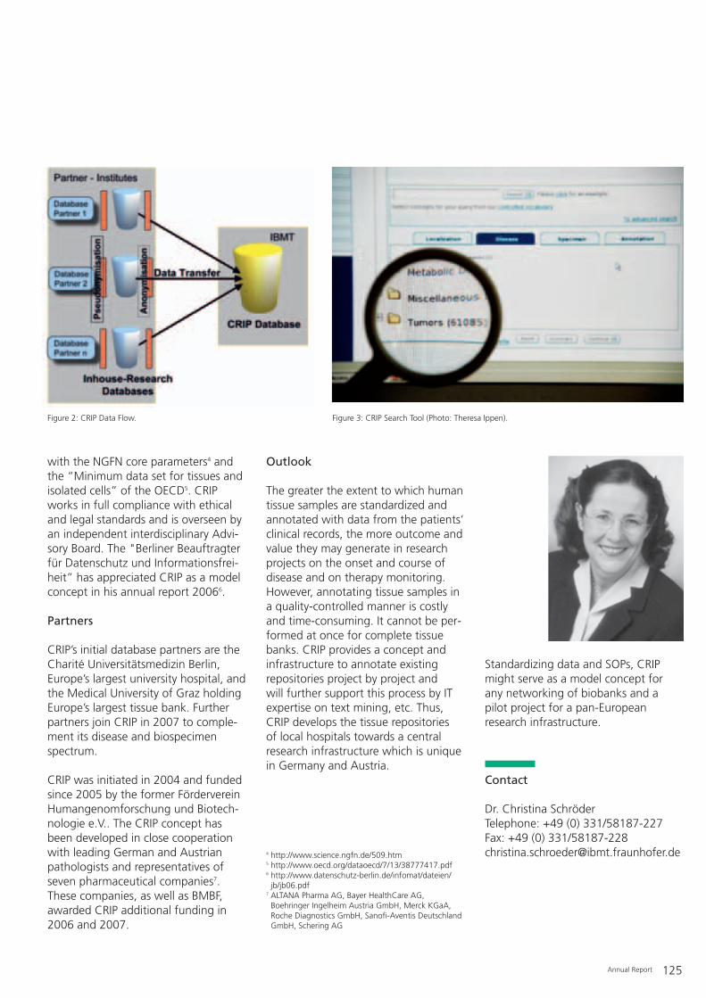
Annual Report 125
with the NGFN core parameters4 andthe “Minimum data set for tissues andisolated cells” of the OECD5. CRIPworks in full compliance with ethicaland legal standards and is overseen byan independent interdisciplinary Advi-sory Board. The "Berliner Beauftragterfür Datenschutz und Informationsfrei-heit” has appreciated CRIP as a modelconcept in his annual report 20066.
Partners
CRIP’s initial database partners are theCharité Universitätsmedizin Berlin,Europe’s largest university hospital, andthe Medical University of Graz holdingEurope’s largest tissue bank. Furtherpartners join CRIP in 2007 to comple-ment its disease and biospecimenspectrum.
CRIP was initiated in 2004 and fundedsince 2005 by the former FördervereinHumangenomforschung und Biotech-nologie e.V.. The CRIP concept hasbeen developed in close cooperationwith leading German and Austrianpathologists and representatives ofseven pharmaceutical companies7.These companies, as well as BMBF,awarded CRIP additional funding in2006 and 2007.
Outlook
The greater the extent to which humantissue samples are standardized andannotated with data from the patients’clinical records, the more outcome andvalue they may generate in researchprojects on the onset and course ofdisease and on therapy monitoring.However, annotating tissue samples ina quality-controlled manner is costlyand time-consuming. It cannot be per-formed at once for complete tissuebanks. CRIP provides a concept andinfrastructure to annotate existingrepositories project by project and will further support this process by ITexpertise on text mining, etc. Thus,CRIP develops the tissue repositories of local hospitals towards a centralresearch infrastructure which is uniquein Germany and Austria.
Standardizing data and SOPs, CRIPmight serve as a model concept forany networking of biobanks and apilot project for a pan-Europeanresearch infrastructure.
Contact
Dr. Christina SchröderTelephone: +49 (0) 331/58187-227Fax: +49 (0) 331/[email protected]
Figure 2: CRIP Data Flow. Figure 3: CRIP Search Tool (Photo: Theresa Ippen).
4 http://www.science.ngfn.de/509.htm 5 http://www.oecd.org/dataoecd/7/13/38777417.pdf 6 http://www.datenschutz-berlin.de/infomat/dateien/jb/jb06.pdf
7 ALTANA Pharma AG, Bayer HealthCare AG,Boehringer Ingelheim Austria GmbH, Merck KGaA,Roche Diagnostics GmbH, Sanofi-Aventis DeutschlandGmbH, Schering AG
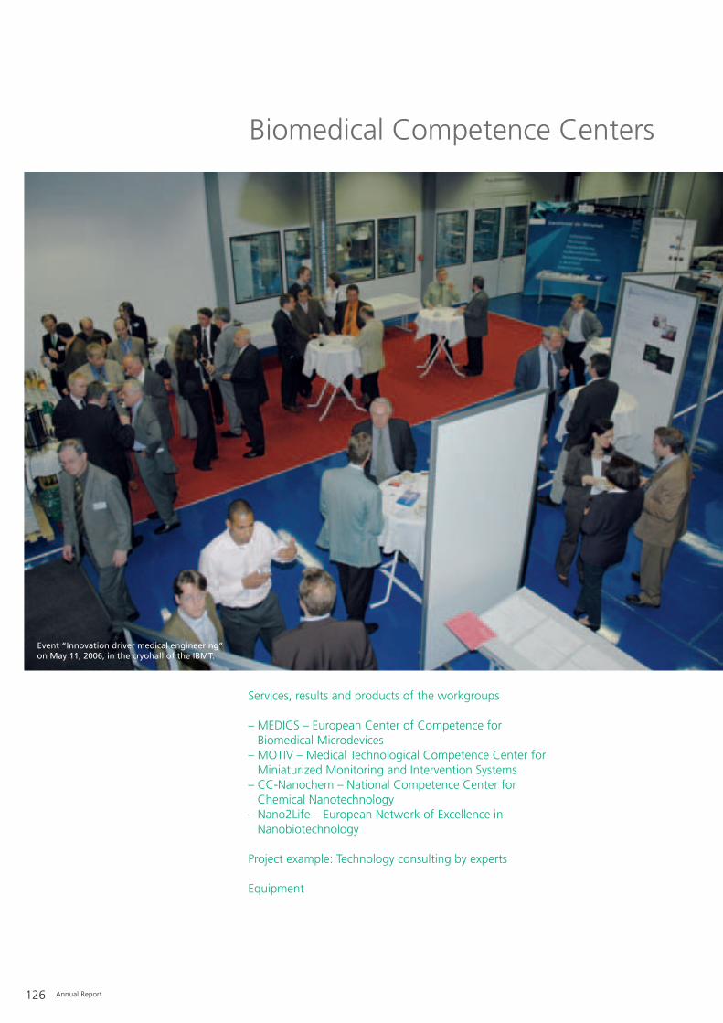
126 Annual Report
Biomedical Competence Centers
Services, results and products of the workgroups
– MEDICS – European Center of Competence for Biomedical Microdevices
– MOTIV – Medical Technological Competence Center forMiniaturized Monitoring and Intervention Systems
– CC-Nanochem – National Competence Center for Chemical Nanotechnology
– Nano2Life – European Network of Excellence inNanobiotechnology
Project example: Technology consulting by experts
Equipment
Event “Innovation driver medical engineering” on May 11, 2006, in the cryohall of the IBMT.
Annual Report 127
Dominated by the USA, Europe andJapan, the global market for biomed-ical technology amounts to around180 billion € annually, and is character-ized by stability with constant growthrates. Despite this apparent attractive-ness, the market for biomedical tech-nology is extremely complex and diffi-cult. Research and development in par-ticular must overcome immense chal-lenges in a tense field determined bycontinuous improvement in medicalcare accompanied by increasing sav-ings in costs, low production volumeswith high quality standards, longdevelopment times facing an increas-ing pace of innovation, cumbersomeapproval procedures and extensiveinterdisciplinary demands.
Micro, nano, optical and biotechnolo-gies are often described as the keytechnologies of the 21st century dueto their enormous possibilities, andthey show great potential to meetsuch complex demands. Consequently,the application of these technologieshas advanced considerably over recentyears: Capsule endoscopy in smallintestine diagnostics, the use ofnanoparticles for treating patients intumor therapy, as well as the wider useof active implants for the treatment ofepilepsy or Parkinson’s disease duringrehabilitation are only a few examplesimpressively demonstrating this.
The use of new technologies alonedoes not guarantee the developmentand production of successful productsand applications in biomedical technol-ogy. Rather, it is necessary to continu-ously assess the usefulness and risks,which can only be accomplished by aninterdisciplinary team of experts.
The workgroup “Biomedical Compe-tence Centers” at the Fraunhofer IBMTspecializes in novel technologies in theapplication areas of biomedical tech-nology. It supports small and medium-sized companies, industry, public cus-tomers, as well as banks and investors
in finding solutions for numerousproblems.
Contact European Center of Com-petence for Biomedical Microde-vices (MEDICS)Dipl.-Ing. Andreas SchneiderTelephone: +49 (0) 6897/[email protected]
Contact National Medical Engineer-ing Competence Center for Miniaturized Monitoring and Intervention Systems (MOTIV)Dipl.-Biol. Jochen SchmidtTelephone: +49 (0) 6897/[email protected]
Spokesman MOTIVProf. Dr. Günter R. FuhrTelephone: +49 (0) 6894/[email protected]
Contact National Competence Cen-ter for Chemical Nanotechnology(CC-Nanochem)Dipl.-Ing. Andreas SchneiderTelephone: +49 (0) 6897/[email protected]
Contact European Network ofExcellence in NanobiotechnologyNano2LifeDr. rer. nat., Dipl.-Biol. Meike Reimann-ZawadzkiTelephone: +49 (0) 6897/[email protected]
128 Annual Report
– Micro, nano, optical and biotech-nologies for biomedical applications
– Technology consulting– Feasibility studies and concept
assessment– Technology, patent and market
enquiries– Finding industrial and scientific
partners– Application, funding and coordina-
tion of R&D projects– Independent project management– Support with business start-up– Support with approval procedures
(MPG, MDD, FDA)– Internet information services– Workshops and training programs
Contact European Center of Com-petence for Biomedical Microde-vices (MEDICS)Dipl.-Ing. Andreas SchneiderTelephone: +49 (0) 6897/[email protected]
Contact National Medical Engineer-ing Competence Center for Minia-turized Monitoring and Interven-tion Systems (MOTIV)Dipl.-Biol. Jochen SchmidtTelephone: +49 (0) 6897/[email protected]
Contact National Competence Cen-ter for Chemical Nanotechnology(CC-Nanochem)Dipl.-Ing. Andreas SchneiderTelephone: +49 (0) 6897/[email protected]
Contact European Network ofExcellence in NanobiotechnologyNano2LifeDr. rer. nat., Dipl.-Biol. Meike Reimann-ZawadzkiTelephone: +49 (0) 6897/[email protected]
Biomedical Competence Centers
Services, results and products of the workgroups

Annual Report 129
Initial situation
The important role of the workgroup“Biomedical Competence Centers”and the Fraunhofer IBMT within thefield of biomedical technology isunderpinned by their being appointedas the European and national compe-tence centers for the following:– Coordination of the European Center
of Competence for BiomedicalMicrodevices “MEDICS” as assignedby the European Union(www.medics-network.com).
– Coordination of the National MedicalEngineering Competence Center“MOTIV” – Miniaturized Monitoringand Intervention Systems as assignedby the Federal Ministry of Educationand Research (www.motiv-medtech.de).
– Taking charge of nanobiotechnologycompetence within the NationalCompetence Center for ChemicalNanotechnology “CC-NanoChem”(www.cc-nanochem.de).
Services
Technology consulting – Ask theexpertsThe multidisciplinary core team of theworkgroup “Biomedical CompetenceCenters” at the Fraunhofer IBMT com-prises biologists, bionics experts, med-ical technology engineers and econo-mists. Moreover, we have access to aninternational network of experts in dif-ferent technology and applicationareas. The workgroup offers services support-ing Research & Development of bio-medical devices and applications.
In addition to project applications,independent project management,brokerage of project partners and sup-port with approval procedures, tech-nology consulting represents a particu-larly important field of expertise of theworkgroup. Technology consultingincludes feasibility and market studies,concept assessment and concept con-sulting as well as technical and patentenquiries.The following case study exemplifiesthe procedures and process of a con-sulting project at the Fraunhofer IBMT.
Case study of technology consultingfor an industrial customerThe goal of this technology consultingproject is to support an industrial cus-tomer in assessing a novel productiontechnology and in identifying potentialapplication areas within biomedicaltechnology.
The first contact with the industrialcustomer is mediated by a partnerwithin the international network of theworkgroup “Biomedical CompetenceCenters”.
Month 1:Establishing contact between the cus-tomer and staff of workgroup “Bio-medical Competence Centers” at theFraunhofer IBMT. First meeting at thecustomer’s location to discuss the chal-lenges and objectives as well as possi-bilities for support by the FraunhoferIBMT.
Month 2:Submission of an offer to the cus-tomer, which includes the compo-nents: design of a consulting concept,conducting interviews with experts,producing a results report, as well asfinal presentation with discussion ofthe results and further steps at the cus-tomer’s location.
Project example: Technology consulting by experts

130 Annual Report
Month 3:The industrial customer places theircommission, the Fraunhofer IBMTaccepts it.
Month 3-5:Work on the industry commission. Thetechnology under investigation is intro-duced with a short presentation, whichis prepared in cooperation with thecustomer, and subsequently discussedwith selected experts from differentdisciplines. A total of 24 expert inter-views are conducted where the follow-ing points are discussed and recordedin writing:– Assessment of the strengths and
weaknesses and working out theunique features of the technology.
– Discussion of possible applicationareas and their potential. More than20 application areas in biomedicaltechnology are examined as well aspotential non-biomedical technologyspin-offs are developed.
– Definition of important industry sup-pliers and downstream customers.
– Discussion of possible access routesto the respective industry branchesand downstream customers.
The contents of the expert interviewsare subsequently backed up by back-ground research and additional litera-ture. The final report concisely presentsall the results and background researchof the technology consultation as wellas presenting recommendations forfurther steps.
Month 5:Handing over of the final report.
Month 7:Presentation of the results at the cus-tomer’s location with subsequent dis-cussion of the results, the necessarymeasures as well as the further stepsand cooperation.
Month 9:Receipt of a consecutive commissionfrom the industrial customer.
ContactDipl.-Ing. Andreas SchneiderBiomedical Competence CentersIndustriestrasse 566280 SulzbachGermanyTelephone: +49 (0)6897/[email protected]

Annual Report 131
Equipment
Facilities and staff of the workgroup “Biomedical Competence Centers” at Fraunhofer IBMT.
Biomedical Competence Centers
– Interdisciplinary team of experts frombiomedical engineering, bionics, biology, micro- & nanotechnologyand economic engineering
– European Competence CenterMEDICS – Biomedical Microdevices
– National Competence Center MOTIV– Miniaturized Monitoring and Inter-vention Systems
– National Competence Center CC-NanoChem – Chemical Nanotechnol-ogy
– European Network of ExcellenceNano2Life – Nanobiotechnology
– Biomedical database– Biomedical Internet search engine– International network of suppliers
and users

132 Annual Report
Facts and Statistics
Names, Dates, Events
– National/international guests: scientists, research fellows and guest lecturers
– Exibition and event list
Scientific Publications
– Diplomas, masters, bachelors and PhD theses– Publications and talks 2006– Patents
IBMT at the joint stand of the Fraunhofer-Gesellschaft at theMEDICA 2006 in Duesseldorf.
Annual Report 133
Visiting scientists 2006
Isabella Guido Stiftung der Deutschen WirtschaftMarkus Küppers RWTH AachenRita M. Malpique Universität LissabonSven Martin JenLab, JenaJuan Martinez Leonardo da Vinci-Programm der EU Dr. Igor Morgenstern Universität RostockUta Siebert RWTH AachenAisada Uchugonova DAADHenrik W. Wagner Justus-Liebig-Universität GießenDr. Pavel Zinin Universität of Hawaii, USA
Guest lecturers 2006
Dr. Wataru Watanabe National Institut of Advanced Industrial Science & Technology (AIST), Japan
Prof. Dr. Albert van den Berg University of Twente, Niederlande
National/international guests: scientists,research fellows and guest lecturers
Names, Dates, Events
Nano2Life Business Day on NanoMed 200616.02.2006, Berlin
NanoMed 2006 – 5th International Workshop on BiomedicalApplications of Nanotechnology16.–17.02.2006, Berlin
MEDTEC 2006 – Fair and Conference07.–09.03.2006, StuttgartCoordination Fraunhofer Gemeinschaftsstandhttp://www.medtecshow.de/
MOTIV/ZPT-Unternehmertag »Innovationsmotor Medizin-technik: Anwendungen über die Medizin hinaus« 11.05.2006, Sulzbach
20. Treffpunkt Medizintechnik, Telemedizin und medizinische Informatik15.06.2006, Berlin
Workshop on Advanced Multiphoton and Fluorescence Lifetime Imaging Techniques19.–21.06.2006, St. Ingbert
Lange Nacht der Wissenschaften 200630.06.2006, Fraunhofer IBMT/AMBT, Institut für Biologie,Humboldt-Universität zu Berlin, Berlin
Health Care Forum Saar »Perspektiven der Krankenhaus-versorgung«04.10.2006, Saarbrücken
Kompetenzzentren für die Medizintechnik – eine Erfolgs-geschichte23.10.2006, Aachen
MEDICA 2006 – Weltforum der Medizin, InternationaleFachmesse mit Kongress 15.–18.11.2006, Düsseldorf, Halle 10 Stand F05http://www.medica.de
NanoTech 200614.–16.11.2006, Montreux
Exibition and event list

Diplomas, masters, bachelors, and PhD theses
Scientific Publications
134 Annual Report
Name University Qualification
Feili, Dara Universität des Saarlandes, Physik und Mechatronik Promotion
Fournelle, Marc Universität des Saarlandes, Physik Diplom
Geismann, Claudia Universität zu Lübeck, Biologie Master
Hanft, Marius HAW Hamburg, Physik/Biophysik Master
Kim, Sohee Universität des Saarlandes, Mechatronik Promotion
Lehmann, André FH Wildau, Biosystemtechnik Bachelor
Lobeda, Peter FH Wildau, Biosystemtechnik Bachelor
Maaß, Kirsten FH Saarbrücken, Mechatronik Diplom
Mietchen, Daniel Universität des Saarlandes, Physik und Mechatronik Promotion
Mundakapadam, Shirin RWTH Aachen, Biomedizinische Technik Master
Olbert, Marion FH Remagen, Biomedizinische Technik Diplom
Petschnik, Anna Universität zu Lübeck, Biologie Master
Rickmann, Christiane FH Jena, Medizintechnik Diplom
Steffen, Jenny Universität Potsdam, Molekularbiologie Promotion
Steinmetz, Oliver Universität des Saarlandes, Mechatronik Promotion
Tagliareni, Fabio Universität des Saarlandes, Mechatronik Promotion
Yildirim, Mehmet HTW Saarbrücken, Elektrotechnik Diplom

Annual Report 135
Department Microsystems/Laser Medicine
BECKER, W., BERGMANN, A., HAUSTEIN, E.,PETRASEK, Z., SCHWILLE, P., BISKUP, C., KELBAUSKAS, L., BENNDORF, K., KLÖCKNER,N., ANHUT, T., RIEMANN, I., KÖNIG, K.: „Fluorescence Lifetime Images and CorrelationSpectra obtained by Multidimensional Time-correlated Single Photon Counting. MicroscopyResearch and Technique”. 69, 186-195 (2006)
CSAKI, A., GARWE, F., STEINBRÜCK, A., MAUBACH, G., FESTAG, G., WEISE, A., RIEMANN, I., KÖNIG, K., FRITZSCHE, W.: „A Parallel Approach for Sub-wavelength Mole-cular Surgery using Gene-specific PositionedMetal Nanoparticles as Laser Light Antennas”, (eingereicht)
EHLERS, A., RIEMANN, I., MARTIN, S., LEHARZIC, R., BARTELS, A., JANKE, C., KÖNIG,K.: „High (1GHz) Repetition Rate Compact Fem-tosecond Laser: a Powerful Multiphoton Tool forNanomedicine and Nanobiotechnology”, (eingereicht)
EHLERS, A., RIEMANN, I., STARK, M., KÖNIG,K.: „Multiphoton Fluorescence Lifetime Imagingof Human Hair”.Microscopy Research and Technique, (in Druck)
HEYE, T., KUNTZ, C., DÜX, M., ENCKE, J., PALMOWSKI, M., AUTSCHBACH, F., VOLKE, F.,KAUFFMANN, G. W., GRENACHER, L.: „NewCoil Concept for Endoluminal MR Imaging“.European Radiology 16 (11), 2401-2409 (2006)
KÖHLER, M. J., KÖNIG, K., ELSNER, P., BÜCKLE,R., KAATZ, M.: „In Vivo Assessment of HumanSkin Aging by Multiphoton Laser ScanningTomography”.Optics Letters 31 (19), 2879-2881 (2006)
KÖNIG, K., EHLERS, A., STRACKE, F., RIEMANN,I.: „In Vivo Drug Screening in Human Skin usingFemtosecond Laser Multiphoton Microscopy”.Skin Pharmacol Physiol 19, 78-88 (2006)
KÖNIG, K., WYSS-DESSERICH, MT., TADIR, Y.,HALLER, U., TROMBERG, B., BERNS, M. W.,WYSS, P.: „Modifications of Protoporphyrin IXFluorescence during ALA-Based PhotodynamicTherapy of Endometriosis”.Medical Laser Application, 21, 291-297 (2006)
LEE, S.-C., CHO, J.-H., MIETCHEN, D., KIM, Y.-S., HONG, K. S., LEE, C., KANG, D.,PARK, K. D., CHOI, B.-S., CHEONG, C.: „Subcellular In Vivo 1H MR Spectroscopy ofXenopus Laevis Oocytes”.Biophysical Journal 90 (5), 1797-1803 (2006)
LUENGO, J., WEISS, B., SCHNEIDER, M.,EHLERS, A., STRACKE, F., KÖNIG, K., KOSTKA,K. H., LEHR, C. M., SCHÄFER, U. F.: „Influenceof Nanoencapsulation on Human Skin Transportof Flufenamic Acid”.Skin Pharmacol Physiol., 19, 190–197 (2006)
MANZ, B., COY, A., DYKSTRA, R., ECCLES, C. D.,HUNTER, M. W., PARKINSON, B. J., CALLAGHAN,P. T.: „A Mobile One-sided NMR-Sensor with aHomogeneous Magnetic Field: The NMR-MOLE“. Journal of Magnetic Resonance 183, 25-31 (2006)
MÜLLER, W. E. G., KALUZHNAYA, O. V., BELIKOV, S. I., ROTHENBERGER, M., SCHRÖDER,H. C., REIBER, A., KAANDORP, J. A., MANZ, B.,MIETCHEN, D., VOLKE, F.: „Magnetic ResonanceImaging of the Siliceous Skeleton of the Demos-ponge Lubomirskia Baicalensis”.Journal of Structural Biology 153, 31-41 (2006)
SCHENKE-LAYLAND, K., MADERSHAHIAN, N.,RIEMANN, I., STARCHER, B., HALBHUBER, J.,KÖNIG, K., STOCK, U. A.: „Impact of Cryo-preservation on Extracellular Matrix Structuresof Heart Valve Leaflets”.The Annals of Thoracic Surgery (2006), (in Druck)
SCHENKE-LAYLAND, K., KÖNIG, K.: „Two-Photon Microscopes and In Vivo MultiphotonTomographs – Novel Diagnostic Tools for TissueEngineering and Drug Delivery.”Special issue: Multiphoton Imaging: Diseasesand Therapies. ADDR, (in Druck)
SCHENKE-LAYLAND, K., XIE, J., HAGVALL, S. H.,KÖNIG, K., ANGELIS, E., HAMM-ALVAREZ, S. F.,STOCK, U. A., BROCKBANK, K.G.M., MACLELLAN, W. R.: „Optimized Preservation ofExtra-Cellular Matrix in Cardiac Tissues: Implications for Long-term Graft Durability”,(eingereicht)
STARK, M., MANZ, B., EHLERS, A., KÜPPERS,M., RIEMANN, I., VOLKE, F., SIEBERT, U.,WESCHKE, W., KÖNIG, K.: „MultiparametricHigh Resolution Imaging of Barley Embryos byMultiphoton Microscopy and Magnetic Resonance Micro-Imaging”.to Microscopy Research and Technique, (in Druck)
STRACKE, F., WEISS, B., LEHR, C. M., KÖNIG, K.,SCHÄFER, U. F., SCHNEIDER, M.: „MultiphotonMicroscopy for the Investigation of DermalPenetration of Nanoparticle-borne Drugs”.Journal of Investigative Dermatology, 126,2224-2233 (2006)
VELTEN, T., SCHUCK, H., KNOLL, T., SCHOLZ,O., SCHUMACHER, A., GÖTTSCHE, T., WOLLF,A., BEISKI, B. Z., and IntelliDrug Consortium:„Intelligent Intraoral Drug Delivery Microsystem”.Journal of Mechanical Engineering Science -Part C, C11 (November 2006)
WANG, B. G., KÖNIG, K., RIEMANN, I., SCHUBERT, H., HALBHUBER, K. J.: „Applicationof Multiphoton Microscopy in Visualizing Intra-Tissue Optical Nanosurgery induced byNon-amplified Femtosecond Lasers”, (eingereicht)
WANG, B. G., KÖNIG, K., RIEMANN, I., KRIEG,R., HALBHUBER, K.-J.: „Intraocular MultiphotonMicroscopy with Subcellular Spatial Resolutionby Femtosecond Lasers“.Histochem Cell Biol. 124, 177-188 (2006)DOI 10.1007/s00418-006-0187-0, 2006
WANG, B. G., KÖNIG, K., HALBHUBER, K.-J.:„Corneal Multiphoton Microscopy and Intratissue Optical Nanosurgery by NanojouleFemtosecond Near Infrared Pulsed Lasers“.Annals of Anatomy, 2006, (eingereicht)
Department Ultrasound
WIKLUND, M., GÜNTHER, C., LEMOR, R. M.,JÄGER, M., FUHR, G. R., HERTZ, H. M.: „Ultra-sonic standing Wave Manipulation Technologyintegrated into a Dielectrophoretic Chip”. Lab Chip, DOI: 10.1039/b612064b, Germany(2006)
ZININ, P. V., WEISS, E.C., ANASTASIADIS, P.,LEMOR, R. M.: “Mechanical Properties of HeLaCells at Different Stages of Cell Cycle by Time-resolved Acoustic Microscope”. The Journal of the Acoustical Society of America, 120(5) 3230 (2006)
Department Medical Engineering & Neuroprothesis
BOSSI, S., MENCIASSI, A., KOCH, K. P., HOFF-MANN, K.-P., YOSHIDA, K., DARIO, P., MICERA,S.: „Shape Memory Alloy Microactuation of tf-LIFEs: Theoretical Study and PreliminaryResults”. IEEE Transactions on Biomedical Engineering,(eingereicht)
CITI, L., CARPANETO, J.,YOSHIDA, K., HOFF-MANN, K.-P., KOCH, K. P., DARIO, D., MICERA,S.: „On the Use of LIFEs to identify Neural Infor-mation: Towards a Neuro-controlled ProstheticHand”. Journal of Neural Engineering, (eingereicht)
FEILI, D., SCHUETTLER, M., DOERGE, T., KAM-MER, S., HOFFMANN, K.-P., STIEGLITZ, T.: „Flexible Organic Field Effect Transistors for Biomedical Microimplants using Polyimide andParylene C as Substrate and Insulator Layer”.J. Micromech. Microeng. 16, 1–7 (2006)
GUIRAID, D., STIEGLITZ, T., KOCH, K. P.,DIVOUX, J.-L., RABISCHONG, P.: „An Implanta-ble Neuroprostheses for Standing and Walkingin Paraplegia: Five Year Patient Follow-up”.Journal of Neural Engineering 3, 268-275(2006)
HSU, J., RIETH, L., VANFLEET, R., KAMMER, S.,KOCH, K. P., NORMANN, R. A., SOLZBACHER,F.: „Characterizations of a-SiCx:H Film as Encapsulation Material for Integrated SiliconBase Neural Interface Devices”.Thin Solid Films, (eingereicht)
1. Articles in Journal (print or online), peer-reviewed
Publications and talks 2006
136 Annual Report
LAGO, N., YOSHIDA, K., KOCH, K. P., NAVARRO, X.: „Assessment of Biocompatibilityof chronically Implanted Polyimide and Platiumum Intrafascicular Electrodes”. IEEE Transactions on Biomedical Engineering,(akzeptiert)
LAGO, N., UDINA, E., RAMACHANDRAN, A.,NAVARRO, X.: „Neurobiological Assessment ofRegenerative Electrodes for Bidirectional Interfacing of Injured Peripheral Nerves”.IEEE Transactions on Biomedical Engineering, (in Druck)
RAMACHANDRAN, A., SCHUETTLER, M., LAGO,N., DOERGE, T., KOCH, K. P., NAVARRO, X.,HOFFMANN, K.-P., STIEGLITZ, T.: „Design, invitro and in vivo Assessment of a Multi-ChannelSieve Electrode with Integrated Multiplexer”. Journal Neural Engineering 3, 114-124 (2006)
RAMACHANDRAN, A., JUNK, M., KOCH, K. P.,HOFFMANN,K.-P.: „A Study of Parylene C Poly-mer Deposition Inside Microscale Gaps”. IEEE Journal of Advanced Packaging, (eingereicht)
Department Cryobiophysics & Cryotechnology
IHMIG, F. R., SHIRLEY, S. G., DURST, C. H. P., ZIMMERMANN, H.: „Cryogenic ElectronicMemory Infrastructure for Physically RelatedContinuity of Care Records of Frozen Cells”.Cryogenics, VOL: 46, S. 312-320 (2006)
KIESEL, M., REUSS, R., ENDTER, J., ZIMMER-MANN, D., ZIMMERMANN, H., SHIRAKASHI, R.,BAMBERG, E., ZIMMERMANN, U., SUKHORUKOV, V. L.: „Swelling-activatedPathways in Human T-Lymphocytes studied byCell Volumetry and Electrorotation“.Biophysical J., VOL: 90, S. 4720-4729 (2006)
MALPIQUE, R., KATSEN-GLOBA, A., CARRONDO, M. J. T., ZIMMERMANN, H.,ALVES, P. M.: „Cryopreservation in Microvolu-mes: Impact upon Caco-2 Colon Adenocarci-noma Cells Proliferation and Differentiation”.Biotechnol. Bioengineering, (eingereicht)
SUKHORUKOV, V. L., REUSS, R., ENDTER, J. M.,FEHRMANN, S., KATSEN-GLOBA, A., GEßNER,P., STEINBACH, A., MÜLLER, K. J., KARPAS, A.,ZIMMERMANN, U., ZIMMERMANN, H.: „A Biophysical Approach to the Optimisation ofDendritic Tumour Cell Electrofusion“.Biochem. Biophys. Res. Comm., VOL: 346, S. 829-839 (2006)
Department Biohybrid Systems
ALUIGI, M. G., ANGELINI, C., FALUGI, C., FOSSA, R., GENEVER, P., GALLUS, L., LAYER, P.G., PRESTIPINO, G., RAKONCZAY, Z., SGRO, M.,THIELECKE, H., TROMBINO, S.: „Interaction between Organophosphate Compounds andCholinergic Functions during Development“. Chemico-Biological Interactions 157-158, 305-316 (2005). Epub 2005 Oct 28.
BOUAZZAOUI, A., KREUTZ, M., EISERT, V., DINAUER, N., BRACHARZ, S., HEINZELMANN, A.,HALLENBERGER, S., STRAYLE, J., ANDREESENR., VON BRIESEN, H.: „Stimulated Trans-actingFactor of 50kD (Staf50) Inhibits HIV-1 Replicati-on in Human Monocyte-derived Macrophages“.Virology (2006), (in Druck)
CHO, S., BECKER, S., VON BRIESEN, H., THIELECKE, H.: „Impedance Monitoring of Herpes Simplex Virus-Induced Cytopathic Effectin Vero Cells”.Sensors & Actuators B (2006), (eingereicht)
CHO, S., THIELECKE, H.: „Micro Hole-based CellChip with Impedance Spectroscopy”.Biosensors & Bioelectronics (2006), (in Druck)
CHO, S., THIELECKE, H.: „Influence of the Electrode Position on the Characterisation ofArtery Stenotic Plaques by using ImpedanceCatheter”.IEEE Transactions on Biomedical Engineering,Vol. 53, No. 11 (2006), (in Druck)
HOOGDUIJIN, M. J., GORJUP, E., GENEVER, P. G.: „Comparative Characterization of Hair Follicle Dermal Stem Cells and Bone MarrowMesenchymal Stem Cells”.Stem Cells Dev. 15, 49-60 (2006)
KUFLEITNER, J., HERMANNS, J., VON BRIESEN,H., KREUTER, J.: „Nanoparticulate Systems forthe Brain Delivery of Oximes. First Loading Studies and Cell Culture Experiments.” Toxicology, (im Druck) (2006)
LEROY, V., SAKAROVITCH, C., CORTINA-BORJA,M., MCINTYRE, J., COOVADIA, H., DABIS, F.,NEWELL, M. L, SABA, J., GRAY, G., NDUGWA,CH., KILEWO, CH., MASSAWE, A., KITUUKA, P.,OKONG, P., GRULICH, A., VON BRIESEN, H.,GOUDSMIT, J., BIBERFELD, G., HAVERKAMP, G.,WEVERLING, G. J., LANGE, J. M., GHENTGROUP ON HIV IN WOMEN AND CHILDREN: „Isthere a Difference in the Efficacy of PeripartumAntiretroviral Regimens in reducing Mother-to-Child Transmission of HIV in Africa?”.AIDS 19, 1865-1875 (2005)
SANCHEZ DE JUAN, B., VON BRIESEN, H., GELPERINA, S., KREUTER, J.: „Investigation ofCytotoxicity of Doxorubicin Bound to Poly(butylcyanoacrylate) Nanoparticles in Rat Glioma CellLines using Different Assays“.J. Drug Targeting (2006), (in Druck)
THIELECKE, H., IMPIDJATI, FUHR, G. R.: „Biopsy on Living Cells by Ultra Slow InstrumentMovement”. Journal of Physics: Condensed Matter 18, S 627-637 (2006)
Project Group Cell Differentiation & Cell Technology
GULDNER, N. W., KAJAHN, J., KLINGER, M., SIEVERS, H.-H., KRUSE, C. : „AutonomouslyContracting Human Cardiomyocytes generatedfrom Adult Pancreatic Stem Cells and enhancedin Cocultures with Myocardial Biopsies”.Int. J. Artif. Org., (in Druck)
KRUSE, C., BODO, E., PETSCHNIK, A. E., DANNER, S., TIEDE, S., PAUS, R.: „Towards theDevelopment of a Pragmatic Technique for iso-lating and differentiating Nestin-positive Cellsfrom Human Scalp Skin into Neuronal and GlialCell Populations: Generating Neurons fromHuman Skin?”.Exp. Dermatol 15, 794-801 (2006)
KRUSE, C., KAJAHN, J., PETSCHNIK, A. E.,MAASS, A., KLINK, E., RAPOPORT, D. H.,WEDEL, T.: „Adult Pancreatic Stem/ProgenitorCells spontaneously Differentiate in vitro intoMultiple Cell Lineages and form Teratoma-like Structures”. Ann. Anat., (in Druck)
Department Cellular Biotechnology & Biochips
BÖTTCHER, M., JÄGER, M., RIEGGER, L.,DUCREE, J., ZENGERLE, R., DUSCHL, C.: „Lab-On-Chip-based Cell Separation by combi-ning Dielectrophoresis and Centrifugation“.Biophysical Reviews and Letters, (eingereicht)
FELTEN, M., GEGGIER, P., JÄGER, M., DUSCHL,C.: „Controlling Electrohydrodynamic Pumpingin Microchannels through Defined TemperatureFields“.Phys. Fluids 18, 051707 (2006)
JÄGER, M., UHLIG, K., CLAUSEN-SCHAUMANN,H., DUSCHL, C.: „An AFM Study of the Struc-ture and Functionality of a Contractile ProteinAggregate“.Biophys. J., (eingereicht)
JÄGER, M., MÜLLER, T., SCHNELLE, T.: „Thermometry in Dielectrophoresis Chips forContact-free Cell Handling“.Journal of Physics D: Applied Physics, (eingereicht)
WIKLUND, M., GÜNTHER, C., LEMOR, R.,JÄGER, M., FUHR, G.R., HERTZ, H. M.:„Ultrasonic Standing Wave Manipulation Technology integrated into a DielectrophoreticChip“.Lab Chip, DOI:10.1039/b612064b (2006)

Annual Report 137
Department Molecular Bioanalytics & Bioelectronics
ANDRESEN, H., GRÖTZINGER, C., ZARSE, K.,KREUZER, O. J., EHRENTREICH-FÖRSTER, E.,BIER, F. F.: „Functional Peptide Microarrays forSpecific and Sensitive Antibody Diagnostics“.Proteomics, 6, 1376-1384 (2006)
ANDRESEN, H., GRÖTZINGER, C., ZARSE, K.,BIRRINGER, M., HESSENIUS, C., KREUZER, O. J.,EHRENTREICH-FÖRSTER, E., BIER, F. F.: „PeptideMicroarrays with Site-specifically ImmobilizedSynthetic Peptides for Antibody Diagnostics“.Sensors and Actuators B, 113, 655-663 (2006)
ANDRESEN, H., ZARSE, K., GRÖTZINGER, C.,EHRENTREICH-FÖRSTER, E., BIER, F. F., KREUZER,O. J.: „Development of Peptide Microarrays forEpitope Mapping of Antibodies against theHuman TSH Receptor”.Journal of Immunological Methods, 315, 11-18(2006)
NÜBEL, U., ANTWERPEN, M., WITTE, W.,EHRENTREICH-FÖRSTER, E., SCHELLHASE, M.,BIER, F. F.: „DNA-Microarray for Detection ofAntibiotic Resistance Determinants in Bacillusanthracis and closely related Bacillus cereus“.Molecular & Cellular Probes, (in Druck)
VON NICKISCH-ROSENEGK, M., MARSCHAN,X., BIER, F. F.: „Quantitative on-Chip-Detectionof mRNA by RT-PCR at Immobilized Primers“.BMC J. Biotechnology, (eingereicht)
Department Microsystems/Laser Medicine
ANHUT, T., HASSLER, K., LASSER T., KÖNIG, K.,RIGLER, R.: „Fluorescence Correlation Spectros-copy on Dielectric Surfaces in Total InternalReflection Geometries”. SPIE-Proceedings, vol. 5699 (2006), (in Druck)
BECKER, W., BERGMANN, A., BISCOTTI, G.,KÖNIG, K., RIEMANN, I., KELBAUSKAS, L., BISKUP, C.: „High-speed FLIM Data Acquisitionby Time-correlated Single-photon Counting”.SPIE-Proceedings, vol. 5323 (2006), (in Druck)
BECKER, W., BERGMANN, A., HAUSTEIN, E.,PETRASEK, Z., SCHWILLE, P., BISKUP, C., ANHUT,T., RIEMANN, I., KÖNIG, K.: „Fluorescence Life-time Images and Correlation Spectra Obtainedby Multidimensional TCPC”. SPIE-Proceedings, vol. 5700 (2006), (in Druck)
CSAKI, A., MAUBACH, G., GARWE, F., STEINBRÜCK, A., KÖNIG, K., FRITZSCHE, W.: „A Novel DNA Restriction Technology based onLaser Pulse Energy Conversion on Sequence-specific Bound Metal Nanoparticles”. SPIE-Proceedings, vol. 5699 (2006), (in Druck)
CSAKI, A., GARWE, F., STEINBRÜCK, A., WEISE,A., KÖNIG, K., FRITZSCHE, W.: „Localization ofLaser Energy Conversion by Metal Nano-particles: Basic Effects and Applications”. SPIE-Proceedings, vol. 6191 (2006), (in Druck)
EHLERS, A., RIEMANN, I., ANHUT, T., KOBOW,J., KÖNIG, K.: „Multiphoton Tomography of Epidermis and Dermis”.SPIE-Proceedings, vol. 5700 (2006), (in Druck)
FISCHER, F., KÖNIG, K., PUSCHMANN, S., WEPF,R., RIEMANN, I., ULRICH, V., FISCHER, P.: „Characterization of Multiphoton Laser Scanning Device Optical Parameters for ImageRestoration”.SPIE-Proceedings, vol. 5463 (2006), (in Druck)
FRITZSCHE, W., CSAKI, A., STEINBRÜCK, A.,GARWE, F., KÖNIG, K., RASCHKE, M.: „MetalNanoparticles as Passive and Active Tools forBioanalytics”.SPIE-Proceedings, vol. 5699 (2006), (in Druck)
KÖNIG, K., SCHUCK, H., SAUER, D., BAUER-FELD, F., STRACKE, F., VELTEN, T., TCHERNOOK,A., MARTIN, S., LEHARZIC, R.: „FemtosecondLaser Nanoprocessing using Near Infrared Nano-joule Pulses at MHZ Repetition Frequency”.SPIE-Proceedings, vol. 6400 (2006), (in Druck)
KÖNIG, K.: „Multiphoton Tomography, Transfec-tion, and Nanosurgery with (less-than) 2-nJ, 80-MHz Femtosecond Laser Pulses”.SPIE-Proceedings, vol. 5340 (2006), (in Druck)
KÖNIG, K., GARWE, F., CZAKI, A., MAUBACH,G., RIEMANN, I., FRITZSCHE, W.: „Nano-processing of DNA with Femtosecond Laser”.SPIE-Proceedings, vol. 5462 (2006), (in Druck)
KÖNIG, K.: „Femtosecond Laser Application inBiotechnology and Medicine”.SPIE-Proceedings, vol. 5662 (2006), (in Druck)
KÖNIG, K., RIEMANN, I., EHLERS, A., BÜCKLE,R., DIMITROV, E., KAATZ, M., FLUHR, J., ELSNER,P.: „In-Vivo Multiphoton Tomography of SkinCancer”.SPIE-Proceedings, vol. 5686 (2006), (in Druck)
KÖNIG, K., RIEMANN, I., SCHUCK, H., SAUER,D., VELTEN, T., LEHARZIC, R.: „Time-resolvedand spectrally Resolved 5D Multiphoton Microscopy for Analysis and Nanoprocessing ofMaterials”.SPIE-Proceedings, vol. 5713 (2006), (in Druck)
KÖNIG, K., WANG, B., RIEMANN, I., KOBOW, J.:„Cornea Surgery with Nanojoule FemtosecondLaser Pulses”.SPIE-Proceedings, vol. 5688 (2006), (in Druck)
KÖNIG, K., RIEMANN, I., EHLERS, A., LEHARZIC,R.: „In vivo Non-invasive Multiphoton Tomography of Human Skin”.SPIE-Proceedings, vol. 5990 (2006), (in Druck)
KÖNIG, K., SCHUCK, H., SAUER, D., BAUER-FELD, F., STRACKE, F., VELTEN, T., TCHERNOOK,A., MARTIN, S., LEHARZIC, R.: „FemtosecondLaser Nanoprocessing using Near Infrared Nano-joule Pulses at MHZ Repetition Frequency”.SPIE-Proceedings, vol. 6400 (2006), invited paper
LEHARZIC, R., BREITLING, D., SOMMER, S.,FOHL, C., VALETTE, S., KÖNIG, K., DAUSINGER,F., AUDOUARD, E.: „Pulse Duration and EnergyDensity Influence on Laser Processing of Metalswith Short and Ultrashort Pulses”.SPIE-Proceedings, vol. 5713 (2006), (in Druck)
LEHARZIC, R., MARTIN, S., BÜCKLE, R., WULLNER, C., DONITZKY, C., RIEMANN, I.,KÖNIG, K.: „New Developments in CornealRefractive Surgery with Femtosecond Laser Pulses”.SPIE-Proceedings, vol. 6138 (2006), (in Druck)
MIETCHEN, D.: „3D Magnetic ResonanceMicroscopy of Dehydrated Biological Specimens”.Doktorarbeit, Fraunhofer-Institut für Biomedizi-nische Technik – Abteilung Magnetische Resonanz und Universität des Saarlandes –Fakultät Physik und Mechatronik, Mai 2006
2. Articles in Journal (print or online), not peer-reviewed(or scientific papers)
138 Annual Report
RIEMANN, I., SCHENKE-LAYLAND, K., EHLERS,A., DIMITROV, E., KAATZ, M., ELSNER, P., MARTIN, S., KÖNIG, K.: „High-resolution Multiphoton Optical Tomography of Tissues –an In Vitro and In Vivo Study”.SPIE-Proceedings, vol. 6142 61420N-1 (2006)
RIEMANN, I., STRACKE, F., SAUER, D., MARTIN,S., KÖNIG, K.: „Multiphoton Nanosurgery inCells and Tissues”.SPIE-Proceedings, vol. 6089, doi:10.1117/12.646096 (2006)
WANG, B., HALBHUBER, K. J., RIEMANN, I.,KÖNIG, K.: „In Vivo Corneal Nonlinear OpticalTomography Based on Second Harmonic andMultiphoton Autofluorescence Imaging inducedby Near Infrared Femtosecond Lasers with Rabbits”.SPIE-Proceedings, vol. 5964 (2006), (in Druck)
WANG, B. G., KÖNIG, K., RIEMANN, I., SCHUBERT, H., HALBHUBER, K.-J.: „Multi-photon Imaging of Corneal Tissue with NearInfrared Femtosecond Laser Pulses: CornealOptical Tomography and its Use in RefractiveSurgery”.SPIE-Proceedings, vol. 6089 (2006), (in Druck)
Department Ultrasound
ZININ, P. V., WEISS, E. C., ANASTASIADIS, P.,LEMOR, R. M.: „Mechanical Properties of HeLaCells by Time-resolved Acoustic Microscope”. Proceedings of the Fifth International Conferen-ce on the Ultrasonic Measurement and Imagingof Tissue Elasticity, p. 42 (2006)
Department Cryobiophysics & Cryotechnology
DURST, C. H. P., IHMIG, F. R., BIEL, M., DAFFERTSHOFER, M., ZIMMERMANN, H.: „A Method and Infrastructure for Long-termManaging of Sample Preparation Knowledge for Cryobiomedical Applications“.Proceedings 6th International Conference onKnowledge Management (I-KNOW), in Graz (Österreich), 06.-08.09.2006
FUCHS, C. C., IHMIG, F. R., SHIRLEY, S. G., ZIMMERMANN, H.: „Optoelectronic SignalTransmission between Liquid Nitrogen andRoom Temperature”. Proceedings 7th European Workshop on LowTemperature Electronics (WOLTE-7), VOL: 264, S. 255-262in Noordwijk (Niederlanden), 21.06.2006
IHMIG, F. R., SHIRLEY, S. G., FUCHS, C. C.,DURST, C. H. P., ZIMMERMANN, H.: „Low Tem-perature Electronics for the Cryopreservation ofLiving Cells”.Vortrag anlässlich des 7th European Workshopon Low Temperature Electronics (WOLTE-7),VOL: 264, S. 13-20in Noordwijk (Niederlanden), 21.06.2006
SHIRLEY, S. G., IHMIG, F. R., ZIMMERMANN, H.:„Prototype Electronic Infrastructure for a Cryo-Repository“.Posterbeitrag anlässlich des 7th EuropeanWorkshop on Low Temperature Electronics(WOLTE-7), VOL: 264, S.263-268in Noordwijk (Niederlanden), 21.-23.06.2006
ZIMMERMANN, H., IHMIG, F. R., KATSEN-GLOBA, A., EHRHART, F., DURST, C. H. P., SHIRLEY, S.G.: „Cryobiotechnology – Low Temperature Microsystems for Biotechnologyand Regenerative Medicine“.Microsystems Technology, VOL: 2006, S. 22-25(2006)
Department Biohybrid Systems
HILDEBRANDT, C., THIELECKE, H.: „Technolo-gien für die Zellforschung und Zelltherapie“.Bio World, Vol. 3, pp. 2-4 (2006)
Department Molecular Bioanalytics & Bioelectronics
STEFFEN, J., BIER, F. F.: „RNA- und Proteinsyn-these auf Oberflächen, Vernetzte Prozesse“.HighChem hautnah, Gesellschaft DeutscherChemiker, GDCh (Hrsg.), 42 (2006)

Annual Report 139
FUHR, G. R.: „Ultra Slow Manipulation - A NewTechnique for Gentle (Single) Cell Handling“.Plenarvortrag anlässlich des 41. Winter-kolloquiums der Universität Bielefeld,in Klosters (Schweiz), 22.01.2006
FUHR, G. R.: „Sanft wie im Körper – In-vitro-Zellhandhabung für die regenerative Medizin“.Vortrag anlässlich der Versammlung der Freundeder Universität des Saarlandes,in Saarbrücken (Saarland), 14.02.2006
FUHR, G. R.: „Ultraslow Instrument Manipulati-on - A New Technique for Gentle Cell Handling“.Vortrag anlässlich des OMNT Seminars „Nano-technologies for Cell Investigation“, in Paris (Frankreich), 14.03.2006
FUHR, G. R.: „Gentle Cell Manipulation forRegenerative Medicine“.Vortrag anlässlich des 12. International Work-shop on Stem Cells and Calcified Tissues,in Tel Aviv (Israel), 21.03.2006
FUHR, G. R.: „Medizintechnik – Initiator undMotor für das Saarland“.Vortrag anlässlich des ZPT-MOTIV Unter-nehmertages,in Sulzbach (Saarland), 11.05.2006
FUHR, G. R.: „Zellmanipulation und -differenzie-rung mittels nanostrukturierter und funktionali-sierter Oberflächen - IP der EU CellPROM“.Vortrag anlässlich des Delegationsbesuchs desWirtschaftsministeriums des Saarlands bei derSchering AG,in Berlin (Berlin), 22.05.2006
FUHR, G. R.: „Frozen Medicine – Cryobanking inBiotechnology”.Eröffnungsvortrag anlässlich des Workshop onAdvanced Multiphoton and Fluorescence Life-time Imaging Techniques, in St. Ingbert/Sulzbach (Saarland), 19.-21.06.2006
FUHR, G. R.: „Cell Surface Interactions andGentle Cell Handling“.Vortrag anlässlich der EST Conference Bio-Systems Berlin 2006, in Berlin (Berlin), 26.06.2006
FUHR, G. R.: „The Role of Surfaces in Stem CellDifferentiation: Theoretical Aspects and FuturePerspectives“.Eröffnungsvortrag anlässlich der 6th Baltic Summer School, in Kiel (Schleswig-Holstein), 20.08.2006
FUHR, G. R.: „Ultralangsame Manipulation tierischer und humaner Zellen“.Vortrag anlässlich der Tagung der DeutschenGesellschaft für Biophysik 2006,in Mainz (Rheinland-Pfalz), 26.-27.09.2006
FUHR, G. R.: „Biotechnologie in Deutschland -Ansätze, Probleme und neue Geschäftsfelder“.Vortrag anlässlich der Veranstaltung für denArbeitskreis Wirtschaft e. V.,in St. Ingbert (Saarland), 17.10.2006
FUHR, G. R.: „Surface Supported Cell Manipula-tion and Differentiation – A New Generation ofCell Handling Devices“.Vortrag anlässlich des Fraunhofer Life ScienceSymposium Leipzig 2006, in Leipzig (Sachsen), 22.-24.10.2006
FUHR, G. R.: „Forschungsfelder des FraunhoferIBMT“.Vortrag anlässlich der Summer School des Fraunhofer IBMT, Abteilung Zelldifferenzierung & Zelltechnologie, in Lübeck (Schleswig-Holstein), 22.-23.11.2006
FUHR, G. R.: „Biotechnologie und Nano-technologie“.Vortrag anlässlich des Kolloquiums der Univer-sität Kaiserslautern,in Kaiserslautern (Rheinland-Pfalz), 27.11.2006
FUHR, G. R.: „Sanfte Zellmanipulation für dieregenerative Medizin und Biokompatibilität“.Ringvorlesung „Wohin steuert die Bundesrepu-blik“ der Technischen Universität Braunschweig,Peter-Lang-Verlag, Frankfurt 2006
Department Microsystems/Laser Medicine
ANHUT, T., DUVENECK, G. L., RIEMANN, I.,KÖNIG, K.: „Evanescent Multiphoton Excitationof Cells grown on Planar Waveguides”.Poster anlässlich der Photonics West Konferenz2006,in San José (USA), 23.01.2006
BECKER, W., BERGMANN, A., KÖNIG, K., BISKUP, C.: „Multispectral Fluorescence LifetimeImaging by TCSPC“.Photonics West Konferenz 2006,in San José (USA), 23.01.2006
EHLERS, A., RIEMANN, I., ANHUT, T., KAATZ, M.,ELSNER, P., KÖNIG, K.: „Fluorescence LifetimeImaging of Human Skin and Hair“.Photonics West Konferenz 2006,in San José (USA), 23.01.2006
EHLERS, A., KÖNIG, K.: „In Vivo MultiphotonEndoscopy based on a GRIN-Lens”.EOS 2006,in Paris (Frankreich), 18.10.2006
KAATZ, M., RIEMANN, I., KÖNIG, K.: „Multiphoton Tomography of Melanoma”.Vortrag beim International Workshop on Advanced Multiphoton and Fluorescence Lifetime Imaging Techniques,in St. Ingbert (Saarland), 19.-21.06.2006
KIM, S., SCHOLZ, O., ZOSCHKE, K., HARRISON,R., SOLZBACHER, F., KLEIN, M., TOEPPER, M.:„FEA Simulation of Thin Film Coils to PowerWireless Neural Interfaces”. Vortrag anlässlich der Nanotech 2006,in Boston (USA), 07.-11.05.2006
KIM, S., KNOLL, T., SCHOLZ, O.: „Feasibility ofInductive Communication between Millimeter-sized Robots”.Vortrag anlässlich der 1st IEEE/RAS-EMBS Inter-national Conference on Biomedical Robotics andBiomechatronics,in Pisa (Italien), 20.-22.02.2006.Proceedings, pp. 1178-1182 (2006)
KÖNIG, K., RIEMANN, I., EHLERS, A.: „Multiphoton Tomography of Skin Cancer”.Photonics West Konferenz, in San José (USA), 23.01.2006
KÖNIG, K.: „Multiphoton Imaging of HumanSkin”.64th Annual Meeting der American Academy of Dermatology,in San Francisco (USA), 03.-07.03.2006
KÖNIG, K., RIEMANN, I., STRACKE, F., LEHARZIC, R.: „Nanoprocessing with NanojouleNear Infrared Femtosecond Laser Pulses”.Focus on Microscopy FOM2006, in Perth (Australien), 10.04.2006
KÖNIG, K.: „Two-Photon Excited Optical Tomography of Human Skin”.1st Annual Advanced Optical Methods Workshop in Shenzhen (China), 26.-28.05.2006
KÖNIG, K.: „Clinical Multiphoton Endoscopy”.International Workshop on Advanced Multi-photon and Fluorescence Lifetime Imaging Techniquesin St. Ingbert (Saarland), 19.-21.06.2006
KÖNIG, K., RIEMANN, I., DIMITROW, E., EHLERS,A., FLUHR, J., ELSNER, P., KOBOW, J., KAATZ,M.: „High Resolution In Vivo Multiphoton Tomography of Melanoma”. Poster anlässlich des International Workshop onAdvanced Multiphoton and Fluorescence Lifetime Imaging Techniques,in St. Ingbert (Saarland), 19.-21.06.2006
KÖNIG, K.: „Multiphoton Analysis and Nano-structuring with the Femtosecond Laser Microscope FemtOcut”. The 3rd International Nanophotonics SymposiumHandai. Nano Biophotonics, in Osaka (Japan), 06.-08.07.2006
KÖNIG, K.: „Research Activities of the Fraunhofer Society in Life Sciences”.Vortrag in St. Petersburg (Russland), 25.-28.09.2006
3. Further Publications (i.a. reviews, contributions to encyclopedias and conferences, lectures, abstracts, poster), not peer-reviewed

140 Annual Report
KÖNIG, K.: „In Vivo Non-invasive MultiphotonTomography of Human Skin with SubcellularSpatial and Picosecond Time Resolution”.12th NSRRC User Meeting & Workshops in Hsinchu, Taipeh (Taiwan), 03.-04.10.2006
KÖNIG, K.: „Multiphoton Tomography in Medicine Using Femtosecond Lasers”.EOS 2006, in Paris (Frankreich), 18.10.2006
MANZ, B.: „NMR in Biomedical Engineering”.Vortrag anlässlich der Industrial Research Limited,in Lower Hutt (Neuseeland), 15.02.2006
MANZ, B., NEU, T. R., VOLKE, F., STAUDT, C.,HAESNER, M., HEMPEL, D., HORN, H.: „Application of MRI and CLSM for the Analysisof Biofilm Detachment”.Fouling, Cleaning & Disinfection in Food Processing, Jesus College, in Cambridge (Großbritannien), 20.-22.03.2006
MANZ, B.: „Applications of NMR in BiomedicalEngineering”.Präsentation an der School of Chemical and Physical Sciences, Victoria University,in Wellington (Neuseeland), 30.03.2006
MANZ, B.: „A Tour of NMR inside and out of the Lab”.Physik-Seminar, Institute of Fundamental Sciences, Massey University,in Palmerston North (Neuseeland), 11.04.2006
RIEMANN, I., STRACKE, F., SAUER, D., MARTIN,S., KÖNIG, K.: „Multiphoton Nanosurgery inCells and Tissues”.Photonics West 2006,in San José (USA), 24.01.2006
RIEMANN, I., EHLERS, A., LEHARZIC, R., MARTIN, S., REIF, A., KÖNIG, K.: „In Vivo Multi-photon Tomography of Wound Healing and ScarForming”.Photonics West 2006,in San José, (USA), 23.01.2006
RIEMANN, I., SCHENKE-LAYLAND, K., EHLERS,A., DIMITROW, E., KAATZ, M., ELSNER, P., MARTIN, S., KÖNIG, K.: „High-resolution Multi-photon Optical Tomography of Tissues – An InVitro and In Vivo Study“.Vortrag anlässlich der SPIE Medical Imaging2006 Konferenz,in San Diego, (USA), 11.-16.02.2006
RIEMANN, I., TCHERNOOK, A.: „MultiphotonMicroscopy”.Vortrag anlässlich des International Workshop onAdvanced Multiphoton and Fluorescence Lifetime Imaging Techniques,in St. Ingbert (Saarland), 19.-21.06.2006
RIEMANN, I., KASENBACHER, A., SHI, S., KÖNIG,K.: „Microscopic Analysis of Human Adult PulpaStem Cells (DPSC) after NIR fs Laser Treatmentand Temperature Increase”.Poster anlässlich des International Workshop onAdvanced Multiphoton and Fluorescence Lifeti-me Imaging Techniques,in St. Ingbert (Saarland), 19.-21.06.2006
SCHOLZ, O., BIEHL, M., MIETHKE, C., KNEBEL,J., SCHAUER, D., SCHNEIDER, J., LUTZE, T.: „Einneuartiges, aktives Ventil-Implantat zur Behand-lung des Hydrozephalus".Vortrag anlässlich der Gemeinsamen Jahres-tagung der Deutschen, Österreichischen undSchweizerischen Gesellschaft für BiomedizinischeTechnik,in Zürich (Schweiz), 06.-09.09.2006
SCHUCK, H., BAUERFELD, F., VELTEN, T., RIEMANN, I., KÖNIG, K.: „Rapid Prototyping of3D Microstructures with Nanostructured Surfaces to Investigate Cell Behaviour”.Poster anlässlich der Konferenz Innano (Advances in Nano-Biotechnology),in Innsbruck (Österreich), 19.-20.10.2006
STARK, M., EHLERS, A., SCHENKL, S., STRACKE,F., UCHUGONOVA, A., RIEMANN, I., KÖNIG, K.:„Two-Photon Microscopy for Medical Applications”.Swiss-German Winter School on CondensedPhase Dynamics III,in Ovronnaz, (Schweiz), Februar 2006
UCHUGONOVA, A., RIEMANN, I., STRACKE, F.,TCHERNOOK, A., KÖNIG, K.: „The Influence ofFemtosecond Laser Radiation on Three-dimen-sional Stem Cell Clusters and Tumor Spheroids”.Poster auf dem International Workshop onAdvanced Multiphoton and Fluorescence Lifetime Imaging Techniques,in St. Ingbert (Saarland), 19.-21.06.2006
VELTEN, T.: „Packaging Aspects of Biochips“.Vortrag anlässlich des Workshops Emerging CADChallenges for Biochip Design of Design, Automation & Test in Europe (DATA 06),in München (Bayern), 10.03.2006
VELTEN, T.: „Micromachined Injection Chip forCell Injections“.Vortrag anlässlich der 8th Expert Evaluation &Control of Compound Semiconductor Materials& Technologies (EXMATEC’06),in Cádiz (Spanien), 17.05.2006
VELTEN, T., SCHUCK, H., BAUERFELD, F., SAUER,D., LEHARZIC, R.: „Multiphoton Assisted Micro- and Nano-Processing of Materials“.Poster anlässlich des Workshop on AdvancedMultiphoton and Fluorescence Lifetime ImagingTechniques,in St. Ingbert (Saarland), 19.-21.06.2006
VELTEN, T., SCHUCK H., KNOLL T., GRAF, N.,HABERER, W.: „Membrane-less Mass Flow MicroSensor”.Vortrag anlässlich der 2nd International Confe-rence on Multi-Material Micro Manufacture (4M),in Grenoble (Frankreich), 20.09.2006,Proceedings, 95-98 (2006)
VELTEN, T., SCHUCK, H., RICHTER, M., KLINK,G., BOCK, K., KHAN MALEK, C., POLSTER, S.,BOLT, P.: „Microfluidics on Foil”.Vortrag anlässlich der 2nd International Confe-rence on Multi-Material Micro Manufacture (4M)in Grenoble (Frankreich), 20.09.2006,Proceedings, 313-317 (2006)
VOLKE, F.: „Biomedical Technology”.Präsentation im Sheraton Frankfurt Hotel &Towers, Conference Center,in Frankfurt (Hessen), 15.02.2006
VOLKE, F.: „Enhanced and Selective Micro-MRI“.Vortrag Fraunhofer TEG,in Stuttgart (Baden-Württemberg), 22.02.2006
VOLKE, F.: „Theranostics, an IntegratedApproach to Patient Treatment”.Vortrag anlässlich der 27th Annual E. NelsonConference,in Coral Gables, Florida (USA), 2006
VOLKE, F.: „Micro-MRI and NMR: A Non-invasiveTool to study Early Stages of Diseases and Morphological Changes of Biological Objects ona Molecular Level”.Vortrag anlässlich des gemeinsamen Kolloqui-ums der Fachbereiche Biologie und Physik, Technische Universität Kaiserslautern,in Kaiserslautern (Rheinland-Pfalz), 15.05.2006
VOLKE, F.: „In Vivo and In Vitro Investigation ofDrug Penetration through Human Skin: AnApproach to Early Skin Cancer Detection usingFT-IR-ATR and µ-MRI”. Shedding Light on Disease: Optical Diagnosis forthe New Millennium, Congress Center GermanCancer Research Center,in Heidelberg (Baden-Württemberg), 2006
VOLKE, F.: „MRI of the Head: New SoftwareDevelopment”.Vortrag anlässlich der ESG München in München (Bayern), 31.05.2006
VOLKE, F.: „Software-supported Skin Inspection”.Vortrag anlässlich des Workshop on AdvancedMultiphoton and Fluorescence Lifetime ImagingTechniques,in St. Ingbert (Saarland), 19.-21.06.2006
VOLKE, F.: „Micro-MRI of Biological Objects on aMolecular Level”.Recherche sans frontières, Workshop “In VivoImaging”in Lüttich (Belgien), 05.-06.07.2006

Annual Report 141
VOLKE, F.: „Nicht-invasive in vitro und in vivo-NMR und MRI in Life- und Materialwissenschaftund Anwendungsbeispiele an Gelen Verbund-materialien, Biopolymeren, Drug DeliverySystems und Biotechnologie“.Vortrag anlässlich des Kooperationsforums Saarland-Henkel, Fritz-Henkel Hausin Düsseldorf (Nordrhein-Westfalen), 31.10.2006
WANG, B., KÖNIG, K., HALBHUBER, K.-J.: „InVivo Multiphoton-mediated Imaging of CornealTissue with Near Infrared Femtosecond Laser Pulses: Corneal Optical Tomography and itsApplication in Refractive Surgery”.Poster auf der Photonics West,in San José (USA), 23.01.2006
WANG, B., KRIEGL, L., GLIESING, M., ASCHOFF,A., EITNER, A., LINDENAU, J., OEHRING, H., SU,H.-Q., RIEMANN, I., SCHUBERT, H., KOENIG, K.,HALBHUBER, K.-J.: „Multiphoton Microscopyand Optical Non-linear Nanosurgery with Fem-tosecond Nanojoule Near Infrared Laser Pulses“.Poster anlässlich des Anatomic Meetingsin Würzburg (Bayern), 27.-29.09.2006
XAVIER, J. B., MOEHLE, R., HEMPEL, D. C.,BAECKER, M., HORN, H., MANZ, B., VOLKE, F.,PICIOREANU, C., VAN LOOSDRECHT, M.C.M.:„Modelling Biofilm Growth, Detachment andFluid Flow in a Cross Section of Tube Reactors”.International Conference on Biofilms,in Leipzig (Sachsen), 23.-24.03.2006
Department Ultrasound
BECKER, F. J.: „Technische Aspekte von Syste-men zur akustischen Zell- und Gewebebe-handlung“.Vortrag anlässlich des 13. Workshops Physikali-sche Akustik der Deutschen Gesellschaft fürAkustik,in Bad Honnef (Nordrhein-Westfalen),20.10.2006
FOURNELLE, M., DEGEL, C., FONFARA, H.,LEMOR, R. M.: „Multichannel Acquisition ofLaser-induced Ultrasound as a Platform for Molecular Imaging”.Vortrag anlässlich des Workshops MolekulareBildgebungin Jena (Thüringen), 03.-04.07.2006
LEMOR, R. M.: „Bestimmung physikalischerGrößen in biologischen Proben mittels akusti-scher Mikroskopie“.Eingeladener Vortrag anlässlich des Vor-kolloquiums der DAGAin Braunschweig (Niedersachsen), 20.03.2006
LEMOR, R. M.: „Anwendungsnahe Technologie-entwicklung“.Präsentation anlässlich des Workshops Innovationsmotor Medizintechnik – Anwendun-gen über die Medizin hinausin Sulzbach (Saarland), 11.05.2006
LEMOR, R. M., FOURNELLE, M., DEGEL, C., FONFARA, H.: „Laser-induced Ultrasound as aTechnology Platform for Molecular Imaging”.Invited Talk anlässlich des Workshops on In VivoImagingin Liege (Belgien), 05-06.07.2006
LEMOR, R. M.: „Ultraschallbildgebung von Zellen“.DFG Rundgespräch – Ultraschall in Diagnose undTherapie, an der Ruhr-Universitätin Bochum (Nordrhein-Westfalen), 14.09.2006
WEBER, P. K., FOURNELLE, M.: „Ultrasound-based Molecular Images“.Vortrag anlässlich des Kongresses Ultraschall2006in Graz (Österreich), 18.-21.10.2006
WEBER, P. K., FOURNELLE, M., LEMOR, R. M.:„Molecular Imaging“.Vortrag anlässlich des Kongresses Ultraschall2006in Graz (Österreich), 18.-21.10.2006
WEISS, E. C., LEMOR, R. M.: „Observation ofCell Division with Acoustic Microscopy”.Präsentation anlässlich der 5th InternationalConference on Ultrasonic Biomedical Micro-scanningin Cargese, Korsika (Frankreich), 12.-15.09.2006
Department Telematics/Tele-Medicine
ALI, S., KIEFER, S.: „Semantic Medical DevicesSpace: An Infrastructure for the Interoperabilityof Ambient Intelligent Medical Devices”.Vortrag anlässlich der International Conferenceon Advanced Information and TelemedicineTechnologies for Health ITAB 2006in Ioannina (Griechenland), 26.-28.10.2006,Proceedings (in Druck)
ALI, S., KIEFER, S.: „Smart Biodiagnostic Devicesfor Multiparameter Cancer Marker Monitoring ina Pervasive Healthcare Environment”.Vortrag anlässlich der International Conferenceon Advanced Information and TelemedicineTechnologies for Health ITAB 2006in Ioannina (Griechenland), 26.-28.10.2006
BRESSER, B., PAUL, V.: „Anbindung der nieder-gelassenen Arztpraxen an das onkologischeKrebsregisters in Münster mit D2D“.Vortrag anlässlich der Sitzung der ArbeitsgruppeeGesundheit NRW der Landesregierung Nordrhein-Westfalenin Düsseldorf (Nordrhein-Westfalen),18.01.2006
BRESSER, B., PAUL, V.: „Strategie der Weiterent-wicklung von D2D/PaDok“.Vortrag anlässlich der PaDok-Klausurtagung derKassenärztlichen Vereinigung Nordrheinin Daun/Eifel (Rheinland-Pfalz), 21.01.2006
BRESSER, B., PAUL, V.: „Integration von HBA undPKI in D2D“.Vortrag anlässlich der Tagung „Heilberufsaus-weis für das deutsche Gesundheitswesen“ derDGN-Service GmbHin Düsseldorf (Nordrhein-Westfalen), 25.01.2006
BRESSER, B., NEUROHR, F., PAUL, V.: „Die Funk-tion des SMC Typ B in D2D“.Vortrag anlässlich der Klausurtagung „HBA undeGK“ der Fa. Giesecke & Devrientin München (Bayern), 01.02.2006
BRESSER, B., PAUL, V.: „PKI und PaDok/D2D“.Vortrag anlässlich der Tagung „Neue Telematik-dienste der Deutschen Post AG“ der Deutschen Post AGin Bonn (Nordrhein-Westfalen), 03.02.2006
BRESSER, B., PAUL, V.: „Mittelfristige Entwick-lung der D2D-Dienste“.Vortrag anlässlich der D2D-Anwenderkonferenzder Kassenärztlichen Vereinigung Nordrheinin Düsseldorf (Nordrhein-Westfalen), 07.03.2006
BRESSER, B., PAUL, V.: „Infrastrukturelle Voraus-setzungen zur Einführung von D2D“.Vortrag anlässlich des D2D-Workshops der Kassenärztlichen Vereinigung Bayernsin München (Bayern), 09.05.2006
BRESSER, B., PAUL, V.: „Anbindung spezialisier-ter Clients an D2D“.Vortrag anlässlich der Klausurkonferenz „Ein-führung der elektronischen Quartalsabrech-nung“ der Kassenärztlichen Vereinigung Bayernsin München (Bayern), 10.05.2006
BRESSER, B., PAUL, V.: „Unterstützung von D2Ddurch die Primärsysteme“.Vortrag anlässlich der D2D-Anwenderkonferenzder Kassenärztlichen Vereinigung Nordrheinin Düsseldorf (Nordrhein-Westfalen), 17.05.2006
BRESSER, B., PAUL, V.: „Interdisciplinary Researchin ICT Law”.Vortrag anlässlich der Tagung „LEDICT – LegalEducation and ICT-Law in Europe“ in Hannover (Niedersachsen), 14.06.2006
BRESSER, B., PAUL, V.: „Voraussetzungen fürden Einsatz des HBA in D2D“.Vortrag anlässlich der Konferenz „Einführungdes elektronischen HBA“ der ÄrztekammerNordrheinin Düsseldorf (Nordrhein-Westfalen), 04.09.2006
BRESSER, B., PAUL, V.: „D2D – Der Support unddie geplanten Weiterentwicklungen“.Vortrag anlässlich des PaDok-Workshops derKassenärztlichen Vereinigung Nordrheinin Düsseldorf (Nordrhein-Westfalen),26.09.2006
142 Annual Report
KIEFER, S.: „SenSAVE – A Wearable Multipara-meter Monitoring Platform for CardiovascularDiseases”.Vortrag anlässlich des EC Consultation Work-shops “Personal Health Systems: the Path fromFP6 to FP7“in Luzern (Schweiz), 02.02.2006
KIEFER, S.: „Personal Health Systems – Sensorsfor Remote Monitoring”.Vortrag anlässlich der pHealth 2006in Luzern (Schweiz), 30.01.-01.02.2006
KIEFER, S., ROHM, K.: „Implementierung vonTelemedizindiensten für unterversorgte Regionenin Entwicklungsländern. Ein Beispiel aus Latein-Amerika zur Bekämpfung von Malaria”.Vortrag anlässlich der Telemed 2006in Berlin (Berlin), 07.-08.04.2006Proceedings, 239-247 (2006)
KIEFER, S.: „Telemedicine for Rural and RemoteZones. Optimizing Healthcare Resources usingPlatforms of eHealth, Experiences with Bad Infra-structures in Latin-America”.Vortrag anlässlich des IV European Union – LatinAmerica and the Caribbean Ministerial Forum onInformation Societyin Lissabon (Portugal), 28.-29.04.2006
KIEFER, S.: „Personal Health Systems – FutureNeeds and Research Trends”.Eingeladener Vortrag anlässlich der eHealth2006in Malaga (Spanien), 10.-12.05.2006
KIEFER, S.: „T@lemed – Evidence-based Telemedicine for Remote and Rural UnderservedRegions in Latin America using E-Health Platforms”.Vortrag anlässlich der T@lemed-Abschluss-veranstaltungin Cali (Kolumbien), 06.09.2006
KIEFER, S.: „The Future of Telemedicine Services”.Vortrag anlässlich des Encuentro Internacional de Telemedicinain Bogotá (Kolumbien), 07.09.2006
PAUL, V., BRESSER, B.: „Jahresrückblick 2005“.Vortrag anlässlich der PaDok-Klausurtagung derKassenärztlichen Vereinigung Nordrheinin Daun/Eifel (Rheinland-Pfalz), 20.01.2006
PAUL, V., NEUROHR, F., BRESSER, B.: „Krypto-graphische Grundfunktionen in D2D“.Vortrag anlässlich der Klausurtagung „HBA undeGK“ der Fa. Giesecke & Devrientin München (Bayern), 01.02.2006
PAUL, V., BRESSER, B.: „Zuweiseranbindung andie Klinik mit D2D“.Vortrag anlässlich der Einweihung des neuenAmbulanzdienstes der Uniklinik Leipzigin Leipzig (Sachsen), 13.02.2006
PAUL, V., BRESSER, B.: „D2D-Praxis -Workshop”.Vortrag anlässlich der D2D-Anwenderkonferenzder Kassenärztlichen Vereinigung Nordrheinin Düsseldorf (Nordrhein-Westfalen), 07.03.2006
PAUL, V., BRESSER, B.: „Was ist neu am 1.8erD2D-Daemon?“.Vortrag anlässlich der D2D-Anwenderkonferenzder Kassenärztlichen Vereinigung Nordrheinin Düsseldorf (Nordrhein-Westfalen), 07.03.2006
PAUL, V., BRESSER, B.: „Adressierte Übertragungals Modell für die elektronische Onlineabrech-nung im ambulanten Sektor des Gesundheits-wesens“.Vortrag anlässlich der Konferenz „Einführungder eAbrechnung“ der Kassenärztlichen Vereini-gung Baden-Württembergin Stuttgart (Baden-Württemberg), 26.04.2006
PAUL, V., BRESSER, B.: „Teilnehmer-Registrierungdurch Post-Ident“.Vortrag anlässlich des D2D-Workshops der Kassenärztlichen Vereinigung Bayernsin München (Bayern), 09.05.2006
PAUL, V., BRESSER, B.: „D2D Systemüberblick“.Vortrag anlässlich der Klausurkonferenz „Ein-führung der elektronischen Quartalsabrech-nung“ der Kassenärztlichen Vereinigung Bayernsin München (Bayern), 10.05.2006
PAUL, V., BRESSER, B.: „AG Telematik – Lösungs-orientierte IT-Werkzeuge”.Vortrag anlässlich des „ZPT – Businessday“ desWirtschaftsministeriums des Saarlandesin Sulzbach (Saarland), 11.05.2006
PAUL, V., BRESSER, B.: „Die neue User-Registrie-rung in D2D“.Vortrag anlässlich der D2D-Anwenderkonferenzder Kassenärztlichen Vereinigung Nordrheinin Düsseldorf (Nordrhein-Westfalen), 18.05.2006
PAUL, V., BRESSER, B.: „Das elektronischeBerichtswesen der Berufsgenossenschaften mitD2D“.Vortrag anlässlich der DALE/UV-Anwenderkonfe-renz des Hauptverbandes der Berufsgenossen-schaftenin St. Augustin (Nordrhein-Westfalen),20.06.2006
PAUL, V., BRESSER, B.: „Die D2D-Infrastruktur –Interconnectivity der Serverstandorte“.Vortrag anlässlich des PaDok-Workshops derKassenärztlichen Vereinigung Nordrheinin Düsseldorf (Nordrhein-Westfalen), 25.09.2006
ROHM, K.: „T@lemed - Telemedicine for Ruraland Remote Regions”.Vortrag anlässlich des Besuchs einer kolumbiani-schen Wissenschaftsdelegation an der Univer-sität des Saarlandesin Saarbrücken (Saarland), 27.09.2006
SACHPAZIDIS, I., KONNIS, G., KIEFER, S., ROHM,K., LOZANO, A., YUNDA, L., SELBY, P., BINOTTO,A., MESSINA, L., SAKAS, G.: „T@LEMED: Medical Imaging Tele-Cooperation Technologiesproviding Medical Services in Latin America”.Vortrag anlässlich der International Conferenceon Advanced Information and TelemedicineTechnologies for Health ITAB 2006in Ioannina (Griechenland), 26.-28.10.2006,Proceedings (in Druck)
Department Medical Engineering & Neuroprostetics
BOSSI, S., MICERA, S., MENCIASSI, A., BECCAI,L., HOFFMANN, K.-P., KOCH, K. P., DARIO, P:„On the Actuation of Thin Film LongitudinalIntrafascicular Electrodes”. The first IEEE/RAS-EMBS International Conferen-ce on Biomedical Robotics and Biomechatronics(BioRob)in Pisa (Italien) 20.-22.02.2006,Proceedings, 483-488 (2006)
CITI, L., CARPANETO, J., YOSHIDA, K., HOFFMANN, K.-P., KOCH, K. P., DARIO, P.,MICERA, S.: „Characterization of tfLIFE NeuralResponse for the Control of a Cybernetic Hand”. The first IEEE/RAS-EMBS International Conferen-ce on Biomedical Robotics and Biomechatronics(BioRob)in Pisa (Italien) 20.-22.02.2006,Proceedings, 477-482 (2006)
FEILI, D., SCHUETTLER, M., STIEGLITZ, T., HOFFMANN, K.-P: „Flexible Pentacene-basedOrganic Thin Film Transistors for Implant Application”.Dispositifs électroniques Organiques DIELOR in Paris (Frankreich) 2006
HOFFMANN, K.-P., KOCH, K. P., DÖRGE, T.:„Schnittstelle zwischen Biologie und Technik:Implantierbare Mikroelektroden“.http://www.dvbs-online.de/horus/1996-4-2664.htm (2006)
HOFFMANN, K.-P., KOCH, K. P., DÖRGE, T.,MICERA, S.: „New Technologies in Manufactu-ring of Different Implantable Microlectrodes asan Interface to the Peripheral Nervous System“.The first IEEE/RAS-EMBS International Conferen-ce on Biomedical Robotics and Biomechatronics(BioRob) in Pisa (Italien) 20.-22.02.2006Proceedings 414-419 (2006)
HOFFMANN, K.-P.: „Lichtpsychologie und -phy-siologie“. Vortrag anlässlich des Fraunhofer Technologie-tages OSRAM Opto Semiconductorsin München (Bayern), 15.03.2006

Annual Report 143
HOFFMANN, K.-P.: „Mikrostrukturen für dieNeuroprothetik und für den Organersatz“.Vortrag anlässlich des 36. Kongresses der Deutschen Gesellschaft für Endoskopie und bild-gebende Verfahren e. V.in München (Bayern), 23.-25.03.2006
HOFFMANN, K.-P.: „Neuroprothetik: ModerneStrategien, Praxis und Ausblick“. Vortrag anlässlich des Orthopädie- und Reha-Technik-Weltkongresses und Fachmesse in Leipzig (Sachsen), 10.-13.05.2006
HOFFMANN, K.-P., CARROZZA, M. C., MICERA,S., KOCH, K. P.: „Die fühlende Handprothese –ein Projekt mit Zukunft“. Orthopädie- und Reha-Technik-Weltkongressund Fachmesse in Leipzig (Sachsen), 10.-13.05.2006
HOFFMANN, K.-P.: „Neuroprothetik: Eine appli-kative und technologische Herausforderung“. Vortrag anlässlich des 4. Niedersächsischen Life-Science-Tages in Hannover (Niedersachsen), 06.07.2006
HOFFMANN, K.-P.: „New Materials for EMG Surface Electrodes“.Vortrag anlässlich des Neurobotic EMG Work-shopsin Pontedera (Italien), 26.-28.07.2006
HOFFMANN K.-P., RUFF, R., POPPENDIECK, W.:„Long-term Characterization of Electrode Materials for Surface Electrodes in BiopotentialRecording“.Vortrag anlässlich der 28th Annual InternationalConference IEEE Engineering in Medicine andBiology Society (EMBS)in New York (USA), 30.08.-03.09.2006,Proceedings, 2239-2242 (2006)
HOFFMANN K.-P.: „Abi, was dann?“Vortrag anlässlich der Rotary-Informationsbörsein Saarbrücken, Congresshalle, 14.-15.09.2006
HSU, J.-M., TATHIREDDY, P., RIETH, L., KAMMER,S., KOCH, K. P., HOFFMANN, K.-P., ROMANN, R.A., SOLZBACHER, F.: „PECVD a-SICX:H Encapsulation for Chronically Implanted NeuralRecording Devices“.Materials Research Society (MRS), Spring Meeting in San Francisco (USA), 17.–21.04.2006
HSU, J.-M., RIETH, L., KAMMER, S., KOCH, K. P.,ALLURI, C. V., TATHIREDDY, P., NORMANN, R. A.,SOLZBACHER, F.: „Characterizations of PECVDa-SiCx:H and Parylene Films as an Encapsulationfor Chronic Neural Interface Devices“.National Institut of Health (NIH), Neural Interfaces Workshop in Bethesda (USA), 21.-23.08.2006
KAMMER, S., GROßE HOLTHAUS, M., HSU, J.-M., KOCH, K. P., SOLZBACHER ,F.:„Implementation of Methods to characteriseEncapsulation Behaviour of Intended ImplantableMaterials”. 1st Electronics Systemintegration TechnologyConference (ESTC) 2006,Proceedings, Vol. 2, 1040-1046 (2006)
KATSEN-GLOBA, A., PETER, L., PFLUEGER, S.,DOERGE, T., DAFFERTSHOFER, M., PRECKEL, H.,ZWANZIG, M., FIEDLER, S., SCHMITT, D., ZIMMERMANN, H.: „Cell Behaviour on theNano- and Microstructured Surfaces: From Fabri-cation, Treatment and Evaluation of Substratestowards Cryopreservation.”Postervortrag anlässlich der Cryo 2006 in Hamburg (Hamburg), 24.-27.07.2006
KOCH, K. P.: „Sensorische Neuroprothesen: Vom künstlichen Tastsinn bis zur Sehprothese”.Eingeladender Vortrag anlässlich DECHEMAFrühjahrstagung Biotechnologie in Frankfurt (Hessen), 31.01.2006
KOCH, K. P., RAMACHANDRAN, A., POPPENDIECK, W., FEILI, D., HOFFMANN, K.-P.:„Polymer-based Implantable Electrodes: State ofthe Art and Future Prospects”. Proceedings of Materials Research Society 2006 Spring Meeting (2006)in San Francisco (USA), 17.–21.04.2006.Proceedings Vol. 926, 0926-CC06-01, 2006Spring Meeting
KOCH, K. P.: „Implantable Electrodes based onFlexible Substrates”. Vortrag anlässlich IMAPS-Benelux Spring Event2006 in Leuven (Belgien), 12.05.2006
KOCH, K. P., STEINMETZ, O., VELTEN, T., BEISKI,B. Z., HOFFMANN, K.-P., SALIWELL STUDYGROUP: „Device for Saliva Stimulation“.Beitrag anlässlich der 11th Annual Conferenceof the International Functional Electrical Stimulation Societyin Miyagi-Zao (Japan), 280-282 (2006)
LAMADÉ, W., BUCHHOLD, CH., MEYDING-LAMADÉ, U., ULMER, C., KOCH, K. P., THON, K.P., UTTENWEILER, V.: „Phoniatrische Befundenach kontinuierlichem Neuro-Monitoring mitdem EMG-Doppelallontubus bei Schilddrüsen-operationen”. Beitrag anlässlich der Gemeinsamen Jahresta-gung der Deutschen, Österreichischen undSchweizerischen Gesellschaft für BiomedizinischeTechnik (DGBMT)in Zürich (Schweiz) 06.-09.09.2006,Proceedings, ISSN 0939-4990
MARTINEZ-GÓMEZ, J., YOSHIDA, K., KAMMER,S., KOCH, K. P., HOFFMANN, K.-P.: „TheoreticalModelling of Microprobe Tips for Insertion intoPeripheral Nerves“. Beitrag anlässlich der 11th Annual Conferenceof the International Functional Electrical Stimula-tion Society in Miyagi-Zao (Japan), 252-254 (2006)
MICERA, S., SERGI, P. N., CARPANETO, J., CIT, L.,BOSSI, S., KOCH, K. P., HOFFMANN, K.-P., MENCIASSI, A., YOSHIDA, K., DARIO, P.: „Experiments on the Development and Use of aNew Generation of Intraneural Electrodes toControl Robotic Artefacts”.Beitrag anlässlich der 28th Annual InternationalConference IEEE Engineering in Medicine andBiology Society (EMBS)in New York (USA) 30.08.-03.09.2006,Proceedings, 2940-2943 (2006)
POPPENDIECK, W., RUFF, R., FEILI, D., HOFFMANN, K.-P.: „Langzeitstabilität von leit-fähigen Polymerbeschichtungen zur Impedanz-senkung von Neuroelektroden“. Beitrag anlässlich der Gemeinsamen Jahres-tagung der Deutschen, Österreichischen undSchweizerischen Gesellschaft für BiomedizinischeTechnik (DGBMT)in Zürich (Schweiz) 06.–09.09.2006,Proceedings, ISSN 0939-4990
RAMACHANDRAN, A., SOLZBACHER, F., KOCH,K. P., HOFFMANN, K.-P.: „Aspects of PolymerEncapsulation: Failure Mode and Effects Analysisof Implantable Microsystems“. Beitrag anlässlich der 11th Annual Conferenceof the International Functional Electrical Stimula-tion Society in Miyagi-Zao (Japan), 246-248 (2006),
RAMACHANDRAN, A., KOCH, K. P., KAMMER,S., HOFFMANN, K.-P.: „A First Investigation onAdhesion in Flexible Microsystems through Leakage Current Probing Method”.Beitrag anlässlich der Gemeinsamen Jahres-tagung der Deutschen, Österreichischen undSchweizerischen Gesellschaft für BiomedizinischeTechnik (DGBMT)in Zürich (Schweiz) 06.–09.09.2006,Proceedings, ISSN 0939-4990
SCHWEIGMANN, M., PAZ, L., LOEW, T., KOCH,K. P.: „An Easy-to-use Portable Impedance Meteras Evaluation Tool for Implantable Micro-electrodes.”Beitrag anlässlich der Gemeinsamen Jahres-tagung der Deutschen, Österreichischen undSchweizerischen Gesellschaft für BiomedizinischeTechnik (DGBMT)in Zürich (Schweiz) 06.–09.09.2006,Proceedings, ISSN 0939-4990

144 Annual Report
THATHIREDDY, P., HSU, J.-M., RIETH, L., KAMMER, S., KOCH, K. P., HOFFMANN, K.-P.,NORMANN, R. A., SOLZBACHER, F.: „PECVD a-SICx:H Encapsulation for chronically ImplantedNeural Recording Devices“. Materials Research Society (MRS), Spring Meetingin San Francisco (USA), 17.-21.04.2006
YOSHIDA, K., HENNINGS, K., KAMMER, S.:„Acute Performance of the Thin-Film Longitudinal Intrafascicular Electrode”. The first IEEE/RAS-EMBS International Conferen-ce on Biomedical Robotics and Biomechatronics(BioRob)in Pisa (Italien) 20.-22.02.2006,Proceedings
Department Cryobiophysics & Cryotechnology
DURST, C. H. P., IHMIG, F. R., EHRHART, F., BIEL,M., DAFFERTSHOFER, M., ZIMMERMANN, H.: „A Method and Technology for Reliable Sample-controlled Execution of Preparation and FreezingProtocols in Biomedical Laboratories and Cryo-banks“.Posterbeitrag anlässlich des 43rd Meeting of theSociety for Cryobiology in Association with theSociety for Low Temperature Biologyin Hamburg (Hamburg), 24.-27.07.2006,Abstractbook S. 152
EHRHART, F., KATSEN-GLOBA, A., REUSS, R.,SUKHORUKOV, V. L., SCHULZ, J. C., STARK, M.,STRACKE, F., KASIMIR-BAUER, S., HAIN, J., ZIMMERMANN, U., ZIMMERMANN, H.:„Towards a New Model System for OptimisingFreezing Protocols for Cryobanking of HumanTumours”.Posterbeitrag anlässlich des 43rd Meeting of theSociety for Cryobiology in Association with theSociety for Low Temperature Biologyin Hamburg (Hamburg), 24.-27.07.2006,Abstractbook S. 108
KATSEN-GLOBA, A., PETER, L., PFLUEGER, S.,DOERGE, T., DAFFERTSHOFER, M., PRECKEL, H.,ZWANZIG, M., FIEDLER, S., SCHMITT, D., ZIMMERMANN, H.: „Cell Behaviour on theNano- and Microstructered Surfaces: From Fabri-cation, Treatment and Evaluation of Substratestowards Cryopreservation“.Posterbeitrag anlässlich des 43rd Meeting of theSociety for Cryobiology in Association with theSociety for Low Temperature Biologyin Hamburg (Hamburg), 24.-27.07.2006,Abstractbook S. 101
KATSEN-GLOBA, A., KOFANOVA, O. A., EHR-HART, F., SUKHORUKOV, V.L., BERNHARDT, I.,ZIMMERMANN, U., ZIMMERMANN, H.: „A FirstCryopreservation of Alginate-encapsulated RedBlood Cells in IBMT-miniaturized Cryo-substrates”.Posterbeitrag anlässlich des 43rd Meeting of theSociety for Cryobiology in Association with theSociety for Low Temperature Biologyin Hamburg (Hamburg), 24.-27.07.2006,Abstractbook S. 103
MALPIQUE, R., KATSEN-GLOBA, A., CARRON-DO, M. J. T., ZIMMERMANN, H., ALVES, P. M.:„Cryopreservation in Microvolumes: Impactupon Caco-2 Human Colon AdenocarconomaCells Viability, Proliferation and Differentiation”.Posterbeitrag anlässlich des 43rd Meeting of theSociety for Cryobiology in Association with theSociety for Low Temperature Biologyin Hamburg (Hamburg), 24.-27.07.2006,Abstractbook S. 11
REUSS, R., ZIMMERMANN, H., EHRHART, F., FEILEN, P. J., WEBER, M. M., SHIRAKASHI, R., ZIMMERMANN, U., SUKHORUKOV, V. L.: „Intracellular Inositol delivered through Swelling-activated Channels offers Cryoprotection to Lan-gerhans Islets“.Posterbeitrag anlässlich des 43rd Meeting of theSociety for Cryobiology in Association with theSociety for Low Temperature Biologyin Hamburg (Hamburg), 24.-27.07.2006,Abstractbook S. 68
ZIMMERMANN, H.: „Aus dem Meer in den Patienten – Mikroverkapselung und Kryokonser-vierung von Langerhans’schen Inseln”.Vortrag anlässlich des Treffens des Lions Club in Saarbrücken (Saarland), 06.03.2006
ZIMMERMANN, H.: „Cryopreservation of Cellsusing Microsystems and Nanostructured Surfaces”.Vortrag anlässlich des Seminars Mikro- undNanosysteme der ETH-Zürichin Zürich (Schweiz), 30.06.2006
ZIMMERMANN, H.: „Cryopreservation of Cellsusing Microsystems and Nanostructured Surfaces”.Vortrag anlässlich des Arbeitsgruppenseminarsder Experimentalphysikin Saarbrücken (Saarland), 12.07.2006
ZIMMERMANN, H., KATSEN-GLOBA, A., EHRHART, F., REUSS, R., FEILEN, P. J., SUKHORUKOV, V. L., SCHNEIDER, S., WEBER, M. M., ZIMMERMANN, U.: „Improved Cryo-preservation of Pancreatic Islets and MulticellularSpheroids in IBMT-miniaturized Cryosubstrates“.Posterbeitrag anlässlich des 43rd Meeting of theSociety for Cryobiology in Association with theSociety for Low Temperature Biologyin Hamburg (Hamburg), 24.-27.07.2006,Abstractbook S. 105
ZIMMERMANN, H., EHRHART, F., BAUNACH, J.,SCHULZ, J., KATSEN-GLOBA, A.: „Neue Verfah-ren zur Kryokonservierung therapeutisch relevan-ter Zellen“.Vortrag anlässlich des 13. Heiligenstädter Kolloquiums in Heilbad Heiligenstadt (Thüringen),26.09.2006, S.171
ZIMMERMANN, H.: „Cryo-Nanobiotechnology:Preservation of Cells with Therapeutic Relevancein Microsystems and on Nanostructured Surfaces”.Vortrag anlässlich des Sino-German Forum onNanoscience and Biomedicinein Peking (China), 12.10.2006
ZIMMERMANN, H.: „Cryo-Nanobiotechnology:Preservation of Cells with Therapeutic Relevancein Microsystems and on Nanostructured Surfa-ces”.Vortrag anlässlich des German-Ukrainian Sympo-sium on Nanobiotechnologyin Kiew (Ukraine), 15.12.2006
Department Biohybrid Systems
CHO, S., BECKER, S., VON BRIESEN, H., THIELECKE, H.: „Impedance Monitoring of Virus-induced Cytopathic Effect in Cells”.Vortrag anlässlich der World Congress on Medi-cal Physics and Biomedical Engineering 2006in Seoul (Korea), 27.08.- 01.09.2006,IFMBE Proceeding, Vol. 14, pp. 597-600 (2006)
FUHR, G., THIELECKE, H., IMPIDJATI: „Ultra-langsame Manipulation tierischer und humanerZellen“. Jahrestagung der Deutschen Gesellschaft fürBiophysik, 24.-27.09. 2006in Mainz (Rheinland-Pfalz), (2006)Proceedings
GORJUP, E.: „Differentiation of Adult Multi-potent Stem Cells.“Vortrag anlässlich des CellPROM Biology Meetingsin Wien (Österreich), 30.-31.10.2006
THIELECKE, H., CHO, S., GORJUP, E.: „Monitoring of Stem Cell Differentiation byusing a Planar Electrode Chip”.Vortrag anlässlich der World Congress on Medi-cal Physics and Biomedical Engineering 2006in Seoul (Korea), 27.08.- 01.09.2006,IFMBE Proceeding, Vol. 14, p. 4242 (2006)
THIELECKE, H.: „Cell-based Test and Manipu-lation Systems for the Evaluation and Applicationof Engineered Nanomaterials/Nanoparticles”.Korea-EU Workshop, 29.09.2006in Saarbrücken (Saarland), (2006),Proceedings

Annual Report 145
VON BRIESEN, H.: „Herausforderung Biomedizin.“Vortrag anlässlich des Forums InnovationsmotorMedizintechnikin Sulzbach (Saarland), 11.05.2006
WAGNER, S.: „Präklinische Testung nanoparti-kulärer Arzneistoffsysteme zur gezielten Tumor-therapie.“Vortrag anlässlich des CC-NanoChem-Work-shops Nanotechnologie im Bereich Life Sciencein Berlin (Berlin), 29.09.2006
Project Group Cell Differentiation & Cell Technology
KRUSE, C.: „Möglichkeiten und Grenzen derStammzelltherapie”.Vortrag anlässlich einer Fortbildungsveranstal-tung des Ärztevereins Segebergin Bad Segeberg (Schleswig-Holstein),17.01.2006
DANNER, S., KAJAHN, J.: „Adult Stem Cell Plasticity and Cariomyocytes generated fromHuman Pancreatic Stem Cells”.Vortrag anlässlich CellPROM Meeting 2006in Paris (Frankreich), 14.03.2006
DANNER, S., KRUSE, C.: „Clonal Analysis ofLong-term Cultures from Pancreatic Stem Cells”.Poster anlässlich der 2nd International Conference Strategies in Tissue Engineeringin Würzburg (Bayern), 30.05.-02.06.2006,Cytotherapy 8, 50 (2006)
KAJAHN, J., CIBA, P., KLINGER, M., KRUSE, C.,GULDNER, N. W.: „Human Cardiomyogenic Cellsgenerated from Adult Human Pancreatic StemCells”.Poster anlässlich der 2nd International Conference Strategies in Tissue Engineeringin Würzburg (Bayern), 30.05.-02.06.2006,Cytotherapy 8, 50 (2006)
KRUSE, C. KLINK, E., KAJAHN, J. WEDEL, T.DANNER, S.: „ Pancreatic Stem Cells form Multicellular Teratoma-like Structures in vitro”.Poster anlässlich der 2nd International Conference Strategies in Tissue Engineeringin Würzburg (Bayern), 30.05.-02.06.2006,Cytotherapy 8, 50 (2006)
KRUSE, C.: „Untersuchungen pankreatischerStamm-/Progenitorzellen”.Vortrag im Rahmen von Weiterbildungsmaß-nahmen im Lübecker Offenen Labor (LOLA)in Lübeck (Schleswig-Holstein), 29.06.06
CIBA, P., PETSCHNIK, A., DANNER, S. GEISMANN, C., RAPOPORT, D., KRUSE, C.: „Differentiation of Adult Pancreatic Stem Cells“.Poster anlässlich der 24. DECHEMA-Jahresta-gung der Biotechnologen in Wiesbaden (Hessen), 26.09.-28.09.2006
DUSCHL, C.: „Dielectrophoresis and Micro-fluidics: Key Methods for the Manipulation ofSingle Cells”.Vortrag anlässlich „The 10th Annual EuropeanConference on Micro & Nanoscale Technologiesfor the Biosciences 2006“ in Montreux (Schweiz) 14.-16.11.2006.
FELTEN, M., JÄGER, M., GEGGIER, P., DUSCHL,C.: „Fluid Transport in Microchannels induced byHigh-frequency Travelling Electric Waves”.Vortrag anlässlich der DPG-Frühjahrstagung desArbeitskreises Festkörperphysikin Dresden (Sachsen), 30.03.2006
FELTEN, M., DUSCHL, C., JÄGER, M., STUKE, M.:„An Electrohydrodynamic Pumping Mechanismfor Biochips”.Poster anlässlich der Bio-Systems-Konferenzin Berlin (Berlin), 28.06.2006
GUIDO, I., JÄGER, M., DUSCHL, C.: „Dielectrophoretic Stretching for Single CellIdentification”.Poster anlässlich der Bio-Systems-Konferenzin Berlin (Berlin), 28.06.2006
JÄGER, M.: „Microfluidic Toolkits for Single CellHandling”.Vortrag anlässlich der Bio-Systems-Konferenzin Berlin (Berlin), 27.06.2006
LEYA, T.: „Schneealgen der Antarktis und Arktis“.Vortrag anlässlich der Konferenz 11. Wissen-schaftliche Tagung der Sektion Phykologie in der DBGin Helgoland (Schleswig-Holstein), 28.-31.08.2006,Proceedings (2006)
LEYA, T., BLEY, U.-S., ZACKE, T.: „AdaptationStrategies of Psychrophilic Snow Algae to theirCold Environment”.Vortrag anlässlich der Konferenz CRYO 2006 –43rd Meeting of the Society for Cryobiology inAssociation with the Society for Low Temperature Biologyin Hamburg (Hamburg), 24.-27.07.2006,Proceedings (2006)
LEYA, T., REMIAS, D.: „Response of Psychrophilicand Mesophilic Snow Algae to High Light Stressby Changes in Pigment and Vitamin E (alpha-Tocopherol) Composition”. (in Vorbereitung) (2006)
ZACKE, T., LEYA, T., LINKE, B., BUCKHOUT, T. J.:„Untersuchungen zu Aktivitätsmaxima verschie-dener Enzyme aus Schneealgen“.Vortrag anlässlich der Konferenz 11. Wissen-schaftliche Tagung der Sektion Phykologie in der DBGin Helgoland (Schleswig-Holstein), 28.-31.08.2006,Proceedings (2006)
KRUSE, C.: „Möglichkeiten und Grenzen derStammzelltherapie.“Vortrag anlässlich des 13. Heiligenstädter Kolloquiums in Heilbad Heiligenstadt (Niedersachsen), 25.-27.09.06 Tagungsband 13. Heiligenstädter Kolloquium, 9(2006)
CIBA, P., PETSCHNIK, A., GEISMANN, C., RAPOPORT, D., KRUSE, C.: „Guided Differentia-tion of Adult Pancreatic Stem Cells into SomaticCell Lines of all Three Germ Layers”.Poster anlässlich des Fraunhofer Life ScienceSymposiums in Leipzig (Sachsen), 22.-24.10.2006
DANNER, S., KAJAHN, J., RAPOPORT, D. KRUSE,C: „Derivation of Oocyte-like Cells from a Clonal Pancreatic Stem Cell Line”.Poster anlässlich des Fraunhofer Life ScienceSymposiums in Leipzig (Sachsen), 22.-24.10.2006
CIBA, P., PETSCHNIK, A., DANNER, S., GEISMANN, C., RAPOPORT, D., KRUSE, C.: „Differentiation of Adult Pancreatic Stem Cells“.Poster anlässlich des 1. Kongresses der Deut-schen Gesellschaft für Stammzellforschungin Köln (Nordrhein-Westfalen), 03.-04.11.2006
KAJAHN, J, KLINK, E., KRUSE, C., PAUS R., DANNER S.: „ Isolated Cells from Human Skinshow Features of Stem-/Progenitor Cells“.Poster anlässlich des 1. Kongresses der Deut-schen Gesellschaft für Stammzellforschungin Köln (Nordrhein-Westfalen), 03.-04.11.2006
Department Cellular Biotechnology & Biochips
BLEY, U.-S., LEYA, T., LINKE, B.: „DifferentielleTranskriptanalysen an einer psychrophilenSchneealge“.Vortrag anlässlich der Konferenz 11. Wissen-schaftliche Tagung der Sektion Phykologie in der DBGin Helgoland (Schleswig-Holstein), 28.-31.08.2006,Proceedings (2006)
DUSCHL, C.: „Offene Fragen der Biophysik –Neue Felder der Biotechnologie“.Vortrag im Rahmen der Ringvorlesung „Biophy-sik im Überblick“ am Institut für Biologie derHumboldt-Universität zu Berlin in Berlin (Berlin), 25.01.2006
DUSCHL, C.: „Tools for the Manipulation andAnalysis of Cells”.Vortrag anlässlich des Besuches des DeutschenRheuma-Forschungszentrums in Berlin (Berlin), 16.05.2006.
DUSCHL, C.: „Novel Tools for the Manipulationand Characterisation of Biological Cells”.Poster anlässlich des Treffpunkts Medizintechnikin Berlin (Berlin), 15.06.2006

146 Annual Report
Department Molecular Bioanalytics & Bioelectronics
ANDRESEN, D.: „Development of a DNA-Chipfor the Detection of Pathogens in Poultry“.Poster anlässlich des DECHEMA-StatusseminarsChiptechnologie 2006in Frankfurt am Main (Hessen), 02.-03.02.2006
ANDRESEN, D., EHRENTREICH-FÖRSTER, E., VONNICKISCH-ROSENEGK, M., KUHN, M., BIER, F. F.:„Development of a Multiplex OnChip-PCR andits Use in Food Allergen Testing”.Poster anlässlich des World Congress on Biosensorsin Toronto (Kanada), 10.-12.05.2006
ANDRESEN, D., EHRENTREICH-FÖRSTER, E.:„Microarray-Diagnostik von Lebensmittelaller-genen“.Vortrag anlässlich der 4. BiotechnologischenUmwelttage in Reisensburg (Bayern), 20.-22.06.06
ANDRESEN, H., GRÖTZINGER, R. C., KREUZER,O. J., EHRENTREICH-FÖRSTER, E., BIER, F. F.:„Comprehensive and Differentiated Immunodia-gnosis of Viral Infections with Peptide Microar-rays“.Vortrag anlässlich des Biosensor World Congressin Toronto (Kanada), 10.-12.05.2006
ANDRESEN, H., GRÖTZINGER R. C., ZARSE, K.,KREUZER, O. J., EHRENTREICH-FÖRSTER, E.,BIER, F. F.: „Peptide Microarrays: Miniaturizedand Multiplexed Immunoassays for AntibodyDetection and Characterization“.Poster anlässlich des Statusseminars Chiptechno-logie 2006in Frankfurt am Main (Hessen), 02.-03.02.2006
ANDRESEN, H., GRÖTZINGER, R. C., KREUZER,O. J., EHRENTREICH-FÖRSTER, E., BIER, F. F.:„Comprehensive and Differentiated Immunodiagnosis of Viral Infections with PeptideMicroarrays“.Keynote Lecture anlässlich des 9th World Congress on Biosensorsin Toronto (Kanada), 10.-12.05.2006
ANDRESEN, H.: „Peptid-Mikroarrays für die spezielle und sensitive Antikörperdiagnostik“.Vortrag anlässlich des 13. Heiligenstädter Kollo-quiums „Technische Systeme für Biotechnologieund Umwelt“in Heilbad Heiligenstadt (Thüringen), 25.-27.09.2006
BIER, F. F.: „Biofunktionalisierung von Nano-partikeln für die Bioanalytik und molekulare Diagnostik“.Vortrag anlässlich des Philips Medical Meetingsin Stuttgart (Baden-Württemberg), 22.02.2006
BIER, F. F.: „DNA-Modifying Enzymes acting onImmobilised Templates – Transcription on theChip and the Concept of Active Arrays“.Vortrag anlässlich der Analytica Conference2006in München (Bayern), 25.-27.04.2006
BIER, F. F.:„Active Arrays – DNA modifying Enzymes Activities on Immobilised Templates“.Vortrag anlässlich des Workshop Molecular Interactionsin Berlin (Berlin), 03.-05.04.2006
EHRENTREICH-FÖRSTER, E., RIMMELE, M.,GLÖKLER, J., BIER, F. F.: „Aptamer Biosensor todetect Environmental Hazards and Explosives inReal Time“.Poster anlässlich der Analytica Conference 2006in München (Bayern), 25.-27.04.2006
EHRENTREICH-FÖRSTER, E.: „Adaption of Micro-array Techniques for Analyzing Poisonous, Cancerous or Hazardous Effects of ChemicalCompounds on the Environment“.Poster und Vortrag anlässlich des IUPAC Kon-gresses „Grüne Chemie“in Dresden (Sachsen), 10.-15.09.2006
HÖLZEL, R., CHRISTMANN, A., GAJOVIC-EICHELMANN, N., REISS, E.,NICKISCH-ROSENEGK, M., BIER, F. F.: „Monitoring Dielectrophoretic Collection of DNAby Electrical Impedance Spectroscopy“.Vortrag anlässlich des Internationalen Sympo-siums „DNA-Based Nanoscale Integration“in Jena (Thüringen), 18.-20.5.2006
NAGEL, T., GAJOVIC-EICHELMANN, N., BIER, F. F.: „Monitoring of Protein-Protein Interactionson a Polymer Film“.Poster anlässlich des 4. Workshops Ellipsometrieder Bundesanstalt für Materialforschung und -prüfungin Berlin (Berlin), 20.-22.02.2006
NAGEL, T., GAJOVIC-EICHELMANN, N., DANZ,N., BIER, F. F.: „Electropolymer Coating for theImmobilisation of Proteins on SPR-Chips“.Poster anlässlich der Europt(r)ode Konferenzin Tübingen (Baden-Württemberg), 02.-05.04.2006
NAGEL, T., GAJOVIC-EICHELMANN, N., BIER, F. F.: „Ultrathin Polymer Layers for the Modifi-cation of Biosensor Surfaces“.Poster anlässlich des 2nd Symposium on Semiconductors Nanowiresin Lund (Schweden), 01.-02.10.2006
REISS, E., HÖLZEL, R., VON NICKISCH-ROSENEGK,M., BIER, F. F.: „Rolling Circle Amplification forSpatially Directed Synthesis of a Solid PhaseAnchored ssDNA Molecule“.Poster anlässlich des internationalen SymposiumsDNA-Based Nanoscale Integrationin Jena (Thüringen), 19.-20.05.2006
REISS, E.: „Phi29 DNA Polymerase-mediated Rolling Circle Amplification for the Creation ofDefined DNA Nanostructures”.Poster anlässlich des 2nd Symposium on Semiconductor Nanowiresin Lund (Schweden), 01.-02.10.2006
VON NICKISCH-ROSENEGK, M., BIER, F. F.:„Transkriptomanalyse durch Exposition interngelabelter aRNA auf einem Mikroarray“.Vortrag anlässlich des BioHyTec-Statusseminars in Luckenwalde (Brandenburg), 28.03.2006
Workgroup Biomedical Competence Centers
SCHMIDT, J.: „Kompetenzzentrum MOTIV – eineErfolgsgeschichte“. Vortrag anlässlich der Festveranstaltung „Kom-petenzzentren für die Medizintechnik – eineErfolgsgeschichte“ der German Medical Technology Alliance (GMTA) in Aachen (Nordrhein-Westfalen), 23.10.2006
4. Review Articles
Department Medical Engineering & Neuroprostetics
HOFFMANN, K.-P., KOCH, K. P., DÖRGE, T.:„Schnittstelle zwischen Biologie und Technik:Implantierbare Mikroelektroden“.Inno, Innovative Technik, Neue Anwendungen10 Nr. 31 (2005) 8–9
HOFFMANN, K.-P.: „Biologie und Technik im Verbund“. Mechatronik F&M 3, 12-15 (2006)
HOFFMANN, K.-P., CARROZZA, M. C., MICERA,S., KOCH, K. P.: „Neuroprothesen – implantier-bare Mikrosysteme auf der Grundlage vonMethoden der Neurobionik“. Orthopädie-Technik 5, 334-339 (2006)
Department Biohybrid Systems
FÜRNROHR, B. G., SHERIFF, A., MUNOZ, L., VONBRIESEN, H., URBONAVICIUTE, V., NEUBERT, K., KALDEN, J. R., HERRMANN, M.,VOLL, R. E.: „Signals, Receptors, and Cytokinesinvolved in the Immunomodulatory and Anti-inflammatory Properties of Apoptotic Cells.“Signal Transduction 5, 356-365 (2005)

Annual Report 147
Department Microsystems/Laser Medicine
KÖNIG, K.: „Scanning“. Scientific Board
KÖNIG, K.: „Journal of Fluorescence“. Scientific Board
KÖNIG, K.: „Medical Laser Application“. Scientific Board
Department Medical Engineering & Neuroprostetics
HOFFMANN, K.-P.: „Das NeurophysiologieLabor“.Wissenschaftlicher Beirat
BRÜSTLE, O., FUHR, G. R.: „Stammzellentechno-logie”.In: Bullinger (editor): Technologie-Führer. Springer-Verlag, 186-191 (2006), ISBN 3-540-33788-1
Department Microsystems/Laser Medicine
KÖNIG, K.: „Cell Damage during MultiphotonMicroscopy”. In. J.B. Pawley (ed.). Handbook of biological confocal microscopy. Third edition. SpringerScience+Business Media, 2006, New York (USA),ISBN 987-0387-25921-5, pp. 680-689
KÖNIG, K.: „Minimal Invasive Medizin”.In: Bullinger (editor): Technologie-Führer. Springer-Verlag, 218-221 (2006), ISBN 3-540-33788-1
KÖNIG, K.: „High Resolution in vivo MultiphotonTomography of Skin”.In: Wilhelm (ed.): Skin imaging and analysis.CRC Press, (in Druck)
KÖNIG, K.: „Femtosecond Laser Nano-processing”.In: P. So and B.R. Masters (eds.): Handbook ofBiological Nonlinear Optical Microscopy. OxfordUniversity Press, (in Druck)
KÖNIG, K.: „Multiphoton-induced Cell Damage”.In: P. So and B.R. Masters (eds.): Handbook ofBiological Nonlinear Optical Microscopy. OxfordUniversity Press, (in Druck)
KÖNIG, K., SCHUCK, H., VELTEN, T., BAUER-FELD, F., SAUER, D., LEHARZIC, R., MARTIN, S.,TCHERNOOK, A.: „Femtosecond Laser Nano-processing and Two-photon Nanolithographie ofSilicon Wafers”.Handai Nanophotonics, Elsevier, Vol. III NanoBiophotonics: Science and Technology 2006, (in Druck)
Department Medical Engineering & Neuroprostetics
HOFFMANN, K.-P.: „Der Ingenieur im Kranken-haus“. In Kramme, R. (Eds.): Medizintechnik – Verfah-ren, Systeme und Informationsverarbeitung. Springer Berlin, Heidelberg, New York (eingereicht, 2006)
HOFFMANN, K.-P.: „Geräte und Methoden derKlinischen Neurophysiologie (EEG, EMG/ENG, EP)“.In Kramme, R. (Eds.): Medizintechnik – Verfah-ren, Systeme und Informationsverarbeitung. Springer Berlin, Heidelberg, New York (eingereicht, 2006)
HOFFMANN, K.-P.: „Schlafdiagnostiksysteme“. In Kramme, R. (Eds.): Medizintechnik – Verfah-ren, Systeme und Informationsverarbeitung. Springer Berlin, Heidelberg, New York (eingereicht, 2006)
HOFFMANN, K.-P.: „Nystagmographie“. In Kramme, R. (Eds.): Medizintechnik – Verfah-ren, Systeme und Informationsverarbeitung. Springer Berlin, Heidelberg, New York (eingereicht, 2006)
HOFFMANN, K.-P.: „Einführung in die Neuro-prothetik “. In Kramme, R. (Eds.): Medizintechnik – Verfah-ren, Systeme und Informationsverarbeitung. Springer Berlin, Heidelberg, New York (eingereicht, 2006)
HOFFMANN, K.-P.: „Biosignale erfassen und verarbeiten“. In Kramme, R. (Eds.): Medizintechnik – Verfah-ren, Systeme und Informationsverarbeitung. Springer Berlin, Heidelberg, New York (eingereicht, 2006)
KOCH, K. P.: „Neural Prostheses and BiomedicalMicrosystems in Neurological Rehabilitation”, in:Sakas, D. E., Simpson, B., Krames, E. (Eds.): Neuromodulation. Springer London, New York(eingereicht)
KOCH, K. P.: „Telemedizin am Beispiel aktiverImplantate“. Kramme, R. (Eds.): Medizintechnik-Verfahren,Systeme und Informationsverarbeitung. SpringerBerlin, Heidelberg, New York (eingereicht, 2006)
Department Molecular Bioanalystics &Bioelectronics
ANDRESEN, D., EHRENTREICH-FÖRSTER, E.:„Microarray-Diagnostik von Lebensmittel-allergenen“.Artikel in Tagungsband anlässlich der 4. Biotechnologischen Umwelttagein Reisensburg (Bayern).
BIER, F. F., ANDRESEN, D., WALTER, A.: „DNA-based Bio-Micro-Electronical-MechanicalSystems”.in Urban, G. A. (Hrsg,): BioMEMS, Springer Verlag, p.167-198 (2006)
REISS, E., HÖLZEL, R., VON NICKISCH-ROSENEGK, M., BIER, F. F.: „Rolling Circle Ampli-fication for spatially Directed Synthesis of a SolidPhase Anchored Single-stranded DNA Molecule“.in Wolfgang Fritzsche (Hrsg.): „DNA-BasedNanoscale Integration“, AIP Conference Proceedings Volume 859, 25-30 (2006)
DU, M.-L., BIER, F. F., HÖLZEL, R.: „QuantifyingDNA Dielectrophoresis“.In Wolfgang Fritzsche (Hrsg.): „DNA-BasedNanoscale Integration“, AIP Conference Proceedings 859, 65-72 (2006)
HEISE, C., BIER, F. F.: „ Immobilization of DNA onMicroarrays“.In Wittmann, C. (Hrsg.) „Immobilization of DNAon Chips II“, Topics in Current Chemistry, 261, 1-25 (2006)
5. Journal (editor) 6. Book Contribution
148 Annual Report
Fuhr, G. R.; Zimmermann H.„Probenträger und Probenspeicher, insbeson-dere zur Kryokonservierung biologischerProben“Patent application 10 2006 003 995.5Filing date 27.01.2006, 06F47022
Fuhr, G. R.; von Briesen, H.; Gorjup, E.; Kruse, C.„Verfahren und Kultureinrichtung zur Kul-tivierung biologischer Zellen“ Patent application 10 2006 006 269.8 Filing date 10.02.2006, 06F47029
Degel, C.; Becker, F.-J.„Streifenschwinger“Patent application 10 2006 013 220.3-2206F47087
Zimmermann, H.; von Briesen, H.; Fuhr, G. R.„Probensammelverfahren und Probensam-meleinrichtung“Patent application 10 2006 007 315.0Filing date 16.02.2006, 06F47104
Guldner, N. W.; Kruse, C.; Kajahn, J.„Verfahren zur Herstellung autonom kon-trahierender Herzmuskelzellen aus adultenStammzellen, insbesondere humanen adultenStammzellen“Patent application 10 2006 003 996.3Filing date 27.01.2006, 06F47272
Zimmermann, H.; Fuhr, G. R.; Shirley, S. G.;Ihmig, F.„Storage Device for Cryopreservation of Biological Samples”EP-Application 06012660.4Filing date 20.06.2006, 06F47397
Zimmermann, H.; Fuhr, G. R. „Hochdruckeinrichtung und Verfahren zu derenHerstellung und Betrieb“ Patent application 10 2006 041 063.7 AT 01.09.2006, 06F47449
Ruff, R.; Hoffmann, K.-P.„Elektrodeneinrichtung und Verfahren zur Über-tragung von bioelektrischen Signalen“Patent application 10 2006 037 788.5AT 11.08.2006, 06F47478
Patents

Fraunhofer Institute for Biomedical Engineering (IBMT)Ensheimer Strasse 4866386 St. IngbertGermanyTelephone: +49 (0) 6894/980-0Fax: +49 (0) 6894/[email protected]://www.ibmt.fraunhofer.de (German/English)
Director:Prof. Dr. Günter R. [email protected]
Marketing/Press and Public Relations:Dipl.-Phys. Annette Eva MaurerTelephone: +49 (0) 6894/980-102Fax: +49 (0) 6894/[email protected]
Print and Layout:O/D Druck. Logistik. Datenservice.Johannes-Gutenberg-Strasse66564 Ottweiler
Imprint
