peptidemappingofbacterialfimbrialepitopesinteracting ... · experimentalprocedures fima...
TRANSCRIPT

Peptide Mapping of Bacterial Fimbrial Epitopes Interactingwith Pattern Recognition Receptors*
Received for publication, July 6, 2005, and in revised form, August 3, 2005 Published, JBC Papers in Press, August 29, 2005, DOI 10.1074/jbc.M507326200
George Hajishengallis1, Pukar Ratti, and Evlambia HarokopakisFrom the Center of Excellence in Oral and Craniofacial Biology and Department of Microbiology and Immunology, Louisiana StateUniversity Health Sciences Center, New Orleans, Louisiana 70119
The fimbriae of the oral pathogen Porphyromonas gingivalisinduce Toll-like receptor 2 (TLR2)-dependent macrophage activa-tion upon their recognition by CD14 and the �2 integrin CD11b/CD18. To map functional epitopes of fimbriae that interact withthese pattern recognition receptors (PRRs), we examined 20 syn-thetic peptides covering the entire length of the 41-kDa fimbrillinsubunit. Using direct or competitive inhibition assays for receptorbinding or cell activation, the CD14 binding activity of fimbriae waslocalized to residues 69–90 and was essential for TLR2-dependentcytokine induction. The CD11b/CD18 binding activity of fimbriaewas localized to two neighboring epitopes defined by residues 166–185 and 206–225. Unlike epitope 69–90 that constitutively boundCD14, the CD11b/CD18 binding activity of epitopes 166–185 and206–225 was inducible by integrin activators. The CD11b/CD18binding activity played a contributory role to TLR2-dependentinduction of tumor necrosis factor-� by fimbriae but was involvedin specific down-regulation of interleukin-12. Cell activation by acombination of fimbrillin peptides corresponding to the CD14 andCD11b/CD18 binding activities resulted in higher tumor necrosisfactor-� responses than would be expected from a simply additiveeffect, attributable to CD14-dependent inside-out signaling leadingto enhanced binding interactions with CD11b/CD18. These datasuggest that P. gingivalis fimbriae display a modular structure thatinteracts through discrete epitopes and in a regulated mode withdistinct PRRs, which in turn differentially modulate the state of cellactivation. Elucidation of pathogen interactions with PRRs at themolecular level may glean insight into host defense mechanisms aswell as into microbial strategies that subvert innate immunity.
Porphyromonas gingivalis is a Gram-negative bacterium that hasbeen strongly associated with periodontal disease (1) and has morerecently been implicated in systemic inflammatory conditions such asatherosclerosis (2–4). Studies in animal models of periodontitis or ath-erosclerosis have established the fimbriae (filamentous appendages onthe cell surface) of P. gingivalis as a major virulence factor of this patho-gen (5, 6). In vitromechanistic studies have shown that fimbriae displayadhesive properties that enable P. gingivalis to bind diverse extracellularsubstrates (7, 8) or to interact with a variety of cell types (9–12). Themultifunctional adhesive capacity of P. gingivalis fimbriae may resultfrom versatile structural motifs, which in turn may offer pattern recog-
nition substrate for the innate host defense. Indeed pattern recognitionreceptors (PRRs)2 of the innate immune system can detect the presenceof fimbriae and respond by inducing release of proinflammatory cyto-kines such as tumor necrosis factor-� (TNF-�) (13–15).
Monocytes/macrophages constitute a major source of TNF-� pro-duction, and their number increases in periodontal inflammation com-pared with healthy periodontal tissue (16, 17). These cells express mul-tiple PRRs, including CD14 and CD11b/CD18, which play an importantaccessory role in Toll-like receptor (TLR)-dependent innate immuneand inflammatory responses (18–20). It is thought that these PRRsfunction as TLR-associated co-receptors that detect microbial patho-gens or components thereof and present them to TLRs for activation ofsignaling pathways (19, 21). In our efforts to determine the PRRs and themechanisms involved in innate immune recognition of P. gingivalis fim-briae, we have found that CD14 directly binds fimbriae (22) and medi-ates their ability to stimulate TLR2-dependent TNF-� release (14, 23).The �2 integrin CD11b/CD18 is also involved in P. gingivalis fimbria-induced cell activation (23), although it does not constitutively recog-nize this bacterial molecule (22). However, the ligand binding activity ofCD11b/CD18 is stimulated through an inside-out signaling pathwaythat is activated by fimbriae and involves the participation of CD14,TLR2, and phosphatidylinositol 3-kinase (PI3K) (22).Although CD14 and CD11b/CD18 function as constitutive and
inducible binding receptors for fimbriae, respectively, it is unknownwhich fimbrial segments interact with which receptor. The objective ofthe current study was to identify fimbrial epitopes that interact withCD14 or CD11b/CD18 leading to cell activation. Our approach wasbased on the use of a series of synthetic peptides covering the entirelength of the fimbrillin (FimA) subunit of P. gingivalis fimbriae. SimilarFimA peptides were used previously to identify fimbrial domains inter-acting with fibronectin (8) or salivary proteins (7). We found that CD14and CD11b/CD18 recognize distinct fimbrial domains; CD14 constitu-tively interacts with a domain corresponding to amino acid residues (aa)69–90, whereas CD11b/CD18 inducibly interacts with fimbrial seg-ments defined by aa 166–185 and 206–225.Moreover CD14was foundto be indispensable for cell activation by fimbrial peptide or intact pro-tein, whereas fimbriae-CD11b/CD18 interactions are involved in bothpositive and negative regulation of innate immune responses. It appears,therefore, that P. gingivalis fimbriae interact through discrete domainswith CD14 and CD11b/CD18, which in turn differentially modulate theactivation state of monocytes/macrophages.
* This work was supported by United States Public Health Service Grant DE015254 fromthe NIDCR, National Institutes of Health. The costs of publication of this article weredefrayed in part by the payment of page charges. This article must therefore behereby marked “advertisement” in accordance with 18 U.S.C. Section 1734 solely toindicate this fact.
1 To whom correspondence should be addressed: Oral Health and Systemic DiseaseResearch Center, Dept. of Periodontics and Endodontics, University of LouisvilleSchool of Dentistry, Rm. 206, 501 S. Preston St., Louisville, KY 40202. Tel.: 502-852-5276; Fax: 502-852-4052; E-mail: [email protected].
2 The abbreviations used are: PRR, pattern recognition receptor; TNF-�, tumor necrosisfactor-�; TLR, toll-like receptor; PI3K, phosphatidylinositol 3-kinase; aa, amino acidresidue(s); CHO, Chinese hamster ovary; mAb, monoclonal antibody; NF-�B, nuclearfactor �B; fMLP, N-formyl-Met-Leu-Phe; BSA, bovine serum albumin; IL, interleukin;ELISA, enzyme-linked immunosorbent assay; PI(3,4,5)P3, phosphatidylinositol 3,4,5-trisphosphate; PI(4,5)P2, phosphatidylinositol 4,5-bisphosphate; FITC, fluorescein iso-thiocyanate; CR3, complement receptor 3.
THE JOURNAL OF BIOLOGICAL CHEMISTRY VOL. 280, NO. 47, pp. 38902–38913, November 25, 2005© 2005 by The American Society for Biochemistry and Molecular Biology, Inc. Printed in the U.S.A.
38902 JOURNAL OF BIOLOGICAL CHEMISTRY VOLUME 280 • NUMBER 47 • NOVEMBER 25, 2005
by guest on August 26, 2019
http://ww
w.jbc.org/
Dow
nloaded from

EXPERIMENTAL PROCEDURES
FimA Peptides—Synthetic peptides (17–22 aa) covering the entirelength of P. gingivalis fimbrillin (FimA) (TABLE ONE), in standard orbiotinylated form were synthesized by SynPep (Dublin, CA) or the Pep-tide Synthesis Core Laboratories of the Louisiana State UniversityHealth Sciences Center, New Orleans, LA. For control purposes, thereverse amino acid sequences were also synthesized for selected FimApeptides that gave positive results in cell activation assays (i.e. peptides5, 11, and 13). All peptides were �95% pure (by high pressure liquidchromatography) and tested negative for endotoxic activity in theLimulus amebocyte lysate assay (BioWhittaker, Walkersville, MD).Native fimbriae were used as a positive control in the peptide experi-ments and were purified from P. gingivalis 381 as described previously(22) based on the method developed by Lee et al. (24).
Cells—Monocytes were purified from the peripheral blood of healthyhuman volunteers as described previously (22). Briefly monocytes wereseparated from lymphocytes upon centrifugation of peripheral bloodover NycoPrepTM 1.068 (Axis-Shield, Oslo, Norway). Incidental non-monocytes were removed by magnetic depletion using a mixture ofbiotin-conjugated monoclonal antibodies (mAbs) and magneticmicrobeads coupled to anti-biotin mAb (monocyte isolation kit II;Miltenyi Biotec, Auburn, CA). Purified monocytes were cultured in96-well polystyrene culture plates at 37 °C and 5% CO2 in RPMI 1640medium (Invitrogen) supplemented with 10% heat-inactivated fetalbovine serum, 2mM L-glutamine, 10mMHEPES, 100 units/ml penicillinG, 100�g/ml streptomycin, and 0.05mM 2-mercaptoethanol (completeRPMI). The human monocytic THP-1 cell line (ATCC TIB-202) wasdifferentiated into a macrophage phenotype after a 3-day incubation incomplete RPMI supplemented with 10 ng/ml phorbol myristate acetate(25). Differentiated THP-1 cells were used for cytokine induction assays(see below). To determine the binding of biotinylated FimA peptides tocell surface CD14 (see below), we used undifferentiated THP-1 cell linesstably transfected with human CD14 (THP-1/CD14) or with emptyvector (THP-1/RSV) (26). Both clones were kindly provided by Dr. R. J.Ulevitch (The Scripps Research Institute, La Jolla, CA). Mouse macro-phages were isolated from the peritoneal cavity of C57BL/6 wild-typecontrol mice or mice deficient in CD14 (27), CD11b (28), or TLR2 (29)(The Jackson Laboratory, Bar Harbor, Maine) upon thioglycollate-in-duced elicitation (30). The cells were cultured in complete RPMI asabove except for the use of autologous serum fromCD14-deficientmicein assays involving comparison of CD14-deficient macrophages againstwild-type controls. Chinese hamster ovary (CHO) cells stably trans-fected with human complement receptor 3 (CR3) (CD11b/CD18) orCR1 (CD35) were kindly provided by Dr. Douglas T. Golenbock (Uni-versity of Massachusetts Medical School, Worcester, MA) (31). Bothcell lines were cultured in Ham’s F-12 nutrient mixture (Invitrogen)supplementedwith 2mML-glutamine, 10%heat-inactivated fetal bovineserum, 100 units/ml penicillin, and 100 �g/ml streptomycin.
Cytokine Induction Assays—Human monocytes, differentiatedTHP-1 cells, or mouse macrophages (1.5 � 105/well) were stimulatedwith FimA peptides (10–500 �g/ml) or native fimbriae (1–10 �g/ml)for 16 h at 37 °C. For control purposes, cells were treated with mediumsupplemented with the appropriate concentration of Me2SO (vehiclecontrol for peptide stimulation). Culture supernatants were collected atthe end of the experiment and stored at�80 °C until assayed for TNF-�,interleukin (IL)-6, or IL-12 p70 responses using ELISA kits (eBioscience,San Diego, CA). None of the peptides or native protein affected cellviability as determined by trypan blue exclusion. In assays involvingpeptide interactions with activated human CD11b/CD18, cells were
pretreated for 30 min with 10 �g/ml VIM12 (Caltag, Burlingame, CA),an activatingmAb toCD11b (32). In certain experiments, cell activationby FimA peptides was performed in the absence or presence of blockingmAbs to CD14 (MEM-18; Caltag), CD11b (2LPM19c; DakoCytoma-tion, Carpinteria, CA), or immunoglobulin isotype-matched control(IgG1; e-Bioscience).
Nuclear Factor �B (NF-�B) Activation Assay—Activation of thetransactivating p65 subunit of NF-�B was determined by means of aNF-�B/p65 transcription factor assay kit (Active Motif, Carlsbad, CA)(33). This is an ELISA-based procedure in which the detecting antibodyrecognizes an epitope of NF-�B p65 that is accessible only when NF-�Bis activated and bound to its target DNA (containing the NF-�B con-sensus binding site 5�-GGGACTTTCC-3�) attached to 96-well plates.Extract preparation and ELISA were carried out according to protocolssupplied by the manufacturer. Monocytes were incubated with FimApeptides or native fimbriae as described above for cytokine inductionassays except for the time used for stimulation; the optimal stimulationtime (90min) and the amount of total protein used in the ELISA (7.5�g)were determined in earlier experiments (33, 34).
PI3K Activation Assay—PI3K activity was measured as enzymaticproduction of PI(3,4,5)P3 from PI(4,5)P2 substrate by means of a PI3KELISA kit following the instructions of the manufacturer (Echelon Bio-sciences, Salt Lake City, UT). Briefly PI3K was immunoprecipitatedfrom cell lysates using anti-PI3K antibody and protein A-agarose beads,and the bead-bound enzymewas subsequently incubatedwith 100 pmolof PI(4,5)P2 substrate in kinase reaction buffer (4 mM MgCl2, 20 mM
Tris-HCl, pH 7.4, 10 mM NaCl, and 25 �M ATP) for 2 h at room tem-perature. The generation of PI(3,4,5)P3 productwas determined in com-petitive ELISA. Specifically the reaction product was incubated withPI(3,4,5)P3 detector protein for 1 h at room temperature in the dark, andthemixture was then added to a PI(3,4,5)P3-coatedmicroplate for com-petitive binding. After a 30-min incubation at room temperature in thedark, the plate was washed. The manufacturer’s peroxidase-linked sec-ondary reagent was then added to colorimetrically probe plate-boundPI(3,4,5)P3 detector protein. The colorimetric signal is inversely propor-tional to the amount of PI(3,4,5)P3 produced by PI3K activity, whichwascalculated from a calibration curve generated using known concentra-tions of PI(3,4,5)P3 standard.
CD11b/CD18 Activation Assay—The CBRM1/5 epitope inductionassay was used to monitor the activation state of CD11b/CD18 as wehave described previously (22). The assay is based on the property of theCBRM1/5 mAb to detect a conformational change on CD11b that sig-nifies the high affinity binding state of CD11b/CD18 (35).
CD14 Binding Assay—The binding of biotinylated FimA peptides(100�g/ml) or native fimbriae (0.25�g/ml) to plate-immobilized CD14was determined as described previously (22) based on the methoddeveloped by Cunningham et al. (36). Briefly 96-well microtiter plateswere coated overnight with 2 �g/ml CD14 or CD40 as receptor control(R&D Systems, Minneapolis, MN). Nonspecific binding sites wereblocked with 5 mg/ml bovine serum albumin (BSA). Biotinylated fim-briae or peptides were allowed to bind to the plates for 30 min at 37 °C.After washing, bound biotinylatedmolecules were detected with perox-idase-conjugated streptavidin followed by addition of tetramethylben-zidine chromogenic substrate. The optical density signal at 450 nm wasread in a Bio-Tek Instruments (Winooski, VT) microplate reader.
Binding of FimA Peptides to Primary Cells or Cell Transfectants—Bi-otinylated FimA peptides (100 �g/ml) were allowed to bind to mono-cytes (1.5 � 105 cells/well) for 30 min at 37 °C in complete RPMI asdescribed previously (22). To activate the ligand binding capacity ofCD11b/CD18, the cells were pretreated for 30 min with a 10 �g/ml
Mapping of Fimbrial Epitopes Interacting with Host Receptors
NOVEMBER 25, 2005 • VOLUME 280 • NUMBER 47 JOURNAL OF BIOLOGICAL CHEMISTRY 38903
by guest on August 26, 2019
http://ww
w.jbc.org/
Dow
nloaded from

concentration of the activating mAb VIM12 (Caltag) or for 10 min with10�7 M N-formyl-Met-Leu-Phe (fMLP; Sigma) (22). Following incuba-tion with the biotinylated peptides, the cells were washed and incubatedon ice with FITC-labeled streptavidin in the dark. After washing, bind-ing was determined by measuring cell-associated fluorescence (in rela-tive fluorescence units) on a microplate fluorescence reader (FL600;Bio-Tek Instruments) with excitation/emission wavelength settings of485/530 nm. Background fluorescence was determined in cells treatedwith medium only and FITC-streptavidin. To investigate the blockingeffect of mAbs to CD14 (MEM-18) or to CD11b (2LPM19c), the cellswere pretreated for 30 min with mAb or IgG1 isotype control prior toaddition of biotinylated peptides or fimbriae. Binding of biotinylatedFimA peptides to cell surface CD14 was determined using the THP-1/CD14 cell line and the CD14-non-expressing control THP-1/RSV (26).This binding assay was performed as outlined above for primary mono-cytes. To determine FimA peptide binding to CD11b/CD18 (alsoknown as CR3), we used CHO/CR3 cells, while CHO/CR1 cells servedas negative control. CHO/CR3 or CHO/CR1 cells were seeded onto96-well plates at a density of 2 � 104 cells/well and incubated overnightat 37 °C in a humidified atmosphere containing 5% CO2. The followingday the adherent cells were washed with Hank’s balanced salt solution(Invitrogen) and incubated with biotinylated FimA peptides in the pres-ence of 100 ng/ml phorbol myristate acetate for 30 min at 37 °C.Unbound peptides were removed by washing, and the cells were thenincubated on icewith FITC-labeled streptavidin in the dark. Afterwash-ing, the binding of peptides was determined by measuring cell-associ-ated fluorescence as described above.
Statistical Analysis—Data were evaluated by analysis of variance andtheDunnettmultiple comparison test using the InStat program (Graph-Pad Software, SanDiego, CA).Where appropriate (comparisons involv-ing two groups only), two-tailed t tests were performed. Statistical dif-ferences were considered significant at the level of p � 0.05.
RESULTS
Screening of FimA Peptides for Induction of Cell Activation—A seriesof synthetic peptides (17–22 aa) covering the entire length of P. gingi-valis fimbrillin (TABLEONE)were examined for their ability to activatehuman primarymonocytes or differentiated (macrophage-type) THP-1cells. In a preliminary screening of the peptides for TNF-� induction inmonocytes, only FimA peptide 5 (aa 69–90) induced TNF-� release atlevels (283 � 36 pg/ml) substantially higher than background levels(�20 pg/ml) in unstimulated cells. However, we thought that FimApeptides that could potentially induce TNF-� release through interac-tion with CD11b/CD18 could not demonstrate this effect under theexperimental conditions used. This is because CD11b/CD18 requiresprior activation to effectively recognize ligands (37), although nativefimbriae are capable of both activating and binding CD11b/CD18 (22).However, if these two activities involve discrete fimbrial regions, itappears somewhat unlikely that a relatively short (17–22-aa) peptidewould contain both regions and thus retain both activities.Therefore, we repeated the preliminary experiment, and the pep-
tides were now allowed to interact with monocytes in the absence orpresence of VIM12mAb, a CD11b/CD18 activator (32) that does notinduce TNF-� release on its own (Fig. 1). Unlike most physiologicstimuli that activate CD11b/CD18 through inside-out signaling,VIM12 activates this integrin by binding to a CD11b site distal to theligand-binding domain (32). We first confirmed that VIM12 inducesthe high affinity conformation of CD11b/CD18 (not shown),detected by the reporter mAb CBRM1/5 as we have describedrecently for other integrin activators (22). In the presence of VIM12
(but not of IgG1 isotype control), there was a dramatic increase inthe ability of two FimA peptides for cell activation (Fig. 1). Specifi-cally peptides 11 (aa 166–185) and 13 (aa 206–225) induced activa-tion of NF-�B p65 (Fig. 1A) and release of TNF-� (Fig. 1, B and C) inVIM12-activated primary monocytes or differentiated THP-1 cells;the level of activation was almost comparable to that induced bynative fimbriae (�50%) (Fig. 1). On the other hand, peptide 5 (aa69–90) induced modest activation of NF-�B p65 (Fig. 1A) andTNF-� release (Fig. 1, B and C) regardless of the presence or absenceof VIM12. These data collectively suggest that the FimA regionsdefined by residues 69–90, 166–185, and 206–225 are involved incell activation interactions. Further experiments were designed tobetter define those interactions and the PRRs involved.
Interactions of FimA Peptides with CD14—We have previouslyshown that CD14 and CD11b/CD18 recognize native fimbriae in a con-stitutive or inducible mode, respectively (22). We hypothesized thatFimA peptide 5 may interact with CD14 because this peptide inducesNF-�B activation andTNF-� release regardless of the activation state ofCD11b/CD18 (Fig. 1). In contrast, FimA peptides 11 and 13 werehypothesized to interact with CD11b/CD18 because their ability for cellactivation is strictly dependent upon theCD11b/CD18 activatorVIM12(Fig. 1).Using a CD14 binding assay developed for native fimbriae (22), we
examined the ability of biotinylated FimA peptide 5 to bind CD14 orCD40 (receptor control) immobilized on plastic plates. Biotinylatedpeptides 11 and 13 served as ligand controls.We found that only peptide5 bound CD14 at levels significantly (p � 0.05) higher than backgroundlevels (Fig. 2A), whereas none of the peptides bound significantly toCD40 or BSA (Fig. 2A). To determine whether peptide 5 can also inter-act with cell surface-bound CD14, we examined its binding to the THP-1/CD14 cell line in comparison to a CD14-non-expressing control(THP-1/RSV). Peptide 5 indeed bound THP-1/CD14 cells but failed tosignificantly bind THP-1/RSV cells (Fig. 2B). Peptides 11 and 13 wereused as negative controls and displayed negligible binding to THP-1/
TABLE ONE
List of amino acid sequences of synthetic peptides corresponding tosegments of P. gingivalis fimbrillinSequences were determined by Dickinson et al. (44).
Peptide no. Position Amino acid sequence
1 1–21 AFGVGDDESKVAKLTVMVYNG2 22–41 EQQEAIKSAENATKVEDIKC3 42–61 SAGQRTLVVMANTGAMELVG4 49–68 VVMANTGAMELVGKTLAEVK5 69–90 ALTTELTAENQEAAGLIMTAEP6 81–98 AAGLIMTAEPKTIVLKAG7 94–110 VLKAGKNYIGYSGTGEG8 106–125 GTGEGNHIENDPLKIKRVHA9 126–146 RMAFTEIKVQMSAAYDNIYTF
10 147–165 VPEKIYGLIAKKQSNLFGA11 166–185 TLVNADANYLTGSLTTFNGA12 186–205 YTPANYANVPWLSRNYVAPA13 206–225 ADAPQGFYVLENDYSANGGT14 226–245 IHPTILCVYGKLQKNGADLA15 246–265 GADLAAAQAANWVDAEGKTY16 266–286 YPVLVNFNSNNYTYDSNYTPK17 280–298 DSNYTPK NKIERNHKYDIK18 292–309 NHKYDIKLTITGPGTNNP19 303–322 GPGTNNPENPITESAHLNVQ20 318–337 HLNVQCTVAEWVLVGQNATW
Mapping of Fimbrial Epitopes Interacting with Host Receptors
38904 JOURNAL OF BIOLOGICAL CHEMISTRY VOLUME 280 • NUMBER 47 • NOVEMBER 25, 2005
by guest on August 26, 2019
http://ww
w.jbc.org/
Dow
nloaded from

CD14 or THP-1/RSV (Fig. 2B). The data from Fig. 2 demonstrate thatpeptide 5 defines a FimA region (aa 69–90) that is involved in bindinginteractions with CD14. These CD14 interactions are specific in thesense that peptide 5 does not bindCD40, BSA, or CD14-non-expressingcells; moreover CD14 binding interactions are not shared by otherFimA peptides (peptides 11 and 13) that induce cell activation.We next determinedwhether the ability of FimApeptide 5 to activate
humanmonocytes is similarly dependent upon CD14. Indeed we foundthat the ability of peptide 5 to induce activation of NF-�B p65 (Fig. 3A)or release of TNF-� (Fig. 3B) was significantly (p � 0.05) inhibited byanti-CD14 but not by anti-CD11b or IgG1 isotype control. Moreover acontrol peptide of the same amino acid composition as peptide 5 but inreverse sequence failed to activate monocytes (Fig. 3). Similar resultswere obtained when differentiated THP-1 cells were used in lieu ofprimary monocytes (not shown). These data demonstrate that CD14
mediates cellular activation in response to a specific FimA sequencedefined by aa 69–90.
Competitive Inhibition by FimA Peptide 5 of Native Fimbriae-CD14Interactions—If the binding of native fimbriae toCD14 involves a regiondefined by FimA peptide 5 (aa 69–90), as suggested above, peptide 5should be able to competitively inhibit the binding of native fimbriae toimmobilized CD14. To investigate this possibility, biotinylated nativefimbriae were allowed to bind to CD14 in the presence of increasingconcentrations of peptide 5 or its reverse sequence peptide control (Fig.4A). We observed a partial but statistically significant (p � 0.05) dose-dependent inhibition of native fimbriae by peptide 5 but not by thecontrol peptide (Fig. 4A). Maximal inhibition (about 53%) was observedat a concentration of peptide 5 that was 500-fold higher than that ofnative fimbriae (Fig. 4A). At this concentration (125 �g/ml), no otherFimApeptide could inhibit the binding of biotinylated fimbriae toCD14
FIGURE 1. Screening of FimA peptides for theirability to activate monocytic cells. Primaryhuman monocytes (A and B) or differentiatedTHP-1 cells (B) were stimulated with a series of 20synthetic peptides (100 �g/ml) corresponding todefined regions of FimA (TABLE ONE). Vehicle con-trol (vc) consisting of 0.5% Me2SO and native fim-briae (NF; 1 �g/ml) were used as negative or posi-tive controls, respectively. Prior to stimulation, thecells were pretreated for 30 min with medium onlyor with a 10 �g/ml concentration of an activatingmAb to CD11b (VIM12) or an equal concentrationof IgG1 isotype control. After a 90-min stimulationof monocytes, cellular extracts were analyzed forNF-�B activation using the Active Motif ELISA-based kit, which quantifies the activated form ofthe p65 subunit (A). After 16 h, culture superna-tants from monocytes (B) or differentiated THP-1cells (C) were assayed for TNF-� release. Results areshown as means � S.D. of triplicate determina-tions. Statistically significant (p � 0.05) differencesin comparison to vehicle control (vc) are indicatedby asterisks.
Mapping of Fimbrial Epitopes Interacting with Host Receptors
NOVEMBER 25, 2005 • VOLUME 280 • NUMBER 47 JOURNAL OF BIOLOGICAL CHEMISTRY 38905
by guest on August 26, 2019
http://ww
w.jbc.org/
Dow
nloaded from

(Fig. 4B). Unlabeled native fimbriae at 100-fold excess resulted in potentcompetitive inhibition (85%) of the labeled molecule (Fig. 4B). Thesedata suggest that peptide 5 defines a CD14-binding region that is sharedwith the native molecule.Because the CD14-binding peptide 5 is a relatively weak cytokine-
inducing agonist (Fig. 1), it may function as an antagonist of nativefimbriae with regard to induction of inflammatory responses. Indeedthe ability of a constant concentration of native fimbriae (1 �g/ml) toinduce TNF-� release was significantly (p � 0.05) inhibited by increas-ing concentrations of peptide 5 (Fig. 5A; maximal inhibition about 43%)but not by the control peptide with the reverse amino acid sequence(Fig. 5B). Moreover the inhibitory effect of a constant concentration(500 �g/ml) of peptide 5 was gradually overcome by increasing concen-trations of native fimbriae (Fig. 5C), further supporting that the antag-onistic effect of peptide 5 involves competitive inhibition. The data ofFig. 5 in conjunctionwith those of Figs. 1–4 demonstrate that the abilityof P. gingivalis fimbriae to bind CD14 and induce CD14-mediatedmonocyte/macrophage activation involves participation of a specificFimA region defined by aa 69–90.
Interactions of FimA Peptides with CD11b/CD18—The FimA pep-tides 11 and 13 failed to induce monocyte activation unless the cellswere pretreated with VIM12 mAb (Fig. 1). Because VIM12 is known toactivate the ligand binding capacity of CD11b/CD18 (32), we hypothe-sized that FimA peptides 11 and 13 bind to activated CD11b/CD18. Ifthis is true, the ability of these peptides to bind VIM12-pretreated
monocytes should be inhibitable by 2LPM19c, a mAb that blocks theCD11b ligand-binding domain (38). In a preliminary experiment, weconfirmed that biotinylated FimA peptides 11 and 13 do not bindmedium only- or IgG1 isotype control-pretreated monocytes butreadily bindVIM12-treatedmonocytes (not shown).We then examinedwhether the binding of these peptides to VIM12-pretreated monocytesis inhibited by 2LPM19c (Fig. 6). We found that the binding of bothpeptides was potently inhibited by 2LPM19c but not by MEM-18 (aCD14-specific mAb) or by IgG1 isotype control (Fig. 6A). Converselythe binding of the CD14-interacting FimA peptide 5 was inhibited byMEM-18 but not by 2LPM19c (Fig. 6A). A similar binding pattern wasobserved when FimA peptides 11 and 13 were allowed to interact withmonocytes exposed to fMLP (10�7 M) (Fig. 6B), a physiologically rele-vant agonist that we have shown to induce the high affinity conforma-tion of CD11b/CD18 (22). We next demonstrated that the ability ofFimA peptides 11 and 13 to induce TNF-� release in activated mono-cytes is similarly inhibited by 2LPM19c but not by MEM-18, whereasthe converse was true for the FimA peptide 5 (Fig. 6C).To conclusively show that peptides 11 and 13 bind to CD11b/
CD18, we examined their binding to CHO cells transfected withCD11b/CD18 (CR3) (“CHO/CR3 cells”). CHO cells transfected withCD35 (CR1) (“CHO/CR1”) were used as negative controls. We foundthat both peptides 11 and 13 bound CHO/CR3 but not CHO/CR1(Fig. 6D). In contrast, FimA peptide 12, which corresponds to theintervening region (TABLEONE), did not exhibit significant bindingto CHO/CR3 compared with CHO/CR1 cells (Fig. 6D). Peptide 5 wasused as control in this assay and did not bind either cell line (Fig. 6D).The Fig. 6 results collectively suggest that the regions defined by
FIGURE 2. Binding of FimA peptides to CD14. A, microtiter wells were coated with CD14or CD40 (receptor control) or were left untreated. After blocking uncoated sites with BSA,100 �g/ml biotinylated FimA peptides (peptides 5, 11, and 13; TABLE ONE) were allowedto bind to the immobilized receptors for 30 min at 37 °C. Bound peptides were colori-metrically detected by ELISA using streptavidin-conjugated peroxidase. Backgroundbinding was determined in cells treated with medium only (no peptide) and streptavi-din-peroxidase (SA-HRP control). B, THP-1/CD14 and CD14-nonexpressing THP-1/RSVcells were incubated with biotinylated FimA peptides (100 �g/ml) for 30 min at 37 °C.Peptide binding was measured as cell-associated fluorescence (in relative fluorescenceunits (RFU)) after cell staining with streptavidin-FITC. Background binding was deter-mined in cells treated with medium only (no peptide) and streptavidin-FITC (SA-FITCcontrol). Data are shown as means � S.D. (n � 3) from one set of experiments that wasperformed three times and yielded similar results. Asterisks indicate statistically signifi-cant (p � 0.05) differences in peptide binding to plate-immobilized CD14 (A) or to CD14-expressing cells (B) compared with corresponding controls.
FIGURE 3. CD14 is involved in monocyte activation in response to FimA peptide 5 (aa69 –90). Primary human monocytes were stimulated with 100 �g/ml FimA peptide 5(pept. # 5), a control peptide displaying the reverse amino acid sequence (rsq pept. # 5), orvehicle control (vc) consisting of 0.5% Me2SO. Prior to stimulation, the cells were pre-treated for 30 min with medium only or with a 10 �g/ml concentration of blocking mAbsto CD14 (MEM-18) or to CD11b (2LPM19c). After a 90-min stimulation, cellular extractswere analyzed for NF-�B activation (A). After 16 h, culture supernatants were assayed forTNF-� release (B). Results are shown as means � S.D. (n � 3) from one of two independ-ent experiments that yielded similar results. Statistically significant (p � 0.05) inhibitionof NF-�B activation (A) or TNF-� release (B) by mAb treatment is indicated by asterisks.
Mapping of Fimbrial Epitopes Interacting with Host Receptors
38906 JOURNAL OF BIOLOGICAL CHEMISTRY VOLUME 280 • NUMBER 47 • NOVEMBER 25, 2005
by guest on August 26, 2019
http://ww
w.jbc.org/
Dow
nloaded from

peptides 11 and 13 (residues 166–185 and 206–225, respectively) areimportant for the ability of P. gingivalis fimbriae to interact withCD11b/CD18.
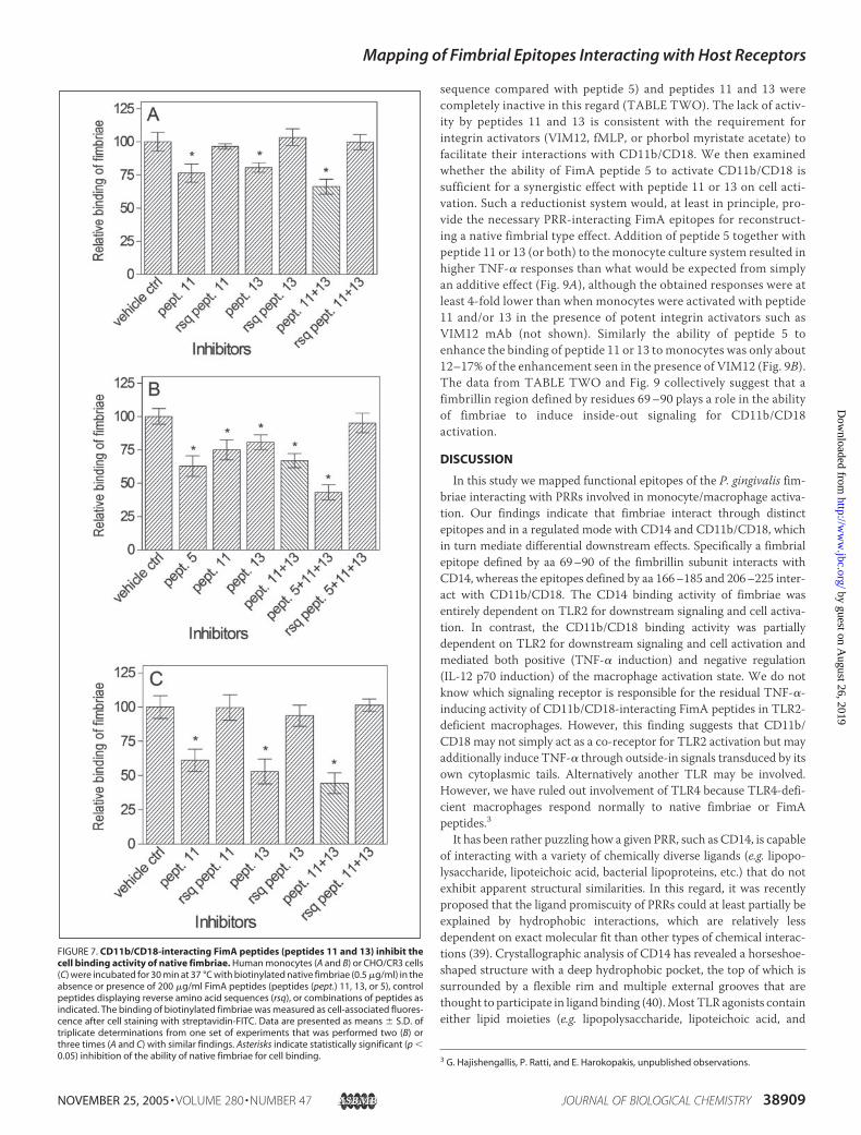
Competitive Inhibition by CD11b/CD18-interacting FimA Peptides ofthe Cell Binding Activity of Native Fimbriae—We then showed thatFimA peptides 11 and 13, but not control peptides displaying reverseamino acid sequences, inhibited partially but significantly (p� 0.05) theability of biotinylated native fimbriae to bind monocytes (Fig. 7A). Inhi-bition was somewhat more effective when the two inhibitor peptideswere combined, although the combination of their reverse sequencecontrol peptides still had no effect (Fig. 7A). The observed modest inhi-bition may, at least partially, be due to the ability of native fimbriae toengage other monocytic receptors such as CD14. Indeed when theCD14-interacting peptide 5 was added together with peptides 11 and 13to competitively inhibit the binding of native fimbriae tomonocytes, theresulting inhibition (�57%; Fig. 7B) was stronger than what wasobserved in the absence of peptide 5 (�34%; Fig. 7, A and B). Moreoverwhen the experiment was repeated using CHO/CR3 cells, which do notexpress CD14, FimA peptides 11 and 13 (and especially their combina-tion) displayed increased inhibitory activity against the binding of nativefimbriae (Fig. 7C) compared with that seen in monocytes (Fig. 7A).These data strongly suggest that the regions defined by peptides 11 and13 (aa 166–185 and 206–225) are important in the interactions ofnative fimbriae with CD11b/CD18.
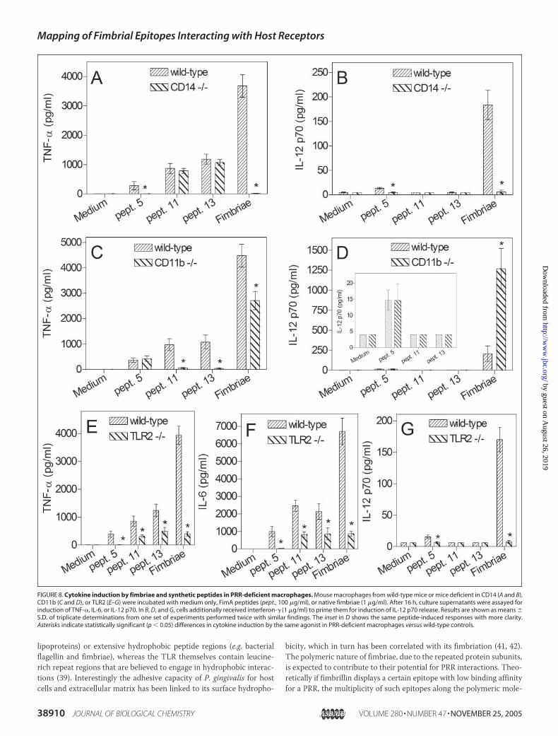
Relative Contribution of CD14 and CD11b/CD18 in CytokineInduction by Fimbriae and Synthetic Peptides—To confirm the PRRspecificities of FimA peptides 5, 11, and 13 using an independentapproach, we determined the ability of macrophages from wild-typemice or mice deficient in CD14 or CD11b to respond to the peptides.A second objective was to determine the relative importance ofCD14 and CD11b/CD18 (the expression of which is undetectable inCD11b-deficient mice (28)) in fimbria-induced cytokine release. Asexpected, the ability of FimA peptide 5 to induce TNF-� was com-pletely abrogated in CD14-deficient cells (Fig. 8A) but was unaf-fected in CD11b-deficient cells (Fig. 8C). The converse was true forFimA peptides 11 and 13, i.e. they could induce TNF-� release inCD14-deficient but not in CD11b-deficient macrophages (Fig. 8, A
FIGURE 4. FimA peptide 5 (aa 69 –90) competitively inhibits the binding of nativefimbriae to CD14. Microtiter wells were coated with CD14, blocked with BSA, and incu-bated with biotinylated native fimbriae (0.25 �g/ml). In A, labeled fimbriae were incu-bated in the presence of increasing concentrations of FimA peptide 5 (pept. # 5) or itscontrol peptide displaying the reverse amino acid sequence (rsq pept. # 5). In B, labeledfimbriae were incubated in the presence of a constant concentration (125 �g/ml) of allFimA peptides (TABLE ONE) or unlabeled native fimbriae (NF; 25 �g/ml). Bound biotiny-lated fimbriae were detected by ELISA using streptavidin-conjugated peroxidase. Dataare shown as means � S.D. of optical density (450 nm) values (n � 3). These experimentswere repeated twice for verification, and similar findings were obtained. Asterisks indi-cate statistically significant (p � 0.05) inhibition of native fimbriae binding to CD14 byFimA peptides.
FIGURE 5. FimA peptide 5 (aa 69 –90) competitively inhibits the ability of nativefimbriae to induce TNF-� release. Primary human monocytes were stimulated withnative fimbriae (1 �g/ml) in the presence of increasing concentrations of FimA peptide 5(pept. # 5) (A) or its control peptide displaying the reverse amino acid sequence (rsq pept.# 5) (B). In C, the concentration of the inhibitor FimA peptide 5 was kept constant (500�g/ml), and the concentration of native fimbriae was varied as indicated. After 16 h ofstimulation, culture supernatants were collected and assayed for TNF-� release. Resultsare shown as means � S.D. of triplicate determinations from a series of experiments(A–C) that were performed twice yielding similar findings. Statistically significant (p �0.05) inhibition of the ability of native fimbriae to induce TNF-� by FimA peptides isindicated by asterisks.
Mapping of Fimbrial Epitopes Interacting with Host Receptors
NOVEMBER 25, 2005 • VOLUME 280 • NUMBER 47 JOURNAL OF BIOLOGICAL CHEMISTRY 38907
by guest on August 26, 2019
http://ww
w.jbc.org/
Dow
nloaded from

and C). As with the human monocyte model (Fig. 6B), mouse macro-phages incubated with FimA peptides 11 and 13 also received fMLP(10�7 M) to activate CD11b/CD18 and enable the cells to respond tothese peptides (fMLP alone did not induce detectable TNF-� release;not shown). Although native fimbriae induced high levels of TNF-�in wild-type macrophages, the production of this cytokine was unde-tectable in CD14-deficient cells (Fig. 8A) and partially reduced (byabout 40%) in CD11b-deficient cells (Fig. 8C). A similar pattern wasobserved for IL-6 (i.e. induction of IL-6 was abrogated in CD14-deficient but only partially reduced in CD11b-deficient cells; datanot shown). Surprisingly, however, we detected low levels (40 � 9pg/ml) of IL-12 p70 in the supernatants of CD11b-deficient macro-phages stimulated with native fimbriae, whereas no IL-12 p70 wasdetectable in similarly stimulated wild-type or CD14-deficient cells.To better investigate this observation, the experiment was repeatedin the presence of interferon-� (1 �g/ml) to prime the cells for IL-12p70 induction. Although fimbriae induced a modest IL-12 p70response in interferon-�-primed wild-type macrophages (�210pg/ml; Fig. 8, B and D), the response was increased about 6-fold inCD11b-deficient cells (Fig. 8D) but was abolished in CD14-deficientcells (Fig. 8B). In interferon-�-primed wild-type cells, FimA peptide5 (but not peptides 11 and 13) induced a weak but detectable IL-12p70 response, which remained unaltered in CD11b-deficient cells(Fig. 8, D and inset) but was abolished in CD14-deficient cells (Fig.8B). Taken together, these data confirm that CD14 and CD11b/CD18 respond to different fimbrial epitopes and suggest that thesePRRs exert differential effects on cell activation by P. gingivalisfimbriae.
Role of TLR2 in Cell Activation by FimA Peptides—The ability of P.gingivalis fimbriae to induce activation of human monocytes isinhibited by anti-TLR2 mAb (22, 23, 34). This was confirmed in thecurrent study because fimbriae lost at least 90% of their cytokine-inducing capacity in TLR2-deficient macrophages in comparisonwith wild-type controls (Fig. 8, E–G). Moreover TLR2-deficientmacrophages completely failed to respond to the CD14-interactingFimA peptide 5 (Fig. 8, E–G). In contrast, the CD11b/CD18-inter-acting peptides (peptides 11 and 13) maintained about 30–40% oftheir ability to induce release of TNF-� (Fig. 8E) or IL-6 (Fig. 8F) inTLR2-deficient macrophages compared with wild-type controls.The remaining FimA peptides (TABLE ONE) were not tested inTLR2-deficient macrophages because preliminary experimentsshowed that they were unable to induce detectable cytokineresponses in wild-typemacrophages (not shown). These data suggestthat peptides 5, 11, and 13 define fimbrial epitopes that induce TLR2-dependent cell activation, although only peptide 5 is entirelydependent on TLR2 signaling for cytokine induction.
Induction of Inside-out Signaling by FimA Peptide 5—Peptide 5 (aa69–90) is capable of binding CD14 and activating TLR2 signaling(Figs. 2, 8, and 9). We thus determined whether peptide 5 can fur-thermore activate the inside-out signaling pathway that is down-stream of CD14/TLR2 and leads to PI3K-dependent activation ofCD11b/CD18 as we have shown earlier for native fimbriae (22). Pep-tide 5 was indeed found to activate PI3K and to induce the highaffinity conformation of CD11b/CD18, although with considerablylower potency (�16%) compared with native fimbriae (TABLETWO). A control peptide (displaying the reverse amino acid
FIGURE 6. Interactions of FimA peptides with activated CD11b/CD18 (CR3). Primary human monocytes were pretreated with CD11b/CD18 activators, VIM12 (A and C) or fMLP (B),in the absence or presence of blocking mAbs to CD11b (2LPM19c) or CD14 (MEM-18) or IgG1 isotype control (all antibodies at 10 �g/ml). In A and B, the cells were either left withoutfurther treatment or were incubated for 30 min at 37 °C with biotinylated FimA peptides 11 and 13 (100 �g/ml) or an equal concentration of peptide 5 (control). Peptide binding wasmeasured as cell-associated fluorescence after cell staining with streptavidin-FITC. Background binding was determined in cells treated with medium only and streptavidin-FITC(SA-FITC control). In C, the cells were treated exactly as in the experiment shown in A except that non-biotinylated peptides were used, and incubation was carried out for 16 h to assayinduction of TNF-� release. In D, biotinylated FimA peptides were examined for binding to CHO/CR3 cells or to CD11b/CD18-nonexpressing controls (CHO/CR1). Data are presentedas means � S.D. (n � 3) of typical experiments that were performed twice and yielded similar results. In A–C, asterisks indicate statistically significant (p � 0.05) inhibition by mAbtreatment of the ability of FimA peptides to bind (A and B) or activate (C) monocytes. In D, asterisks indicate significant (p � 0.05) binding of FimA peptides to CHO/CR3 compared withCHO/CR1 control cells. ctrl, control; RFU, relative fluorescence units.
Mapping of Fimbrial Epitopes Interacting with Host Receptors
38908 JOURNAL OF BIOLOGICAL CHEMISTRY VOLUME 280 • NUMBER 47 • NOVEMBER 25, 2005
by guest on August 26, 2019
http://ww
w.jbc.org/
Dow
nloaded from
sequence compared with peptide 5) and peptides 11 and 13 werecompletely inactive in this regard (TABLE TWO). The lack of activ-ity by peptides 11 and 13 is consistent with the requirement forintegrin activators (VIM12, fMLP, or phorbol myristate acetate) tofacilitate their interactions with CD11b/CD18. We then examinedwhether the ability of FimA peptide 5 to activate CD11b/CD18 issufficient for a synergistic effect with peptide 11 or 13 on cell acti-vation. Such a reductionist system would, at least in principle, pro-vide the necessary PRR-interacting FimA epitopes for reconstruct-ing a native fimbrial type effect. Addition of peptide 5 together withpeptide 11 or 13 (or both) to the monocyte culture system resulted inhigher TNF-� responses than what would be expected from simplyan additive effect (Fig. 9A), although the obtained responses were atleast 4-fold lower than when monocytes were activated with peptide11 and/or 13 in the presence of potent integrin activators such asVIM12 mAb (not shown). Similarly the ability of peptide 5 toenhance the binding of peptide 11 or 13 to monocytes was only about12–17% of the enhancement seen in the presence of VIM12 (Fig. 9B).The data from TABLE TWO and Fig. 9 collectively suggest that afimbrillin region defined by residues 69–90 plays a role in the abilityof fimbriae to induce inside-out signaling for CD11b/CD18activation.
DISCUSSION
In this study we mapped functional epitopes of the P. gingivalis fim-briae interacting with PRRs involved in monocyte/macrophage activa-tion. Our findings indicate that fimbriae interact through distinctepitopes and in a regulated mode with CD14 and CD11b/CD18, whichin turn mediate differential downstream effects. Specifically a fimbrialepitope defined by aa 69–90 of the fimbrillin subunit interacts withCD14, whereas the epitopes defined by aa 166–185 and 206–225 inter-act with CD11b/CD18. The CD14 binding activity of fimbriae wasentirely dependent on TLR2 for downstream signaling and cell activa-tion. In contrast, the CD11b/CD18 binding activity was partiallydependent on TLR2 for downstream signaling and cell activation andmediated both positive (TNF-� induction) and negative regulation(IL-12 p70 induction) of the macrophage activation state. We do notknow which signaling receptor is responsible for the residual TNF-�-inducing activity of CD11b/CD18-interacting FimA peptides in TLR2-deficient macrophages. However, this finding suggests that CD11b/CD18 may not simply act as a co-receptor for TLR2 activation but mayadditionally induce TNF-� through outside-in signals transduced by itsown cytoplasmic tails. Alternatively another TLR may be involved.However, we have ruled out involvement of TLR4 because TLR4-defi-cient macrophages respond normally to native fimbriae or FimApeptides.3
It has been rather puzzling how a given PRR, such as CD14, is capableof interacting with a variety of chemically diverse ligands (e.g. lipopo-lysaccharide, lipoteichoic acid, bacterial lipoproteins, etc.) that do notexhibit apparent structural similarities. In this regard, it was recentlyproposed that the ligand promiscuity of PRRs could at least partially beexplained by hydrophobic interactions, which are relatively lessdependent on exact molecular fit than other types of chemical interac-tions (39). Crystallographic analysis of CD14 has revealed a horseshoe-shaped structure with a deep hydrophobic pocket, the top of which issurrounded by a flexible rim and multiple external grooves that arethought to participate in ligand binding (40).Most TLR agonists containeither lipid moieties (e.g. lipopolysaccharide, lipoteichoic acid, and
3 G. Hajishengallis, P. Ratti, and E. Harokopakis, unpublished observations.
FIGURE 7. CD11b/CD18-interacting FimA peptides (peptides 11 and 13) inhibit thecell binding activity of native fimbriae. Human monocytes (A and B) or CHO/CR3 cells(C) were incubated for 30 min at 37 °C with biotinylated native fimbriae (0.5 �g/ml) in theabsence or presence of 200 �g/ml FimA peptides (peptides (pept.) 11, 13, or 5), controlpeptides displaying reverse amino acid sequences (rsq), or combinations of peptides asindicated. The binding of biotinylated fimbriae was measured as cell-associated fluores-cence after cell staining with streptavidin-FITC. Data are presented as means � S.D. oftriplicate determinations from one set of experiments that was performed two (B) orthree times (A and C) with similar findings. Asterisks indicate statistically significant (p �0.05) inhibition of the ability of native fimbriae for cell binding.
Mapping of Fimbrial Epitopes Interacting with Host Receptors
NOVEMBER 25, 2005 • VOLUME 280 • NUMBER 47 JOURNAL OF BIOLOGICAL CHEMISTRY 38909
by guest on August 26, 2019
http://ww
w.jbc.org/
Dow
nloaded from
lipoproteins) or extensive hydrophobic peptide regions (e.g. bacterialflagellin and fimbriae), whereas the TLR themselves contain leucine-rich repeat regions that are believed to engage in hydrophobic interac-tions (39). Interestingly the adhesive capacity of P. gingivalis for hostcells and extracellular matrix has been linked to its surface hydropho-
bicity, which in turn has been correlated with its fimbriation (41, 42).The polymeric nature of fimbriae, due to the repeated protein subunits,is expected to contribute to their potential for PRR interactions. Theo-retically if fimbrillin displays a certain epitope with low binding affinityfor a PRR, the multiplicity of such epitopes along the polymeric mole-
FIGURE 8. Cytokine induction by fimbriae and synthetic peptides in PRR-deficient macrophages. Mouse macrophages from wild-type mice or mice deficient in CD14 (A and B),CD11b (C and D), or TLR2 (E–G) were incubated with medium only, FimA peptides (pept., 100 �g/ml), or native fimbriae (1 �g/ml). After 16 h, culture supernatants were assayed forinduction of TNF-�, IL-6, or IL-12 p70. In B, D, and G, cells additionally received interferon-� (1 �g/ml) to prime them for induction of IL-12 p70 release. Results are shown as means �S.D. of triplicate determinations from one set of experiments performed twice with similar findings. The inset in D shows the same peptide-induced responses with more clarity.Asterisks indicate statistically significant (p � 0.05) differences in cytokine induction by the same agonist in PRR-deficient macrophages versus wild-type controls.
Mapping of Fimbrial Epitopes Interacting with Host Receptors
38910 JOURNAL OF BIOLOGICAL CHEMISTRY VOLUME 280 • NUMBER 47 • NOVEMBER 25, 2005
by guest on August 26, 2019
http://ww
w.jbc.org/
Dow
nloaded from

cule could result in engagement ofmultiple receptormolecules and thuswould increase the avidity of the overall interaction. This may explainwhy the FimA peptides identified to interact with CD14 or CD11b/CD18 are less potent than native fimbriae for inducing cell activationdespite their use at higher concentrations. Alternatively or in addition,interactionswith PRRsmay involve three-dimensional structuralmotifsthat are not adequately formed in relatively short protein segments,such as the peptides tested.However, the relatively low agonistic activity
of FimA peptide 5 enables it to act (when in excess) as a specific antag-onist of native fimbria-induced TNF-� responses (Fig. 5A) by compet-itively blocking binding toCD14 (Fig. 4A). Thismight be a usefulmolec-ular strategy to control excessive fimbria-induced periodontalinflammation without completely inhibiting the innate immunedefense.In terms of evolution, PRRs are thought to have evolved to recognize
conserved pathogen-associatedmolecular patterns such as lipopolysac-charide and lipoteichoic acid (43). Pathogen-associated molecular pat-terns and virulence factors are not equivalent terms and generally rep-resent distinct microbial molecules. Unlike the relatively conservednature of pathogen-associated molecular patterns, genes encodingmicrobial virulence proteins may mutate to prevent innate immunerecognition. However, if exploitation of PRRs is essential for the survivalof the pathogen (e.g. for colonization), the virulence factors involved areexpected to display some relatively invariant motifs that are potentiallyrecognizable by PRRs. The fimbriae of P. gingivalis function as a majoradhesin and constitute an important virulence factor of this pathogen(5, 6). It seems unlikely that PRRs have evolved to specifically recognizethis fimbrial structure, which is not shared by any other bacteria (44).Rather it seems more plausible to speculate that P. gingivalis fimbriaehave evolved to recognize and interact with host receptors for the ben-efit of the pathogen. For example, the fimbriae promote the uptake ofP. gingivalis into host epithelial cells (12, 45) where the pathogen cansurvive and replicate (46). Other pathogens, such as Mycobacteriumtuberculosis, exploit CD11b/CD18 as a mechanism for entrance andintracellular parasitism (47). It is currently unknown whether P. gingi-valis can similarly induce its uptake by monocytes through fimbriae-CD11b/CD18 interactions resulting in survival rather than postphago-cytosis killing. If this is true, the fimbria-induced inside-out signalingpathway leading to CD11b/CD18 activation (22) may represent a strat-egy for access into a nutritionally rich and immunologically safe site forthe pathogen. Although inflammation is a potentially protective hostresponse, induction of fimbria-induced periodontal inflammation couldalso benefit P. gingivalis through acquisition of crucial nutrients derivedfrom serum exudate into the crevicular area of the teeth. Moreover asdiscussed in greater detail below, we have obtained initial evidence thatthe interaction of P. gingivalis fimbriae with CD11b/CD18 leads todown-regulation of biologically active IL-12 (p70), a major cytokine inmediating bacterial clearance (48).In contrast to CD14, which is essential for TLR2-dependent cytokine
release by native fimbriae (or by the FimA peptide 5), CD11b/CD18plays a contributory role but is not required for induction of TNF-�release by native fimbriae (Fig. 8B). Interestingly in the absence of thisreceptor (i.e. in CD11b-deficient macrophages), fimbriae induce
TABLE TWO
Ability of FimA peptide 5 to activate PI3K and CD11b/CD18
FimA peptide testedActivity (means � S.D.; n � 3)
compared with native fimbriae in assay forPI3K activationa CD11b/CD18 activationb
%
Peptide 5 (aa 69–90) 15.6 � 3.5c 13.4 � 4.5c
Control peptide 5 (reverse) 3.8 � 3.3 3.3 � 2.1Peptide 11 (aa 166–185) 1.6 � 1.8 2.8 � 2.3Peptide 13 (aa 206–225) 2.5 � 3.2 1.7 � 1.6
a Assayed as enzymatic production of PI(3,4,5)P3 from PI(4,5)P2 substrate. Induction of the lipid kinase activity of PI3K by native fimbriae resulted in production of 36.7 � 4.2pmol of PI(3,4,5)P3.
b Assayed as induction of an activation-specific epitope probed by FITC-labeled CBRM1/5 mAb. The value obtained upon induction using native fimbriae was 76,768 � 9,895relative fluorescence units.
c Statistically significant (p � 0.05) differences compared to reverse sequence control or peptides 11 and 13.
FIGURE 9. Modulation of the activities of CD11b/CD18-interacting FimA peptides(peptides 11 and 13) in the presence of CD14-interacting FimA peptide (peptide 5).A, human monocytes were incubated with vehicle control (vc) or synthetic FimA pep-tides (pept., 100 �g/ml) either alone or in combinations, and culture supernatants werecollected after 16 h to assay induction of TNF-� release. B, the ability of biotinylatedpeptide 11 or 13 to bind monocytes was assayed in the absence or presence of non-biotinylated peptide 5 or VIM12 mAb (integrin activator control). Binding was measuredas cell-associated fluorescence after cell staining with streptavidin-FITC. Backgroundbinding was determined in cells treated with medium only and streptavidin-FITC (SA-FITC control). Results are shown as means � S.D. (n � 3) from one set of experimentsperformed twice with similar results. Asterisks in B indicate statistically significant (p �0.05) increase of the binding activity of peptide 11 or 13 in the presence of integrinactivators. RFU, relative fluorescence units.
Mapping of Fimbrial Epitopes Interacting with Host Receptors
NOVEMBER 25, 2005 • VOLUME 280 • NUMBER 47 JOURNAL OF BIOLOGICAL CHEMISTRY 38911
by guest on August 26, 2019
http://ww
w.jbc.org/
Dow
nloaded from

increased levels of IL-12 p70 (Fig. 8D). This suggests that binding toCD11b/CD18 down-regulates IL-12 p70. Furthermore we have foundthat the ability of lipopolysaccharide to induce IL-12 p70 in humanmonocytes is suppressed in the presence of the CD11b/CD18-interact-ing FimA peptides.3 It is tempting to speculate that fimbriae may haveevolved to subvert innate immunity by exploiting CD11b/CD18, andfurther studies are warranted to clarify the mechanisms involved inIL-12 down-regulation by fimbriae. Our working hypothesis is that theability of P. gingivalis fimbriae to activate CD11b/CD18 through inside-out signaling (22) leads to down-regulation of IL-12 p70 through out-side-in signaling initiated by the binding of fimbriae to activatedCD11b/CD18. Therefore, the capacity of fimbriae to interact withCD11b/CD18 may provide intracellular access for P. gingivalis, as dis-cussed above, and moreover inhibit induction of IL-12 p70, an impor-tant cytokine in defense against intracellular pathogens (48).In addition to cytokine induction, another downstream effect of
CD14 binding by P. gingivalis fimbriae involves activation of the ligandbinding capacity of CD11b/CD18 through an inside-out signaling path-way involving TLR2 and PI3K (22). Our current findings from theepitope mapping studies allowed us to update and improve the modeldescribing the interactions of P. gingivalis fimbriae with PRRs. Accord-ing to the updated model, a fimbrial region involving residues 69–90binds to CD14 leading to TLR2 activation and PI3K-dependent induc-tion of the high affinity conformation of CD11b/CD18, which canthereby bind fimbriae at a region involving residues 166–185 and 206–225 (Fig. 10). The intervening region defined by residues 186–205 doesnot seem to interact with CD11b/CD18 (Figs. 1 and 6D), but it isunknown at present whether residues 166–185 and 206–225 define asingle discontinuous epitope. Although the binding of fimbriae to CD14would necessarily occur prior to any interactions with CD11b/CD18(Fig. 10), the latter appears to be an important monocytic receptor forfimbriae; this is suggested by the ability of FimA peptides 11 and 13 (Fig.7A) or a blocking mAb to CD11b (22) to competitively inhibit the bind-ing of native fimbriae to monocytes. On the other hand, substantialbinding of fimbriae to CD14 is detectable even after activation of theligand binding capacity of CD11b/CD18.3 Thus, both CD14 andCD11b/CD18 could simultaneously function asmajor cellular receptorsfor fimbriae, suggesting that the affinity/avidity of the interactionsinvolved may not be radically different.In summary, we have defined two different epitopes from the same
microbial molecule interacting with two distinct PRRs, leading to differen-
tial downstream effects. Specifically P. gingivalis fimbriae seem to display amodular structure that enables them to interact with CD14 and CD11b/CD18. Synthetic peptides representing fimbrial epitopes that interact withCD14 or CD11b/CD18 appear tomimic (alone or in combination) immu-nomodulatory effects induced by native fimbriae. Because CD14 andCD11b/CD18 differentially regulate cell activation, the modular structureof fimbriaemayallowthedevelopmentofmolecular approaches thatwouldselectively interfere with interactions that favor the pathogen.
Acknowledgment—We thank Dr. Seth Pincus for critical review of themanuscript.
REFERENCES1. Zambon, J. J., Grossi, S., Dunford, R., Harazsthy, V. I., Preus, H., and Genco, R. J.
(1994) in Molecular Pathogenesis of Periodontal Disease (Genco, R. J., Hamada, S.,Lehrer, J. R., McGhee, J. R., and Mergenhangen, S., eds) pp. 3–12, American Societyfor Microbiology, Washington, D. C.
2. Haraszthy, V. I., Zambon, J. J., Trevisan, M., Zeid, M., and Genco, R. J. (2000) J.Periodontol. 71, 1554–1560
3. Chun, Y. H., Chun, K. R., Olguin, D., and Wang, H. L. (2005) J. Periodontal Res. 40,87–95
4. Desvarieux,M., Demmer, R. T., Rundek, T., Boden-Albala, B., Jacobs, D. R., Jr., Sacco,R. L., and Papapanou, P. N. (2005) Circulation 111, 576–582
5. Malek, R., Fisher, J. G., Caleca, A., Stinson, M., van Oss, C. J., Lee, J. Y., Cho, M. I.,Genco, R. J., Evans, R. T., and Dyer, D. W. (1994) J. Bacteriol. 176, 1052–1059
6. Gibson, F. C., III, Hong, C., Chou, H. H., Yumoto, H., Chen, J., Lien, E., Wong, J., andGenco, C. A. (2004) Circulation 109, 2801–2806
7. Amano, A., Sharma, A., Lee, J. Y., Sojar, H. T., Raj, P. A., andGenco, R. J. (1996) Infect.Immun. 64, 1631–1637
8. Sojar, H. T., Lee, J.-Y., and Genco, R. J. (1995) Biochem. Biophys. Res. Commun. 216,785–792
9. Weinberg, A., Belton, C. M., Park, Y., and Lamont, R. J. (1997) Infect. Immun. 65,313–316
10. Deshpande, R. G., Khan,M. B., andGenco, C. A. (1998) Infect. Immun. 66, 5337–534311. Jotwani, R., and Cutler, C. W. (2004) Infect. Immun. 72, 1725–173212. Giacona, M. B., Papapanou, P. N., Lamster, I. B., Rong, L. L., D’Agati, V. D., Schmidt,
A. M., and Lalla, E. (2004) FEMS Microbiol. Lett. 241, 95–10113. Ogawa, T., Asai, Y., Hashimoto, M., and Uchida, H. (2002) Eur. J. Immunol. 32,
2543–255014. Hajishengallis, G., Martin, M., Sojar, H. T., Sharma, A., Schifferle, R. E., DeNardin, E.,
Russell, M. W., and Genco, R. J. (2002) Clin. Diagn. Lab. Immunol. 9, 403–41115. Zhou, Q., Desta, T., Fenton,M., Graves, D. T., and Amar, S. (2005) Infect. Immun. 73,
935–94316. Muthukuru, M., Jotwani, R., and Cutler, C. W. (2005) Infect. Immun. 73, 687–69417. Delima, A. J., and Van Dyke, T. E. (2003) Periodontol. 2000 31, 55–7618. Perera, P.-Y., Mayadas, T. N., Takeuchi, O., Akira, S., Zaks-Zilberman, M., Goyert,
S. M., and Vogel, S. N. (2001) J. Immunol. 166, 574–58119. Triantafilou, M., Brandenburg, K., Gutsmann, T., Seydel, U., and Triantafilou, K.
(2002) Crit. Rev. Immunol. 22, 251–26820. Underhill, D. M. (2003) Eur. J. Immunol. 33, 1767–177521. Akira, S., and Takeda, K. (2004) Nat. Rev. Immunol. 4, 499–51122. Harokopakis, E., and Hajishengallis, G. (2005) Eur. J. Immunol. 35, 1201–121023. Hajishengallis, G., Sharma, A., Russell, M. W., and Genco, R. J. (2002) Ann. Period-
ontol. 7, 72–7824. Lee, J.-Y., Sojar, H. T., Amano, A., and Genco, R. J. (1995) Protein Expr. Purif. 6,
496–50025. Hajishengallis, G., Nawar, H., Tapping, R. I., Russell, M.W., and Connell, T. D. (2004)
Infect. Immun. 72, 6351–635826. Pugin, J., Kravchenko, V. V., Lee, J. D., Kline, L., Ulevitch, R. J., and Tobias, P. S. (1998)
Infect. Immun. 66, 1174–118027. Moore, K. J., Andersson, L. P., Ingalls, R. R., Monks, B. G., Li, R., Arnaout, M. A.,
Golenbock, D. T., and Freeman, M. W. (2000) J. Immunol. 165, 4272–428028. Coxon, A., Rieu, P., Barkalow, F. J., Askari, S., Sharpe, A. H., von Andrian, U. H.,
Arnaout, M. A., and Mayadas, T. N. (1996) Immunity 5, 653–66629. Wooten, R.M.,Ma, Y., Yoder, R. A., Brown, J. P.,Weis, J. H., Zachary, J. F., Kirschning,
C. J., and Weis, J. J. (2002) J. Immunol. 168, 348–35530. Hajishengallis, G., Tapping, R. I., Martin,M. H., Nawar, H., Lyle, E. A., Russell, M.W.,
and Connell, T. D. (2005) Infect. Immun. 73, 1343–134931. Levitz, S. M., Tabuni, A., Kozel, T. R., MacGill, R. S., Ingalls, R. R., and Golenbock,
D. T. (1997) Infect. Immun. 65, 931–93532. Stockl, J., Majdic, O., Pickl, W. F., Rosenkranz, A., Prager, E., Gschwantler, E., and
FIGURE 10. Epitopes of P. gingivalis fimbrillin (FimA) interacting with PRRs. CD14recognizes a fimbrillin region defined by aa 69 –90. Following activation through aninside-out signaling pathway dependent upon CD14, TLR2, and PI3K, the CD11b/CD18integrin is induced to recognize a fimbrillin region involving two neighboring epitopesdefined by aa 166 –185 and 206 –225.
Mapping of Fimbrial Epitopes Interacting with Host Receptors
38912 JOURNAL OF BIOLOGICAL CHEMISTRY VOLUME 280 • NUMBER 47 • NOVEMBER 25, 2005
by guest on August 26, 2019
http://ww
w.jbc.org/
Dow
nloaded from

Knapp, W. (1995) J. Immunol. 154, 5452–546333. Hajishengallis, G., Martin, M., Schifferle, R. E., and Genco, R. J. (2002) Infect. Immun.
70, 6658–666434. Hajishengallis, G., and Genco, R. J. (2004) Infect. Immun. 72, 1188–119135. Diamond, M. S., and Springer, T. A. (1993) J. Cell Biol. 120, 545–55636. Cunningham, M. D., Seachord, C., Ratcliffe, K., Bainbridge, B., Aruffo, A., and
Darveau, R. P. (1996) Infect. Immun. 64, 3601–360837. Shimaoka, M., Takagi, J., and Springer, T. A. (2002) Annu. Rev. Biophys. Biomol.
Struct. 31, 485–51638. Diamond, M. S., Garcia-Aguilar, J., Bickford, J. K., Corbi, A. L., and Springer, T. A.
(1993) J. Cell Biol. 120, 1031–104339. Seong, S. Y., and Matzinger, P. (2004) Nat. Rev. Immunol. 4, 469–47840. Kim, J. I., Lee, C. J., Jin,M. S., Lee, C. H., Paik, S. G., Lee, H., and Lee, J. O. (2005) J. Biol.
Chem. 280, 11347–1135141. Watanabe, K., Yamaji, Y., and Umemoto, T. (1992) Oral Microbiol. Immunol. 7,
357–36342. Naito, Y., Tohda, H., Okuda, K., and Takazoe, I. (1993) Oral Microbiol. Immunol. 8,
195–20243. Medzhitov, R. (2001) Nat. Rev. Immunol. 1, 135–14544. Dickinson, D. P., Kubiniec, M. A., Yoshimura, F., and Genco, R. J. (1988) J. Bacteriol.
170, 1658–166545. Yilmaz, O., Watanabe, K., and Lamont, R. J. (2002) Cell Microbiol. 4, 305–31446. Lamont, R. J., Chan, A., Belton, C.M., Izutsu, K. T., Vasel, D., andWeinberg, A. (1995)
Infect. Immun. 63, 3878–388547. Ernst, J. D. (1998) Infect. Immun. 66, 1277–128148. Trinchieri, G. (2003) Nat. Rev. Immunol. 3, 133–146
Mapping of Fimbrial Epitopes Interacting with Host Receptors
NOVEMBER 25, 2005 • VOLUME 280 • NUMBER 47 JOURNAL OF BIOLOGICAL CHEMISTRY 38913
by guest on August 26, 2019
http://ww
w.jbc.org/
Dow
nloaded from

George Hajishengallis, Pukar Ratti and Evlambia HarokopakisRecognition Receptors
Peptide Mapping of Bacterial Fimbrial Epitopes Interacting with Pattern
doi: 10.1074/jbc.M507326200 originally published online August 29, 20052005, 280:38902-38913.J. Biol. Chem.
10.1074/jbc.M507326200Access the most updated version of this article at doi:
Alerts:
When a correction for this article is posted•
When this article is cited•
to choose from all of JBC's e-mail alertsClick here
http://www.jbc.org/content/280/47/38902.full.html#ref-list-1
This article cites 47 references, 27 of which can be accessed free at
by guest on August 26, 2019
http://ww
w.jbc.org/
Dow
nloaded from