peptide secondary structure modulates single-walled carbon ... · peptide secondary structure...
TRANSCRIPT
Peptide secondary structure modulates single-walledcarbon nanotube fluorescence as a chaperonesensor for nitroaromaticsDaniel A. Heller, George W. Pratt, Jingqing Zhang, Nitish Nair, Adam J. Hansborough, Ardemis A. Boghossian,Nigel F. Reuel, Paul W. Barone, and Michael S. Strano1
Department of Chemical Engineering, Massachusetts Institute of Technology, Cambridge, MA 02139-4307
Edited* by Mildred Dresselhaus, Massachusetts Institute of Technology, Cambridge, MA, and approved March 31, 2011 (received for review May 3, 2010)
A class of peptides from the bombolitin family, not previouslyidentified for nitroaromatic recognition, allows near-infrared fluor-escent single-walled carbon nanotubes to transduce specificchanges in their conformation. In response to the binding ofspecific nitroaromatic species, such peptide–nanotube complexesform a virtual “chaperone sensor,” which reports modulation ofthe peptide secondary structure via changes in single-walled car-bon nanotubes, near-infrared photoluminescence. A split-channelmicroscope constructed to image quantized spectral wavelengthshifts in real time, in response to nitroaromatic adsorption, resultsin the first single-nanotube imaging of solvatochromic events. Thedescribed indirect detection mechanism, as well as an additionalexciton quenching-based optical nitroaromatic detection method,illustrate that functionalization of the carbon nanotube surfacecan result in completely unique sites for recognition, resolvableat the single-molecule level.
bionanotechnology ∣ explosives detection ∣ pesticides ∣ spectroscopy ∣optical sensors
Single-walled carbon nanotubes (SWNT) emit near-infrared(NIR) bandgap photoluminescence (PL) (1, 2), which is
highly responsive to its physical and chemical environment (3–6).SWNT are unique among nanoscale sensor platforms in theirability to detect the adsorption of as few as a single molecule ofan analyte (7, 8). For such a capability to be extended to specificclasses of organic molecules, chemical approaches must be devel-oped that allow for selective molecular recognition. Stepwisequenching of SWNT PL by single-molecule adsorption eventsto the nanotube surface (7) has been demonstrated (8, 9). Ourprevious findings extend this resolution to biologically importantreactive oxygen species and demonstrate multiplexed detection ofredox-active analytes by two optical modes, leading to speciesidentification (9).
The current work investigates the selective optical detectionof binding events by single-SWNT PL modulation, employingboth intensity- and wavelength-based signal transduction. We findthat specific noncovalently bound polymers can be harnessedto change the properties of the nanotube–polymer complex,resulting in complete modulation of the nanotube sensitivity tocertain analytes. Resolution of an entire class of molecules canbe achieved by the nanotube via reporting the conformationalstate of a peptide, for example. Nanotube emission undergoessolvatochromic shifts due to nitroaromatic compound-mediatedsecondary structure changes of the amphipathic bombolitin IIoligopeptide. In this work, we probe solvatochromic interactionsat the single-nanotube level by a strategy in which two spectrallyadjacent optical channels measure anticorrelated, quantizedfluctuations, signifying molecular binding events. In addition, wefind that an oligonucleotide of single-stranded DNA with thesequence ðATÞ15 [ssðATÞ15] oligonucleotide imparts optical selec-tivity of SWNT for trinitrotoluene (TNT) via intensity modula-tion. Although nanotubes do not normally detect this analyte,
the electronic and steric effects of this encapsulating sequence al-low single-molecule detection by reversible excitonic quenching.
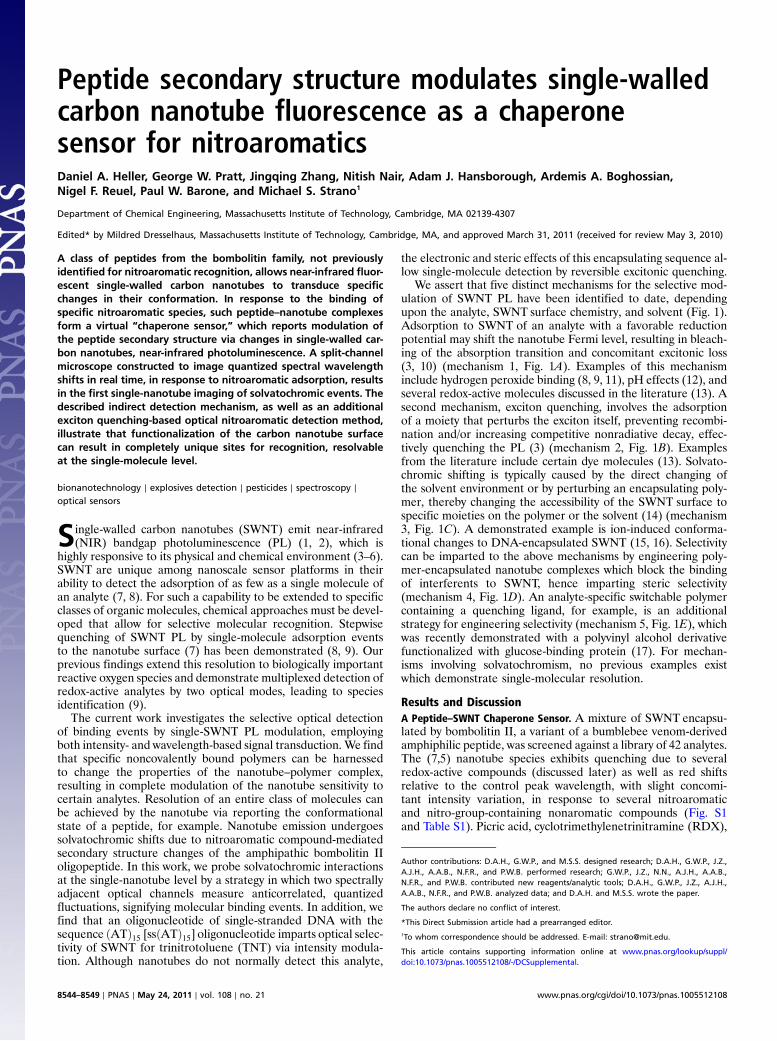
We assert that five distinct mechanisms for the selective mod-ulation of SWNT PL have been identified to date, dependingupon the analyte, SWNT surface chemistry, and solvent (Fig. 1).Adsorption to SWNT of an analyte with a favorable reductionpotential may shift the nanotube Fermi level, resulting in bleach-ing of the absorption transition and concomitant excitonic loss(3, 10) (mechanism 1, Fig. 1A). Examples of this mechanisminclude hydrogen peroxide binding (8, 9, 11), pH effects (12), andseveral redox-active molecules discussed in the literature (13). Asecond mechanism, exciton quenching, involves the adsorptionof a moiety that perturbs the exciton itself, preventing recombi-nation and/or increasing competitive nonradiative decay, effec-tively quenching the PL (3) (mechanism 2, Fig. 1B). Examplesfrom the literature include certain dye molecules (13). Solvato-chromic shifting is typically caused by the direct changing ofthe solvent environment or by perturbing an encapsulating poly-mer, thereby changing the accessibility of the SWNT surface tospecific moieties on the polymer or the solvent (14) (mechanism3, Fig. 1C). A demonstrated example is ion-induced conforma-tional changes to DNA-encapsulated SWNT (15, 16). Selectivitycan be imparted to the above mechanisms by engineering poly-mer-encapsulated nanotube complexes which block the bindingof interferents to SWNT, hence imparting steric selectivity(mechanism 4, Fig. 1D). An analyte-specific switchable polymercontaining a quenching ligand, for example, is an additionalstrategy for engineering selectivity (mechanism 5, Fig. 1E), whichwas recently demonstrated with a polyvinyl alcohol derivativefunctionalized with glucose-binding protein (17). For mechan-isms involving solvatochromism, no previous examples existwhich demonstrate single-molecular resolution.
Results and DiscussionA Peptide–SWNT Chaperone Sensor. A mixture of SWNT encapsu-lated by bombolitin II, a variant of a bumblebee venom-derivedamphiphilic peptide, was screened against a library of 42 analytes.The (7,5) nanotube species exhibits quenching due to severalredox-active compounds (discussed later) as well as red shiftsrelative to the control peak wavelength, with slight concomi-tant intensity variation, in response to several nitroaromaticand nitro-group-containing nonaromatic compounds (Fig. S1and Table S1). Picric acid, cyclotrimethylenetrinitramine (RDX),
Author contributions: D.A.H., G.W.P., and M.S.S. designed research; D.A.H., G.W.P., J.Z.,A.J.H., A.A.B., N.F.R., and P.W.B. performed research; G.W.P., J.Z., N.N., A.J.H., A.A.B.,N.F.R., and P.W.B. contributed new reagents/analytic tools; D.A.H., G.W.P., J.Z., A.J.H.,A.A.B., N.F.R., and P.W.B. analyzed data; and D.A.H. and M.S.S. wrote the paper.
The authors declare no conflict of interest.
*This Direct Submission article had a prearranged editor.1To whom correspondence should be addressed. E-mail: [email protected].
This article contains supporting information online at www.pnas.org/lookup/suppl/doi:10.1073/pnas.1005512108/-/DCSupplemental.
8544–8549 ∣ PNAS ∣ May 24, 2011 ∣ vol. 108 ∣ no. 21 www.pnas.org/cgi/doi/10.1073/pnas.1005512108
2,4-dinitrophenol, and 4-nitro-3(trifluoromethyl)phenol (TFM)induce spectral shifts without significant signal attenuation.Other shifting analytes induce large-intensity diminutions.
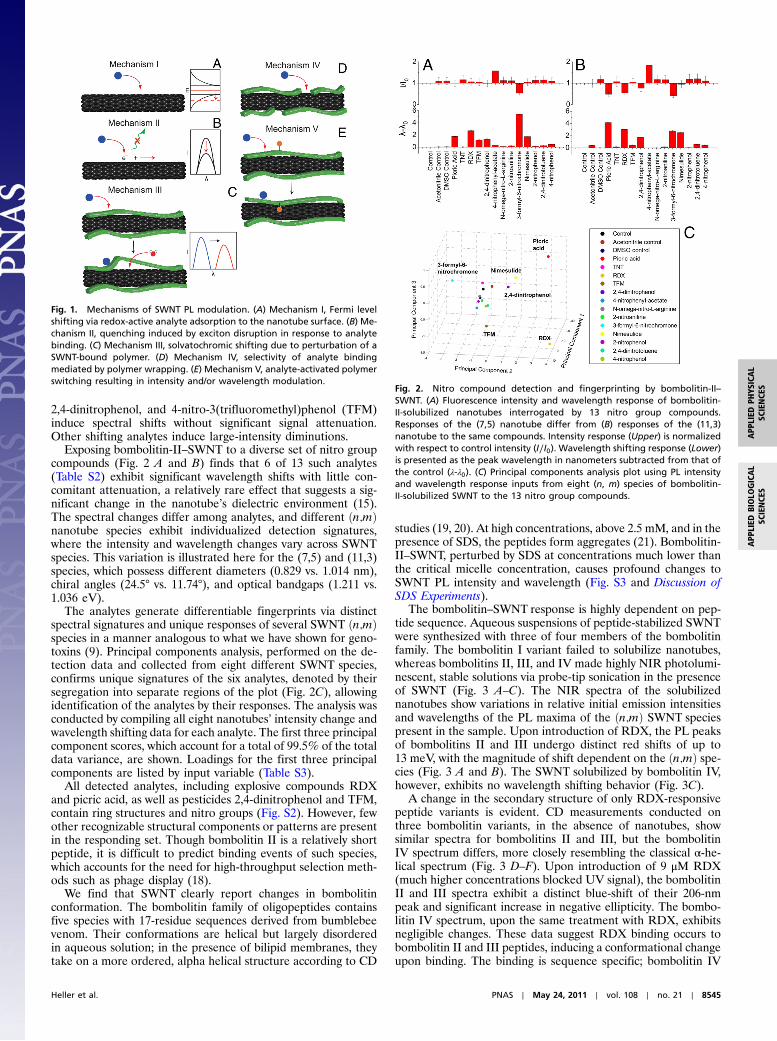
Exposing bombolitin-II–SWNT to a diverse set of nitro groupcompounds (Fig. 2 A and B) finds that 6 of 13 such analytes(Table S2) exhibit significant wavelength shifts with little con-comitant attenuation, a relatively rare effect that suggests a sig-nificant change in the nanotube’s dielectric environment (15).The spectral changes differ among analytes, and different ðn;mÞnanotube species exhibit individualized detection signatures,where the intensity and wavelength changes vary across SWNTspecies. This variation is illustrated here for the (7,5) and (11,3)species, which possess different diameters (0.829 vs. 1.014 nm),chiral angles (24.5° vs. 11.74°), and optical bandgaps (1.211 vs.1.036 eV).
The analytes generate differentiable fingerprints via distinctspectral signatures and unique responses of several SWNT ðn;mÞspecies in a manner analogous to what we have shown for geno-toxins (9). Principal components analysis, performed on the de-tection data and collected from eight different SWNT species,confirms unique signatures of the six analytes, denoted by theirsegregation into separate regions of the plot (Fig. 2C), allowingidentification of the analytes by their responses. The analysis wasconducted by compiling all eight nanotubes’ intensity change andwavelength shifting data for each analyte. The first three principalcomponent scores, which account for a total of 99.5% of the totaldata variance, are shown. Loadings for the first three principalcomponents are listed by input variable (Table S3).
All detected analytes, including explosive compounds RDXand picric acid, as well as pesticides 2,4-dinitrophenol and TFM,contain ring structures and nitro groups (Fig. S2). However, fewother recognizable structural components or patterns are presentin the responding set. Though bombolitin II is a relatively shortpeptide, it is difficult to predict binding events of such species,which accounts for the need for high-throughput selection meth-ods such as phage display (18).
We find that SWNT clearly report changes in bombolitinconformation. The bombolitin family of oligopeptides containsfive species with 17-residue sequences derived from bumblebeevenom. Their conformations are helical but largely disorderedin aqueous solution; in the presence of bilipid membranes, theytake on a more ordered, alpha helical structure according to CD
studies (19, 20). At high concentrations, above 2.5 mM, and in thepresence of SDS, the peptides form aggregates (21). Bombolitin-II–SWNT, perturbed by SDS at concentrations much lower thanthe critical micelle concentration, causes profound changes toSWNT PL intensity and wavelength (Fig. S3 and Discussion ofSDS Experiments).
The bombolitin–SWNT response is highly dependent on pep-tide sequence. Aqueous suspensions of peptide-stabilized SWNTwere synthesized with three of four members of the bombolitinfamily. The bombolitin I variant failed to solubilize nanotubes,whereas bombolitins II, III, and IV made highly NIR photolumi-nescent, stable solutions via probe-tip sonication in the presenceof SWNT (Fig. 3 A–C). The NIR spectra of the solubilizednanotubes show variations in relative initial emission intensitiesand wavelengths of the PL maxima of the ðn;mÞ SWNT speciespresent in the sample. Upon introduction of RDX, the PL peaksof bombolitins II and III undergo distinct red shifts of up to13 meV, with the magnitude of shift dependent on the ðn;mÞ spe-cies (Fig. 3 A and B). The SWNT solubilized by bombolitin IV,however, exhibits no wavelength shifting behavior (Fig. 3C).
A change in the secondary structure of only RDX-responsivepeptide variants is evident. CD measurements conducted onthree bombolitin variants, in the absence of nanotubes, showsimilar spectra for bombolitins II and III, but the bombolitinIV spectrum differs, more closely resembling the classical α-he-lical spectrum (Fig. 3 D–F). Upon introduction of 9 μM RDX(much higher concentrations blocked UV signal), the bombolitinII and III spectra exhibit a distinct blue-shift of their 206-nmpeak and significant increase in negative ellipticity. The bombo-litin IV spectrum, upon the same treatment with RDX, exhibitsnegligible changes. These data suggest RDX binding occurs tobombolitin II and III peptides, inducing a conformational changeupon binding. The binding is sequence specific; bombolitin IV
Fig. 1. Mechanisms of SWNT PL modulation. (A) Mechanism I, Fermi levelshifting via redox-active analyte adsorption to the nanotube surface. (B) Me-chanism II, quenching induced by exciton disruption in response to analytebinding. (C) Mechanism III, solvatochromic shifting due to perturbation of aSWNT-bound polymer. (D) Mechanism IV, selectivity of analyte bindingmediated by polymer wrapping. (E) Mechanism V, analyte-activated polymerswitching resulting in intensity and/or wavelength modulation.
Fig. 2. Nitro compound detection and fingerprinting by bombolitin-II–SWNT. (A) Fluorescence intensity and wavelength response of bombolitin-II-solubilized nanotubes interrogated by 13 nitro group compounds.Responses of the (7,5) nanotube differ from (B) responses of the (11,3)nanotube to the same compounds. Intensity response (Upper) is normalizedwith respect to control intensity (I∕I0). Wavelength shifting response (Lower)is presented as the peak wavelength in nanometers subtracted from that ofthe control (λ-λ0). (C) Principal components analysis plot using PL intensityand wavelength response inputs from eight (n, m) species of bombolitin-II-solubilized SWNT to the 13 nitro group compounds.
Heller et al. PNAS ∣ May 24, 2011 ∣ vol. 108 ∣ no. 21 ∣ 8545
APP
LIED
BIOLO
GICAL
SCIENCE
SAPP
LIED
PHYS
ICAL
SCIENCE
S

shows little conformational change upon exposure to RDX. Thebombolitin IV sequence differs in only three residues that areconserved between bombolitin II and III variants: Asn2, Val11,and Gly15, which are responsible for the differences in peptidesecondary structure modulation and sensor response (Fig. 3G).
The wavelength shifting of bombolitin-II-solubilized SWNTexhibits concentration dependence, which fits well to a first-orderLangmuir adsorption isotherm (Fig. 3H), implying that the tran-sition is reversible. The peptide is found to exhibit low affinityto SWNT because it can be dialyzed away, causing aggregationof the nanotubes, which is evident from PL quenching (Fig. 3H,Inset). This behavior suggests that the nanotube reports theconformation of the largely free peptide, which individuallysuspends the nanotubes as would a surfactant (22). The nano-tube, although solubilized by the peptide, acts as a chaperonesensor, which indirectly detects the binding event by transducingchanges to the secondary structure of the bombolitin variant. Inaddition to the usage described herein, it may allow researchersto detect misfolded proteins, among other applications. We pro-pose that, in an aqueous solution, the lowest energy conformationresults in the formation of a “binding pocket” within the bom-bolitin II where RDX noncovalently interacts. We computedenergy-minimized structures of bombolitin II interacting withRDX in a water medium, resulting in the images presented inFig. 3I and confirming an RDX-modulated peptide secondarystructure change. The binding was calculated to yield a net freeenergy change of −6.35 kcal∕mol, signifying an energeticallyfavorable interaction.
Different ðn;mÞ nanotube species respond distinctly to RDX-induced conformational changes of the peptide. Certain SWNTspecies exhibit higher sensitivity to RDX interrogation, yet othersexhibit a greater maximum shifting response (Fig. S4A). Thedissociation constant, Kd, of the RDX response of the (11,3)nanotube species, calculated as the inverse of the Langmuir equi-librium constant, is 7.7 μM, which is on the order of the Kd ofa TNT-binding antibody fragment interaction. Quantum dot-mediated detection of TNT using such a system resulted in areported Kd of 15.7 μM (23). The maximum wavelength shiftðλ-λ0 maxÞ displays a different dependence on SWNT species(Fig. S4B). The λ-λ0 max maximizes when both the SWNT speciesdiameter and chiral angle are large. Such a relationship follows ifthe bombolitin II, in its RDX-bound conformation, exhibitedpoorer stabilization of the larger, more chiral nanotubes ascompared to the smaller SWNTwith lower chiral angles. Absorp-tion spectroscopy of bombolitin-II–SWNT-RDX interactionsconfirms variations in solvatochromic behavior among SWNTðn;mÞ species (Fig. S4C).
To image NIR PL spectral shifts of individual nanotubes, a de-tection scheme was developed (Fig. 4A). A split-channel micro-scope was constructed to divide an image into two channels thatproject adjacent spectral regions. The light from one isolatednanotube emission band is split into two beams that are filteredto permit only half of the peak’s emission to appear in eachchannel. One channel on a single NIR detector array projectsthe long-wavelength half of the emission band, and the otherdisplays the short-wavelength half (Fig. 4B). Two images of thespectrally isolated (7,5) SWNTare collected transiently; concomi-
Fig. 3. Peptide–SWNT chaperone sensor for RDX. Photoluminescence spectra of peptide-suspended nanotubes before (blue) and after (red) addition of RDX.PL spectra of nanotubes suspended by (A) bombolitin II, (B) bombolitin III, (C) bombolitin IV. (D–F) CD spectra of the same peptides in the absence of nanotubesbefore (blue) and after (red) addition of RDX. (G) Sequences of the three bombolitin peptides highlighting differences between the variants. (H) Centerwavelength of the (11,3) nanotube peak of bombolitin-II-solubilized SWNT plotted versus RDX concentration (red circles). The data are fit to a first-orderLangmuir adsorption isotherm (blue curve). (Inset) PL spectra of bombolitin-II-solubilized SWNT before (green) and after (red) dialysis using a 20,000 Da mo-lecular weight cutoff membrane. (I) Energy-minimized structures of bombolitin II in water and in the presence of both water and RDX; a conformationalchange with RDX is computed.
8546 ∣ www.pnas.org/cgi/doi/10.1073/pnas.1005512108 Heller et al.

tant intensity changes of the same nanotube in both channels areobserved.
This microscope detects minute SWNT spectral shifts whichsignify RDX binding to bombolitin II. Nanotubes are immobi-lized on a glass surface and imaged in the presence of 8 μMof the peptide in Tris buffer. The emission of each PL spot inthe detector is binned in a 2 × 2 pixel area and measured in eachchannel over the course of a movie captured at one frame persecond. An aliquot of RDX is introduced to the peptide solutionabove the immobilized nanotubes during the course of the movie,resulting in a final concentration of 9 μM. The aggregate behaviorof the emission of the nanotubes in each channel shows expectedanticorrelated behavior. Upon averaging all data from 100 traces,the normalized data show that the short-wavelength channelemission decreases concomitantly with the increase of thelong-λ channel after the addition of RDX (Fig. 4C), whereas the100 control traces show no changes (Fig. 4D). The simultaneousanticorrelated behavior of the split-channel nanotube emissionafter RDX addition demonstrates the effect of solvatochromicshifting of individual, surface-adsorbed SWNT by RDX. Anexample trace of a single nanotube observed with the split-chan-nel microscope upon introduction of RDX exhibits stepwisecorrelated and anticorrelated behavior (Fig. 4E and Fig S5A).The data were fit by an error-minimizing algorithm which distin-guishes true steps from gradual intensity changes (24). The resultsuggests that solvatochromic shifts occur as discrete eventson single carbon nanotubes and can be employed for moleculardetection of analytes.
Polymer-Mediated Analyte Selectivity. The polymer encapsulatinga carbon nanotube significantly modulates analyte response
and selectivity. Nanotubes solubilized by poly(vinyl alcohol)(PVA–SWNT), and screened against the aforementioned 42analyte library, undergo intensity attenuation without shiftingupon exposure to certain redox-active compounds (ascorbicacid, NADH, dopamine, L-thyroxine, melatonin, and seratonin)(Fig. S6A). We have previously hypothesized that such reducingagents quench excitons in nanotubes via nonradiative Augerrecombination (25). Introduction of the same analytes to nano-tubes encapsulated by the ssðATÞ15 oligonucleotide (ssðATÞ15–SWNT) (Fig. S6B) results in a different profile, with riboflavin,alpha tocopherol, and TNTexhibiting a quenching response andconcomitantly preventing others. The disparities in PL responsesof ssðATÞ15–SWNT relative to those of PVA–SWNT are likelycaused by a combination of steric effects, due to the tight, nucleo-base-mediated wrapping of short oligonucleotides (26–28) and pdoping of the nanotube by DNA, which has been documen-ted (29).
We find that encapsulation of SWNT by ssðATÞ15 allows selec-tive molecular recognition of TNT among nitroaromatics. Uponprobing the complex with 13 nitro group compounds (Fig. 5A),the PL exhibits quenching in response to TNT and attenuatesto a lesser degree by 2,4-dinitrotoluene and 2-nitrophenol, to theexclusion of the other analytes. Relative responses are predictedby literature values (30–32) of three analytes’ reduction poten-tials plotted relative to SWNT Fermi levels of several ðn;mÞSWNT species (from ref. 10) in Fig. 5B. The SWNT Fermi levelshave not been adjusted according to polymer type. The PL-at-tenuating analytes TNT and 2,4-dinitrotoluene are positionedto withdraw electron density from several SWNTspecies, whereas2-nitroaniline is not; the PL data show agreement.
Absorption spectroscopy of ssðATÞ15–SWNT interacting withTNT does not exhibit attenuation of the SWNT bands (Fig. 6A),as compared to a significant drop in PL emission on exposure tothe same conditions (Fig. 6A, Inset), denoting a lack of spectralbleaching. We thus infer the sensing mechanism to be excitonicPL quenching of SWNT (13) (mechanism 2 from Fig. 1) andconclude that doping of the adsorbed oligonucleotide modulatesthe exciton quenching. The response fits a first-order Langmuiradsorption isotherm (Fig. 6B), suggesting reversible behaviorand confirming the picture of direct binding of TNT to theSWNT surface without interacting with the encapsulating DNA(Fig. 6C).
Fig. 4. Single-molecule detection of RDX via split-channel microscope.(A) NIR split-channel microscope schematic. The microscope image showsthe same location of the microscope field on both the short-λ and long-λchannels (red boxes), but the spectral region is adjacent in wavelength space.(B) Absorption curves of edgepass filters used in the dual-channel microscopemeasurements, plotted with the (7,5) SWNT PL curves before (blue) and after(red) introduction of 90 uM RDX. (C) Normalized intensity of short-λ (green)and long-λ (blue) channels of 100 averaged nanotube time traces upon ad-dition of 9 μM RDX to surface-adsorbed bombolitin-II-bound SWNT. (D) Aver-aged normalized time traces of 100 nanotubes without introduction of RDX.(E) Time trace of the intensity of a single nanotube’s PL fit by an iterativeerror minimization. Addition of 9 μM RDX occurred at time 100 s (red arrow).
Fig. 5. Screening of analyte responses against polymer-encapsulated SWNT.(A) Photoluminescence intensity andwavelength responses of the (7,5) SWNTspecies, encapsulated by ssðATÞ15, to nitro group compounds. (B) The reduc-tion potentials of three nitroaryl compounds compared to the SWNT Fermilevel. The reduction potentials of TNT and 2,4-dinitrotoluene suggest theywill oxidize several SWNT species, whereas 2-nitroanaline will not.
Heller et al. PNAS ∣ May 24, 2011 ∣ vol. 108 ∣ no. 21 ∣ 8547
APP
LIED
BIOLO
GICAL
SCIENCE
SAPP
LIED
PHYS
ICAL
SCIENCE
S

Stochastic, single-molecule detection of TNT is achieved byreal-time PL measurement of ssðATÞ15–SWNTadhered to a glasssurface. The time trace of the emission of one SWNT (Fig. 6Dand Fig. S5B) exhibits discrete quenching and dequenching stepswith quantized heights, signifying single-molecule adsorption anddesorption events, and confirming sensor reversibility. Tracescontaining at least one step were fit by an error-minimizing,step-finding algorithm as used previously (24). A histogram offitted step heights upon exposure to 1.1 μM TNT (Fig. 6E) illus-trates quantization—three separate regions of probability densityoccur due to single, double, and triple exciton quenching/dequenching events occurring within the time resolution of theexperiment (500 ms), denoted by clustering at integer-multiplestep heights. The plot was fit by a series of Gaussian curves usingan automated curve-fitting function. Such quantization of nor-malized step heights is observed by our group (8) and others (33)in the case of single-molecule fluorescence quenching of SWNT.However, the histogram herein combines steps from multipletraces of individual nanotubes; therefore, we expect a greater dis-persity of step sizes due to the presence of different ðn;mÞ species,diverse biomacromolecule wrapping conformations, and inhomo-geneities in the excitation field.
Exposure of surface-adsorbed ssðATÞ15–SWNT to three differ-ent TNTconcentrations results in differentiable quenching rates.Histograms (Fig. 6F) illustrate the quenching rate (kf ) of eachPL-emitting nanotube during the course of a 1,000-s measure-ment upon introduction of TNT. (Data analysis is detailed inSI Methods.) The clustering of rates from each TNT perfusionexperiment into different regions denotes an increase of stepwisequenching rate with TNT concentration. Control experiments
found relatively few quenching steps of any rate compared toTNT exposure. Reverse quenching (dequenching) rates ofSWNT emission (kr) show less dependence on TNT concentra-tion, consistent with earlier studies (8) (Fig. S5C).
ConclusionsThis work has introduced the concept of a chaperone sensor,where an analyte is detected indirectly via the optical transduc-tion of the secondary structure changes to a polypeptide in solu-tion. Variants of the bombolitin class of amphipathic, bee-venom-derived peptides, not previously known for nitroaromatic recog-nition, undergo a unique sequence-dependent conformationalchange upon binding, resulting in a specific analyte response in-volving wavelength shifting of the SWNTemission. The inducedwavelength shift permits both the fingerprinting of the analyte viaanalysis of the response of different SWNT species, as well asimaging of the solvatochromic shifting of single nanotubes. Theimaging of single-nanotube shifts was conducted using a uniquesplit-channel microscope to observe solvatochromic events byconverting a wavelength shift into an anticorrelated intensityfluctuation that can be monitored spatially and in real time. Inaddition to the above mechanism, electronic and steric effectsof an adsorbed biopolymer are shown to create a binding sitefor selective detection of a nitroaromatic analyte via excitonicquenching on the nanotube sidewall. In this case, the ssðATÞ15oligonucleotide encapsulation of SWNT results in a selectiveoptical sensor for TNT with single-molecule resolution.
MethodsPreparation of SWNT Suspensions. Nanotubes were suspended with DNA andpeptides by sonication with PVA by dialysis (details provided in SI Methods).
Fig. 6. Single-molecule, reversible excitonic quenching of ssðATÞ15–SWNT by TNT. (A) Absorption spectra of ssðATÞ15–SWNT before (blue) and after (red)introduction of 44 μM TNT. (Inset) PL spectra under same conditions. (B) Langmuir adsorption isotherm of TNT binding to the ssðATÞ15-encapsulated (8,7)SWNT species. (C) Depiction of direct TNT binding to the SWNT surface of the ssðATÞ15–SWNT complex. (D) Single-molecule TNT detection by ssðATÞ15–SWNTcomplexes bound to glass. The trace shows the PL of a single complex acquired under 658-nm excitation. TNT (220 nM) was added at frame 100 (red arrow); oneframe was acquired every 500 ms. Intensity fluctuations were fit by an error-minimizing algorithm (red). (E) A histogram of fitted intensity fluctuations frommultiple nanotube intensity traces upon exposure of SWNT to 1.1 μM TNT. The histogram, fit by a series of Gaussian curves, elucidates quantization into single,double, and triple steps. (F) Histogram of quenching rates of individual nanotubes exposed to three different concentrations of TNT.
8548 ∣ www.pnas.org/cgi/doi/10.1073/pnas.1005512108 Heller et al.

Bombolitin sequences used were bombolitin I, IKITTMLAKLGKVLAHV;bombolitin II, SKITDILAKLGKVLAHV; bombolitin III, IKIMDILAKLGKVLAHV;and bombolitin IV, INIKDILAKLVKVLGHV (AnaSpec).
Spectroscopy. NIR PL spectra were obtained using 658- or 785-nm excitationand an Acton SP-150 spectrograph coupled to a Princeton Instruments OMAV InGaAs detector. Absorption measurements were obtained using a Shimad-zu UV-3101PC UV-visible-NIR scanning spectrophotometer in a cuvette with a1-cm path length. CD measurements were obtained using an Aviv Model 202CD Spectrometer in a 1-mm path length strain-free cuvette.
High-Throughput Analyte Screening. Analyte screening was conducted in a96-well plate containing polymer-encapsulated nanotubes and interrogatedby 42 analytes plus controls, or 13 nitro group compounds and controls(Tables S1 and S2), and measured via NIR PL spectroscopy with 785-nmexcitation. Spectra were fit to a series of eight Lorentzian peaks to giveanalyte responses of each ðn;mÞ SWNT species.
RDX Binding Experiments. Bombolitin–SWNTsolutions were interrogated withconcentrations of up to 90 uM RDX dissolved in acetonitrile. NIR PL spectrawere obtained 30 min to 1 h after mixing. Control spectra included the sameacetonitrile concentrations.
Microscopy of Single SWNT. Nanotubes were imaged on a glass-bottomPetri dish (MatTek) under 658-nm excitation using a Zeiss Axio ObserverD1 microscope. Light was collected using a 256 × 320 pixel InGaAs arraydetector (PI Acton). Additional experimental details appear in SI Methods.
ACKNOWLEDGMENTS. The authors thank D. Pheasant for experimental assis-tance and P. Jena for aiding instrumentation design. M.S.S. acknowledgessupport from the National Science Foundation, an Arnold and MabelBeckman Young Investigator Award, and the Institute for Soldier Nano-technologies at Massachusetts Institute of Technology. The latter is fundedby a grant from the Army Research Office. The Biophysical InstrumentationFacility for the Study of Complex Macromolecular Systems (National ScienceFoundation 0070319 and National Institutes of Health GM68762) is gratefullyacknowledged.
1. O’Connell MJ, et al. (2002) Band gap fluorescence from individual single-walledcarbon nanotubes. Science 297:593–596.
2. Lefebvre J, Fraser JM, HommaY, Finnie P (2004) Photoluminescence from single-walledcarbon nanotubes: A comparison between suspended and micelle-encapsulatednanotubes. Appl Phys A Mater Sci Process 78:1107–1110.
3. Barone PW, Baik S, Heller DA, StranoMS (2005) Near-infrared optical sensors based onsingle-walled carbon nanotubes. Nat Mater 4:86–92.
4. Strano MS, et al. (2003) The role of surfactant adsorption during ultrasonication inthe dispersion of single-walled carbon nanotubes. J Nanosci Nanotechnol 3:81–86.
5. Kim JH, et al. (2009) The rational design of nitric oxide selectivity in single-walledcarbon nanotube near-infrared fluorescence sensors for biological detection. NatChem 1:473–481.
6. Shan W, et al. (2004) Pressure dependence of optical transitions in semiconductingsingle-walled carbon nanotubes. Phys Status Solidi B 241:3367–3373.
7. Cognet L, et al. (2007) Stepwise quenching of exciton fluorescence in carbonnanotubes by single-molecule reactions. Science 316:1465–1468.
8. Jin H, Heller DA, Kim JH, Strano MS (2008) Stochastic analysis of stepwise fluorescencequenching reactions on single-walled carbon nanotubes: Single molecule sensors.Nano Lett 8:4299–4304.
9. Heller DA, et al. (2009) Multimodal optical sensing and analyte specificity usingsingle-walled carbon nanotubes. Nat Nanotechnol 4:114–120.
10. O’Connell MJ, Eibergen EE, Doorn SK (2005) Chiral selectivity in the charge-transferbleaching of single-walled carbon-nanotube spectra. Nat Mater 4:412–418.
11. Tu XM, Pehrsson PE, Zhao W (2007) Redox reaction of DNA-Encased HiPCO carbonnanotubes with hydrogen peroxide: A near infrared optical sensitivity and kineticsstudy. J Phys Chem C 111:17227–17231.
12. Strano MS, et al. (2003) Reversible, band-gap-selective protonation of single-walledcarbon nanotubes in solution. J Phys Chem B 107:6979–6985.
13. Satishkumar BC, et al. (2007) Reversible fluorescence quenching in carbon nanotubesfor biomolecular sensing. Nat Nanotechnol 2:560–564.
14. Choi JH, Strano MS (2007) Solvatochromism in single-walled carbon nanotubes. ApplPhys Lett 90:223114.
15. Heller DA, et al. (2006) Optical detection of DNA conformational polymorphism onsingle-walled carbon nanotubes. Science 311:508–511.
16. Jin H, et al. (2007) Divalent ion and thermally induced DNA conformationalpolymorphism on single-walled carbon nanotubes. Macromolecules 40:6731–6739.
17. Yoon H, et al. (2011) Periplasmic binding proteins as optical modulators ofsingle-walled carbon nanotube fluorescence: Amplifying a nanoscale actuator.Angew Chem, Int Ed 50:1828–1831.
18. Fairbrother WJ, et al. (1998) Novel peptides selected to bind vascular endothelialgrowth factor target the receptor-binding site. Biochemistry 37:17754–17764.
19. Schievano E, Mammi S, Monticelli L, Ciardellaj M, Peggion E (2003) Conformationalstudies of a bombolitin III-derived peptide mimicking the four-helix bundle structuralmotif of proteins. J Am Chem Soc 125:15314–15323.
20. Battistutta R, Pastore A, Mammi S, Peggion E (1995) Conformational properties ofthe amphipathic lytic polypeptide bombolitin II—a circular-dichroism NMR andcomputer-simulation study. Macromol Chem Phys 196:2827–2841.
21. Peggion E, Mammi S, Schievano E (1997) Conformation and interactions of bioactivepeptides from insect venoms: The bombolitins. Biopolymers 43:419–431.
22. Dieckmann GR, et al. (2003) Controlled assembly of carbon nanotubes by designedamphiphilic peptide helices. J Am Chem Soc 125:1770–1777.
23. Goldman ER, et al. (2005) A hybrid quantum dot-antibody fragment fluorescenceresonance energy transfer-based TNT sensor. J Am Chem Soc 127:6744–6751.
24. Kerssemakers JWJ, et al. (2006) Assembly dynamics of microtubules at molecularresolution. Nature 442:709–712.
25. Zhang J, et al. (2011) Single molecule detection of nitric oxide enabled by dðATÞ15 DNAadsorbed to near infrared fluorescent single-walled carbon nanotubes. J Am Chem Soc133:567–581.
26. Zheng M, et al. (2003) DNA-assisted dispersion and separation of carbon nanotubes.Nat Mater 2:338–342.
27. Dukovic G, et al. (2006) Racemic single-walled carbon nanotubes exhibit circulardichroism when wrapped with DNA. J Am Chem Soc 128:9004–9005.
28. Strano MS, et al. (2004) Understanding the nature of the DNA-assisted separation ofsingle-walled carbon nanotubes using fluorescence and Raman spectroscopy. NanoLett 4:543–550.
29. Shoda M, Bandow S, Maruyama Y, Iijima S (2009) Probing interaction between ssDNAand carbon nanotubes by Raman scattering and electron microscopy. J Phys Chem C113:6033–6036.
30. Hofstetter TB, Heijman CG, Haderlein SB, Holliger C, Schwarzenbach RP (1999)Complete reduction of TNT and other (poly)nitroaromatic compounds under ironreducing subsurface conditions. Environ Sci Technol 33:1479–1487.
31. Kumagai Y, Kikushima M, Nakai Y, Shimojo N, Kunimoto M (2004) Neuronal nitricoxide synthase (nNOS) catalyzes one-electron reduction of 2,4,6-trinitrotoluene,resulting in decreased nitric oxide production and increased nNOS gene expression:Implication for oxidative stress. Free Radic Biol Med 37:350–357.
32. Meaney MS, McGuffin VL (2008) Investigation of common fluorophores for the detec-tion of nitrated explosives by fluorescence quenching. Anal Chim Acta 610:57–67.
33. Cognet L, et al. (2007) Stepwise quenching of exciton fluorescence in carbonnanotubes by single-molecule reactions. Science 316:1465–1468.
Heller et al. PNAS ∣ May 24, 2011 ∣ vol. 108 ∣ no. 21 ∣ 8549
APP
LIED
BIOLO
GICAL
SCIENCE
SAPP
LIED
PHYS
ICAL
SCIENCE
S