peptide bond - uf macromolecular structure group
TRANSCRIPT

What amino acids really look like

Tetrahedral carbon Cα Page 13 (12)

Molecular Asymmetry Page 18 (16)
Carbon with 4 different substituent groups (hand)
Chiral Achiral

Looking along the H-Cα bond with H atom closest to youReading clockwise, the groups attached to the Cα spell CORN
Amino acid StructureThe central carbon (Cα-atom)is a chiral center
Encoded proteins have the L-configuration at this chiral center
Configuration can be remembered as the CORN law

When an amino acid is incorporated into a polypeptide by the ribosome at position i in the sequence, it undergoes a condensation reaction in which the carboxyl group of the preceding amino acid(i-1) forms an amide (or peptide) bond with the amino group residue i. In the next elongation cycle of the ribosome, the carboxyl group of residue i becomes covalently linked to the amino group of residue i+1 in the final sequence by another peptide bond
Peptide BondAll amino acids have amino and carboxyl groups

The Polypeptide chain
Amino acids in proteins (or polypeptides) are joined together bypeptide bonds and have different properties:
acidic, basic, neutral, hydrophobic, etc (see L5)The amino acid side-chains also direct the folding of the nascent polypeptide and stabilize its final conformation
AminoterminusNH2
CarboxylterminusHOOC
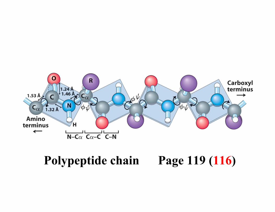
Polypeptide chain Page 119 (116)

204 =160,000
Unique sequences:= (number of possible amino acids) (amino acid length)

Primary structure: the sequence of amino acids along the chain Secondary structure: the local folding of the polypeptide chain Tertiary structure: the arrangement of secondary structure elements in 3 dimensionsQuaternary structure: the arrangement of proteins forming a function unitAmino acids confer important properties on the protein such as the ability to bind ligands and catalyze biochemical reactions

Peptide Bond Structure
Linus Pauling and Robert Corey analyzed the geometry and dimensions of the peptide bonds in the crystal structures of molecules containing one or a few peptide bonds
Summary: The consensus bond lengths are shown in Angstrom units Bond angles in degrees are also shown for the peptide N and C atoms

Note that the C-N bond length of the peptide is 10% shorter than that found in usual C-N amine bonds. This is because the peptide bond has some double bond character (40%) due to resonance which occurs with amides. The two canonical structures are:
As a consequence of this resonance all peptide bonds are found to be almost planar, i.e. atoms, C(i), O(i), N(i+1) and H(i+1) are approximately co-planar. This rigidity of the peptide bond reduces the degrees of freedom of the polypeptide during folding

Peptide Torsion AnglesThe three main chain torsion angles of a polypeptide are:phi, psi and omega.
The planarity of the peptide bond restricts ω to 180o in very nearly all of the main chain peptide bonds. In rare cases ω = 0o
for a cis peptide bond which usually involves proline
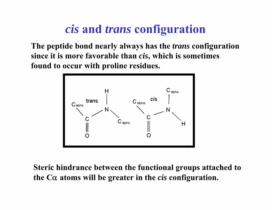
The peptide bond nearly always has the trans configuration since it is more favorable than cis, which is sometimes found to occur with proline residues.
Steric hindrance between the functional groups attached to the Cα atoms will be greater in the cis configuration.
cis and trans configuration

However for proline residues, the cyclic nature of the side chain means that both cis and trans configurations have more equivalent energies. Thus proline is found in the cisconfiguration more frequently than other amino acids. The omega torsion angle of proline will be close to zero degrees forthe cis configuration, or most often, 180 degrees for the transconfiguration
(trans)
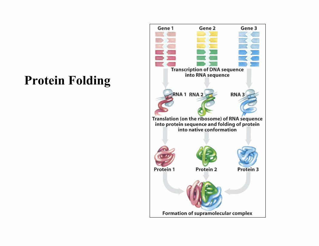
Protein Folding

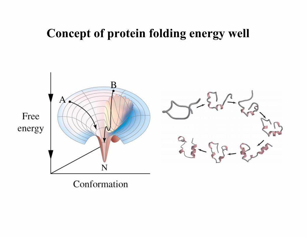
Concept of protein folding energy well

Many weak interactions
Only 10 KJmol-1
differentiates a folded functional to a precipitated protein
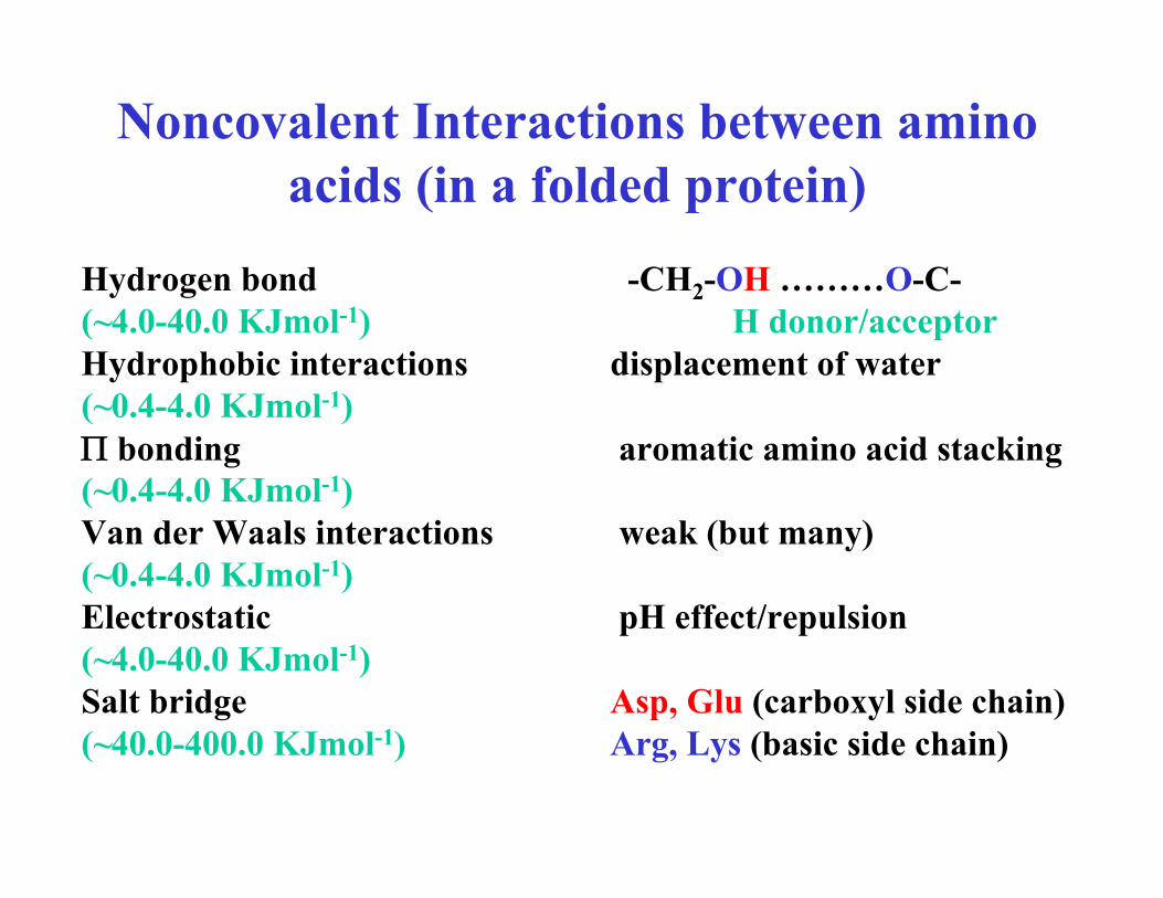
Noncovalent Interactions between amino acids (in a folded protein)
Hydrogen bond -CH2-OH ………O-C-(~4.0-40.0 KJmol-1) H donor/acceptorHydrophobic interactions displacement of water(~0.4-4.0 KJmol-1)Π bonding aromatic amino acid stacking(~0.4-4.0 KJmol-1)Van der Waals interactions weak (but many)(~0.4-4.0 KJmol-1)Electrostatic pH effect/repulsion(~4.0-40.0 KJmol-1)Salt bridge Asp, Glu (carboxyl side chain)(~40.0-400.0 KJmol-1) Arg, Lys (basic side chain)

Hydrogen bond

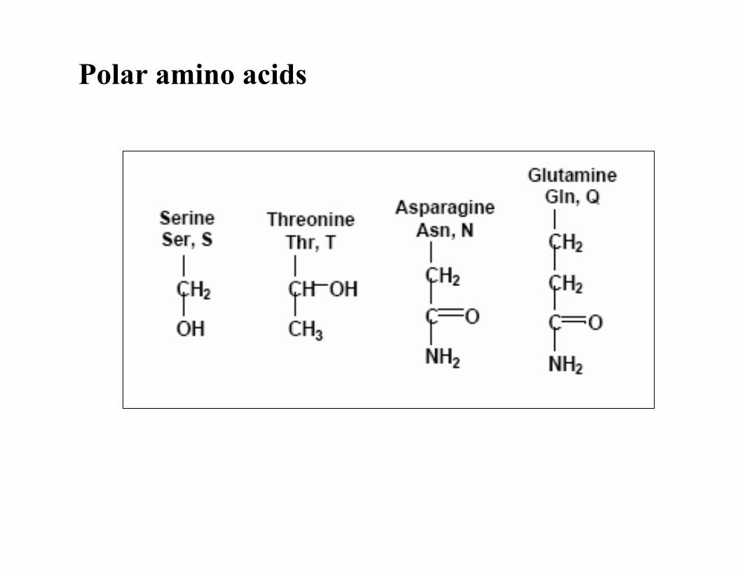
Polar amino acids


Trp Phe
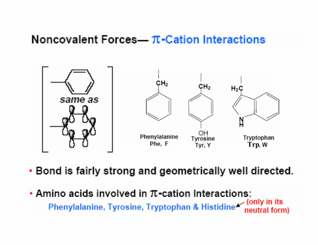
Trp


Charged amino acids


If pH of environment less than pKa amino acid + chargeIf pH of environment greater than pKa amino acid - charge
Examples:
Arg at pH 7.4Arg pKa 12.5 (pH less than pKa) + charge
Asp at pH 7.4Asp pKa 3.9 (pH greater than pKa) - charge
Simple rule:

Disulfide BridgeWhen two cysteine are close to each other in the folded protein
(important in protein folding)
-CH2-SH + HS-CH2-
2H+ + 2e-
(oxidation/reduction)
-CH2-S-S-CH2 (oxidized)(oxidized)
Forms a covalent bond(~200-800.0 KJmol-1)Free rotation about S-S bond
Stabilizes the 3 dimensional structure

Example of a protein with disulfide bond


Summary