pelagia research library - imedpub screening for foreign matter the shade-dried roots were ground to...
TRANSCRIPT

Available online at www.pelagiaresearchlibrary.com
Pelagia Research Library
Der Pharmacia Sinica, 2011, 2 (2): 131-141
ISSN: 0976-8688
CODEN (USA): PSHIBD
131
Pelagia Research Library
Morphological, Microscopical and Physico-chemical Investigations on the roots of Viburnum punctatum Buch.-Ham.ex D.Don
Kathiresan Prabhu1,*, Pradip Kumar Karar2, Siva Hemalatha3, Kathiresan Ponnudurai4
and Prakash Mankar5
1Department of Pharmacognosy, Nandini Nagar Mahavidyalaya College of Pharmacy, Nawabganj, Gonda, Uttar Pradesh, India
2Department of Pharmaceutical Chemistry, DOABA College of Pharmacy, Kharar, Mohali, Punjab, India 3Department of Pharmaceutics, Pharmacognosy Division, IT-Banaras Hindu University, Varanasi, Uttar
Pradesh, India 4Department of Pharmacology, Nandini Nagar Mahavidyalaya College of Pharmacy, Nawabganj,
Gonda, Uttar Pradesh, India 5Department of Pharmaceutical Biotechnology, Nandini Nagar Mahavidyalaya College of Pharmacy,
Nawabganj, Gonda, Uttar Pradesh, India ______________________________________________________________________________ ABSTRACT The main objective of this study is to conduct morphological, microscopical, physico-chemical and a preliminary organic investigation on the roots of a species under the genus-Viburnum Linn., belonging to Adoxaceae family, which covers about 17 species of medicinally valuable and corroborated plants in India. Macroscopic studies and various microscopic investigations were carried out to record the parameters of diagnostic values, in addition to an exploration on various physico-chemical features. An organic analysis conducted on the solvent extracts revealed the presence of bio-active molecules such as phyto-sterols, triterpenes; phenolic compounds and their derivatives such as tannins, flavonoes, simple and poly-phenolic compounds. The findings of the current study can be useful to progress and surge further scientific investigation on the roots of this species. Keywords: Viburnum, Adoxaceae, Xylem fibre, Phenolics, Druses. ______________________________________________________________________________
INTRODUTION
India has a rich heritage of using medicinal plants and hosting several thousands of medicinally valuable plants belonging to hundreds of families. One cannot assure that all of these plants possess a long recorded history, although they have been reported to contain medicinally valuable phyto-pharmaceuticals and subjected to formulate, ayurvedic, unani, siddha, Chinese

Kathiresan Prabhu et al Der Pharmacia Sinica, 2011, 2 (2):131-141 _____________________________________________________________________________
132
Pelagia Research Library
system of medicine. For many of them, an authentic protocol derived from multidisciplinary approach is very scant. In particular, the plants, which are growing at elevated altitude more than 2000 ft and forest dominated hilly areas. They are not often exposed to plant vendors, botanists, plant collectors, pharmacognosists and traditional therapists due to inaccessibility as well as climatic conditions of the locations [1]. The genus Viburnum Linn., belonging to the family Caprifoliaceae (formerly) and Adoxaceae (presently), is a typical example of such a kind, which is dwelling at a high altitude. The genus Viburnum Linn. includes about 17 species in India and about 200 species distributed throughout the world [2, 3]. Viburnum Linn. Species have been investigated and reported to contain sesquiterpenoids [4], triterpenoids and sterols; phenolic compounds and their glycosides (tannins, flavonoids and anthocyanins and irridoid glycosides) in their stem, root and leaves, and investigated to posses uterine sedative, diuretic, cardiovascular stimulant, antimicrobial, anti-inflammatory, anti-nociceptive , antispasmodic [5], anti-asthmatic and astringent activities [6]. In the late 1960s and early 1980s, scientific studies on the genus Viburnum Linn. were voluminous [7-9]. However, the number of species subjected for studies and areas of investigations were absolutely narrow. After a couple of decades, a few Viburnum species re-emerged to be subjected for some extensive phytochemical and pharmacological investigations. The typical examples are: iridoid aldehyde and their glycosides in Viburnum luzonicum [10] and their cytotoxic effect; vibsane type diterpene from Viburnum awabuki [11]; iridoid glycosides from Viburnum tinos; anti-nociceptive and anti-inflammatory activities of Viburnum lanata [12] and Viburnum opulus [13]; iridoid glucosides from Viburnum rhytidophyllum [14]. In addition to the above, a questionnaire and verbal enquiries to the local dwellers, tribals and herbalists of Nilgiri hills and Coimbatore hills (Top Slip) Tamilnadu, about some Viburnum species, were conducted; for it has been noted that the leaves, stem bark and root barks of mature plants had been reliably in usage to uterus and GIT related ailments, and still in practice, as an ideal healing aid as well as one of the best home remedies. As far as the species of priority is concerned Viburnum punctatum (stem and roots), Viburnum coriaceum (Leaves and Stem bark) and Viburnum erubescens (Stem and roots) are significant. The current study high lights the pharmacognostical cum botanical characteristics on the roots of one of the above species namely: Viburnum punctatum (synonymously: Viburnum acuminatum).
MATERIALS AND METHODS
Collection of specimens The plant specimens for the study were collected from Nilgiri Hills, Tamil Nadu, India,and authenticated by Dr. V. Chelladurai, former Professor of Botany, Medicinal Plant Survey for Siddha, Government of India, as Viburnum punctatum Buch.-Ham.ex D.Don. A voucher specimen (V181) was deposited in the herbal museum at Nandini NagarMahavidyalaya College of Pharmacy, UttarPradesh. A care was taken to select healthy plants for the study. The plant parts for the study were collected fresh from the plant and placed in FAA (Formalin-Acetic acid-70 % ethyl alcohol) in a ratio of 1:1:18. Twenty four hours later, the specimens were dehydrated with a graded series of tertiary-butyl alcohol (TBA). Infiltration of the specimens was carried out by gradual addition of paraffin wax (melting point 58 - 60 ºC) until TBA solution attained super saturation. The specimens were then cast into paraffin blocks.

Kathiresan Prabhu et al Der Pharmacia Sinica, 2011, 2 (2):131-141 _____________________________________________________________________________
133
Pelagia Research Library
Sectioning The paraffin-embedded specimens were sectioned with the aid of an MC 930 advanced precision rotary microtome. The thickness of the sections was 10 - 12 µm. Dewaxing of the sections was performed by treating the specimen slides sequentially with xylol, xylol-alcohol, alcohol, water and finally, with staining fluid [15]. Wherever necessary, sections were also stained with safranin, fast-green and iodine in potassium iodide in order to evaluate starch, calcium oxalate crystals and other ergastic cell content and their distribution. Some hand sections were prepared and cleaned with 5% sodium hydroxide and treated with Jeffrey’s maceration fluid [16] to study the histo-morphology of vascular elements. Glycerin mounted temporary preparations were made for macerated/cleared materials. Powdered materials of different parts were cleared with NaOH and mounted in glycerine. After staining with Toluidine blue, different root components were studied and measured as described below. Microscopic examination With the aid of a compound microscope (Focus (ISI), JPM-1, India) and an eye-piece micrometer calibrated with a stage micrometer, the individual character of each specimen was studied under both low- (10x ×10x) and high-power (10x × 45x) magnification. Powder microscopy The roots were dried for a minimum of 15 days under the sun shade, powdered and screened through sieves with aperture size of 180 µm and 125 µm separately to obtain fine and very fine powders, respectively, then subjected to microscopic examination. The specimens were treated with the following reagents in order to evaluate components of diagnostic value: 50 % glycerin as temporary mountant; 2% phloroglucinol in 90% ethanol and conc. HCl (1:1) for lignin; 5% alcoholic ferric chloride solution for phenolic compounds; 2 % iodine solution for starch grains and suberin; and 0.08% ruthenium red in 10% lead acetate solution for mucilage [17]. Photomicrography Photomicrographs were taken in addition to microscopic examination, where necessary, at different magnifications using a Nikon Labphot 2 microscope. For normal observations, a bright field was used while for the study of calcium oxalate crystals, starch grains and lignified cells, polarized light was employed. Since these structures have bi-refringent properties under polarized light, they appear bright against dark background. Magnifications of the figures were indicated by scale-bars [18,19]. Numerical and physical standards Determination of extractives Approximately 5 g of the air-dried crude drug was macerated with 100 ml of the solvents for 24 h. After filtration, 25 ml of the filtrate was evaporated to dryness and the extractives were calculated with reference to the air-dried drug [20]. Determination of ash values Approximately 3 g of the air-dried crude drug was incinerated in a tared silica crucible (Vitrosil, India) in a furnace at a temperature not exceeding 450 °C until free from carbon. It was washed in hot water to exhaust the charred mass, and the residue was incinerated on an ashless filter paper and weighed. Further treatment was carried out to derive water soluble, acid insoluble and sulphated ash values [21].

Kathiresan Prabhu et al Der Pharmacia Sinica, 2011, 2 (2):131-141 _____________________________________________________________________________
134
Pelagia Research Library
Histochemical studies Histochemical analysis was carried out on the specimens using, separately, dilute iodine solution, Lacto phenol, Dragendorff”s reagent, dilute ferric chloride solution and phloroglucinol-HCl (1:1). The reagent treated hand sections of the plant tissue were observed under microscope to detect the presence of histochemical components [22]. Fluorescence studies Fluorescence analysis was carried out in an ultraviolet cabinet (MAC, MSW-508, Long UV, India) at 365 nm [23]. Micrometric studies Micrometric evaluation was carried out with the aid of a compound microscope fitted with a camera lucida (Swift Ive’s) as described elsewhere [24-26]. Preliminary screening for foreign matter The shade-dried roots were ground to a moderately coarse powder in a mechanical grinder. Aprrox. 100 g of the powder was extracted successively with petroleum ether (60 – 80 ºC), benzene, chloroform and ethanol (95 %) using a Soxhlet apparatus. Each solvent extraction was carried out for 24h. Finally, the marc left was extracted with water by digesting over a boiling water bath. The extraction was continued until a few drops of the last portion of the extract left no residue on drying. The extracts were taken in a tarred porcelain dishes, evaporated to dryness over a water bath and dried in an oven at 105 ºC to a constant weight. The extractives (%) were calculated with reference to the air-dried drug [27].
RESULT
A mature root appeared with a yellowish brown bark containing enormous number of longitudinal fissure which were irregular; the root stem appeared yellowish and extremely hard. As a whole the roots were fibrous and hard in fracture; odour less and astringent in taste, when dried; but valerian root like odour, when freshly cut out and smelled. The microscopic characteristics of the roots revealed, through transverse sections (T.S.), the presence of histological zones such as pariderm, cortex, secondary phloem and then secondary xylem from the outside in. These histological zones were common for both a thin as well as a thick root selected for the study. However, histo-morphological aspects of both the roots were quiet different in characters. The powder microscopy showed the presence of calcium oxalate crystals (Druses) xylem fibres, thin walled xylem vessel, circular and oval shaped starch grains and sclereids (Brachy-sclereids) as their diagnostic features. Histochemical analysis performed using hand sections of the roots represented the distribution of starch, tannins, saponin, lignins and calcium oxalate crystals in their histological zones. Upon the microscopic analysis of the root it was revealed that the grains crystals, fibres, stone cells and vessels were the salient features whose semi-quantitative and quantitative analysis were needed to be performed, since it may supplement some additional information to recognize those species from the roots of its co-species. The physicochemical analysis such as extractive values, various ash figures, crude fibre content, loss on drying, fluorescence analysis and preliminary phytochemical analysis were also carried out to result remarkable observations, which deserve an elaborate discussion in this article.

Kathiresan Prabhu et al Der Pharmacia Sinica, 2011, 2 (2):131-141 _____________________________________________________________________________
135
Pelagia Research Library
DISCUSSION
Morphology Both a thin root (measuring less than 2.5 mm diameter) and a thick root (measuring more than 10 mm diameter) were observed. The barks are yellowish brown and the stems, yellowish to pale yellow; longitudinal fissures on the surface which are prominent over the thick root. The secondary growth, evident by growth rings with thick roots; hard and brittle in fracture; the broken surface observed to be fibrous (remains of the broken fibre); slightly astringent and bitter in taste; smelled like valerian root odour, when fresh (Figure 1). Morpho-anatomy Thin root measuring 2.3 mm in diameter has narrow periderm, wide cortex, thin continuous secondary phloem and dense solid secondary xylem cylinder. The periderm is 60 µm wide comprising of three to five layers of thin walled tabular cells. Cortex consists of seven or eight layers of circular, thin walled compact parenchyma cells. Secondary phloem has funnel-shaped dilated rays and conical bands of sieve elements and phloem parenchyma cells. Secondary xylem has no growth rings. The vessels are diffuse in distribution. They are mostly solitary, angular and thin walled. The density of the vessels is low. The vessel diameter is 70 µm in average. Xylem fibres are wide and thick walled (Figure 2). A thick root of more than 3 mm in thickness has thick, deeply fissured periderm of 250 µm thick. It consists of dark tubular, narrow phellem cells. The cortex has radially compressed oblong parenchyma cells. Secondary phloem has funnel shaped, dilated rays. The cells are being large rectangular in shape. The sieve elements and phloem parenchyma occur in conical bands. The sieve elements are polygonal in outline; the companion cells are small and occur in a corner. Secondary xylem has angular, wide, diffuse thin walled vessels. They are solitary. The wide vessels are 130 µm in diameter. Xylem rays are prominent, thick walled and lignified walls. Starch grains are abundant within ray cells. Thick masses sclereids are seen in between phloem and cortex (Figure 3). Powder Microscopy Organoleptic examination
Colour : yellowish brown Odour : root characteristics Taste : slightly bitter and astringent Texture : fibrous With water : mucilaginous nature absent Dilute FeCl3 treatment : powder turned blackish Phloroglucinol-HCl (1:1) treatment : powder turned pinkish Particle size : passing through No. 40, 60 and 90 sieves
Microscopical Features (Figure 4) Xylem fibre: Wide and thick walled; leaving a thin central lumen with no striations. The shape and width were similar to that of the stem fibres and were as long as 1 mm. Vessels: Vessels, showing annular thickening and were from 70 – 140 µm in width. Starch grains: Starch grains were circular and oval in shape containing central hilum and eccentric; single grain is 4 – 17 µm in diameter.
Kathiresan Prabhu et al Der Pharmacia Sinica, 2011, 2 (2):131-141 _____________________________________________________________________________
136
Pelagia Research Library
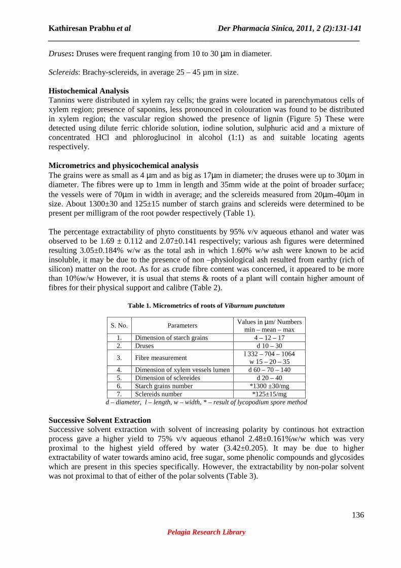
Druses: Druses were frequent ranging from 10 to 30 µm in diameter. Sclereids: Brachy-sclereids, in average 25 – 45 µm in size. Histochemical Analysis Tannins were distributed in xylem ray cells; the grains were located in parenchymatous cells of xylem region; presence of saponins, less pronounced in colouration was found to be distributed in xylem region; the vascular region showed the presence of lignin (Figure 5) These were detected using dilute ferric chloride solution, iodine solution, sulphuric acid and a mixture of concentrated HCl and phloroglucinol in alcohol (1:1) as and suitable locating agents respectively. Micrometrics and physicochemical analysis The grains were as small as 4 µm and as big as 17µm in diameter; the druses were up to 30µm in diameter. The fibres were up to 1mm in length and 35mm wide at the point of broader surface; the vessels were of 70µm in width in average; and the sclereids measured from 20µm-40µm in size. About 1300±30 and 125±15 number of starch grains and sclereids were determined to be present per milligram of the root powder respectively (Table 1). The percentage extractability of phyto constituents by 95% v/v aqueous ethanol and water was observed to be 1.69 ± 0.112 and 2.07±0.141 respectively; various ash figures were determined resulting 3.05±0.184% w/w as the total ash in which 1.60% w/w ash were known to be acid insoluble, it may be due to the presence of non –physiological ash resulted from earthy (rich of silicon) matter on the root. As for as crude fibre content was concerned, it appeared to be more than 10%w/w However, it is usual that stems & roots of a plant will contain higher amount of fibres for their physical support and calibre (Table 2).
Table 1. Micrometrics of roots of Viburnum punctatum
S. No. Parameters Values in µm/ Numbers min – mean – max
1. Dimension of starch grains 4 – 12 – 17 2. Druses d 10 – 30
3. Fibre measurement l 332 – 704 – 1064
w 15 – 20 – 35 4. Dimension of xylem vessels lumen d 60 – 70 – 140 5. Dimension of sclereides d 20 – 40 6. Starch grains number *1300 ±30/mg 7. Sclereids number *125±15/mg
d – diameter, l – length, w – width, * – result of lycopodium spore method Successive Solvent Extraction Successive solvent extraction with solvent of increasing polarity by continous hot extraction process gave a higher yield to 75% v/v aqueous ethanol 2.48±0.161%w/w which was very proximal to the highest yield offered by water (3.42±0.205). It may be due to higher extractability of water towards amino acid, free sugar, some phenolic compounds and glycosides which are present in this species specifically. However, the extractability by non-polar solvent was not proximal to that of either of the polar solvents (Table 3).

Kathiresan Prabhu et al Der Pharmacia Sinica, 2011, 2 (2):131-141 _____________________________________________________________________________
137
Pelagia Research Library
Table 2. Physico-chemical parameters of roots of Viburnum punctatum
S.No. Parameters Values in % w/w/ nm 1 Alcoholic extractive values (95%v/v)ethanol 1.69±0.112 2. Aqueous extractive value (Double distilled) 2.07±0.141 3. Ash figures
a. Total ash value b. Water soluble ash c. Acid insoluble ash d. Sulphated ash
3.05±0.184 2.60±0.126 1.85±0.105 4.80±0.241
4. Crude fibre content 10.95±0.311 5. Loss on drying 3.27±0.241 6. λmax of 80% methanolic extracts (peak maxima and
sub-maxima) 250 nm, 270 nm
7. Fluorescence analysis (365 nm) of alcoholic extracts (80%)
Reddish brown in the day light and yellowish under UV (long)
Values are represented as mean±S.D, n=3, Wavelength in nanometres Preliminary organic analysis The presence of phenolic compound (by dilute ferric chloride test); saponins (by haemolytic test);free sugars(By reducing agents); phenolics glycosides (acid hydrolysis after exhausting free sugar followed by test for sugar & Aglycone part); flavonoids (By shinoda’s test and UV- quenching of TLC with a sample of 80% methanolic extract) were detected in the alcoholic and aqueous extracts; but in case of non-polar layers, a test for phyto-sterols and triterpenes were positive by a preliminary organic analysis of the successive extracts (Table 3).
Table 3. Preliminary phyto-chemical screening of root extracts of Viburnum punctatum
Extract Percentage Extractives
Sterol Triterpenes Sugars Glycosides Flavones Saponins Phenolics
Petroleum ether 1.06±0.147 ( - ) (+++) ( - ) ( - ) ( - ) ( - ) ( - ) ( - ) Benzene 0.52±0.043 ( - ) ( + ) ( - ) ( - ) ( - ) ( - ) ( - ) ( - ) Chloroform 0.36±0.029 ( - ) ( + ) ( + ) ( - ) ( - ) ( - ) ( - ) ( + ) Ethanol (75% v/v) 2.48±0.161 ( - ) ( - ) ( - ) (+) (++) ( - ) (+++) (+++) Water 3.42±0.205 ( - ) ( - ) ( - ) (++) ( + ) (++) (++) (+++)
Values are represented as mean±S.D, n=3, ( + )-Test Positive, ( ++ )-present in moderate amount, (+++)-Relatively larger amount, ( - )-Test negative
Figure 1. Roots of Viburnum punctatum
RP – Root Pieces, FT –Fruiting Twig

Kathiresan Prabhu et al Der Pharmacia Sinica, 2011, 2 (2):131-141 _____________________________________________________________________________
138
Pelagia Research Library
Figure 2. T.S. of thin root of Viburnum punctatum 2.1. Co – Cortex; Pe – Periderm; SX – Secondary Xylem; Sph – Secondary phloem; 2.2. Co – Cortex; DR – Dilated
Ray; Pe – Periderm; SE – Sieve Element; Sph – Secondary phloem, Ve – Vessel, XF – Xylem Fibres
Figure 3. T.S. of thick root of Viburnum punctatum Dr – Druses; DR – Dilated Ray; Sph – Secondary phloem; SE – Sieve Element; SX – Secondary Xylem; Ve – Vessel;
XR – Xylem Ray; XF – Xylem Fibres

Kathiresan Prabhu et al Der Pharmacia Sinica, 2011, 2 (2):131-141 _____________________________________________________________________________
139
Pelagia Research Library
Figure 4. Powder Characteristics of Roots of Viburnum punctatum
Dr – Druses in leaf, SG, SGR – Starch grains, XV – Xylem vessel, Ve – Vessel fragment, LXV – Lignified vessel and
fibres sheath, Sc–Sclereid, SGR – Starch grains, XFR – Xylem fibre
Figure 5. Histochemical analysis of T.S. of Roots of Viburnum punctatum

Kathiresan Prabhu et al Der Pharmacia Sinica, 2011, 2 (2):131-141 _____________________________________________________________________________
140
Pelagia Research Library
TXR – Tannins in Xylem Ray cells; STXP – Starch grains in Xylem Parenchyma; LX – Lignin in Xylem; SX –
Saponins in Xylem region
CONCLUSION
The roots were collected from Nilgiri hills for subjecting to morphological and various microscopical investigations, in addition to a preliminary organic analysis on the successive solvent extracts prepared. The entire study has resulted in to some findings which will be useful to progress with further phyto-chemical and biological studies on Viburnum Linn. species besides acting as a tool, in future, to develop a criterion of differentiating these roots from the roots of its co-species.
REFERENCES [1] The Wealth of India, A Dictionary of Indian Raw materials and Industrial Products – Raw Material Series, Publication and Information Directorate, CSIR, New Delhi, 2003, 10: 437 - 446. [2] J.S. Gamble, Flora of the Presidency of Madras, Vol. I, II & III. Botanical Survey of India, Calcutta, India, 1935. [3] W.C. Evans, Pharmacognosy, 15th ed, W.B. Saunders, London, 2002, 37 – 547. [4] R.L. Khosa, A.K. Wahi, Y. Mohan and A.B. Ray, Ind J Pharm, 1979, 41(3), pp.120. [5 ] K. Prabhu, P.K. Karar, S. Hemalatha and K. Ponnudurai, Int J Curr Tr Sci Tech, 2010, 1(3): 175–186. [6] K.M. Nadkarni, Indian Materia Medica, 2nd ed, Popular Prakashan, Bombay, India, 2002, 1: 1271 - 1272. [7] L. Hoerhammer, H. Wagner and H. Reinhardt, Apothekerzer, 1965, 105(40): 1371. [8] S.G. Yunusova, A.R. Karimova, E.M. Tsyrlina, M.S. Chem Nat Comp, 2004, 40(5): 423 – 426. [9] A.K. Wahi, R.L. Khosa and Y. Mohan, Bot Res, 1981, 3: 205. [10] L. Tomassini, J. Gao, S. Foddai, M. Serafini, Nat Prod Res, 2006, 20(8), pp.697 - 700. [11] Y. Fukuyama, M. Kubo, H. Minami, H. Yuasa, A. Matsuo, T. Fujii, M. Morisaki and K. Harada, Chem Pharm Bull, 2005, 53(1): 72 - 80. [12] Y.B. Sever, C.G. Saltan, M.L. Altun and H. Ozbek, Pharm Biol, 2007, 45(3), 241-245. [13] M.L. Altun, C.G. Saltan, Y.B. Sever and H. Ozbek, Pharmaceutical Biology, 2009, 47 (7), 653-658. [14] L.Tomassini, B. Dejan, F. Sebastiano and M. Nicoletti, Phytochemistry, 1997, 44 (4), 751-753. [15] D.A. Johansen, Plant Microtechnique, Mc Graw Hill Book Co., New York, 1940, 523.

Kathiresan Prabhu et al Der Pharmacia Sinica, 2011, 2 (2):131-141 _____________________________________________________________________________
141
Pelagia Research Library
[16] J.E. Sass, Elements of Botanical Microtechnique, Mc Graw Hill Book Co., New York, 1940, 222. [17] K.R. Khandelwal, Practical Pharmacognosy Techniques and Experiments, Nirali Prakashan, India, 2006, 16, 15 - 163. [18] K. Easu, Plant Anatomy, John Wiley and Sons, New York, 1964, 767. [19] A.N. Henry, G.R. Kumari, V. Chitra, Flora of Tamilnadu, India. Vol.I, II & III Botanical Survey of India, Southern Circle, Coimbatore, India, 1987, 3: 258. [20] Pharmacopoeia of India, Ministry of Health and Family Welfare, The Controller of Publications, New Delhi, India, 1996, Vol. 2, A47 - A89. [21] British Pharmacopoeia, Ministry of Health and Social Services for Northern Ireland, 1988, 2: A139 - A140. [22] T.P. O’Brien, N. Feder and M.E. Mc Cull, Protoplasma, 1964, 59, 364 - 373. [23] World Health Organization, Quality control methods for medicinal plant materials, WHO/PHARM/92.559, 1992, 11 - 36. [24] P.K. Lala, Practical Pharmacognosy, Lina Guha Publication, India, 1981, 1: 136-153. [25] G.E. Trease, W.C. Evans, Pharmacognosy, 10th ed, Berilliee, Tindal, London, 2002, 519 - 547. [26] T.E. Wallis, Text Book of Pharmacognosy, 5th ed, CBS Publishers and Distributors, New Delhi, India, 2005, 559 - 618. [27] J.B. Harborne, Phytochemical methods, 3rd ed, Chapman and Hall, London, 2005, 49-244.