pediatric renal tumors: update for 2014 - pathology · pediatric renal tumors: update for 2014 john...
TRANSCRIPT

1
Pediatric Renal Tumors:Update for 2014
John HicksTexas Children’s Hospital
& Baylor College of Medicine
Renal Tumors of Childhood6th Most Common Group of Tumors
Most Occur in 1st Decade of Life
Diverse Group of Tumors– Benign Mesoblastic Nephroma to Aggressive
and Often Fatal Rhabdoid Tumor
– Most Common Tumor - Wilms Tumor

2
Other Renal Tumors
Anaplastic Sarcoma of Kidney
Primitive Neuroectodermal Tumor (PNET/EWS)
Desmoplastic Small Round Cell Tumor
Rhabdomyosarcoma
Synovial Sarcoma
Primary Neuroblastoma
Oncocytoid Post-Neuroblastoma Carcinoma
“Others”
3
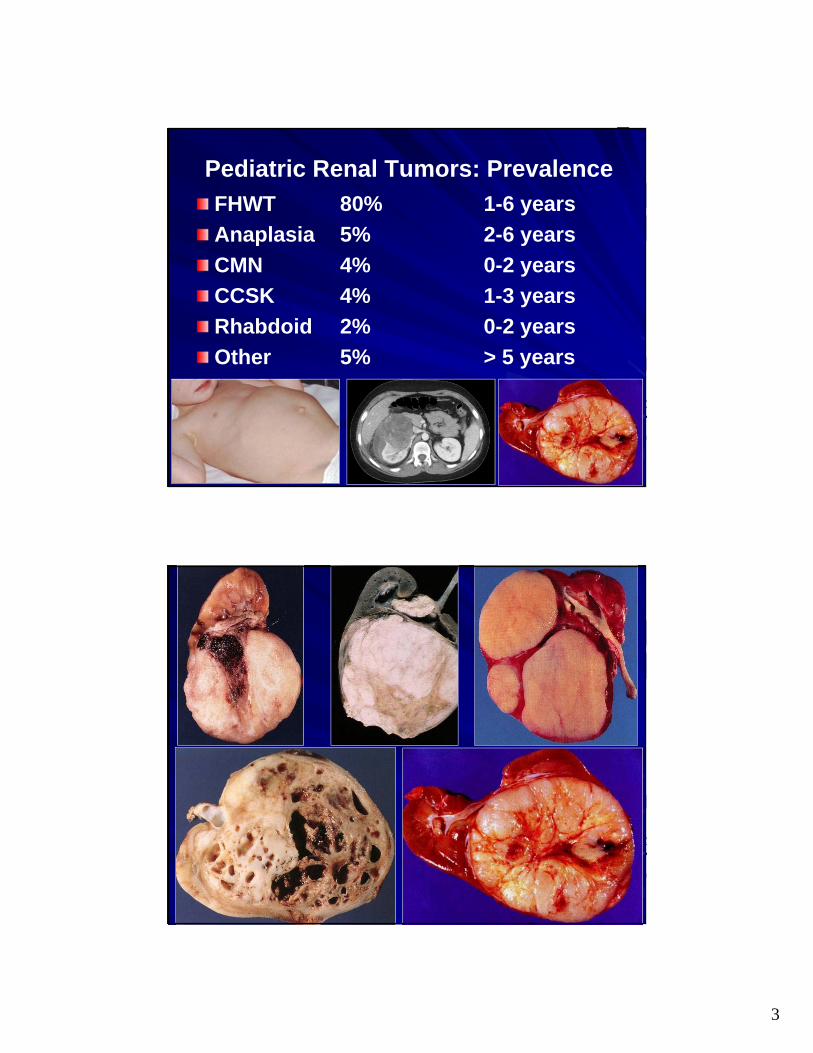
Pediatric Renal Tumors: Prevalence
FHWT 80% 1-6 years
Anaplasia 5% 2-6 years
CMN 4% 0-2 years
CCSK 4% 1-3 years
Rhabdoid 2% 0-2 years
Other 5% > 5 years

4
NEPHROBLASTOMA (WILMS TUMOR)

5

6

7
PERILOBAR NEPHROGENIC RESTS
LOCATED AT PERIPHERY
USUALLY NUMEROUS
MARGINS WELL DEFINED
NO NEPHRONS WITHIN REST
COMPOSED OF BLASTEMA AND TUBULES; STROMA SCANTY OR SCLEROTIC

8

9
“Adenomatous” Change Within Hyperplastic Perilobar Nephroblastomatosis
Nodules with Cells Composed of Pale Eosinophilic Cytoplasm
Often With Papillary Architecture
Confused With Renal Cell Carcinoma & Metanephric Adenoma

10
INTRALOBAR NEPHROGENIC
RESTS
RANDOM LOCATIONOFTEN SINGLEILL-DEFINED MARGINSDISPERSED BETWEEN NORMAL KIDNEYCOMPOSED OF TUBULES, BLASTEMA AND CYSTS; STROMA USUALLY PREDOMINATES

11
SSSYYYNNNDDDRRROOOMMMEEESSS AAANNNDDD CCCOOONNNGGGEEENNNIIITTTAAALLL DDDIIISSSOOORRRDDDEEERRRSSS AAASSSSSSOOOCCCIIIAAATTTEEEDDDWWWIIITTTHHH NNNEEEPPPHHHRRROOOGGGEEENNNIIICCC RRREEESSSTTTSSSaaa
WWWiiilllmmmsss tttuuummmooorrr,,, aaannniiirrriiidddiiiaaa,,, gggeeennniiitttooouuurrriiinnnaaarrryyy aaannnooommmaaallliiieeesss,,, rrreeetttaaarrrdddaaatttiiiooonnn (((WWWAAAGGGRRR)))
BBBeeeccckkkwwwiiittthhh---WWWiiieeedddeeemmmaaannnnnn sssyyynnndddrrrooommmeee (((cccooommmpppllleeettteee aaannnddd iiinnncccooommmpppllleeettteee)))
PPPeeerrrlllmmmaaannn sssyyynnndddrrrooommmeee
DDDeeennnyyysss---DDDrrraaassshhh sssyyynnndddrrrooommmeee
KKKllliiippppppeeelll---TTTrrreeennnaaauuunnnaaayyy sssyyynnndddrrrooommmeee
BBBrrraaaccchhhmmmaaannnnnn---DDDeee LLLaaannngggeee sssyyynnndddrrrooommmeee
RRReeennnaaalll dddyyysssppplllaaasssiiiaaa aaannnddd ooobbbssstttrrruuuccctttiiivvveee uuurrrooopppaaattthhhyyy
TTTrrriiisssooommmyyy 111333
TTTrrriiisssooommmyyy 111888
CCCooonnngggeeennniiitttaaalll hhheeeaaarrrttt dddiiissseeeaaassseee
SSSpppllleeennniiiccc aaagggeeennneeesssiiisss wwwiiittthhh llliiivvveeerrr mmmaaalllfffooorrrmmmaaatttiiiooonnnsss
BBBiiilllaaattteeerrraaalll rrraaadddiiiaaalll aaappplllaaasssiiiaaa aaannnddd ooottthhheeerrr ssskkkeeellleeetttaaalll aaabbbnnnooorrrmmmaaallliiitttiiieeesss
- (10% of WT)

12

13
Anaplasia & Outcome

14
Anaplastic Wilms Tumor
NWTS-4 :stage I focal and diffuse anaplasia behaved similarly to FHWT
NWTS-5: decreased survival for stage I patients with focal and diffuse anaplasia
Treatment for stage 1 anaplasia now more aggressive in new protocols (FA=DA)
Focal anaplasia has very specific criteria; tumors not meeting the criteria must be classified as diffuse
ANAPLASIA– Polypoid, Multipolar Mitotic Figures– Nuclear Enlargement (>3 times) with Hyperchromasia– Epithelial, Blastemal or Stromal Elements with Anaplasia– Note: Skeletal Muscle May Have Nuclear Enlargement,
Pleomorphism and Hyperchromasia Representing Regenerative Features & Not Anaplasia
FOCAL ANAPLASIA– Circumscribed & Perimeter Completely Examined (requires
mapping of anaplastic area that extends to tissue section edges)– Confined to Renal Parenchyma (Vascular Invasion Precludes
Focal Anaplasia)– Absence of Severe Nuclear Pleomorphism and Hyperchromasia in
Non-Anaplastic Tumor (severe “nuclear unrest”)

15
Processing of Renal Tumors
Avoid frozen sections of biopsies for diagnosis– Beware nephrogenic rests
Ink prior to bivalving– tumor displacement
during grossing procedure
Search carefully for lymph nodes
Take Most Sections From Periphery
Demonstrate Tumor Relationship To:– Renal capsule– Renal sinus– Normal Kidney
Critical for Accurate– Staging– Rest determination– Diagnosis