pediatric dermatology pearls u044... · pityriasis rubra pilaris •prp in children is a...
TRANSCRIPT
Pearls for Challenging Cases in Skin of Color in Pediatric Dermatology
Assistant Professor of Dermatology and Pediatrics
University of Colorado School of Medicine
Children's Hospital Colorado

Disclosure
• I have no conflicts of interest or financial relationships to disclose.

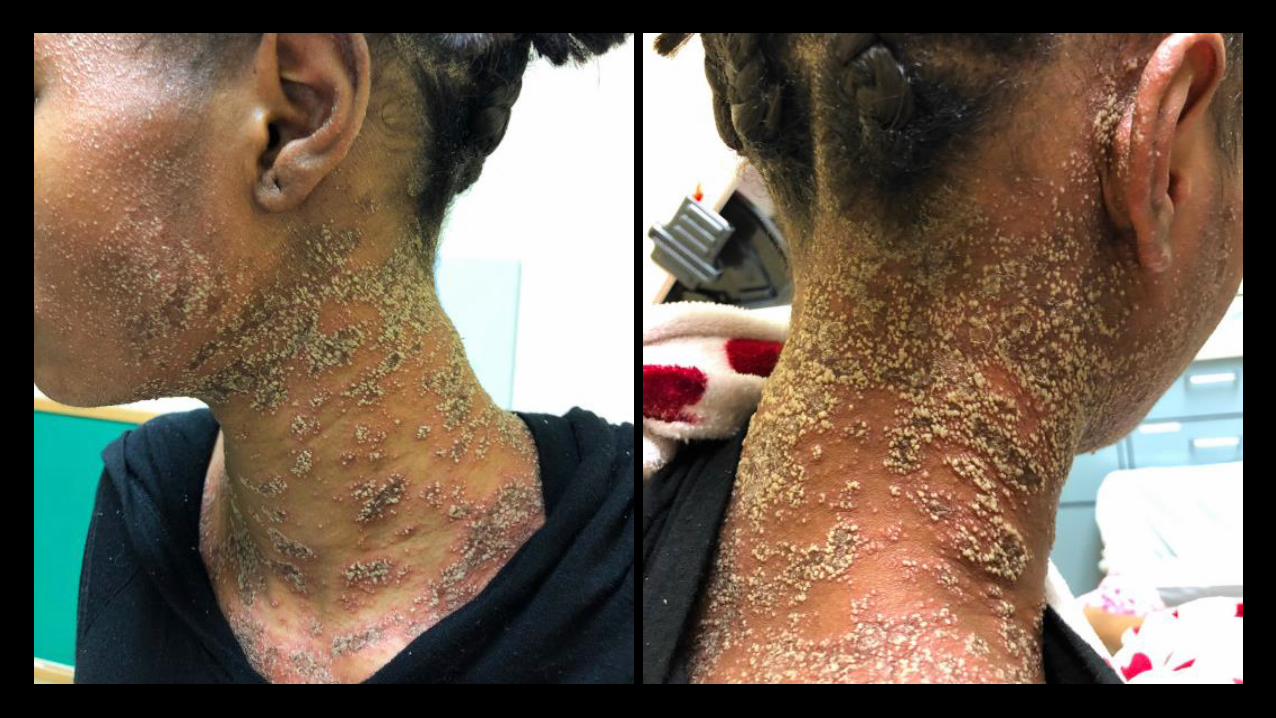
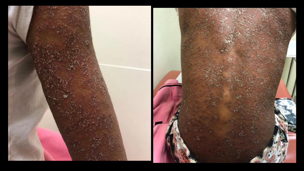
Case 1
Inpatient Consult for “generalized rash”
Case 1
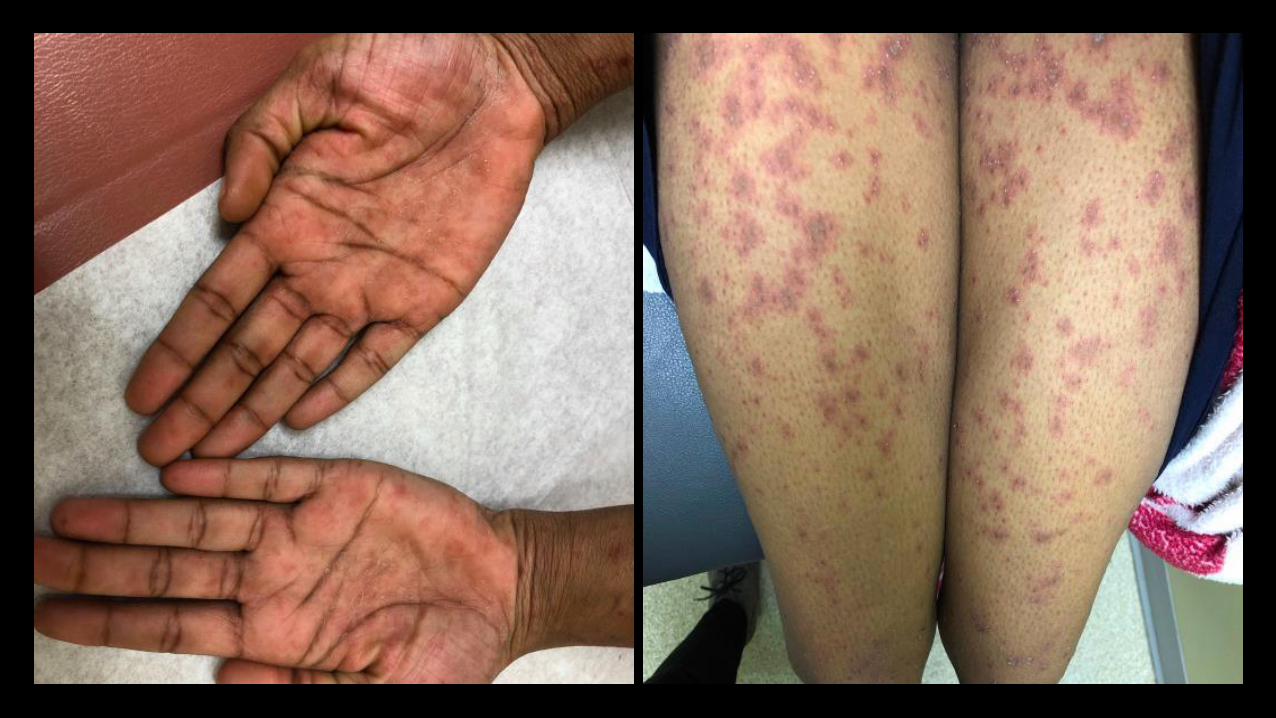
• 16 yo Somali female with presented to the ER for 10 day history of worsening rash affecting entire body.
• Prior treatments include Permethrin for suspected scabies without improvement and triple antibiotic therapy for newly diagnosed gonorrhea, and empiric therapy for chlamydia and syphilis (Penicillin, Ceftriazone and Azythromycin).
• Patient acutely decompensated and became febrile, tachycardic and hypotensive and was transferred to PICU. She received Vancomycin, blood cultures and skin cultures from pustules noted on admission were negative.
• Dermatology was consulted on hospital day 2.
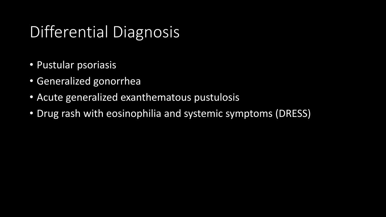
Differential Diagnosis
• Pustular psoriasis
• Generalized gonorrhea
• Acute generalized exanthematous pustulosis
• Drug rash with eosinophilia and systemic symptoms (DRESS)
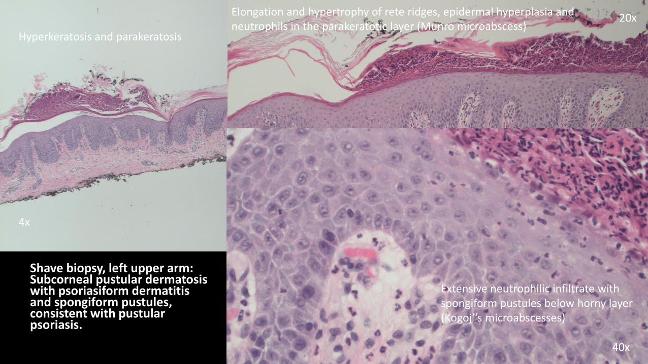
Pathology
Shave biopsy, left upper arm: Subcorneal pustular dermatosis with psoriasiform dermatitis and spongiform pustules, consistent with pustular psoriasis.
4x
20x
40x
Hyperkeratosis and parakeratosis
Elongation and hypertrophy of rete ridges, epidermal hyperplasia and neutrophils in the parakeratotic layer (Munro microabscess)
Extensive neutrophilic infiltrate with spongiform pustules below horny layer (Kogoj’’s microabscesses)
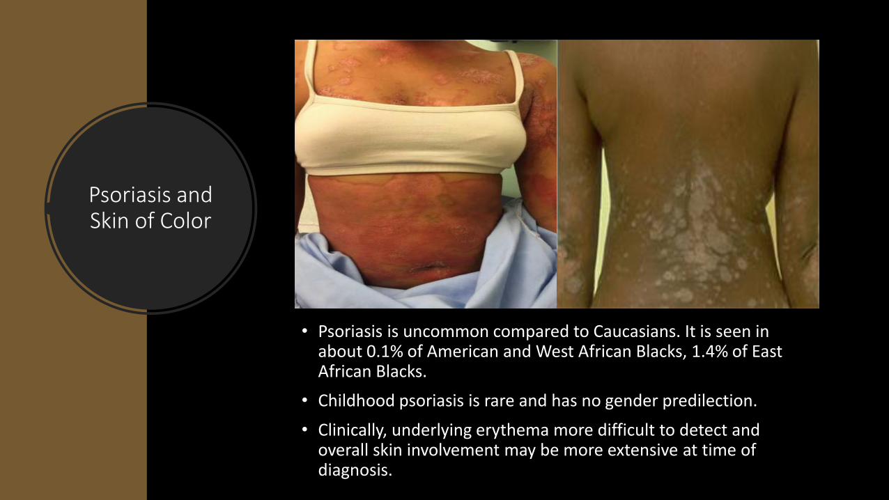
Psoriasis and Skin of Color
• Psoriasis is uncommon compared to Caucasians. It is seen in about 0.1% of American and West African Blacks, 1.4% of East African Blacks.
• Childhood psoriasis is rare and has no gender predilection.
• Clinically, underlying erythema more difficult to detect and overall skin involvement may be more extensive at time of diagnosis.
Pustular Psoriasis
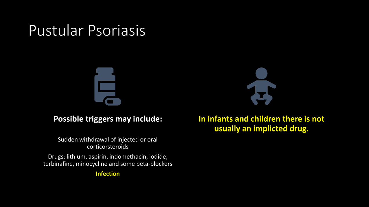
Possible triggers may include:
Sudden withdrawal of injected or oral corticorsteroids
Drugs: lithium, aspirin, indomethacin, iodide, terbinafine, minocycline and some beta-blockers
Infection
In infants and children there is notusually an implicted drug.
Pustular Psoriasis
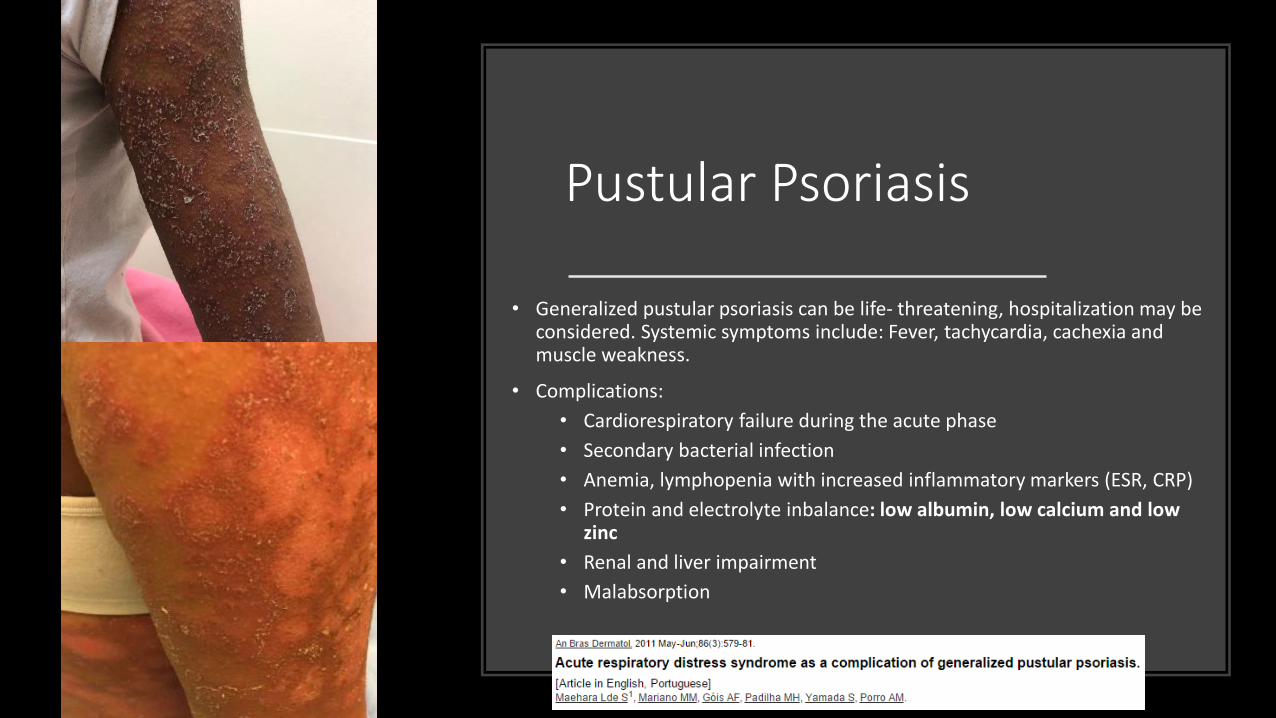
• Generalized pustular psoriasis can be life- threatening, hospitalization may be considered. Systemic symptoms include: Fever, tachycardia, cachexia and muscle weakness.
• Complications:
• Cardiorespiratory failure during the acute phase
• Secondary bacterial infection
• Anemia, lymphopenia with increased inflammatory markers (ESR, CRP)
• Protein and electrolyte inbalance: low albumin, low calcium and lowzinc
• Renal and liver impairment
• Malabsorption
Pustular Psoriasis - Treatment
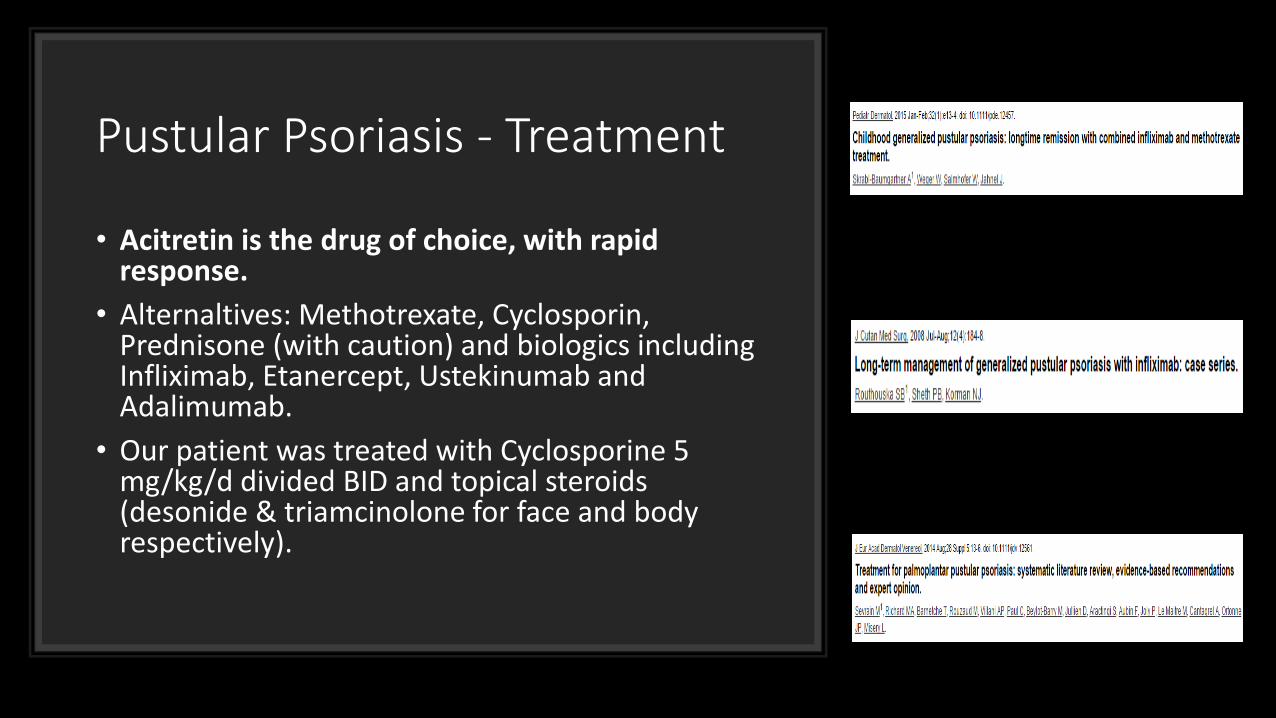
• Acitretin is the drug of choice, with rapidresponse.
• Alternaltives: Methotrexate, Cyclosporin, Prednisone (with caution) and biologics includingInfliximab, Etanercept, Ustekinumab and Adalimumab.
• Our patient was treated with Cyclosporine 5 mg/kg/d divided BID and topical steroids(desonide & triamcinolone for face and bodyrespectively).
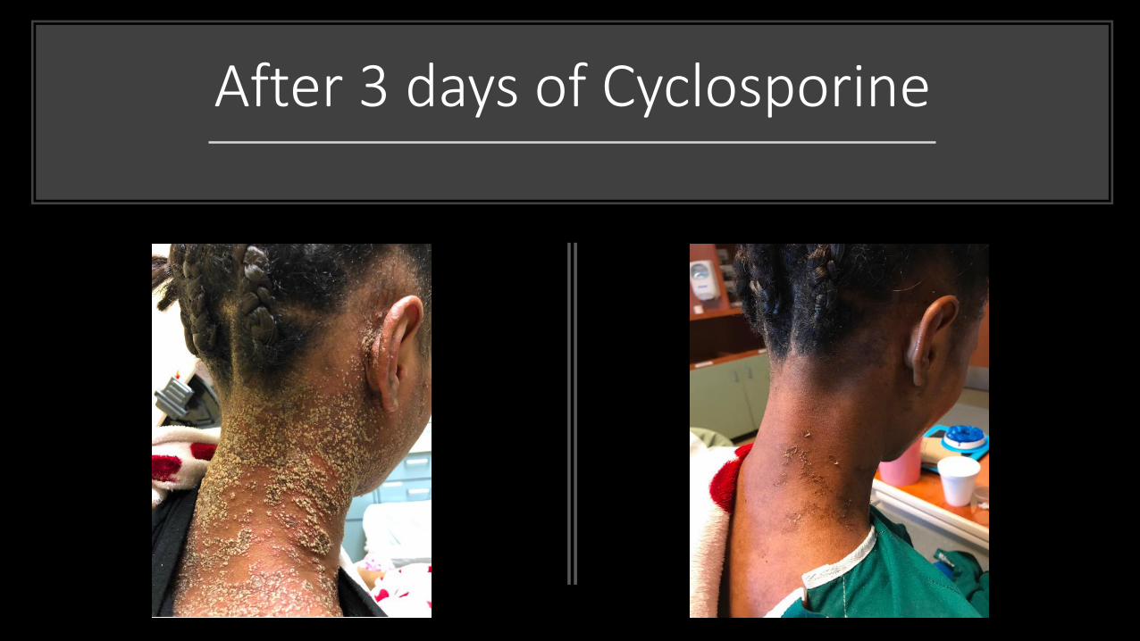
After 3 days of Cyclosporine
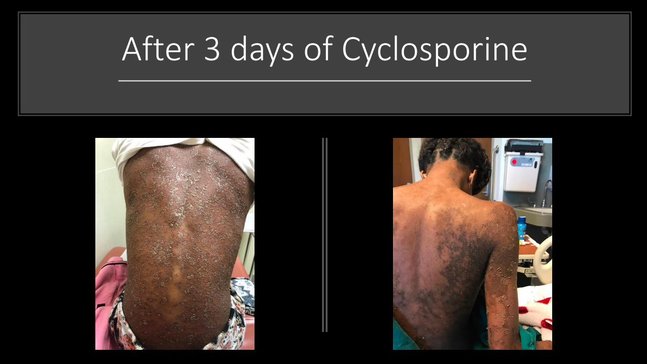
After 3 days of Cyclosporine
Pearls: Psoriasis & Skin of Color
• Erythema is not prominent, lesions appear violaceous or hyperpigmented.
• Post-inflammatory hypo- or hyperpigmentation is very common.
• Consider potential clinical mimickers: lichen planus (especially hypertrophic type), cutaneous lupus erythematosus (discoid and subacute).
• Potential extensive involvement at initial presentation.
• Managing scalp psoriasis: Consider impact of hair texture, styling practices, and washing frequency when evaluating severity and selecting topical therapy.
Case 2
Outpatient visit for “eczema flare”
Case 2
• 9 year old African American male presented to Dermatology clinic for follow-up evaluation of eczema. He was last seen 2 years ago and prescribed Desonide 0.05% ointment and Triamcinolone 0.1% ointment BID for face and body respectively, and oral antihistamines (Cetirizine 5 mg and Hydroxyzine 20 mg) for pruritus.
• Parents used topical therapy for less than 2 months, after noticing his skin was “turning white with the treatment”.
• He is currently using an OTC moisturizer lotion about once to twice daily. His scalp is very itchy, sleep is not disturbed.
• Eczema is now flaring. He is otherwise healthy, had had no preceedingillness or fever.
•
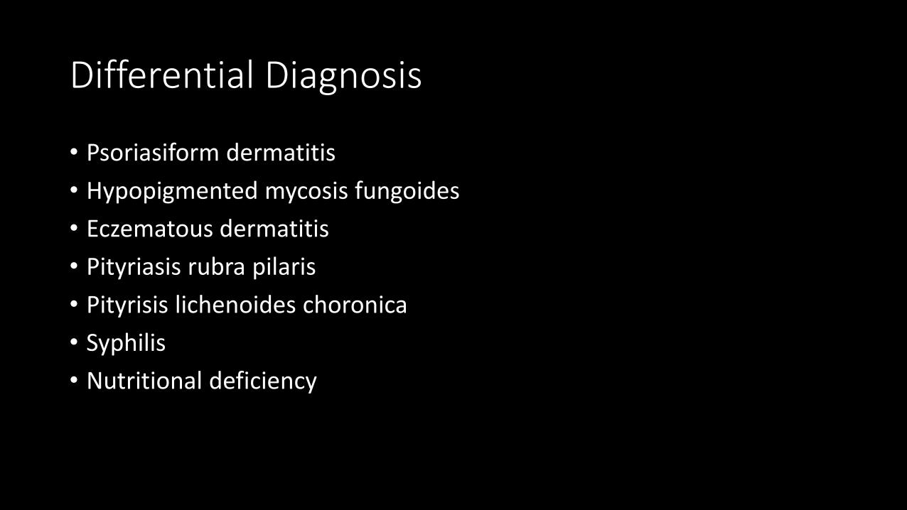
Differential Diagnosis
• Psoriasiform dermatitis
• Hypopigmented mycosis fungoides
• Eczematous dermatitis
• Pityriasis rubra pilaris
• Pityrisis lichenoides choronica
• Syphilis
• Nutritional deficiency
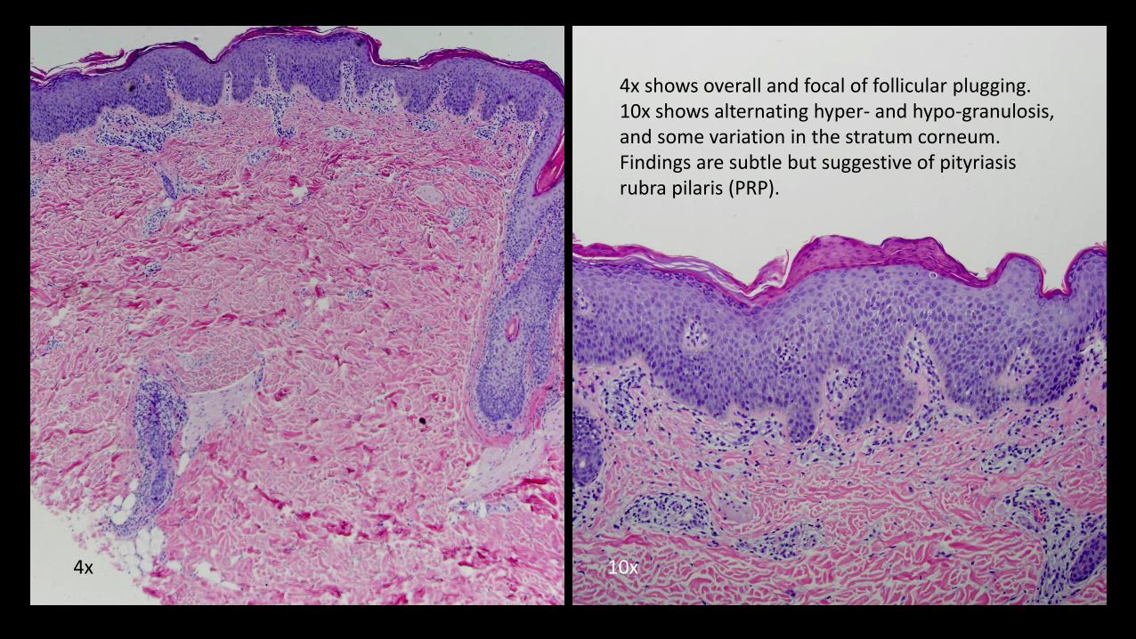
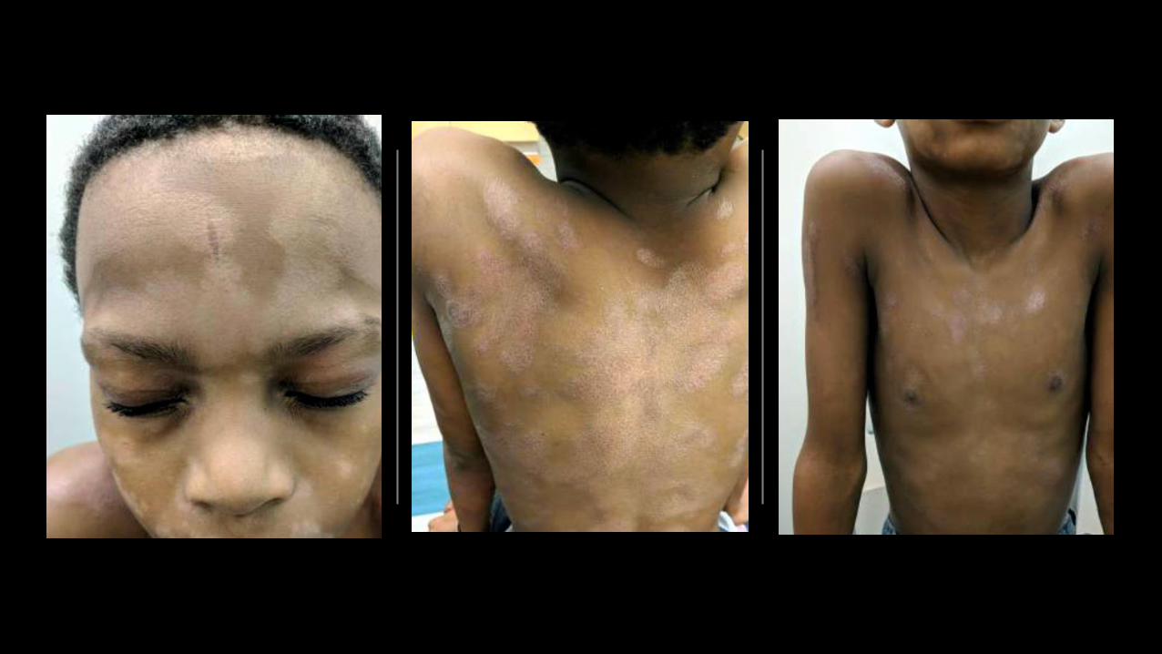
4x shows overall and focal of follicular plugging.10x shows alternating hyper- and hypo-granulosis, and some variation in the stratum corneum.Findings are subtle but suggestive of pityriasis rubra pilaris (PRP).
4x 10x
PityriasisRubra Pilarisand Skin of
Color
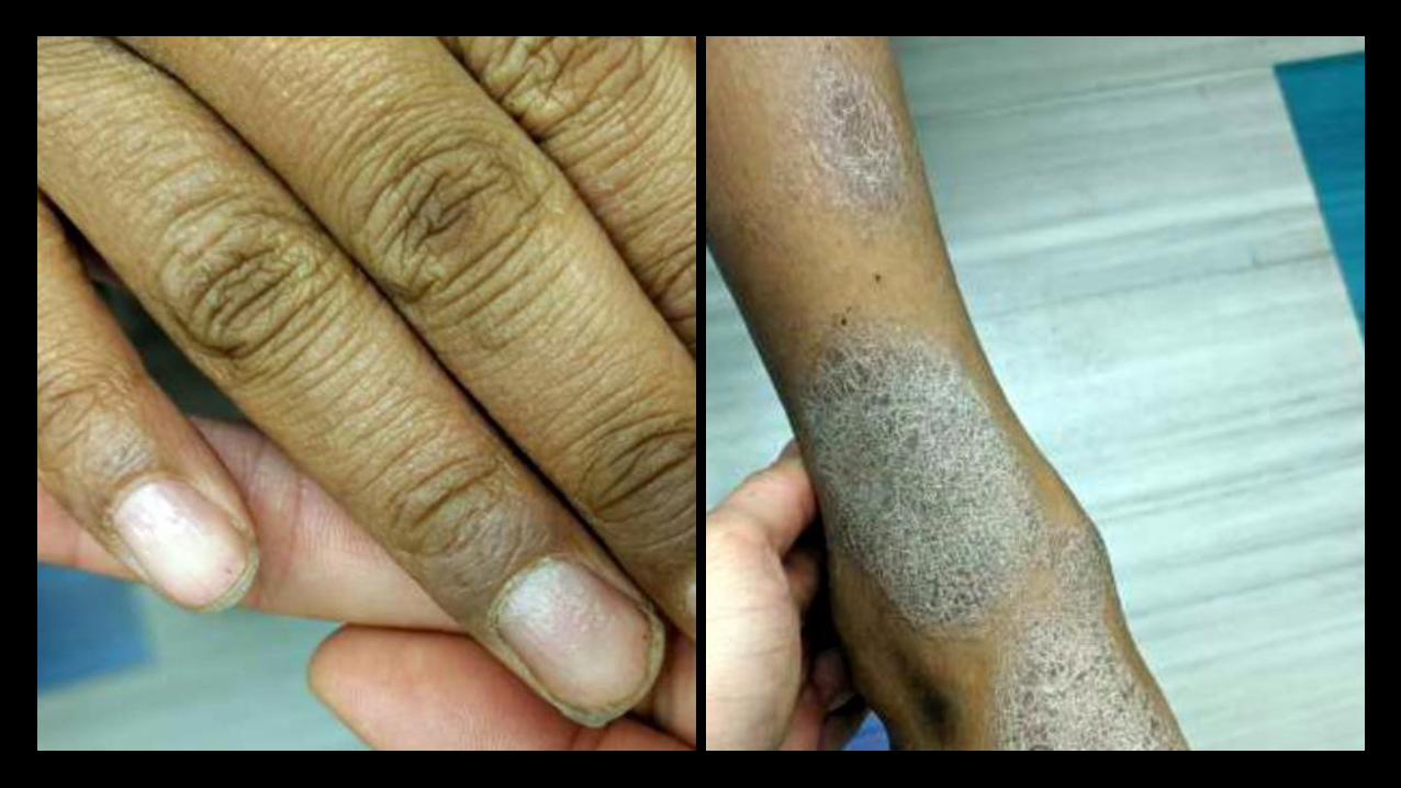
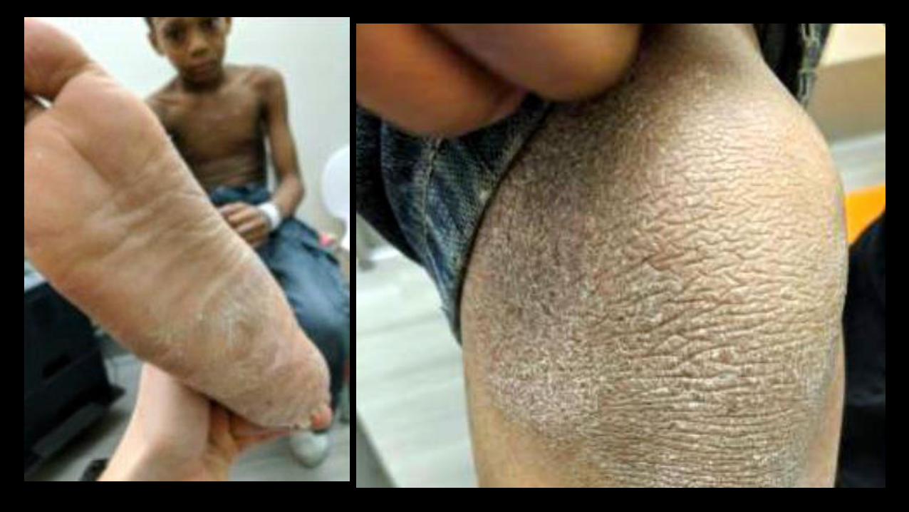
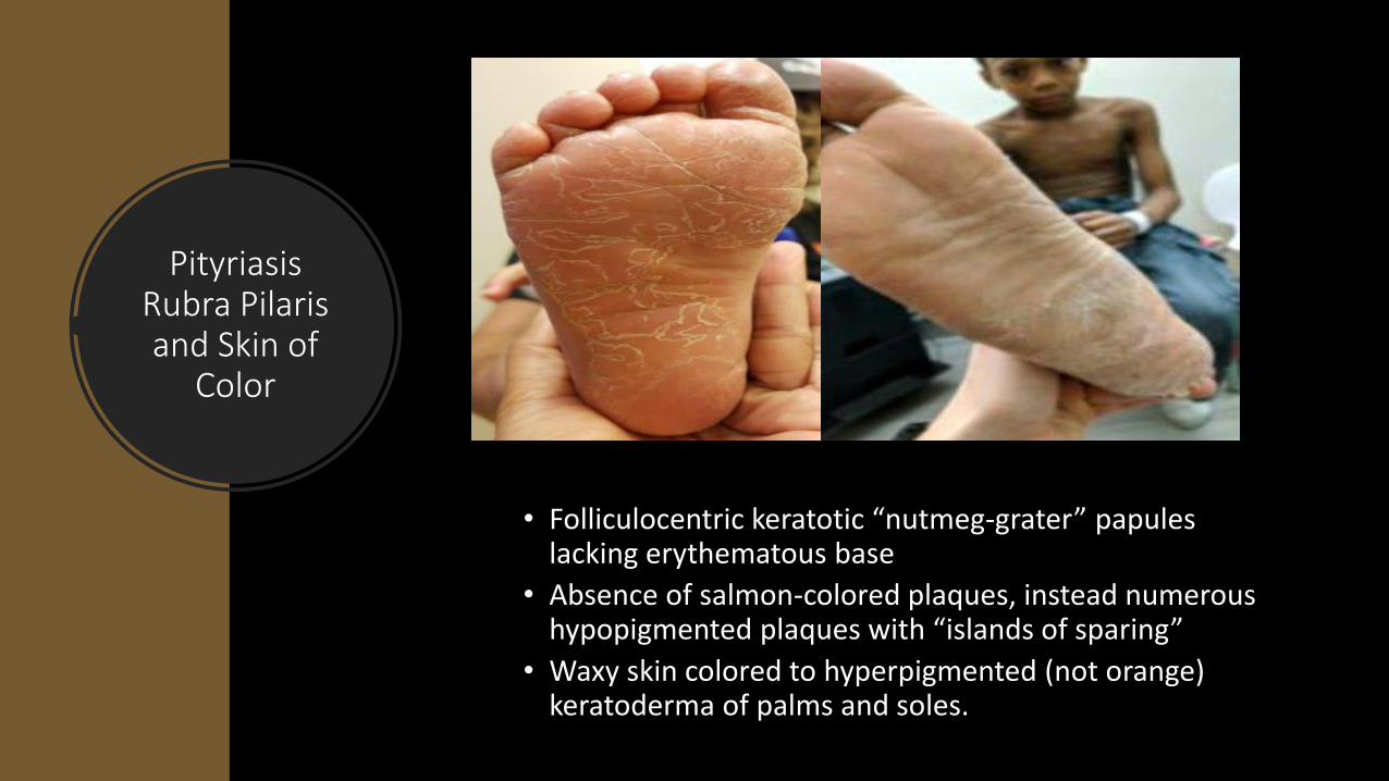
• Folliculocentric keratotic “nutmeg-grater” papules lacking erythematous base
• Absence of salmon-colored plaques, instead numerous hypopigmented plaques with “islands of sparing”
• Waxy skin colored to hyperpigmented (not orange) keratoderma of palms and soles.
Pityriasis Rubra Pilaris
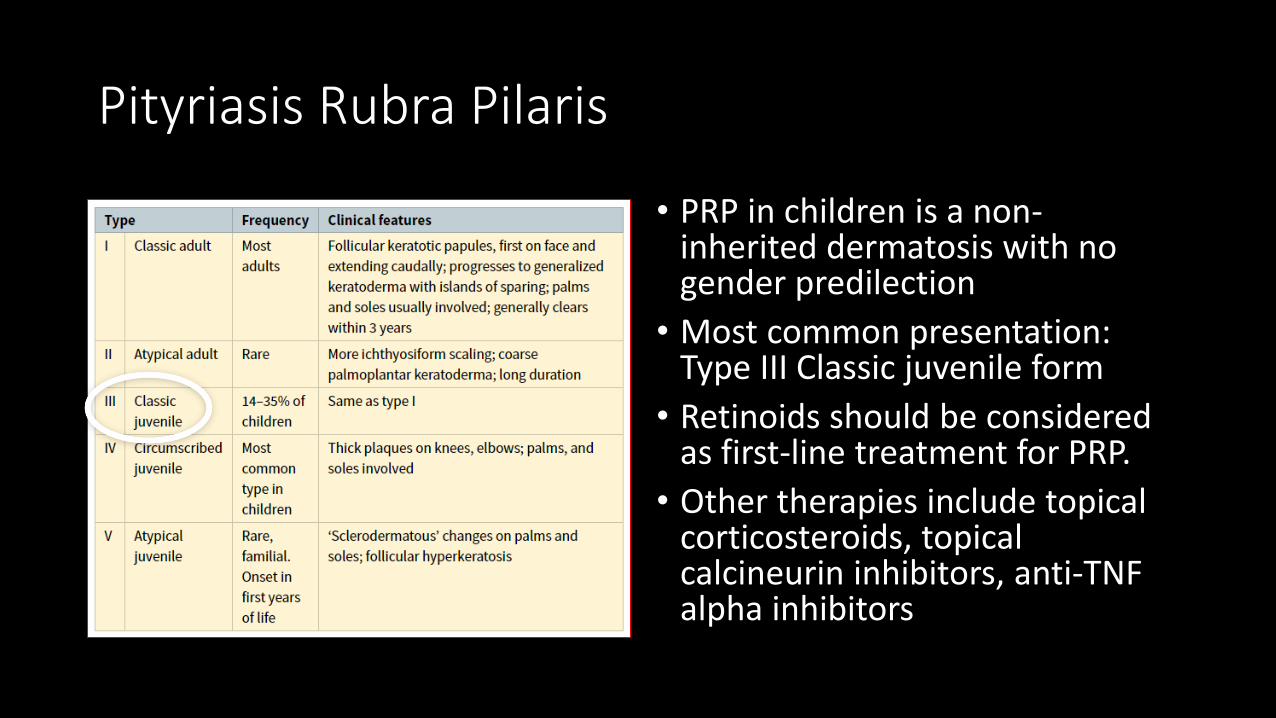
• PRP in children is a non-inherited dermatosis with no gender predilection
• Most common presentation: Type III Classic juvenile form
• Retinoids should be considered as first-line treatment for PRP.
• Other therapies include topical corticosteroids, topical calcineurin inhibitors, anti-TNF alpha inhibitors
Pearls: Pityriasis Rubra Pilaris & Skin of Color
• High index of suspicion with presentation of psoriasiform dermatitis and keratoderma
• Erythema is not prominent, lesions appear hypo or hyperpigmented.
• Post-inflammatory hypo- or hyperpigmentation is very common in comparison to classic salmon colored plaques with “islands of sparing”
• Consider potential clinical mimickers: hypopigmented mycosis fungoides, psoriasis

Case 3
Inpatient consult for “swollen face”
Case 3
• 16 year old African American female presented with 1 week of fever (102-104 °C) and general malaise
• Recently treated with amoxicillin for acute otitis media
• Following admission, she developed facial rash associated to rapidly progressive periorbital edema
• Labwork was remarkable for pancytopenia

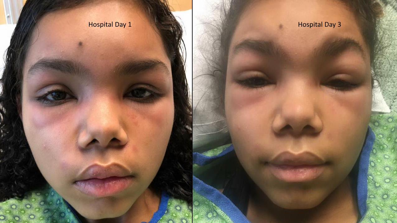
Hospital Day 1 Hospital Day 3
Differential Diagnosis
• Reactive process (drug or viral exanthem)
• Manifestation of an autoimmune disorder
Work-up
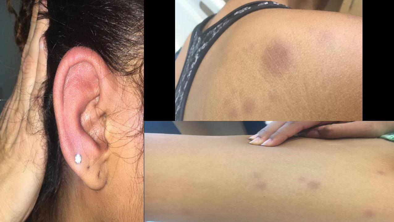
• Skin biopsy from nodule on right leg revealed superficial and deep mixed perivascular and interstitial dermatitis. Histotologic features were not diagnostic. Pattern of inflammation favor a variant of palisaded and neutrophilic granulomatous dermatitis, which can be associated with underlying systemic and connective tissue disorders. A drug eruption, reactive erythema, exuberant urticarial or reactive process, and other similar dermatoses could also present with these features.”
Diagnosis
• During hospitalization, she developed arthritis, hemolytic anemia, leukopenia, thrombocytopenia, hypocomplementemia, ANA, Anti-Sm, Antiphospholipid Antibodies.
• Diagnosis: Systemic lupus erythematosus.
• Management : IV Steroids and Plaquenil.
• Rash and periorbital edema quickly improved. Fevers stopped
Systemic Lupus Erythematosus
• Severe organ involvement and significant disease activity are primarycharacteristics in children with juvenile SLE.
• African Americans are at highest risk for severe organ involvement.
• The clinical manifestations of lupus nephritis (LN) in children are diverse.
• Early treatment may result in significant disease remission.
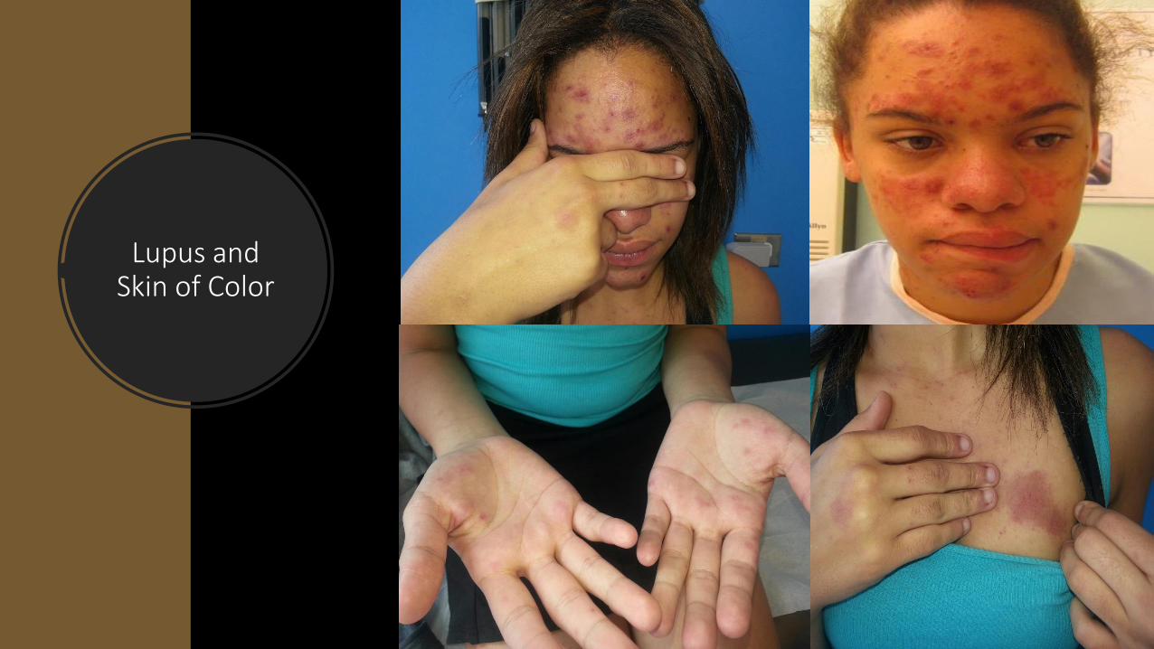
Lupus and Skin of Color
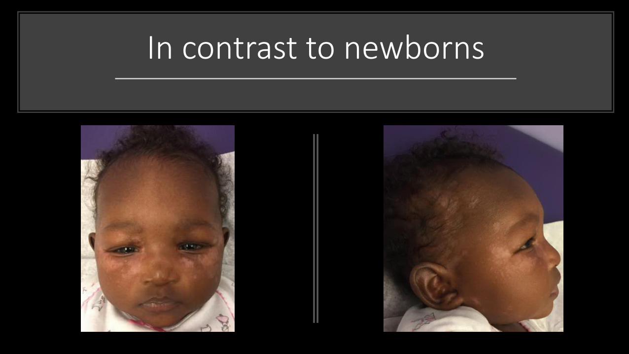
In contrast to newborns
Neonatal Lupus Erythematosus
• Caused by transplacental passage of maternal antibodies, most commonly anti-Ro/SS-A
• 50% of women with a child with NLE are asymptomatic at time of child’s birth
• Cardiac disease results in 20% mortality -> Congenital heart block
• Women who have a prior child with NLE have a 25% risk of NLE in subsequent children
Pearls: Lupus and Skin of Color
• Chronic cutaneous lupus Erythematosus is a scarring subtype, more prevalent in blacks
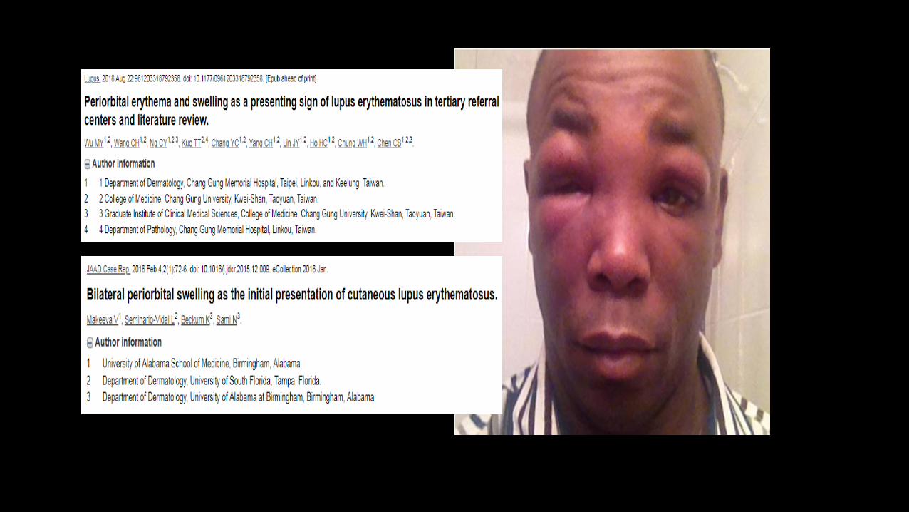
• Periorbital edema has been described as a presenting sign of lupus
• Skin findings tend to be exaggerated with an increased risk of post-inflammatory hyperpigmentation and hypertrophic scarring
• Consider potential clinical mimickers in neonatal lupus: seborrheic dermatitis, pityriasis alba and atopic dermatitis