pectoralis major myocutaneous flap in …repository-tnmgrmu.ac.in/3932/1/240320413shanmugharaj.pdf1...
TRANSCRIPT

1
PECTORALIS MAJOR MYOCUTANEOUS FLAP IN
ORAL & MAXILLOFACIAL RECONSTRUCTION
Dissertation submitted to
THE TAMILNADU Dr.M.G.R. MEDICAL UNIVERSITY
In partial fulfillment for the Degree of
MASTER OF DENTAL SURGERY
BRANCH III
ORAL AND MAXILLOFACIAL SURGERY
MARCH 2013

2

3
ACKNOWLEDGEMENT
This thesis would not have been possible without the efforts of a large
number of people to whom I am greatly indebted.
No words can express what I feel when I sit down to express my
gratitude to my teacher Dr.A.T.Vigneshwaran, M.D.S., Professor and Head
of the Department, Department of Oral & Maxillofacial Surgery,
Sri Ramakrishna Dental College and Hospital, Coimbatore. It has been a
privilege to have associated with him as a student during my post graduation.
I avail this opportunity to express my deep sense of gratitude and
thanks to my teacher and guide Dr. L.Deepanandan, M.D.S., Professor
Department of Oral and Maxillofacial Surgery, Sri Ramakrishna Dental
College and Hospital, Coimbatore. His constant help, encouragement and
initiative guidance has made this study possible for me.
I thank Dr. Guhan, MD, DM, Director, Dr. Karthikesh, Mch,
Dr. Bhargavi, Mch, Surgical Oncologists, Sri Ramakrishna Institute of
Oncology for their guidance and constant encouragement throughout the
study.
I sincerely thank my teacher Dr.M.S.Senthilkumar, M.D.S., Reader
Department of Oral and Maxillofacial Surgery, Sri Ramakrishna Dental
College and Hospital, Coimbatore for constant support and encouragement
throughout my course.
4
I am greatful to my teachers Dr. M.A.I.Munshi, M.D.S.,
Dr. R.S.Karthik, M.D.S., Senior Lecturers, Department of Oral and
Maxillofacial Surgery, Sri Ramakrishna Dental College and Hospital,
Coimbatore, for their everlasting guidance, cooperation and support
throughout my course.
I am thankful to Dr. Vasista and Dr. Sundari, Anesthetists,
Sri Ramakrishna Hospital, for their help rendered towards this study.
I express my gratitude and heartfelt thanks to Dr.V.Prabhakar,
M.D.S., Principal, Sri Ramakrishna Dental College and Hospital,
Coimbatore, for giving me an opportunity to utilize the facilities in the
institution for the study.
My Heartfelt thanks goes to my batch mate Dr.Naveena, my senior and
junior postgraduates who stood by me throughout my course and for their
valuable help.
At personal front, I express special thanks to my parents, wife and
children for their innumerable sacrifice, love, understanding and support
towards me.
Above all I thank the almighty god for showering his blessings and
love on me which has provided me the inspiration and zest to tread the path of
the life.

5
CONTENTS
S.NO. TITLE PAGE NO.
1. INTRODUCTION 6
2. AIMS AND OBJECTIVES 9
3. REVIEW OF LITERATURE 11
4. MATERIALS AND METHODS 39
5. RESULTS 61
6. DISCUSSION 83
7. SUMMARY & CONCLUSION 96
8. BIBLIOGRAPHY 99

ABSTRACT
AIM
Pectoralis major myocutaneous flap is a versatile flap and used in head and neck
reconstruction for ablative tumor surgery. This study is aimed to evaluate the Functional
outcome, aesthetic outcome, and recipient & donor site morbidity after Pectoralis Major
Myocutaneous flap reconstruction in Oral & Maxillofacial region.
METHODOLOGY
The data of 20 cases of pectoralis major flap for reconstruction of post cancer resection
defects of the oral & maxillofacial regions were analyzed. All data concerning functional
outcome, aesthetic outcome, site of tumours, types of defects, donor site and recipient site
complications and surgical treatment of these patients with pectoralis major myocutaneous
flap for reconstruction were analyzed in the follow up period at first week, first month, third
month, sixth month and ninth months.
RESULT
In patients who underwent pectoralis major myocutaneous flap reconstruction for oncological
resection males (64.7%) were affected more commonly than female, mostly in the age of 40-
60 (53.0%) years, the left side (52.9%) of the oral cavity is more commonly affected than the
right side, and the alveolus (35.3%) is the most commonly affected site . T2 (47%) size lesion
was more common and 52.9% had systemic disease. The results showed significant
improvement in the speech, oral spintcher function, tolerance of full diet and satisfactory
aesthetic outcome at the end of nine month follow up period. In donor and recipient site
complications there was one case of total flap loss with reconstruction plate exposure which
had a significant association with ischemic heart disease. In 2/4 patients with T4 lesion, there
was significant association with fistula. In 6% patient had wound dehiscence, 11.8% had

partial flap necrosis, 11.7% had implant infection, 17.7% had reconstruction plate exposure
and 15% patient died of unknown cause within three months after reconstructive surgery, it
was not due to flap related complications.
CONCLUSION
The pectoralis major myocutaneous flap is a major flap for reconstruction of large head and
neck defects. The results proved that the flap its continual usage in T3, T4 patients and
provides better functional, aesthetic and less donor and recipient site complications.
KEY WORDS:
pectoralis major, reconstruction, function, aesthetic

6
INTRODUCTION
Reconstruction of any tissue defect poses a unique challenge to the
surgeon especially after a more ablative surgery in the head and neck region.
When a local tissue cannot cover a Head and neck defect, the surgeon has to
seek a distant tissue for reconstruction. The choice lies between a pedicled and
free tissue transfer. Free tissue transfer involves micro vascular anastomosis,
which requires expertise and facilities. Reconstructive surgeon therefore has to
often settle for the use of pedicled flaps to repair defects of head & neck.1
The pectoralis major myocutaneous flap is the most commonly
employed pedicled flap for the reconstruction of tumor and trauma-related
defects in the oral and maxillofacial region.2 The pectoralis major
myocutaneous flap is an axial pattern flap, which means that it is based on a
dominant vascular supply that runs axially along the length of the muscle. The
skin in such flaps receives its blood supply from perforating vessels of the
axial artery system.19
The pectoralis major myocutaneous flap was described by Hueston and
McConchie in 1968 for reconstruction of a large midline chest wall defect.19,41
The concept of a pectoralis major island flap was introduced by Brown in
1977. The pectoralis major myocutaneous flap was introduced into head and
neck reconstruction by Ariyan in 1979.6 Ariyan extensively used pectoralis
major myocutan flap in reconstruction of oropharynx, the cervicofacial

7
region, the orofacial complex, the orbit and the temporal region. This
experience led surgeons to consider the pectoralis major myocutaneous flap as
the gold standard for head and neck reconstruction. This flap was thought to
be more versatile than the deltopectoral flap that had enjoyed widespread use
up to that point.31
Nowadays, free flaps are more common due to improved microsurgical
techniques, esthetic and early functional results.18 Since most patients in our
country report only when the disease is in the advanced stage where follow up
and prognosis is poor .In medically compromised patients free flap is not ideal
due to increased donor site morbidity and high cost involved with salvage
surgery if required later. But in several cases the pectoralis major
myocutaneous flap still has its advantages, including its proximity to the head
and neck, the simplicity of harvesting, and its use as an alternative when
microsurgical flap failure occurs.4 For cases like coverage of a reconstruction
plate and carotid artery, the bulkiness of the flap can be an advantage. The
pectoralis major myocutaneous flap is characterized by a simple procedure and
a short time to harvest.19
Disadvantages include reduced neck mobility and the need to rotate the
vascular pedicle of the flap 180° when using the skin paddle to resurface the
neck. Another disadvantage can be the thickness of the flap, which is
determined by the amount of subcutaneous fat between the pectoralis muscle
and the overlying skin paddle, leading to possible reduced swallowing or

8
speech function. Complications such as partial or complete flap necrosis,
fistula formation, dehiscence, infection, hematoma and complications in
patients after radiotherapy have been described earlier. The complication rate
seems to be higher than in free flap reconstructions. Several issues related to
the development of the pectoralis major myocutaneous flap should be
considered preoperatively. These issues include the timing of flap
development, the arc of rotation of the flap, the size of the recipient defect, the
color match of the skin paddle and the recipient tissue bed and the potential
trauma to the thoracoacromial axis.7

9
AIMS AND OBJECTIVES
The purpose of this study is to
� Evaluate the Functional outcome with Pectoralis Major Myocutaneous
flap in Oral & Maxillofacial Reconstruction
� Evaluate the Esthetic outcome with Pectoralis Major Myocutaneous
flap in Oral & Maxillofacial Reconstruction
� To evaluate the Recipient & Donor site morbidity after Pectoralis
Major Myocutaneous flap reconstruction in Oral & Maxillofacial
region
SURGICAL ANATOMY
The pectoralis major muscle is a broad, flat, fan shaped muscle that
covers the pectoralis minor, subclavius, serratus anterior and intercostal
muscles on the anterior thoracic wall. Its origination is the medial one half to
two thirds of the clavicle, the lateral portion of the entire sternum and the
adjacent cartilages of the first six ribs and the bony portions of the fourth, fifth
and sixth ribs. The blood supply to this muscle includes the pectoral branch of
the thoracoacromial artery, the lateral thoracic artery, the superior thoracic
artery and the intercostal artery. Three major segmental subunits have been
described, including a clavicular segment, a sternocostal segment and a
laterally placed external segment. Each has its own vascular and motor nerve
10
supply. The clavicular segment arises from the clavicular area, receives its
blood supply from the deltoid branch of the thoracoacromial artery and is
innervated by branches of the lateral pectoral nerve. The sternocostal segment
accounts for most of the pectoralis major muscle mass and receives its blood
supply from the pectoral branch of the thoracoacromial artery. This segment
receives motor innervation from the lateral pectoral and the medial pectoral
nerves. The external segment is innervated by the branches of the medial
pectoral nerve and has a variable blood supply, with the lateral thoracic artery
the exclusive source in 49% of cases. In 18% of cases, the external segment is
supplied by branches of the pectoral branch of the thoracoacromial artery; rest
of the 33% of cases is supplied by a combination of the lateral thoracic artery
and the thoracoacromial artery. The motor action of the pectoralis major
muscle is to rotate medially and adduct the humerus. The development of the
myocutaneous flap is generally well tolerated, because the latissimusdorsi
muscle compensates for otherwise lost adductor activity.19

11
REVIEW OF LITERATURE
Ariyan S (1979)6 was the first person to apply the principles of
pectoralis major muscle for the reconstruction of head and neck defects. In his
study, with 14 cases of Pectoralis major myocutaneous flap found that the flap
is reliable for repair of defects after ablative surgery in head & neck region and
can be transferred immediately.
R T Gregor (1982)24
analysed the use of pectoralis major
myocutaneous island flap, for reconstruction in head and neck surgery. He
explained there are two types of cutaneous blood supply: direct vessels and
musculocutaneous vessels. He advocated the paddle should be situated medial
to the nipple, but may include the nipple if this is essential. It should lie along
the acromio-xiphisternal line. He discussed the disadvantage as the skin used
is usually hair-bearing in men and the bulkiness of the muscle may be
undesirable in certain cases, especially in muscular males; however the muscle
can usually be trimmed to fit the defect but it should not be smaller than the
skin paddle.
Weaver AW et al (1982)56 designed the bilobular or gemini type of
pectoralis major myocutaneous flap to simplify the closure of large surgical
defects of both mucosa and skin that could not be satisfactorily closed
primarily. The mucosal and skin defects are closed by two skin paddles
supported by a single muscular vascular pedicle. In this design the skin

12
paddles are fashioned side by side and separated from each other as the muscle
is folded between them parallel to the vascular axis. All of the defects were
successfully closed with this technique and the major portion of all grafts
survived. This design permits single-stage reconstruction after ablation of
tumors or treatment of complications which produced large through-and-
through muco-cutaneous defects of the head and neck area.
Berktold RE et al (1986)9 developed a simple, single-stage, primary
procedure for chin reconstruction. It is based on a simple modification of the
pectoralis major myocutaneous flap with seven cases, including two with total
chin reconstructions. The number and type of complication was low and
consistent with the magnitude of the surgical procedure. This technique
provided acceptable aesthetic and functional results to patients undergoing
partial or total resection of the chin.
ROBERT E et al (1990)48
made an improved technique for the
development of the Pectoralis Major Myocutaneous Flap. The modifications
introduced in this approach to the pectoralis major myocutaneous flap are
based on the known vascular supply to this surgical area and sound principles
of flap development and rotation. When flap elevation normally there is a
severe restriction due to the muscle insertion on the humerus. All or most of
this insertion should be divided just medial to the axilla. This maneuver
extends the arc of rotation by about 8 cm cephalad. The technique described in
this report usually maintains all three axial vessels of the pectoralis muscle and

13
suspect that the flap development increases the perfusion pressure in the
anatomic vascular territory of the flap itself and decreases the perfusion
pressure in the adjacent territory. These modifications of the basic approach to
developing a pectoralis major myocutaneous flap have resulted in a more
predictable transfer of tissue in even heavily irradiated areas. This technique
produced fewer complications and less residual deficiencies and being more
cosmetic for the patient.
L E Loh (1992)30 compared two modalities of treatment in the case of
buccal carcinoma. In one case reconstruction was done by using pectoralis
major flap by turning it externally to provide skin coverage for the cheek
defect. In other case pectoralis major flap was turned internally to repair the
buccal mucosal defect and cervicopectoral advancement flap to repair the
cheek skin defect. Of the several modalities of reconstruction available the
author feels that two modalities are promising.
S Nagral et al (1992)38
reported their experience with the pectoralis
major myocutaneous flap for head and neck reconstruction in a general
surgical unit. A tube Pectoralis major myocutaneous flaps was used to
reconstruct the pharynx and the cervical esophagus. Cosmetically, the donor
site scar is totally hidden by clothing; the functional loss is negligible. The flap
was employed before or subsequent to the use of chemotherapy or
radiotherapy. Disadvantages of the flap include excessive bulk in obese or

14
muscular individuals and troublesome hair growth in the oral cavity. The final
functional and cosmetic results were satisfactory.
Richard Crosher, Roy Mitchell, John Llewelyn (1995)45 described a
modification of the pectoralis major myocutaneous flap that provide sizeable
skin paddle and allows a primary closure of the chest wound with good
cosmetic results. Here the incision is made along the lower limb of the
deltopectoral flap and continues into the crescentic shaped skin paddle. The
myocutaneous flap is then raised in the conventional manner and transferred to
reconstruct the primary defect. With this technique there is minimal distortion
of the breast and the nipple and can be positioned at the same level as the
opposite side with good cosmetic results.
Robert A Ord (1996)
41 analysed the reliability and complication of
pectoralis major myocutaneous flap in oral & maxillofacial reconstruction
with respect to reconstruction of post cancer resection defects of the oral
cavity & maxillofacial region. In his technique of flap elevation electric
cautery was not used to coagulate vessels on the paddle to avoid retrograde
thrombosis and ties were used even small bleeders. The lateral thoracic artery
was incorporated in the flap whenever possible. He concludes that the
pectoralis major flap is reliable and remains excellent choice for large soft
tissue defects in oral cavity despite increased use of microvascular flap.
Kiyokawa K et al (1998)29 describe a method that preserves
circulation during the preparation of the pectoralis major myocutaneous flap

15
used in head and neck reconstruction. The major disadvantage of this flap is its
poor circulation and consequent partial necrosis. They analyzed the circulation
and hemodynamics of the pectoralis major myocutaneous flap (the perforator
of the anterior intercostal branch located about 1 to 2 cm medial to the areola
in the fourth intercostal space is important), evaluated the safe donor sites in
the chest wall for a skin island (the perforator is included on the skin island's
central axis), improved the surgical procedure for elevating flaps (for
preventing perforator injuries) and devised a means to transfer flaps, thereby
increasing the range of the flaps (the transfer route is under the clavicle).
Using this technique, the partial or marginal necrosis of the flap caused by
circulatory problems was detected only in 5% cases. Using this method, the
problems associated with inadequate circulation in the pectoralis major
myocutaneous flap were greatly alleviated, thus reconfirming the usefulness of
this flap in head and neck reconstruction.
G R Williams et al (2000)57 reported the surgical technique and the
outcome of use of this flap in a patient with poor soft-tissue coverage,
following multiple operations for a clavicular fracture complicated by
nonunion and infection. In this technique, the vascular pedicle,
thoracoacromial artery and axillary artery were identified and the length of the
vascular pedicle from the axillary artery to the muscle was measured. The
angle of rotation of the flap about its intact clavicular origin was measured
before and after division of the acromial branch of the thoracoacromial artery.

16
The clavicular origin was then incised and the overall length, width and
thickness of the muscle as well as the distance from each end of the muscle to
the vascular pedicle were measured. The result was that average length of the
vascular pedicle from the axillary artery to the pectoralis muscle belly was 5.3
centimeters. The rotational flap was successfully used to reconstruct the
defect.
O M Oluwatosin, Abikoye F O, Adegboye V O
(2000)
40 in his study
raised the flap in conventional way with one skin paddle, which is turned in for
lining. The muscle component of the flap is covered with skin graft and claimed
in this way donor site morbidity and bulk is reduced. The medial paddle retains
the blood supply from the pectoral branch of the acromiothoracic artery and the
lateral paddle derives its supply from the lateral thoracic artery. Transferring the
pectoralis major as a true island flap with the skin paddle raised as far distally
produces an increased pedicle length and greater arc of rotation, which in some
patients reach the zygoma and contralateral aspect of lip at the same time.
Liu R, Gullane P, Brown D, Irish J (2001)
31 did a retrospective study
on reconstruction procedures using the pectoralis major pedicled flap
reconstructions after ablation of oral cancer. In his study 35% were affected by
complications such as dehiscence, infection, hematoma, seroma, partial flap
failure, total flap failure, fistula and donor site complications. The duration of
admission for cases with complications was longer and higher complication
rates were associated with salvage procedures in oral cavity reconstructions.

17
Marco Luigi Castelli et al (2001)33 made a study on Pectoralis major
myocutaneous flap and analyzed complications in difficult patients and found
that pectoralis major myocutaneous flap is simple to perform and is suitable
for immediate repair of large oral and pharyngeal defects, even in difficult
patients with various medical problems known to increase complication rates.
Patients who had radiotherapy prior to the operation had no such complication.
Despite these problems that were encountered in the first postoperative month,
after 3 months all patients were able to eat solid food or to drink without
aspiration. They recommend the use of PMMF as a first option in frail patients
who had large oro-pharyngo-laryngeal excisions and radical neck dissections
and who may require postoperative radiotherapy.
Collin Rol, Prabir K josh, R N Podder (2002)14 presented a case of
55-year-old lady with trismus and full thickness oral defect exposing upper
and lower row of teeth diagnosed as cancrum oris. The operative intervention
consisted of radical excision of scar tissue, release of bony block of right
temporo-mandibular joint ankylosis. The cheek fistula defect was corrected in
two layers by bipaddled island pectoralis major myocutaneous flap were the
correction of jaw ankylosis and the flap cover was done in a single stage.
A Croce, Moretti, L D’Agostino, Neri G (2003)16 studied the
Continuing validity of pectoralis major muscle flap, 25 years after its first
application. They concluded, the pectoralis major flap is indicated in patients

18
who have advance disease, systemic diseases and where micro vascular
surgery is contraindicated.
Eric R Carlson (2003)
19 Pectoralis major myocutaneous flap is a
reliable and predictable transfer of soft tissue for reconstruction of the patient
undergoing tumor ablation or sustaining avulsive trauma. In men, one can
transpose a skin paddle of approximately 6 × 6 cm without having to skin graft
the chest wall or without significant anatomic distortion of the ventral chest
wall following primary closure, when developing a pectoralis major
myocutaneous flap. An arteriogram can be obtained if any question exists
regarding the integrity of the pedicle. This flap possesses a large volume of
vascularized muscle and a skin paddle of sufficient size to satisfy 90% of soft
tissue reconstructive needs. Soft tissue flaps of sufficient vascularity will
promote healing without complication. He suggests the inclusion of the lateral
thoracic artery to the flap is important for flap viability.
Roy L H Ng et al (2003)49
reported a case of immediate reconstruction
of upper and lower lips with mandible using multiple flaps following resection
of extensive squamous cell carcinoma. The patient was reconstructed with free
fibula osteocutaneous flap, pedicled scalp flap, tongue flap, Palmaris longus
tendon sling and pectoralis major myocutaneous flap. At 4 weeks, patient
demonstrated good speech and swallowing with an acceptable aesthetic result.
Douglas B et al (2004)
18 evaluated the factors related to surgical
complications, rate of gastrostomy tube (G-tube) dependence in patients
19
undergoing reconstruction with a pectoralis myocutaneous flap against a soft
tissue revascularized flap. G-tube dependence is very reliable indication of
swallowing function. The minor complication rate was higher in the pectoralis
group at 57% than in the revascularized flap group at 21%. The revascularized
flaps helped to ameliorate the effects of radiation before surgery; 56% of the
patients who received pectoralis flaps were G-tube dependent, while the rate
of G-tube dependence in the revascularized flap group was 23%. He
concluded that patients who undergo reconstruction with a pectoralis flap have
significantly higher minor complication rates, a higher rate of G-tube
dependence than patients who undergo reconstruction with a soft-tissue
revascularized flap.
M Ethunandan et al (2004)20 made their important study on Skin
necrosis of a pectoralis major myocutaneous flap, caused by methicillin-
resistant Staphylococcus aureus, with a bi-paddled pectoralis major
myocutaneous flap in which both the pectoral branch of the thoracoacromial
artery and the lateral thoracic artery was preserved. They found that the
MRSA was colonisation rather than an active infection that would lead to skin
necrosis. The main stay of treatment of MRSA infection is vancomycin or
teicoplanin or both, though in less severe infections fusidic acid and
rifampacin may be used. They suggested when MRSA is isolated, it should be
treated early and aggressively with appropriate antibiotics.

20
Chaudhary A, Wakhlu A (2005)10 reported a case large
paraganglioma arising from right side of the neck and extending to the scalp.
The tumor was treated with radical excision and the resultant tissue defect was
resurfaced with a pectoralis major flap with split thickness skin graft. There
was no recurrence or metastasis with the follow-up for 2years.
M Jog, Meckenzie K, Dempster (2005)27
reported two cases where
metastatic spread at the donor site of the PMMC flap was identified without
any apparent persistence or recurrence at the index site. The modes of tumor
spread could be due to implantation theory and to prevent this, consideration
to extend the radiotherapy field to include the donor site may be given.
Mohammed Tahir, Tahmeedullah, Amir Taimur Khan (2005)36 did
a clinical evaluation of pectoralis major myocutaneous flap in Head and Neck
Reconstruction. The infection rate observed in this study was 10%. The
significant risk factors for infection were diadetes mellitus and haemoglobin
level (less then 10gm %). He also found Oral continence was normal in 5% of
patients, occasional drooling in 75% cases and continous drooling in 20%
cases and concluded that pectoralis major myocutaneous flap can be used for
reconstruction of large Head and Neck defects with acceptable esthetic and
functional outcome results.
Aleksandar Milenovic et al
(2006)4 studied the usage of the
Pectoralis major flap in head and neck reconstruction in advanced malignant
tumor of head & neck and concluded that the pectoralis major myocutaneous
21
flap is still an acceptable method. Despite the increasing application of
microvascular reconstruction, it still has many advantages like it is fast,
reliable, provides safe repair and is indicated especially where bulk is needed.
C M E Avery et al (2006)8 retrospectively studied the use of the
pectoralis major flap for advanced and recurrent head and neck malignancy in
the medically compromised patient. They concluded that the flap retains a
major role in the management of advanced primary or recurrent disease,
extensive metastatic neck disease and after failure of a free flap when in
conjunction with significant co morbidity.
Hamdy-el-marakby (2006)
25 evaluated the Reliability of Pectoralis
Major Myocutaneous Flap in Head and Neck Reconstruction. The results
showed that the flap is usually associated with a high incidence of
complications in addition to its large bulk compared with the free
fasciocutaneous flaps. The final functional and the aesthetic results are inferior
to free flaps in head and neck reconstruction. In pectoralis flap the
complications such as wound dehiscence, infection, hematoma, seroma, partial
flap failure, total flap failure, fistula and donor site complications may occur.
A higher complication rates were associated when the flap was used for
reconstruction, in salvage surgical procedures.
Nath S et al (2006)39 used the pectoralis myocutaneous flap for
mandibular reconstruction and concluded that pectoralis myocutaneous flap is
still the workhorse in head and neck reconstruction. Their case series had very

22
few complications and no disability from the loss of the flap. Locally the
donor defect can be primarily closed and recommended the Pectoralis major
osteomusculocutaneous flap for reconstructing mandibular defects and
suggested reconstruction of the soft palate, floor of the mouth and lateral
pharyngeal wall defects with this flap is also possible.
P B Mariani (2006)
34 evaluated the mandibular reconstruction by
means of reconstructive plates and myocutaneous flaps. He concluded that
mandibular reconstruction plates are effective for the reconstruction of lateral
and small defects and are not effective in bridging large defects. He claimed
that early radiation therapy may have some influence on loss of the
reconstruction plate, because it can compromise the healing process of the
pectoralis major flap. The ideal time to start postoperative radiotherapy is
within 6 to 8 weeks postoperatively after reconstruction. In non radiated
patients after benign tumor resection, with a steel prosthesis, there was an
increase of the dose on the external site of the plate and a decrease on the
internal side when mean dose of 50 Gy was applied. The Titanium is a more
suitable material when a radiation therapy is required as complications were
less severe than with stainless steel plates as it showed less modification of
dose on either side. He also recommended that the stabilization of the
reconstruction plate in the mandible must be carried out with atleast 3 screws
in each reminiscent side for correct plate banding to provide a good aesthetic

23
result and not compress the myocutaneous flap causing ischemia and wound
dehiscence.
Po-Wing Yuen (2006)
44 did a study on Preservation of lateral thoracic
artery to improve vascular supply of distal skin without compromising pedicle
length in harvesting pectoralis major myocutaneous flap. The pectoralis major
myocutaneous flap is supplied by three arterial systems. This simple technique
preserves the lateral thoracic artery without compromising the pedicle length
of the Pectoralis major flap. The lateral thoracic artery is usually seen coming
out underneath the lateral border of the pectoralis minor muscle. The
pectoralis minor muscle overlying the lateral thoracic artery can be divided
completely to release the lateral thoracic artery up to the clavicle. Both the
lateral thoracic artery and the pectoral branch of the thoracoacromial artery
will have the same centre of rotation on the clavicle. There is no compromise
of the pedicle length with this technique. There was no flap loss and it is a
recommended technique to improve the blood supply to the distal skin of
Pectoralis major flap.
O Goktas et al (2007)
23 used a pectoralis major myocutaneous flap to
close the defect after achieving tumor-free resection margins in parotid
surgery. They discussed treating previously irradiated malignant salivary
gland tumors and repeated irradiation or chemotherapy with palliative intent
patient possibility, as Pectoralis flap provides a reconstructive option of
performing complex surgical procedures.
24
Anna Karinne et al (2007)5 made a study on analysis of Swallowing
after retromolar or oropharynx Resection and Reconstruction with
Myocutaneous or Microvascular Free Flaps. Microvascular free flaps present
low rate of postoperative complication, allowing early beginning to speech
swallowing and functional rehabilitation process but needs high specialized
surgical team to perform it. The dysphagia severity level was evaluated as bad
for the two groups when surgical resection was extended to 2 or 3 adjacent
structures. In free flap group, patients with three or more adjacent structures
resected presented no function and swallowing difficulties. When pharynx,
tongue base or soft palate resection was associated with primary lesion
dysphagia severity was more. The final results of oropharyngeal swallowing
after retromolar or oropharyngeal cancer surgery seem to differ depending on
the type of reconstruction. Microvascular free flaps seemed to allow a more
efficient oropharyngeal deglutition.
Hao Zou, DD Sin et al (2007)26 conducted study on salvage
reconstruction of extensive recurrent oral cancer defects with the pectoralis
major myocutaneous flap. Fourteen flaps were used for mucosal lining of the
mouth and 10 flaps were used for reconstruction of the cutaneous defects. The
reconstruction of the base of the tongue, the floor of the mouth and the
oropharynx emerged as a significant risk factor for flap necrosis. The major
complications correlated with the site of reconstruction and from salvage
25
surgery, survival rate was increased to 2 to 4 years postoperatively in some
patients.
P Salvatori (2007)51 studied the locking-screw titanium plates and
pedicled pectoralis major myocutaneous flaps as a valid alternative to complex
reconstruction with bony free flaps in poor prognosis or poor performance
status oncological patients with mandibular defects. He explain the use of the
pedicled flap was necessary to obliterate the dead space under the newly-
reconstructed bony arch due to the risk of infection and fistulas in the
immediate post-operative period and scar retraction with distortion of the
newly reconstructed mandible in the later post-operative period. In more
lateral/posterior defects, in older and patients with poor general conditions he
advocate to perform pedicled flap or myocutaneous free flap in association
with a reconstructive plate, if dentition is present. He also suggests for partial
glossectomy patients with resection of the floor of the mouth, a simultaneous
fascio-cutaneous free flap is the better choice. Most patients with
myocutaneous free flap in association with a reconstructive plate considered
the aesthetic outcome acceptable, however all edentulous patients complained
of unsatisfactory dental rehabilitation. From the acceptable success rate, it may
be concluded that bridging plates represent a useful reconstruction method,
provided they are well covered by viable muscular tissue. They should be
offered to patients contraindicated for more invasive procedures or with
limited functional needs or poor prognosis.

26
Vicente A Resto et al (2007)
54 reported their experience with the
pectoralis major myocutaneous flap for the reconstruction of composite lateral
temporal bone defects extending beyond the temporal line. Complete healing
of the reconstructed surgical defect with no flap loss was achieved in all cases.
It concluded that with specific technical modifications, the pectoralis major
myocutaneous flap can be reliably used for the reconstruction of composite
lateral skull base defects extending up to and beyond the temporal line,
making this flap an important alternative to free flap reconstruction in selected
cases.
Ahmed F El-Kased et al (2007)
2 studied the use of the Pectoralis
major myocutaneous flap for oral cavity Reconstruction in females and
difficult cases requiring double island flaps and concluded that pectoralis
major flap is still an acceptable method and has advantages in spite of
increasing application of free flaps. He concluded that Pectoralis major
myocutaneous flap in females is safe when using the breast sparing medially
based skin paddle
Abid H Ahmad S, Warraich R A (2008)1 evaluated the outcomes of
this myocutaneous flaps in term of survival, complications, donor site
morbidity, primary closure and chest expansion and shoulder movements. He
concluded that Pectoralis major myocutaneous flap cannot only provide skin
and mucosal cover simultaneously but also provide adequate muscle cover for
through and through defects. It doesn’t cause any hindrance in mandibular

27
movements when used over mandibular reconstruction plate. Its arc of rotation
limits its use only for the defects below zygomatic arch and inferior orbital rim
and donor site is closed mostly by primary closure, with minimum morbidity.
Asong M (2008)3 defined the operational indications for using PMMF
in head and neck defect reconstruction and summarized how to further
increase the success rate. The reconstruction size ranged from 15 cm x 12 cm
to 8 cm x 5 cm. His results showed that no morbidity during operation. The
success rate of reconstruction was 95.5%. Postoperative complications
occurred in 2 (9.1%) patients and concluded that pectoralis major
myocutaneous flap is a good donor for head and neck reconstruction.
Corten E M L (2008)15 evaluated the possibilities of Pectoralis
muscle-preservation methods, by transferring one segment of this muscle to
reduce donor-site morbidity through microvascular transfer. The nerve supply
to the clavicular part of the pectoralis major muscle was investigated in order
to maintain its function at the donor site. To determine its feasibility as a
segmental microsurgical free flap, the length and diameter of the vascular
pedicle of the pectoralis major muscle were investigated. A separate nerve
innervated the clavicular and upper medial sternocostal parts of the pectoralis
major muscle, based on these anatomical findings they proposed a surgical
technique for transfer of the pectoralis major island flap through a tunnel in
the deltopectoral groove. They advocated that the technique is reliable with
clinical results comparable to conventional techniques, in addition to function

28
preservation at the donor site. They also concluded that the length and arterial
diameter of the vascular pedicle of the sternocostal part were sufficient for
microvascular anastomosis and the segmental pectoralis major free flap is a
useful and justifiable adjunct to the microsurgical armamentarium for flat or
wide craniofacial defects.
Rikimaru H, Kiyokawa K, Watanabe K (2009)
46 reported a new
method of preparing a pectoralis major myocutaneous flap for a small thin
skin paddle with stable blood circulation. A skin paddle is designed just above
the third intercostal perforating branch of internal thoracic artery. With this
method it is possible to prepare the pectoralis flap using a small thin skin
paddle with stable blood circulation. Donor site closure is done by performing
a Z-plasty near axilla and advancing the thoracic skin flap to cover the skin
defect this Z-plasty preventing scar contracture in axilla after the surgery.
Vijay Ramakrishnan, William Yao, John P Campana (2009)
55 did
a study to examine the outcome of the skin paddle survival using pectoralis
myocutaneous flaps in reconstruction of the head and neck. The pectoralis
major myocutaneous flap has been associated with a notable incidence of
distal skin necrosis and flap loss. Total flap loss was not encountered in any
patient. The overall major complication rate in myocutaneous flaps was 4%
with these cases consisting of significant skin paddle loss. Donor site
complications of the chest wall occurred in 6% of cases. Study concluded that
Skin paddle necrosis may be minimized by modifying the classic technique of

29
extending skin flap over the rectus sheath which is the cause for distal skin
flap necrosis and the pectoralis major myocutaneous flap remains a valuable
reconstructive option in the head and neck.
Y. Mallet et al (2009)32
compared the free vascularized flap and the
pectoralis major pedicled flap options for reconstruction of the tongue. He
favored the free soft-tissue transfer for head and neck reconstruction following
cancer resection. He was not able to conclude the choice, either free tissue
transfer or the pedicled Pectoralis Major Musculocutaneous Flap as the
primary option for head and neck reconstruction.
Mohamed A F El-zohairy et al (2009)
35 did a study to evaluate
outcomes of mandibular reconstruction using titanium plates covered with a
pedicled pectoralis major myocutaneous flap after ablative surgery for locally
advanced tumors of the oral cavity. He discussed that the free flaps involves
high cost and may not be justified in patients with advanced disease and poor
prognosis, or poor performance status. In such cases he advacated simpler
mandibular reconstruction using pectoralis major myocutaneous flap and
titanium plates usage, providing a good possibility of restoring function,
including masticatory function, improving appearance, and thereby improving
quality of life. He discussed that the titanium plates/screw do not interfere
with planned radiotherapy and have no significant influence on the radiation
doses received by surrounding tissues. Early plate exposure has been related to
wound breakdown following infection or soft tissue necrosis. Delayed plate
30
failure is due to sliding of the pectoralis major and uncovering of part of the
plate leading to friction with the overlying skin and plate exposure. All
patients achieved good functional and acceptable aesthetic outcome. He
concluded that Titanium plate and pedicled pectoralis major myocutaneous
flap is a safe and reliable option for composite mandibular defects.
C M E Avery et al (2010)8 made a study on the use of the pectoralis
major flap for advanced and recurrent head and neck malignancy in the
medically compromised patient. Normally lateral segmental defects of the
mandible were not reconstructed with a plate because of the high incidence of
exposure and infection, which reduces the quality of life with further
morbidity. The pectoralis major flap is allowed reasonable function in the
context of limited life expectancy due to its bulk. The use of the pectoralis
major flap reflects an increasing number of patients presenting with significant
co-morbidity and advanced disease. It is the flap of choice for patients
compromised by factors like advanced primary or recurrent oral disease and
extensive neck disease, following previous major surgery and/or radiotherapy
and in conjunction with significant medical co-morbidity.
Fábio Roberto Pinto, Carina Rosa M, Chirstiana M S V (2010)
22
made his study on factors influencing occurrences of complications and the
final outcome in Pectoralis major myocutaneous flaps for head and neck
reconstruction following cancer resection. He positioned the skin island just
medially to the nipple, over the fourth, fifth and sixth intercostal spaces, with
31
the skin perforator vessels that arise from the intercostal branches of the
internal thoracic artery. They suggested that below the seventh rib, the
vascular supply for the skin comes from the cutaneous branches of the
superior epigastric artery and therefore, when portions of skin beyond this
limit are included in the flap, creates an axial flap with a distal random
portion, thereby increasing the risk of partial flap necrosis. Data prove that
pectoralis major myocutaneous flap tolerate radiotherapy well.
Astrid L Kruse et al (2011)7 made a study on evaluation of the
pectoralis major flap for reconstructive head and neck surgery. The
disadvantages can include reduced neck mobility and the need to rotate the
vascular pedicle of the flap 180° when using the skin paddle to resurface the
neck, the thickness of the flap, which is determined by the amount of
subcutaneous fat between the pectoralis muscle and the overlying skin paddle,
leading to possible reduced swallowing or speech function. For cases like
coverage of a reconstruction plate or coverage of the carotid artery, the
bulkiness of pectoralis major flap can be an advantage. The size of the defect
that could be covered in men is 6 cm squared without the need of a further
skin graft for closure: In females this size can be doubled due to greater
redundancy of the female breast. Special attention should be given to the skin
paddles in order to incorporate enough perforators but the complication rate
should not be underestimated in particular after radiotherapy.
32
Chih-Yu Hsing et al
(2011)12 made a quality of life analysis
comparison between free flap and pectoralis major pedicled flap for
reconstruction in oral cavity cancer patients. Microsurgical reconstructions too
have potential morbidities, requiring specialized surgical skills and are often
lengthy procedures. Complication rate was higher in the pectoralis major
myocutaneous flap group. Wound healing problems were more frequently
observed in the pedicled flap group when compared with the free flap group.
A lower rate of positive margins was found in patients who underwent free
flap reconstruction when compared with that of patients who received
pectoralis major myocutaneous flap reconstruction but the statistical difference
was not significant. Patients who underwent free flap reconstruction reported
better average scores than those who underwent pectoralis major
myocutaneous flap reconstruction in the speech, shoulder and mood domains.
Firdous khan et al (2011)21 studied the versatility of the flap in head
and neck reconstruction which exceeded its utilization for the oral cavity and
in covering a soft tissue defects in the face. Women who underwent pectoralis
major myocutaneous flaps had a higher rate of flap necrosis. Pectoralis major
myocutaneous flap has been utilized in extensive deep defects that have
resulted from resection of stage III- IV cancer. Study concluded that most
complications were minor and did not require a second salvage procedure.
Despite the use of free flaps, this flap is still considered the mainstay of head

33
and neck reconstruction. It is fast, reliable, provides safe repair and is
indicated especially where bulk is needed.
Parag sahasrabudhe et al (2011)42 reported their experience of the
pectoralis major flap as the treatment modality for post coronary artery bypass
sternal wound dehiscence. Unilateral or bilateral pectoralis major muscle flap
by the double breasting technique using rectus extension was used in the
management of these patients and found that double breasting technique of the
pectoralis major muscle flaps with rectus sheath extension is efficient in
covering the entire length of the defect and can reduce the morbidity, without
affecting the function of the shoulder joint.
Plazak et al (2011)43 reported a subcutaneous calcification in the
pectoralis major flap as late complication after radiotherapy. He claims that
the aetiological factors for heterotopic tissue calcification are hypercalcemia,
ischemia, trauma, inflammatory vascular damage, thereby causing a
thickening of the vessel walls and proliferation of intimal, subintimal cells
leading to circulatory inefficiency by fibrotic and sclerotic changes of the
vessels later.
Sagayaraj et al (2011)50 studied different methods of raising a
pectoralis major myocutaneous flap island flap, to overcome its drawbacks
like bulk, flap length and the difficulty of developing this flap in female
patients. Three patients had minor complications like margin necrosis and

34
wound dehiscence, which were managed conservatively. One patient
developed orocutaneous fistula, which required secondary suturing. None of
our patients had a total necrosis of the flap. They concluded, in institutions
where microvascular expertise is not available, island pectoralis major
myocutaneous flap can be used as an alternative with results comparable to
that of free tissue transfer.
Schneider et al (2011)
52 studied about the Pectoralis major flap usage
without the skin and found there is rapid re-epithelialization of the muscle and
a satisfactory long-term result. They advocate preparing the vascular pedicle
of the Pectoralis major myocutaneous flap like the pedicle of a free flap and
claims than a correctly prepared Pectoralis major myocutaneous flap, in a not
too fat patient, is not inferior to a free flap. They used the pectoralis major flap
only to salvage a free flap complication and with simultaneous free flap for
additional soft tissue filler in primary reconstruction of compromised host and
for great vessel coverage after radical neck dissection. The authors believes
that drawbacks ascribed to the Pectoralis major myocutaneous flap are the
consequence of a non-correct harvesting of the flap and can be avoided with
some well-known flap modifications, mainly as regards to the vascular
pedicle.
Takeshi Wada et al (2011)
53 studied the usefulness of myofascial flap
without Skin in contemporary oral and maxillofacial reconstruction. When
only myofascial flap was used, the regenerated mucosa seems to allow better

35
mobility of the intraoral structures compared with the more rigid and less
flexible skin. In cases of mandibular reconstruction using a reconstruction
plate alone, the pectoralis major flap was wrapped around the reconstruction
plate, completely covering it. The axial pattern flaps survived completely and
mucosal regeneration progressed favorably. The regenerated mucosa allowed
good mobility of the intraoral structures and wearing of a partial denture was
possible even when mandibular reconstruction was carried out with only a
reconstruction plate. The period between surgery and complete
epithelialisation of the grafted myofascia was approximately 2 months,
varying with the size of the defect. Final prosthetic reconstruction was
possible within 3 months of operation. There were no complications, such as
necrosis of the flap, secondary infection, or dehiscence of the wounds.
Chen Xiao - Hong, Zhao Han-Xue, Fang Ju-Gao (2012)11 made a
study to develop a safe and fast method for preparing pectoralis major flap
island flaps using preoperative ultrasonography for vessel detection.
Ultrasonography was used to mark out the course of the thoracic branches of
the thoracoacromial artery, the lower end of this artery perforating from the
fascia into the muscles and the largest perforating branch of the fourth or fifth
internal mammary artery entering the pectoralis major flap. A line from the
lower end of the thoracic branch to the largest perforating branch of the fourth
or fifth internal mammary artery, was drawn to determine the axis of the
PMMC flap. According to the ultrasonic marks, the distance from lower end

36
of thoracic branch to the midpoint of the margin of the inferior clavicular was
(5.1±1.2) cm. The time from designing to transferring the island flap was
significantly shorter. The rate of partial necrosis was less. They claim that the
preoperative vessel detection by ultrasonography facilitates easy, fast and safe
harvesting of the true PMMC island flap.
Christiana Maria Riberio et al (2012)13 evaluated the factors that
determine complications and influence the final outcome of the
reconstructions with Pectoralis major myocutaneous flap in salvage cancer
surgery or in salvage reconstruction. Smoking and diabetes had been
associated with a higher incidence of complications. Patients with the highest
risk of complications were those over 53 years of age and needing
reconstruction of the hypopharynx. Successful reconstruction was achieved in
70.8% of the cases and the rate of complications was 62.5%, with three cases
of marginal necrosis of the flap, three dehiscence of the operative wound, five
cases of necrosis of up to 40% of the flap and two cases of necrosis beyond
40% of the flap. The preservation of the deltopectoral flap was taken care
during elevation of the pectoralis major myocutaneous flap in order to reserve
the flap in the event of failure of the pectoralis major myocutaneous flap.
Another important policy is to avoid any situation that could place the viability
of the pectoralis major myocutaneous flap at risk such as including random
areas in the skin island or placing it outside the area of its perforating vessels.
The study concluded that older patients, with hypopharyngeal defects

37
submitted to previous surgery plus radiation therapy, presented a higher risk of
complications and reconstruction failure with pectoralis major myocutaneous
flap.
G Montemari A Rocco et al (2012)37 did a retrospective analysis of
hypopharyngeal reconstruction using pectoralis major myofascial flap which is
a simple variant of the pectoralis major myocutaneous flap. The absence of the
skin paddle makes Pectoralis major myofascial flap thinner, more stretchable
and easy to tube, allowing the surgeon to easily create a physiologic thin-
walled pharynx with no need for previous flap shape planning. They observed
postoperative flap-related complications in 6.7% of cases. Total or partial
necrosis did not occur in any case. There were four postoperative deaths and
were not related to flap complications. Oral intake started within postoperative
10–12 days, without swallowing problems of liquid or solid food.
Postoperative radiotherapy performed in 30 patients was well tolerated. He
claimed that pectoralis major myofascial flap is safe one-step procedure with
low morbidity and particularly useful for partial hypopharyngeal
reconstructions, overcoming the disadvantages of the Pectoralis major
myocutaneous flap.
Rithin Suvarna et al (2012)47 evaluated the reconstruction methods of
primary oral squamous cell carcinoma cases. They reported that the Pectoralis
major flap was commonly used, followed by free fibular flaps, free forearm
radial artery flaps and skin grafting. The study concluded that for a given

38
surgical defect the reconstruction could be with a combination of different
flaps or modification of a single flap which will give esthetics and function
with minimum donor site morbidity.47
V D Kekatpure et al (2012)28 conducted a study to evaluate factors
affecting the selection of pectoralis major flap in the era of free tissue
reconstruction for post ablative head and neck defects and flap associated
complications. He found that pectoralis major flap has reliable vascularity and
the chances of complete flap loss are reduced. Bipaddled can be used for
reconstructing full thickness cheek defects. They indicated that pectoralis
major flap can be used in medically compromised patients, free flap salvage
surgery, extended neck dissections and providing cover to pharyngeal repair
following salvage laryngectomy. They also suggested that pectoralis major
flap is a reliable option for head and neck reconstruction and has a major role
even in this era of free flaps.
39
MATERIALS AND METHODS
STUDY DESIGN
The data of 20 cases of pectoralis major flap for reconstruction of post
cancer resection defects of the Oral & Maxillofacial regions will be analyzed.
All data concerning functional outcome, esthetic outcome, site of tumours,
types of defects, donor site, recipient site complications and surgical treatment
of these patients with pectoralis major myocutaneous flap for reconstruction
were analyzed. All patients were treated at Sri Ramakrishna General Hospital,
Coimbatore.
MATERIAL
The records of all patients treated with a Pectoralis major
myocutaneous flap between September 2010 to September 2012 in Sri
Ramakrishna Hospital were systematically reviewed. Twenty patients with
carcinoma of oral cavity having T-2, T-3, T-4 lesion with stage-III, stage-IV
disease involving bone or Skin of the maxillofacial region which requires
composite resection & reconstruction with pedicled flap were selected.
Composite resection and reconstruction with 2.5mm reconstruction plate and
pectoralis major myocutaneous flap was done.

40
INCLUSION CRITERIA
1. Patient who had large oral and maxillofacial defect after oncologic
resection which cannot be closed primarily.
2. Patient who needs muscle bulk to cover reconstruction plate after
segmental resection of mandible.
3. Extended or salvage radical neck dissections in patients with skin
involvement over nodes.
4. Patient who has undergone neo adjuvant & adjuvant radiotherapy.
EXCLUSION CRITERIA
1. If the patient is medically compromised who could not tolerate general
anesthesia.
2. A defect that is too large or outside the potential reach of the
reconstructive tissue.
3. Very obese patients will have a difficult-to-handle and possibly
nonviable skin paddle.
4. Patients with prior chest wall trauma and/or prior chest wall surgery
(mastectomy, breast implants, subclavian lines, cardiac pacemaker, etc)
may have absent, scarred or poorly vascularized pectoralis major
muscle.
5. If the pectoralis major myocutaneous pedicle is damaged.
6. Patient unwilling to undergo data collection procedures.

41
All the patients are informed as to the nature of the surgical and
experimental procedures and consent being obtained before surgery.
PREOPERATIVE INVESTIGATION AND SEQUENCE OF PATIENT
CARE
On initial presentation to the department, all patients were evaluated
clinically and incisional biopsy was done and sent for histopathological study.
Patients were also evaluated with CT scan for the tumor involvement of
mandible, masticatory muscles, infra temporal fossa, lymphnodes and skin of
the face. All the Patients underwent preoperative hematological investigation.
Patient with T4 lesions underwent pre-operative radiotherapy. Abalative and
reconstructive procedures were performed in a single stage. The specimen was
sent for histopathological study, if it reveals close margins or multiple node
involvement, post operative radiotherapy was given at total dose of 60Gy
divided into 30 fractions. Post operatively we assessed both the functional and
esthetic outcome of Pectoralis Major Myocutaneous Flap reconstruction.
Functional outcome was measured in terms of the quality of speech,
oral splinter function and the ability to tolerate a full diet.
Speech was classified as:
1. Normal
2. Easily intelligible (understood without difficulties)
3. Poor but intelligible (understood and difficulties)
4. Unintelligible (impossible to understand even with concentration)

42
Oral splinter function was classified according to the degree of drooling:
1. Normal (no drooling)
2. Occasional drooling (episodic drooling)
3. Continual drooling (producing considerable inconvenience)
The ability to tolerate a full diet after a major oral reconstruction was
considered a major success. Patients were assessed to determine whether:
1. Could tolerate a full diet (no limitations)
2. Could tolerate soft diet (pureed food or fluids)
3. Were dependent on a nasogastric tube/gastrostomy tube (entirely
dependent on tube feeding for nutritional support)
Aesthetic outcome was judged by both the patient and the surgeon, in
terms of color, contour and form of reconstruction. It is purely subjective. It
was classified as:
1. Good
2. Acceptable
3. Poor
4. Failure

43
The complications associated with Pectoralis Major Myocutaneous Flap
reconstruction were categorized into two groups:
1. Donor site complications
a) Haematoma
b) Seroma
c) Wound dehisence
2. Recipient site complications
a) Partial flap necrosis
b) Complete flap necrosis
c) Fistula
d) Infection
e) Reconstruction plate exposure
Patient’s folders were assigned a numerical reference specific for each
case. Clinical examination was performed. Extra oral and intra oral
photographs were taken and CT scan evaluation was done. Neo adjuvant
radiotherapy and chemotherapy was completed in patients with T4 lesion,
stage III and stage IV disease. Pre-operative data forms were completed and
placed into patient’s case folder. Subsequently the patient was scheduled for
composite resection and reconstruction with pectoralis major myocutaneous
flap under general anesthesia. Tracheostomy was done for all the patients. Post
operatively the patients were given IV antibiotics and analgesics (Inj. stelcef
1.5gm, Inj. Tramadol 50mg). Cleaning and dressing of the donor & recipient

44
area was done. Vitality of the flap was checked periodically. On post
operative period patient underwent clinical examination that included
functional assessments, esthetic outcome, donor and recipient site
complications. The patient was given follow-up appointments postoperatively
on 1st week, 1
st month, 6
th month and on 9
th month. The discussion criteria’s
such as Functional assessments under which speech, oral spintcher function,
tolerance of full diet, esthetic outcome, donor site complications such as
hematoma, seroma, wound dehiscence and recipient site complications such
partial flap necrosis, complete necrosis, fistula, implant infection,
reconstruction plate exposure were evaluated. Data’s were collected and
statistically analyzed and compared.
STATISTICAL ANALYSIS
All the categorical variables were analysed using the Chi-squared test
and continuous variables using students ‘t’ test. Patients who died with in 90
days of surgery were excluded for the long-term esthetic and functional
complication analysis. Statistical significance was defined as P <0.05.
Statistical analyses were performed using Statistical Package for the Social
Sciences 14.0 (SPSS, Chicago, Illinois, USA).
45
SURGICAL DEVELOPMENT OF THE PECTORALIS MAJOR
MYOCUTANEOUS FLAP
SURFACE MARKINGS OF THE VASCULAR PEDICLE
First, the clavicle, xiphoid, ipsilateral sternal border are identified, and
then the size and location of the skin paddle being located at the inferior-
medial border of the pectoralis major muscle are marked. The vascular axis is
drawn on the skin of the chest (figure 3).
FLAP ELEVATION
The initial incision is made at the lateral part toward the anterior
axillary line down to the pectoralis major muscle. The maximum amount of
muscle should be harvest, because the larger the muscle volume, the safer the
flap due to the increased number of myocutaneous perforators. The inferior,
medial and lateral incisions are made through the skin, subcutaneous fat and
Pectoralis fascia down to the chest wall (figure 8). An electro cautery is used
to divide and elevate the flap. The muscle is elevated inferiorly too superiorly,
the pedicle should be identified by palpation and visualization on the deep
surface of the muscle (figure 9). The pectoralis major muscle derives its blood
supply from the pectoral branch of the thoraco acromial artery and lateral
thoracic artery. The thoraco acromial artery divides into four branches:
pectoral, acromial, clavicular and deltoid. The lateral thoracic is normally
cauterized mainly to achieve length and greater arc of rotation. After

46
dissection the flap off the chest wall, a subcutaneous tunnel is formed under
the skin between neck and the chest and the flap is passed underneath the skin
bridge and flap mobilised to the recipient area to close the defect (figure 11).
When mandible has to be reconstructed with titanium plate the muscle is used
to wrap around the plate from inward to outwards to cover the plate entirely,
when the defect is laterally (figure 10). When the defect is medially the muscle
is wrapped from outside to inside enclosing the reconstruction plate.

47
SRI RAMAKRISHNA DENTAL COLLEGE & HOSPITAL
DEPARTMENT OF ORAL AND MAXILLOFACIAL SURGERY
REVIEW PROFORMA
PATIENT NAME: AGE/SEX:
IP NO:
DIAGNOSIS:
STAGING:
HAEMATOLOGICAL ASSESMENT:
ADJUVANT THERAPY:
TREATMENT DONE:

48
FUNCTIONAL ASSESMENTS
SPEECH
1ST
WEEK
1ST
MONTH 6TH
MONTH 9TH
MONTH
NORMAL
EASILY
INTELLIGIBLE
POOR BUT
INTELLIGIBLE
UNINTELLIGIBLE
ORAL SPLINTER FUNCTION
1st WEEK 1
st MONTH 6
th MONTH 9
th MONTH
DEPENDENT ON
NASOGASTRIC
TUBE
NORMAL
OCCASIONAL
DROOLING
CONTINUAL
DROOLING
49
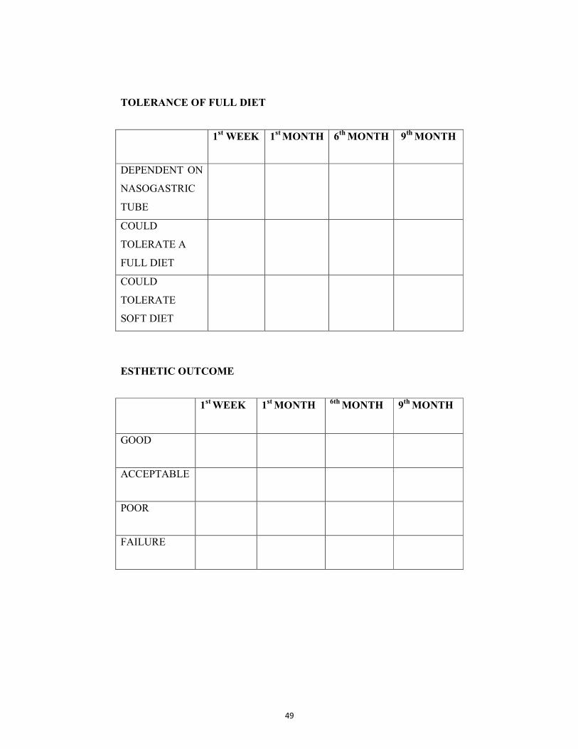
TOLERANCE OF FULL DIET
1st WEEK 1
st MONTH 6
th MONTH 9
th MONTH
DEPENDENT ON
NASOGASTRIC
TUBE
COULD
TOLERATE A
FULL DIET
COULD
TOLERATE
SOFT DIET
ESTHETIC OUTCOME
1st
WEEK 1st
MONTH 6th
MONTH 9th
MONTH
GOOD
ACCEPTABLE
POOR
FAILURE
50
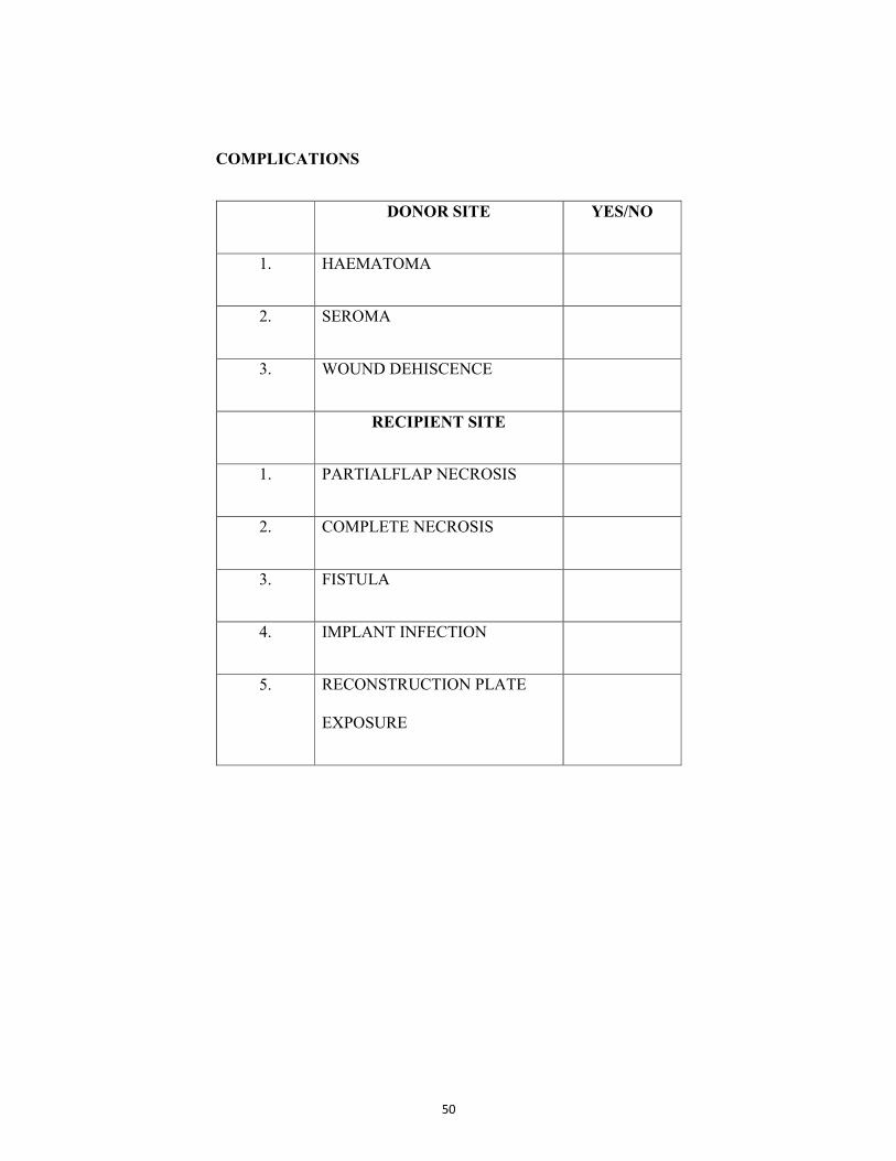
COMPLICATIONS
DONOR SITE YES/NO
1. HAEMATOMA
2. SEROMA
3. WOUND DEHISCENCE
RECIPIENT SITE
1. PARTIALFLAP NECROSIS
2. COMPLETE NECROSIS
3. FISTULA
4. IMPLANT INFECTION
5. RECONSTRUCTION PLATE
EXPOSURE

51
FIGURES
Figure 1: Armamentarium–Resection and
Reconstruction Kit
Figure 2: Micro Saw and Drill

52
SURFACE MARKINGS
Figure 3: Donor Site Marking
Figure 4: Recipient Site Marking
53
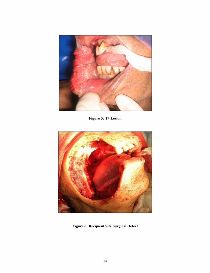
Figure 5: T4 Lesion
Figure 6: Recipient Site Surgical Defect

54
Figure 7: Resection & Reconstruction with Titanium Plate
Figure 8: Exposure of Pectoralis Major Muscle with Skin Paddle

55
Figure9: Vascular Pedicle Visible On Deep Aspect of PectoralisMajor
Figure 10: Flap Wrapping the Reconstruction Plate

56
Figure 11: Reconstruction ofRecipient Site Defect with Reconstruction
Plate and Bipaddle Flap
Figure 12: Donor Site Closed Primarily

57
Figure 13: Esthetic out Come- 9th
month Post Operative
Figure 14: Donor Site Outcome -9th
MonthsPost Operative

58
Figure15:Flap in situ-9th
Month Postoperative
Figure 16: Wound Dehisence

59
Figure 17: Orocutaneous Fistula
Figure 18: Implant Infection- 3rd
month Post Operative Radiograph

60
Figure 19:Reconstruction Plate Exposure
Figure 20: Total Flap Necrosis with Reconstruction Plate Exposure

61
RESULT
A two year clinical study was conducted on the functional, esthetic and
donor and recipient site complications in 20 patients in whom resection and
reconstruction was done with pectoralis major myocutaneous flap from
September 2010 to September 2012 in Department of Oral and Maxillofacial
Surgery at Sri Ramakrishna Hospital, Coimbatore.
The result of this study are shown under the following sub headings
1. Age and Gender distribution
2. Side of the tumor
3. Site of the lesion
4. Primary T-status
5. History of systemic disease
6. Functional outcome
7. Esthetic outcome
8. Donor and Recipient site complications
Out of 20 patients, 17 patients were taken for analysis as three patients
did not survive the study period of 9 months. They were excluded from the
results.

62
AGE AND GENDER DISTRIBUTION
In 17 cases in whom resection and reconstruction was done with
pectoralis major myocutaneous flap gender distribution of the study
population over two years showed that 64.7% of male were affected compared
with 35.3% female. The peak incidence of tumor was noted in patients in 40-
60 years of age accounting for 53% cases (Table-2 and 3).
SIDE OF THE LESION
In 17 cases, 52.9% of patients with lesion on the left side and 47.1% on
the right side of the oral cavity were resected and reconstructed with pectoralis
major myocutaneous flap. There was no significant association between the
side of the lesion and associated complications (P > 0.05) (Table-4 and 20,
graph 2).
SITE OF THE LESION
In 17 cases the lesion were 35.3% in lower alveolus, 23.5% in buccal
mucosa, 17.6% in retromolar trigone,11.8% in lateral border of the tongue and
11.8% in floor of the mouth were resected and reconstructed with pectoralis
major myocutaneous flap. There is no significant association between the site
of the lesion & complications (P > 0.05) (Table-5 and 24, graph-1 and 6).

63
T-SIZE OF THE TUMOR
In 17 cases in whom resection and reconstruction was done with
pectoralis major myocutaneous, 12.5% (1/8) patient with T2 status had seroma
& wound dehiscence. 20% (1/5) in T3, 25% (1/5) in T4 had partial flap
necrosis, 25% (1/4) in T4 status had complete necrosis, 20% (1/5) in T3 status
had implant infection 40% (2/5) in T-3 status, 25% (1/4) in T4 status had
reconstruction plate exposure.50% (2/4) in T4 status had fistula and Chisquare
value for association between T-status with fistula is 7.367 (P < 0.05) which is
significant (Table-6 and 22, graph 4).
HISTORY OF SYSTEMIC DISEASE
In 17 cases in whom resection and reconstruction was done with
pectoralis major myocutaneous flap. 47.1% (8/17) had history of systemic
disease in which 29.4% (5/17) had diabetes, 29.4% (5/17) had hypertension
and 17.6% (3/17) had ischemic heart disease. There is significant association
between ischemic heart disease and complete flap necrosis with Chi square
value 4.958 (P < 0.05) (Table-7 and 23, graph 5).
FUNCTIONAL OUTCOME
In 17 cases in whom resection and reconstruction was done with
pectoralis major myocutaneous flap, at the end of nine months speech was
normal in 11.8%, easily intelligible in 70.6%, poor but intelligible in 11.8%
and unintelligible in 5.9% of the patients. There is significant improvement in

64
the quality of speech between initial stage and end of nine months, t - value of
3.405 (P < 0.05) (Table-8).
Oral spintcher function was normal in 70.6%, continual drooling in
17.6%, occasional drooling in 5.9% and ryles tube feeding in 5.9% patients.
There is improvement in the oral spintcher function at the end of 9 months as
t - value of 3.405 for the mean difference in the oral spintcher function
between the initial stage and the 9th month is significant (P < 0.05) (Table-9).
Tolerance of full diet in 23.5% could tolerate soft diet in 70.6%
dependent on nasogastric tube in 5.9%. There is improvement in tolerance of
full diet at the end of 9 months, as the t - value of 3.405 for the mean
difference in the tolerance of full diet between the initial stage and the 9th
month is significant (P < 0.05) (Table-10).
AESTHETIC OUT COME
Patient satisfaction on aesthetic outcome is good in 23.5%, acceptable
in 52.9% and poor in 23.5% at the 9th month. There was improvement in
aesthetics of the recipient and donor site as t - value of 2.384 for the mean
difference in the aesthetic outcome between the initial stage and the 9th month
is significant (P < 0.05) (Table-11).

65
DONOR & RECIPIENT SITE COMPLICATION
SEROMA
Among 17 cases in whom resection and reconstruction was done with
pectoralis major myocutaneous flap 33.3% (1/3) had disease in retromolar
trigone, 5.9% (1/8) with T - II status, 9.1%(1/10) patients stage - III 1 out of
8 (12.5%) patients with systemic disease, 1 patients without radiotherapy had
seroma. There is no significant association between side, radiotherapy, T-size
and systemic disease with seroma (Table-12).
WOUND DEHISCENCE
In our study we found, 33.3% (1/3) had disease in retromolar trigone,
9.1% (1/10) cases in stage III, 5.9% (1/8) cases in T2 status, 12.5% (1/8) of
patient with systemic disease, 62.5% (10/16) of patients without radiotherapy
had wound dehiscence. There is no significant association between
radiotherapy, T-size and systemic disease with wound dehiscence (Table-13).
FISTULA
In our study we found 11.8% (1/ 2 ) patients with disease in lateral
border of the tongue and floor of the mouth, 33.3% (2/6) patient with stage -
IV, 50% 25% (2/8) patients with history of systemic disease had fistula. In
50% (2/ 4) patients with T4 status there was significant association with fistula
with Chi square value being 7.367 (P < 0.05) (Table-14 and 22, Graph-4).

66
PARTIAL FLAP NECROSIS
In our study we found 50% (1/2) patients in lateral border of the
tongue and 16.7% ( 1/6) patients with carcinoma in alveolus, 9.1% (1/11)
patients in stage III and 6.7% (1/6) in stage IV 12.5% (1/8) patient with
medical history, 20% (2/10) patients who underwent radiotherapy had partial
flap necrosis. There is no significant association between radiotherapy, T-size
and systemic disease with partial flap necrosis (Table-15).
COMLETE NECROSIS
In our study we found 50% (1/2) who had disease in floor of the
mouth, 16.7% (1/6) in stage IV disease, 25% (1/4) patients with T4 size,
12.5% (1/8) patient with systemic disease, 10% (1/10) who underwent
radiotherapy had complete necrosis There is a significant association between
ischemic heart disease and complete flap necrosis with Chi square value 4.958
(P < 0.05) (Table-16 & 23, Graph-5).
IMPLANT INFECTION
In our study we found 50% (1/2) each who underwent radiotherapy,
50% (1/2) without radiotherapy, 33.3% (1/3) in retromolartrigone,16.7% (1/6)
in alveolus, 12.5% (1/8) in T-2 status, 20% (1/5) in T-3status, 12.5% (1/8)
with history of systemic disease, 11.1% (1/9) without systemic disease, 12.5%
(1/8) on right side, 11.1%(1/9) on left side implant infection. There is no

67
significant association between radiotherapy, T-size and systemic disease with
Implant infection (Table-17).
RECONSTRUCTION PLATE EXPOSURE
In our study we found 30% (3/10) who underwent radiotherapy, 33.3%
(2/6) in alveolus, 50%(1/2) in floor of the mouth, 40% (2/5) in T-3, 25% (1/4)
in T-4 status,12.5% (1/8) with history of systemic disease, 22.2% (2/9)
without systemic disease, 25% (2/8) on right side, 11.1% (1/9) on left side had
reconstruction plate exposure. There is no significant association between
radiotherapy, T-size and systemic disease with reconstruction plate exposure
(Table-18).
MORTALITY
Among 20 cases in whom resection and reconstruction was done with
pectoralis major myocutaneous flap three patients died of unknown cause with
in 3months of follow up period. It was not due to flap related complications.
(Table-19).
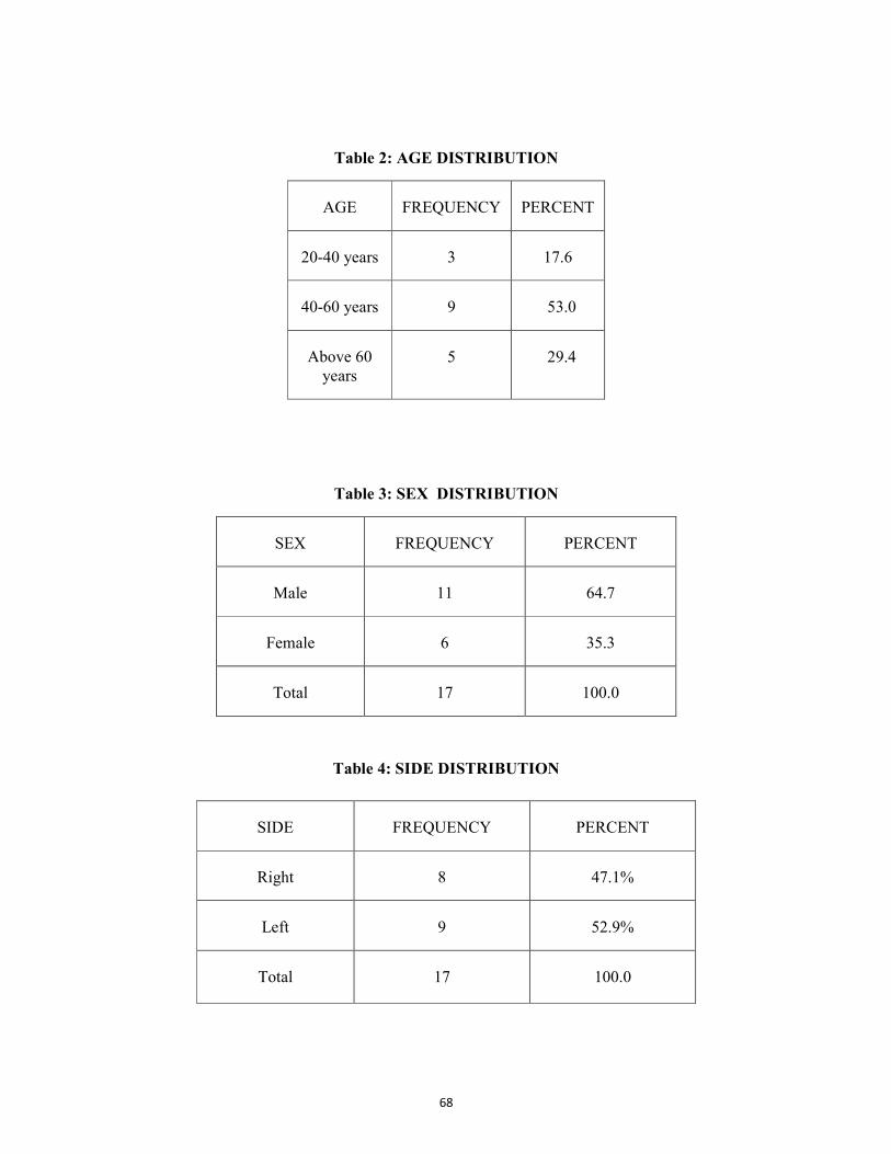
68
Table 2: AGE DISTRIBUTION
AGE FREQUENCY PERCENT
20-40 years 3 17.6
40-60 years 9 53.0
Above 60
years
5 29.4
Table 3: SEX DISTRIBUTION
SEX FREQUENCY PERCENT
Male 11 64.7
Female 6 35.3
Total 17 100.0
Table 4: SIDE DISTRIBUTION
SIDE FREQUENCY PERCENT
Right 8 47.1%
Left 9 52.9%
Total 17 100.0

69
Table 5: SITE OF LESION DISTRIBUTION
SITE FREQUENCY PERCENT
Alveolus 6 35.3
Buccal mucosa 4 23.5
Retromolar trigone 3 17.6
Lateral border of the
tongue
2 11.8
Floor of the mouth 2 11.8
Total 17 100.0
Table 6: PRIMARY-T STATUS DISTRIBUTION
STAGE FREQUENCY PERCENTAGE
T2 8 47.0
T3 5 30.0
T4 4 23.0
TOTAL 17 100.0

70
Table 7: SYSTEMIC DISEASE DISTRIBUTION
SYSTEMIC DISEASE FREQUENCY PERCENT
NO 9 52.9
YES 8 47.1
TOTAL 17 100
Table 8: MEAN DIFFERENCE IN SPEECH
SPEECH MEAN N STD
DEVIATION t-VALUE P
1st month
9th month
2.1176
2.8824
17
17
.60025
.69663 4.747 0.01
P<0.05=SIGNIFICANT
Table 9: MEAN DIFFERENCE IN ORAL SPINTCHER FUNCTION
Oral spintcher
function Mean N
Std
Deviation t-value P
1st month
9th month
3.0000
3.5294
17
17
1.00000
.87447 3.405 0.01
P<0.05=SIGNIFICANT
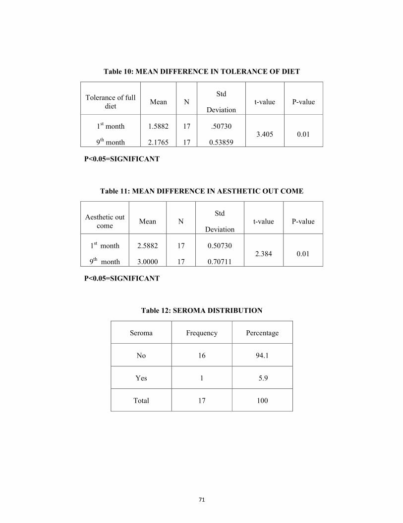
71
Table 10: MEAN DIFFERENCE IN TOLERANCE OF DIET
Tolerance of full
diet Mean N Std
Deviation t-value P-value
1st month
9th month
1.5882
2.1765
17
17
.50730
0.53859 3.405 0.01
P<0.05=SIGNIFICANT
Table 11: MEAN DIFFERENCE IN AESTHETIC OUT COME
Aesthetic out
come Mean N Std
Deviation t-value P-value
1st month
9th month
2.5882
3.0000
17
17
0.50730
0.70711 2.384 0.01
P<0.05=SIGNIFICANT
Table 12: SEROMA DISTRIBUTION
Seroma Frequency Percentage
No 16 94.1
Yes 1 5.9
Total 17 100

72
Table 13: WOUND DEHISCENCE DISTRIBUTION
Wound
Dehiscence
Frequency Percentage
No 16 94
Yes 1 6
Total 17 100
Table 14: FISTULA DISTRIBUTION
Fistula Frequency Percentage
No 15 88.2
Yes 2 11.8
Total 17 100

73
Table 15: PARTIAL FLAP NECROSIS DISTRIBUTION
Partial Flap
Necrosis
Frequency Percentage
No 15 80.2
Yes 2 11.8
Total 17 100
Table 16: COMPLETE NECROSIS DISTRIBUTION
Complete
Necrosis
Frequency Percentage
No 16 94.1
Yes 1 5.9
Total 17 100
Table 17: IMPLANT INFECTION DISTRIBUTION
Implant Infection Frequency Percentage
No 15 88.3
Yes 2 11.7
Total 17 100
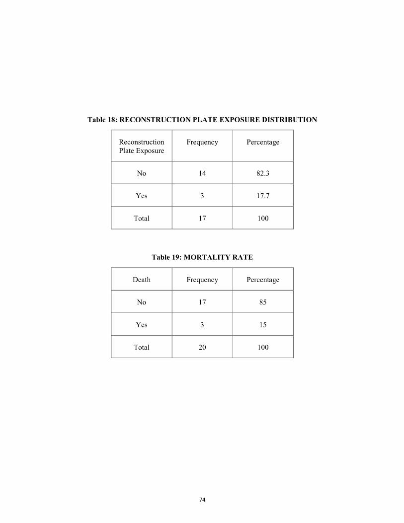
74
Table 18: RECONSTRUCTION PLATE EXPOSURE DISTRIBUTION
Reconstruction
Plate Exposure
Frequency Percentage
No 14 82.3
Yes 3 17.7
Total 17 100
Table 19: MORTALITY RATE
Death Frequency Percentage
No 17 85
Yes 3 15
Total 20 100
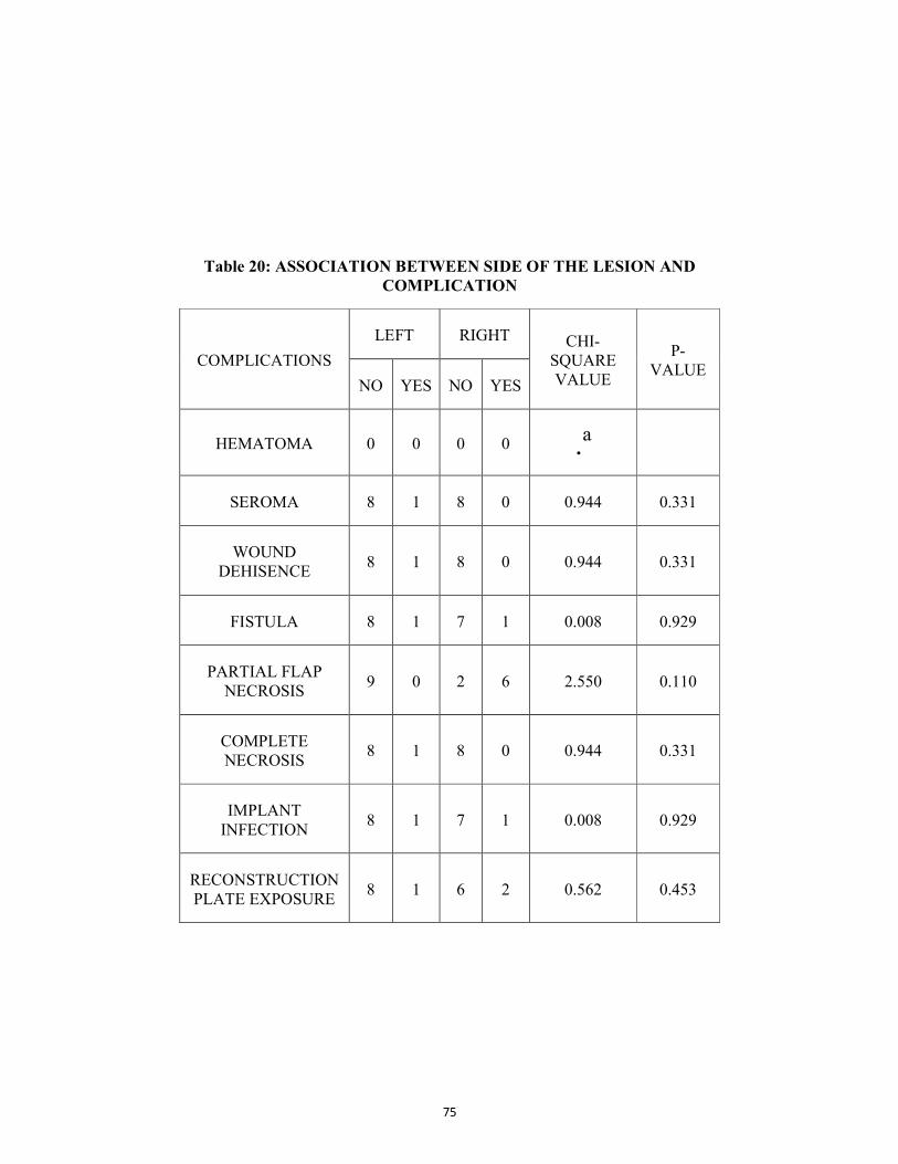
75
Table 20: ASSOCIATION BETWEEN SIDE OF THE LESION AND
COMPLICATION
COMPLICATIONS LEFT RIGHT CHI-
SQUARE
VALUE
P-
VALUE NO YES NO YES
HEMATOMA 0 0 0 0 .a
SEROMA 8 1 8 0 0.944 0.331
WOUND
DEHISENCE 8 1 8 0 0.944 0.331
FISTULA 8 1 7 1 0.008 0.929
PARTIAL FLAP
NECROSIS 9 0 2 6 2.550 0.110
COMPLETE
NECROSIS 8 1 8 0 0.944 0.331
IMPLANT
INFECTION 8 1 7 1 0.008 0.929
RECONSTRUCTION
PLATE EXPOSURE 8 1 6 2 0.562 0.453

76
Table 21: ASSOCIATION BETWEEN RADIOTHERAPY AND
COMPLICATIONS
COMPLICATIONS
RADIOTHERAPY CHI-
SQUARE
VALUE
P-
VALUE NO YES
HEMATOMA NO 0
0
.a YES 0
0
SEROMA
NO 6 10
1.518 0.218 YES 1 0
WOUND DEHISENCE
NO 6 10
1.518 0.218 YES 1 0
FISTULA
NO 7 8
1.587 0.208 YES 0 2
PATIALFLAP NECROSIS
NO 7 8
1.587 0.208 YES 0 2
COMPLETE NECROSIS
NO 7 9
0.744 0.388 YES 0 1
IMPLANT INFECTION
NO 6 9
0.073 0.787 YES 1 1
RECONSTRUCTION
PLATE EXPOSURE
NO 7 7
2.550 0.110
YES 0 3
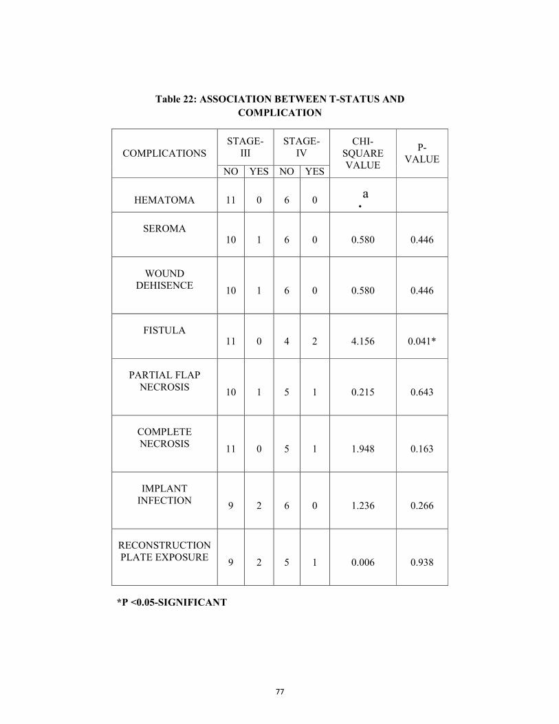
77
Table 22: ASSOCIATION BETWEEN T-STATUS AND
COMPLICATION
COMPLICATIONS STAGE-
III STAGE-
IV CHI-
SQUARE
VALUE
P-
VALUE NO YES NO YES
HEMATOMA 11 0 6 0 .a
SEROMA
10 1 6 0 0.580 0.446
WOUND
DEHISENCE
10 1 6 0 0.580 0.446
FISTULA
11 0 4 2 4.156 0.041*
PARTIAL FLAP
NECROSIS
10 1 5 1 0.215 0.643
COMPLETE
NECROSIS
11 0 5 1 1.948 0.163
IMPLANT
INFECTION
9 2 6 0 1.236 0.266
RECONSTRUCTION
PLATE EXPOSURE
9 2 5 1 0.006 0.938
*P <0.05-SIGNIFICANT

78
Table 23: ASSOCIATION BETWEEN SYSTEMIC DISEASE AND
DONOR&RECIPIENT SITE COMPLICATIONS
DIABETES HYPERTENSION ISCHEMIC
HEART
DISEASE
CHI- SQUARE
VALUE
P-VALUE
NO YES NO YES NO YES
HEMATOMA 0 0 0 0 0 0 .a
SEROMA 1 0 1 0 1 0 0.443 0.056
WOUND
DEHISENCE 0 1 1 0 1 0 2.550 0.110
FISTULA 1 1 1 1 1 1 0.463 0.496
PARTIAL FLAP
NECROSIS 1 1 2 0 2 0 0.463 0.496
COMPLETE
NECROSIS 1 0 0 1 0 1 4.958 0.026*
IMPLANT
INFECTION 1 1 2 0 2 0 0.463 0.496
RECONSTRUCTION
PLATE EXPOSURE 3 0 2 0 2 1 1.518 0.218
* P <0.05-SIGNIFICANT

79
Table 24: ASSOCIATION BETWEEN SITE OF THE LESION AND
COMPLICATION
RETROMLAR
TRIGONE
LATERAL
BORDER
OF
TONGUE
ALVEOLUS BUCCAL
MUCOSA
FLOOR OF
THE
MOUTH
CHI
SQUARE
VALUE
P
VALUE
NO YES NO YES NO YES NO YES NO YES
HEMATOMA 0 0 0 0 0 0 0 0 0 0 .a
SEROMA 2 1 2 0 6 0 4 0 2 0 4.958 0.292
WOUND
DEHISENCE 2 1 2 0 6 0 4 0 2 0 4.958 0.292
FISTULA 3 0 1 1 6 0 4 0 1 1 7.367 0.118
PARTIAL
FLAP
NECROSIS
3 0 1 1 5 1 4 0 2 0 4.156 0.385
COMPLETE
NECROSIS 3 0 2 0 6 0 4 0 1 1 7.969 0.093
IMPLANT
INFECTION 2 1 2 0 5 1 4 0 2 0 2.550 0.636
RECONSTRUCT
ION PLATE
EXPOSURE 3 0 2 0 4 2 4 0 1 1 4.385 0.356

80
Graph 1: SITE OF LESION DISTRIBUTION
Graph 2: ASSOCIATION BETWEEN SIDE OF THE LESION AND
COMPLICATION

81
Graph 3: ASSOCIATION BETWEEN RADIOTHERAPY AND
COMPLICATIONS
Graph 4: ASSOCIATION BETWEEN T-STATUS AND
COMPLICATION

82
Graph 5: ASSOCIATION BETWEEN SYSTEMIC DISEASE AND
ASSOCIATED COMPLICATION
Graph 6: ASSOCIATION BETWEEN SITE OF THE LESION AND
COMPLICATION

83
DISCUSSION
In head and neck region, reconstruction of maxillofacial defects after a
ablative cancer surgery is a challenge to the surgeon.4,26,40
In oncologic
surgery, the goal is to achieve tumour-free margins with functional and
esthetic restoration, so that the quality of life is as little impaired as possible.15
The applicability of Pectoralis major flap in head and neck reconstructive
surgery have proved to be an advance over previously used flaps such as the
deltopectoral flap.2 All the previous literatures have very well established that
pectoralis major myocutaneous flap is a reliable and practical transfer of soft
tissue for the reconstruction of extirpative and avulsive defects of the oral
cavity and face32 and continued to enjoy as the main reconstructive option for
a variety of reasons including preference, costs and lack of expertise in free
flaps.5,35 The flap elevation procedure is simple and shows predictable results
when the scientific principles of myocutaneous flap surgery are adhered.
Sufficient evidence substantiates the numerous indications, diverse
applicability, techniques and great versatility of this soft tissue flap. Recent
concepts of micro vascular flaps for reconstruction have proven its efficacy in
reconstruction on head and neck region not outdating the pectoralis major flap
completely due to its usage as secondary flap after free flap failure.32
Reconstruction of the soft palate, floor of the mouth and lateral
pharyngeal wall can also be done with this flap.43 The pectoralis major
myocutaneous flap allowed blood supply to the grafted iliac bone.8 The

84
discussion involves various modification of pectoralis major flap. The study
also analyses the influence of functional, esthetic outcomes and complications
of donor and recipient site with factors like systemic disease, radiotherapy,
site, stage of disease and T status influence.2
Robert E. Marx and Brian R. Smith, 1990 included the lateral thoracic
artery and showed good reduction in complications, a greater range of use and
consistent healing in radiated and non irradiated tissues and not requiring
sectioning of the clavicle causing significant chest deformities.48
Richard Crosher 1995, described a modification where the incision is
made along the lower limb of the deltopectoral flap and continues into the
crescentic skin paddle of the pectoralis that provides a sizeable skin paddle
and allows primary closure of the chest wound with good cosmetic results.45
Kiyokawa. K et al, included the third intercostal perforating branch of
internal thoracic artery along with the skin paddle which provided the flap a
stable blood circulation29, Anthony Po-Wing Yuen, preserved the lateral
thoracic artery by dividing the pectoralis major muscle over the artery at the
clavicular part for complete release without compromising pedicle length in
harvesting pectoralis major myocutaneous flap.44
Rikimaru also preserved the internal thoracic artery but modified the
incision which extended to the axilla and a Z- plasty was used to close the
donor defecy.46 Vijay R. Ramakrishnan et al believed that the skin flap over

85
the rectus sheath is the cause of distal skin flap necrosis and avoided rectus
sheath with the skin paddle, also designed the skin paddle within curvilinear
incision in males and infra mammary skin paddle in females.55
The further modification of the flap includes an osteomyocutaneous
variety where the flap is raised along with the anterior part of the fifth or sixth
rib to reconstruct the mandible. The final functional and cosmetic results were
satisfactory. The pectoralis major myocutaneous flap provided natural mucosa
when transferred intra orally.13 In our study the lateral pedicle of
thoracoacromial artery was sacrificed to attain length and greater arc of
rotation, holding the basic of the early technique of raising pectoralis major
myocutaneous flap with electrocautery dissection.
Astrid L Kruse et al (2011) reported that male to female ratio was 17:3,
with a mean age of 60 years.7,15,16
In our study the male to female ratio was
11:6. In literature the most common type of tumor was squamous cell
carcinoma 90%.26 All the cases in our study were squamous cell carcinoma.
In literature, most commonly the tumor involved area was right
mandibular region (66.6%) followed by left mandibular region (22.2%) and
right buccal mucosa 11.1%.16
In our study left mandibular region (52.9%) was more commonly
affected than right mandibular (47.1%) region. The site of involvement in
86
alveolus was 35.3%, buccal mucosa 23.5% and retromolar area 17.6%, floor
of the mouth and lateral border of tongue 11.8% each (Table-5).
The reconstruction of the base of the tongue, the floor of the mouth,
and the oropharynx emerged as a significant risk factor for flap necrosis.26 In
our study a patient with carcinoma floor of the mouth had total flap loss. The
end of the study didn’t show any significant association between site, side of
the lesion with donor & recipient site complications (P > 0.05).
For primary oral squamous cell carcinoma cases pectoralis major flap
was commonly used, thus restoring esthetics and function with minimal donor
site morbidity.16,38,48
Earlier studies showed that speech was normal in
45.4%.36 In our study at the end of 9 months there is significant improvement
in speech (P < 0.05), being normal in 11.8%, easily intelligible in 70.6% poor
but intelligible in 11.8%, unintelligible in 5.9% patients (Table-8).
In comparison to pectoralis major flap, free flap reconstruction reported
better intelligible speech.5,35
In literature oral continence was normal in 5% of patients, occasional
drooling in 75% cases and continuous drooling in 20% cases.
16,19,36
In our study at end of 9 months there is significant improvement in oral
sphincter function (P < 0.05) being normal in 70.6%, continual drooling in
17.6%, occasional drooling in 5.9%, patients (Table-9).

87
Early studies showed, 54.5% was able to tolerate full diet, 27.3% were
able to tolerate soft diet and dependant on nasogastric tube was in 18.2% of
cases on pectoralis major flap reconstruction.36
In our study, there was significant improvement in tolerance of full diet
in 23.5% and 5.9% patients were nasogastric tube dependent at the end of 9
months (Table-10).
The thickness of the flap in obese and muscular individuals, which is
determined by the amount of subcutaneous fat between the pectoralis muscle
and the overlying skin paddle, leads to reduced swallowing or speech
function.5,7,35,33,35
Mohammed Tahir et al showed 15% had good result; majority had
acceptable result 60%, poor result in15% patients in his analysis on esthetic
outcome on patients who had under gone pectoralis major flap
reconstruction.36
In our study esthetic outcome was good in 23.5%, acceptable in 52.9%,
poor in 23.5% and there was marked improvement in the esthetic outcome in
patients reconstructed with Pectoralis major flap (Table-11).
The large bulk of the flap reduced neck mobility and needed the
vascular pedicle of the flap to rotate 180° when the skin paddle is used to
resurface the neck.7 The resection of the pedicle, even a few weeks after
transplant, together with the subclavicular passage, may avoid the majority of
88
the well-known functional and esthetic problems related to this reparative
technique.40 In mandibular reconstruction by means of reconstructive plates
and myocutaneous flaps, reconstruction plates should be stabilized with atleast
3 screws in each reminiscent side for correct plate banding to provide a good
aesthetic result.34,35 Aesthetic results are improved at the harvesting site in
males in whom the thoracic skin can be re-sutured without any tension
cervical mobility and obtaining a good appearance of the neck and decreasing
the traction downwards of the flap.57
Common complications cited in donor & recipient site after Pectoralis
major myocutaneous flap reconstruction are, haematoma, seroma, wound
dehiscence, fistula, implant infection, reconstruction plate exposure and total
or partial skin necrosis secondary to vascular compromise.55
In literature overall success rate ranged from 70.8% to 95.5%.16,38,48
The overall complications ranged from 6% to 63%.18,25,37
The overall
complication rate in our study was 5.8% .
The total loss of flap ranged from 2% to 6%.19,22,30,31,36,43
In our study
complete necrosis was in 5.9% (Table-16). At the recipient site, rough
handling of a very small skin paddle (2 x 3 cm) seemed to be responsible for
the totally necrosed skin paddle and extensive peripheral vascular disease with
postoperative hypotension seemed to be the major factor in the only partially
necrotic skin paddle.3 There is a significant association between ischemic

89
heart disease and complete flap necrosis with chi square value 4.958
(P < 0.05).
When total flap loss is realized, resurrection of the flap is impossible
even when hyperbaric oxygen therapy is administered.2 The lateral thoracic
artery’s contribution to the flap is most important for flap viability,19,44 but in
our study we didn’t include lateral thoracic artery. There was one total flap
loss case in our study.
Partial flap loss ranged from 3.3% to 32%.2,19,21,22,30,36,43
In our study
partial flap necrosis was in 11.8% of cases (Table-15). The most difficult
complication of a pectoralis major myocutaneous flap is partial or total flap
necrosis. This complication requires additional surgery to attempt salvage of
the reconstruction.
The possible cause for flap loss could be the variations in vascular
supply or a possible technical error while harvesting the flap, like skin paddle
beyond the seventh rib, external compression caused by the tracheostomy
cannula strap.21 Skin paddle away from the area of the main skin perforating
vessels that arise from the intercostal branches of the internal thoracic artery
and small skin paddle which did not encompass sufficient skin perforating
vessels are also the reasons for partial or total flap loss.
40
Fistula complication in head and neck surgery cases reconstructed with
pectoralis major muscle ranged from 2% to 45%.2,21,30,35,36,43
In our study
90
fistula was in 11.8% cases (Table-14). There was significant association
between patients with T4 status and fistula with Chi square value being 7.367
(Table-22).
The oro-cutaneous fistulas may be avoided by elevating a slightly
larger cutaneous island which allows tension-free suture of the flap to the
oropharyngeal excision margins.
Wound dehiscence after reconstruction ranged from 5.2%-
26%.25,19,21,35
In our study wound dehiscence were in 6% of cases. (Table-13)
There is no significant association between wound dehiscence and
radiotherapy, systemic disease and T-size of the disease. A tension free
closure could have allowed and compensated for fibrosis and contracture.
Accumulation of hematoma ranged from 5.4% to 7%8,12,19,38
in
literature. In our study there was no case of hematoma under the flap. Dead
space can develop between the graft bed and the myofascia, leading to
development of hematoma that blocks blood flow to the grafted flap.8 An
uninterrupted suture over the residual resected muscles of the major pectoral
has avoided the formation of hematoma in the donor site.38,49 Meticulous care
in suturing a tension free flap can avoid hematoma formation. Two vacuum
drain were kept as routine in the resected and reconstructed site.
G. Montemari, A. Rocco reported that pectoralis major myofascial flap
has tolerated the Postoperative radiotherapy well.37 Revascularized flaps

91
helped ameliorate the effects of radiation in surgery undergone patients.18
Robert .E. Marx reported no radiation related complications.48 In our study
among 10 patients, who underwent radiotherapy 1 patient (10%) had complete
necrosis of flap, 11.8% had fistula, 10% had implant infection and 30% had
reconstruction plate exposure. (Table-21).
The complication could be due to early radiotherapy and excessive
fibrosis. The protocol of following radiation therapy after 6-8 weeks could be
advisable, to avoid post radiation complications.
Radical surgical procedure should be carried out with caution in old
patients with pre-exsisting disease as the chances of life threatening
complication are very high in these patients.8 This makes an indication for the
pectoralis major myocutaneous flap to be used in medically compromised
patients who are known to have increased complication rates.8,26 The systemic
diseases have to be treated pre and postoperatively with care.33 Systemic
diseases like hypertension, ischemic heart disease, insulin dependent diabetes,
cirrhosis were common among the patients who undergo surgery and have
very advanced squamous cell carcinomas (T3-T4) in which reconstruction
with pectoralis major myocutaneous flap were performed.47
In our study out of 17 patients in whom resection & reconstruction was
done 47.1% suffered from various systemic illness like diabetes 29.4%,
hypertension 29.4% and ischemic heart disease 17.6% of patients.
92
In our study out of 5 patients with diabetes, none had seroma, 20%
(1/5) patients had wound dehiscence, 20% (1/5) patient had partial necrosis,
10% (1/5) patient had implant infection & there was no case of reconstruction
plate exposure or total flap necrosis. There was no significant association
between diabetes with donor & recipient site complication (Table-23).
Out of 5 patients with hypertension, 10% (1/5) patients had seroma,
10% (1/5) patient had fistula, none had implant infection, 10% (1/5) patient
had reconstruction plate exposure, 10% (1/5) patient had complete necrosis.
There was no significant association of hypertension with donor and recipient
site complication (Table-23).
Out of 3 patients with Ischemic heart disease, none had seroma and
partial flap necrosis, and 33% (1/3) had complete necrosis, 33% (1/3) had
fistula, 33% (1/3) had implant infection, 33.3% (1/3) had reconstruction plate
exposure. There was a significant association between ischemic heart disease
and complete necrosis with Chi square value being 4.958 (Table-23).
In our study there was no significant association of donor & recipient
site complication in patients with systemic disease like diabetes and
hypertension and who have undergone radiotherapy. Higher complication
rates were associated with the utilization of the flap as a salvage procedure.
Women who underwent Pectoralis Major Myocutaneous Flap had a higher rate
of flap necrosis than did men presumably because of interposition of breast
tissue between muscle & skin paddle.2 There were no significant differences
93
between free flap and Pectoralis Major Myocutaneous Flap groups in age,
surgical site infection rate, partial flap necrosis rate, tumor stage, positive
surgical margin rate.12,28 The bulk of the Pectoralis Major Myocutaneous flap
is sufficient to allow reasonable function in the context of limited life
expectancy. Long-term survivors may be offered a delayed free flap bone
reconstruction.18,32
Bridging plates represents a useful reconstruction method when the
tumor size, age and prognosis did not allow usage of free flaps.34 The
reconstruction plate is used to reconstruct, lateral and small defects but are not
effective in bridging large defects. When reconstruction plates were placed
across 2 areas with muscle stress concentration (chin and angle of mandible)
failure rate was as higher as 68%. Early radiation therapy may have some
influence on loss of the reconstruction plate, because it can compromise the
healing process of the pectoralis major flap. Bridging plates alone is associated
with considerable morbidity, including fracture and loosening of fixation, and
most importantly plate exposure. Plate exposure rates can be markedly
reduced by covering the plate with a myocutaneous flap.22, 34, 35
Fábio Roberto Pinto et al (2010) found that titanium plate exposure in
17.7% cases.22 In our study 50% (1/2) of cases in floor of the mouth, 33.3%
(2/6) cases in alveolus, 18.2% (2/11) patients in stage III, 16.7% (1/6) patients
in stage IV, 2 out of 5 patients in T3, 1 out of 4 patients in T4, 12.5% (1/8) with
systemic disease, 22.2% (2/7) patients without systemic disease, 30% (3/7)

94
patients who underwent radiotherapy had reconstruction plate exposure. In
literature 18.8% of cases Patients in whom the plate had to be removed have
been submitted to radiation therapy in the first 2 postoperative months, and 6
had been submitted to radiation therapy after the second postoperative
month.22,35
In our study we found no significant association between
radiotherapy, systemic disease, T-size and reconstruction plate exposure
(Table 21, graph 3). In the cases which had gone for radiation had invariably
plate related complications. The possible explanation could be of early
radiation, the muscle raping around the plate which could have pulled the plate
in vertical direction, causing loosening of screws.
The ideal time to start postoperative radiotherapy is within 6 to 8
weeks postoperatively. Titanium is a more suitable material when a radiation
therapy is required.34 From the acceptable success rate, it may be concluded that
bridging plates represent a useful reconstruction method, provided they are well
covered by viable muscular tissue. They should be offered to patients
contraindicated for more invasive procedures or with limited functional needs,
or poor prognosis.25
Mohamed A Zohairy (2009) in his study reported that fourteen patients
died from their disease at the end of 2 years follow-up due to systemic
metastases or local recurrence.35
The mortality rate within 30 days was high as 7%. In 1-year, 3-years
and 5-years, overall survival rates were 65.5%, 39.1% and 11.0%

95
respectively.15, 36
The bulk of the Pectoralis myocutaneous flap is sufficient to
allow reasonable function in the context of limited life expectancy.44 In our
study 15% (3/20) died due to unknown cause within three months after surgery
(Table-19). The functional and social outcome to pectoralis major flap was good
and provided a reasonable alternative for reconstruction in advanced disease.

96
SUMMARY AND CONCLUSION
Pectoralis major myocutaneous pedicled flap has been considered
suitable choice for patients in whom reconstructing large defects resulting
from excision of head and neck malignancies associated with poor general
conditions and radiotherapy. This flap possesses a large volume of
vascularized muscle and skin paddle of sufficient size to satisfy the soft tissue
reconstructive needs.
Various modalities like free flap, faciomyocutaneous and skin islands
of different muscles have been tried, but single stage reconstruction with
myocutaneous flap like pectoralis major flap has proved to be advantage in
using at, the any form above. The free flap from pectoralis major sounds
interesting and has to be tried in larger number to known the possible
alternative in soft tissue reconstruction.
In our study, the technique used for Pectoralis major flap elevation
was the popular Ariyan technique, by sacrificing the lateral thoracic artery
with using of electrocautery for dissection. It proved to have much lesser
complication (5.8%) related to flap. The complication rate increased only
when the mandible has to be reconstructed with reconstruction plate (29.4%).
Pectoralis major flap vascular pedicle i.e. pectoral branch of
thoracoacromial artery is easily identifiable and possibly an early marking
with ultrasonography can reduce the flap rising time. The lateral thoracic

97
artery can be preserved whenever possible. The Pectoralis major flap provides
adequate coverage for the vital structures of neck and lies at a less distance
from the resected site when the patient has to undergo post operative radiation.
The flap provides large area for defect coverage, and can be used as
both mucosal lining and coverage of skin or combination of both. The bulk of
Pectoralis major provides good cosmetic benefit after composite resection.
The selection of pectoralis major flap over free flap was influenced by
patient factors in most cases. A correctly prepared Pectoralis major
myocutaneous flap is nothing inferior to the free flaps. It serves as an alternate
flap in head and neck reconstruction when free tissue transfer fails.
The following variables which were related to a higher rate of
complications are male sex, age over than 50 years, immediate radiation
therapy, presence of co morbidities (hypertension and diabetes), larger flaps,
advanced stages (T3 or T4) and the site of reconstruction being the tongue,
floor of the mouth, or the oropharynx.
In salvage surgeries, Pectoralis major myocutaneous flap could be the
choice comparing to its complications. For the best results, the size and shape
of the flap, careful surgical technique avoiding damage to the vascular pedicle
and to the musculocutaneous perforating vessels, tension free closure, delayed
post radiation (after 6-8 weeks) can be followed.

98
To conclude our study the Pectoralis major myocutaneous flap remains
a valuable reconstructive option in the head and neck owing to its technical
ease, reliability and versatility, function, esthetics, reduced postoperative
complications in donor and recipient site.

99
BIBLIOGRAPHY
1. Abid H, Ahmad S, Warraich R A. The Versatility of Pectoralis
Major Myocutaneous Flap in Head and Neck Reconstruction. Annals,
2008; 14(3): 45-50.
2. Ahmed F.El-Kased, Hossam El- Fol, Tarek, Solaiman Elshakhs.
Use of the pectoralis major myocutaneous flap for oral cavity
Reconstruction in females and difficult cases requiring double island
flaps. Menoufiya Medical Journal, 2007; 20(2): 27-40.
3. Asong M. Reconstruction with pectoralis major myocutaneous flap for
surgical defect in advanced head and neck cancer patients. Ai Zheng,
2008; 27(1): 58-61.
4. Aleksandar, Miso Virag, Vedran Uglesic. Pectoralis major flap in
head & neck reconstruction: first 500 patients. Journal of
craniomaillofacial surgery, 2006: l34(6): 340-343.
5. Anna Karinne C, Bandeira, Maria de Fatima, Luciana P . Analysis
of Swallowing After Retromolar or Oropharynx Resection and
Reconstruction with Myocutaneous or Microvascular Free Flaps.
Applied cancer research, 2007; 27(1): 23-29.
6. Ariyan S. The pectoralis major myocutaneous flap for reconstruction
in head and neck defects. Plastic Reconstructive Surgery, 1979; 63:73.

100
7. Astrid L Kruse, Heinz T Luebbers, J O Obwegeser. Evaluation of
the pectoralis major flap for reconstructive head and neck surgery.
Head & Neck Oncology, 2011; 3:12.
8. Avery C M E, Crank S T, Neal C P, Hayter J P,. The use of the
pectoralis major flap for advanced and recurrent head and neck
malignancy in the medically compromised patient. Oral Oncology,
2010; 46: 829–833.
9. Berktold R E, Ossoff R H, Sisson G A, Wurster C F. Pectoralis
major flap for chin augmtation. Otolaryngol Head Neck Surg, 1986,
94(2):181-186.
10. Chaudhary A, Wakhlu A. Massive paraganglioma in an infant.
Journal of Indian association of paediatric surgery, 2005; 10(3): 56-59.
11. Chen Xiao-Hong, Zhao Han-Xue, Fang Ju-Gao. Use of preoperative
ultrasound in designing the true pectoralis major myocutaneous island
flap. Chinese Medical Journal, 2012; 125(4): 667-670.
12. Chih-Yu Hsing, Yong- Kie Wong, Ching Ping Wang, Chen-Chi
Wang. Comparison between free flap and pectoralis major pedicled
flap for reconstruction in oral cavity cancer patients – A quality of life
analysis. Oral Oncology, 2011; 47: 522–527.

101
13. Christiana Maria R S V, Leandro L de Matoos, Mario Paulo S
Junior. Enhanced Morbidity of Pectoralis Major Myocutaneous Flap
Used for Salvage after Previously Failed Oncological Treatment and
Unsuccessful Reconstructive Head and Neck Surgery. The Scientific
World Journal, 2012; 58:120-126.
14. Collin Roy, Prabir K josh, R N Podder. Cancrum Oris: A Single
Stage Correction of Ankylosis of Jaw and Cheek Fistula Defect. Indian
Journal of Surgery, 2002; 64(6): 520-522.
15. Corten E M L. The versatility of the pectoralis major flap in
reconstructive surgery, publisher. Utrecht University, 2008.
16. Croce A, Moretti, L D’Agostino, Neri G. Continuing validity of
pectoralis major muscle flap 25 years after its first application.
Actaotorhinolaryngol ital, 2003; 23:297-304.
17. De Bree, Reith R, Quak J J, Uyl- de Groot, Van Agthoven. Free
radial forearm flap versus pectoralis major myocutaneous flap
reconstruction of oral and oropharyngeal defects: a cost analysis, R
Clin. Otolaryngol, 2007; 32:275-282.
18. Douglas B, Gail Annich, Melissa A P, Jill B. Pectoralis Major
Myocutaneous Flap Vs Revascularized Free Tissue Transfer. Arch
otolaryngology Head Neck surg, 2004; 130.

102
19. Eric R Carlson. Pectoralis major myocutaneous flap. Oral
Maxillofacial Surgical Clinics of North America, 2003;15:565–575.
20. Ethunandan M, Ansell M, Mellor T K, Brennan P A. Skin necrosis
of a pectoralis major myocutaneous flap, caused by methicillin-
resistant Staphylococcus aureus. British Journal of Oral and
Maxillofacial Surgery, 2004;42:38-40.
21. Firdous Khan, Hifsa Hameed, Syed A Shah, Naji ullah khan . Head
and neck reconstruction; our experience of pectoralis major
myocutaneous pedicled flap. Professional Medical Journal,
2011;18(2):310-316.
22. Fábio Roberto Pinto, Carina Rosa M, Chirstiana M S V. Pectoralis
major myocutaneous flaps for head and neck reconstruction: factors
influencing occurrences of complications and the final outcome. Med
J, 2010; 128:6: 336-341.
23. Göktas O, T Schrom. Extensive Recurrence of a Parotid Carcinoma
After Primary Radiotherapy. The Open Otorhinolaryngology Journal,
2007; 1: 1-4.
24. Gregor R T, Sa Mediese Tydskrif. The use of the pectoralis major
myocutaneous island flap. For reconstruction after major head and
neck surgery, 1982; 61: 788.

103
25. Hamdy H El-marakby. The Reliability of Pectoralis Major
Myocutaneous Flap in Head and Neck Reconstruction, Journal of the
Egyptian Nat. Cancer Inst, 2006; 18(1): 41-50.
26. Hao Zou, Wen-Feng Zhang, Qi-Bin Han, Yi-Fang Zhao. Salvage
reconstruction of extensive recurrent oral cancer defects with
Pectoralis major myocutaneous flap. Journal of Oral Maxillofacial
Surgery, 2007; 65:1935-1939.
27. Jog J M, Meckenzie K, Dempster. Tumour Recurrence at the
donor Site of the pectoralis major myocutaneous flap with a tumour
free index site. Journal of Otorhinolaryngology, 2005; 3(2): 150-156.
28. Kekatpure V D. Pectoralis major flap for head and neck
reconstruction in era of free flaps. International Journal of Oral
Maxillofacial Surgery, 2012; 41: 453–457.
29. Kiyokawa K A, Tai , Tanabe H Y, Inoue Y . Method that preserves
circulation during preparation of the pectoralis major myocutaneous
flap in head and neck reconstruction. Plast Reconstr Surg, 1998;
102(7): 2336-2345.
30. Loh L E. Buccal carcinoma-A comparison of two modalities of
reconstruction. Singapore med journal, 1992; l33: 244-246.

104
31. Liu R, Gullane P, Brown D, Irish J. Pectoralis major myocutaneous
pedicled flap in head and neck reconstruction: retrospective review of
indications and results in 244 consecutive cases at the Toronto General
Hospital. Jornal Otolaryngol, 2001; 30(1): 34-40.
32. Mallet Y, El Bedoui S, Penel N, Ton Van, Fournier C. The free
vascularized flap and the pectoralis major pedicled flap options:
Comparative results of reconstruction of the tongue. Oral Oncology,
2009; 45(56): 1028–1031.
33. Marco Luigi Castelli, Giancarlo pecorari, Succo G. Pectoralis major
myocutaneous flap: analysis of complications in difficult patients. Eur
Arch Otorhinolaryngol, 2001; 258: 542–545.
34. Mariani.P. B. Reconstruction of large defects postmandibulectomy for
oral cancer using plates and myocutaneous flaps: a long term follow
up. International journal of oral maxillofacial. surgery,
2006; 35: 427-432.
35. Mohamed A.F. El-Zohairy. Mandibular Reconstruction Using
Pectoralis Major Myocutaneous Flap and Titanium Plates after
Ablative Surgery for Locally Advanced Tumors of the Oral Cavity.
Journal of the Egyptian Nat Cancer Inst, 2009; 21: 4; 299-307.
36. Mohammed Tahir, Tahmeedullah, Amir Taimur Khan. Clinical
evaluation of pectoralis major myocutaneous flap in Head and Neck

105
Reconstruction. Journal of postgraduate medical institute, 2005; 19:
71-75.
37. Montemari G, Rocco A, Galla S, Damiani V, Bellocchi G.
Hypopharynx reconstruction with pectoralis major myofascial flap: our
experience in 45 cases. Acta otorhinolaryngoica italic, 2012; 32: 93-97.
38. Nagral S, Sankhe M, Patel C V. Experience with the pectoralis
major myocutaneous flap for head and neck reconstruction in a general
surgical unit. Postgrad Med journal, 1992; 38:119-121.
39. Nath S, Joshi K D, Shakya s, Shrestha S, Koirala U . Mandibular
reconstruction. Kathmandu University Medical Journal, 2006; 4(16):
497-500.
40. Oluwatosin O M, Abikoye F O, Adegboye V O. A review of
pectoralis major musculocutaneous flap in head & neck reconstruction
in idadan. The Nigerian Journal of Surgical Research, 2000; 2: 16-20.
41. ORD R.A. The pectoralis major myocutaneous flap in oral &
maxillofacial reconstruction. A retrospective analysis of 50 cases.
Journal of oral & maxillofacial surgery, 1996; 54: 1292-1295.
42. Parag sahasrabudhe, Ranjeet Jagtap, Pankaj Waykole, Nikhil
Panse, Pallavi B. Our experience with pectoralis major flap for

106
management of sternal dehiscence a review of 25 cases. Indian Journal
of Plastic Surgery, 2011; 44(3): 405–413.
43. Plazak J, Kalitova P, Urbanova M, Betka J. Subcutaneous
calcification in the pectoralis major flap: a late complication of
radiotherapy. The British Journal of Radiology, 2011; 84: 223–225.
44. Po-Wing Yuen A. Preservation of lateral thoracic artery to improve
vascular supply of distal skin without compromising pedicle length in
harvesting Pectoralis major myocutaneous flap. Journal of Plastic,
Reconstructive & Aesthetic Surgery, 2006; 59: 1433-1435.
45. Richard Crosher, Roy Mitchell, John Llewelyn. A modification of
the pectoralis major myocutaneous flap that reduces the defect at the
donor site. Ann R Coll Surg Engl, 1995; 77: 389-391.
46. Rikimaru H, Kiyokawa K, Watanabe K. New method of preparing a
pectoralis major myocutaneous flap with a skin paddle that includes
the third intercostals perforating branch of internal thoracic artery.
Plastic reconstructive surgery, 2009; 123: 1220-1228.
47. Rithin suvarna, Aravind P, Ranjith R, Ashok H, . Reconstructive
surgeries in oral cancers international journal of A. J Institute of
Medical science, 2012; 47-54.

107
48. Robert E. Marx, Brian R. Smith. An Improved Technique for
Development of the Pectoralis Major Myocutaneous Flap. Journal of
Oral Maxillofacial Surgery, 1990; 48: 1168-1180.
49. Roy L H Ng, Potochny J, Lemon J M, Meyers J, David W Chang.
Reconstruction of Upper and Lower Lips and Mandible With Multiple
Flaps Following Resection of Extensive Squamous Cell Carcinoma.
The Internet Journal of Plastic Surgery, 2003; 1, 2.
50. Sagayaraj A, R P Deo, Azeem mohijuddin G Oommen Modayil.
Island Pectoralis Major Myocutaneous Flap. An Indian Perspective
Indian journal of oto laryngology and head & neck surgery, 2011: 64;
3: 270 - 274.
51. Salvatori. Oromandibular reconstruction using titanium plate and
pectoralis major myocutaneous flap. Acta Otorhinolaryngol Ital, 2007;
27(5): 227-32.
52. Schneider D S, Wu V, Wax M K. Indications for pedicled pectoralis
major flap in a free tissue transfer practice. Head Neck, 2012;
34(8)1106-10.
53. Takeshi Wada, Ken N, Yukihiro H, Kenji Negoro. Usefulness of
Myofascial Flap without Skin in Contemporary Oral and Maxillofacial
Reconstruction. Journal of Oral Maxillofacial Surgery, 2011; 69: 1815-
1825.

108
54. Vicente A Resto, Michael J McKenna, Daniel G Deschler.
Pectoralis Major Flap in Composite Lateral Skull Base Defect
Reconstruction. Arch Otolaryngol Head Neck Surg, 2007; 133: 490-
494.
55. Vijay Ramakrishnan, William Yao, John P Campana. Improved
Skin Paddle Survival in Pectoralis Major Myocutaneous Flap
Reconstruction of Head and Neck Defects. Arch facial plastic surgery,
2009; 11(5): 563-569.
56. Weaver A M, Wandenberg H J, Atkinson D P, Wallace J R.
Modified bilobular (Gemini) pectoralis major myocutaneous flap.
American journal of surgery, 1982; 144(4): 482-488.
57. Williams G R, Koffler K, Pepe M, Wong K. Rotation of the
Clavicular Portion of the Pectoralis Major for Soft-Tissue Coverage of
the Clavicle an anatomical study and case report. Journal of bone and
joint surgery, 2000; 82(12): 567-572.