molecular physiology - niscairnsdl.niscair.res.in/jspui/bitstream/123456789/754/1/...in the target...
TRANSCRIPT

MOLECULAR PHYSIOLOGY
Endocrine System
Najma Z. Baquer Emeritus Professor
School of Life Sciences Jawaharlal Nehru University
New Delhi – 110 067
07-Apr-2007 (Revised 08-Jun-2007)
CONTENTS
IntroductionStructure of hormonesSynthesis of hormonesOrganization of the mammalian endocrine systemRegulation of secretion GTP-binding proteins: The hormonal missing linkSteroid hormonesInteraction of estrogen with estrogen receptors. InvitroThyroid hormones and basal metabolic rate
Keywords Hormone receptor; Intracellular messenger; Hormone receptor complex; Steroid hormones; Thyroid hormone

Introduction
Hormones are chemical signals, which help to “coordinate” the metabolism of multicellular organism. They are synthesized in endocrine glands (Fig. 1) and secreted into the circulation. Changes in the external and internal environment alter the amount of circulating hormones. The hormones react with special target tissues to initiate chemical changes, which will constitute an organism’s response to environmental fluctuations.
Fig. 1. Endocrine glands-hormones they secrete Structure of hormones
Many different chemical species act as hormones. Steroid hormones are derived from cholesterol and they regulate metabolism, salt and water balances, inflammatory processes and sexual functions. Secretion and functions of steroid hormones has been given in Table 1. Peptide hormones are a large and still expanding group of hormones that appear to regulate processes in all the body tissues including the release of yet other hormones. Origin, Target tissue and function of major peptide hormones have been given in Table 2. Several hormones are amino acid derivatives. Among these are epinephrine and norepinephrine, which regulate smooth muscle contraction and relaxation, blood pressure, cardiac rate and the process of lipolysis and glycogenolysis and the thyroid hormones, which stimulate metabolism.
2

Table 1: Secretion and functions of steroid hormones
Hormone Endocrine
gland Target tissue Physiological response
Glucocorticoids: cortisol
Adrenal cortex General Metabolism of carbohydrates, proteins and lipids; mediation of inflammatory response
Mineralocorticoids: Aldosterones, Deoxycorticosterone
Adrenal cortex Kidney, parotid gland, sweat and salivary glands, gastrointestinal tract
Regulates transepithelial sodium transport
Estrogens:estradiol, Estrone
Ovary (follicle) Breast, uterus, vagina Maturation and normal cyclic function
Androgens: Testosterone, Dihyrotestosterone
Testis, adrenal cortex
Prostate gland, seminal vesicle Bone, brain, hair bulb
Maturation and normal function Development of secondary sex characteristics
Progestin: Progesterone
Ovary (corpus luteum)
Uterine endometrium Preparation for zygote implantation
Table 2: Origin, Target tissue and function of major peptide hormones
Hormone Target tissue(s) Principle phenomena
affected Secreted from the adenohypophysis (anterior pituitary)
Growth hormone (Somatotropin) Liver, kidney Muscle, bone
Stimulation of protein synthesis and production of somatomedins Stimulation of growth and mineral metabolism
Prolactin Mammary gland Proliferation and initiation of milk secretion
Luteinizing or interstitial cell stimulating hormone (LH or ICSH)
Ovary Interstitial (Leydig) cells of testis
Estrogen and progesterone secretion Testosterone secretion
Thyrotropin (thyroid stimulating hormone, TSH)
Adipose tissue Thyroid
Release of lipid Formation and secretion of thyroid hormone
Follicle stimulating hormone (FSH) Ovary Testis
Development of follicles; with LH, secretion of progesterone Development of seminiferous tubules; spermatogenesis
3
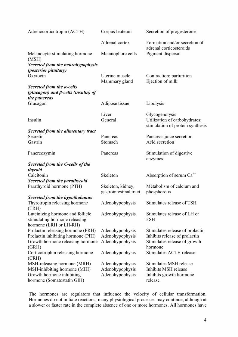
Adrenocorticotropin (ACTH) Corpus leuteum Adrenal cortex
Secretion of progesterone Formation and/or secretion of adrenal corticosteroids
Melanocyte-stimulating hormone (MSH)
Melanophore cells Pigment dispersal
Secreted from the neurohypophysis (posterior pituitary)
Oxytocin Uterine muscle Mammary gland
Contraction; parturition Ejection of milk
Secreted from the α-cells (glucagon) and β-cells (insulin) of the pancreas
Glucagon Adipose tissue Liver
Lipolysis Glycogenolysis
Insulin General Utilization of carbohydrates; stimulation of protein synthesis
Secreted from the alimentary tract Secretin Pancreas Pancreas juice secretion Gastrin Stomach Acid secretion
Pancreozymin Pancreas Stimulation of digestive
enzymes Secreted from the C-cells of the thyroid
Calcitonin Skeleton Absorption of serum Ca++
Secreted from the parathyroid Parathyroid hormone (PTH) Skeleton, kidney,
gastrointestinal tract Metabolism of calcium and phosphorous
Secreted from the hypothalamus Thyrotropin releasing hormone (TRH)
Adenohypophysis Stimulates release of TSH
Luteinizing hormone and follicle stimulating hormone releasing hormone (LRH or LH-RH)
Adenohypophysis Stimulates release of LH or FSH
Prolactin releasing hormone (PRH) Adenohypophysis Stimulates release of prolactin Prolactin inhibiting hormone (PIH) Adenohypophysis Inhibits release of prolactin Growth hormone releasing hormone (GRH)
Adenohypophysis Stimulates release of growth hormone
Corticotrophin releasing hormone (CRH)
Adenohypophysis Stimulates ACTH release
MSH-releasing hormone (MRH) Adenohypophysis Stimulates MSH release MSH-inhibiting hormone (MIH) Adenohypophysis Inhibits MSH release Growth hormone inhibiting hormone (Somatostatin GIH)
Adenohypophysis Inhibits growth hormone release
The hormones are regulators that influence the velocity of cellular transformation. Hormones do not initiate reactions; many physiological processes may continue, although at a slower or faster rate in the complete absence of one or more hormones. All hormones have
4

relatively short physiological half-lives. Therefore, to function as regulators in the maintenance of the normal state, and to achieve their regulation in feedback systems, hormones must be continually synthesized and secreted, must exert their effect rapidly, and in turn be rapidly inactivated.
Synthesis of hormones
A number of hormones are synthesized in their respective endocrine glands as larger molecules known as “prohormones”. The latter undergo proteolysis generally in the gland in which they are synthesized and it may occur in the blood. The prohormone-hormone relationship is now established for a number of hormones namely, the thyroid hormone, insulin, glucagon, adrenocorticotropin and growth hormone. A chemical example of the prohormone is the protein, thyroglobulin synthesized and stored in the thyroid gland. Proteolysis of the thyroglobulin releases the “active” hormone, triiodothyronine and thyroxine with relatively low molecular weight amino acids, which are the active circulatory hormones. Regulation of Secretion
In addition to the hormones present in the secretion, regulatory “factors” secreted by the hypothalamus govern the secretion of hormones by other endocrine glands. The secretory rates of hypothalamic regulatory factors as well as of the hypophyseal (pituitary) hormones are subject to both positive and negative feed back control. Concentration
Hormones generally function at blood levels that are less than 10-8M. The mechanism of action of hormones in addition to the broadly descriptive phenomenon seen in the intact organism has been presently derived from studies in vitro following addition of the hormone to the incubating target tissues or cells. Organization of the mammalian endocrine system
The synthesis/release of several of the hormones is controlled in a hierarchal manner involving three successive stages of hormone-target cell interactions. When the hypothalamus at the base of the brain, receives specific normal messages, it secretes minute quantities of “hormones” called releasing factors, which are passed down nerve fibers to the anterior pituitary gland. There, each of the releasing factors can trigger the release of a specific hormone from the anterior pituitary gland. For example, as shown in Fig. 2 thyrotropin releasing factor (TRF) triggers the release of thyrotropic hormone and corticotropin releasing factor (CRF) causing the release of the adrenocorticotropin (ACTH) hormone. The hypothalamus also secretes hormone like substances called “inhibitory” factors, which can inhibit the release of some of the pituitary hormones. The various hormones released from the anterior pituitary pass via the systemic blood to specific target glands. The target of ACTH is the adrenal cortex and that of thyrotropic hormone is the thyroid gland. These glands as well as other target glands are in turn stimulated to produce their characteristic hormones, which act on a number of final target tissues. Besides secreting various releasing and inhibitory factors acting on the anterior pituitary, the hypothalamus also generates at
5

least two other hormones: i). Oxytocin and ii). Vasopressin or antidiuretic hormone important in the regulation of lactation and of water balance respectively. Neural input (external stimuli) hypothalamus (base of the brain)
Passed down through nerve Oxytocin
fiber Releasing CRF Posterior Pituitary Factor Vasopressin Anterior Pituitary (tropic hormone) Pass through blood
ACTH TSH FSH LH PRL GH Adrenal Thyroid Testis & Ovaries α-cells of pancreas Cortex
Estrogen thyronine & progesterone Glucagon Adrenal testosterone Cortical steroids Muscle, liver accessory sex Mammary Bones Liver & other tissues tissues gland
Primary Target
Secondary Target
Final Target
Fig. 2: Three stages of Hormone Target Cell Interactions
Hormones whose release is under less direct pituitary control include the following polypeptides: 1. Calcitonin formed by certain types of cells in the thyroid and parathyroid gland
and 2. Parathyroid hormone, which regulates calcium and phosphorous metabolism. Also in
this group are: 3. Insulin and Glucagon polypeptides formed by the β-cells and the α-cells respectively in
the islets of Langerhans, specialized endocrine region of the pancreas as well as epinephrine (adrenalin) and nor-epinephrine (nor-adrenalin) formed by the adrenal medulla.
6

Regulation of secretion
The secretion of hormones is regulated by a complex network of the following controls (Fig. 3): 1. External stimuli transmitted by the nervous system, modulate the activity of the
hypothalamus 2. Secretion of epinephrine by the adrenal medulla. 3. The secretion of the tropic hormone by the anterior pituitary is in turn modulated in a
feed back relationship by the characteristic secretions of their target glands. 4. The secretions of some hormones are modulated by the concentration of the specific
metabolites in the blood e.g., the release of insulin from the pancreas is stimulated when the concentration of glucose in the blood rises. Moreover, high blood concentration of thyroxine inhibits the action of thyrotropin releasing factor in the anterior pituitary.
Releasing Factors
Tropic Hormone
End Organ Hormone
+
+
Long Loop
Hypothalamus Short Loop
Fig. 3: Regulation of secretion. General feed back control of endocrine system involving the hypothalamus, anterior pituitary and end organ
General
Hormone Receptors and Intracellular Messenger
Hormones and other signal molecules in biological systems bind with very high affinity to their receptors, displaying KD values in the range 10-12 to 10-6M. The concentrations of hormones are maintained at levels equivalent to or slightly above these Kd values. Once hormonal effects have been induced, the hormone is usually rapidly metabolized. The low concentrations and short life times of secreted hormones hence, typically made identification of hormones and elucidation of their mechanisms of action extremely difficult. Modern biochemical methods, including molecular biological methods for cloning and expression of the genes encoding hormone receptors and radioimmunoassay techniques for detection and quantitation of hormones have stimulated rapid advances in this field in recent years. Two basic principles of “Hormone Action” have emerged from research. The first is that the responsive target cells for any given hormone contain specific hormone receptors
7

specialized proteins capable of binding the hormone molecule with very high “specificity” and “affinity”. Such hormone receptors occur in only very small amounts in the “non-target” cells. In the target cells of the water-soluble hormones such as epinephrine, glucagon and insulin, which do not pass through cell membranes readily, the hormone receptors are located on the cell surface. In target cells of the sex and adrenal cortex hormones, which are lipid soluble steroids, and thus can pass through the cell membranes, the primary receptors are located within the cell in the cytosol. The second principle is that binding of hormone to the specific receptors causes the formation of an intracellular “messenger” molecule, which then “stimulates” or “depresses” some characteristic biochemical activity of the target tissue. For the polar water-soluble hormones epinephrine and glucagon, the intracellular messenger is 3'5' cyclic AMP, often called the second messenger. With lipid soluble steroids, the hormone receptor complex HRC itself becomes the intracellular messenger. The structure, biosynthesis, release and mode of action of these hormones are important because they exert major regulatory effects on central metabolic pathways.
Signal transducing receptors transmit the hormonal message
Very often in life, the message is more important than the messenger and this is certainly true for hormones. The structure and chemical properties of a hormone are only important for specific binding of the hormone to its appropriate receptor. Of much greater interest and importance, however, is the metabolic information carried by the hormonal signal. The information implicit in the hormonal signal is interpreted by the cell and an intricate pattern of cellular response ensues. Steroid hormones may either bind to plasma membrane receptors or exert their effects within target cells, entering the cell and migrating to their sites of action via specific cytoplasmic receptor proteins. The non-steroid hormones which act by binding to outward-facing plasma membrane receptors activate various signal transduction pathways that mobilize various second messengers-cyclic nucleotides, Ca2+ ions and other substances that activate or inhibit enzymes or cascades of enzymes in very specific ways (Fig. 4). Cyclic AMP and the second messenger model
Epinephrine and glucagon activate glycogen breakdown in the liver but the mechanism of this activation was a mystery until Earl Sutherland and his colleagues showed that the glycogen phosphorylase reaction, the initial step in glycogen breakdown was stimulated by epinephrine and glucagon. Activation of the phosphorylase was eventually shown to be done to an ATP-dependent phosphorylation of the enzyme. Sutherland also showed that a phosphatase from the liver cells inactivated phosphorylase, eliminating the activation due to phosphorylation. A significant breakthrough was achieved with Sutherland’s demonstration that hormone activated phosphorylase only in the presence of plasma membrane fragments. He hypothesized that binding of epinephrine or glucagon to a receptor in the membrane activated synthesis or release of a substance that activated phosphorylation of
8

phosphorylase. This crucial substance was eventually shown (in 1956) to be adenosine 3'5' cyclic AMP, denoted by c' AMP.
Fig. 4: Non-steroidal hormones bind exclusively to plasma membrane receptors, which mediate cellular responses to the hormone. Steroid hormones exert their effects either by binding to plasma membrane receptors or by diffusing to the nucleus, where they
modulate transcriptional events
Fig. 5: Earl Sutherland’s model of hormone action
9
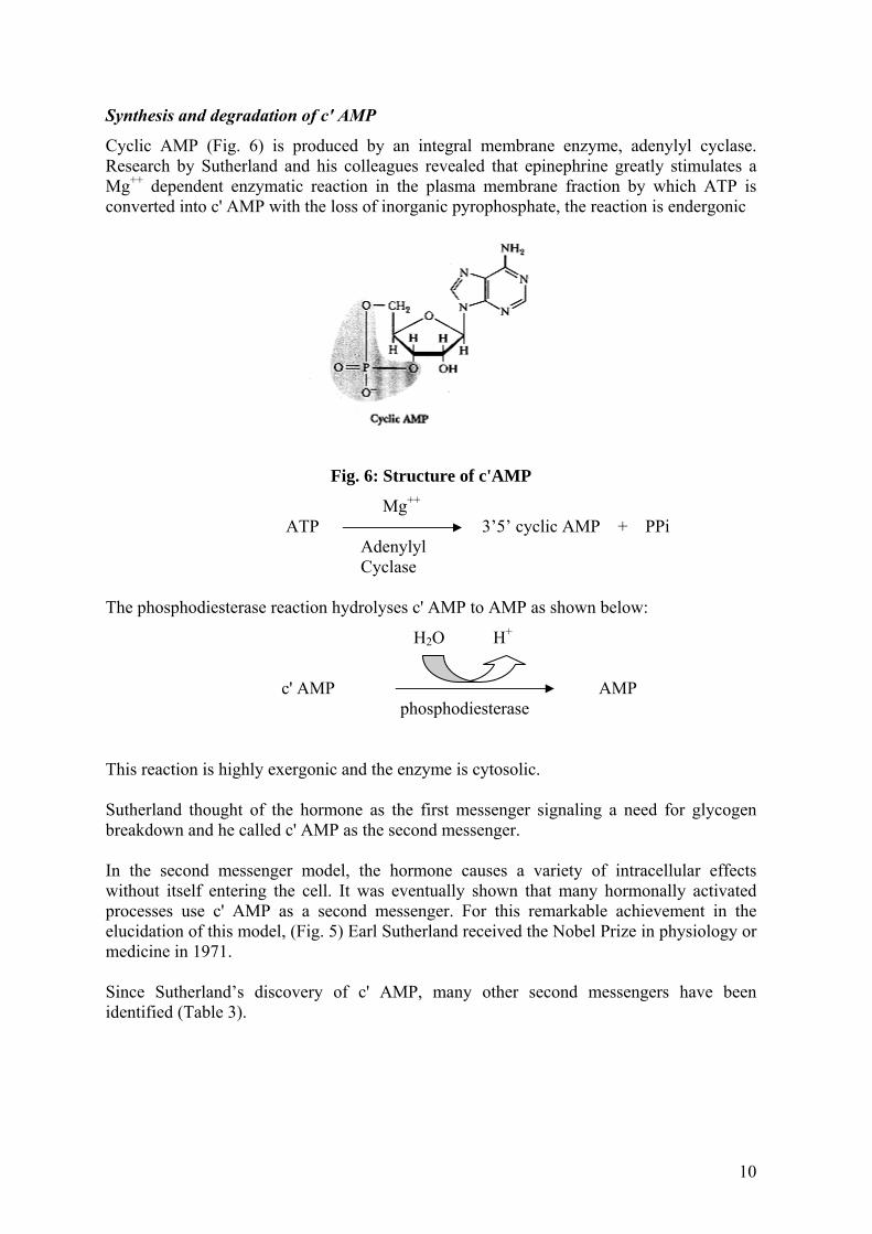
Synthesis and degradation of c' AMP
Cyclic AMP (Fig. 6) is produced by an integral membrane enzyme, adenylyl cyclase. Research by Sutherland and his colleagues revealed that epinephrine greatly stimulates a Mg++ dependent enzymatic reaction in the plasma membrane fraction by which ATP is converted into c' AMP with the loss of inorganic pyrophosphate, the reaction is endergonic
Fi
ATP The phosphodiesterase reaction hy
c' AMP
This reaction is highly exergonic a Sutherland thought of the hormobreakdown and he called c' AMP a In the second messenger modelwithout itself entering the cell. Iprocesses use c' AMP as a secoelucidation of this model, (Fig. 5) medicine in 1971. Since Sutherland’s discovery ofidentified (Table 3).
g. 6: Structure of c'AMP
Mg++
3’5’ cyclic AMP + PPi Adenylyl Cyclase
drolyses c' AMP to AMP as shown below:
H2O H+
AMP phosphodiesterase
nd the enzyme is cytosolic.
ne as the first messenger signaling a need for glycogen s the second messenger.
, the hormone causes a variety of intracellular effects t was eventually shown that many hormonally activated nd messenger. For this remarkable achievement in the Earl Sutherland received the Nobel Prize in physiology or
c' AMP, many other second messengers have been
10

Table 3: Intracellular second messengers*
Messenger Source Effect cAMP Adenylyl cyclase Activates protein kinases cGMP Guanylyl cyclase Activates protein kinases,
Regulates ion channels, Regulates phosphodiesterases
Ca2+ Ion channel in ER and plasma membrane
Activates protein kinases, Activates Ca2+ modulated proteins
IP3 PLC action on PI Activates Ca2+ channels DAG PLC action on PI Activates protein kinase
C Phosphatidic acid Membrane component
andProduct of PLD Activates Ca2+ channels, Inhibits adenylyl cyclase
Ceramide PLC action on sphingomyelin
Activates protein kinases
Nitric oxide (NO) No synthase Activates guanylyl cyclase, Relaxes smooth muscle
Cyclic ADP-ribose cADP-ribose synthse Activates Ca2+ channels *IP3 is inositol-1,4,5-triphospahte; PLC is phospholipase C; PLD is phospholipase D; PI is phosphoinositol; DAG is diacylglycerol
GTP-binding proteins: The hormonal missing link
Another protein was implicated in the hormonal activation of adenylyl cyclase (AC). Firstly, purification of adenylyl cyclase and the hormone receptor resulted in a loss of hormone stimulation of cyclase activity. Secondly, Martin Rodbell and his colleagues showed that GTP was necessary for hormone activation of adenylyl cyclase. He also showed that analogue of GTP was a superactivator of adenylyl cyclase giving higher AC activities than GTP itself, also showing that GTP-binding site was the active site of a GTPase. In 1977, Ross and Gilman reported that partial purification of a GTP-binding protein, which when constituted with the cyclase and hormone-receptor, restored hormone stimulation to the AC reaction. Thus AC is not directly activated by the hormone receptor complex. Instead, binding of hormone to the receptor stimulates a GTP-binding protein. (Abbreviated now as G-Protein), which in turn activates adenylyl-cyclase. Glucagon and Insulin
Glucagon
In addition to epinephrine, a number of other hormones are capable of increasing the concentration of c'AMP in specific target cells. Among these is glucagon also called the “hyperglycemic glycogenolytic” hormone. It is a polypeptide hormone of the pancreas secreted by the α-cells of the islet of Langerhans into the blood, whenever the blood glucose
11

levels drop below the normal value of 80mg/100ml. The glucagon so released causes the liver to breakdown glycogen to restore the blood glucose to its normal value. Glucagon thus counterbalances the action of insulin which is secreted into the blood when blood glucose is high and subsequently causes the glucose to be removed from the blood by the peripheral tissues. Glucagon acts primarily on the liver and does not affect glycogen breakdown in muscle, whereas epinephrine on the other hand promotes glycogen breakdown in both. Like epinephrine, glucagon exerts its action by binding to specific glucagon binding sites, “receptors” on the liver cell membranes resulting in stimulation of adenylate cyclase. The resulting increase in c'AMP causes an increase in the concentration of phosphorylase a in the liver and then breakdown of glycogen. Insulin
The concentration of insulin in the blood and tissues of man is very small, so that standard chemical methods and bioassays procedures are inadequate to carry out studies of its secretion and utilization. Sensitive radioimmunoassays have now been developed for the assay of insulin levels. Synthesis
Insulin is synthesized in the β-cells of islet tissues (Fig. 7). Insulin is first made in the ribosomes in the form of proinsulin, which is translocated via the cristae of the endoplasmic reticulum to the golgi apparatus. The proinsulin is cleaved to form insulin and a c peptide which are packed in golgi vesicles, where the insulin and c peptide crystallize with Zn++ in an ordered array. Ultimately, on secretion of certain signals triggered by an increase in the blood glucose level, the content of these vesicles are released by exocytosis through the plasma membrane into the blood; Ca++ plays an important role in insulin release. The release of insulin from the pancreas depends on the glucose concentration in the blood and certain other factors when the blood glucose increases significantly above its normal levels of 80-100mg/100ml. After a meal, the contents of the secretion vesicles closest to the plasma membrane of the β-cells are ejected into the blood. The insulin concentration then declines to the normal level in an hour or two after a meal. The half-life of insulin in the blood is only about 3 to 4 minutes; the release of insulin from the pancreas is therefore very responsive to fluctuations in blood glucose concentrations. The release of insulin is also stimulated by increased levels of certain amino acids, and by specific factors secreted by the stomach and intestines. Insulin receptors
Earlier studies on insulin receptors showed the following observations: The insulin receptor protein was extracted from fat cells with “non-ionic” detergents and purified by means of affinity chromatography on insulin agarose beads. In this way, the high degree of specificity of the interaction between insulin and the insulin receptor was utilized to retain only the solubilized insulin receptor molecules, thus separating them from most of the proteins in the cell extract. A high degree of purification of the Insulin-receptor has been achieved and its affinity for insulin is very high, the dissociation constant of the insulin-receptor complex is about 10-8 M.
12
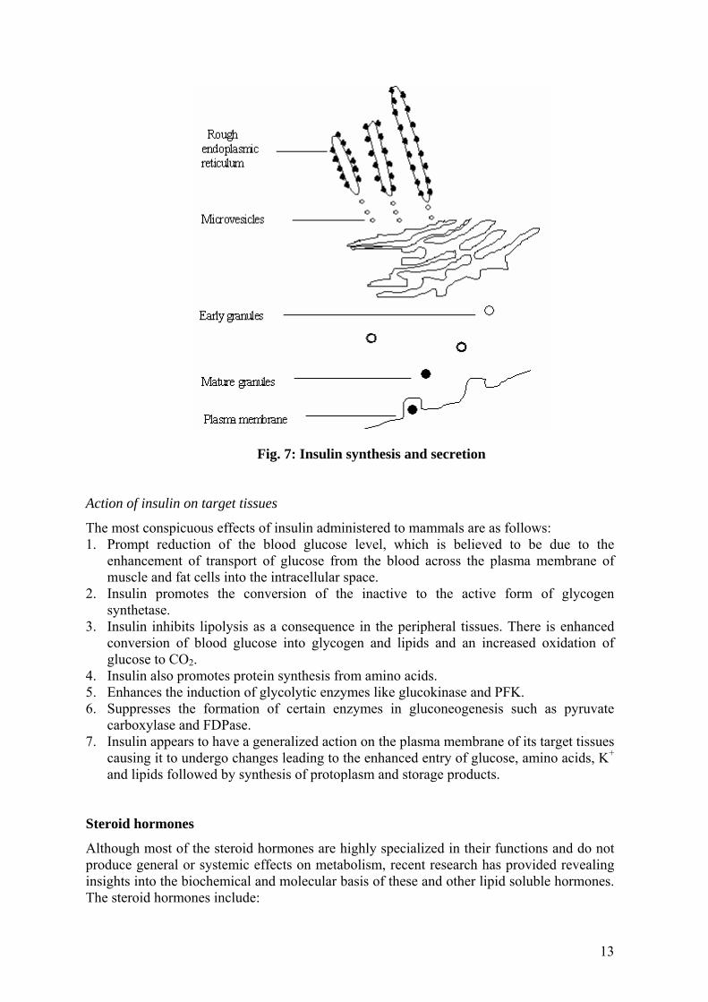
Fig. 7: Insulin synthesis and secretion Action of insulin on target tissues
The most conspicuous effects of insulin administered to mammals are as follows: 1. Prompt reduction of the blood glucose level, which is believed to be due to the
enhancement of transport of glucose from the blood across the plasma membrane of muscle and fat cells into the intracellular space.
2. Insulin promotes the conversion of the inactive to the active form of glycogen synthetase.
3. Insulin inhibits lipolysis as a consequence in the peripheral tissues. There is enhanced conversion of blood glucose into glycogen and lipids and an increased oxidation of glucose to CO2.
4. Insulin also promotes protein synthesis from amino acids. 5. Enhances the induction of glycolytic enzymes like glucokinase and PFK. 6. Suppresses the formation of certain enzymes in gluconeogenesis such as pyruvate
carboxylase and FDPase. 7. Insulin appears to have a generalized action on the plasma membrane of its target tissues
causing it to undergo changes leading to the enhanced entry of glucose, amino acids, K+ and lipids followed by synthesis of protoplasm and storage products.
Steroid hormones
Although most of the steroid hormones are highly specialized in their functions and do not produce general or systemic effects on metabolism, recent research has provided revealing insights into the biochemical and molecular basis of these and other lipid soluble hormones. The steroid hormones include:
13

1. Estrogens or female sex hormones of which the most important are 17-β estradiol and estrone.
2. Androgens or male hormones (testosterone, dihydrotestosterone). 3. The progestational hormone, Progesterone. 4. The steroid hormone of the adrenal cortex (major form cortisol, aldosterone and
corticosterone). Although the sex hormones act largely on the sex accessory organs, some of the steroids secreted by the adrenal cortex have profound effects on the metabolism of carbohydrates and proteins in many tissues. Steroid hormone receptors, earlier experimental evidence, estrogen
The steroid hormones, lipid soluble molecules derived from cholesterol include (Fig. 8) the glucocorticoids (cortisol and corticosterone), the mineralocorticoids (aldosterone) Vitamin D and the sex hormones (for example, progesterone and testosterone). The steroid hormones exert their effects in two ways. First by entering cells and migrating to the nucleus, steroid hormones act as transcription regulators, modulating gene expression. These effects of steroid hormones occur on time scales of hours and involve synthesis of new proteins. Considerable evidence has accumulated, however, that steroids can also act at the cell membrane, directly regulating ligand-gated ion channels and perhaps other processes. These latter processes take place very rapidly, on time scale of seconds and minutes.
Fig. 8: Structure of cholesterol Extracellular effects of the steroid hormones
Several effects of steroid hormones appear to involve action at the plasma membrane. For example, progesterone modules Ca++ channels in membranes of brain stem neurons and also activates processes in Xenopus laevis oocytes by binding to the plasma membrane. Certain steroid actions occur so rapidly that activation of protein synthesis cannot be involved. The male steroid hormone testosterone quickly stimulates the transport of glucose, Ca++ ions and amino acids into kidney cells. Similar rapid induction of Ca++ influx into heart cells by testosterone has also been demonstrated.
14

The steroid hormones, which combine with specific receptors were first emerged from earlier studies of E.V. Jensen on estrogen action. They studied the fate of very small amounts of highly radioactive estradiol injected into immature female animals. The labelled hormone became localized in the uterus and vagina, the “target” of estrogen action. The uptake of labelled estrogen closely paralleled the timing of the earliest detectable hormonal effects of estrogen on the target tissues (Fig. 9).
Fig. 9: A model for steroid hormone action. The hormone (for example, estrogen) dissociated from plasma membrane proteins, diffuses into the cell, and binds to
receptor proteins. The active hormone receptor complex migrates to the nucleus where it interacts with DNA or transcription factors or both. (Adapted from Welshons, W.,
and Jordan, V., 1987) Interaction of estrogen with estrogen receptors. Invitro.
In vitro studies, incubation of minute amounts of labelled estradiol with intact uterine tissue or homogenates of the uterus revealed that the hormone became bound in two cell fractions. The estradiol in the soluble cytosol fraction binds to a specific binding protein sedimenting at 4S. The labelled estrogen found in the “nuclear fraction” has been localized in the chromatin. No estrogen receptors were found in the tissues unresponsive to the hormones. Estrogen receptor protein normally occurs in the cytosol in a form sedimenting at 4S, upon binding estrogen, the 4S receptor undergoes a change into a form sedimenting at 5S. The 5S estrogen receptor complex then finds its way to the cell nucleus, where it binds to the chromatin and brings about different effects like increase in mRNA for protein synthesis.
15

Receptor proteins carry steroids to nucleus
Intracellular effects of the steroid hormones are initiated when the steroid diffuses across the plasma membrane and binds to specific receptor proteins. The binding of the steroids to these receptors is typically very high with dissociation constants in the nanometer range. Nearly all the receptor molecules in a steroid sensitive cell are located in the nucleus. Nonetheless because of the highly hydrophobic character of the steroid themselves and the low likelihood that they could migrate through the cytoplasm to the nucleus without the help of receptor proteins, it is believed that small concentration of receptor proteins are available in cytoplasm to ferry the steroids from the plasma membrane to the nucleus. The steroid receptor complex has two functions in the nucleus. It can bind directly to DNA in order to regulate transcription or it may combine with transcription factors. In these latter interactions, the steroid hormone receptors regulate gene expression without interacting directly with DNA. The receptor proteins for thyroid hormones are highly homologous with those of steroid hormones. The primary structures of nuclear steroid and thyroid receptors
These thyroid receptors, which mediate the effects of thyroxine (T4) and triiodothyronine (T3) possess DNA binding domains with the same Cys-X-X-Cys motif and identically conserved Lys and Arg residues. Inspite of the high degree of homology in their DNA binding domains, these receptor proteins specifically recognize unique DNA sequences on target genes. Thyroid hormones and basal metabolic rate
Thyroxine, tetraiodothyronine and 3,5,3' triiodothyronine, iodinated derivative of tyrosine, characteristic hormones secreted by the thyroid gland have profound physiological effects on Basal Metabolic Rate (BMR) of man and animals. Excessive secretion of thyroid hormones called hyperthyroidism is responsible for “Graves Disease” or “exopthalmic goitre”, while deficiency in thyroid hormones like hypothyroidism is characteristic of the disease “myxedema”. The basal metabolic rate (BMR) is the rate of oxygen consumption measured in a subject lying at complete rest but not asleep. Such measurements are made in the post absorptive state, at least 12 hours after the meal. The BMR is expressed as either in terms of kilocalories of heat energy produced, or volume of oxygen consumed per sq. meter per hour. Since there is a direct relationship between the energy released from the consumption of food and oxygen consumed, Respiratory quotient RQ, is the molar ratio of CO2 produced to oxygen consumed. CO2 produced O2 consumed In chemical practice, it is assumed that the RQ is 0.82 corresponding to combination of a normal fuel mixture.
16
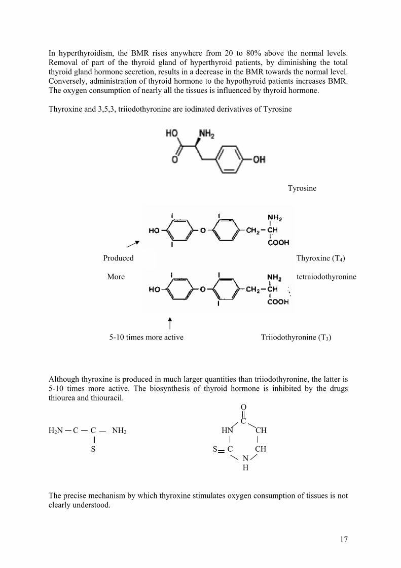
In hyperthyroidism, the BMR rises anywhere from 20 to 80% above the normal levels. Removal of part of the thyroid gland of hyperthyroid patients, by diminishing the total thyroid gland hormone secretion, results in a decrease in the BMR towards the normal level. Conversely, administration of thyroid hormone to the hypothyroid patients increases BMR. The oxygen consumption of nearly all the tissues is influenced by thyroid hormone. Thyroxine and 3,5,3, triiodothyronine are iodinated derivatives of Tyrosine
Tyrosine
Produced mo Thyroxine (T4)
More tetraiodothyronine
5-10 times more active Triiodothyronine (T3)
Although thyroxine is produced in much larger quantities than triiodothyronine, the latter is 5-10 times more active. The biosynthesis of thyroid hormone is inhibited by the drugs thiourea and thiouracil. O
C H2N C C NH2 HN CH S S C CH N H
The precise mechanism by which thyroxine stimulates oxygen consumption of tissues is not clearly understood.
17

Functions of thyroid hormone
1. Thyroxine is used as a metabolic marker; it promotes an increased concentration, synthesis of several enzymes concerned in respiration, particularly glycerol-3-p dehydrogenase of mitochondria, a flavin dehydrogenase functioning in the glycerol-p-shuttle.
2. Thyroxine increases the rate of respiration of liver mitochondria in the absence of ADP and causes mitochondrial swelling.
3. Minute amount of thyroxine induces metamorphosis in the tadpole. Parathyroid hormone (PTH), 1,25 dihydroxycholicalceferol and Calcitonin play an important role in the regulation of phosphate and calcium metabolism, the mode of action of which is not well understood. Parathyroid hormone is secreted when the calcium content falls below normal levels. It stimulates release of Ca++ from bone. It is a polypeptide having 84 amino acid residues, acts on kidney, stimulates adenylate cyclase thereby increasing c' AMP and more phosphate (PO4) is excreted in the urine, thereby, it is lowered in blood. 1,25 dihydroxy cholicalceferol, hormone secreted by kidney acts on small intestines and bones. It promotes absorption of calcium (Ca++) by stimulating the synthesis of a Ca++ binding protein by derepression of certain structural genes. Firstly calcitonin acts on bones and secondly on kidney. It inhibits loss of Ca++ from bone to the blood; its action is opposite to the action of PTH. Calcitonin was discovered, isolated, synthesized and sequenced, yet its role in human physiology is still uncertain. Suggested Reading
1. Biochemistry by Garret and Grisham, Latest edition. 2. Harpers Biochemistry, Latest edition.
18