chest imaging guidelines cigna - evicore care professionals in making medical necessity and other...
TRANSCRIPT

Cigna Medical Coverage Policies – Radiology
Chest Imaging Effective July 14, 2017
_____________________________________________________________________________________________ Instructions for use The following coverage policy applies to health benefit plans administered by Cigna. Coverage policies are intended to provide guidance in interpreting certain standard Cigna benefit plans and are used by medical directors and other health care professionals in making medical necessity and other coverage determinations. Please note the terms of a customer’s particular benefit plan document may differ significantly from the standard benefit plans upon which these coverage policies are based. For example, a customer’s benefit plan document may contain a specific exclusion related to a topic addressed in a coverage policy. In the event of a conflict, a customer’s benefit plan document always supersedes the information in the coverage policy. In the absence of federal or state coverage mandates, benefits are ultimately determined by the terms of the applicable benefit plan document. Coverage determinations in each specific instance require consideration of: 1. The terms of the applicable benefit plan document in effect on the date of service 2. Any applicable laws and regulations 3. Any relevant collateral source materials including coverage policies 4. The specific facts of the particular situation Coverage policies relate exclusively to the administration of health benefit plans. Coverage policies are not recommendations for treatment and should never be used as treatment guidelines. This evidence-based medical coverage policy has been developed by eviCore, Inc. Some information in this coverage policy may not apply to all benefit plans administered by Cigna. These guidelines include procedures eviCore does not review for Cigna. Please refer to the Cigna CPT code list for the current list of high-tech imaging procedures that eviCore reviews for Cigna. CPT® (Current Procedural Terminology) is a registered trademark of the American Medical Association (AMA). CPT® five digit codes, nomenclature and other data are copyright 2016 American Medical Association. All Rights Reserved. No fee schedules, basic units, relative values or related listings are included in the CPT® book. AMA does not directly or indirectly practice medicine or dispense medical services. AMA assumes no liability for the data contained herein or not contained herein.
.
©2017 eviCore, Inc. Chest Imaging Guidelines

CHEST IMAGING GUIDELINES
CHEST IMAGING GUIDELINES CH-1~General Guidelines 4 CH-2~Lymphadenopathy 8 CH-3~Cough 10 CH-4~Non-Cardiac Chest Pain 11 CH-5~Dyspnea/Shortness Of Breath 13 CH-6~Hemoptysis 14 CH-7~Bronchiectasis 15 CH-8~Bronchitis 16 CH-9~Asbestos Exposure 17 CH-10~Chronic Obstructive Pulmonary Disease (COPD) 18 CH-11~Interstitial Disease 19 CH-12~Multiple Pulmonary Nodules 20 CH-13~Pneumonia 21 CH-14~Other Chest Infections 22 CH-15~Sarcoid 23 CH-16~Solitary Pulmonary Nodule (SPN) 24 CH-17~Pleural-Based Nodules And Other Abnormalities 27 CH-18~Pleural Effusion 28 CH-19~Pneumothorax/Hemothorax 29 CH-20~Mediastinal Lymphadenopathy 30 CH-21~Mediastinal Mass 31 CH-22~Chest Trauma 32 CH-23~Chest Wall Mass 33 CH-24~Pectus Excavatum And Pectus Carinatum 34 CH-25~Breast Abnormalities 35 CH-26~Pulmonary Arteriovenous Fistula (AVM) 44 CH-27~Pulmonary Embolism (PE) 45 CH-28~Subclavian Steal Syndrome 49 CH-29~Superior Vena Cava (SVC) Syndrome 50 CH-30~Thoracic Aorta 51 CH-31~Elevated Hemidiaphragm 55 CH-32~Thoracic Outlet Syndrome (TOS) 56 CH-33~Newer Imaging Techniques 57 CH-34~Lung Transplantation 59
V.19.0; Effective July 14, 2017 – Chest Imaging 2 of 59 RETURN
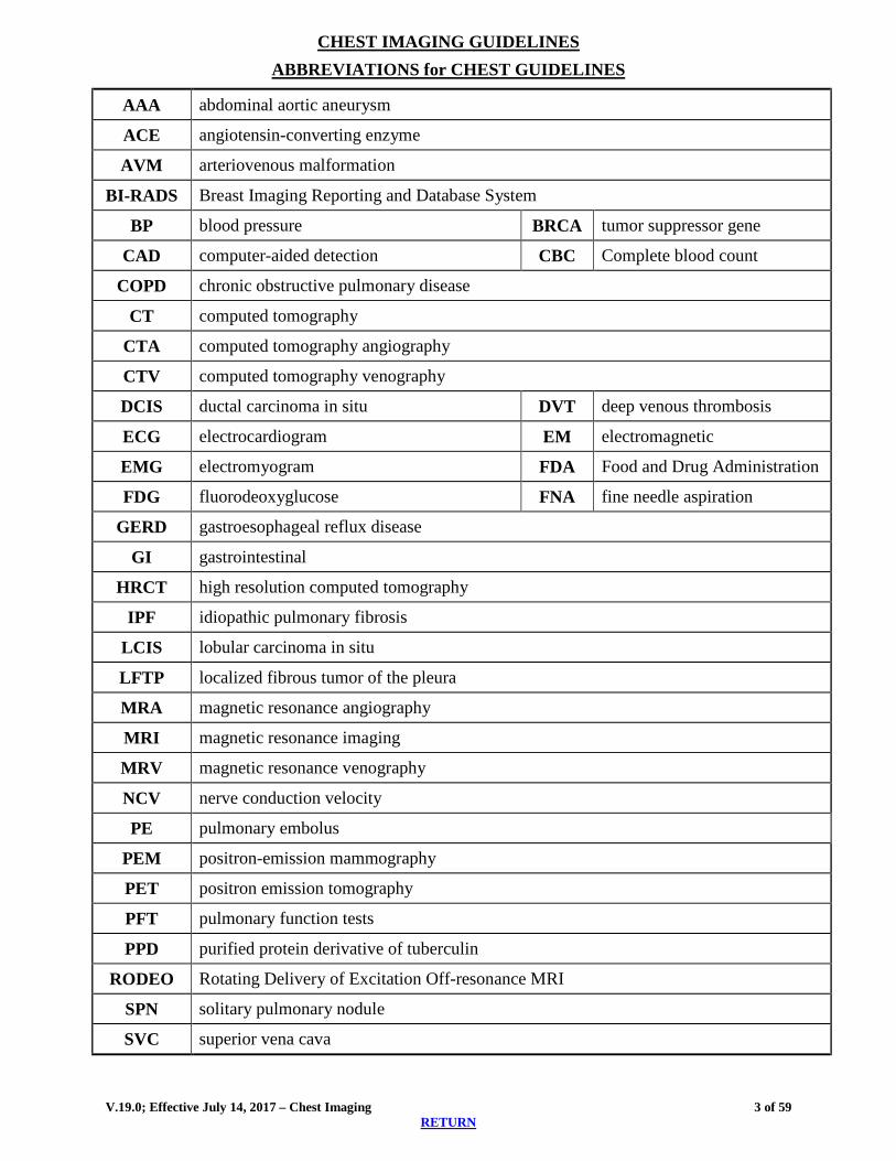
CHEST IMAGING GUIDELINES ABBREVIATIONS for CHEST GUIDELINES
AAA abdominal aortic aneurysm
ACE angiotensin-converting enzyme
AVM arteriovenous malformation
BI-RADS Breast Imaging Reporting and Database System
BP blood pressure BRCA tumor suppressor gene
CAD computer-aided detection CBC Complete blood count
COPD chronic obstructive pulmonary disease
CT computed tomography
CTA computed tomography angiography
CTV computed tomography venography
DCIS ductal carcinoma in situ DVT deep venous thrombosis
ECG electrocardiogram EM electromagnetic
EMG electromyogram FDA Food and Drug Administration
FDG fluorodeoxyglucose FNA fine needle aspiration
GERD gastroesophageal reflux disease
GI gastrointestinal
HRCT high resolution computed tomography
IPF idiopathic pulmonary fibrosis
LCIS lobular carcinoma in situ
LFTP localized fibrous tumor of the pleura
MRA magnetic resonance angiography
MRI magnetic resonance imaging
MRV magnetic resonance venography
NCV nerve conduction velocity
PE pulmonary embolus
PEM positron-emission mammography
PET positron emission tomography
PFT pulmonary function tests
PPD purified protein derivative of tuberculin
RODEO Rotating Delivery of Excitation Off-resonance MRI
SPN solitary pulmonary nodule
SVC superior vena cava
V.19.0; Effective July 14, 2017 – Chest Imaging 3 of 59 RETURN
CHEST IMAGING GUIDELINES
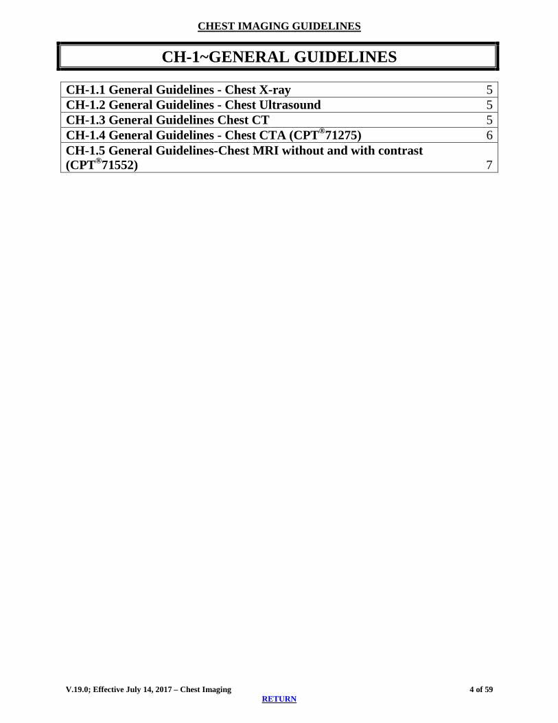
0BCH-1~GENERAL GUIDELINES
CH-1.1 General Guidelines - Chest X-ray 5 CH-1.2 General Guidelines - Chest Ultrasound 5 CH-1.3 General Guidelines Chest CT 5 CH-1.4 General Guidelines - Chest CTA (CPT®71275) 6 CH-1.5 General Guidelines-Chest MRI without and with contrast (CPT®71552) 7
V.19.0; Effective July 14, 2017 – Chest Imaging 4 of 59 RETURN

CHEST IMAGING GUIDELINES A current clinical evaluation (within 60 days), which includes a relevant history and physical examination and appropriate laboratory studies and non-advanced imaging modalities, such as plain x-ray or ultrasound, are required prior to considering advanced imaging. Other meaningful contact (telephone call, electronic mail or messaging) by an established individual can substitute for a face-to-face clinical evaluation. A Pulmonary or Thoracic Surgical Specialist can be helpful in evaluating thoracic disorders.
CH-1.1 General Guidelines - Chest X-ray A recent chest x-ray (generally within the last 60 days) that has been overread by a
radiologist would be performed in many of these cases prior to considering advanced imaging. o Identify and compare with previous chest films to determine presence and stability o Chest x-ray can help identify previously unidentified and may direct proper advanced
imaging for (2): pneumothorax, pneumomediastinum, fractured ribs, acute and chronic infections, and malignancies
o Exceptions to preliminary chest x-ray may include: Supraclavicular lymphadenopathy Known bronchiectasis Suspected interstitial lung disease Positive PPD or tuberculosis Suspected pulmonary AVM
CH-1.2 General Guidelines - Chest Ultrasound Chest ultrasound (CPT®76604) includes transverse, longitudinal, and oblique images of the
chest wall with measurements of chest wall thickness, and also includes imaging of the mediastinum.
Chest Ultrasound Coding Notes Chest ultrasound: CPT®76604
Breast ultrasound o CPT®76641: unilateral, complete o CPT®76642: unilateral, limited o CPT®76641 and CPT®76642 should be reported only once per breast, per imaging
session
Axillary ultrasound: CPT®76882 (unilateral); if bilateral can be reported as CPT®76882 x 2
CH-1.3 General Guidelines –Chest CT– Intrathoracic abnormalities found on chest x-ray, fluoroscopy, abdominal CT scan, or other
imaging modalities may be further evaluated with chest CT with contrast (CPT®71260). o “Abnormalities” through these guidelines may include suspected lung or pleural nodules
or masses, pleural effusion, adenopathy or other findings that are not considered benign.
V.19.0; Effective July 14, 2017 – Chest Imaging 5 of 59 RETURN

CHEST IMAGING GUIDELINES o Lung nodule(s) identified incidentally on Chest CTA without and with contrast
(CPT®71275), Chest MRI without contrast (CPT®71550), Chest MRI without and with contrast (CPT®71552) or Chest MRA without and with contrast (CPT®71555) can replace Chest CT with contrast (CPT®71260) or Chest CT without contrast (CPT®71250) as the initial dedicated study.
o See also: CH-16~Solitary Pulmonary Nodule (SPN) o See also: ONC-8.2 Non-Small Cell Lung Cancer, Suspected/Diagnosis
Chest CT without contrast (CPT®71250) can be used for the following: o Individual has contraindication to contrast o Follow-up of pulmonary nodule(s) o High Resolution CT (HRCT) o Low-dose chest CT (CPT®G0297) may be approved for lung cancer screening if all of the
following criteria are met: Individual has not received a low-dose CT lung screening in less than 12 months; and Individual has NO signs or symptoms suggestive of underlying lung cancer, and is
able and willing to undergo curative lung surgery; and Individual is between 55 and 80 years of age; and Individual has at least a 30 pack-year history of cigarette smoking; and Currently smokes or quit less than 15 years ago
o Other circumstances as specified in the guidelines
Chest CT without and with contrast (CPT®71270) does not add significant diagnostic information above and beyond that provided by chest CT with contrast, unless a question regarding calcification, most often within a lung nodule, needs to be resolved.
Chest CT Coding Notes High resolution chest CT should be reported only with an appropriate code from the set CPT®71250-CPT®71270. No additional CPT® codes should be reported for the “high resolution” portion of the scan. The “high resolution” involves additional slices, which are not separately billable.
CH-1.4 General Guidelines - Chest CTA (CPT®71275) Chest CTA can be considered for suspected Pulmonary Embolism and Thoracic Aortic
disease
CTA prior to minimally invasive or robotic surgery. (See: CD-1.10 in the Cardiac Imaging Guidelines.)
V.19.0; Effective July 14, 2017 – Chest Imaging 6 of 59 RETURN

CHEST IMAGING GUIDELINES
CH-1.5 General Guidelines-Chest MRI without and with contrast (CPT®71552) Indications for chest MRI are infrequent and may include concerns about CT contrast, such
as renal insufficiency, contrast allergy. Another infrequent use may be for clarification of equivocal findings on previous imaging studies, which are often in the thymic mediastinal region or determining margin (vascular/soft tissue) involvement with tumor and determined on a case by case basis.
MRI Chest may be considered : o Chest wall mass (CH-23~Chest Wall Mass) o Chest muscle tendon injuries (MS-11~Muscle/Tendon Injuries) o Brachial plexopathy (PN-4~Brachial Plexus) and Thymoma (ONC-10.2
Thymoma)
References 1. Moyer VA. Screening for Lung Cancer: U.S. Preventive Services Task Force
Recommendation Statement. Annals of Internal Medicine, 2014 Mar 4; 160(5):330-338. 2. Hoffman U, Akers SR, Brown RKJ, et al. ACR Appropriateness Criteria® Acute Nonspecific
Chest Pain–Low Probability of CAD. Date of origin: 1998. Last review date: 2015. https://acsearch.acr.org/docs/69401/Narrative/
V.19.0; Effective July 14, 2017 – Chest Imaging 7 of 59 RETURN
CHEST IMAGING GUIDELINES
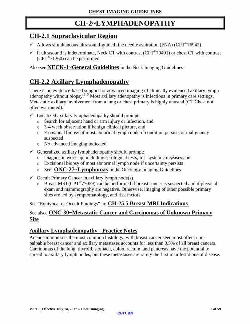
CH-2~LYMPHADENOPATHY
CH-2.1 Supraclavicular Region Allows simultaneous ultrasound-guided fine needle aspiration (FNA) (CPT®76942)
If ultrasound is indeterminate, Neck CT with contrast (CPT®70491) or chest CT with contrast (CPT®71260) can be performed.
Also see NECK-1~General Guidelines in the Neck Imaging Guidelines
CH-2.2 Axillary Lymphadenopathy There is no evidence-based support for advanced imaging of clinically evidenced axillary lymph adenopathy without biopsy.2, 3 Most axillary adenopathy is infectious in primary care settings. Metastatic axillary involvement from a lung or chest primary is highly unusual (CT Chest not often warranted).
Localized axillary lymphadenopathy should prompt: o Search for adjacent hand or arm injury or infection, and o 3-4 week observation if benign clinical picture, and o Excisional biopsy of most abnormal lymph node if condition persists or malignancy
suspected o No advanced imaging indicated
Generalized axillary lymphadenopathy should prompt: o Diagnostic work-up, including serological tests, for systemic diseases and o Excisional biopsy of most abnormal lymph node if uncertainty persists o See: ONC-27~Lymphomas in the Oncology Imaging Guidelines
Occult Primary Cancer in axillary lymph node(s) o Breast MRI (CPT®77059) can be performed if breast cancer is suspected and if physical
exam and mammography are negative. Otherwise, imaging of other possible primary sites are led by symptomatology, and risk factors
See “Equivocal or Occult Findings” in: CH-25.5 Breast MRI Indications. See also: ONC-30~Metastatic Cancer and Carcinomas of Unknown Primary Site
Axillary Lymphadenopathy - Practice Notes Adenocarcinoma is the most common histology, with breast cancer seen most often; non-palpable breast cancer and axillary metastases accounts for less than 0.5% of all breast cancers. Carcinomas of the lung, thyroid, stomach, colon, rectum, and pancreas have the potential to spread to axillary lymph nodes, but these metastases are rarely the first manifestations of disease.
V.19.0; Effective July 14, 2017 – Chest Imaging 8 of 59 RETURN

CHEST IMAGING GUIDELINES
CH-2.3 Mediastinal Lymphadenopathy Chest CT with contrast (CPT®71260) can be performed if mediastinal abnormalities are
detected on a chest x-ray (over read by a radiologist) or other non-dedicated advanced chest imaging.
Follow-up chest CT (CPT®71260) can be performed at 4 weeks4 if o Enlarged lymph nodes are in the mediastinum with no other thoracic abnormalities; and o Low risk or no clinical suspicion for malignancy o Thereafter, stability does not require further advanced imaging
Further evaluations o Lymph node biopsy (see methods below) should be considered for: Persistent lymphadenopathy on follow-up chest CT; or Suspected malignancy
Practice Notes Lymphadenopathy from neoplasms as well as from benign sources of inflammation can result in a positive PET scan. Therefore, the use of PET may not be helpful prior to histologic diagnosis. Less invasive methods of mediastinal biopsies are percutaneous biopsy, transbronchial biopsy, transbronchial biopsy using endobronchial ultrasound, and endoscopic ultrasound-guided FNA.
More invasive and traditional methods are mediastinoscopy or thoracoscopy/thoracotomy. References 1. van Overhagen H, Brakel K, Heijenbrok MW, et al. Metastases in Supraclavicular Lymph
Nodes in Lung Cancer: Assessment with Palpations, US, and CT. Radiology. 2004 Jul;232(1):75-80.
2. Lehman CD, DeMartini W, Anderson BO, et al. Indications for Breast MRI in the Patient with Newly Diagnosed Breast Cancer. J Natl Compr Canc Netw. 2009 Feb;7(2):193-201.
3. Yamaguchi H, Ishikawa M, Hatanaka K, et al. Occult Breast Cancer Presenting as Axillary Metastases. The Breast. 2006 Apr;15(2):259-262.
4. Stigt, JA, Boers JE, Oostdijk AH, et al. Mediastinal Incidentalomas, J Thoracic Oncol. 2011 Aug;6(8):1345-1349.
5. English BS, Ray Jr, CE, Chang JY et al. ACR Appropriateness Criteria® Radiologic Management of Thoracic Nodules and Masses. Date of origin 1995. Last review date 2015. https://acsearch.acr.org/docs/69343/Narrative/
V.19.0; Effective July 14, 2017 – Chest Imaging 9 of 59 RETURN

CHEST IMAGING GUIDELINES
1BCH-3~COUGH
CH-3.1 Cough Initial evaluation should include a recent chest x-ray1
o Discontinue all medications known to cause coughing (e.g. ACE inhibitors) after the current episode of cough started or changed 1
If the initial chest x-ray is without abnormalities, a chest CT (either with contrast [CPT®71260] or without contrast [CPT®71250]) can be performed for the following:
o Cough in non-smoker after the following sequence for a total 3 week trial and investigation: Antihistamine and decongestant treatment 1 Bronchoprovocation challenge (e.g. methacholine challenge, exhaled nitric oxide test)
and spirometry should be performed to rule out asthma 1 Empiric trial of corticosteroids 1 Treatment of gastroesophageal reflux disease (GERD) 1 See: HD-29~Sinusitis
o Current or past cigarette smokers with: New cough lasting greater than 2 weeks (URI based cough can be prolonged)
Changed chronic cough in worsening frequency or character See: CH-6~Hemoptysis
For any abnormalities present on the initial chest x-ray, advanced chest imaging can be performed according to the relevant Chest Imaging Guidelines section.1
Practice Notes
The resolution of cough usually will occur at a median time of 26 days of stopping use of the angiotensin-converting enzyme (ACE) inhibitor drug.1 Smoking cessation is “almost always effective” in resolving cough in smoker. 1
It should be realized that cough after URI (Upper Respiratory Infection) can typically last beyond 2-3 weeks. 2
References 1. Pratter M, Brightling CE, Boulet LP, et. al. An Empiric Integrative Approach to the
Management of Cough: ACCP Evidence-Based Clinical Practice Guidelines. Chest. 2006 Jan; 129(1_suppl):222S-231S.
2. Ebell MH, Lundgren J and Youngpairoj S. How Long Does a Cough Last? Comparing Patients’ Expectations with Data from a Systematic Review of the Literature. Ann Fam Med. 2013 Jan/Feb;11(1):5-13.
V.19.0; Effective July 14, 2017 – Chest Imaging 10 of 59 RETURN

CHEST IMAGING GUIDELINES
2BCH-4~NON-CARDIAC CHEST PAIN
See also the following guidelines: CH-27~Pulmonary Embolism CH-30.1 Aortic Dissection CD-1~General Guidelines CD-8~CT Heart and Coronary Computed Tomography Angiography (CCTA)
“Evidence is not conclusive whether Triple-rule-out CT (CAD, PE, and AD) will improve efficiency of individual management” with acute chest pain.1
MRI is not supported, in the evaluation of chest pain1,2,4
CH-4.1 Non-Cardiac Chest Pain - Imaging Initial evaluation should include a chest x-ray.1, 2
If x-ray is abnormal, chest CT with contrast (CPT®71260) or CTA chest with contrast (CPT®71275) can be performed1, 2, 3, 4
If x-ray is normal, an individual should undergo evaluation of other possible causes of pain prior to advanced imaging (CT chest with contrast or CTA chest with contrast) including: 1, 2,
3, 4 o Cardiac (ECG, echocardiogram, stress test) 1, 2, and o GI (trial of anti-reflux medication, possible upper endoscopy, pH probe, esophageal
manometry),1 either a barium swallow esophageal pH monitoring, manometry, or endoscopy should be done in all after cardiac causes have been ruled out since GERD is the cause in almost 60%,1,2 and
o Pulmonary (PFT’s) 1, 2
Chest CT with contrast (CPT®71260) can be performed if persistent: o The initial chest x-ray reveals no abnormalities; and either Sickle cell disease2 Suspected lung mass in an individual with chest pain, cough, and weight loss.2
CH-4.2 Costochondritis/Other Musculoskeletal Chest Wall Syndrome Costochondritis or other suggested musculoskeletal chest wall syndrome does not require advanced imaging (CT or MRI) unless it meets other criteria in these guidelines. Costochondritis can be readily diagnosed with palpation tenderness and/or hooking maneuver and imaging is non-specific. 3, 4 Practice Notes Chest x-ray could identify pneumothorax, pneumomediastinum, fractured ribs, acute and chronic infections, and malignancies.1
V.19.0; Effective July 14, 2017 – Chest Imaging 11 of 59 RETURN

CHEST IMAGING GUIDELINES References 1. Hoffman U, Venkatesh V, White RD, et al. ACR Appropriateness Criteria® Acute
Nonspecific Chest Pain - Low Probability of Coronary Artery Disease. Date of origin 1998. Last review date 2015.
2. Woodard PK, White RD, Abbara S, et al. ACR Appropriateness Criteria® Chronic Chest Pain - Low to Intermediate Probability of Coronary Artery Disease. Date of origin 1998. Last review date 2012.
3. Proulx AM and Zryd TW. Costochondritis: Diagnosis and Treatment. Am Fam Physician. 2009 Sep 15;80(6):617-620.
V.19.0; Effective July 14, 2017 – Chest Imaging 12 of 59 RETURN

CHEST IMAGING GUIDELINES
3BCH-5~DYSPNEA/SHORTNESS OF BREATH
CH-5.1 Dyspnea/Shortness of Breath Dyspnea is the subjective experience of breathing discomfort. Initial evaluation should include a recent chest x-ray.1, 2 If x-ray is abnormal, chest CT
without contrast (CPT®71250) can be performed 1, 2.
If the initial chest x-ray is indeterminate, Chest CT without contrast (CPT®71250, including HRCT), or Chest CT with contrast (CPT®71260) can be performed if the following evaluations have been conducted and are indeterminate 2: o ECG, echocardiogram or stress testing 2, and o Pulse oximetry and pulmonary function studies (PFT’s) 2, and/or o Blood work including CBC and thyroid function tests 2 if appropriate
If pulmonary embolus (PE) is suspected, see CH-27~Pulmonary Embolism
References 1. Dyer DS, Mohammed TLH, Kirsch J, et al. ACR Appropriateness Criteria® Chronic Dyspnea
- Suspected Pulmonary Origin. Date of origin 1995. Last review date 2012.
V.19.0; Effective July 14, 2017 – Chest Imaging 13 of 59 RETURN

CHEST IMAGING GUIDELINES
4BCH-6~HEMOPTYSIS
CH-6.1 Hemoptysis Chest CT with contrast (CPT®71260) OR without contrast (CPT®71250) OR CTA chest
(CPT®71275) may be performed after: o Abnormal chest x-ray, or o No chest x-ray needed if any of the following: High risk for malignancy with >40 years of age and >40 pack-year smoking history,
or Persistent/recurrent with > 40 years of age or > 30 pack year smoking history, or Massive hemoptysis (>30cc per episode or unable protect airway) 1
Reference
1. Ketai LH, Kirsch J, Kanne JP, et al. ACR Appropriateness Criteria® Hemoptysis. Date of origin 1995. Last review date 2014.
V.19.0; Effective July 14, 2017 – Chest Imaging 14 of 59 RETURN

CHEST IMAGING GUIDELINES
BRONCHIAL TREE
CH-7~BRONCHIECTASIS
CH-7.1 Bronchiectasis - Imaging High resolution chest CT scan (HRCT) without contrast (CPT®71250):
o To confirm suspected diagnosis of bronchiectasis after an initial x-ray 1, 2; or o For known bronchiectasis with worsening symptoms or worsening PFT’s 2. o For hemoptysis with known or suspected bronchiectasis3
References 1. Schneebaum N, Blau H, Soferman R, et al. Use and Yield of Chest Computed Tomography
in the Diagnostic Evaluation of Pediatric Lung Disease. Pediatrics, 2009 Sep 2;124(2):472-479.
2. Rosen MJ. Chronic Cough Due to Bronchiectasis: ACCP Evidence-based Clinical Practice Guidelines. Chest, 2006 Jan;(1 Suppl) 129: 122S-131S.
3. British Thoracic Society Bronchiectasis. Guidelines for Non-CF Bronchiectasis, Thorax, 2010 July; 65(1 Suppl).
4. Expert Panel on Thoracic Imaging. ACR Appropriateness Criteria® Hemoptysis. Reston (VA): American College of Radiology (ACR); 2014.
5. Hansell DM. Bronchiectasis. Radiologic Clinics of North America, 1998 Jan;36(1):107-28.
V.19.0; Effective July 14, 2017 – Chest Imaging 15 of 59 RETURN

CHEST IMAGING GUIDELINES
5BCH-8~BRONCHITIS
CH-8.1 Bronchitis Advanced imaging is not needed for bronchitis1,2
Chest x-ray to determine if any abnormality is present
References 1. Braman, SS. Chronic cough due to Acute Bronchitis: ACCP Evidence-based Clinical
Practice Guidelines, Chest. 2006 Jan;129(1 Suppl): 95S-103S. 2. Michigan Quality Improvement Consortium. Management of Uncomplicated Acute
Bronchitis in Adults. Southfield (MI), Michigan Quality Improvement Consortium. 2012.
V.19.0; Effective July 14, 2017 – Chest Imaging 16 of 59 RETURN

CHEST IMAGING GUIDELINES
LUNG PARENCHYMA (ALPHABETICAL ORDER)
6BCH-9~ASBESTOS EXPOSURE
CH-9.1 Asbestos Exposure Chest x-ray as radiographic screening for asbestos exposure.1,2
o Stable calcified pleural plaques on chest x-ray do not require advanced imaging of the chest2
CT of the chest should not be used to screen populations at risk for asbestos-related diseases.2
High resolution chest CT (HRCT) (CPT®71250) is considered for2: o Any change seen on chest x-ray Send requests for additional follow-up imaging to Medical Director for review
Practice Notes Asbestosis and asbestos-related diseases include: pleural effusion, pleural plaques, lung cancer, and malignant mesothelioma. The risk of developing mesothelioma increases with increasing intensity and duration of exposure.
References 1. OSHA, Occupational Safety and Health Standards, Medical surveillance guidelines for
asbestos, 1910.1001 App H. 2. Daniel E. Banks, et. al., American College of Chest Physicians Consensus Statement on the
Respiratory Health Effects of Asbestos: Results of a Delphi Study, Chest. 2009;135(6):1619-1627. doi:10.1378/chest.08-1345
3. http://www.atsdr.cdc.gov/asbestos/site-kit/docs/clinscrguide_32205_lo.pdf
V.19.0; Effective July 14, 2017 – Chest Imaging 17 of 59 RETURN

CHEST IMAGING GUIDELINES
7BCH-10~Chronic Obstructive Pulmonary Disease (COPD)
CH-10.1 COPD - Imaging Chest X-ray should be performed initially
Chest CT without contrast (CPT®71250) or with contrast (CPT®71260)1,2,3,4 can be performed if emphysema is suspected and either: o Pre-operative study for Lung Volume Reduction Surgery (LVRS)1 o Definitive diagnosis is not yet determined by laboratory studies and chest x-ray and one
on the following is suspected: bronchiectasis, sarcoidosis, emphysema, pneumoconiosis, idiopathic pulmonary fibrosis, Langerhans cell histiocytosis, hypersensitivity pneumonitis, bronchiolitis obliterans, lipoid pneumonia, drug toxicity, and lymphangitic cancer
o Lung cancer screening is discussed in the following guideline: o See “Screening Indications” in ONC-8~Non-Small Cell Lung Cancer
Practice Notes COPD includes asthmatic bronchitis, chronic bronchitis, and emphysema. COPD is airflow reduction (FEV1/FVC ratio < 0.7 or FEV1 ≥ 80% predicted) in the presence of respiratory symptoms, such as dyspnea. Advanced chest imaging is not typically indicated in COPD exacerbation, which is an acute change in baseline dyspnea, cough, and/or sputum beyond normal day-to-day variations.2, 3 References 1. Dyer DS, Mohammed TLH, Kirsch J, et al. ACR Appropriateness Criteria® Chronic Dyspnea
- Suspected Pulmonary Origin. Date of origin 1995. Last review date 2012. 2. Austin JHM. Pulmonary Emphysema: Imaging Assessment of Lung Volume Reduction
Surgery. Radiology. 1999 Jul;212(1):1-3.
V.19.0; Effective July 14, 2017 – Chest Imaging 18 of 59 RETURN

CHEST IMAGING GUIDELINES
8BCH-11~INTERSTITIAL DISEASE
CH-11.1 Interstitial Disease High resolution chest CT (HRCT) without contrast (CPT®71250) is the diagnostic modality
of choice to evaluate for o Interstitial changes identified on other imaging (including chest x-ray) in individuals with
pulmonary symptoms and abnormal pulmonary function studies (PFT’S) (See: CH-5~Dyspnea)
o Initial request to identify interstitial disease with a connective tissue disease diagnosis, including rheumatoid arthritis, scleroderma and the myopathies
o New or worsening pulmonary symptoms or worsening PFTs in any type of interstitial disease, including connective tissue diseases, or
o Once a year in individuals with known idiopathic pulmonary fibrosis (IPF) if showing progression or regression of disease will change individual management4
References 1. Dyer DS, Mohammed TLH, Kirsch J, et al. ACR Appropriateness Criteria® Chronic Dyspnea
- Suspected Pulmonary Origin. Date of origin 1995. Last review date 2012. 2. Misumi S and Lynch DA. Idiopathic Pulmonary Fibrosis/Usual Interstitial Pneumonia:
Imaging Diagnosis, Spectrum of Abnormalities, and Temporal Progression. Proceedings of the American Thoracic Society, 2006;3(4):307-314.
3. Wells AU, Hirani N, et al. Interstitial Lung Disease Guideline: The British Thoracic Society in Collaboration with the Thoracic Society of Australia and New Zealand and the Irish Thoracic Society. Thorax, 2008 Sep;63(Suppl 5):v1-v58.
4. Dempsey OJ, Kerr KM, Remmen H, et al. How to Investigate a Patient with Suspected Interstitial Lung Disease. BMJ. 2010 Jun 9;340(7759):1294-1299.
5. Castelino F and Varga J. Interstitial Lung Disease in Connective Tissue Diseases: Evolving Concepts of Pathogenesis and Management. Arthritis Research & Therapy. 2010 Aug 23, 12:213.
V.19.0; Effective July 14, 2017 – Chest Imaging 19 of 59 RETURN

CHEST IMAGING GUIDELINES
9BCH-12~MULTIPLE PULMONARY NODULES
CH-12.1 Multiple Pulmonary Nodules The largest of multiple pulmonary nodules should be imaged based on guideline:
CH-16~Solitary Pulmonary Nodule (SPN) Suspected infection with multiple pulmonary nodules can be followed with chest CT without
contrast (CPT®71250) or chest CT with contrast (CPT®71260). Follow-up imaging should not exceed 3 studies in 3 months.
Practice Notes More than 6 nodules and clustering of multiple nodules in a single location usually indicate inflammatory lung disease although a dominant nodule with adjacent small satellite nodules can be seen in primary lung cancer. References 1. Libby DM, Smith JP, Altorki NK, et al. Managing the Small Pulmonary Nodule Discovered
by CT. Chest. 2004 Apr;125(4):1522-1529. 2. MacMahon H, Austin JHM, Gamsu G, et al. Guidelines for Management of Small
Pulmonary Nodules Detected on CT scans: A Statement from the Fleischner Society. Radiology. 2005 Nov;237(2):395-400.
3. Mandel J and Stark P. Differential diagnosis and evaluation of multiple pulmonary nodules. UpToDate, 2016 Jan 6; http://www.uptodate.com/contents/differential-diagnosis-and-evaluation-of-multiple-pulmonary-nodules
4. Naidich DP, Bankier AA, MacMahon H, et al. Recommendations for the Management of Subsolid Pulmonary Nodules Detected at CT: A Statement from the Fleischner Society. Radiology 2013 Jan;266(1):304.
V.19.0; Effective July 14, 2017 – Chest Imaging 20 of 59 RETURN

CHEST IMAGING GUIDELINES
10BCH-13~PNEUMONIA
CH-13.1 Pneumonia Chest x-ray would be performed initially in all individuals with suspected pneumonia, prior
to considering advanced imaging.1,2
Chest CT with contrast (CPT®71260) if initial or repeat chest x-ray findings reveal: o Complication of pneumonia (e.g. abscess, effusion, hypoxemia, respiratory distress,
necrotizing pneumonia, pneumothorax)1,2 o Possible lung mass associated with the infiltrate2
References 1. Mandell LA, Wunderink RG, Anzueto A, et al. Infectious Diseases Society of
America/American Thoracic Society Consensus Guidelines on the Management of Community-Acquired Pneumonia in Adults. Clin Infect Dis. 2007 Mar 1;44(Suppl 2):S27-72.
2. Bradley JS, Byington CL, Shah SS, et al. The Management of Community-Acquired Pneumonia in Infants and Children Older Than 3 Months of Age: Clinical Practice Guidelines by the Pediatric Infectious Diseases Society and the Infectious Diseases Society of America. Clin Infect Dis. 2011 Oct;53(7):e25-76.
3. Kirsch J, Ramirez J, Mohammed TH, et al. ACR Appropriateness Criteria® Acute Respiratory Illness in Immunocompetent Patients. J Thorac Imaging, 2011 May;26(2):W42-44.
V.19.0; Effective July 14, 2017 – Chest Imaging 21 of 59 RETURN
CHEST IMAGING GUIDELINES
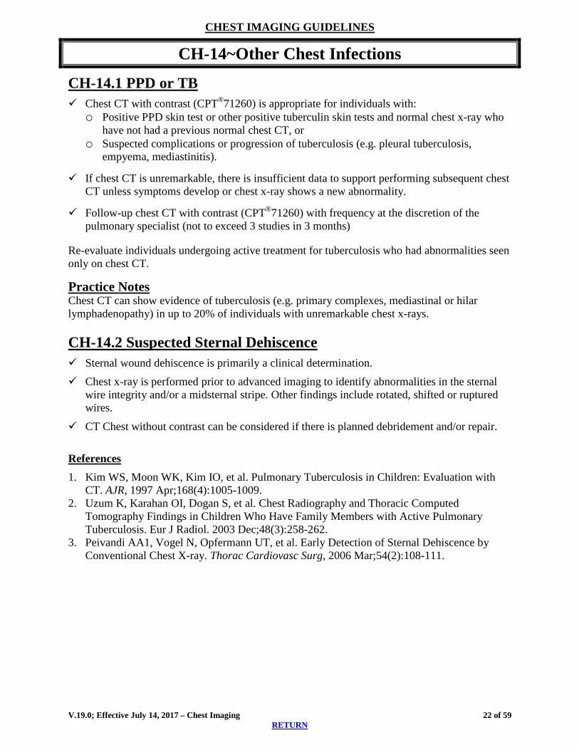
11BCH-14~Other Chest Infections
CH-14.1 PPD or TB Chest CT with contrast (CPT®71260) is appropriate for individuals with:
o Positive PPD skin test or other positive tuberculin skin tests and normal chest x-ray who have not had a previous normal chest CT, or
o Suspected complications or progression of tuberculosis (e.g. pleural tuberculosis, empyema, mediastinitis).
If chest CT is unremarkable, there is insufficient data to support performing subsequent chest CT unless symptoms develop or chest x-ray shows a new abnormality.
Follow-up chest CT with contrast (CPT®71260) with frequency at the discretion of the pulmonary specialist (not to exceed 3 studies in 3 months)
Re-evaluate individuals undergoing active treatment for tuberculosis who had abnormalities seen only on chest CT.
Practice Notes Chest CT can show evidence of tuberculosis (e.g. primary complexes, mediastinal or hilar lymphadenopathy) in up to 20% of individuals with unremarkable chest x-rays. CH-14.2 Suspected Sternal Dehiscence Sternal wound dehiscence is primarily a clinical determination. Chest x-ray is performed prior to advanced imaging to identify abnormalities in the sternal
wire integrity and/or a midsternal stripe. Other findings include rotated, shifted or ruptured wires.
CT Chest without contrast can be considered if there is planned debridement and/or repair. References 1. Kim WS, Moon WK, Kim IO, et al. Pulmonary Tuberculosis in Children: Evaluation with
CT. AJR, 1997 Apr;168(4):1005-1009. 2. Uzum K, Karahan OI, Dogan S, et al. Chest Radiography and Thoracic Computed
Tomography Findings in Children Who Have Family Members with Active Pulmonary Tuberculosis. Eur J Radiol. 2003 Dec;48(3):258-262.
3. Peivandi AA1, Vogel N, Opfermann UT, et al. Early Detection of Sternal Dehiscence by Conventional Chest X-ray. Thorac Cardiovasc Surg, 2006 Mar;54(2):108-111.
V.19.0; Effective July 14, 2017 – Chest Imaging 22 of 59 RETURN

CHEST IMAGING GUIDELINES
12BCH-15~SARCOID
CH-15.1 Sarcoid Chest CT either with contrast (CPT®71260) or without contrast (CPT®71250) is appropriate
for the following:o Establish or rule out the diagnosis when suspected,o Development of worsening symptoms,o New symptoms appear after a period of being asymptomatic, oro Treatment change is being considered in known sarcoid
If CT is equivocal, definitive diagnosis can only be made by biopsy.o Requests for PET to confirm diagnosis of sarcoid should be sent for Medical Director
review.
Gallium scan can be considered as an alternative or supplemental to CT Chest with:o Suspected sarcoido Suspected inflammatory reaction
There is currently no evidence-based data to support performing serial PET scans to monitordisease activity while tapering steroid therapy.
For Cardiac PET (CPT®78459), See CD-7~CARDIAC PET See also: HD-22~Cerebral Vasculitis in the Head Imaging Guidelines
References 1. Hantous-Zannad S, Charrada L, Zidi A, et al. Value of CT Scanning in the Investigation of
Thoracic Sarcoidosis. Rev Mal Respir. 2003 April;20(2 pt 1):207-213. 2. Okumura W, Iwasaki T, Toyama T, et al. Usefulness of Fasting 18F-FDG PET in
Identification of Cardiac Sarcoidosis. J Nucl Med 2004 Dec;45(12):1989-1998.
V.19.0; Effective July 14, 2017 – Chest Imaging 23 of 59 RETURN
CHEST IMAGING GUIDELINES
13BCH-16~Solitary Pulmonary Nodule (SPN)
CH-16.1 SPN - Imaging Chest CT with contrast (CPT®71260) or chest CT without contrast (CPT®71250) (with
contrast is preferred for initial evaluation) can be performed for discrete nodule(s) in the following scenarios: o Lung nodule(s) seen on an imaging study other than a “dedicated” chest CT or MR (chest
x-ray, abdominal CT, spine MRI, coronary artery CTA, etc.). Examples of other studies: chest x-ray, abdominal CT, spine MRI, coronary CTA (See: CH-1.3 General Guidelines - Chest CT)
o Lung nodule(s) identified incidentally on any of the following dedicated chest studies canreplace Chest CT with contrast (CPT®71260) or Chest CT without contrast (CPT®71250)as the initial dedicated study: (See: CH-1.3) Chest CT without and with contrast (CPT® 72170) Chest CTA without and with contrast (CPT®71275) Chest MRI without contrast (CPT®71550) Chest MRI without and with contrast (CPT®71552) Chest MRA without and with contrast (CPT®71555)
o Low Dose CT Chest (CPT®G0297, CPT®S8032) can be used instead of CT Chest(CPT®71250, CPT®71260) for follow-up of lung nodule(s) identified on Lung CancerScreening After preliminary comparison with any available previous chest films to determine if
nodule was present and stable Using largest measurement of multiple lung nodule (See: CH-12~Multiple Pulmonary Nodules)
o Similar-sized pleural nodule is treated as a pulmonary nodule, except does not require PET scan (See: CH-12~Multiple Pulmonary Nodules)
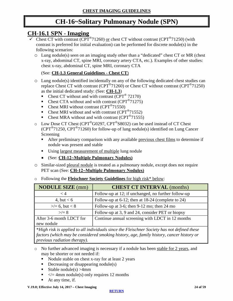
o Following the Fleischner Society Guidelines for high risk* below:
NODULE SIZE (mm) CHEST CT INTERVAL (months) < 4 Follow-up at 12; if unchanged, no further follow-up
4, but < 6 Follow-up at 6-12; then at 18-24 (complete to 24) >/= 6, but < 8 Follow-up at 3-6; then 9-12 mo; then 24 mo
>/= 8 Follow-up at 3, 9 and 24, consider PET or biopsy After 3-6 month LDCT for new nodule
Continue annual screening with LDCT in 12 months
*High risk is applied to all individuals since the Fleischner Society has not defined thesefactors (which may be considered smoking history, age, family history, cancer history or previous radiation therapy).
o No further advanced imaging is necessary if a nodule has been stable for 2 years, andmay be shorter or not needed if: Nodule stable on chest x-ray for at least 2 years Decreasing or disappearing nodule(s) Stable nodule(s) >4mm </= 4mm nodule(s) only requires 12 months At any time, if.
V.19.0; Effective July 14, 2017 – Chest Imaging 24 of 59 RETURN

CHEST IMAGING GUIDELINES classically benign characteristics by chest x-ray or previous CT (e.g. benign
calcification pattern typical for a granuloma or hamartoma) Decreasing or disappearing nodule(s)
Except ground glass or sub-solid densities, which can be imaged beyond 2 years
SPECIAL SITUATIONS Chest CT imaging interval:
CLINICAL FINDINGS CHEST CT INTERVAL (months) Negative PET 3 (after PET), 9, and 24 Previous or current malignancy and pulmonary nodule(s)that would reasonably metastasize to the lungs
3, 6, 12, and 24
Ground Glass or Subsolid Nodules 3, then every 6 months and beyond 2 years
PET (CPT®78812 or CPT®78815) is appropriate for a distinct lung nodule ≥8 mm on chestCT(A) or MR(A).o If there is a history of malignancy, refer to the appropriate Oncology restaging/recurrence
guideline for indications for PET imagingo Not pleural nodule, infiltrate, ground glass opacity, or hilar enlargemento Serial PET studies are not considered appropriate
Practice Notes A nodule is any pulmonary or pleural lesion that is a discrete, spherical opacity 2-30 mm in diameter surrounded by normal lung tissue. A larger nodule is called a mass. Entities that are not nodules, and are considered benign, include non-spherical linear, sheet-like, two-dimensional or scarring opacities.
Malignant nodule features can include spiculation, abnormal calcification, size greater than 7-10 mm, ground glass opacity, interval growth, history of a cancer that tends to metastasize to the lung or mediastinum, and/or smoking history.
o A nodule that grows at a rate consistent with cancer (doubling time 30 to 360 days) maybe sampled for biopsy or resected.
o Less than 1% of <7mm nodules are malignant.o A nodule that does not grow in 6 months has a risk of malignancy at <10%.
Benign features can include benign calcification (80% granuloma, 10% hamartoma), multiple areas of calcification, small size, multiple nodules, negative PET, and stability of size over 2 years.
Ground glass or subsolid opacities, which can harbor indolent adenocarcinoma, may require longer follow-up time than 2 years and may be resected if greater than 2 cm or if are more dominantly nodular (part-solid or solid). According to the Fleischner Society (2013), focal nodular areas of increased lung attenuation mostly identified on CT scan, which have typically been separated as either “pure” or “part-solid” ground glass”, should better be unified as “subsolid nodules.”
Repeat PET is discouraged, since if the original PET is positive, biopsy may be performed. If the original PET is negative but subsequent chest CT shows increase in size of the nodule, biopsy may be performed.
False positive PET can occur with infection or inflammation; false negatives can occur with small size nodule, ground glass lesions and indolent cancers such as bronchoalvealor or carcinoid. V.19.0; Effective July 14, 2017 – Chest Imaging 25 of 59
RETURN

CHEST IMAGING GUIDELINES
References 1. Kanne JP, Jensen LE, Mohammed TLH, et al. ACR Appropriateness Criteria®.
Radiographically Detected Solitary Pulmonary Nodule. Date of Origin 1995. Revised 2012.2. Benjamin MS, Drucker EA, McLoud TC, et al. Small Pulmonary Nodules: Detection at
Chest CT and Outcome. Radiology. 2003 Feb; 226(2):489-493.3. Fletcher JW, Kymes SM, Gould M, et al. A Comparison of the Diagnostic Accuracy of 18F-
FDG PET and CT in the Characterization of Solitary Pulmonary Nodules. J Nucl Med. 2008Feb;49(2):179-185.
4. Henschke CI, McCauley DI, Yankelevitz DF, et al. Early Lung Cancer Action Project:Overall Design and Findings from Baseline Screening. Lancet. 1999 Jul 10;354(9173):99-105.
5. Henschke CI, Yankelevitz DF, Mirtcheva R, et al. CT Screening for Lung Cancer: Frequencyand Significance of Part-solid and Nonsolid Nodules. AJR. 2002 May;178(5):1053-1057.
6. Henschke CI, Yankelevitz DF, Naidich DP, et al. CT Screening for Lung Cancer:Suspiciousness of Nodules According to Size on Baseline Scans. Radiology. 2004 Apr;231(1):164-168.
7. Libby DM, Smith JP, Altorki NK, et al. Managing the Small Pulmonary Nodule Discoveredby CT. Chest. 2004 Apr;125(4):1522-1529.
8. Lindell RM, Hartman TE, Swensen SJ, et al. Lung Cancer Screening Experience: ARetrospective Review of PET in 22 Non-small Cell Lung Carcinomas Detected on ScreeningChest CT in a High-risk Population. AJR. 2005 Jul;185(1):126-131.
9. MacMahon H, Austin JHM, Gamsu G, et al. Guidelines for Management of SmallPulmonary Nodules Detected on CT Scans: a Statement from the Fleischner Society.Radiology. 2005 Nov;237(2):395-400.
10. McCarville MB, Lederman HM, Santana VM. Distinguishing Benign From MalignantPulmonary Nodules with Helical Chest CT in Children with Malignant Solid Tumors.Radiology. 2006 May;239(2):514-520.
11. Gould MK, Fletcher J, et al. Evaluation of Patients With Pulmonary Nodules: When Is ItLung Cancer? ACCP Evidence-Based Clinical Practice Guidelines (2nd Edition). Chest.2007 Sep;132(3_Suppl):108S-130S.
12. Naidich DP, Bankier AA, Schaefer-Prokop CM, et al. Recommendations for theManagement of Subsolid Pulmonary Nodules Detected at CT: A Statement from theFleischner Society. Radiology. 2013;266(1):304-317.
13. Swensen SJ, Jett JR, Hartman TE, et al. CT Screening for Lung Cancer: Five-YearProspective Experience. Radiology. 2005 Apr;235(1):259-265.
14. Winer-Muram HT. The Solitary Pulmonary Nodule. Radiology. 2006 Apr;239(1):34-49.
V.19.0; Effective July 14, 2017 – Chest Imaging 26 of 59 RETURN

CHEST IMAGING GUIDELINES
CH-17~Pleural-Based Nodules and Other Abnormalities
CH-17.1 Pleural-Based Nodules and Other Abnormalities Chest CT with contrast (CPT®71260) or chest CT without contrast (CPT®71250) (with
contrast is preferred for initial evaluation) can be performed for pleural nodule(s) 1, 2:o Pleural nodule(s) seen on an imaging study other than a “dedicated” chest CT or MR
(See: CH-1.3) 1, 2
o Pleural nodule(s) identified incidentally on any of the following dedicated chest studiescan replace Chest CT as the initial dedicated study 1, 2
(See: CH-1.3) o After preliminary comparison with any available previous chest films to determine
presence and stability o Using largest measurement of multiple nodule(s) 1
(See: CH-12~Multiple Pulmonary Nodules) o Following the Fleischner Society Guidelines for high risk see CH-16.11
PET can be considered if dedicated CT or MRI Chest identifies a pleural nodule/mass ordefined area of pleural thickening that is > 8 mm when there is a likelihood of malignancyincluding current or previous malignancy, pleural effusion, bone erosion, chest pain. 2
Practice Notes Pleural nodule/mass or thickening without suggestion of malignancy would undergo surveillance or biopsy.2
References 1. MacMahon H, Austin JHM, Gamsu G, et al. Guidelines for Management of Small
Pulmonary Nodules Detected on CT Scans: A Statement from the Fleischner Society. Radiology. 2005 Nov;237(2):395-400.
2. Rivera MP, Mehta AC, Wahidi MM. Establishing the Diagnosis of Lung Cancer: Diagnosisand Management of Lung Cancer, 3rd Ed: American College of Chest Physicians Evidence-based Clinical Practice Guidelines. Chest. 2013 May;143(5 Suppl):e142S-65S.
V.19.0; Effective July 14, 2017 – Chest Imaging 27 of 59 RETURN

CHEST IMAGING GUIDELINES DISORDERS INVOLVING THE PLEURAL SPACE
CH-18~Pleural Effusion
CH-18.1 Pleural Effusion Chest CT with contrast (CPT®71260) can be performed after both:
o Chest x-ray including lateral decubitus films; ando Thoracentesis to determine if fluid is exudative and remove as much as possible (fluid
obscures underlying lung parenchyma and mass).
Chest ultrasound (CPT®76604) can be used as an alternative to chest X-ray to evaluate forthe presence of fluid within the pleural spaces.
Practice Notes Bilateral effusions are more often systemic related transudates (CHF, RF, liver insufficiency, etc) and advanced imaging is rarely needed. Large unilateral effusions can be malignant. Analysis of fluid may include cytology, culture, cell count, biochemical studies.
References 1. Light RW, MacGregor MI, Luchsinger PC, et al. Pleural Effusions: The Diagnostic
Separation of Transudates and Exudates. Ann Intern Med. 1972 Oct 1; 77(4):507-513. 2. British Thoracic Society Pleural Disease Guideline Group. BTS Pleural Disease Guideline
2010: Thorax, 2010 Aug; 65(Suppl 2).
V.19.0; Effective July 14, 2017 – Chest Imaging 28 of 59 RETURN

CHEST IMAGING GUIDELINES DISORDERS INVOLVING THE PLEURAL SPACE
CH-19~PNEUMOTHORAX/HEMOTHORAX
CH-19.1 Pneumothorax/Hemothorax Chest X-ray should be performed initially
Chest CT with contrast (CPT®71260) or without contrast (CPT®71250) if:o Diagnosis of a small pneumothorax is in doubt, and the presence of a pneumothorax will
affect individual treatment decisionso Preoperative study for treatment of pneumothorax Pneumothorax associated with hemothorax Suspected complications from hemothorax (e.g. empyema)
Practice Notes Expiration chest x-ray can enhance evaluation of equivocal plain x-ray. There is no data supporting the use of serial chest CT to follow individuals with known pneumothorax or hemothorax who are asymptomatic or have stable symptoms. With the exception of the indications above, advanced imaging of the chest is rarely indicated in the diagnosis or management of pneumothorax. Inspiratory/expiratory chest x-rays are helpful in defining whether a pneumothorax is present.
References 1. Manes N, Hernandez-Rodriguez H, Lopez-Martin S, et al. Pneumothorax--Guidelines of
Action. Chest. 2002 Feb;21(2):669. 2. Mowery NT, Gunter OL, Collier BR, et al. Practice Management Guidelines for
Management of Hemothorax and Occult Pneumothorax. J Trauma. 2011 Feb;70(2): 510-518.
V.19.0; Effective July 14, 2017 – Chest Imaging 29 of 59 RETURN

CHEST IMAGING GUIDELINES MEDIASTINUM
CH-20~Mediastinal Lymphadenopathy
See: CH-2.3 Mediastinal Lymphadenopathy
V.19.0; Effective July 14, 2017 – Chest Imaging 30 of 59 RETURN
CHEST IMAGING GUIDELINES
CH-21~MEDIASTINAL MASS
CH-21.1 Mediastinal Mass Chest CT with contrast (CPT®71260) is the imaging study of choice to evaluate mediastinal
abnormalities on chest x-ray and can be done once initially if there is a concern formediastinal cyst including bronchogenic, thymic, pericardial or esophageal
Subsequent evaluations either with CT Chest or MRI Chest can be performed if:o New signs or symptoms oro Preoperative
For Adenopathy; see CH-2 For Thymus abnormality; see ONC-10.5 For Goiter; see NECK-9
References 1. Kuhlman JE, Bouchardy L, Fishman EK, et al. CT and MR Imaging Evaluation of Chest
Wall Disorders. RadioGraphics. 1994 May;14(3):571-595. 2. Juanpere S, Cañete N, Ortuno P, et al. Diagnostic Approach to the Mediastinal Masses.
Insights Imaging. 2013 Feb;4(1):29-52. 3. Berry MF. Approach to the Adult Patient with a Mediastinal Mass. UpToDate. Topic last
updated: Sep 28, 2016. Literature review current through: May 2017. http://www.uptodate.com/contents/evaluation-of-mediastinal-masses#H16465732.
4. Komanapalli C, Schipper P and Sukumar M. Pericardial Cyst. CTSNet, 2010 Aug 30:http://www.ctsnet.org/article/pericardial-cyst.
V.19.0; Effective July 14, 2017 – Chest Imaging 31 of 59 RETURN
CHEST IMAGING GUIDELINES CHEST WALL AND RIBS (ALPHABETICAL ORDER)
CH-22~CHEST TRAUMA
CH-22.1 Chest Trauma Chest X-ray should be performed initially
Chest CT without contrast (CPT®71250) or with contrast (CPT®71260) is appropriate for thefollowing situations:1
Rib1 or Sternal2 Fracture:o With associated complications identified clinical or by other imaging, including
pneumothorax, hemothorax, pulmonary contusion, atelectasis, flail chest, cardiovascularinjury and/or injuries to solid of hollow abdominal organs1
o Single fractures, multiple fractures, non-acute fractures, or occult rib fractures are NOTan indication for chest CT unless malignancy is suspected in the etiology1
Routine follow-up advanced imaging of rib or sternal fractures is not indicated1
No advanced imaging of the abdomen or pelvis is indicated when there is chest trauma andno physical examination or laboratory evidence of injury
References 1. Henry TS, Kirsch J, Kanne JP, et al. ACR Appropriateness Criteria® Rib Fractures. Date of
origin: 1995. Last review date: 2014. 2. Clancy K, Velopulos C, Bilaniuk JW, et al. Screening for Blunt Injury: An Eastern
Association for the Surgery of Trauma Practice Management Guideline. J Trauma Acute Care Surg. 2012 Nov;73(5 Suppl 4):s301-e306.
V.19.0; Effective July 14, 2017 – Chest Imaging 32 of 59 RETURN

CHEST IMAGING GUIDELINES CHEST WALL AND RIBS (ALPHABETICAL ORDER)
14BCH-23~CHEST WALL MASS
CH-23.1 Chest Wall Mass Chest x-ray or chest ultrasound (CPT® 76604) should be performed initially in all cases of
chest wall mass.
Chest CT with contrast (CPT®71260) or Chest CT without contrast (CPT®71250) or MRIchest without and with contrast (CPT®71552) can be considered when the following are met:o Chest x-ray completed and does not demonstrate any of the following: Obvious lipoma Clearly benign entity No mass identified (radiographically or palpated)
Practice Notes Chest x-rays of chest wall masses can detect calcification, ossification, or bone destruction as well as location and size.3
References 1. Tateishi, U, Gladish GW, Kusumoto M, et al. Chest Wall Tumors: Radiologic Findings and
Pathologic Correlation. Part 2. Malignant Tumors. RadioGraphics. 2003 Nov-Dec;23(6):1491-1508.
2. Nam SJ, Sungjun K, Beom JL et al. Imaging of Primary Chest Wall Tumors with Radiologic-Pathologic Correlation. RadioGraphics. 2011 May-Jun;31(3):749-771.
3. David EA and Marshall MB. Review of Chest Wall Tumors: A Diagnostic, Therapeutic, andReconstructive Challenge. Semin Plast Surg, 2011 Feb;25(1):16–24.
V.19.0; Effective July 14, 2017 – Chest Imaging 33 of 59 RETURN

CHEST IMAGING GUIDELINES CHEST WALL AND RIBS (ALPHABETICAL ORDER)
CH-24~Pectus Excavatum and Pectus Carinatum
CH-24.1 Pectus Excavatum and Carinatum Chest CT without contrast (CPT®71250) or MRI chest without and with contrast
(CPT®71552) and 3-D reconstruction (CPT®76377) if requested can be considered if:o Candidate for surgical correction including to determine Haller Index1,2
o Cardiac or pulmonary dysfunction has been identified1,2
ECG and echocardiography if cardiac symptoms or evidence of abnormalities of cardiacfunction.1
Chest x-ray and PFT’s if increasing shortness of breath.1
See also PACCH-11~Pectus Deformities in the Pediatric Chest Imaging Guidelines
References 1. Mayer OH. Pectus Excavatum: Etiology and Evaluation. UpToDate. Topic last updated: Dec
17, 2015. Literature review current through: May 2017. 2. Marcovici PA, LoSasso BE, Kruk P, et al. MRI for the Evaluation of Pectus Excavatum.
Pediatr Radiol. 2011 Jun; 41(6):757-758. 3. Goretsky MJ, Kelly Jr RE, Croitoru D, et al. Chest Wall Anomalies: Pectus Excavatum and
Pectus Carinatum. Adolesc Med Clin. 2004 Oct; 15(3):455-471. http://citeseerx.ist.psu.edu/viewdoc/download;jsessionid=7DD97FE6F66579625B7B71C87FB27586?doi=10.1.1.599.2646&rep=rep1&type=pdf
V.19.0; Effective July 14, 2017 – Chest Imaging 34 of 59 RETURN

CHEST IMAGING GUIDELINES CHEST WALL AND RIBS (ALPHABETICAL ORDER)
15BCH-25~BREAST ABNORMALITIES
CH-25.1 Breast Ultrasound 37 CH-25.2 Breast MRI 37 CH-25.3 Breast Reconstruction 38 CH-25.4 CAD for Breast MRI 38 CH-25.5 Breast MRI is NOT Indicated 38 CH-25.6 Breast MRI Indications 39 CH-25.7 Nipple Discharge/Galactorrhea 40 CH-25.8 Breast Pain (Mystodynia) 41 CH-25.9 Newer Breast Imaging Techniques 41 CH 25.10 Suspected Breast Cancer in Males 41 CH-25.11 Digital Breast Tomosynthesis 41
V.19.0; Effective July 14, 2017 – Chest Imaging 35 of 59 RETURN

CHEST IMAGING GUIDELINES
BI-RADS™ Categories Chart
Category 0: Incomplete Need additional imaging evaluation or prior mammograms for comparison.
Category 1: Negative There is nothing to comment on. The breasts are symmetrical and no masses, architectural disturbances or suspicious calcifications are present.
Category 2: Benign Finding This is also a negative mammogram, but the interpreter may wish to describe a finding. Involuting, calcified fibroadenomas, multiple secretory calcifications, fat containing lesions such as oil cysts, lipomas, galactoceles, and mixed density hamartomas all have characteristic appearances, and may be labeled with confidence. The interpreter might wish to describe intramammary lymph nodes, implants, etc. while still concluding that there is no mammographic evidence of malignancy.
Category 3: Probably Benign Finding – Short Interval Follow-up Suggested A finding placed in this category should have a very high probability of being benign. It is not expected to change over the follow-up interval, but the radiologist would prefer to establish its stability. Data is becoming available that sheds light on the efficacy of short interval follow-up. At the present time, most approaches are intuitive. These will likely undergo future modification as more data accrue as to the validity of an approach, the interval required, and the type of findings that should be followed.
Category 4: Suspicious Abnormality – Biopsy Should Be Considered There are lesions that do not have the characteristic morphologies of breast cancer but have a definite probability of being malignant. The radiologist has sufficient concern to urge a biopsy. If possible, the relevant possibilities should be cited so that the individual and her physician can make the decision on the ultimate course of action.
Category 5: Highly Suggestive of Malignancy-Appropriate Action Should Be Taken These lesions have a high probability of being cancer and should be biopsied or treated surgically
Category 6: Known Biopsy-Proven Malignancy – Appropriate Action Should Be Taken These lesions have been biopsied and are known to be malignant.
V.19.0; Effective July 14, 2017 – Chest Imaging 36 of 59 RETURN
CHEST IMAGING GUIDELINES
16BCH-25~BREAST ABNORMALITIES
See BI-RADS™ Categories Chart for full description of BI-RADS™ categories.
CH-25.1 Breast Ultrasound Routine performance of breast ultrasound as stand-alone screening or with screening
mammography is inappropriate.o Do NOT use breast ultrasound to screen general population as either a stand-alone study
or a combined study with screening mammography.
Breast ultrasound (CPT 76641: unilateral, complete OR CPT 76642: unilateral, limited) canbe used to further evaluate abnormalities found on mammogram, especially in differentiatingcysts from solid lesions.o Bilateral should be coded CPT 76641 x 2 OR CPT 76642 x 2
Palpable breast masses should be evaluated with mammography and breast ultrasound, in anyorder, regardless of age. Ultrasound can enhance biopsy.
Axilla ultrasound (CPT®76882)o For women with clinically suspicious lymph nodes, preoperative axillary ultrasound with
a FNA or biopsy can help identify individuals who have positive nodes.o Bilateral should be coded CPT®76882 x 2
References 1. Mainiero MB, Bailey L, D’Orsi, C, et al. ACR Appropriateness Criteria® Breast Cancer
Screening. Date of origin: 2012. Last review date: 2016.
CH-25.2 Breast MRI Breast MRI is usually bilateral (CPT®77059) or can be unilateral (CPT®77058) in some after
mastectomy, per physician request.
MRI guided breast biopsy (CPT®19085) includes the imaging component. Additional lesionsshould be billed using CPT®19086.
MRI Breast can be repeated at least 6 months after an MRI directed breast biopsy todocument successful lesion sampling if histology is benign and nonspecific, equivocal oruncertain.
Breast MRI - Practice Notes Although breast MRI has superior sensitivity in identifying new unknown malignancies, it carries a significant false positive risk when compared to mammogram and ultrasound. Incidental lesions are seen on 15% of breast MRI’s and increase with younger age The percentage of incidental lesions that turn out to be malignant varies from 3% to 20% depending on the individual population. Cancer is identified by breast MRI in only 0.7% of those with “inconclusive mammographic lesions.”1
V.19.0; Effective July 14, 2017 – Chest Imaging 37 of 59 RETURN

CHEST IMAGING GUIDELINES
CH-25.3 Breast Reconstruction CTA or MRA of the body part from which the free tissue transfer flap is being taken, can be
performed for breast reconstruction preoperative planning.2,3 o For example, CTA (CPT®74175 and CPT®72191) or MRA (CPT®74185 and
CPT®72198) of the abdomen and pelvis for Deep Inferior Epigastric Perforators (DIEP) flap
There is currently insufficient evidence-based data to support the need for routine advanced imaging for TRAM flaps or other flaps performed on a vascular pedicle.
CH-25.4 CAD for Breast MRI The use of CAD with breast MRI is currently considered investigational,
experimental, and/or unproven. o 3D rendering codes (CPT®76376 or CPT®76377) should not be used in
conjunction with code 0159T. See: Preface-4.1 3D Rendering
CH-25.5 Breast MRI is NOT Indicated Breast MRI should not be used to determine biopsy recommendations for suspicious or
indeterminate lesion(s) that can be readily biopsied, either using imaging guidance or physical exam, such as palpable masses and microcalcifications.
MRI should not be used for routine surveillance in individuals with history of breast cancer, unless there are physical exam, imaging findings, recurrent, or residual disease at the mastectomy site o Annual screening breast MRI study is indicated for high risk individuals as outlined in
CH-25.6 Breast MRI Indications Individual with dense breasts as determined by mammogram
o To date, evidence does not suggest improved outcomes for women whose only risk factor is breast density9 (see heading “Equivocal or Occult Findings” (Radiologist Report) in CH-25.6 Breast MRI Indications
Low risk, probably benign (BI-RADS™ 3) lesions o Repeat the original type study (mammogram, US or MRI) in 6 months, thereafter,
screening or surveillance does not require MRI
Suspicious (BI-RADS™ 4 or 5) lesion on mammogram and/or ultrasound o Bilateral total breast ultrasound (CPT®76641: unilateral, complete), and bilateral axillary
ultrasound (CPT®76882) are recommended for individuals who have BI-RADSTM 4 or 5 abnormalities. If additional suspicious breast lesions or more extensive malignant breast disease is detected by ultrasound, the extent of disease can be mapped with ultrasound-guided biopsies (CPT®76942).
o A lesion categorized as have BI-RADSTM 4 or 5 should be biopsied. o A palpable lesion should be considered for biopsy.
V.19.0; Effective July 14, 2017 – Chest Imaging 38 of 59
RETURN
CHEST IMAGING GUIDELINES
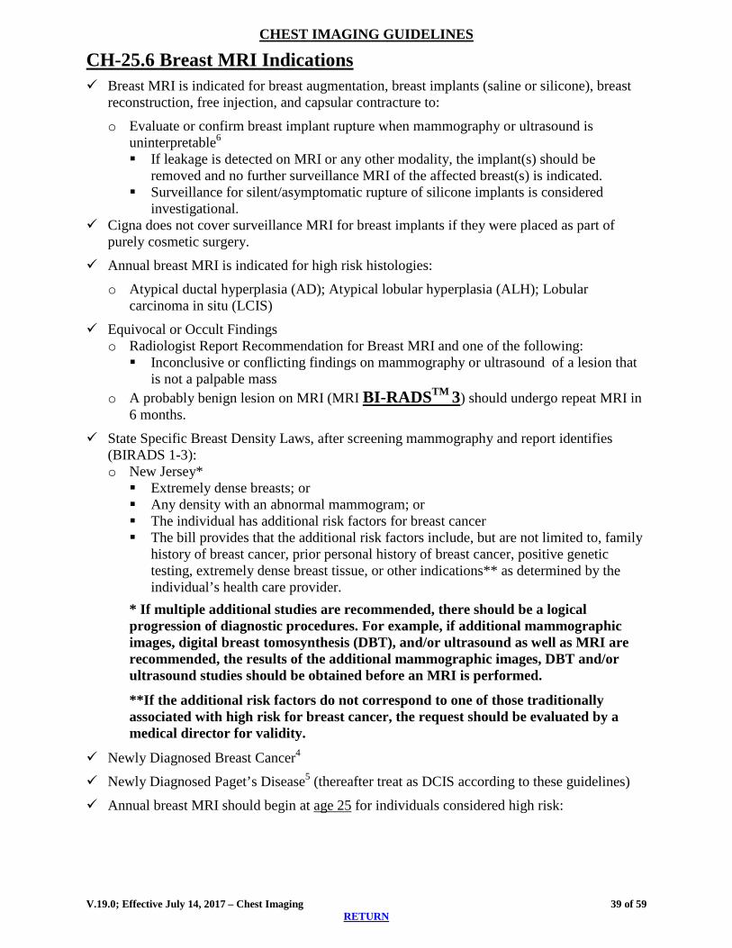
CH-25.6 Breast MRI Indications Breast MRI is indicated for breast augmentation, breast implants (saline or silicone), breast
reconstruction, free injection, and capsular contracture to:
o Evaluate or confirm breast implant rupture when mammography or ultrasound is uninterpretable6 If leakage is detected on MRI or any other modality, the implant(s) should be
removed and no further surveillance MRI of the affected breast(s) is indicated. Surveillance for silent/asymptomatic rupture of silicone implants is considered
investigational. Cigna does not cover surveillance MRI for breast implants if they were placed as part of
purely cosmetic surgery.
Annual breast MRI is indicated for high risk histologies:
o Atypical ductal hyperplasia (AD); Atypical lobular hyperplasia (ALH); Lobular carcinoma in situ (LCIS)
Equivocal or Occult Findings o Radiologist Report Recommendation for Breast MRI and one of the following: Inconclusive or conflicting findings on mammography or ultrasound of a lesion that
is not a palpable mass o A probably benign lesion on MRI (MRI BI-RADSTM 3) should undergo repeat MRI in
6 months.
State Specific Breast Density Laws, after screening mammography and report identifies (BIRADS 1-3): o New Jersey* Extremely dense breasts; or Any density with an abnormal mammogram; or The individual has additional risk factors for breast cancer The bill provides that the additional risk factors include, but are not limited to, family
history of breast cancer, prior personal history of breast cancer, positive genetic testing, extremely dense breast tissue, or other indications** as determined by the individual’s health care provider.
* If multiple additional studies are recommended, there should be a logical progression of diagnostic procedures. For example, if additional mammographic images, digital breast tomosynthesis (DBT), and/or ultrasound as well as MRI are recommended, the results of the additional mammographic images, DBT and/or ultrasound studies should be obtained before an MRI is performed.
**If the additional risk factors do not correspond to one of those traditionally associated with high risk for breast cancer, the request should be evaluated by a medical director for validity.
Newly Diagnosed Breast Cancer4
Newly Diagnosed Paget’s Disease5 (thereafter treat as DCIS according to these guidelines)
Annual breast MRI should begin at age 25 for individuals considered high risk:
V.19.0; Effective July 14, 2017 – Chest Imaging 39 of 59 RETURN

CHEST IMAGING GUIDELINES
High Risk Indications For 1 through 3, begin MRI screening at age 25 1. BRCA 1 or BRCA 2 mutation
2. Presence of Cowden, Bannayan-Riley-Ruvalcaba7
Genetic factors also associated with > 20% risk of breast cancer include ATM, CDH, CHEK2, PALB2, PTEN, STK11
For 3 through 9 above, MRI screening begins at age 40, or 10 years before the age of relative when he/she was first diagnosed with breast cancer, whichever is earlier 3. First degree relative (parent, sibling, child) with BRCA 1 or BRCA 2, even if an individual has not
been tested for BRCA mutation 4. Two or more first degree relatives with breast or ovarian cancer 5. One first degree relative with breast cancer or ovarian cancer that was diagnosed < age 50 6. One first degree relative with bilateral breast cancer, or both breast and ovarian cancer 7. A first or second degree male relative (father, brother, uncle) diagnosed with breast cancer 8. Clinical lifetime risk estimated at greater than or equal to 20% using clinical risk estimator such as
the Gail, Claus, Tyrer-Cuzick or BRCAPRO models 9. Ashkenazi Jewish women from families with onset of breast cancer before age 40
Additional Risks:
10. Women with history of radiation to the chest between ages 10 and 30; breast screening should start 8 to 10 years post-therapy, or at age 25, whichever comes first.
11 Li-Fraumeni Syndrome (TP53 mutation) should start annual breast screening MRI starting at age 20, or at the age of the earliest diagnosed breast cancer in the family, if below age 20 years of age
Residual or Recurrent Malignancy o Assessment of residual tumor in individuals who have undergone lumpectomy and have
close or positive margins, when the findings may indicate a significant change in surgical management.
o Evaluate clinical suspicion of recurrence, following evaluations with mammography and/or ultrasound, if those evaluations are inconclusive or conflict with physical examination or other clinical indicators. This applies to intact breasts, reconstructed breasts, and possible chest wall recurrences following mastectomy.
Breast MRI Indications - Practice Notes MRI should not be used in lieu of mammographically, clinically, and/or sonographically suspicious findings (ACR Practice Guidelines). CH-25.7 Nipple Discharge/Galactorrhea Mammogram should be obtained and ultrasound (CPT®76641: unilateral, complete or
CPT®76642: unilateral, limited) as initial imaging: o If mammogram and ultrasound are negative, a ductal excision is indicated. A ductogram
may be useful to exclude multiple lesions and to localize lesions before surgery. o Ductal excision is indicated even if the ductogram is negative. o An MRI may be considered if a ductogram is technically limited o For a Birads 4 or 5 based on mammogram and/or ultrasound, biopsy is indicated.
V.19.0; Effective July 14, 2017 – Chest Imaging 40 of 59 RETURN

CHEST IMAGING GUIDELINES Practice Notes - Nipple Discharge/Galactorrhea For milky discharge, prolactin and TSH levels are recommended to diagnose prolactinoma; pituitary imaging is not needed if normal serum Prolactin CH-25.8 Breast Pain (Mystodynia) Mammogram and ultrasound are the initial imaging for breast pain
Advanced imaging is NOT routinely indicated in individuals with breast pain and negative evaluation (evaluation includes individual history and physical exam, pregnancy test, mammogram and ultrasound (CPT®76641: unilateral, complete or CPT®76642: unilateral, limited). o If evaluation is not negative, see CH-25.5 Breast MRI Indications
Breast Pain – Practice Notes The risk of malignancy following a negative examination has been estimated to be only 0.5%.9
CH-25.9 Newer Breast Imaging Techniques Positron-Emission Mammography (PEM) or Naviscan® (See: CH-33.3) Scintimammography
o Nuclear medicine study that uses a radioisotope such as Tc-99 tetrofosmin to image the breast. Breast cancer typically shows increased uptake of the radioisotope compared to benign lesions.
o There is insufficient data currently to generate appropriateness criteria for the use of scintimammography
o Scintimammography is not currently an eviCore contracted service
CH-25.10 Suspected Breast Cancer in Males For men < 25 years of age with an indeterminate palpable mass, ultrasound is recommended
as initial imaging followed by mammography if ultrasound is inconclusive or suspicious For men > 25 years of age with an indeterminate palpable mass or with a concerning physical
examination, mammography is recommended initially followed by ultrasound if mammography is inconclusive or suspicious
There is limited evidence on the use of MRI in the evaluation of male breast disease.
CH-25.11 Digital Breast Tomosynthesis Cigna considers digital breast tomosynthesis (DBT), also called 3D mammography, a medically appropriate imaging option in the screening of breast cancer.
V.19.0; Effective July 14, 2017 – Chest Imaging 41 of 59 RETURN
CHEST IMAGING GUIDELINES Coding Notes: CPT®77061: Digital breast tomosynthesis; unilateral
CPT®77062: Digital breast tomosynthesis; bilateral
CPT®+77063: Screening digital breast tomosynthesis (used in conjunction only with screening bilateral mammography code CPT®77057)
3D rendering (CPT®76376 or CPT®76377) should not be assigned with any 3-D mammography code.
References 1. Rosen DJ, McCord K. State of New Jersey. Office of Legislative Services. Fiscal Note.
Senate, No. 792. December 23, 2013. http://www.njleg.state.nj.us/2012/Bills/S1000/792_F5.HTM
2. Sedgwick EL, Ebuoma L, Hamame A, et al. BI-RADS Update for Breast Cancer Caregivers. Breast Cancer Res Treat. 2015 April;150(2):243-254.
3. van Gelder L, Bisschops RHC, Menke-Pluymers MB, et al. Magnetic Resonance Imaging in Patients with Unilateral Bloody Nipple Discharge; Useful When Conventional Diagnostics are Negative? World J Surg. 2015 Jan;39(1):184-186.
4. Emaus MJ, Bakker MF, Peeters PHM, et al. MR Imaging as an Additional Screening Modality for the Detection of Breast Cancer in Women Aged 50-75 Years with Extremely Dense Breasts: The DENSE Trial Study Design. Radiology. 2015 Nov;277(2): 527-537.
5. Mainiero MB, Lourenco AP, Barke LD, et al. ACR Appropriateness Criteria® Evaluation of the Symptomatic Male Breast. Date of origin: 2014.
6. Moy L, Elias K, Pate V, et al. Is Breast MRI Helpful in the Evaluation of Inconclusive Mammographic Findings? Am J Roentgenol. 2009 Oct;193(4):986-993.
7. Pinel-Giroux FM, El Khoury MM, Trop I, et al. Breast Reconstruction: Review of Surgical Methods and Spectrum of Imaging Findings. RadioGraphics. 2013 Mar-Apr;33(2): 435-453.
8. Lehman CD, Gatsonis C, Kuhl CK, et al. MRI evaluation of the Contralateral Breast in Women with Recently Diagnosed Breast Cancer. N Engl J Med. 2007 March 29; 356(13):1295-1303.
9. Lim HS, Jeong SJ, Lee JS et al. Paget Disease of the Breast: Mammographic, US, and MR Imaging Findings with Pathologic Correlation. Radiographics. 2011 Nov-Dec;31(7):1973-1987.
10. Saslow D, Boetes C, Burke W, et al. American Cancer Society Guidelines for Breast Screening with MRI as an Adjunct to Mammography. CA Cancer J Clin. 2007 Mar-Apr;57(2):75-89.
11. US National Library of Medicine. TP53 gene: Tumor Protein p53. Genetics Home Reference. Reviewed October 2015. Published June 13, 2017. http://ghr.nlm.nih.gov/gene/TP53
12. Morrogh M, Morris EA, Liberman L, et al,. The Predictive Value of Ductography and Magnetic Resonance Imaging in the Management of Nipple Discharge. Ann Surg Oncol. 2007 Dec;14(2):3369-3377.
13. Institute for Clinical Systems Improvement (ICSI). Diagnosis of Breast Disease. Fourteenth Edition, 2012; https://www.icsi.org/guidelines__more/catalog_guidelines_and_more/catalog_guidelines/catalog_womens_health_guidelines/breast_disease/
14. Committee on Gynecologic Practice. ACOG Committee Opinion No. 593: Management of Women with Dense Breasts Diagnosed by Mammography. Obstet Gynecol. 2014 Apr;123(4):910-911.
V.19.0; Effective July 14, 2017 – Chest Imaging 42 of 59 RETURN

CHEST IMAGING GUIDELINES 15. Siu AL. Screening for Breast Cancer: US Preventive Services Task Force Recommendation
Statement. Ann of Int Med. 2016 Feb 16;164(4):279-296. 16. Mainiero MB, Bailey L, D’Orsi C, et al. ACR Appropriateness Criteria® Breast Cancer
Screening. Date of origin: 2012. Last review date: 2016. 17. Sprague BL, Stout NK, Schechter C, et al. Benefits, Harms, and Cost-effectiveness of
Supplemental Ultrasonography Screening for Women with Dense Breasts. Ann Intern Med. 2015 Feb 3;162(3):157-166.
V.19.0; Effective July 14, 2017 – Chest Imaging 43 of 59 RETURN

CHEST IMAGING GUIDELINES
THORACIC VASCULAR DISORDERS
17BCH-26~Pulmonary Arteriovenous Fistula (AVM)
CH-26.1 Pulmonary AVM Chest CT with contrast, chest CTA (preferred modality) (CPT®71275), or chest MRA
(CPT®71555) or can be obtained for evaluation of: o Suspected pulmonary AVM o First degree relatives of an individual with a primary pulmonary AVM o Evaluation of individuals with paradoxical embolus/stroke and no evidence of patent
foreman ovale on echocardiogram.
Practice Notes Pulmonary AVMs are abnormal connections between pulmonary arteries and veins, usually found in the lower lobes, that can be either primary or acquired (such as trauma, bronchiectasis). They can be identified in up to 98% of chest x-rays by a peripheral, circumscribed, non-calcified lesion connected by blood vessels to the hilum of the lung. Treatment is often by surgery or embolization of the feeding artery using platinum coils or detachable balloons.
References 1. De Cillis E, Burdi N, Bortone AS, et al. Endovascular Treatment of Pulmonary and Cerebral
Arteriovenous Malformations in Patients Affected by Hereditary Haemorrhagic Teleangiectasia. Curr Pharm Des. 2006;12 (10):1243-1248.
2. Gossage JR and Kanj G. Pulmonary Arteriovenous Malformations: A State of the Art Review. Am J Respir Crit Care Med. 1998 Aug 1;158(2):643-661.
3. Lee EY, Boiselle PM, and Cleveland RH. Multidector CT Evaluation of Congenital Lung Anomalies. Radiology. 2008 Jun;247(3):632-648.
V.19.0; Effective July 14, 2017 – Chest Imaging 44 of 59 RETURN

CHEST IMAGING GUIDELINES THORACIC VASCULAR DISORDERS
18BCH-27~PULMONARY EMBOLISM (PE)
CH-27.1 Pulmonary Embolism Chest CT with contrast with PE protocol (CPT®71260) or chest CTA (CPT®71275) would be
considered with any one of the 3 from each of both sets.
With any one of the 3 1. Dyspnea, new onset and otherwise unexplained; 2. Chest Pain, pleuritic; 3. Tachypnea
AND with any one of the 3: 1. Abnormal D-dimer test; 2. Wells Criteria score* higher than 4 points; 3. One Risk Factor** or Symptom** of new onset demonstrating high clinical probability of
PE RISK FACTORS** SYMPTOMS ATTRIBUTED TO PE** Immobilization at least 3 days or surgery in last 4 weeks or recent trauma Signs or symptoms of DVT
Previous history of DVT or PE Hemoptysis Cancer actively treated in last 6 months or receiving palliative treatment Right heart strain or failure
Recent history of a long airplane flight Systolic BP<90 Use of estrogen-based contraceptives (birth control pills, the patch, and vaginal ring)/Oral estrogen Syncope Advanced age (>/=70) Cough Congestive heart failure Heart Rate >100 Obesity (BMI >/= 35) Palpitations
Well’s Criteria for Clinical Probability of PE* Clinical signs/symptoms of DVT (at minimum: leg swelling and pain with palpation of the deep veins) 3
PE is likely or equally likely diagnosis 3 Heart rate >100 1.5 Immobilization at least 3 days or surgery in last 4 weeks 1.5 Previous history of DVT or PE 1.5 Hemoptysis 1 Cancer actively treated in last 6 months or receiving palliative treatment 1
Calculate Probability: Low <2 Moderate 2 to 6 High >6
Using the above criteria, only 3% of individuals with a low pretest probability had PE versus 63% of those with a high pretest probability.
Non-urgent cases which do not meet above 2-step criteria, should undergo prior to advanced
imaging: o Chest x-ray (to rule out other causes of acute chest pain) o Primary cardiac and pulmonary etiologies should be eliminated.
V.19.0; Effective July 14, 2017 – Chest Imaging 45 of 59 RETURN

CHEST IMAGING GUIDELINES Pregnant women with suspected PE are suggested to proceed with
o D-dimer and/or; o Doppler studies of the lower extremities; o V/Q preferred if Doppler negative; Chest CTA (CPT®71275) or chest MRA
(CPT®71555) can be performed if V/Q scanning is not available.
Follow-up imaging in stable or asymptomatic individuals with known PE is not warranted o Chest CT with contrast with PE protocol (CPT®71260) or chest CTA (CPT®71275) can
be performed for any of the following indications: Recurrent signs or symptoms such as dyspnea, or Elevated d-dimer which is persistent or recurrently elevated, or Right heart strain or failure identified by EKG, ECHO or Heart catheterization
Practice Notes Pulmonary embolism is found in approximately 10% of all those that present with suspicion of PE. Dyspnea, pleuritic chest pain and tachypnea occur with about 50% incidence with leg swelling or pain just over 50%.
D-dimer level has a high sensitivity and low specificity for diagnosing PE. o A negative D-dimer in combination with low or moderate PE risk classification has a
negative predictive value approaching 100%. o D-dimer can be falsely elevated with recent surgery, injury, malignancy, sepsis, diabetes,
pregnancy, or other conditions where fibrin products are likely to be present.
CT imaging has supplanted V/Q scanning since the latter is difficult to obtain quickly, does not provide a substantial cost savings, and does not diagnose other pulmonary pathology.
The decision to terminate anticoagulation treatment after previous pulmonary embolism (PE) with absent or stable symptoms is based on clinical evaluation and risk factors. o Repeat studies do not allow one the ability to distinguish new from residual clot, with
luminal diameter and clot character poorly correlated to symptoms and ECHO findings. o Two thirds after primary thromboembolism have residual pulmonary artery clot at 6 months
and 50% remains at one year. o Subsequent persistence or elevation of D-dimer is associated with increased risk of recurrent
PE. ECHO and Right Heart Catheterization (RHC) can identify those with pulmonary hypertension. Yet, half of all have persistent or new pulmonary hypertension after primary thromboembolism and only half of this latter group has dyspnea at rest or exercise intolerance.
References 1. Abcarian PW, Sweet JD, Watabe JT, et al. Role of a Quantitative D-Dimer Assay in
Determining the Need for CT Angiography of Acute Pulmonary Embolism. Am J Roetgenol. 2004 Jun;182(6):1377-1381.
2. Canonico M, Plu-Bureau G, Lowe GDO, et al. Hormone Replacement Therapy and Risk of Venous Thromboembolism in Postmenopausal Women: Systematic Review and Meta-analysis. BMJ. 2008 May 31;336(7655):1227-1231.
3. Courtney DM, Steinberg JM, and McCormick JC. Prospective Diagnostic Accuracy Assessment of the HemosIL HS D-Dimer to Exclude Pulmonary Embolism in Emergency Department Patients. Thromb Res. 2010 Jan;125(1):79-83.
4. Di Nisio M, Squizzato A, Rutjes AW, et al. Diagnostic Accuracy of D-Dimer Test for Exclusion of Venous Thromboembolism: A Systematic Review. J Thromb Haemost. 2007 Jan 22;5(2):296-304.
V.19.0; Effective July 14, 2017 – Chest Imaging 46 of 59 RETURN

CHEST IMAGING GUIDELINES 5. Fedullo PF, Auger WR, Kerr KM, et al. Chronic Thromboembolic Pulmonary Hypertension.
N Engl J Med. 2001 Nov 15;345(20):1465-1472. 6. Kass SM, Williams PM, and Reamy BV. Pleurisy. Am Fam Physician, 2007 May 1;
75(9):1357-1364. 7. Kavanagh EC, O’Hare A, Hargaden G, et al. Risk of Pulmonary Embolism After Negative
MDCT Pulmonary Angiography Findings. Am J Roetgenol. 2004 Feb;182(2):499-504. 8. Kline JA, Steuerwald MT, Marchick MR, et al. Prospective Evaluation of Right Ventricular
Function and Functional Status 6 Months After Acute Submassive Pulmonary Embolism: Frequency of Persistent or Subsequent Elevation in Estimated Pulmonary Artery Pressure. Chest. 2009 Nov;136(5):1202-1210.
9. Kruip MJ, Slob MJ, Schijen JA, et al. Use of a Clinical Decision Rule in Combination with D-Dimer Concentration in Diagnostic Workup of Patients With Suspected Pulmonary Embolism: A Prospective Management Study. Arch Intern Med. 2002 Jul 22;162(14):1631-1635.
10. Nijkeuter M, Hovens MMC, Davidson BL, et al. Resolution of Thromboemboli in Patients with Acute Pulmonary Embolism: A Systematic Review. Chest. 2006 Jan;129(1):192-197.
11. Palareti G, Cosmi B, Legnani C, et al. D-Dimer Testing to Determine the Duration of Anticoagulation Therapy. N Engl J Med. 2006 Oct 26;355(17):1780-1789.
12. Ramzi DW and Leeper KV. DVT and Pulmonary Embolism: Part II. Treatment and Prevention. Am Fam Physician. 2004 Jun 15;69(12):2841-2848.
13. Stein PD, Terrin ML, Hales C, et al. Clinical, Laboratory, Roentgenographic, and Electrocardiographic Findings in Patients with Acute Pulmonary Embolism and No Pre-existing Cardiac or Pulmonary Disease. Chest. 1991 Sep;100(3):598-603.
14. Stein PD, Woodard PK, Weg JG, et al. Diagnostic Pathways in Acute Pulmonary Embolism: Recommendations of the PIOPED II Investigators. Radiology. 2007 Jan;242(1):15-21.
15. Stein PD, Fowler SE, Goodman LR, et al. Multidetector Computed Tomography for Acute Pulmonary Embolism. N Engl J Med. 2006 Jun 1;354(22):2317-2327.
16. Wells PS, Anderson DR, Rodger M, et al. Derivation of a Simple Clinical Model to Categorize Patients Probability of Pulmonary Embolism: Increasing the Models Utility with the SimpliRED D-Dimer. Thromb Haemost, 2000 Mar;83(3):416-420.
17. Wells PS, Anderson DR, Rodger M, et al. Excluding Pulmonary Embolism at the Bedside Without Diagnostic Imaging: Management of Patients with Suspected Pulmonary Embolism Presenting to the Emergency Department by Using a Simple Clinical Model and D-dimer. Ann Intern Med. 2001 Jul 17:135(2): 98-107.
18. Wolf SJ, McCubbin TR, Feldhaus KM, et al. Prospective Validation of Wells Criteria in the Evaluation of Patients with Suspected Pulmonary Embolism. Ann Emerg Med. 2004 Nov;44(5): 503-510.
19. Writing Group for the Christopher Study Investigators. Effectiveness of Managing Suspected Pulmonary Embolism Using an Algorithm Combining Clinical Probability, D-dimer Testing, and Computed Tomography. JAMA. 2006 Jan 11;295(2):172-179.
20. Fesmire FM, Brown MD, and Espinosa JA, et al. Critical Issues in the Evaluation and Management of Adult Patients Presenting with Suspected Pulmonary Embolism. Annals Emerg Med. 2011 Jun;57(6):628-652.e75.
21. Campbell IA, Fennerty A, and Miller AC. British Thoracic Society Guidelines for the Management of Suspected Acute Pulmonary Embolism. Thorax. 2003;58:470-484.
22. Torbicki A, van Beek EJR, Charbonnier B, et al. Guidelines on Diagnosis and Management of Acute Pulmonary Embolism. Eur Heart J. 2000 Aug 1;21(16):1301-1336.
V.19.0; Effective July 14, 2017 – Chest Imaging 47 of 59 RETURN

CHEST IMAGING GUIDELINES 23. Drescher FS, Chandrika S, Weir ID, et al. Effectiveness and Acceptability of a Computerized
Decision Support System Using Modified Wells Criteria for Evaluation of Suspected Pulmonary Embolism. Ann Emerg Med. 2011 Jun;57(6):613-621.
24. Kirsch J, Brown RKJ, Henry TS, et al. ACR Appropriateness Criteria® Suspected Pulmonary Embolism. Revised 2016. https://acsearch.acr.org/docs/69404/Narrative/
25. Hoffman U, Akers SR, Brown RKJ, et al. ACR Appropriateness Criteria® Acute Nonspecific Chest Pain—Low Probability of Coronary Artery Disease. Date of origin: 1998. Last review date: 2015. https://acsearch.acr.org/docs/69401/Narrative/
26. Dupras D, Bluhm J, Hansen C, et al. Health Care Guideline: Venous Thromboembolism Diagnosis and Treatment. Institute for Clinical Systems Improvement. Thirteenth Ed. January 2013. https://pdfs.semanticscholar.org/3f06/d88a25a6888f86198fab7c3ed9951cac6a94.pdf
V.19.0; Effective July 14, 2017 – Chest Imaging 48 of 59 RETURN

CHEST IMAGING GUIDELINES PULMONARY HYPERTENSION
See PVD-5~Pulmonary Artery Hypertension in the Peripheral Vascular Disease Imaging Guidelines
19BCH-28~SUBCLAVIAN STEAL SYNDROME Occurs from blood flowing up the contralateral vertebral artery to the basilar artery and retrograde down the ipsilateral vertebral artery (reversal of flow) to supply collateral circulation to the arm on the side and past the stenotic or occluded proximal subclavian or innominate artery to perfuse that arm.
CH-28.1 Subclavian Steal Syndrome Initial evaluation should include clinical findings satisfying the symptom complex and initial
imaging with carotid duplex study (CPT®93882) o Satisfying the symptom complex Physical examination findings suggestive of subclavian stenosis include a
discrepancy of > 15 mmHg in blood pressure readings taken in both upper extremities, delayed or decreased amplified pulses in the affected side, and a bruit in the supraclavicular area on the affected side.
Symptoms include vertebral basilar artery insufficiency, vertigo, limb paresis, and paresthesias. Bilateral cortical visual disturbances, ataxia, syncope, and dysarthria occur less frequently.
Symptoms of cerebral ischemia may be produced by exercise of the affected arm. o Carotid duplex study (CPT®93882) is the initial and definitive imaging study Reversal of flow in the ipsilateral vertebral artery. If the carotid duplex is not diagnostic for reversal of flow in the ipsilateral vertebral
artery, then neurological symptoms should be evaluated according to the Head guidelines.
Neck and chest MRA (CPT®70548 and CPT®71555) or CTA (CPT®70498 and CPT®71275) can be performed for diagnosis in individuals with symptoms of vertebrobasilar ischemia if the clinical exam and duplex study are positive, indeterminate, or as preoperative studies if they will substitute for invasive angiography.
Upper extremity MRA (CPT®73225) or CTA (CPT®73206) can be performed in symptomatic individuals if needed to exclude pathology distal to the subclavian artery and if they will substitute for invasive angiography.
See also HD-21.1 Stroke/TIA in the Head Imaging Guidelines.
Treatment options include ligation of the ipsilateral vertebral artery, aorta-subclavian artery bypass graft, or subclavian endarterectomy. References 1. Van Grimberge F, Dymarowski S, Budts W, et al. Role of Magnetic Resonance in the
Diagnosis of Subclavian Steal Syndrome. J Magn Reson Imaging. 2000 Aug;12(2):339-342. 2. Potter BJ and Pinto DS. Subclavian steal syndrome. Circulation. 2014 Jun 3;129(22):2320-
2323.
V.19.0; Effective July 14, 2017 – Chest Imaging 49 of 59 RETURN

CHEST IMAGING GUIDELINES
20BCH-29~Superior Vena Cava (SVC) Syndrome
CH-29.1 SVC Syndrome Chest CT with contrast (CPT®71260) is the initial imaging studies of choice for the
evaluation of suspected SVC syndrome based on the facial cyanosis and UE swelling without anasarca
MRV (CPT®71555) or CTV (CPT®71275) of the chest may be indicated when stenting of the SVC is being considered.
Practice Notes SVC syndrome is caused by acute or subacute, intrinsic or extrinsic obstruction of the SVC, most commonly from lung cancer (80-85%) and less often benign (fibrosis, mediastinitis, indwelling devices). Other symptoms include dyspnea, headache and dizziness.
References 1. Wilson LD, Detterbeck FC, Yahalom J. Superior Vena Cava Syndrome with Malignant
Causes. New Engl J Med. 2007 May 3;356(18):1862-1869. 2. Lepper PM, Ott SR, Hoppe H, Schumann C et al. Superior Vena Cava Syndrome in
Thoracic Malignancies. Respir Care, 2011 May;56(5): 653-666.
V.19.0; Effective July 14, 2017 – Chest Imaging 50 of 59 RETURN

CHEST IMAGING GUIDELINES
21BCH-30~THORACIC AORTA
CH-30.1 Aortic Dissection 52 CH-30.2 Thoracic Aortic Aneurysm (TAA) 52 CH-30.3 Screening Guidelines for Familial Syndromes 53 CH-30.4 Thoracic Aorta in Indiviuals with Bicuspid Aortic Valve 53
V.19.0; Effective July 14, 2017 – Chest Imaging 51 of 59 RETURN
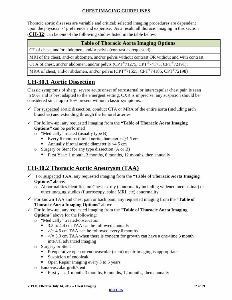
CHEST IMAGING GUIDELINES Thoracic aortic diseases are variable and critical; selected imaging procedures are dependent upon the physicians’ preference and expertise. As a result, all thoracic imaging in this section (CH-32) can be one of the following studies listed in the table below:
Table of Thoracic Aorta Imaging Options CT of chest, and/or abdomen, and/or pelvis (contrast as requested); MRI of the chest, and/or abdomen, and/or pelvis without contrast OR without and with contrast; CTA of chest, and/or abdomen, and/or pelvis (CPT®71275, CPT®74175, CPT®72191); MRA of chest, and/or abdomen, and/or pelvis (CPT®71555, CPT®74185, CPT®72198)
CH-30.1 Aortic Dissection Classic symptoms of sharp, severe acute onset of retrosternal or interscapular chest pain is seen in 96% and is best adapted to the emergent setting. CXR is imprecise; any suspicion should be considered since up to 10% present without classic symptoms.
For suspected aortic dissection, conduct CTA or MRA of the entire aorta (including arch branches) and extending through the femoral arteries
For follow-up, any requested imaging from the “Table of Thoracic Aorta Imaging Options” can be performed o “Medically” treated (usually type B) Every 6 months if total aortic diameter is ≥4.5 cm Annually if total aortic diameter is <4.5 cm
o Surgery or Stent for any type dissection (A or B) First Year: 1 month, 3 months, 6 months, 12 months, then annually
CH-30.2 Thoracic Aortic Aneurysm (TAA) For suspected TAA, any requested imaging from the “Table of Thoracic Aorta Imaging
Options” above: o Abnormalities identified on Chest –x-ray (abnormality including widened mediastinal) or
other imaging studies (fluoroscopy, spine MRI, etc) abnormality
For known TAA and chest pain or back pain, any requested imaging from the “Table of Thoracic Aorta Imaging Options” above
For follow-up, any requested imaging from the “Table of Thoracic Aorta Imaging Options” above for the following: o “Medically” treated/observation 3.5 to 4.4 cm TAA can be followed annually >/= 4.5 cm TAA can be followed every 6 months >/= 3.0 cm TAA when there is concern for growth can have a one-time 3 month
interval advanced imaging o Surgery or Stent Preoperative open or endovascular (stent) repair imaging is appropriate Suspicion of endoleak Open Repair imaging every 3 to 5 years
o Endovascular graft/stent First year: 1 month, 3 months, 6 months, 12 months, then annually
V.19.0; Effective July 14, 2017 – Chest Imaging 52 of 59
RETURN

CHEST IMAGING GUIDELINES Screening with Abdominal Aortic Aneurysm (AAA)
o Known TAA can be screened for AAA using Abdominal Imaging Guidelines (usuallyUS) (see: AB-17.1 Abdominal Aortic Aneurysm)
o Known AAA screening for TAA is not supported by sufficient evidence
For educational information on the normal size of the aortic arch and descending thoracic, see Practice Notes.
CH-30.3 Screening Guidelines for Familial Syndromes Screening for Familial Syndromes and Genetic Syndromes
o Suspected Familial Thoracic Aortic Aneurysm ECHO (CPT®93306, CPT®93307, or CPT®93308) and CXR for all First-degree
relatives (parents, siblings, children) of individuals with TAA and/or dissectiono Any imaging listed can be performed if these studies identify a TAA or are equivocal or
do not visualize the ascending aorta adequatelyo Follow-Up per TAA Follow-Up guidelines
Screening for Marfan Syndrome or Ehlers-Danlos Syndrome, Vascular form or Type IVo Suspected, ECHO (CPT®93306, CPT®93307, or CPT®93308) at the time of diagnosis.o Follow-up: Annual ECHO (CPT®93306, CPT®93307, or CPT®93308) or per TAA Follow-Up
guidelines
For educational information on familial TAA, see Practice Notes.
CH-30.4 Thoracic Aorta in Indiviuals with Bicuspid Aortic Valve Screening for Bicuspid Aortic Valve
o Suspected, any requested imaging from the “Table of Thoracic Aorta ImagingOptions” and/or ECHO (CPT®93306, CPT®93307, or CPT®93308) Additional imaging such as cardiac MRI, cardiac CT, or CCTA is NOT generally
indicated. There is no evidence-based data to support screening relatives of individuals with
bicuspid aortic valve.o Follow-up per TAA Follow-Up guidelines If no dilatation of the aortic root or ascending thoracic aorta is found, there is no
evidence-based data to support continued surveillance imaging.
For more educational information on the Bicuspid Aortic Valve, see Practice Notes
For Coarctation; see CD-6.2, PEDPVD-4.1, PEDAB-14, PEDCD-2.3
Practice Notes Aortic Dissection There are two general types of aortic dissection: 1. Type A: Those that begin in the ascending aorta2. Type B: Those that begin from just distal to the left subclavian artery branch of the aorta
Type A often requires urgent surgical intervention with placement of an aortic graft or endovascular stent graft.
V.19.0; Effective July 14, 2017 – Chest Imaging 53 of 59 RETURN

CHEST IMAGING GUIDELINES Type B can usually be treated medically with careful blood pressure control. Surgery is reserved for distal dissections that are leaking, ruptured, or compromising blood flow to a vital organ, or if there is inability to control the blood pressure. Transesophageal echo may be equally diagnostic compared to CT or MRI. Routine follow-up imaging is important because 30%-40% of chronic dissections will become aneurysmal in 5 years and will require intervention, with less patent false lumina at higher risk.
Penetrating ulcer (through the intima) and intramural hematoma (no intimal tear) are variant forms of aortic dissection and should follow that of aortic dissection, since they are considered precursors of aortic dissection. TAA The normal size of the aortic arch and descending thoracic aorta is 3 cm. The aortic root is normally 3.5 cm:
o TAA occurs most often in the descending (50%) and then equally likely in the ascending or arch aorta.
o Risk factors include atherosclerosis, prolonged hypertension and trauma with mean age 65.
o Risk of rupture is 0% if < 4 cm and 31% if > 6 cm, which is when surgery is often recommended.
Familial TAA Familial TAA presents at an earlier age, has a faster aortic growth rate, is seen in about 20% or non-Marfan TAA and has autosomal dominant inheritance, when compared to non-familial TAA.
Bicuspid Aortic Valve Since 20% of individuals who underwent bicuspid aortic valve surgery had concurrent ascending aortic aneurysms that needed repair. All individuals with bicuspid aortic valve should have both the aortic root and ascending thoracic aorta evaluated for evidence of aortic dilatation.
References 1. Albornoz G, Coady MA, Roberts M, et al. Familial Thoracic Aortic Aneurysms and
Dissections—Incidence, Modes of Inheritance, and Phenotypic Patterns. Ann Thorac Surg. 2006 October;82(4):1400-1405.
2. Elefteriades JA. Natural History of Thoracic Aortic Aneurysms: Indications for Surgery, and Surgical Versus Nonsurgical Risks. Ann Thorac Surg. 2002 Nov;74(5):S1877-S1880.
3. Evangelista A and Eagle KA. Is the Optimal Management of Acute Type A Aortic Intramural Hematoma Evolving? Circulation. 2009 Nov 24;120(21):2029-2032.
4. Ganaha F, Miller DC, Sugimoto K, et al. Prognosis of Aortic Intramural Hematoma With and Without Penetrating Atherosclerotic Ulcer: A Clinical and Radiological Analysis. Circulation. 2002 Jul 16;106(3):342-348.
5. Hiratzka LF, Bakris GL, Beckman JA, et al. 2010 ACCF / AHA / AATS / ACR / ASA / SCA / SCAI / SIR / STS / SVM Guidelines for the Diagnosis and Management of Patients with Thoracic Aortic Disease. Circulation. 2010 Apr 10;121(13):e266-e369.
6. Song JK, Yim JH, Ahn JM, et al. Outcomes of Patients with Acute Type A Aortic Intramural Hematoma. Circulation. 2009 Nov 24;120(21);2046-2052.
7. Tadros TM, Klein MD, Shapira OM. Ascending Aortic Dilatation Associated with Bicuspid Aortic Valve. Circulation. 2009 Feb 17;119(6):880-890.
V.19.0; Effective July 14, 2017 – Chest Imaging 54 of 59 RETURN

CHEST IMAGING GUIDELINES
CH-31~Elevated Hemidiaphragm
CH-31.1 Elevated Hemidiaphragm Chest CT with contrast (CPT®71260) and neck CT with contrast (CPT®70491) (if requested)
with new diaphragmatic paralysis after: o Previous available chest x-rays reviewed to determine if the diaphragm was previously
elevated, and/or o Fluoroscopic examination (“sniff test”) to differentiate true paralysis from weakness
CT abdomen with contrast (CPT®74160) to rule out liver or abdominal process if Chest CT is negative
Repeat advanced imaging studies in the absence of new signs or symptoms are not indicated.
Practice Notes The right hemidiaphragm sits about 2 cm higher than the left. “Eventration” is thin membranous replacement of muscle, usually on the left, as the most
common cause of elevation. Any injury to the phrenic nerve from neck to diaphragm can lead to paralysis. Common phrenic causes are traumatic or surgical injury or malignancy involving the
mediastinum. Any loss of lung volume or increased abdominal pressure can lead to diaphragm elevation.
References 1. Khan AN, Al Ghanem S, Irion KL, et al. Imaging in Diaphragm Injury and Paresis. Medscape.
Updated Oct 19, 2015. http://emedicine.medscape.com/article/355284-overview#a20. 2. Ko MA and Darling GE. 2009. Acquired Paralysis of the Diaphragm. Thorac Surg Clin. 2009
Nov;19(4):501-510. 3. Qureshi A. Diaphragm Paralysis. Semin Respir Crit Care Med. 2009 Jun;30(3):315-320.
V.19.0; Effective July 14, 2017 – Chest Imaging 55 of 59 RETURN

CHEST IMAGING GUIDELINES THORACIC VASCULAR DISORDERS
22BCH-32~Thoracic Outlet Syndrome (TOS)
CH-32.1 Thoracic Outlet Syndrome Chest X-ray after the current episode of symptoms started or changed should be performed
initially in all cases since it can identify bone abnormalities or other causes of right upper extremity pain.1
MR imaging is the preferred imaging modality in individuals with suspected TOS1 o Chest MRI (CPT®71550) or upper extremity other than joint MRI (CPT®73218)1 o Neck and chest MRA (CPT®70548 and CPT®71555)) can be used in place of MRI with
suspected arterial or venous TOS1 o CT Chest with contrast or CT Neck with contrast can be used in place of MRI if:1 Suspected anomalous ribs or fractures, as bone anatomy is more easily definable with
CT Postoperative individuals in whom there is a question of a remnant first rib. Dialysis-dependent renal failure, claustrophobia, or implanted device incompatibility
Also see PN-4~Brachial Plexus in the Peripheral Nerve Disorders Guidelines
Practice Notes TOS refers to compression of the subclavian vessels and/or brachial plexus at the thoracic outlet of the chest (the area bounded by the two scalene muscles and the first rib).
There are 3 types, with neurogenic seen in 80%, venous (also called effort thrombosis) 15% and the remaining 5% arterial.
Since this is such a rare entity and diagnosis is difficult, specialist evaluation by a vascular surgeon or thoracic surgeon is helpful in determining the appropriate imaging pathway.
Reference 1. Raptis CA, Sridhar S, Thompson RW, et al. Imaging of the Patient with Thoracic Outlet
Syndrome. RadioGraphics. 2016 Jul-Aug;36(4):984-1000.
V.19.0; Effective July 14, 2017 – Chest Imaging 56 of 59 RETURN

CHEST IMAGING GUIDELINES
23BCH-33~NEWER IMAGING TECHNIQUES
CH-33.1 Virtual Bronchoscopy There is insufficient data currently to generate appropriateness criteria for the use of virtual
bronchoscopy, and this procedure should be considered investigational at this time.
Virtual bronchoscopy uses multidetector CT with 3D rendering (CPT®71260 and CPT®76377) to generate an image of the tracheobronchial tree down to the level of the sixth- to seventh-generation bronchi, and can visualize areas inaccessible to the flexible bronchoscope.
CH-33.2 EM-Guided Peripheral Bronchoscopy EM Guided Peripheral Bronchoscopy is currently an experimental / investigational
procedure. Current evidence does support its use in performing biopsies of peripheral lesions of the lungs.
Peripheral bronchoscopy using electromagnetic (EM) navigational guidance on a CT road map is a technology for performing biopsies of peripheral lesions of the lungs.
Clinical trials are currently underway to evaluate this technique for mediastinal lymph node biopsies.
Coding Notes Planning is included in the navigational bronchoscopy code (CPT®+31627). Neither separate unlisted codes, (CPT®76499 or CPT®76497), nor other diagnostic CT codes
are reported for the planning phase and pre-procedure imaging acquisition. 3D Rendering, (CPT®76376 and CPT®76377), is not reported in conjunction with
CPT®+31627.
CH-33.3 Positron Emission Mammography There is currently insufficient data to generate appropriateness criteria for this modality, and
this procedure should be considered investigational at this time.
High-resolution positron-emission mammography (PEM) by Naviscan™ PET Systems, also referred to as Naviscan™ or PET mammography, performs high-resolution metabolic imaging of breast cancer using FDG tracer. The PEM detectors are integrated into a conventional mammography system, allowing acquisition of the emission images immediately after the mammogram.
Requesting providers often ask for PEM as CPT®78811 or “PET scan of the breast.”
The spatial resolution of this technique is at the individual duct level (1.5 mm) and allows visualization of intraductal as well as invasive breast cancers. This technique is especially adept at detecting ductal carcinoma in situ.
Early clinical trials have shown high clinical accuracy in characterizing lesions identified as suspicious on conventional imaging or physical examination, as well as detecting incidental breast cancers not seen on other imaging modalities.
A prospective multi-center clinical trial for women with newly diagnosed breast cancer anticipating breast-conservation surgery was performed. These women underwent both high-
V.19.0; Effective July 14, 2017 – Chest Imaging 57 of 59 RETURN

CHEST IMAGING GUIDELINES resolution PEM imaging and breast MRI. Results showed that PEM and MRI had comparable breast-level sensitivity, although MRI had greater lesion-level sensitivity and more accurately depicted the need for mastectomy. PEM had greater specificity at the breast and lesion levels. 3.6% of the women had tumors seen only at PEM.
The radiation exposure from a PEM study is 23 times higher than for digital mammography.
References 1. Tafra L, Cheng Z, Uddo J, et al. Pilot Clinical Trial of 18F-Fluorodeoxyglucose Positron-
Emission Mammography in the Surgical Management of Breast Cancer. Am J Surg. 2005 Oct;190(4):628-632.
2. Murthy K, Aznar M, Thompson CJ et al. Results of Preliminary Clinical Trials of the Positron Emission Mammography System PEM-I: A Dedicated Breast Imaging System Producing Glucose Metabolic Images Using FDG. J Nucl Med. 2000 Nov;41(11):1851-1858.
3. Hendrick RE. Radiation Doses and Cancer Risks from Breast Imaging Studies. Radiology. 2010 Oct;257(1):246-253. http://pubs.rsna.org/doi/full/10.1148/radiol.10100570
V.19.0; Effective July 14, 2017 – Chest Imaging 58 of 59 RETURN

CHEST IMAGING GUIDELINES
24BCH-34~LUNG TRANSPLANTATION
CH-34.1 Pre-Transplant Imaging Studies Individuals on the waiting list or being considered for the lung transplant can undergo
advanced imaging per that institution’s protocol as long as the studies do not exceed the following: o Chest CT with and without contrast (CPT®71270), Chest CT with (CPT®71260), or Chest
CT without contrast (CPT®71250) o ECHO o Imaging Stress Test (MPI, SE, MR) or Heart Catheterization (Right and Left); Heart
catheterization can also be done after a positive stress test
Other studies that will be considered include V/Q scan, Six Minute Walk
Initial post-transplant follow-up: CT chest with and without contrast (CPT®71270), CT chest with (CPT®71260), or CT chest without contrast (CPT®71250) o Requests for subsequent follow-up imaging will go to medical director review.
See: CD-1.6 Transplant Individuals Reference 1. Ng YL, Paul N, Patsios D, et al. Imaging of Lung Transplantation: Review. Am J Roentgenol.
2009 Mar;192(3 suppl):S1-S13.
V.19.0; Effective July 14, 2017 – Chest Imaging 59 of 59 RETURN