-aminobutyric acid-containing sympathetic … acid-containing sympathetic preganglionic neurons in...
TRANSCRIPT

�-Aminobutyric Acid-ContainingSympathetic Preganglionic Neurons inRat Thoracic Spinal Cord Send Their
Axons to the Superior Cervical Ganglion
TETSUFUMI ITO,1* HIROYUKI HIOKI,2 KOUICHI NAKAMURA,2,3
YASUYO TANAKA,2 HIROYUKI NAKADE,1 TAKESHI KANEKO,2,3 SATOSHI IINO,1
AND YOSHIAKI NOJYO1
1Department of Anatomy, Faculty of Medical Sciences, University of Fukui,Fukui 910-1193, Japan
2Department of Morphological Brain Science, Graduate School of Medicine,Kyoto University, Kyoto 606-8501, Japan
3CREST of Japan Science and Technology Agency, Kawaguchi 332-0012, Japan
ABSTRACT�-Aminobutyric acid (GABA)-containing fibers have been observed in the rat superior cer-
vical ganglion (SCG) and, to a lesser extent, in the stellate ganglion (STG). The aim of presentstudy is to clarify the source of these fibers. No cell body showed mRNAs for glutamic aciddecarboxylases (GADs) or immunoreactivity for GAD of 67 kDa (GAD67) in the cervical sympa-thetic chain. Thus, GABA-containing fibers in the ganglia are suggested to be of extraganglionicorigin. GAD67-immunoreactive fibers were found not in the dorsal roots or ganglia, but in theventral roots, so GABA-containing fibers in the sympathetic ganglia were considered to originatefrom the spinal cord. Furthermore, almost all GAD67-immunoreactive fibers in the sympatheticganglia showed immunoreactivity for vesicular acetylcholine transporter, suggesting that GABAwas utilized by some cholinergic preganglionic neurons. This was confirmed by the followingresults. 1) After injection of Sindbis/palGFP virus into the intermediolateral nucleus, someanterogradely labeled fibers in the SCG were immunopositive for GAD67. 2) After injection offluorogold into the SCG, some retrogradely labeled neurons in the thoracic spinal cord werepositive for GAD67 mRNA. 3) When the ventral roots of the eighth cervical to the fourth thoracicsegments were cut, almost all GAD67- and GABA-immunoreactive fibers disappeared from theipsilateral SCG and STG, suggesting that the vast majority of GABA-containing fibers in thoseganglia were of spinal origin. Thus, the present findings strongly indicate that some sympatheticpreganglionic neurons are not only cholinergic but also GABAegic. J. Comp. Neurol. 502:113–125,2007. © 2007 Wiley-Liss, Inc.
Indexing terms: nerve transection; colocalization; anterograde tracing; retrograde tracing;
immunohistochemistry; in situ hybridization
Sympathetic postganglionic neurons receive manykinds of chemical inputs in the sympathetic ganglia.The chemical inputs are classified into two majorgroups: 1) cholinergic excitatory or facilitatory afferentsand 2) inhibitory or suppressive afferents [e.g.,�-aminobutyric acid (GABA), enkephalin, and dopa-mine]. These inputs are derived not only from sympa-thetic preganglionic neurons (SPNs) in the spinal cordbut also from primary sensory neurons in the dorsalroot ganglia (DRGs), enteric neurons in the gastrointes-tinal tract, and small intensely fluorescent (SIF) cellswithin the sympathetic ganglia. It is very important forunderstanding the function of sympathetic ganglia (i.e.,neural computation, integration of inputs, etc.) to inves-
Grant sponsor: Ministry of Education, Science and Culture of Japan;Grant number: 18700339 (to T.I.); Grant number: 14580726 (to Y.N.);Grant number: 16200025 (to T.K.); Grant number: 16500217 (to T.K.);Grant number: 17022020 (to T.K.); Grant number: 17022024 (to T.K.);Grant number: 15-5638 (to T.K.); Grant number: 18700341 (to H.H.).
*Correspondence to: Tetsufumi Ito, Department of Anatomy, Faculty ofMedical Sciences, University of Fukui, Fukui 910-1193, Japan.E-mail: [email protected]
Received 8 July 2007; Revised 4 October 2007; Accepted 26 December2007
DOI 10.1002/cne.21309Published online in Wiley InterScience (www.interscience.wiley.com).
THE JOURNAL OF COMPARATIVE NEUROLOGY 502:113–125 (2007)
© 2007 WILEY-LISS, INC.

tigate both the chemical characteristics and the originof afferent fibers projecting to the ganglia.
The superior cervical ganglion (SCG) is located in themost rostral part of the sympathetic chain and is the onlysympathetic ganglion that projects to various organs inthe head and face (e.g., pineal body, iris, salivary glands,cochlea, lacrimal gland). Because these organs have quiteunique roles in autonomic functions, the neural circuit ofthe ganglia is likely also to be specialized. For understand-ing the function of the SCG, it is very important to identifyspecific features (e.g., chemical properties of preganglionicfibers and postganglionic neurons, physiological proper-ties, morphology of postganglionic neurons) in the gan-glion.
The use of GABA, an inhibitory neurotransmitter in thecentral nervous system, in the sympathetic nervous sys-tem, including the SCG (Amenta et al., 1992; Dobo et al.,1990, 1993; Guillermo et al., 1994; Liu and Burt, 1999;Wolff et al., 1989, 1993a,b), has been reported. GABA-immunopositive fibers are distributed in the rostral sym-pathetic trunk with a rostrally deviated gradient, and thenumber of GABA-immunopositive fibers reaches a maxi-mum in the cervical sympathetic trunk (Wolff et al.,1993a). The GABA-containing fibers make numerous axonbaskets with varicosities in the SCG (Wolff et al., 1989).These observations imply that GABA has an unique rolein SCG-specific neural functions.
Wolff and colleagues argued that, in the cervical sym-pathetic chain, which is composed of the SCG, STG, andcervical sympathetic trunk, SIF-like cells extended theiraxons and projected to the SCG (Dobo et al., 1990; Wolff etal., 1993a). We recently reported that, in the mouse SCG,almost all GABA-containing varicosities were immunopo-sitive for vesicular acetylcholine transporter (VAchT),suggesting a spinal origin of the GABA-containing cholin-ergic fibers (Ito et al., 2005). The DRG neurons may beanother candidate for the origin of GABA-containing fi-bers, because a few axons of the DRG fibers pass throughthe cervical sympathetic trunk (Murata et al., 1982), andGABA-immunopositive DRG neurons have been reported(Szabat et al., 1992). Therefore, there are three candidatesfor the source (SIF-like cells, SPNs, and the DRG neurons)of GABA-containing fibers in the SCG.
To reveal the source of GABA-containing fibers in theSCG, we first investigated the distribution of mRNAs forGADs of 65 and 67 kDa (GAD65 and GAD67). Second, westudied possible sources by double immunohistochemis-try, nerve transection, and anterograde and retrogradetracer labelings.
MATERIALS AND METHODS
Animals
Twenty-two adult male Sprague-Dawley rats (bodyweight 200–250 g, Japan SLC, Shizuoka Japan) wereused in this study. All animals were maintained andtreated according to the guidelines for animal experi-ments of the University of Fukui and Kyoto University.All efforts were made to minimize animal suffering andthe number of animals used.
Surgery
Anterograde tracing. Three rats were deeply anes-thetized with chloral hydrate (400 mg/kg body weight, i.p.)
and fixed in a stereotaxic apparatus in the prone position.A laminectomy of vertebra T1 was carried out to exposethe dorsal surface of the spinal cord. About 0.6 �l of viralsolution containing Sindbis/palGFP virus (Furuta et al.,2001) was injected by pressure (0.7 kg/cm2) of N2 gasthrough a glass micropipette. The injection coordinateswere at 0.5 mm right of the posterior median sulcus and0.6 mm below the dorsal surface of the spinal cord. Over-flowing liquid was carefully absorbed with kimwipe (Cre-cia, Tokyo, Japan). After the surgery, animals were al-lowed to survive for 18 hours. Throughout the survivalperiod, a large amount of palmitoylated GFP was pro-duced in the infected neurons and outlined the completeneuronal morphology (i.e., Golgi-like staining). Withinthis survival period, the death of infected neurons wasminimal (Furuta et al., 2001).
Retrograde tracing. Three rats were deeply anesthe-tized with chloral hydrate (400 mg/kg body weight, i.p.),supinely fixed in a stereotaxic apparatus, and injectedwith 1 �l of 4% (w/v) FG in the right SCG by pressure (0.5kg/cm2) of N2 gas through a glass micropipette. After theinjection, animals were allowed to survive for 2 days.
Nerve transection. Seven rats were deeply anesthe-tized with chloral hydrate (400 mg/kg body weight, i.p.).The inferior articular processes of vertebrae C8–T4 wereremoved to expose the dorsal surface of the right DRGs,and dorsal and ventral roots (DRs and VRs) of C8–T4were cut with a spring scissors. After the surgery, animalswere allowed to survive for 4 days. We dissected theseanimals after the perfusion and confirmed that the tran-section was complete.
Immunohistochemistry
Antibodies. In this study, we used a mouse monoclo-nal antibody for GAD67 (MAB5406; Chemicon, Temecula,CA), a goat polyclonal antibody for VAchT (AB1578;Chemicon), and rabbit polyclonal antibodies for FG(AB153; Chemicon), GFP (Tamamaki et al., 2000) andGABA (A2052; Sigma-Aldrich, St. Louis, MO) were usedas primary antibodies (Table 1).
Although the specificity of the mouse monoclonal anti-body for GAD67 had already been checked by immuno-blotting (Fong et al., 2005), an absorption test had notbeen conducted. In the present study, we carried out anabsorption test with the glutathione-S-transferase (GST)-GAD67 fusion protein as an antigen. First, the entirecoding sequence of the rat GAD67 cDNA (GenBank acces-sion No. M76177) was subcloned into the SmaI site ofpGEX-4T2 (GE Healthcare Bioscience, Fairfield, CT).Then, expression of GST-GAD67 was induced in Esche-richia coli by adding isopropyl-l-thio-�-D-galactopyranosideto the medium. The fusion protein was extracted from themedium with CelLytic B (Sigma) and purified on a GST-column (GE Healthcare Bio-Sciences, Piscataway, NJ). Inimmunoblotting, the fusion protein was recognized by theanti-GAD67 antibody, giving a single band of about 100kDa. This molecular weight was in good agreement withthe estimated molecular weight of GST-GAD67 (approxi-mately 97 kDa).
We preincubated the anti-GAD67 antibody (1:3,000)with this fusion protein (180 �g/ml) diluted in 0.3% (w/v)Triton X-100, 1% (v/v) normal donkey serum in 0.05 Mphosphate-buffered saline (PBS-XD; pH 7.4) for 1 hour atroom temperature (RT). The sections immunostained withthe anti-GAD67 antibody preabsorbed with GST-GAD67
The Journal of Comparative Neurology. DOI 10.1002/cne
114 T. ITO ET AL.

showed no immunopositive signal in the SCG (Fig. 1e2),STG (Fig. 1f2), or brain (Fig. 1h2). Furthermore, GAD67immunoreactivity in the brain (Fig. 1h1) was very similarto that in a previous study (Mugnaini and Oertel, 1985),and the distribution of GAD67-immunopositive cell bodieswas quite consistent with that of GAD67-expressing cellbodies (Fig. 2f1). These observations strongly indicatedthat the antibody was specific for GAD67.
To check the specificity of the goat polyclonal antibodyfor VAchT, we again performed an absorption test. Wepreincubated the anti-VAchT antibody (1:4,000) withVAchT control peptide (1:50; AG260; Chemicon) diluted inPBS-XD for 1 hour at RT and confirmed the absence ofimmunostaining (not shown). Likewise, to check the spec-ificity of the rabbit polyclonal antibody for GABA, wepreincubated the antibody (1:10,000) with GABA (0.5 M)diluted in PBS-XD for 1 hour at RT. The sections of theSCG incubated with the anti-GABA antibody preabsorbedwith GABA showed no immunoreactivity in any regionexamined (Fig. 1d). The specificity of antibodies for GFPand FG was confirmed by a lack of stainings in the sec-tions of animals not injected with Sindbis/palGFP or FG.
Tissue preparation. Animals were deeply anesthe-tized with an overdose of chloral hydrate (800 mg/kg bodyweight, i.p.) and perfused transcardially with saline, fol-lowed by a fixative containing 4% (w/v) paraformaldehydediluted with 0.1 M phosphate buffer (PB; pH 7.4). In theimmunohistochemistry for GABA, we instead used 4%(w/v) paraformaldehyde, 0.05% (v/v) glutaraldehyde di-luted with 0.1 M PB as a fixative. The SCGs and STGs ofboth sides, the spinal cord, DRs, VRs, and DRGs from C8to T4 levels were dissected out and postfixed with thesame fixative for 2 hours at RT. The specimens wereimmersed in 30% (w/v) sucrose in 0.1 M PB overnight at4°C. After embedding in OCT compound (Pelco, Redding,CA), frozen sections of the spinal cord were made trans-versely at a thickness of 30 �m by cryostat and collected in0.05 M phosphate-buffered saline (PBS; pH 7.4). Sectionsof the DRs, VRs, DRG, STG, and SCG were made pallarel
to the longitudinal axis at a thickness of 14 �m by cryo-stat, mounted on APS-coated glass slides, and air dried.Sections for the immunoperoxidase reaction were im-mersed in PBS containing 0.3% (v/v) H2O2 for 15 minutesto remove the endogenous peroxidase reactivity.
Immunohistochemistry for confocal microscopy.
Sections were incubated overnight with goat anti-VAchT(1:1,000), rabbit anti-FG (1:2,000), rabbit anti-GFP (1:100), mouse anti-GAD67 (1:1,000), or rabbit anti-GABA(1:3,000) diluted in PBS-XD, followed by fluorescein iso-thiocyanate (FITC) donkey anti-goat IgG (1:200; RocklandImmunochemicals, Gilbertsville, PA) to visualize VAchTimmunoreactivity, Cy3 donkey anti-rabbit IgG (1:400;Chemicon) for FG, Cy3 donkey anti-mouse IgG (1:400;Rockland Immunochemicals) for GAD67, and FITC don-key anti-rabbit IgG (1:200; Chemicon) for GFP and GABAdiluted in PBS-XD. GAD immunoreactivity in neuronalsomata increases when the sections are incubated withoutTriton X-100 (Mugnaini and Oertel, 1985), so we incu-bated some sections without Triton X-100 to enhance theGAD67 immunoreactivity of neuronal somata. To amplifyimmunopositive signals, some sections incubated withanti-GAD67 or anti-GABA were then incubated withhorseradish peroxidase (HRP)-conjugated donkey anti-bodies for mouse or rabbit IgG (1:100; Chemicon), respec-tively, and reacted with a tyramide signal amplification(TSA) fluorescein system (Perkin Elmer, Wellesley, MA).Then, sections were mounted on glass slides with Vectash-ield (Vector Laboratories, Burlingame, CA).
Immunohistochemistry for brightfield microscopy.
Some sections were preincubated with an Avidin/BiotinBlocking Kit (Vector Laboratories) to block endogenousbiotin. The sections were then incubated with goat anti-VAchT (1:4,000), rabbit anti-GFP (1:400), mouse anti-GAD67 (1:3,000), and rabbit anti-GABA (1:3,000) dilutedin PBS-XD, followed by biotinylated donkey secondaryantibodies (biotinylated anti-mouse IgG, biotinylatedanti-rabbit IgG, or biotinylated anti-goat IgG; 1:200; Rock-land Immunochemicals) diluted in PBS-XD and further
TABLE 1. Details of Antibodies Used for Immunohistochemistry
Antibody HostMono- orpolyclonal Source
CatalogNo. Lot No. Antigen Specificity
Anti-vesicularacetylcholinetransporter(VAchT)
Goat Poly Chemicon AB1578 18112610 Synthetic peptide correspondingto C-terminus of cloned ratVAchT(CSPPGPFDGCEDDYNYYSRS;suplied by Chemicon onrequest)
Absorbed with control peptide(AG260, Chemicon); singleband around 65-70 kDa onimmunoblot (http://www.chemicon.com/browse/productdetail.asp?ProductID�ab1578)
Anti-�-aminobutyricacid (GABA)
Rabbit Poly Sigma-Aldrich
A2052 101K4837 GABA conjugated with bovineserum albumin (GABA-BSA)
Absorbed with GABA; onlyGABA-BSA, but not glycine-BSA, was detected on dotblot (Ligorio et al., 2000)
Anti-glutamicaciddecarboxylaseof 67 kDa(GAD67)
Mouse Mono Chemicon MAB5406 25080061 Recombinant GAD67 protein Absorbed with GST-GAD67fusion protein; single bandaround 67 kDa on theimmunoblot (Fong et al.,2005); immunoreactivitywas in very good agreementwith a previous study(Mugnaini and Oertel, 1985)
Anti-Fluoro-gold Rabbit Poly Chemicon AB153 0509010863 Fluoro-gold Lack of immunoreactivity inthe sections of animalswhich were not injectedwith Fluoro-gold
Anti-enhancedgreenfluorescentprotein (GFP)
Rabbit Poly Prof. T.Kaneko(KyotoUniversity)
N/A N/A Recombinant GFP protein Lack of immunoreactivity inthe sections of animals thatdo not express GFP; affinitypurified (Tamamaki et al.,2000)
The Journal of Comparative Neurology. DOI 10.1002/cne
115GABA-ERGIC SYMPATHETIC PREGANGLIONIC NEURONS

incubated with avidin-biotinylated peroxidase complex (1:50; ABC-Elite; Vector) in PBS containing 0.3% (w/v) Tri-ton X-100 (PBS-X). For some sections of the SCG, weomitted primary antibodies and confirmed an absence ofstaining. To reveal GAD67 immunoreactivity in the neu-ronal somata, we processed some sections without TritonX-100. These sections were used for a diaminobenzidine(DAB) or nickel-DAB reaction. The sections were dehy-drated with graded alcohols, cleared with xylene, andmounted on glass slides with Entellan (Merck).
In situ hybridization histochemistry
Single in situ hybridization. Complementary DNAfragments corresponding to a region of the GAD67 (nucle-otides 227–845; GenBank accession No. BC_027059) andGAD65 (69–693; D42051) cDNAs were cloned into pBlue-script II SK� (Stratagene, La Jolla, CA). With this plas-mid as a template, sense and antisense single-strand RNAprobes were synthesized with a digoxigenin labeling kit(Roche Diagnostics, Mannheim, Germany). The procedurefor nonradioactive in situ hybridization has been de-scribed elsewhere (Liang et al., 2000). Briefly, three ratswere anesthetized with an overdose of chloral hydrate(800 mg/kg body weight, i.p.) and perfused transcardiallywith saline, followed by 4% paraformaldehyde diluted inPB. The whole brain, spinal cord from C8 to T5, rightDRGs from C8 to T5, right SCG, and right STG withcervical sympathetic trunk were dissected out and post-fixed with the same fixative for 4 hours at RT. The speci-mens were immersed in DEPC-treated cryoprotectant [0.1M PB containing 30% (w/v) sucrose] overnight at 4°C andcut parallel to the longitudinal axis at a thickness of 45�m by cryostat. The free-floating sections were washed in0.1 M PB twice for 5 minutes each, immersed in 0.1 M PBcontaining 0.3% (w/v) Triton X-100, and washed in 0.1 M PB.The sections were treated with acetylation solution [0.003%(v/v) acetic acid anhydrate/1.3% (v/v) triethanolamine/6.5%(w/v) HCl in DEPC-treated water] for 10 minutes at RT.After two washes in PB, they were incubated in a prehybrid-ization solution containing 50% (v/v) formamide (NacalaiTesque, Kyoto, Japan)/5� SSC/2% (w/v) blocking reagents(Roche)/0.1% (w/v) N-lauroylsarcosine (NLS)/0.1% (w/v) so-dium dodecyl sulfate (SDS) for 1 hour at 60°C. The sectionswere hybridized with 1 �g/ml digoxigenin-labeled sense orantisense RNA probe for GAD67 and GAD65 in the prehy-bridization solution for 20 hours at 60°C and 72°C, respec-tively. After two washes in 2� SSC/50% (v/v) formamide/0.1% (w/v) NLS for 20 minutes at 60°C or 72°C, the sectionswere incubated with 20 �g/ml RNase A (Nacalai) for 30minutes at 37°C and washed in 2� SSC/0.1% (w/v) NLS for20 minutes twice at 37°C, followed by 0.2� SSC/0.1% (w/v)NLS for 20 minutes twice at 37°C. The sections were blockedwith 1% (w/v) blocking reagent (Roche) diluted in Tris-HCl(pH7.5), 0.15 M NaCl (TS7.5) for 1 hour at RT and incubatedwith an alkaline phosphatase-conjugated antidigoxigeninantibody Fab fragment (1:2,000; Roche) in 1% (w/v) blockingreagent (Roche) diluted in TS7.5 at RT overnight. The boundphosphatase was visualized by a reaction with NBT/BCIPfor 4 hours at 37°C in Tris-HCl (pH 9.5), 0.15 M NaCl.Sections were mounted on glass slides, dehydrated, clearedwith xylene, and coverslipped.
In situ hybridization combined with immunohisto-
chemistry. Three rats that received FG in the right SCGas described above were anesthetized with an overdose ofchloral hydrate (800 mg/kg body weight, i.p.) and perfused
transcardially with saline, followed by 4% paraformalde-hyde diluted with 0.1 M phosphate buffer. The spinal cordfrom C8 to T5 was dissected out and postfixed with thesame fixative for 4 hours at RT. The specimens wereimmersed in DEPC-treated cryoprotectant [PB containing30% (w/v) sucrose] overnight at 4°C and cut parallel to thelongitudinal axis at a thickness of 45 �m by cryostat.Every second section was processed for a combination ofimmunoperoxidase and alkaline phosphatase reactions,and the rest were processed for a combination of immuno-fluorescent and fluorescent in situ hybridization histo-chemistries. For double labeling brightfield microscopy,the sections were washed in DEPC-treated PBS twice andincubated with rabbit anti-FG (1:3,000) diluted in 1%(w/v) blocking reagent (Roche) in DEPC-treated PBS-X atRT overnight. After three washes in DEPC-treated PBS,the sections were incubated for 1 hour in 1% (w/v) blockingreagent (Roche) in DEPC-treated PBS-X with biotinylatedanti-rabbit IgG donkey antibody (1:200; Rockland Immu-nochemicals) and further incubated with ABC-Elite di-luted in DEPC-treated PBS-X. FG immunoreactivity wasrevealed as the DAB reaction product. Then, these sec-tions were processed with brightfield in situ hybridizationhistochemistry as described above.
For double fluorescent labeling, sections were washed inDEPC-treated PBS twice and incubated with rabbitanti-FG (1:500) diluted in 1% (w/v) blocking reagent(Roche) in DEPC-treated PBS-X at RT overnight. Afterthree washes in DEPC-treated PBS, sections were incu-bated for 1 hour in 1% (w/v) blocking reagent (Roche) inDEPC-treated PBS-X with FITC-labeled anti-rabbit IgGdonkey antibody (1:200; Chemicon), and fixed with 4%(w/v) paraformaldehyde in 0.1 M PB for 15 minutes. Thesesections were processed with in situ hybridization. Tovisualize alkaline phosphatase by fluorescent microscopy,sections were developed with 0.005% (w/v) fast red(Roche), 1% (v/v) HNPP (Roche) diluted in Tris-HCl(pH8.0), 0.15 M NaCl for 2 hours at RT. The sections weremounted on glass slides with Permafluor (BeckmanCoulter, Fullerton, CA).
Micrographs
All micrographs were collected digitally. Brightfield mi-crographs were taken by CCD-camera (HC2500; Fujifilm).Fluorescent micrographs were taken by confocal laserscanning microscope (TCS-SP2-AOBS; Leica Microsys-tems). FITC was excited with a 488-nm laser beam andobserved through a 510–560-nm AOBS emission filter.Fast red and Cy3 were excited with a 543-nm laser beamand observed through � 570–nm AOBS emission filters.We used a �63 oil-immersive objective (N.A. � 1.4; Leica)for the determination of axonal colocalization in doubleimmunostained sections. Digital images were stored inTIFF format and edited for optimal contrast and bright-ness in Photoshop CS2 (Adobe Systems, San Jose, CA) forboth brightfield and confocal micrographs.
Counting procedures
When we counted the number of varicosities, we used alaser confocal microscope, observed the specimen with a�63 oil-immersive lens through a �4 zoom, and tookdigital images with a resolution of 512 � 512. Under theseconditions, pixel size (0.12 �m) was smaller than the res-olution of the lens (approximately 200 nm). We countedthe number of varicosities from randomly chosen areas of
The Journal of Comparative Neurology. DOI 10.1002/cne
116 T. ITO ET AL.

three ganglia. When we counted the number of axon bas-kets, we chose only cells that were completely surroundedby varicose fibers (inset in Fig. 1e1). With regard to cellcounting, only those cells whose nuclei were observedwere chosen. Because we chose every second section forcounting FG-positive cells, and the thickness of these sec-tions (45 �m) was much greater than the mean diameterof FG-positive neuronal nuclei (13.7 � 3.0 �m; mean �SD, obtained from 30 neurons of three animals), the oc-currence of double-counting error should have beenavoided.
RESULTS
GABA and GAD67 immunoreactivities in theSCG and STG
In the SCG and STG, many GABA-immunopositive var-icose fibers were observed, which often made basket-likestructures (Fig. 1a). These basket-like structures werefewer in the STG (not shown) than in the SCG. As re-ported previously (Dobo et al., 1990, Wolff et al., 1993a), afew SIF-like cells showed GABA immunoreactivity (ar-rows in Fig. 1b,c). Most of them had GABA-immunopositive processes and appeared to be the type Icells described by Dobo et al. (1990). GABA-immunopositive SIF-like cells were often in close proxim-ity to GABA-immunopositive varicosities and fibers (ar-rowheads in Fig. 1b,c).
In the SCG, almost all GAD67-immunopositive struc-tures were varicose fibers, which frequently made basket-like structures, as observed with GABA immunostaining(Fig. 1e1). We counted the numbers of neurons that weresurrounded by GAD67-positive varicose fibers (inset ofFig. 1e1). One SCG contained 387.7 � 58.4 neurons(mean � SD; n � 7) surrounded densely by GAD67-immunopositive axon baskets. In the STG, only a fewGAD67-positive axon baskets were observed (Fig. 1f1),and 35.3 � 26.4 neurons (mean � SD; n � 4) were sur-rounded by GAD67-immunopositive baskets. In contrastto the GABA immunoreactivity, no GAD67-immunopositivecell body was found in the ganglia (Fig. 1e1,f1,g). Evenwhen sections were incubated in the primary antibodysolution without Triton X-100 and processed with the TSAmethod to enhance GAD67 immunoreactivity in neuronalsomata, no immunoreactive cell body was observed (Fig.1g). These findings imply that, although some SIF-likecells contain GABA, they do not produce enough GAD67for it to be detected in their cell bodies.
GAD expression in the SCG and STG
Because SIF-like cells showed GABA immunoreactivity,we examined whether these small cells express GADmRNA by in situ hybridization histochemistry, but wedetected no GAD67 or GAD65 mRNA signal in the SCG,STG (Fig. 2a–d), or cervical sympathetic trunk (notshown). With the same antisense probes and histochem-istry, strong signals for GAD65 (Fig. 2e1) and GAD67 (Fig.2f1) mRNAs were observed in many brain regions, asreported previously (Esclapez et al., 1993). These resultsindicate that the paravertebral ganglionic cells expresslittle, if any, mRNA for the GADs.
GABAergic neuronal somata in the thoracicspinal cord and GABAergic fibers in the
ventral roots
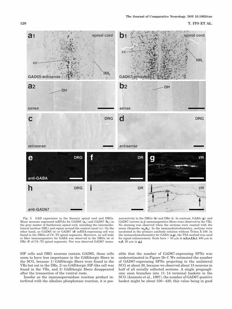
In the gray matter of the thoracic spinal cord, includingthe intermediolateral nucleus (IML), many neurons ex-pressed mRNAs for GAD65 and GAD67 (Fig. 3a1,b1).However, no mRNA signal for GAD65 or GAD67 wasobserved in the DRGs (Fig. 3c,d), indicating that no DRGcell has synthetic mechanisms for GABA. Moreover, al-though the presence of GABA-immunopositive DRG neu-rons was reported previously (Szabat et al., 1992), we didnot detect any GABA-immunopositive (Fig. 3e) or GAD67-immunopositive (Fig. 3h) structures in the DRGs, norGABA- or GAD67-immunopositive fibers in the DRs of theproximal or distal side of the DRGs (Fig. 3f,i). In contrast,many GABA- and GAD67-immunopositive fibers werefound in the VRs (Fig. 3g,j), suggesting that someGABAergic efferents ran from the spinal cord to the pe-riphery.
Origin of GABAergic fibers in the SCG andSTG
The presence of GABA- and GAD67-immunopositive fi-bers in the VRs implies that GABAergic fibers in the SCGoriginate in the spinal cord. Because all spinal cord neu-rons that send axons to the periphery are known to becholinergic, GAD67-immunopositive fibers in the SCG andSTG were expected to display immunoreactivity forVAchT. Actually, almost all GAD67-immunopositive var-icosities showed immunoreactivity for VAchT in theganglia (97.1%; 268/276 randomly chosen GAD67-immunopositive varicosities; n � 3; Fig. 4a–c��).
In an attempt to confirm the spinal origin of GAD67-immunopositive fibers in the SCG and STG, GAD67 im-munoreactivity was studied in the anterogradely labeledpreganglionic fibers. After a small injection of Sindbis/palGFP virus into the lateral horn of the T1 segment,many neurons were infected with the virus (Fig. 4d), andsome of them were located in the IML (box in Fig. 4d; seealso Fig. 4e). Some GFP-labeled axons were observed inthe ipsilateral STG and SCG and had extensive varicosebranches and basket-like structures (Fig. 4f). In all threecases injected with the virus, GAD67 immunoreactivitywas observed in some GFP-positive varicose structures(arrowheads in Fig. 4g–g��).
We next examined whether retrogradely labeled SPNsshowed GAD67 mRNA signals. After the injection of FGinto the SCG, many neurons in the IML of C8-T5 spinalsegments and a few neurons around the central canalwere retrogradely labeled, which is very consistent withprevious reports (Rando et al., 1981; Strack et al., 1988).With the combination of FG immunofluorescence and flu-orescent in situ hybridization for GAD67, some retro-gradely labeled neurons showed mRNA signals for GAD67(arrow in Fig. 5a–a��). To clarify further the distribution ofGAD67-expressing SPNs, we performed combined stain-ing with an immunoperoxidase reaction for FG and analkaline phosphatase reaction for GAD67 mRNA (Fig.5b–d) from every other section collected. In all, 540 � 42(mean � SD; n � 3) neurons showed FG immunoreactiv-ity, and 15 � 3 neurons (2.78% � 0.5%) were positive forboth FG and GAD67. They were located mainly in therostral part of the IML and, to a much lesser extent, inother parts of the IML and around the central canal (Fig.
The Journal of Comparative Neurology. DOI 10.1002/cne
117GABA-ERGIC SYMPATHETIC PREGANGLIONIC NEURONS

5d–f). Actually, the number of these neurons was signifi-cantly larger in the most rostral part of the IML (bar 0–2.3mm in Fig. 5e) than any other part of the IML and aroundthe central canal (Tukey’s multiple analysis, P 0.05).
GAD67-expressing SPNs tended to be located in the cen-tral and lateral part of the gray matter (Fig. 5f), althoughno significance was detected (ANOVA, P � 0.073). Al-though the morphological reconstruction was limited by
Fig. 1. Immunostaining for GABA and GAD67 in the SCG andSTG. Many GABA-immunopositive varicose fibers (a) and a few smallGABA-immunopositive cells (arrows in b,c) were observed in theganglia after staining with the TSA method. These GABA-immunopositive cells were often in close proximity to GABA-immunopositive varicose fibers (arrowheads in b,c). No immunoreac-tivity was observed when sections were incubated with anti-GABAantibody preabsorbed with GABA (d). GAD67-immunoreactive vari-cose fibers were observed frequently in the SCG (e1) and less fre-
quently in the STG (f1), and often made basket-like structures (insetin e1), but no immunoreactive cell body was found in the ganglia evenwhen the sections were incubated in the primary antibody solutionwithout Triton X-100 and processed with the sensitive TSA method(g). No immunoreactivity was observed in the ganglia (e2,f2) or brain(h2) when the sections were incubated with anti-GAD67 antibodypreabsorbed with GAD67-GST fusion protein. Scale bars � 25 �m ina,d–g; 10 �m in b,c; 2 mm in h.
The Journal of Comparative Neurology. DOI 10.1002/cne
118 T. ITO ET AL.

insufficient labeling of FG, GAD67-expressing (A-6, B-4-18, C-10 in Fig. 5c) and nonexpressing SPNs (A-N1, B-N1,2, C-N1 in Fig. 5c) displayed no obvious differences in theshape of cell bodies and proximal dendrites. Thus, at leastsome GABA- and GAD67-immunopositive fibers in theSCG and STG were considered to be of spinal origin.
Effect of VR transection
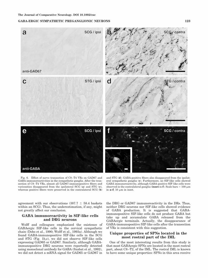
The ventral roots of C8–T4 were transected to examinewhether the SCG and STG had sources of GAD67-immunopositive fibers other than the spinal cord. Almostall GAD67-immunopositive structures disappeared fromthe SCG and STG on the operated side (Fig. 6a,c), whereasGAD67-immunopositive structures remained almost un-changed in the contralateral ganglia (Fig. 6b,d). Further-more, when the sections were immunostained for GABA,not only GABA-immunopositive fibers but also GABA-immunopositive SIF-like cells disappeared from the sym-pathetic ganglia of the operated side (Fig. 6e). On thecontralateral side, however, GABA-immunopositive struc-tures, including varicose fibers (Fig. 6f) and SIF-like cells(inset in Fig. 6f), were observed, as in the normal ganglia.These results indicate that almost all GABAergic fibers inthe SCG come from the spinal cord through the ipsilateralVRs.
DISCUSSION
In this study, we demonstrated that 1) GABAergic neu-rons were present in the IML, but not in the sympatheticchain or DRGs, 2) GABAergic fibers were present in theVRs but not in the DRs, 3) almost all GAD67-positivevaricosities showed immunoreactivity for VAchT in thesympathetic ganglia, 4) GAD67 was expressed in the ter-minals and somata of SPNs, and 5) GABAergic fibers inthe sympathetic ganglia disappeared after the VR tran-section. These observations clearly indicate that almost allGABAergic fibers in the cervical sympathetic chain origi-nate from SPNs. The fact that some SPNs express GAD67implies that these neurons corelease both acetylcholineand GABA.
Technical considerations
Because there is a limitation to the sensitivity of in situhybridization histochemistry, especially in the case ofGAD65, the number of GAD-expressing cells might beunderestimated. Therefore, we cannot exclude the possi-bility that SIF cells and DRG neurons express a smallamount of GAD65, but not GAD67. However, this is un-likely because GAD65 and GAD67 are known to be ex-pressed in the same neuron (Esclapez et al., 1993). Even if
Fig. 2. GAD expression in the SCG and STG. No cell body thatexpresses GAD65 mRNA was found in the SCG (a) or STG (b). Norwas found GAD67 mRNA expression in the SCG (c) and STG (d).Brain sections reacted with GAD65 (e1) and GAD67 (f1) antisenseriboprobes showed strong signals in many regions as reported previ-
ously. No signal was observed in the brain sections reacted with thesense probe for GAD65 (e2) or GAD67 (f2). Note: the olfactory bulb andinferior colliculus are not included in the section of e1. Scale bars � 50�m in a–d; 2 mm in e,f.
The Journal of Comparative Neurology. DOI 10.1002/cne
119GABA-ERGIC SYMPATHETIC PREGANGLIONIC NEURONS
SIF cells and DRG neurons contain GAD65, these cellsseem to have less importance in the GABAergic fibers inthe SCG, because 1) GABAergic fibers were found in theVRs but not in the DRs, 2) no GABAergic SIF-like cell wasfound in the VRs, and 3) GABAergic fibers disappearedafter the transection of the ventral roots.
Insofar as the immunoperoxidase reaction product in-terfered with the alkaline phosphatase reaction, it is pos-
sible that the number of GAD67-expressing SPNs wasunderestimated in Figure 5b–f. We estimated the numberof GAD67-expressing SPNs projecting to the unilateralSCG at about 30, because we observed about 15 neurons inhalf of all serially collected sections. A single pregangli-onic axon branches into 11–14 terminal baskets in theSCG (Asamoto et al., 1997), the number of GAD67-positivebasket might be about 330–420, this value being in good
Fig. 3. GAD expression in the thoracic spinal cord and DRGs.Many neurons expressed mRNAs for GAD65 (a1) and GAD67 (b1) inthe gray matter of thethoracic spinal cord, including the intermedio-lateral nucleus (IML) and region around the central canal (cc). On theother hand, no GAD65 (c) or GAD67 (d) mRNA-expressing cell wasfound in the DRGs of C8–T5 spinal segments. Moreover, no cell bodyor fiber immunopositive for GABA was observed in the DRGs (e) orDRs (f) of C8–T5 spinal segments. Nor was observed GAD67 immu-
noreactivity in the DRGs (h) and DRs (i). In contrast, GABA (g)- andGAD67 (arrows in j)-immunopositive fibers were observed in the VRs.No staining was observed when the sections were reacted with thesense riboprobe (a2,b2). In the immunohistochemistry, sections wereincubated in the primary antibody solution without Triton X-100. Inthe immunohistochemistry for GABA (e,g), the TSA method was usedfor signal enhancement. Scale bars � 50 �m in a,b,e,f,h,i; 400 �m inc,d; 20 �m in g,j.
The Journal of Comparative Neurology. DOI 10.1002/cne
120 T. ITO ET AL.

Fig. 4. Origin and chemistry of GAD67-immunopositive fibers inthe sympathetic ganglia. Almost all GAD67-immunopositive varicos-ities in the SCG and STG exhibited VAchT immunoreactivity (singlearrowheads in a–c), although many varicosities showed VAchT im-munoreactivity alone (double arrowheads in a–c). After the injectionof recombinant Sindbis/palGFP virus into the T1 IML, many neuronsaround the injection site in the longitudinal section of the spinal cord
showed GFP-immunoreactivity (dark brown structures in d), andsome of them were located in the IML (box in d; e). In the SCG, someanterogradely labeled fibers (f) showed GAD67 immunoreactivity (ar-rowheads in g–g��). Note: The section was counterstained withneutral red in d and e. LF, lateral funiculus; PF, posterior funicu-lus. Scale bars � 10 �m in a,c,g; 5 �m in b; 300 �m in d; 50 �m in e;20 �m in f.
The Journal of Comparative Neurology. DOI 10.1002/cne
121GABA-ERGIC SYMPATHETIC PREGANGLIONIC NEURONS

Fig. 5. GABA-producing SPNs in the thoracic spinal cord. In sec-tions stained with fluorescent dyes (a; FG immunoreactivity is repre-sented by green in a, and GAD67 mRNA signal is represented bymagenta in a��), some retrogradely labeled SPNs showed signal forGAD67 mRNA (arrows in a–a��) after the injection of FG into the rightSCG. Combined immunoperoxidase staining for FG (visualized withDAB; brown structures in b) and alkaline phosphatase in situ hybrid-ization for GAD67 mRNA (visualized with nitroblue tetrazolium; darkblue structures in b) were performed to investigate the morphology ofGAD67-expressing SPNs (b). The camera lucida reconstructionshowed that the GAD67-expressing SPNs (cells A-6, B-4, B-11, B-14,B-18, and C-10 in c) were not distinguishable from GAD67-negativeSPNs (cells A-N1, B-N1, B-N2, and C-N1 in c). As seen in the super-imposed drawing of the longitudinal sections of C8–T5 spinal cord (d),
a majority of GAD67-expressing SPNs were located in the rostral partof the IML, and a histogram of GAD67-expressing SPNs along therostrocaudal axis (e) clearly displayed that the numbers of theseneurons were significantly larger in the most rostral part (bar 0–2.3mm in e) than any other part of the IML (Tukey’s multiple analysis,P 0.05). In a histogram of GAD67-expressing SPNs along therelative distance from the midline of the spinal cord (f; zero indicatesthat a neuron is located at the midline, and 1 indicates that a neuronis located in the most lateral part of the IML at the single rostrocaudallevel), GAD67-expressing SPNs tended to be located in the central(0–0.2) and lateral (0.6–0.8) part of the gray matter, although nosignificance was detected (ANOVA, P � 0.073). Scale bars � 20 �m ina,b; 50 �m in c; 1 mm in d.
agreement with our observations (387.7 � 58.4 basketswithin an SCG). Thus, the underestimation, if any, mightnot greatly affect our conclusion.
GABA immunoreactivity in SIF-like cellsand DRG neurons
Wolff and colleagues emphasized the existence ofGABAergic SIF-like cells in the cervical sympatheticchain (Dobo et al., 1990; Wolff et al., 1993a). Although wefound GABA-immunopositive SIF-like cells in the SCGand STG (Fig. 1b,c), we did not observe SIF-like cellsexpressing GAD65 or GAD67. Similarly, although GABA-immunopositive DRG neurons were reportedly detectedusing monoclonal antibody for GABA (Szabat et al., 1992),we did not detect a mRNA signal for GAD65 or GAD67 in
the DRG or GAD67 immunoreactivity in the DRs. Thus,neither DRG neurons nor SIF-like cells showed evidenceof GABA production. It is suggested that GABA-immunopositive SIF-like cells do not produce GABA buttake up and accumulate GABA released from theGABAergic terminals. Actually, the disappearance ofGABA-immunopositive SIF-like cells after the transectionof VRs is consistent with this suggestion.
Unique properties of SPNs located in themost rostral part of the IML
One of the most interesting results from this study isthat most GABAergic SPNs are located in the most rostralpart, about C8–T2, of the IML. The rostral IML is knownto have some unique properties: SPNs in this area receive
Fig. 6. Effect of nerve transection of C8–T4 VRs on GAD67 andGABA immunoreactivities in the sympathetic ganglia. After the tran-section of C8–T4 VRs, almost all GAD67-immunopositive fibers andvaricosities disappeared from the ipsilateral SCG (a) and STG (c),whereas positive fibers were preserved in the contralateral SCG (b)
and STG (d). GABA-positive fibers also disappeared from the ipsilat-eral sympathetic ganglia (e). Furthermore, no SIF-like cells showedGABA immunoreactivity, although GABA-positive SIF-like cells wereobserved in the contralateral ganglia (inset in f). Scale bars � 100 �min a–f; 10 �m in inset.
The Journal of Comparative Neurology. DOI 10.1002/cne
123GABA-ERGIC SYMPATHETIC PREGANGLIONIC NEURONS
strong orexin input (Llewellyn-Smith et al., 2003) andmuch less serotonin input than SPNs in more caudalsegments (Jensen et al., 1995). In addition, neurokinin-1receptor-immunopositive SPNs are fewer in T1 and T2than in the more caudal segments (Llewellyn-Smith et al.,1997). These findings suggest that rostral SPNs are chem-ically coded and may have a specific function. These char-acteristics of rostral SPNs, which mainly project to theSCG, are likely to be associated with the functions of head-and face-specific organs (e.g., the pineal body, iris, sali-vary glands, cochlea, and lacrimal gland).
Functional considerations
Wolff and colleagues (1993a) proposed the “feed-forwardinhibition” mechanism, in which SPNs excited SIF-likecells in the cervical sympathetic chain and GABA-immunopositive SIF-like cells sent the axons to the SCGand inhibited the postganglionic neurons. However, in thepresent study, we showed that 1) no signal for GAD mR-NAs was detected in the cervical sympathetic chain and 2)all GAD67-immunopositive varicosities were cholinergicand of spinal origin. Thus, the “feed-forward inhibition”mechanism seems unlikely in the sympathetic ganglia.
Because both acetylcholine receptors and GABAA recep-tors are expressed in the SCG (Amenta et al., 1992; Liuand Burt, 1999), the present findings imply the coreleaseof excitatory (i.e., acetylcholine with nicotinic receptor)and “inhibitory” (i.e., GABA with GABAA receptor) neuro-transmitters from some preganglionic fibers in the SCG.Dobo and colleagues (1993) reported that GABA-immunopositive fibers innervated mainly neuropeptide Y(NPY)-negative large postganglionic neurons. About halfof all postganglionic neurons are NPY-positive vasocon-strictors (Gibbins, 1995), so it can be postulated that halfof all GAD-positive baskets would encircle NPY-positivepostganglionic neurons if GABAergic fibers randomlychoose the postganglionic target. However, only 10% ofGAD-immunopositive baskets actually encircled NPY-positive postganglionic neurons (Ito et al., 2005), suggest-ing that GABAergic baskets do not prefer NPY-positivevasoconstrictor neurons. Because most NPY-negativelarge postganglionic neurons are involved in the secreto-motor pathway (Gibbins, 1995), and postganglionic neu-rons that are encircled by GABA-positive baskets exit theSCG through the internal carotid nerve (Wolff et al.,1989), postganglionic neurons encircled by GABA-positivepreganglionic fibers may regulate the activity of glandslocated in the cranium.
The resting membrane potential is –45 to –90 mV (Ad-ams and Harper, 1995) and the reversal potential forchloride ion is –40 to –50 mV in the SCG neurons (Sacchiet al., 1999), so GABA may depolarize these neurons. Onthe other hand, because the threshold for action potentialproduction is 12.2 mV in the SCG neurons (Adams andHarper, 1995), activation of GABAA receptors may inhibitaction potential production in postganglionic neurons. Itseems strange that fibers from a single SPN coreleaseneurotransmitters that have opposite effects on actionpotential production.
Nevertheless, the coexpression of excitatory and inhib-itory neurotransmitters has been reported in other re-gions of the brain, such as granule cells in the hippocam-pus (Gutierrez, 2003), interneurons in the neocortex(Hioki et al., 2004), and immature lateral superior olivaryneurons (Gillespie et al., 2005), although the function
remained unclear. It has also been reported that GABAstrongly inhibits long-term potentiation via GABAA recep-tors in the rat SCG (Guillermo et al., 1994). As notedabove, GABAergic preganglionic fibers preferentially in-nervate NPY-negative secretomotor neurons and associ-ate with only a small number of NPY-positive postgangli-onic neurons. Therefore, postganglionic neuronsinnervated by GABAergic fibers, which may be associatedwith secretomotor functions, may be inhibited from pro-ducing long-term potentiation.
However, there is no direct evidence of targets thatreceive sympathetic postganglionic innervation related tothe spinal preganglionic GABAergic input, or that theLTP blockade phenomenon caused by stimulatingGABAergic fibers exists. The question also arises ofwhether the chloride equilibrium potential for cells sur-rounded by GABAergic baskets differs from that for theother postganglionic neurons. The identification of post-ganglionic neurons richly innervated by GABAergic fibersand morphological, electrophysiological, and pharmaco-logical studies of these neurons are needed.
ACKNOWLEDGMENTS
The authors are grateful to Dr. Munenori Ono for crit-ical discussion and to Mr. Taro Okunomiya for technicalassistance.
LITERATURE CITED
Adams DJ, Harper AA. 1995. Electrophysiological properties of autonomicganglion neurons. In: McLachlan EM, editor. Autonomic ganglia. NewYork: Harwood Academic Publishers.
Amenta F, Bronzetti, Cavallotti C, Flici L, Ferrante F, Colier WL. 1992.Autoradiographic localization of the gamma-aminobutyric acid type Areceptor agonist 3H-muscimol in the rat superior cervical ganglion.Pharmacology 44:107–112.
Asamoto K, Tamamaki N, Nojyo Y. 1997. Arborization pattern of sympa-thetic preganglionic axons in the rat superior cervical and stellateganglia. Neurosci Res 28:235–241.
Dobo E, Kasa P, Joo F, Wenthold RJ, Wolff JR. 1990. Structures withGABA-like and GAD-like immunoreactivity in the cervical sympatheticganglion complex of adult rats. Cell Tissue Res 262:351–361.
Dobo E, Joo F, Wolff JR. 1993. Distinct subsets of neuropeptide Y-negativeprincipal neurons receive basket-like innervation from enkephalinergicand gabaergic axons in the superior cervical ganglion of adult rats.Neuroscience 57:833–844.
Esclapez M, Tillakaratne NJK, Tobin AJ, Houser CR. 1993. Comparativelocalization of mRNAs encoding two forms of glutamic acid decarbox-ylase with nonradioactive in situ hybridization methods. J Comp Neu-rol 331:339–362.
Fong AY, Stornetta RL, Foley CM, Potts JT. 2005. Immunohistochemicallocalization of GAD67-expressing neurons and processes in the ratbrainstem: subregional distribution in the nucleus tractus solitarius.J Comp Neurol 493:274–290.
Furuta T, Tomioka R, Taki K, Nakamura K, Tamamaki N, Kaneko T. 2001.In vivo transduction of central neurons using recombinant Sindbisvirus: Golgi-like labeling of dendrites and axons with membrane-targeted fluorescent proteins. J Histochem Cytochem 49:1497–1508.
Gibbins IL. 1995. Chemical neuroanatomy of sympathetic ganglia. In:McLachlan EM, editor. Autonomic ganglia. New York: Harwood Aca-demic Publishers.
Gillespie DC, Kim G, Kandler K. 2005. Inhibitory synapses in the devel-oping auditory system are glutamatergic. Nature Neurosci 8:332–338.
Guillermo R, Burgos G, Biali FI, Nichola Siri LC, Cardinali DP. 1994.Effect of gamma-aminobutyric acid on synaptic transmission and long-term potentiation in rat superior cervical ganglion. Brain Res 658:1–7.
Gutierrez R. 2003. The GABAergic phenotype of the “glutamatergic” gran-ule cells of the dentate gyrus. Prog Neurobiol 71:337–358.
Hioki H, Fujiyama F, Nakamura K, Wu SX, Matsuda W, Kaneko T. 2004.
The Journal of Comparative Neurology. DOI 10.1002/cne
124 T. ITO ET AL.

Chemically specific circuit composed of vesicular glutamate transporter3- and preprotachykinin B-producing interneurons in the rat neocor-tex. Cereb Cortex 14:1266–1275.
Ito T, Iino S, Nojyo Y. 2005. A part of cholinergic fibers in mouse superiorcervical ganglia contain GABA or glutamate. Brain Res 1046:234–238.
Jensen I, Llewellyn-Smith IJ, Pilowsky P, Minson JB, Chalmers J. 1995.Serotonin inputs to rabbit sympathetic preganglionic neurons project-ing to the superior cervical ganglion or adrenal medulla. J CompNeurol. 353:427–438.
Liang F, Hatanaka Y, Saito H, Yamamori T, Hashikawa T. 2000. Differ-ential expression of gamma-aminobutyric acid type B receptor-1a and-1b mRNA variants in GABA and non-GABAergic neurons of the ratbrain. J Comp Neurol 416:475–495.
Ligorio MA, Akmentin W, Gallery F, Cabot JB. 2000. Ultrastructual local-ization of the binding fragment of tetanus toxin in putative gamma-aminobutylic acidergic terminals in the intermediolateral cell column:a potential basis for sympathetic dysfunction in generalized tetanus.J Comp Neurol 419:471–484.
Liu ZF, Burt DR. 1999. GABAA receptor subunit mRNAs in rat superiorcervical ganglia. Pharmacology 58:51–8.
Llewellyn-Smith IJ, Martin CL, Minson JB, Pilowsky PM, Arnolda LF,Basbaum AI, Chalmers JP. 1997. Neurokinin-1 receptor-immunoreactive sympathetic preganglionic neurons: target specificityand ultrastructure. Neuroscience 77:1137–1149.
Llewellyn-Smith IJ, Martin CL, Marcus JN, Yanagisawa M, Minson JB,Scammell TE. 2003. Orexin-immunoreactive inputs to rat sympatheticpreganglionic neurons. Neurosci Lett 351:115–119.
Mugnaini E, Oertel WH. 1985. An Atlas of the distribution of GABAergicneurons and terminals in the rat CNS as revealed by GAD immuno-histochemistry. In: Bjorklund A, Hokfelt T, editors. Handbook of chem-ical neuroanatomy, vol 4. New York: Elsevier.
Murata Y, Shibata H, Chiba T. 1982. A correlative quantitative studycomparing the nerve fibers in the cervical sympathetic trunk and thelocus of the somata from which they originate in the rat. J Auton NervSyst 6:323–333.
Rando TA, Bowers CW, Zigmond RE. 1981. Localization of neurons in therat spinal cord which project to the superior cervical ganglion. J CompNeurol 196:73–83.
Sacchi O, Rossi ML, Canella R, Fesce R. 1999. Participation of a chlorideconductance in the subthreshold behavior of the rat sympathetic neu-ron. J Neurophysiol 82:1662–1675.
Strack AM, Sawyer WB, Marubio LM, Loewy AD. 1988. Spinal origin ofsympathetic preganglionic neurons in the rat. Brain Res 455:187–191.
Szabat E, Soinila S, Happola O, Linnala A, Virtanen I. 1992. A newmonoclonal antibody against the GABA-protein conjugate shows im-munoreactivity in sensory neurons of the rat. Neuroscience 47:409–420.
Tamamaki N, Nakamura K, Furuta T, Asamoto K, Kaneko T. 2000. Neu-rons in Golgi-stain-like images revealed by GFP-adenovirus infectionin vivo. Neurosci Res 38:231–236.
Wolff JR, Kasa P, Dobo E, Wenthold RJ, Joo F. 1989. Quantitative analysisof the number and distribution of neurons richly innervated by GABA-immunoreactive axons in the rat superior cervical ganglion. J CompNeurol 282:264–273.
Wolff JR, Kasa P, Dobo E, Romgens HJ, Parducz A, Joo F, Wolff A. 1993a.Distribution of GABA-immunoreactive nerve fibers and cells in thecervical and thoracic paravertebral sympathetic trunk of adult rat:evidence for an ascending feed-forward inhibition system. J CompNeurol 334:281–293.
Wolff JR, Joo F, Kasa P. 1993b. Modulation by GABA of neuroplasticity inthe central and peripheral nervous system. Neurochem Res 18:453–461.
The Journal of Comparative Neurology. DOI 10.1002/cne
125GABA-ERGIC SYMPATHETIC PREGANGLIONIC NEURONS