paw inflammation model in dogs for preclinical...
TRANSCRIPT

Paw Inflammation Model in Dogs for PreclinicalPharmacokinetic/Pharmacodynamic Investigations ofNonsteroidal Anti-Inflammatory Drugs
E. C. Jeunesse, I. A. Bargues, C. E. Toutain, M. Z. Lacroix, I. M. Letellier, J. M. Giraudel,and P. L. ToutainTOXALIM, Institut National de la Recherche Agronomique, Toulouse, France (E.C.J., I.A.B., M.Z.L., P.L.T.); Institut NationalPolytechnique de Toulouse, Ecole Nationale Veterinaire de Toulouse, and Ecole d’Ingenieurs de Purpan, Universite deToulouse, Toulouse, France (E.C.J., I.A.B., M.Z.L., P.L.T.); Novartis Centre de Recherche Sante Animale, Saint-Aubin,Switzerland (C.E.T., J.M.G.); and Novartis Sante Animale, Rueil-Malmaison, France (I.M.L.)
Received December 17, 2010; accepted April 26, 2011
ABSTRACTThe goal of the present study was to develop and validate anew canine model of inflammation. The motivation was to makeavailable a scientifically appropriate and ethically acceptablemodel to conduct pharmacokinetic/pharmacodynamic investi-gations for testing nonsteroidal anti-inflammatory drugs indogs. A kaolin-induce paw inflammation model previously de-veloped in cats was adapted to the dog. The paw inflammationdeveloped within a few hours, reached maximum values 24 hand up to 3 days after kaolin administration, and then progres-sively resolved over 2 months. Five end points of clinical inter-est (body temperature, creeping time under a tunnel, paw with-drawal latency to a standardized thermal stimulus, lamenessscore, and vertical force developed during walking on a forceplate) were measured regularly over the next 24 h and beyond
to characterize the time development of the inflammation eitherin control conditions (placebo period) or after the administrationof meloxicam (test period) according to a crossover design.Pharmacodynamic data were modeled using an indirect re-sponse pharmacokinetic/pharmacodynamic model. This modeldescribed three effects of meloxicam, namely, classic anti-inflammatory, analgesic, and antipyretic effects. The meanplasma meloxicam IC50 values were 210 ng/ml for the anti-pyretic effect, 390 ng/ml for the analgesic effect, and 546 ng/mlfor the vertical force exerted by the paw on the ground asmeasured by force plates. These in vivo IC50 values requireapproximately 80 (antipyretic effect) to 90% (all other effects)cyclooxygenase-2 inhibition as calculated ex vivo whole-bloodassay data.
IntroductionFew preclinical studies have attempted to model plasma
concentration-time profiles with the time course of nonsteroi-dal anti-inflammatory drug (NSAID) effects in small carni-vores (Toutain et al., 2001; Giraudel et al., 2009). A requisiteto carry out a pharmacokinetic/pharmacodynamic (PK/PD)dose determination is the availability of an animal model inwhich the measured end points are clinically relevant, mea-surable with good metrological performances, and longenough to prevent (or minimize) confusion between the ac-tion of the investigated NSAID and the natural time devel-opment of the inflammatory process. Currently, only acuteknee joint synovitis using intra-articular injection of sodium
urate crystals has been well described in dogs, and its appli-cation to the preclinical evaluation of different NSAIDs isnow well documented (Cross et al., 1997). However, thismodel is of too short in duration (less than 24 h) to allow aPK/PD investigation for drugs having a duration of action ofapproximately 24 h. Likewise, the carrageenan-inducedacute paw inflammation in dogs model proposed by Brooks etal. (1991) was too short in duration for this purpose. This iswhy we developed a Freund’s complete adjuvant osteoarthri-tis model, resulting in a sustained and relatively stable sec-ondary inflammatory response (Botrel et al., 1994; Toutain etal., 2001). This Freund’s model was scientifically attractive,allowing repeatable measurements of relevant clinical endpoints, but the inflammation was irreversible, and it wasmandatory to sacrifice dogs for ethical reasons. Because weare committed to a program of rehabilitating dogs by donat-ing them as companion animals at the end of the trials, weadopted the 3R principles to avoid unnecessary suffering to
This work was supported by Novartis Animal Health, Inc.Article, publication date, and citation information can be found at
http://jpet.aspetjournals.org.doi:10.1124/jpet.110.178350.
ABBREVIATIONS: NSAID, nonsteroidal anti-inflammatory drug; COX, cyclooxygenase; PK/PD, pharmacokinetic/pharmacodynamic; CV, coeffi-cient of variation.
0022-3565/11/3382-548–558$25.00THE JOURNAL OF PHARMACOLOGY AND EXPERIMENTAL THERAPEUTICS Vol. 338, No. 2Copyright © 2011 by The American Society for Pharmacology and Experimental Therapeutics 178350/3699870JPET 338:548–558, 2011 Printed in U.S.A.
548
at ASPE
T Journals on June 3, 2018
jpet.aspetjournals.orgD
ownloaded from

animals [i.e., replace (use alternatives to animal testingwhenever possible), reduce (improve existing methods so thatfewer laboratory animals are required in an experiment), andrefine (improve existing methods so that animals experienceas little discomfort and distress as possible)]. The PK/PDapproach itself can be considered as a refinement of a dosetitration allowing a reduction in the required number ofanimals to establish a full dosage regimen (dose and dosinginterval), but to achieve our ultimate goal of rehabilitatingexperimental animals, it was necessary to develop and vali-date a new reversible inflammation model in dogs. Such amodel was developed recently in cats (Giraudel et al., 2005a).It consisted of administering kaolin (an inert foreign body) asa phlogistic agent in the paw. The resulting inflammationwas sustained between 1 and 3 days after kaolin injection,allowing administration of the NSAID on day 2. The modelwas found to be suitable for simultaneously studying theanalgesic, anti-inflammatory, and antipyretic effects ofNSAIDs in cats by measuring different end points such asbody temperature or gait scoring (Giraudel et al., 2005a,b).Moreover, because the kaolin was administered extra-articu-larly, the inflammation progressively vanished with eitherthe physical wasting of the administered kaolin by directskin exudation or the encystment of the remnant kaolinfraction, and within a few weeks, most animals returned tothe control condition without any apparent sequelae.
The objective of the present study was to transfer andvalidate this reversible inflammation model in dogs, a speciesused extensively in regulatory studies in both human andveterinary medicine. We selected meloxicam as a testNSAID, because it is a well established NSAID in dogs (Ara-gon et al., 2007) and also used in man.
Materials and MethodsAnimals. Eight healthy Beagle dogs (two females and six males)
were selected after clinical examination and biochemical analysis. Thebody weights and ages of the dogs ranged from 12.5 to 17.5 kg and 1 to3 years, respectively. Between experimental phases, the dogs werehoused in large boxes. During the different phases of the trials, theanimals were kept in individual stainless steel cages in a controlledenvironment. On the days of the measurements, the dogs were placed intheir cages at least 2 h before any measurement. Dogs were fed eachevening after the last measurements with 250 � 50 g of commercial dryfood (medium; Royal Canin, Aimargues, France). Animal care and con-duct of the study were performed according to the Guide for the Careand Use of Laboratory Animals (Institute of Laboratory Animal Re-sources, 1996). The study has been carried out in accordance with theDeclaration of Helsinki and with the Guide for the Care and Use ofLaboratory Animals as adopted and promulgated by the U.S. NationalInstitutes of Health. The protocol was approved by the Animal Experimen-tation Ethics Committee of Midi-Pyrenees. The study was performed incompliance with the Principles of Good Clinical Practice (CVMP/VICH/595/98; http://www.ema.europa.eu/docs/en_GB/document_library/Scientific_guideline/2009/10/WC500004343.pdf) and according to theGuideline for the Conduct of Efficacy Studies for NSAIDs (EMEA/CVMP/237/01; http://www.ema.europa.eu/docs/en_GB/document_library/Scientific_guideline/2009/10/WC500004423.pdf). Because the model is to-tally reversible without sequelae, all of the dogs that participated in thistrial were rehabilitated as companion animals at the end of the study.
Animal Preparation and Induction of Inflammation. Bothhind paws were shaved from the toes up to the hock joint, and targetswere marked for the pain assessment and paw circumferencemeasures (Fig. 1).
The preparation of kaolin (hydrated aluminum silicate; Sigma-Aldrich, St. Louis, MO) was done using standardized procedures(Giraudel et al., 2005b). For the induction of inflammation, the dogswere given a general anesthetic with alfaxalone 3 mg/kg (Alfaxan;Vetoquinol, Lure, France), and anesthesia was maintained with iso-flurane (2% v/v; Aerrane; Baxter, Lessines, Belgium). A local anes-thetic was added: approximately 2.5 ml of lidocaine was injectedsubcutaneously into the paw (Lurocaïne; Vetoquinol), after which1.85 � 0.10 g of kaolin was injected under aseptic conditions subcu-taneously at eight different points in the metatarsal pad. The totalvolume of the kaolin suspension (25% w/w) was 6.4 ml.
Experimental Design. The eight dogs and the investigatorswere trained regularly for all of the experimental conditions and allof the end point measurements for at least 1 month. This trainingperiod guaranteed the eligibility for the trial of the eight selecteddogs and the investigators’ skills. The trial consisted of two phases.In the first phase, the metrological performances of the different endpoints were assessed in all of the dogs. Investigated end points werebody temperature, paw circumference, time to perform a creepingtest under a tunnel, vertical force of the paw on force plates, andthermal pain threshold. Three series of end point measurementswere obtained each day and for 5 consecutive days, and the repeat-ability and reproducibility of the different end points were computed.Lameness scoring was not validated during this first control phasebecause all of these control measurements were obtained in nonin-flammatory conditions. Then, during a second phase, a two-period,two-sequence crossover design was carried out. The eight dogs wereallocated randomly into four pairs of the same sex and of similarbody weight. Dogs of each pair then were allocated into one of twogroups (sequences) corresponding to the order of the administrationof the test articles (placebo then NSAID or, conversely, NSAID thenplacebo). The washout period between the two periods of the cross-over was 8 weeks. Kaolin inflammation was induced for each dog ofa pair and in the two periods (right paw period 1 and left paw period2). The experiment was blinded to the investigators. The placebocorresponded to a sham meloxicam administration. Meloxicam(Metacam; Boehringer Ingelheim Vetmedica, Ingelheim/Rhein, Ger-many) was administered by the subcutaneous route 26 h after kaolininflammation induction. The dose of meloxicam administered wasthe marketed dose for dogs (0.2 mg/kg). Blood samples were obtainedfrom the jugular vein by direct puncture at time 0 (control) and then5, 15, 30, and 60 min and, 2, 4, 6, 9, 12, 21, 48, and 72 h after the testarticle administration (meloxicam or placebo). Before kaolin admin-istration, the PD end points were measured twice the same day andthen twice at 22 and 24 h after the induction of the kaolin inflam-mation. Subsequently, the PD end points were measured at 30 min,2.25, 4, 6, 9, 12, 15, 18, 21, 30, and 48 h, and 3, 4, 5, 6, 7, 10, 14, 17,21, and 24 days after test article administration (i.e., placebo or
Fig. 1. Cutaneous reference marks on the hind paw of the dog for thecircumference ( ) and analgesia (F) measurements and view of thedifferent sites of subcutaneous kaolin injection (3).
PK/PD Modeling of NSAIDs in a Dog Model 549
at ASPE
T Journals on June 3, 2018
jpet.aspetjournals.orgD
ownloaded from

meloxicam). Two supplementary measures were obtained on the35th day after test article administration (body temperature andpaw circumference) and at 64 days (paw circumference).
End Point Measurements. The six clinical end points selected toassess inflammation were as follows: 1) body temperature (a singlemeasurement was obtained at each time using an electronic ther-mometer), 2) paw circumference (duplicate measures per time point)measured just above the pad using a measuring tape (DMC, Colmar,France), 3) lameness scored using a numerical rating scale as devel-oped by Giraudel et al. (2005b) (Table 1), 4) creeping time (i.e., thetime required by the dog to creep under a 6-m-long tunnel) measuredin triplicate at each measurement time and with the mean timecomputed, 5) vertical normalized force (i.e., the maximal verticalforce applied on the ground by a hind limb measured with forceplates) (SATEL Veto; Patrick Savet, Blagnac, France); for each mea-surement time, three valid measures were recorded and the meannormalized to the dog’s body weight (Fmax/b.wt., kg/kg) used in thedata analyses, and (6) pain as assessed using a hind paw thermalescape model (Hargreaves et al., 1988). The model consisted of ex-posing the hind limb to a light beam delivered by a Hargreavesapparatus (model 390; IITC Inc., Woodlands Hills, CA) (Giraudel etal., 2005b). This equipment has a light source (halogen 24 V, 150 W)that creates and controls heat intensity. A heat intensity correspond-ing to 15% of the peak heating value of 150 W was selected. Dogswere placed on a table equipped with a glass top that enabled thebeam to be applied from underneath onto a target selected area onthe plantar surface of the hind limb. Dogs were trained to stay quietin control conditions in this environment with minimal manual re-straint. When applying the heat stimulus to the top of the metatarsalpad (Fig. 1), a lateral leg movement or any sign of retraction of thepaw was considered as a response, and the pain stimulation wasstopped immediately. A maximal stimulation time of 90 s was usedto prevent any tissue damage through burning. The paw withdrawaltime (in seconds) was measured. For each trial, three measures wererecorded per measurement time, and the mean of the three measureswas used in the data analysis.
Analysis of Meloxicam in Plasma. Plasma samples were ana-lyzed by a high-performance liquid chromatography method usingUV detection. In brief, an internal standard (piroxicam) and meloxi-cam were extracted from the plasma by solid-phase extraction. Thehigh-performance liquid chromatography apparatus consists of apump system equipped with an automatic injector and an UV detec-tor (360 nm). Separation was achieved by reverse-phase column(150 � 2.0 mm, 3 �m; Diphenyl Pursuit XRs; Varian, Lake Forest,CA) using a guard column (4.0 � 2.0 mm; Phenyl). The mobile phasewas a 50:50 mixture of water with 1% acetic acid and acetonitrile(Sigma-Aldrich) at a flow rate of 0.2 ml/min. Under these conditions,meloxicam (Sigma-Aldrich) and piroxicam (Sigma-Aldrich) wereeluted at retention times of 7.2 and 5.6 min, respectively. Themethod was linear over the calibration range from 10 to 1500 ng/mlusing a linear model weighted by 1/X2. Within-day and day-to-daycoefficients of variation were less than 15%, and the accuracy rangedfrom 101 to 103%, indicating appropriate precision and accuracy forthe analytical method. The lack of interference from endogenous
compounds was verified on blank plasma from untreated dogs, es-tablishing the specificity of the method. The validated limit of quan-tification was 10 ng/ml.
Data Analysis. PK and PK/PD modeling were performed byleast-squares regression analysis using WinNonlin Professionalsoftware (WinNonlin, version 5.2; Pharsight Corporation, Moun-tain View, CA).
Individual plasma meloxicam concentrations (ng/ml) were fitted topolyexponential equations. The data points were weighted by theinverse of the squared-fitted value. The number of exponents (two orthree) needed to obtain the best fit for each data set was determinedby the Akaike’s information criterion (Yamaoka et al., 1978) and byinspection of the plot of residuals. On the basis of this approach, abiexponential equation corresponding to a monocompartmentalmodel for extravascular administration with a lag time was selected(eq. 1).
C(t) �FDk01
V�k01 � k10��exp��k10 � �t � tlag�� � exp��k01 � �t � tlag��
(1)
where C(t) is the meloxicam plasma concentration (ng/ml) at time t(h), V/F (l/kg) is the apparent volume of distribution, k01 (1/h) is therate constant of the initial ascending phase, k10 (1/h) is the rateconstant of the terminal phase, and D is the meloxicam dose (mg/kg).The parameters (V/F, k01, k10, and tlag) were estimated.
For PK/PD modeling, raw measured data were used directly asdependent variables for body temperature, lameness score, andcreeping time. Because kaolin decreased the vertical force exerted onthe force plates or the paw withdrawal time, these variables weretransformed to be expressed as a percentage of reduction (100% isthe maximal effect observed after kaolin administration) to use thesame PK/PD model for all of end points. Individual PK/PD relation-ships were described using indirect PD response models (Dayneka etal., 1993). In these models, the measured response (R) is assumed toresult from factors controlling either the input or the dissipation ofthe measured response. Different models were explored, includingprecursor-dependent indirect response models (Sharma et al., 1998),to capture not only the main effect of meloxicam but also the devel-opment of rebound phenomena, which were observed after meloxi-cam action had ceased. Finally, PD data were described by thefollowing sets of differential equations (eqs. 2–4).
In the control condition (i.e., before kaolin administration), forboth periods
dR⁄dt � kin � koutR (2)
where dR/dt is the rate of change of the response over time, kin
represents the zero-order rate constant for the production of theresponse, and kout is the first-order rate constant for the loss of theresponse.
The time development of the kaolin action, noted as kao(t), on thedifferent measured responses was incorporated additively to kin inthe model described by eq. 3
dR⁄dt � kin � kao�t� � koutR (3)
where kao(t) is the zero-order input rate function (same units as kin)representing the action of the kaolin accounting for the temporalincrease in the response after kaolin administration. Such a modelalready has been selected by other researchers for the PK/PD mod-eling of the antipyretic effect of naproxen in a model of endotoxin-challenged rats (Josa et al., 2001). After consideration of the inverseU shape of the time development of the responses in the placeboperiod, kao(t) was modeled using an empirical biexponential equa-tion of the form (eq. 4)
kao�t� � P1�exp��P2�t � tlag� � exp��P3�t � tlag�� (4)
TABLE 1Numerical rating scale for the evaluation of lameness in the inflamedpaw in dogs
Score Definition of the Lameness Score
0 No lameness1 Barely detectable lameness over most of the observation period2 Mild lameness, substantial weight bearing3 Moderate lameness, minimal weight bearing4 Severe lameness, the animal uses his paw (walking movement
initiated and/or touches lightly the ground) but does not bearweight
5 The animal could not be more lame, refuse to move and/oravoid any contact of the inflamed paw with the ground
550 Jeunesse et al.
at ASPE
T Journals on June 3, 2018
jpet.aspetjournals.orgD
ownloaded from

with P1 the intercept (response unit per hour), P2 (1/h) the slope ofthe decreasing phase of the kaolin action, P3 (1/h) the rate constantreflecting the increasing phase of the kaolin action after kaolinadministration, and tlag the time of kaolin administration. The threeparameters (P1, P2, and P3) were estimated simultaneously with thePD parameters of ultimate interest (i.e., kin, IC50, and kout; seebelow), and the lag time was fixed at the actual time of kaolinadministration for both placebo and test periods.
The effect of meloxicam (melox) was described as the consequenceof the inhibition of the phlogogenic effect of kaolin, that is, kao(t) andwas incorporated in eq. 3 as (eq. 5)
dR⁄dt � kin � kao�t� � �1 � melox� � kout � R (5)
where melox is the classic fractional Imax function of the form (eq. 6)
Melox �Imax � C�t�n1
IC50An1 � C(t)n1 (6)
where melox is a Hill equation in which C(t) (ng/ml) is the meloxicamplasma concentration at time t (h) as obtained by fitting eq. 1, IC50A
(ng/ml) is the meloxicam plasma concentration producing half themaximum meloxicam effect (i.e., 50% of Imax), Imax is a scalar with amaximal value of 1, and n1 is the slope of the concentration-effectrelationship. For the test period, it was nearly always observed thatafter the meloxicam action vanished the response increased again(indicating that kaolin action was still present), and unexpectedly,there was an overshoot of the response compared with measuresmade at the same time in the placebo period. To take into accountthis second effect of meloxicam on the fate of the kaolin inflammationitself, we rendered time-dependent P2, the rate constant reflectingthe kaolin inflammation disappearance (see eq. 4), according to eq. 7
P2, melox � P2 � �1 �C(t)n2
IC50Bn2 � C�t�n2� (7)
where P2,melox is the time-dependent rate constant reflecting thedisappearance of the kaolin action, the value of which was controlledby the plasma meloxicam concentration [C(t)] throughout a frac-tional Imax function with IC50B being the meloxicam plasma concen-tration halving P2. Equation 7 says that P2,melox decreases whenthe plasma meloxicam concentration increases (i.e., meloxicam isdelaying the disappearance of the kaolin action, and the natural timecourse of inflammation resumes when meloxicam is no longer pres-ent) (see Discussion).
Finally, 10 parameters were estimated by simultaneously fittingthe placebo period (with data collected before and after kaolin ad-ministration) and the test periods (with data collected before kaolinadministration, after kaolin administration but before meloxicamadministration, and after meloxicam administration), namely, kin,kout, IC50A for the main anti-inflammatory effect of meloxicam, IC50B
for effect of meloxicam on the time development of the kaolin, n1, n2,Imax, and P1 to P3.
Through the use of mean PK and PD parameters, simulationswere performed to predict the time development of the response forkaolin alone or kaolin followed by meloxicam for each end point andfor doses ranging from 0.05 to 2 mg/kg. Data from all of the individ-ual animals were used to compute the mean PK parameters, but onlythose dogs with an acceptable PD fit (six to eight animals dependingon the PD end points) were taken into account for the calculation ofthe mean PD parameters.
For each simulation, the maximal response obtained for eachresponse profile was used as a metric to further characterize thedose-response relationship. Then, the dose (mg/kg) versus simulatedmaximal response was fitted using a classic inhibitory sigmoid model(model of the form of eq. 8)
Effect � Baseline � �1 �Imax � Dosen
ED50n Dosen� (8)
where ED50 is the dose giving half the maximal response and n is theslope of the dose-response relationship.
To estimate the extent of cyclooxygenase (COX)-1 and COX-2enzyme inhibition corresponding to the different estimated in vivoIC50 values, we computed the percentage of COX-1 and COX-2 inhi-bition from ex vivo results of a whole-blood assay performed inanother group of Beagle dogs. COX-1 activity was determined bymeasuring serum thromboxane B2 synthesis in blood samples.COX-2 activity was determined by measuring prostaglandin E2 syn-thesis in blood samples incubated at 37°C for 24 h in the presence oflipopolysaccharide. The data were fitted to Hill plots, and the slope() and IC50 values were calculated. For prostaglandin PGE2 inhi-bition, was estimated to be 0.986, and the IC50 value was estimatedto be 0.1454 �M (51 ng/ml); corresponding values for thromboxane B2
were 1.024 and 1.215 �M (427 ng/ml). Through the use of theseparameters kindly provided by Novartis, the Imax model was used toestimate the extent of COX-1 and COX-2 inhibition corresponding toour in vivo estimated IC50 value.
Statistical Analysis. Metrological performances of the differentend points were assessed using the following statistical model (Sys-tat 10; Systat Software, Inc., San Jose, CA)
Yijk � � � Ai � Dj � Tk � A * Dij � A * Tik � A * Tjk � εijk
where Yijk is the end point value for animal i at day j and time k, �is the overall mean, Ai is the animal factor, Dj is the day factor, Tk isthe measurement time factor, C�Dij, C�Tik, and D�Tjk are the corre-sponding interactions, and εijk is the residual error for i � 1 to 8, j �1 to 5, and k � 1 to 3.
All of the variables in this model were considered as fixed factors,and a factor was considered significant when p � 0.05.
An analysis of variance was used to test the significance of thedifferent factors and to calculate the repeatability of measurements(as a coefficient of variation) using the residual of the model:
Repeatability: CV% ��Mean square of residual error
mean � 100
Repeatability is the highest level of precision obtained in a given dogfor a given day and a given measurement time. Reproducibility (as acoefficient of variation) was calculated based on all of the valuesrecorded during the five days of the study:
Reproducibility: CV% �SD
mean � 100
Reproducibility (always higher than repeatability) is the lowest levelof precision encompassing all of the factors of variability in ameasurement.
The PK and PD results are presented as mean or median and S.D.For the half-lives, the harmonic mean was calculated. An ANOVAanalysis was used to test the significance of the difference betweendifferent IC50 values.
ResultsMetrological Performance of the PD End Points (Re-
peatability and Reproducibility). The results are sum-marized in Table 2. The dog factor was highly significant forall of the end points (p � 0.001, data not shown), reflectingthe interanimal variability and justifying a crossover designto assess the dogs’ responses in both placebo and test condi-tions. For several end points, there were significant animalper day interactions, meaning that the day-to-day variationthat could be observed for an end point measurement did notfollow a systematic trend as would have happened if a “train-ing” effect had occurred during our validation tests. Themeasurement time (within day) effect was significant for the
PK/PD Modeling of NSAIDs in a Dog Model 551
at ASPE
T Journals on June 3, 2018
jpet.aspetjournals.orgD
ownloaded from

body temperature. This observed trend was interpreted asreflecting an endogenous circadian rhythm, the body temper-ature in the afternoon always being higher than that in themorning. This possible confounding factor was not consideredin our PK/PD modeling because the absolute difference asso-ciated with this factor was only 0.1°C.
Repeatability (i.e., variability within a given measurementtime) was appropriate for all of the end points (�5% for themeasures of creeping time and vertical normalized maximalforce and �0.5% for measures of body temperature and pawcircumference). For the withdrawal time measuring analge-sia, the CV of repeatability was 15.1%, which is low consid-ering the expected amplitude of the kaolin and meloxicameffect. Reproducibility (i.e., the overall variability betweendogs, days, and measurement times) was �5% for the mea-sures of body temperature and paw circumference, �10% forthe measure of the vertical normalized maximal force, and�25% for the creeping time and the paw withdrawal time.The animal factor was the main contributor to the overallreproducibility, thus justifying the use of a crossover designfor this kind of investigation rather than a parallel design.
PK Results. The meloxicam plasma concentration profileafter subcutaneous administration of 0.2 mg/kg was inter-preted as a monocompartmental model with a first-order rateconstant of absorption and a lag time (Fig. 2). The apparenttotal plasma clearance (Cl/F, clearance scaled by the un-known bioavailability) was low (6.33 � 1.95 ml � h�1 � kg�1),and the apparent volume of distribution (V/F, volume ofdistribution scaled by the unknown bioavailability) was
small 192.1 � 34.4 ml/kg. Peak meloxicam plasma concen-tration (890.2 � 166.4 ng/ml) was attained at 5.97 � 1.73 hafter administration, and the harmonic mean of the apparenthalf-life of absorption was 1.32 � 0.59 h. The harmonic meanof the terminal half-life was relatively high at 21.26 � 4.83 h.
Time Development of the Kaolin Paw InflammationModel with and without Meloxicam Administration.Figure 3 illustrates the time course and magnitude of theinflammatory response for the six investigated end points inboth placebo and treated conditions. The end point valuesreached maxima 22 and 24 h after the kaolin injection. Forthe placebo period (without meloxicam), body temperature,creeping time, and paw withdrawal time still were altered56 h after kaolin administration (spontaneous improvementwas �50%, a 100% improvement meaning return to theprekaolin administration condition). Paw edema (measuredby the paw circumference) disappeared slowly, a delay of 15days being necessary to reduce the kaolin-induced edema byapproximately 50%. The vertical force exerted by the hindlimb still was decreased by approximately 50% on the 10thday after the kaolin administration, indicating a rather sus-tained inflammation that enables NSAID effects to be fol-lowed over a prolonged time period. However, the naturaltime course of the inflammation was not stable enough to beignored when modeling the NSAID effect, and a crossoverdesign including a placebo period was mandatory to assessthe net effect of the NSAID (see Discussion). In terms ofanimal welfare, it was observed that for the six investigatedend points an improvement superior to 85% was observedafter kaolin administration between the 14th and 17th days(body temperature), the 24th and 35th days (paw circumfer-ence), the 17th and 21st days (lameness score), the 10th and14th days (creeping time), the 10th and 14th days (verticalforce of the paw exerted on the ground), and the 7th and 10thdays for the allodynia as assessed by paw withdrawal time.
In treated conditions (after a subcutaneous injection ofmeloxicam at a dose of 0.2 mg/kg), a clear-cut drug responsewas obtained for all but one end point, namely, paw circum-ference. The unresponsiveness of paw circumference tomeloxicam was expected (see Discussion) and was not con-sidered for further analysis. The mean time of the peakresponse occurred for all of the end points between 4.6 and7.3 h after meloxicam administration. The mean time of themaximum decrease in body temperature was slightly shorter(4.6 h) than the mean time of the peak response for other endpoints. The mean paw withdrawal time obtained 6 h aftermeloxicam administration (16.54 � 6.44 s) was similar to themean baseline withdrawal time without inflammation(16.79 � 3.03 s), indicating that complete suppression ofallodynia was achieved with 0.2 mg/kg meloxicam. The bodytemperature, lameness score, creeping time, and verticalforce of the paw values did not return to baseline values, themaximal percentages of improvement being of 70, 59, 79, and66%, respectively. An average duration of drug response wasobserved for approximately 20 h for all of the end points.After completion of the meloxicam action, the time course ofthe inflammation did not return to the one observed at thesame time during the placebo period, but rather there was forall of the measured end points an overshoot from the 21sthour after meloxicam administration and the following 2 or 3days.
Fig. 2. Spaghetti plot of meloxicam plasma concentration (ng/ml) versustime in eight dogs after a subcutaneous administration of meloxicam (0.2mg/kg).
TABLE 2Repeatability and reproducibility of the selected endpointsResults are expressed as the coefficient of variation of repeatability andreproducibility (%).
RepeatabilityCV
ReproducibilityCV
%
Body temperature 0.41 0.5Circumference of the paw 0.44 3.2Creeping time under a tunnel 4.66 24.2Vertical normalized maximal force of
the paw as obtained with a forceplate
3.26 7.1
Paw withdrawal time to a heatstimulus
15.14 20.1
552 Jeunesse et al.
at ASPE
T Journals on June 3, 2018
jpet.aspetjournals.orgD
ownloaded from

PK/PD Analysis. Figure 4 shows the fit of the responsesfor all of the end points (body temperature, lameness score,creeping time, vertical force of the paw on the ground, andpaw withdrawal time) for a representative dog. For the eightdogs investigated and the five measured end points, a suc-cessful fit was obtained for 34 of the 40 recorded time courses.
Table 3 gives the mean PD parameters for all of the endpoints. Mean values of IC50A for the meloxicam effect onkao(t) ranged from 210 ng/ml for body temperature to 546ng/ml for the vertical force measured by the force plate. TheIC50A value for body temperature was lower than the otherIC50A values, but the differences were not significant
Fig. 3. Time course of six end points(mean � S.D.). A, body temperature, pawcircumference, and lameness score. B,creeping time and vertical normalizedforce of the paw and pain expressed aswithdrawal time of the paw. Data ob-tained in eight dogs in treated (meloxi-cam, 0.2 mg/kg) and placebo conditionsafter an injection of 1.85 g of kaolin in thehind paw. T� are control values obtainedbefore kaolin administration; T are con-trol values obtained after administrationof kaolin but before meloxicam or placeboadministration.
PK/PD Modeling of NSAIDs in a Dog Model 553
at ASPE
T Journals on June 3, 2018
jpet.aspetjournals.orgD
ownloaded from

(analysis of variance, p 0.05). The interanimal variabil-ity of the estimated IC50A value was estimated using acoefficient of variation that ranged from 28 (for analgesia)to 86% (creeping time) with intermediary values for theother end points.
The meloxicam sensitivity (i.e., the slope of the concen-tration-effect relationship) was relatively high for all of theend points (from 2.56 to 4.37), indicating that the effectwas rapidly reaching Imax with increasing meloxicamplasma concentrations.
Fig. 4. Observed (F, E) and fitted values during the placebo (E, -• -• - ) and the test period (F, __) of the five investigated end points in a representativedog after a single subcutaneous meloxicam administration at a concentration of 0.2 mg/kg. A–E, body temperature (°C) (A), lameness score (B),creeping time (s) (C), vertical force exerted by the paw expressed as a percentage of the maximal observed response to kaolin (D), and paw withdrawal timeexpressed as a percentage of the maximal observed response to kaolin to a heat stimulus during the placebo and the treated (meloxicam, 0.2 mg/kg) period(E). Kaolin was administered at 71 and 71.967 h during the test and placebo period, respectively; meloxicam was administered at 97.5 h.
TABLE 3Estimated mean pharmacodynamic parameters describing meloxicam anti-inflammatory, analgesic, and antipyretic effects after a subcutaneousadministration of meloxicam at a dose of 0.2 mg/kg to eight dogs
Units Definition Body Temperature(CV%)
LamenessScore (CV%)
Creeping(CV%)
Force Plate(CV%)
Analgesia(CV%)
kin Response perhour
Zero-order rate constant forproduction of the response
94.6 (14) 0.0057 (25) 13.8 (57) 7.64 (119) 43 (63)
IC50B Nanograms permilliliter
Plasma meloxicam IC50 on thetime development of kaolininflammation
69 (51) 56 (28) 72 (27) 68 (37) 61 (28)
n2 No unit Hill coefficient associated with IC50A 2.61 (51) 1.87 (23) 2.91 (83) 2.27 (74) 1.13 (77)kout h�1 First-order rate constant for loss of
response2.51 (14) 4.99 (49) 3.45 (63) 3.03 (123) 5.37 (45)
ImaxA No unit Maximal possible clinical effect(between 0 and 1)
0.84 (11) 0.82 (16) 1 (N.A.) 1 (N.A.) 1 (N.A.)
IC50A Nanograms permilliliter
Plasma meloxicam concentration forImaxA/2 for the clinical endpoints
210 (64) 466 (55) 400 (86) 546 (64) 390 (65)
n1 No unit Hill coefficient associated with IC50B 4.37 (66) 3.93 (85) 2.56 (91) 3.03 (95) 3.88 (105)P1 Same as kin Parameters characterizing time
development of kaolin inflammatoryaction (see eq. 4)
5.10 (35) 24.3 (46) 32.8 (42) 303 (125) 483 (56)P2 h�1 0.023 (47) 0.0038 (100) 0.015 (86) 0.0052 (130) 0.0112 (101)P3 h�1 0.96 (20) 1.05 (10) 0.94 (16) 0.71 (72) 0.95 (21)
N.A., not applicable.
554 Jeunesse et al.
at ASPE
T Journals on June 3, 2018
jpet.aspetjournals.orgD
ownloaded from

In the current modeling, meloxicam developed a secondaction through a modulation of kao(t). The estimated IC50B
values for this secondary effect were systematically lowerthan the corresponding IC50A values of the main effect ofmeloxicam [mean IC50A/IC50B ratio from 6 (for analgesia) to8 (for vertical forces)]. The IC50B value expressed the potencyof meloxicam to delay the disappearance of kaolin inflamma-tion, whereas the IC50A value expressed the potency ofmeloxicam to mitigate the clinical expression of the inflam-mation (see Discussion).
Mean PK and PD parameters were used to simulatemeloxicam dosage regimens ranging from 0.05 to 2 mg/kg forbody temperature, lameness score, vertical force on a forceplate, and paw withdrawal time (Fig. 5). Considering themaximal simulated responses, the dose-response relation-ships were modeled (Fig. 6), and the estimated ED50 valueswere calculated for the different end points. The ED50 valuewas 0.0507 mg/kg for body temperature and from 0.091 to0.127 mg/kg for the other end points.
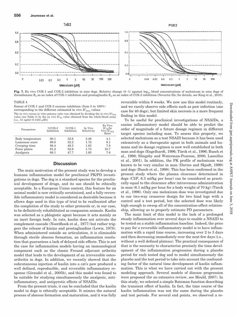
The whole-blood ex vivo assay is useful for predicting theclinical relevance of given levels of COX-1 and COX-2 inhi-bition (Fig. 7). For the present experiment, it was calculatedthat the plasma concentration corresponding to our esti-mated in vivo IC50A value would ensure ex vivo an inhibitionfrom approximately 80 (body temperature) to 90% (others
end points) of the COX-2 isoenzyme and from 32.6 to 52.9%of the COX-1 isoenzyme (Table 4). The in vivo COX-2versus COX-1 selectivity, as evaluated by the ratio of COX-2/COX-1 inhibition for a plasma concentration correspond-ing to the in vivo IC50A value, was from 1.72 to 2.46depending on the end point. The in vitro IC50 value (51ng/ml) versus the in vivo IC50A ratio was from 4.1 to 10.7depending on the end point.
Fig. 5. Simulated values of four investigated end points after a single subcutaneous meloxicam administration at 0 (placebo), 0.055, 0.1, 0.2 (thickline), 0.3, 0.5, and 2 mg/kg b.wt. A–D, body temperature (°C) (A), lameness score (B), vertical force exerted by the paw expressed as a percentage ofthe maximal observed response to kaolin (C), and paw withdrawal time expressed as a percentage of the maximal observed response to kaolin to a heatstimulus (D). Kaolin and meloxicam were administered at 24 and 48 h, respectively. The presence of a rebound (compared with the placebo period)is clearly seen for the four end points.
Fig. 6. Dose-response relationships for meloxicam. The effects [bodytemperature (—), withdrawal time (••••) of the paw as a measure ofanalgesia, and lameness score (-• -• - )] were simulated with the PK/PDmodel using mean PK and PD parameters; the relationship between thedose of meloxicam (mg/kg) and the maximal response obtained for eachdose were fitted with a inhibitory sigmoidal model (see eq. 8 in the text).
PK/PD Modeling of NSAIDs in a Dog Model 555
at ASPE
T Journals on June 3, 2018
jpet.aspetjournals.orgD
ownloaded from
DiscussionThe main motivation of the present study was to develop a
humane inflammation model for preclinical PK/PD investi-gations in dogs. The dog is a standard species for the preclin-ical development of drugs, and its use should be ethicallyacceptable. In a European Union context, this feature for ananimal model is now critically scrutinized, and a fully revers-ible inflammatory model is considered as desirable because itallows dogs used in this type of trial to be reallocated afterthe completion of the study to other protocols or, in our case,to be definitively rehabilitated as companion animals. Kaolinwas selected as a phlogistic agent because it acts mainly asan inert foreign body. In rats, kaolin does not activate thecomplement cascade (Noordhoek et al., 1977) but rather trig-gers the release of kinins and prostaglandins (Lewis, 1978).When administered outside an articulation, it is eliminablethrough sterile abscess formation, an inflammation resolu-tion that guarantees a lack of delayed side effects. This is notthe case for inflammation models having an immunologicalcomponent such as the classic Freund adjuvant arthritismodel that leads to the development of an irreversible osteo-arthritis in dogs. In addition, we recently showed that thesubcutaneous injection of kaolin into a cat’s paw produced awell defined, reproducible, and reversible inflammatory re-sponse (Giraudel et al., 2005b), and this model was found tobe suitable for studying simultaneously the analgesic, anti-inflammatory, and antipyretic effects of NSAIDs.
From the present trials, it can be concluded that the kaolinmodel in dogs is ethically acceptable. It mimics the naturalprocess of abscess formation and maturation, and it was fully
reversible within 8 weeks. We now use this model routinely,and we rarely observe side effects such as paw infection (onecase for 40 dogs), but limited skin necrosis is a more frequentfinding in this model.
To be useful for preclinical investigations of NSAIDs, acanine inflammatory model should be able to predict theorder of magnitude of a future dosage regimen in differenttarget species including man. To assess this property, weselected meloxicam as a test NSAID because it has been usedextensively as a therapeutic agent in both animals and hu-mans and its dosage regimen is now well established in bothman and dogs (Engelhardt, 1996; Turck et al., 1996; Busch etal., 1998; Slingsby and Waterman-Pearson, 2000; Lascelleset al., 2001). In addition, the PK profile of meloxicam wasknown to be very similar in man (Davies and Skjodt, 1999)and dogs (Busch et al., 1998). This has been confirmed in thepresent study where the plasma clearance determined indogs (Cl/F, 6.3 ml/kg per hour) can be considered as practi-cally equal to the clearance after intravenous administrationin man (6.1 ml/kg per hour for a body weight of 70 kg) (Turcket al., 1996). Only one meloxicam dose was investigated dueto our two-way crossover design for two hind legs with acontrol and a test period, but the selected dose was likelyhigh enough to sweep all of the concentration-effect relation-ship, allowing us to properly estimate PD parameters.
The main limit of this model is the lack of a prolongedsteady inflammation over several days to enable a NSAID tobe tested on a stable inflammation baseline. Indeed, the priceto pay for a reversible inflammatory model is to have inflam-mation with a rapid time course, increasing over 2 to 3 daysand then decreasing immediately over the next few days (i.e.,without a well defined plateau). The practical consequence ofthat is the necessity to characterize precisely the time devel-opment of the inflammation progression during a placeboperiod for each tested dog and to model simultaneously theplacebo and the test period to take into account the confound-ing factor of the natural time development of kaolin inflam-mation. This is what we have carried out with the presentmodeling approach. Several models of disease progressionwere proposed (for an extensive review, see Mould, 2007). Inthis study, we selected a simple Bateman function describingthe transient effect of kaolin. In fact, the time course of thekaolin inflammation was not the same during the placeboand test periods. For several end points, we observed a re-
Fig. 7. Ex vivo COX-1 and COX-2 inhibition in nine dogs. Relative change (0–1) against log10 blood concentrations of meloxicam in nine dogs ofthromboxane B2 as an index of COX-1 inhibition and prostaglandin E2 as an index of COX-2 inhibition (Novartis file; for details, see King et al., 2010).
TABLE 4Extent of COX-1 and COX-2 enzyme inhibition (from 0 to 100%)corresponding to the different estimated in vivo IC50A valuesThe in vivo versus in vitro potency ratio was obtained by dividing the in vivo IC50Avalue (see Table 3) by the ex vivo IC50 value obtained from the whole-blood assay�i.e., 51 ng/ml (0.1454 �M)�.
Parameters %COX-2Inhibition
%COX-1Inhibition
In VivoSelectivity
Ex VivoIC50/In
Vivo IC50ARatio
Body temperature 80.1 32.6 2.46 4.1Lameness score 89.8 52.2 1.72 9.1Creeping time 88.4 48.3 1.83 7.8Force plates 91.2 52.9 1.73 10.7Analgesia 88.1 47.7 1.85 7.6
556 Jeunesse et al.
at ASPE
T Journals on June 3, 2018
jpet.aspetjournals.orgD
ownloaded from

bound effect (i.e., that after cessation of meloxicam action thelevels of paw inflammation were greater than those obtainedat the same time after placebo dosing). A possible explana-tion for this delaying action of meloxicam on the disappear-ance of the inflammation is the classically reported impedingaction of NSAIDs on abscess formation that is required tophysically eliminate the administered kaolin. Therefore, wetook into account in our mathematical model this effect ofmeloxicam on the progression of the inflammation by modu-lating the actual value of P2 (see eq. 4), the parameter re-flecting the rate of inflammation disappearance. P2 was mod-ulated by the actual meloxicam concentration through a Hillinhibitory function (see eq. 7). This part of our model pre-dicted that for a high meloxicam plasma concentration thetime development of the kaolin action was slowed down tem-porarily or even stopped. It should be noted that our model isnot equivalent to classic paw edema as observed in rats afterthe intra-articular administration of a phlogistic agent. Inour case, the increase in the paw volume was associated withlymph node hypertrophy (popliteal), which can be inter-preted as a lymphatic drainage blockade protecting the ani-mal from systemic exposure to kaolin particles. This explainswhy meloxicam (as other NSAIDs) has no short-term effecton the paw diameter. As an alternative to this model, we alsoexplored a precursor-dependent indirect PD response model,as outlined by Sharma et al. (1998), to describe tolerance andrebound phenomena. This model fit our data well and alsowas able to capture adequately the rebound phenomena.However, it was associated with generally poor precision inthe individual estimates of IC50 values, and this was proba-bly due to a problem of numerical identifiability. It is possiblethat investigating several dose rates with a population mod-eling approach might have rendered this kind of PK/PDmodel usable, because only a single dose could be tested foreach dog based on our crossover design with two periods(placebo and test article) corresponding to the two hindlimbs. This is a possible limitation to this paw model, becausestudying a single dose rate for a NSAID may lead to someestimation difficulties for PD parameters. This would ariseespecially if the tested dose was too low. Using our PK/PDmodel, we estimated the meloxicam potency for different endpoints of clinical relevance, and we also confirmed the gen-eral statement that potency is usually similar between spe-cies (Levy, 1993; Busch et al., 1998), because the presentresults are very similar to those obtained in cats using thesame model (Giraudel et al., 2005a) and in man (Davies andSkjodt, 1999). Because the meloxicam kinetic parameters(clearance and half-life) and meloxicam potency are verysimilar in dogs and man, the present canine model should beable to predict a relevant dosage regimen in man. In thisstudy, simulations for the different end points using thePK/PD model for different dose levels allowed us to estimatean ED50 value of 0.05 mg/kg for an antipyretic effect, an ED50
value of 0.09 for analgesia, and an ED50 value of 0.127 mg/kgfor the locomotion end points (vertical force assessed by forceplate). These doses are very similar to the current clinicallyrecommended dose not only in dogs (0.1 or 0.2 mg/kg) but alsoin humans (7.5–15 mg, i.e., between 0.1 and 0.3 mg/kg for abody weight of 50–70 kg). This comparison demonstrates thepotential usefulness of this preclinical PK/PD modeling ap-proach for predicting a dosage regimen, not only in the dog asa target species but also in other species including man. In
both instances, well designed clinical studies will be neededto confirm or adapt the dosage regimen to clinical conditionsthat might be less severe than those in this acute paw in-flammation model.
Because the in vivo determined potencies calculated in thepresent study are likely to reflect meloxicam effects on pros-taglandin synthesis, it is relevant to compare these in vivoIC50A values with the potency for COX-2 inhibition obtainedin vitro or ex vivo with the whole-blood assay. We showedthat the plasma concentration corresponding to our esti-mated in vivo IC50A value would ensure, in a canine ex vivowhole-blood assay, an inhibition of approximately 80% forbody temperature and 90% for the other end points of theCOX-2 isoenzyme. These results are consistent with what weobserved for several other NSAIDs (i.e., that an inhibitionclose to 90% of the COX-2 isoenzyme is predictive of clinicalefficacy) (Toutain, 2002). Thus, the kaolin model could berelevant for in vitro (ex vivo) to in vivo extrapolations.
In conclusion, we have developed an ethical dog inflamma-tion model and using meloxicam as a reference NSAID. Weshowed that this model would be useful to investigate dose-effect relationships of NSAIDs and also could be relevant forin vitro to in vivo extrapolations. The limitation of the modelis the requirement of a rather advanced PK/PD analysis toaccurately estimate the PD parameters of interest.
Acknowledgments
We thank Jean-Pierre Gau and Simone Baures for skilled techni-cal assistance.
Authorship Contributions
Participated in research design: Jeunesse, C. Toutain, Letellier,Giraudel, and P. Toutain.
Conducted experiments: Jeunesse, Bargues, and C. Toutain.Contributed new reagents or analytic tools: Lacroix.Performed data analysis: Jeunesse and P. Toutain.Wrote or contributed to the writing of the manuscript: Jeunesse,
Letellier, and P. Toutain.
ReferencesAragon CL, Hofmeister EH, and Budsberg SC (2007) Systematic review of clinical
trials of treatments for osteoarthritis in dogs. J Am Vet Med Assoc 230:514–521.Botrel MA, Haak T, Legrand C, Concordet D, Chevalier R, and Toutain PL (1994)
Quantitative evaluation of an experimental inflammation induced with Freund’scomplete adjuvant in dogs. J Pharmacol Toxicol Methods 32:63–71.
Brooks RR, Carpenter JF, Jones SM, Ziegler TC, and Pong SF (1991) Caninecarrageenin-induced acute paw inflammation model and its response to nonsteroi-dal antiinflammatory drugs. J Pharmacol Methods 25:275–283.
Busch U, Schmid J, Heinzel G, Schmaus H, Baierl J, Huber C, and Roth W (1998)Pharmacokinetics of meloxicam in animals and the relevance to humans. DrugMetab Dispos 26:576–584.
Cross AR, Budsberg SC, and Keefe TJ (1997) Kinetic gait analysis assessment ofmeloxicam efficacy in a sodium urate-induced synovitis model in dogs. Am J VetRes 58:626–631.
Davies NM and Skjodt NM (1999) Clinical pharmacokinetics of meloxicam. A cyclo-oxygenase-2 preferential nonsteroidal anti-inflammatory drug. Clin Pharmacoki-net 36:115–126.
Dayneka NL, Garg V, and Jusko WJ (1993) Comparison of four basic models ofindirect pharmacodynamic responses. J Pharmacokinet Biopharm 21:457–478.
Engelhardt G (1996) Pharmacology of meloxicam, a new non-steroidal anti-inflammatory drug with an improved safety profile through preferential inhibitionof COX-2. Br J Rheumatol 35 (Suppl 1):4–12.
Giraudel JM, Diquelou A, Laroute V, Lees P, and Toutain PL (2005a) Pharmacoki-netic/pharmacodynamic modelling of NSAIDs in a model of reversible inflamma-tion in the cat. Br J Pharmacol 146:642–653.
Giraudel JM, Diquelou A, Lees P, and Toutain PL (2005b) Development and valida-tion of a new model of inflammation in the cat and selection of surrogate endpointsfor testing anti-inflammatory drugs. J Vet Pharmacol Ther 28:275–285.
Giraudel JM, King JN, Jeunesse EC, Lees P, and Toutain PL (2009) Use of apharmacokinetic/pharmacodynamic approach in the cat to determine a dosageregimen for the COX-2 selective drug robenacoxib. J Vet Pharmacol Ther32:18–30.
Hargreaves K, Dubner R, Brown F, Flores C, and Joris J (1988) A new and sensitive
PK/PD Modeling of NSAIDs in a Dog Model 557
at ASPE
T Journals on June 3, 2018
jpet.aspetjournals.orgD
ownloaded from

method for measuring thermal nociception in cutaneous hyperalgesia. Pain 32:77–88.
Institute of Laboratory Animal Resources (1996) Guide for the Care and Use ofLaboratory Animals 7th ed. Institute of Laboratory Animal Resources, Commis-sion on Life Sciences, National Research Council, Washington DC.
Josa M, Urizar JP, Rapado J, Dios-Vieitez C, Castaneda-Hernandez G, Flores-Murrieta F, Renedo MJ, and Troconiz IF (2001) Pharmacokinetic/pharmacody-namic modeling of antipyretic and anti-inflammatory effects of naproxen in therat. J Pharmacol Exp Ther 297:198–205.
King JN, Rudaz C, Borer L, Jung M, Seewald W, and Lees P (2010) In vitro and exvivo inhibition of canine cyclooxygenase isoforms by robenacoxib: a comparativestudy. Res Vet Sci 88:497–506.
Lascelles BD, Henderson AJ, and Hackett IJ (2001) Evaluation of the clinical efficacyof meloxicam in cats with painful locomotor disorders. J Small Anim Pract 42:587–593.
Levy G (1993) The case for preclinical pharmacodynamics, in Integration of Phar-macokinetics, Pharmacodynamics, and Toxicokinetics in Rational Drug Develop-ment (Yacobi A, Skelly JP, Shah VP, and Benet LZ, eds) pp 7–13, Plenum Press,New York.
Lewis AJ (1978) A comparison of the anti-inflammatory effects of copper aspirinateand other copper salts in the rat and guinea pig. Agents Actions 8:244–250.
Mould DR (2007) Developing models of disease progression, in Pharmacometrics: TheScience of Quantitative Pharmacology (Ette EI and Willimas PJ eds) pp 547–582,John Wiley & Sons, Inc., New York.
Noordhoek J, Nagy MR, and Bonta LL (1977) Involvement of complement and kinins
in some non-immunogenic paw inflammations in rats. Agents Actions Suppl2:109–121.
Sharma A, Ebling WF, and Jusko WJ (1998) Precursor-dependent indirect pharma-codynamic response model for tolerance and rebound phenomena. J Pharm Sci87:1577–1584.
Slingsby LS and Waterman-Pearson AE (2000) Postoperative analgesia in the catafter ovariohysterectomy by use of carprofen, ketoprofen, meloxicam or tolfenamicacid. J Small Anim Pract 41:447–450.
Toutain PL (2002) Pharmacokinetic/pharmacodynamic integration in drug develop-ment and dosage-regimen optimization for veterinary medicine. AAPS PharmSci4:E38.
Toutain PL, Cester CC, Haak T, and Laroute V (2001) A pharmacokinetic/pharmacodynamic approach vs. a dose titration for the determination of a dosageregimen: the case of nimesulide, a Cox-2 selective nonsteroidal anti-inflammatorydrug in the dog. J Vet Pharmacol Ther 24:43–55.
Turck D, Roth W, and Busch U (1996) A review of the clinical pharmacokinetics ofmeloxicam. Br J Rheumatol 35 (Suppl 1):13–16.
Yamaoka K, Nakagawa T, and Uno T (1978) Application of Akaike’s informationcriterion (AIC) in the evaluation of linear pharmacokinetic equations. J Pharma-cokinet Biopharm 6:165–175.
Address correspondence to: Pierre-Louis Toutain, UMR1331 ToxalimPhysiologie, Ecole Nationale Veterinaire de Toulouse, 23 Chemin des Capelles,BP87614, 31076 Toulouse cedex 03, France. E-mail: [email protected]
558 Jeunesse et al.
at ASPE
T Journals on June 3, 2018
jpet.aspetjournals.orgD
ownloaded from