paul herzmark - university of california, berkeley
TRANSCRIPT

Paul Herzmark [email protected]
Ellen Robey lab
Molecular and Cell Biology
http://tinyurl.com/PaulHerzmark-microscopy
Microscopy

Microscopy Background-Koehler illumination, Numerical Aperture
fluorescence
Optimizing imaging
Comparing the different microscopes
Test:
• How can you make your fluorescent samples brighter?
• How can you keep them from bleaching and dying?

Lens and
rays

Ray tracing
•At infinity lines are parallel
•Light through the center is unbent
F1
LENS

Ray tracing
•At infinity lines are parallel
•Light through the center is unbent

Ray tracing
•At infinity lines are parallel
•Light through the center is unbent
•At infinity lines are parallel
•Light through the center is unbent
} Focal length

Ray tracing
} Focal length
F1
F2


Determine focal length of a lens
F1


Koehler
illumination The nuts and bolts
(But why Paul?)

Now make the specimen bright.

Light bulb illumination:
even, dim

Critical illumination: Focus the light onto the specimen
bright but uneven

Kohler illumination: Infinity focused light
even, bright
Focal length

Focal length
The light bulb is out of focus.
What is in focus?

The light bulb is out of focus.
What is in focus?


Numerical
Aperture
(NA)

NA=(sine of half angle) X (refractive index of immersion medium)
Air=1 oil =1.51


NA=(sine of half angle) X (refractive index of immersion medium)
High NA = high light gathering
High NA = high resolution


High NA = high resolution
XY resolution = 0.61 wavelength / NA
Z resolution = 2 X wavelength X refractive index medium / NA2
resolution means separation
This is different from see or detect.

High NA = high resolution
demonstration


Resolution means separation

Rayleigh d = 0.61wavelength (in air) / NA
Delta Vision
magnification NA Camera Pixel size (nm)
calculated resolution
(nm)
2 pixels equal (nm)
60 X 1.4 170 550 nm 240 340
40 X 1.3 260 550 nm 258 520
20 X 0.75 520 550 nm 447 1040
10 X 0.3 1010 550 nm 1118 2020
(Bin 1, no Optivar)
X-Y resolution (theoretical!)

d = 2 (wavelength)(refractive index medium) / NA 2
magnification NA calculated resolution (nm)
60 X 1.4 550 nm 847
40 X 1.3 550 nm 983
20 X 0.75 550 nm 2,953
10 X 0.3 550 nm 18,456
4 X 0.13 550 nm 98,284
Z resolution

A 50,000 volt electron has a wavelength of 0.0055nm.
The numerical aperture calculation:
1 degree aperture
Sine of 1o = 0.017
Index of refraction (vacuum) = 1
Rayleigh d = 0.61wavelength (in air) / NA
Resolution is 0.20nm.
Electron microscope

NA and magnification effects on image brightness
Brightness increases (NA)4 (epifluorescence)
Brightness decreases 1/magnification2
relative brightness
60 X 1.4 1.0
40 X 1.3 1.7
20 X 0.75 0.7
10 X 0.3 0.1


Fluorescence
microscopy

Fluorescence
demo

Jablonski diagram


Quantum Yield
Photons emitted
Photons absorbed Q=


Quinine •Anti malaria
•Anti fever
•tasty
•And fluorescent!
Fluorescent structures

GFP


A Comparison of methods of microscopy
But first a digression

Spherical Aberration
Light going near the edges of the lens focuses at
A different plane than light going near the center

You can see
this easily.
Rings on one side
Blur on the other.
Spherical Aberration
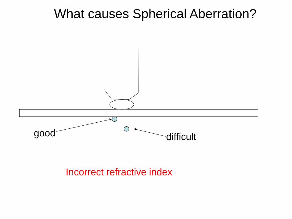
What causes Spherical Aberration?
Incorrect refractive index
good difficult

Confocal microscopy

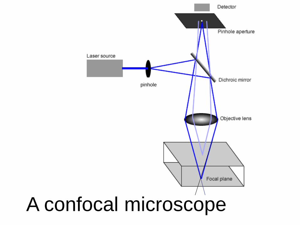
A confocal microscope
pinhole

A confocal microscope

What happens when you open
or close the pinhole?

Pinhole Diameter and Confocal slice thickness

Test:
Your fluorescence is too dim, what
can you do?

Test:
Your sample is bleaching, what can
you do?

Optional quiz question:
When should you use a
confocal microscope?


Ray tracing
•At infinity lines are parallel
•Light through the center is unbent
} Focal length
F1