pattern-specific loss of aquaporin-4 immunoreactivity - brain
TRANSCRIPT

Pattern-specific loss of aquaporin-4 immunoreactivitydistinguishes neuromyelitis optica frommultiplesclerosisShanu F. Roemer,1 Joseph E. Parisi,2 Vanda A. Lennon,1^3 Eduardo E. Benarroch,1Hans Lassmann,4
Wolfgang Bruck,5 Raul N. Mandler,6 Brian G.Weinshenker,1 Sean J. Pittock,1,2 Dean M.Wingerchuk7 andClaudia F. Lucchinetti1
Departments of 1Neurology, 2Laboratory Medicine and Pathology and 3Immunology, Mayo Clinic College of Medicine,Rochester, MN,USA, 4Center for the Clinical Trials Network, National Institutes of Health, Bethesda, MD , 5Department ofNeurology, Mayo Clinic College of Medicine, Scottsdale, AZ,USA, 6Center for Brain Research, Medical University of Vienna,Vienna, Austria and 7Department of Neuropathology, Institute for Multiple Sclerosis Research,Georg-August University,Gottingen,Germany
Correspondence to: Claudia F. Lucchinetti, MD, Neurology, Mayo Clinic, College of Medicine, 200 First St. SW, Rochester,MN 55905, USAE-mail: [email protected]
Neuromyelitis optica (NMO) is an inflammatory demyelinating disease that typically affects optic nerves andspinal cord. Its pathogenic relationship tomultiple sclerosis (MS) is uncertain.Unlike MS,NMOlesions are char-acterized by deposits of IgG and IgM co-localizing with products of complement activation in a vasculocentricpattern around thickened hyalinized blood vessels, suggesting a pathogenic role for humoral immunity targetingan antigen in the perivascular space. A recently identified specific serum autoantibody biomarker, NMO-IgG,targets aquaporin-4 (AQP4), themost abundant water channel protein in the CNS, which is highly concentratedin astrocytic foot processes.We analysed and compared patterns of AQP4 immunoreactivity in CNS tissues ofnine patients with NMO, 13 with MS, nine with infarcts and five normal controls. In normal brain, optic nerveand spinal cord, the distribution of AQP4 expression resembles the vasculocentric pattern of immune complexdeposition observed in NMOlesions. In contrast to MS lesions, which exhibit stage-dependent loss of AQP4, allNMO lesions demonstrate a striking loss of AQP4 regardless of the stage of demyelinating activity, extent oftissue necrosis, or site of CNS involvement.We identified a novel NMO lesion in the spinal cord and medullarytegmentum extending into the area postrema, characterized by AQP4 loss in foci that were inflammatory andoedematous, but neither demyelinated nor necrotic. Foci of AQP4 loss coincided with sites of intensevasculocentric immune complex deposition.These findings strongly support a role for a complement activatingAQP4-specific autoantibody as the initiator of the NMOlesion, and further distinguish NMO from MS.
Keywords: neuromyelitis optica; aquaporin-4; multiple sclerosis; demyelination; astrocyte; blood^brain barrier
Abbreviations: AQP4¼ aquaporin-4; HIVE¼human immunodeficiency virus encephalitis; GFAP¼ glial fibrillary acidicprotein; NMO¼Neuromyelitis optica; PML¼ progressive multifocal leucoencephalopathy
Received August 25, 2006. Revised November 3, 2006. Accepted December 12, 2006. Advance Access publication February 5, 2007
IntroductionNeuromyelitis optica (NMO) is an idiopathic and usuallyrelapsing inflammatory demyelinating disease of the CNScharacterized by severe attacks of optic neuritis andmyelitis. It can be distinguished from classical multiplesclerosis (MS) by clinical, neuroimaging, CSF and
serological criteria (Wingerchuk et al., 1999, 2006).In contrast to classical MS, which is thought to bemediated by effector T cells, increasing evidence supportsa role for an autoantibody-mediated pathogenesis in NMO(Lucchinetti et al., 2002). NMO is often associated with
doi:10.1093/brain/awl371 Brain (2007), 130, 1194^1205
� The Author (2007). Published by Oxford University Press on behalf of the Guarantors of Brain. All rights reserved. For Permissions, please email: [email protected]
Dow
nloaded from https://academ
ic.oup.com/brain/article/130/5/1194/282288 by guest on 27 N
ovember 2021

other organ-specific or non-organ-specific autoimmunedisorders, and a variety of serum autoantibodies(O’Riordan et al., 1996; Wingerchuk et al., 2006). Inaddition, plasmapheresis is therapeutically effective in NMOpatients with severe steroid-unresponsive relapses (Keeganet al., 2002). NMO lesions are characterized by extensivedemyelination with partial necrosis in the spinal cord,involving both grey and white matter, and usuallyextending over multiple segments (Lucchinetti et al.,2005). The lesions are associated with acute axonal injury,and inflammatory cell infiltrates containing eosinophils andgranulocytes. Deposits of IgG and IgM co-localize withproducts of complement activation in a vasculocentric patternaround thickened hyalinized blood vessels, suggesting apathogenic role for humoral immunity targeting an antigenin the perivascular space (Lucchinetti et al., 2002). Thishypothesis is strengthened by the recent identification of aspecific serum autoantibody biomarker, NMO-IgG (Lennonet al., 2004), which targets the most abundant water channelprotein in the CNS aquaporin-4 (AQP4), (Lennon et al., 2005).The anatomical distribution and cellular sites of AQP4
expression in normal mammalian tissues, including brainand spinal cord, have been investigated extensively. AQP4 isa homotetrameric integral plasma membrane proteinanchored in the astrocytic foot process membrane by thedystroglycan complex (Amiry-Moghaddam and Ottersen,2003). Prior studies demonstrate intense AQP4 immuno-reactivity in the plasma membrane of the astrocytic endfeetthat abut capillaries and pia in the brain and spinal cord,particularly in the subpial and subependymal zones, as wellas in the glial lamellae of the supraoptic nucleus in thehypothalamus (Jung et al., 1994; Frigeri et al., 1995; Nielsenet al., 1997; Venero et al., 1999; Amiry-Moghaddam andOttersen, 2003).The localization of AQP4 in the astrocytic foot processes
surrounding endothelial cells is consistent with the role ofastrocytes in the development, function and integrity of theinterface between brain parenchyma and perivascular space,and between brain and cerebrospinal fluid, and serves tomediate water flux (Nicchia et al., 2004). Experimental rodentmodels of ischaemia, trauma and hyponatraemia implicateAQP4 in the development of brain oedema regardless ofcause (Manley et al., 2000; Taniguchi et al., 2000; Saadounet al., 2002; Vajda et al., 2002; Warth et al., 2005).Enhanced AQP4 expression has been reported in a
wide spectrum of human neuropathological conditions,including ischaemia, trauma, brain tumours, bacterialmeningitis, progressive multifocal leucoencephalopathy(PML), human immunodeficiency virus encephalitis(HIVE), MS and Creutzfeldt–Jakob disease (Saadounet al., 2002; Aoki et al., 2003, 2005; Misu et al., 2006). Arecent report (Aoki et al., 2005), suggests that thedistribution of intensely AQP4-positive astrocytes differsamong disease states. In PML and HIVE, abundant AQP4-expressing astrocytes were noted at the boundary betweengrey and white matter apparently unrelated to
inflammatory foci. The absence of a comparative analysisof AQP4 expression in normal control brain tissue in thisstudy, however, may have led to erroneous attribution ofAQP4 expression patterns to disease states. Additionally,rostral–caudal regional differences in AQP4 mRNA expres-sion levels have been described (Venero et al., 1999, 2001).Thus, it is essential to compare similar regions in diseasedand normal control brain, before inferring a ‘pathologicalchange’ in AQP4 distribution and expression level.Upregulated AQP4 was reported in astrocytes located at theperiphery of MS plaques, but the plaque centre andunaffected normal appearing white matter areas wereessentially devoid of AQP4 (Aoki et al., 2005; Misu et al.,2006). However, the stage of demyelinating activity within theMS lesions was not specified, precluding determination ofany association of lesional stage with AQP4 expression.
The distribution of AQP4 at glial–fluid interfaces in themouse spinal cord coincides with sites to which NMO-IgGbinds, and is similar to the deposition pattern of Ig andcomplement activation products observed in activelydemyelinating NMO lesions (Lucchinetti et al., 2002;Lennon et al., 2004). These observations make a compellingbut circumstantial case for AQP4-IgG being a primaryeffector of NMO lesions. A recent immunohistochemicalstudy of a single NMO case reported perivascular loss ofAQP4 immunoreactivity in spinal cord lesions (Misu et al.,2006). The authors contrasted this observation withhigh AQP4 expression in a section of normal cervicalspinal cord grey matter, and with an apparent increase ofAQP4 expression in reactive astrocytes of MS lesions.This study, however, did not specify the number of MScases, the number of lesions analysed, or the stage ofdemyelinating activity in any of the NMO or MS lesions.Furthermore, the observed loss of AQP4 in somelesions corresponded to regions of tissue necrosis andcavitation. Although the authors suggested that functionalimpairment of astrocytes by AQP4-specific antibodymay underlie NMO lesion pathogenesis, they did not addressthe potential influence of other biological factors on AQP4expression, such as demyelinating or remyelinating activity,the extent of tissue necrosis, or the degree of astrocyte loss.
The present study describes AQP4 expression in a largeseries of NMO cases that are well characterized clinicallyand pathologically (Lucchinetti et al., 2002). We compareexpression patterns of AQP4 immunoreactivity in NMOlesions with patterns observed in normal brain, optic nerveand spinal cord; in acute, subacute and chronic infarcts ofbrain, optic nerve and spinal cord; and in acute andchronic MS lesions. We observed that the pattern of AQP4expression in normal tissues is similar to the rim androsette pattern of Ig deposits and products of complementactivation observed in NMO lesions. In contrast to a stage-dependent loss of AQP4 in MS lesions, we observed in allNMO lesions a striking loss of AQP4 regardless of the stageof demyelinating activity, extent of tissue necrosis, orCNS region involved. We further observed AQP4 loss at
Aquaporin-4 immunoreactivity, neuromyelitis optica and MS Brain (2007), 130, 1194^1205 1195
Dow
nloaded from https://academ
ic.oup.com/brain/article/130/5/1194/282288 by guest on 27 N
ovember 2021

sites of vasculocentric Ig deposition and complement activa-tion in NMO lesions that lacked demyelination, but wereinflammatory. These lesions were identified in both the spinalcord and medulla at the floor of the fourth ventricle,particularly involving the area postrema, a region lackinga blood–brain barrier (BBB) and rich in osmoreceptors. Thepathogenic implications of these observations are discussed.
Material and methodsArchival materialThe study was performed on archival brain, optic nerve andspinal cord material from nine patients with NMO, 13 withMS, nine with infarcts and five neurologically normalpatients without CNS histopathological abnormalities. Thestudy was approved by the Mayo Clinic Institutional ReviewBoard (2067–99). Causes of death in the control cases wereacute myocardial infarct (3), pneumonia (1) and unknown (1).All materials were obtained at autopsy except for two acute MScases that were obtained surgically (diagnostic brain biopsy).Detailed clinical histories were available for all cases. The NMOcohort comprised eight women and one man, with an average ageof 50 years (range 16–80 years). The clinical course was relapsingin eight patients, and monophasic in a single case. Mean diseaseduration was 2.4 years (SEM� 0.8 years). All patients died fromrespiratory compromise directly attributable to attacks of NMO.The presenting syndrome was optic neuritis in four patients, andmyelitis in five patients, with a mean interval of 19months (range4–41months) between optic neuropathy and myelopathy. The MScohort comprised: (i) 11 acute MS cases (nine women and twomen) with an average age of 43 years (range 26–70 years). Meandisease duration was 4.1months (SEM� 13.6months). Death wasattributable to an acute MS attack associated with herniation infive cases, sepsis due to pneumonia in three and unknown in one.Two patients are still living. (ii) Two chronic cases, both women(aged 36 and 71 years) who had a secondary progressivecourse and mean disease duration of 18 years (6 and 30 years).
Both patients died of respiratory compromise associated withpneumonia.
Neuropathological evaluation andimmunohistochemistrySpecimens were fixed in 10–15% formalin and embedded inparaffin. Sections were stained with haematoxylin and eosin (HE),Luxol-fast blue-periodic acid-Schiff (LFB/PAS) and Bielschowskysilver impregnation. Immunohistochemistry was performedwithout modification using an avidin–biotin or an alkaline-phosphatase/anti-alkaline phosphatise (APAAP) technique asdescribed previously (Vass et al., 1986). The primary antibodieswere specific for myelin proteins (myelin basic protein [MBP;Boehringer Mannheim, Germany], proteolipid protein [PLP,polyclonal; Serotec, Oxford, USA], myelin oligodendrocyteprotein [MOG; Dr S Piddlesden, Department of Biochemistry,Cardiff, UK], 2030-cyclic-nucleotide 30-phosphodiesterase,[CNPase; Sternberger, USA], myelin associated glycoprotein[MAG; Serotec]), glial fibrillary acidic protein (GFAP; Dako),neurofilament protein (NF; Dako), T lymphocytes (all T cells;CD3 and cytotoxic CD8 T cells; [Dako]), B lymphocytes (CD20;Dako), plasma cells (CD138; Dako), macrophages/microglialcells (KiM1P; Dr Radzun, University of Gottingen, Germany),activated complement antigen (C9neo; Dr Paul Morgan,Department of Biochemistry, Cardiff, UK), immunoglobulin G(IgG; Dako), immunoglobulin M (IgM; Dako) and AQP4(C-terminal residues 249–323, affinity purified rabbit IgG;Sigma-Aldrich). Table 1 lists the antibodies and specific conditionsused for immunohistochemistry. The primary antibodies wereomitted in controls. All antibodies were incubated at 4�Covernight.
Staging of demyelinating activity in NMOand MSlesionsLesions were classified with respect to demyelinating activity, aspreviously described (Bruck et al., 1995). ‘Early active
Table 1 Antibodies used for immunohistochemistry
Antibody Clone Dilution Antigen retrieval Company/source
GFAP Polyclonal 1: 4000 NA Dako, DenmarkAQP4 Polyclonal 1: 250 NA Sigma-Aldrich, USANF Monoclonal 1: 800 Steam/EDTA Dako, DenmarkMBP Monoclonal 1:150 NA Boehringer, Mannheim; Roche,GermanyPLP Monoclonal 1:1000 NA Serotec, USACNPase Monoclonal 1: 2000 Steam/citric buffer Sternberger, USAMOG Monoclonal 1: 200 NA Dr Sarah Piddlesden; Cardiff, UKMAG Monoclonal 1:10 Proteinase K Chemicon, USAKiM1P Monoclonal 1:1000 Steam Dr Radzun; Goettingen,GermanyCD3 Monoclonal 1: 400 Steam/citric buffer Serotec, USACD8 Monoclonal 1: 50 Steam/citric buffer Dako, DenmarkCD20 Monoclonal 1: 60 Steam/EDTA Dako, DenmarkCD138 Monoclonal 1: 50 Steam/EDTA Dako, DenmarkIgG Polyclonal 1:10,000 Steam/EDTA Dako, DenmarkIgM Polyclonal 1: 750 Steam/EDTA Dako, DenmarkC9neo Polyclonal 1: 400 Steam Dr Paul Morgan; Cardiff, UKC9neo Monoclonal 1: 200 Protease 0.03% Dr Paul Morgan; Cardiff, UK
All monoclonal antibodies are from mouse and all polyclonals from rabbit. EDTA¼ ethylenediaminetetraacetic acid; NA¼not applicable.
1196 Brain (2007), 130, 1194^1205 S. F. Roemer et al.
Dow
nloaded from https://academ
ic.oup.com/brain/article/130/5/1194/282288 by guest on 27 N
ovember 2021

demyelinating lesions’ were diffusely infiltrated by macrophages
containing immunoreactive products for all myelin proteins. ‘Late
active demyelinating lesions’ were more advanced with respect to
myelin degradation, and were immunoreactive for major myelin
proteins (MBP and PLP), but not for MOG or CNPase.
‘Remyelinating lesions’ were characterized by uniformly thin and
irregularly arranged myelin sheaths. ‘Inactive demyelinated lesions’
were completely demyelinated without evidence of active demye-
lination. We examined 77 lesions from 9 autopsy cases of clinically
confirmed NMO. Demyelinating activity in the NMO lesions was
classified immunohistochemically as ‘early active’ in 22, ‘late
active’ in 18, ‘inactive’ in 37 and ‘remyelinating’ in 0.
Demyelinating activity in 57 lesions from the 11 acute MS cases
was classified as ‘early active’ in 45, ‘early remyelinating’ in 6 and
‘inactive’ in 6. The 2 chronic MS cases had no ‘active’ lesion, 24
‘inactive’, and 3 ‘late remyelinated’ lesions (i.e. shadow plaques).
Early active MS lesions in the acute MS cohort were further
classified immunopathologically based on previously published
criteria (Lucchinetti et al., 2000), into patterns I (T cell/
macrophage associated) (n¼ 2 lesions from two MS biopsy
cases), II (antibody/complement associated) (n¼ 30 lesions from
six cases), III (distal oligodendrogliopathy) (n¼ 11 lesions from
two cases) or IV (primary oligodendrogliopathy) (n¼ 2 lesions
from one case). We also evaluated AQP4 expression in nine NMO
lesions and seven MS lesions with cavitation.
ResultsTable 2 summarizes AQP4 immunoreactivity patterns ininfarct, MS and NMO tissues relative to baseline expressionin normal CNS control tissues. Specific characteristics ofAQP4 immunoreactivity are discussed subsequently.
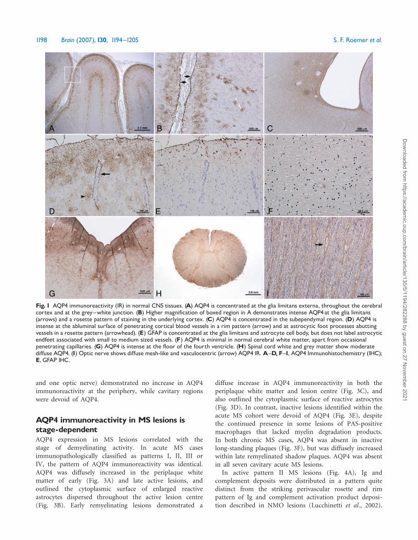
AQP4 immunoreactivity in normal CNStissuesAQP4 immunoreactivity in normal tissues, at all levels ofthe CNS, was most intense at the glia limitans externa andthe subependyma (Fig. 1A–C). Within the cerebral cortex,AQP4 immunoreactivity was concentrated in astrocytic footprocesses extending to the abluminal surface of bloodvessels (Fig. 1D). As is typical of astrocytes, GFAPimmunoreactivity was concentrated in the cell body andsoma, and did not extend to the astrocytic endfeetassociated with either small or medium-sized blood vessels(Simard et al., 2003), (Fig. 1E). Cerebral white mattershowed limited AQP4 immunoreactivity, which wasrestricted to the abluminal surface of occasional penetratingblood vessels (Fig. 1F). Within the brainstem (midbrain tocaudal medulla), AQP4 immunoreactivity was most intensein subependymal regions at the floor of the fourth ventricle(Fig. 1G). Brainstem white matter exhibited a mesh-likepattern of AQP4 immunoreactivity, with prominentperivascular staining in a rim and rosette pattern. In thespinal cord, AQP4 immunoreactivity was prominent withinboth grey and white matter (Fig. 1H). AQP4 immunor-eactivity in the optic nerve exhibited meshwork and rim
and rosette vasculocentric patterns (Fig. 1I), similar tothose seen in brainstem and spinal cord white matter.
AQP4 immunoreactivity in otherneurological disordersThe intensity and distribution of AQP4 immunoreactivityin brain, optic nerve and spinal cord infarcts werecompared with the baseline expression patterns observedin regionally matched normal control tissues. In acuteinfarcts (512 h; one cerebral and two spinal cord) andsubacute infarcts (57 days; one cerebral and two spinalcord), AQP4 was lost in the necrotic centre and diffuselyincreased at the periphery of the infarct (Fig. 2A and E).A similar pattern of GFAP immunoreactivity was seen inthe necrotic centre and ischaemic periphery (Fig. 2B).Higher magnification demonstrated increased AQP4 out-lining the cytoplasmic surface of reactive astrocytes(Fig. 2C), whereas GFAP was more concentrated in thecell body (Fig. 2D). AQP4 was lost in the necrotic zone(Fig. 2A, E and F). Chronic infarcts (42 years; two cerebral
Table 2 Aquaporin-4 immunoreactivity in infarct, MS andNMO relative to baseline expression in normal controls
Normal controls n¼ 5 GM WMCerebrum þþþ þ/�Brainstem þþþ þþ
Spinal cord þ þþ
Optic nerve NA þþ
Neurological disorders n¼ 31 Peri lesion LesionInfarct n¼ 9Acute (n¼ 3) """ 0Subacute (n¼ 3) """ 0Chronic (n¼ 3) $ 0
Chronic multiple sclerosis n¼ 2Inactive (n¼ 24) $ 0Late remyelinating (n¼ 3) "" ""
Acute multiple sclerosis� n¼11Active (n¼ 45) """ "
Inactive (n¼ 6) " 0Early remyelination (n¼ 6) "" ""
Cavitary (n¼ 7) "" 0Neuromyelitis opticay n¼ 9Active (n¼ 40) $ 0Inactive (n¼ 37) $ 0Cavitary (n¼ 9) $ 0
N ¼ number of cases; n, number of lesion areas; GM ¼ greymatter; WM ¼ white matter; NA ¼ not applicable; þ/�, þ, þþ,þþþ¼ intensity scale of AQP4 immunoreactivity in CNS controltissue; ", "", """ ¼ degree of increase in AQP4 immunoreactivityrelative to baseline expression in regionally matched CNScontrol tissue; $ ¼ no change in AQP4 immunoreactivity relativeto baseline expression in regionally matched CNS control tissue;0 ¼ complete loss of AQP4 immunoreactivity. �There is nodifference in AQP4 immunoreactivity pattern between immuno-pathologically classified acute MS cases (patterns I¼ 2, II¼ 6,III¼ 2, IV¼1). yAQP4 immunoreactivity is also absent ininflammatory NMO foci lacking demyelination associated withvasculocentric immune complex deposition.
Aquaporin-4 immunoreactivity, neuromyelitis optica and MS Brain (2007), 130, 1194^1205 1197
Dow
nloaded from https://academ
ic.oup.com/brain/article/130/5/1194/282288 by guest on 27 N
ovember 2021
and one optic nerve) demonstrated no increase in AQP4immunoreactivity at the periphery, while cavitary regionswere devoid of AQP4.
AQP4 immunoreactivity in MS lesions isstage-dependentAQP4 expression in MS lesions correlated with thestage of demyelinating activity. In acute MS casesimmunopathologically classified as patterns I, II, III orIV, the pattern of AQP4 immunoreactivity was identical.AQP4 was diffusely increased in the periplaque whitematter of early (Fig. 3A) and late active lesions, andoutlined the cytoplasmic surface of enlarged reactiveastrocytes dispersed throughout the active lesion centre(Fig. 3B). Early remyelinating lesions demonstrated a
diffuse increase in AQP4 immunoreactivity in both theperiplaque white matter and lesion centre (Fig. 3C), andalso outlined the cytoplasmic surface of reactive astrocytes(Fig. 3D). In contrast, inactive lesions identified within theacute MS cohort were devoid of AQP4 (Fig. 3E), despitethe continued presence in some lesions of PAS-positivemacrophages that lacked myelin degradation products.In both chronic MS cases, AQP4 was absent in inactivelong-standing plaques (Fig. 3F), but was diffusely increasedwithin late remyelinated shadow plaques. AQP4 was absentin all seven cavitary acute MS lesions.
In active pattern II MS lesions (Fig. 4A), Ig andcomplement deposits were distributed in a pattern quitedistinct from the striking perivascular rosette and rimpattern of Ig and complement activation product deposi-tion described in NMO lesions (Lucchinetti et al., 2002).
Fig. 1 AQP4 immunoreactivity (IR) in normal CNS tissues. (A) AQP4 is concentrated at the glia limitans externa, throughout the cerebralcortex and at the grey^white junction. (B) Higher magnification of boxed region in A demonstrates intense AQP4at the glia limitans(arrows) and a rosette pattern of staining in the underlying cortex. (C) AQP4 is concentrated in the subependymal region. (D) AQP4 isintense at the abluminal surface of penetrating cortical blood vessels in a rim pattern (arrow) and at astrocytic foot processes abuttingvessels in a rosette pattern (arrowhead). (E) GFAP is concentrated at the glia limitans and astrocyte cell body, but does not label astrocyticendfeet associated with small to medium sized vessels. (F) AQP4 is minimal in normal cerebral white matter, apart from occasionalpenetrating capillaries. (G) AQP4 is intense at the floor of the fourth ventricle. (H) Spinal cord white and grey matter show moderatediffuse AQP4. (I) Optic nerve shows diffusemesh-like and vasculocentric (arrow) AQP4 IR.A^D, F^I, AQP4 Immunohistochemistry (IHC);E, GFAP IHC.
1198 Brain (2007), 130, 1194^1205 S. F. Roemer et al.
Dow
nloaded from https://academ
ic.oup.com/brain/article/130/5/1194/282288 by guest on 27 N
ovember 2021

The inflammatory infiltrates in pattern II MS lesionswere lymphocytic and products of complement activationwere less pronounced in degree, and were detected ondegenerating myelin sheaths, within macrophages and onoligodendrocytes along the active plaque edge (Fig. 4B)(Lucchinetti et al., 2000). AQP4 immunoreactivity wasless intense in the plaque centre relative to its increasedexpression in the periplaque white matter, and outlinedthe cytoplasm of reactive astrocytes, as well as astrocyticfoot processes surrounding blood vessels (Fig. 4C).
AQP4 immunoreactivity in NMOlesionsThe pattern of AQP4 expression in NMO lesions wasfundamentally different. No AQP4 immunoreactivity wasdetectable in any lesion, irrespective of stage of demyelinat-ing activity. Actively demyelinated NMO lesions (Fig. 4D)
contained granulocytes and eosinophils, and exhibited astriking vasculocentric deposition of Ig and the C9neoproduct of complement activation (Fig. 4E) in a rim androsette pattern (Fig. 4E, inset), in regions of AQP4 loss(Fig. 4F). Periplaque white matter demonstrated a similardegree of AQP4 immunoreactivity as normal regionallymatched control tissue (Fig. 4F). The identical pattern ofAQP4 loss independent of demyelinating stage wasalso observed in the optic nerve of a single NMO case[early active (Fig. 5A and B), inactive, and cavitary lesionareas], in contrast to the stage-dependent pattern ofperiplaque and lesion AQP4 expression observed in earlyactive (Fig. 5C, D), remyelinated, inactive and cavitary opticnerve lesion areas from a single acute MS case.
An unanticipated observation was loss of AQP4 in spinalcord (Fig. 6C) and brainstem regions characterized byeosinophil and plasma cell infiltrates and vasculocentricdeposits of IgM, IgG (Fig. 6A) and complement activationproducts (Fig. 6B), but lacking evidence of demyelination(n¼ 5 cases) (Fig. 6D and E). These regions appearedrarefied, particularly around blood vessels (Fig. 6D),and despite AQP4 loss, they retained normal staining of allmyelin proteins [MBP, PLP, CNPase, MAG, MOG (Fig. 6E)],and lacked macrophages containing myelin degradationproducts. Axons were structurally preserved with noevidence of acute axonal pathology (Fig. 6F).
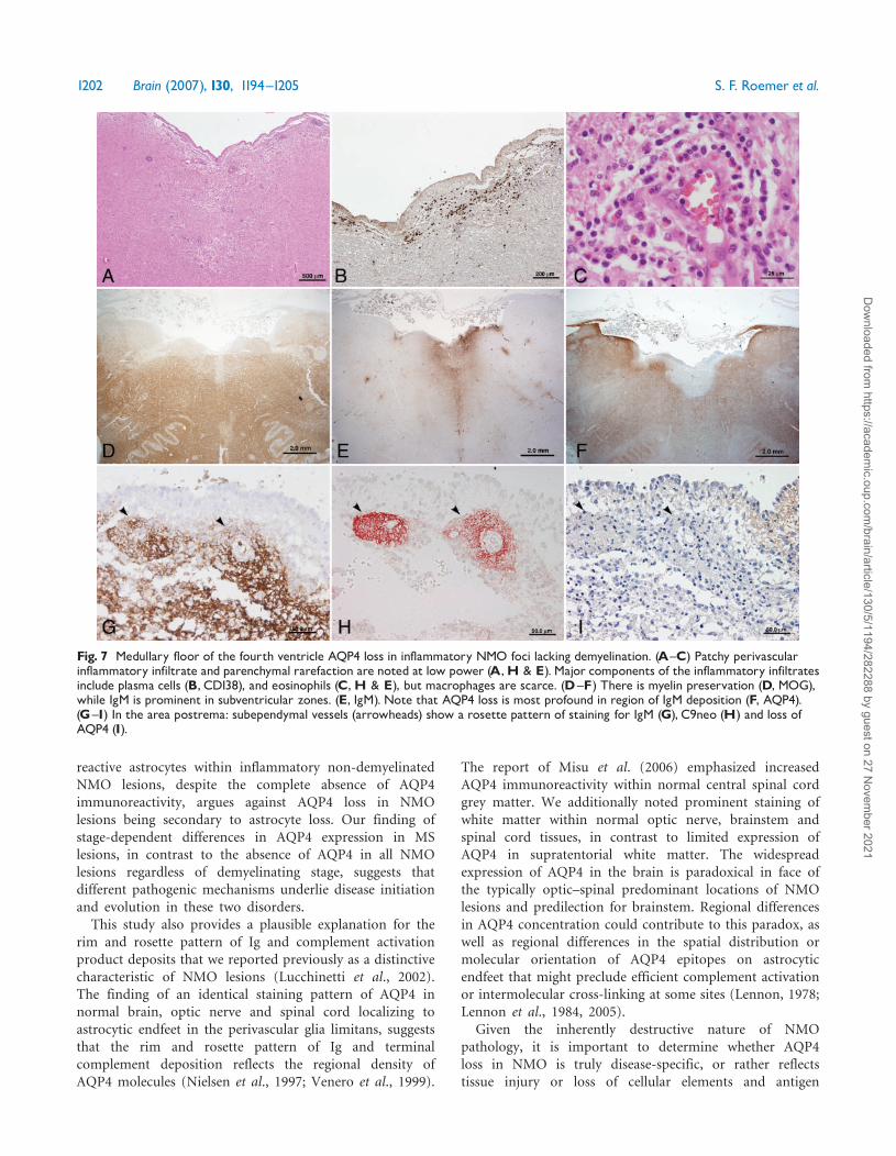
Of particular note, three of the NMO cases hadinflammatory foci lacking demyelination associated withAQP4 loss situated below the floor of the fourth ventricleextending laterally along the subependymal surface (Fig. 7).The involved tissue appeared rarefied (Fig. 7A), andcontained numerous perivascular and parenchymalCD138þ plasma cells (Fig. 7B) and eosinophils (Fig. 7C).Myelin was preserved in the subependymal white matter(Fig. 7D). IgG and IgM deposits were diffuse (Fig. 7E),as well as vasculocentric (Fig. 7G), and serial sectionsrevealed they co-localized with products of complementactivation (Fig. 7H) and regions of AQP4 loss (Fig. 7F andI). These inflammatory foci associated with AQP4 lossextended into the adjacent area postrema (Fig. 7G–I), a siteknown to lack a BBB.
Despite a complete loss of AQP4 immunoreactivity(Fig. 8A and C), reactive GFAP positive astrocyteswere dispersed throughout these inflammatory, non-demyelinated NMO foci (Fig. 8B and D). This is incontrast to active (Fig. 8E and F; Fig. 3B) and remyelinating(Fig. 3C and D) MS lesions, as well as acute and subacuteinfarcts (Fig. 2A, C and E), in which AQP4 immuno-reactivity is diffusely increased, and outlines the cytoplasmicsurface of GFAP positive astrocytes.
DiscussionThe relationship between MS and NMO has long beendebated. We previously reported a unique pattern of tissuedestruction in active NMO lesions characterized by
Fig. 2 AQP4 immunoreactivity (IR) in early acute (A^D) andsubacute (E^F) infarcts. (A) The early acute infarct demonstratesloss of AQP4 in the necrotic centre (*), with increased IR inthe periphery. (B) With GFAP, there is a similar pattern anddistribution of IR, absent in the necrotic centre (*), and increasedat the periphery. (C) Higher magnification of the infarct peripheryin A demonstrates increased AQP4 outlining the cytoplasmicsurface of reactive astrocytes (arrowheads). (D) Higher magnifica-tion of the infarct periphery in B demonstrates GFAP is similarlyincreased in reactive astrocytes, but concentrated in cell bodies(arrowheads). (E) The subacute infarct also demonstratesincreased AQP4 IR in the periphery, with relative lack of AQP4IR in macrophage-rich necrotic areas (*). (F) There is absenceof AQP4 in the necrotic zone. A,C, E, F, AQP4 IHC;B,D, GFAP IHC.
Aquaporin-4 immunoreactivity, neuromyelitis optica and MS Brain (2007), 130, 1194^1205 1199
Dow
nloaded from https://academ
ic.oup.com/brain/article/130/5/1194/282288 by guest on 27 N
ovember 2021

eosinophil and neutrophil infiltration, vascular fibrosis andintense vasculocentric deposition of Ig and products ofcomplement activation in a characteristic ‘rim’ and ‘rosette’pattern (Lucchinetti et al., 2002). Based on those findings,we suggested that humoral autoimmunity targeting anantigen in the perivascular space may play a role in thepathogenesis of NMO. Lennon et al. subsequently identified
a specific marker autoantibody (NMO-IgG), which interactsselectively with the AQP4 water channel (Lennon et al.,2005). AQP4 is concentrated at the astrocytic endfeet,which are typically GFAP-negative (Frigeri et al., 1995;Bushong et al., 2002; Simard et al., 2003). Whereas GFAP-positive processes do not systematically cover smallervessels (516 mm) and capillaries, AQP4 expression outlines
Fig. 4 AQP4 immunoreactivity (IR) in acute pattern II MS (A^C) and NMO (D^F) lesions. (A) Numerous macrophages containing myelindebris are dispersed throughout the active lesion (arrowheads and inset; LFB/PAS). (B) C9neo antigen is present within macrophages(arrowheads), but absent around blood vessels (arrow). (C) Higher magnification reveals AQP4 IR is prominent in a rosette patternsurrounding a penetrating blood vessel in the lesion. (D) In NMO, there is extensive demyelination involving both grey and white matter(LFB/PAS); *indicates preserved myelin in the PPWM. (E) C9neo is deposited in a vasculocentric rim and rosette pattern (inset) withinthe active lesions, but not in the PPWM. (F) The lesions lack AQP4, which is retained in the PPWM (*) and grey matter. A,D, LFB/PAS;B, E, C9neo IHC; C, F, AQP4 IHC.
Fig. 3 Stage-dependent pattern of AQP4 immunoreactivity (IR) in active (A, B), early remyelinated (C, D) and inactive (E, F) MS lesions.(A) An active MS lesion demonstrates increased AQP4 IR that is marked in the adjacent cortical grey matter (*) and periplaque whitematter (PPWM), and moderate in the lesion centre (þ). (B) Higher magnification of the active lesion centre demonstrates AQP4 outliningthe cytoplasmic surface of reactive astrocytes (arrowheads) and their processes. (C) An early remyelinated MS lesion (circle) showsdiffuse increase in AQP4 extending throughout the lesion, and surrounding PPWM. (D) AQP4 stains the cytoplasmic surface of reactiveastrocytes (arrowheads) in an early remyelinated lesion. (E^F). Inactive MS lesions from an acute case (E) and a chronic case (F) showcomplete loss of AQP4; (*) indicates normal grey matter AQP4 IR. A^F, AQP4 IHC.
1200 Brain (2007), 130, 1194^1205 S. F. Roemer et al.
Dow
nloaded from https://academ
ic.oup.com/brain/article/130/5/1194/282288 by guest on 27 N
ovember 2021

the entire network of vessels (Simard et al., 2003; Lennonet al., 2005). A pathogenic role for NMO-IgG remains to beproven, but the strategic location of AQP4 at the BBBstrengthens our original hypothesis of a perivascular targetantigen.The present study describes a unique pattern of AQP4
loss in nine of nine NMO cases. This pattern is unrelated to
stage of demyelinating activity and is distinct from thepatterns of AQP4 expression observed in MS, infarcts andnormal controls. Changes in the intensity of AQP4immunoreactivity in MS lesions were dependent on thestage of demyelination. The increase in AQP4 immunor-eactivity observed in the periplaque white matter andwithin reactive astrocytes of active MS lesions is consistentwith published studies (Aoki et al., 2005; Misu et al., 2006).However, we also observed complete loss of AQP4immunoreactivity in inactive MS lesions sampled fromboth the acute and chronic phases of the disease.This contradicts the findings of Misu et al. (2006) whosestudy concluded that AQP4 is not lost in MS lesions, butin which the stage of demyelinating activity was notreported.
It is not unexpected to find AQP4 expression increasedin actively demyelinating and remyelinating MS lesions,since astrocytic proliferation is a physiological host responseto inflammation. Similarly, it is not unexpected for AQP4expression to be reduced or undetectable in inactive glioticand quiescent non-inflammatory MS lesions. IncreasedAQP4 immunoreactivity outlining GFAP positive reactiveastrocytes and their processes was observed in theperiplaque and lesion centre of active and remyelinatingMS lesions, and in the periphery of infarcts. However, bothactive MS and NMO lesions are characterized by aproliferative astrocytic response, yet AQP4 immunoreactiv-ity was not observed in either the periplaque or lesioncentre of any NMO lesion, regardless of location, or stageof demyelinating activity. These findings suggest a targetedattack against AQP4. The presence of GFAP positive
Fig. 6 Spinal cord AQP4 loss in inflammatory NMO foci lacking demyelination. (A^B) Immune complexes (IgG, A; C9neo,B) aredeposited in a rosette pattern surrounding thickened, hyalinized vessels (arrows). (C^F) The lesion is characterized by completeloss of AQP4 IR (C; note residual AQP4 in grey matter at lower left), with preservation of myelin (D, LFB/PAS; E, MOG) and axons(F, Bielschowsky).
Fig. 5 Comparison of AQP4 immunoreactivity (IR) in active NMO(A, B) and MS (C,D) optic nerve lesions. (A) Active demyelinationwith macrophages containing MOG-immunoreactive myelin debris(arrowheads), adjacent to PPWM (*). (B) AQP4 is lost in the activelesion, but retained in the PPWM (*). (C) Active demyelinationwith macrophages containing PLP-immunoreactive myelin debris(arrowheads), adjacent to PPWM (*). (D) AQP4 IR is increased inboth the active lesion and PPWM (*). A, MOG IHC; B, D, AQP4IHC; C, PLP IHC.
Aquaporin-4 immunoreactivity, neuromyelitis optica and MS Brain (2007), 130, 1194^1205 1201
Dow
nloaded from https://academ
ic.oup.com/brain/article/130/5/1194/282288 by guest on 27 N
ovember 2021
reactive astrocytes within inflammatory non-demyelinatedNMO lesions, despite the complete absence of AQP4immunoreactivity, argues against AQP4 loss in NMOlesions being secondary to astrocyte loss. Our finding ofstage-dependent differences in AQP4 expression in MSlesions, in contrast to the absence of AQP4 in all NMOlesions regardless of demyelinating stage, suggests thatdifferent pathogenic mechanisms underlie disease initiationand evolution in these two disorders.This study also provides a plausible explanation for the
rim and rosette pattern of Ig and complement activationproduct deposits that we reported previously as a distinctivecharacteristic of NMO lesions (Lucchinetti et al., 2002).The finding of an identical staining pattern of AQP4 innormal brain, optic nerve and spinal cord localizing toastrocytic endfeet in the perivascular glia limitans, suggeststhat the rim and rosette pattern of Ig and terminalcomplement deposition reflects the regional density ofAQP4 molecules (Nielsen et al., 1997; Venero et al., 1999).
The report of Misu et al. (2006) emphasized increasedAQP4 immunoreactivity within normal central spinal cordgrey matter. We additionally noted prominent staining ofwhite matter within normal optic nerve, brainstem andspinal cord tissues, in contrast to limited expression ofAQP4 in supratentorial white matter. The widespreadexpression of AQP4 in the brain is paradoxical in face ofthe typically optic–spinal predominant locations of NMOlesions and predilection for brainstem. Regional differencesin AQP4 concentration could contribute to this paradox, aswell as regional differences in the spatial distribution ormolecular orientation of AQP4 epitopes on astrocyticendfeet that might preclude efficient complement activationor intermolecular cross-linking at some sites (Lennon, 1978;Lennon et al., 1984, 2005).
Given the inherently destructive nature of NMOpathology, it is important to determine whether AQP4loss in NMO is truly disease-specific, or rather reflectstissue injury or loss of cellular elements and antigen
Fig. 7 Medullary floor of the fourth ventricle AQP4 loss in inflammatory NMO foci lacking demyelination. (A^C) Patchy perivascularinflammatory infiltrate and parenchymal rarefaction are noted at low power (A,H & E). Major components of the inflammatory infiltratesinclude plasma cells (B, CD138), and eosinophils (C,H & E), but macrophages are scarce. (D^F) There is myelin preservation (D, MOG),while IgM is prominent in subventricular zones. (E, IgM). Note that AQP4 loss is most profound in region of IgM deposition (F, AQP4).(G^I) In the area postrema: subependymal vessels (arrowheads) show a rosette pattern of staining for IgM (G), C9neo (H) and loss ofAQP4 (I).
1202 Brain (2007), 130, 1194^1205 S. F. Roemer et al.
Dow
nloaded from https://academ
ic.oup.com/brain/article/130/5/1194/282288 by guest on 27 N
ovember 2021

degradation associated with necrosis. Therefore, we exam-ined AQP4 immunoreactivity in non-necrotic and necroticregions sampled from acute, subacute and chronic infarcts.Our observations of enhanced AQP4 immunoreactivity inthe periphery of acute and subacute non-necrotic ischaemicfoci are consistent with published studies (Aoki et al.,2003). In a rat ischaemia model, AQP4 mRNA expressionafter middle cerebral artery occlusion was increased in theregion surrounding infarcted cortex during the observationperiod (1–7 days, maximal at day 3), and the change wasrelated to the generation and resolution of brain oedema.The increase of AQP4 immunoreactivity in the periphery ofacute and subacute infarcts may therefore reflect theparticipation of AQP4 in the development of oedema.In contrast, all necrotic ischaemic lesions lacked AQP4immunoreactivity, regardless of infarct stage. AQP4 loss wasalso observed in cavitated demyelinated regions analysedfrom both NMO and acute fulminant MS lesions. Loss ofAQP4 immunoreactivity in these circumstances is notsurprising, because astrocytes are destroyed during necrosis,regardless of the initiating disease. It is, therefore, critical toevaluate non-necrotic NMO lesions in order to confirmdisease-specific associations.Our findings in spinal and brainstem NMO lesions of
AQP4 loss in foci that were inflammatory but neither
demyelinated nor necrotic, and co-localizing with intensevasculocentric deposition of immune complexes, stronglyimplicate a complement-activating AQP4-specific autoanti-body as the initiator of the NMO lesion. Small amounts ofcirculating IgG normally gain access to the CNS because theBBB is not absolutely impenetrable (Brimijoin et al., 1990).NMO may indeed be an antibody-mediated disease.However, our findings to date have not excluded a rolefor effector T cells in NMO pathogenesis.
Our study revealed another novel pathological finding inNMO, namely lesions in the medullary tegmentum at thefloor of the fourth ventricle involving the subependymalregion and the area postrema. Recent MRI studies haveestablished conclusively that lesions in NMO may target thebrain, even relatively early in the course of the disease, andthat certain brain lesions are far more common in NMOthan in MS (Pittock et al., 2006a,b). In 10% of the cases,NMO brain lesions affect the hypothalamus and brainstem,especially the periventricular and subependymal regions.Clinical reports have described endocrine dysfunction inNMO (Vernant et al., 1997), as well as intractable hiccupsand nausea in 8 out of 47 cases of relapsing NMO (Misuet al., 2005). In six of the latter report’s cases, MRI revealedmedullary lesions involving the ventricular and spinal canalregions, the nucleus tractus solitarius and the areapostrema. Our observations likely reflect the pathologicalsubstrate underlying these clinical and imaging correlatesof NMO.
The area postrema, like other circumventricular organs,shows intense AQP4 expression and has characteristic‘hypendymal’ features, namely a neurovascular plexus witha loose glial bed, a thin ependymal cover and lack of BBB(Goren et al., 2006). Expression of AQP4 in astrocyticendfeet and the glia limitans is critical for normalregulation of water fluxes at the blood–brain andCSF–brain interfaces. Neurons and astrocytes of the areapostrema are an important target for circulating signalsregulating blood pressure, cerebral blood flow and osmo-larity, including angiotensin II, arginine, vasopressin andatriopeptins (Simard and Nedergaard, 2004). This regionalso serves as an interface between the immune system andthe brain. It contains receptors for circulating cytokines andfollowing peripheral challenges with immunostimulants,may harbour immunocytes expressing cytokine immuno-reactivity, such as IL-1b. In the area postrema, mast cellsare located subependymally and in close proximity to bloodvessels, suggesting that their products may regulate localblood flow and blood vessel permeability. The areapostrema has robust connections with other CNS areasinvolved in osmoregulation and brain volume homeostasis,including the magnocellular hypothalamic nuclei, andwith areas involved in immunomodulation, such as theparaventricular nucleus. Thus autoimmune targeting ofthis region in NMO may disrupt several homeostaticmechanisms which may result in impaired cerebral
Fig. 8 AQP4 and astrocytes in NMO and MS. (A^D) There is lossof AQP4 in the medullary subependyma (A) and raphe (C), despiteastrogliosis associated with the presence of GFAP immunoreactiveastrocytes (B,D). (E^F) Early MS lesion shows increased AQP4(E) in the PPWM (*), the expanding macrophage rich borderand lesion centre.With GFAP (F), a similar increased distributionassociated with multiple reactive astrocytes is noted. *PPWM.A,C, E, AQP4 IHC; B,D, F, GFAP IHC.
Aquaporin-4 immunoreactivity, neuromyelitis optica and MS Brain (2007), 130, 1194^1205 1203
Dow
nloaded from https://academ
ic.oup.com/brain/article/130/5/1194/282288 by guest on 27 N
ovember 2021

blood flow autoregulation, cerebral oedema and immunedysregulation.In summary, we have documented two basic pathologies
in NMO, both associated with loss of AQP4 immuno-reactivity. The most prevalent lesion type involved thespinal cord and optic nerves, and AQP4 loss was in thecontext of vasculocentric immune complex deposition,active demyelination and vascular hyperplasia with hyalini-zation. These lesions were often cavitary, and involved bothgrey and white matter in the spinal cord. The less frequentlesion type was found in the spinal cord and medullaextending into the area postrema, and was highlyinflammatory. AQP4 loss was associated with vasculocentricIgG and IgM deposits and complement activation, andtissue rarefaction, but there was no evidence of demyelina-tion. Whether these inflammatory non-demyelinated brain-stem lesions progress to demyelinated cavitary NMO lesionsis uncertain, but unlikely in light of recent MRI reportsdescribing reversible T2-weighted non-enhancing signalabnormalities corresponding to the area postrema inpatients who recovered from intractable hiccups andvomiting (Misu et al., 2005). Furthermore, in contrast tothe severe clinical manifestations of lesions involving theoptic nerves and spinal cord in NMO patients, brainstemlesions in NMO are often asymptomatic, and have beenobserved by imaging to resolve rapidly in some patients(Pittock et al., 2006a,b). It is plausible, therefore, that thesemedullary inflammatory non-demyelinated NMO lesionsreflect a transient functional impairment of the astrocyte’scapacity to mediate water flux on initial binding of IgG toAQP4 that may be rapidly compensated in regions richlyendowed with AQP4. It remains to be determined whetherthese two NMO lesion types reflect different pathogenicmechanisms in lesion formation, or alternatively representdifferent anatomical region-specific responses to the samepathogenic mechanism. Proof of the pathogenicity ofAQP4-specific IgG will require the induction of thecharacteristic vasculocentric lesions in the spinal cord andoptic nerve of animals by passive transfer of AQP4-IgG orby active immunization with AQP4.
AcknowledgementsThe authors thank Patricia Ziemer for her expert technicalassistance and Peggy Chihak for photographic assistance.Study supported in part by MSIF Du Pre Grant (SFR),NIH RO1-NS049577-01-A2 (CFL), NMSS RG 3185-B-3(CFL) and the Ralph C. Wilson Medical ResearchFoundation (VL).
ReferencesAmiry-Moghaddam M, Ottersen OP. The molecular basis of water
transport in the brain. [Review]. Nat Rev Neurosci 2003; 4: 991–1001.
Aoki K, Uchihara T, Duyckaerts C, Nakamura A, Hauw JJ, Wakayama Y.
Enhanced expression of aquaporin-4 in human brain with inflammatory
diseases. Acta Neuropathol (Berl) 2005; 110: 281–8.
Aoki K, Uchihara T, Tsuchiya K, Nakamura A, Ikeda K, Wakayama Y.
Enhanced expression of aquaporin-4 in human brain with infarction.
Acta Neuropathol (Berl) 2003; 106: 121–4.
Brimijoin S, Balm M, Hammond P, Lennon VA. Selective complexing of
acetylcholinesterase in brain by intravenously administered monoclonal
antibody. J Neurochem 1990; 54: 236–41.
Bruck W, Porada P, Poser S, Rieckmann P, Hanefeld F, Kretzschmar HA,
et al. Monocyte/macrophage differentiation in early multiple sclerosis
lesions. Ann Neurol 1995; 38: 788–96.
Bushong EA, Martone ME, Jones YZ, Ellisman MH. Protoplasmic
astrocytes in CA1 stratum radiatum occupy separate anatomical
domains. J Neurosci 2002; 22: 183–92.
Frigeri A, Gropper MA, Turck CW, Verkman AS. Immunolocalization of
the mercurial-insensitive water channel and glycerol intrinsic protein in
epithelial cell plasma membranes. Proc Natl Acad Sci USA 1995; 92:
4328–31.
Goren O, Adorjan I, Kalman M. Heterogeneous occurrence of aquaporin-4
in the ependyma and in the circumventricular organs in rat and chicken.
Anat Embryol (Berl) 2006; 211: 155–72.
Jung J, Bhat R, Preston G, Guggino W, Baraban J, Agre P. Molecular
characterization of an aquaporin cDNA from brain: candidate
osmoreceptor and regulator of water balance. Proc Natl Acad Sci USA
1994; 91: 13052–6.
Keegan M, Pineda AA, McClelland RL, Darby CH, Rodriguez M,
Weinshenker BG. Plasma exchange for severe attacks of CNS
demyelination: predictors of response. Neurology 2002; 58: 143–6.
Lennon VA. Myasthenia gravis: a prototype immunopharmacological
disease. In: Miescher PA, editor. The Menarini series on immuno-
pathology. Vol. . Basel: Schwabe & Co; 1978. p. 178–98.
Lennon VA, Kryzer TJ, Pittock SJ, Verkman AS, Hinson SR. IgG marker of
optic-spinal MS binds to the aquaporin-4 water channel. J Exp Med
2005; 202: 473–7.
Lennon VA, Lambert EH, Griesmann GE. Membrane array of
acetylcholine receptors determines complement-dependent mononuclear
phagocytosis in experimental myasthenia gravis. Fed Proc 1984; 43:
1764.
Lennon VA, Wingerchuk DM, Kryzer TJ, Pittock SJ, Lucchinetti CF,
Fujihara K, et al. A serum autoantibody marker of neuromyelitis optica:
Distinction from multiple sclerosis. Lancet 2004; 364: 2106–12.
Lucchinetti C, Parisi J, Bruck W. The pathology of multiple sclerosis.
Neurol Clin 2005; 23: 77–105.
Lucchinetti CF, Bruck W, Parisi J, Scheithauer B, Rodriguez M,
Lassmann H. Heterogeneity of multiple sclerosis lesions: implications
for the pathogenesis of demyelination. Ann Neurol 2000; 47: 707–17.
Lucchinetti CF, Mandler RN, McGavern D, Bruck W, Gleich G,
Ransohoff RM, et al. A role for humoral mechanisms in the
pathogenesis of Devic’s neuromyelitis optica. Brain 2002; 125: 1450–61.
Manley GT, Fujimura M, Ma T, Noshita N, Filiz F, Bollen AW, et al.
Aquaporin-4 deletion in mice reduces brain edema after acute water
intoxication and ischemic stroke. Nat Med 2000; 6: 159–63.
Misu T, Fujihara K, Nakamura M, Murakami K, Endo M, Konno H, et al.
Loss of aquaporin-4 in active perivascular neuromyelitis optica: a case
report. Tohoku J Exp Med 2006; 209: 269–75.
Misu T, Fujihara K, Nakashima I, Sato S, Itoyama Y. Intractable hiccup
and nausea with periaqueductal lesions in neuromyelitis optica.
Neurology 2005; 65: 1479–82.
Nicchia GP, Nico B, Camassa LMA, Mola MG, Loh N, Dermietzel R, et al.
The role of aquaporin-4 in blood-brain barrier development and
integrity: studies in animal and cell culture models. Neuroscience 2004;
129: 935–45.
Nielsen S, Nagelhus EA, Amiry-Moghaddam M, Bourque C, Agre P,
Ottersen OP. Specialized membrane domains for water transport in glial
cells: high resolution immunogold cytochemistry of aquaporin-4 in rat
brain. J Neurosci 1997; 17: 171–80.
O’Riordan JI, Gallagher HL, Thompson AJ, Howard RS, Kingsley DP,
Thompson EJ, et al. Clinical, CSF, and MRI findings in Devic’s
neuromyelitis optica. J Neurol Neurosurg Psychiatry 1996; 60: 382–7.
1204 Brain (2007), 130, 1194^1205 S. F. Roemer et al.
Dow
nloaded from https://academ
ic.oup.com/brain/article/130/5/1194/282288 by guest on 27 N
ovember 2021

Pittock SJ, Weinshenker BG, Lucchinetti CF, Wingerchuk DM, Corboy JR,
Lennon VA. Neuromyelitis optica brain lesions localized to sites of high
aquaporin-4 expression. Arch Neurol 2006a; 63: 964–8.
Pittock SJ, Lennon VA, Krecke K, Wingerchuk DM, Lucchinetti CF,
Weinshenker BG. Brain abnormalities in neuromyelitis optica.
Arch Neurol 2006b; 63: 390–6.
Saadoun S, Papdopoulos MC, Davies DC, Krishna S, Bell BA. Aquaporin-4
expression is increased in oedematous human brain tumors. J Neurol
Neurosurg Psychiatry 2002; 72: 262–5.
Simard M, Arcuino G, Takano T, Liu Q, Nedergaard M. Signaling at the
gliovascular interface. J Neurosci 2003; 23: 9254–62.
Simard M, Nedergaard M. The neurobiology of glia in the context of water
and ion homeostasis. Neuroscience 2004; 129: 877–96.
Taniguchi M, Yamashita T, Kumura E, Tamatani M, Kobayashi A,
Yokawa T, et al. Induction of aquaporin-4 water channel
mRNA after focal ischemia in rat. Brain Res Mol Brain Res 2000; 78:
131–7.
Vajda Z, Pedersen M, Fuchtbauer EM, Wertz K, Stodkilde-Jorgensen H,
Sulyok E, et al. Delayed onset of brain edema and mislocalization of
aquaporin-4 in dystrophin-null transgenic mice. Proc Natl Acad Sci
USA 2002; 99: 13131–6.
Vass K, Lassmann H, Wekerle H, Wisniewski HM. The distribution of Ia
antigen in the lesions of rat acute experimental allergic encephalomye-
litis. Acta Neuropathol (Berl) 1986; 70: 149–60.
Venero JL, Vizuete ML, Ilundain AA, Machado A, Echevarra M, Cano J.
Detailed localization of aquaporin-4 messenger RNA in the CNS; prefer-
ential expression in periventricular organs. Neuroscience 1999; 94: 239–50.
Venero JL, Vizuete ML, Machado A, Cano J. Aquaporins in the central
nervous system. [Review]. Prog Neurobiol 2001; 63: 321–36.
Vernant JC, Cabre P, Smadja D, Merle H, Caubarrere I, Mikol J, et al.
Recurrent optic neuromyelitis with endocrinopathies: a new syndrome.
Neurology 1997; 48: 58–64.
Warth A, Mittlebronn M, Wolburg H. Redistribution of the water channel
protein aquaporin-4 and the Kþ channel protein Kir4.1 differs in low
and high grade human brain tumors. Acta Neuropathol (Berl) 2005;
109: 418–26.
Wingerchuk DM, Hogancamp WF, O’Brien PC, Weinshenker BG.
The clinical course of neuromyelitis optica (Devic’s syndrome).
Neurology 1999; 53: 1107–14.
Wingerchuk DM, Lennon VA, Pittock SJ, Lucchinetti CF,
Weinshenker BG. Revised diagnostic criteria for neuromyelitis optica.
Neurology 2006; 66: 1485–9.
Aquaporin-4 immunoreactivity, neuromyelitis optica and MS Brain (2007), 130, 1194^1205 1205
Dow
nloaded from https://academ
ic.oup.com/brain/article/130/5/1194/282288 by guest on 27 N
ovember 2021