patient management
TRANSCRIPT


RADIOTHERAPY ON HEAD AND NECK
� Radiotherapy has the ability to destroy neoplastic cells
while sparing normal cells. However in practice, normal
tissues experience some undesirable effect.
� Radiation affected hematopoietic cells, epithelial cells,
and endothelial cells soon after radiotherapy begins
� Salivary glands and bone are relatively radioresistant, but
intense vascular compromise may result in salivary glands
and bone damage

RADIATION EFFECTS ON
ORAL MUCOSA
� Initial effect on oral mucosa (first 1 or 2 weeks) :
� erythema that may progress into severe mucositis
with or without ulceration
� Pain
� Dysphagia that may lead to inadequate nutritional
intake
� Loss of taste
� Long term effect: Submucosal fibrosis, which
make mucosal lining less pliable and less resilient.
So, minor trauma may create ulcerations and
take weeks or months to heal

RADIATION EFFECTS ON
MANDIBULAR MOBILITY
� Radiation may lead :
� Pterygomasseteric sling and periauricular
connective tissues become inflamed
� Muscles become fibrotic and tends to contract
� Articular surfaces degenerate
� Usually occuring over the first year after radiation
therapy and painless
TRISMUS

RADIATION EFFECTS ON
SALIVARY GLANDS� Salivary glands damage will result to atrophy, fibrosis, and degeneration →
Xerostomia
� Xerostomia leads to:
� Difficulty with tasting, chewing, and swallowing
� Sleeping difficulty
� Esophageal dysfunction (including chronic esophagitis)
� Nutritional compromises
� Higher frequency of intolerance to medications
� Increased incidence of glossitis, candidiasis, angular cheilitis, halitosis, and bacterial sialadenitis
� Decreased resistance to loss of tooth structure from atrition, abrasion and erosion
� Loss of buffering capacity
� Increase susceptibility to mucosal injury
� Inability to wear dental prostheses
� Rampant (radiation) caries → decay around the entire circumference of the cervical portion
� Increase in oral infections such as candidiasis


TREATMENT OF XEROSTOMIA
� Replacement / Stimulation of saliva:
REPLACEMENT
� Water
� Glycerin (contains several ions in saliva, mimic the
lubricating action of saliva)
� Carboxymethylcellulose (mucin-based products
which animal-derived)
STIMULATION
� Sugar-free chewing gum
� FDA approved:
� Pilocarpine hydrochloride (4 x 5mg / day)
� Cevimeline hydrochloride (3 x 30mg / day)

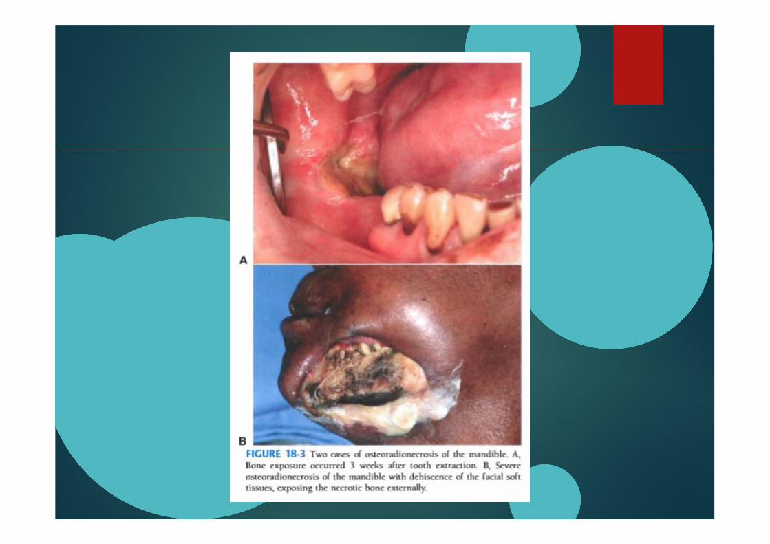
RADIATION EFFECTS ON
BONE
� Osteoradionecrosis is devitalization of the bone by
cancericidal doses of radiation
� The bone virtually nonvital from an endarteritis
because of elimination of the fine vasculature
within the bone.
� Continual process of remodeling does not occur
(e.g sharp areas will not smooth themselves)
� Mandible is denser and poorer blood supply, so
mandible is the most commonly affected with
nonhealing ulcerations and osteoradionecrosis
Other effects of Radiation
� Alteration normal oral flora
� Overgrowth of anaerobic species and fungi
� This may because of radiation and or xerostomia
� Candida albicans commonly thrives, frequently
needed nystatin or 0,1% chlorhexidine (Peridex)
which has antibacterial and antifungal effects
EVALUATION OF DENTITION
BEFORE RADIOTHERAPY� SHOULD TEETH BE EXTRACTED? Consideration:
� Condition of Residual Dentition
� Poor prognosis teeth should be extracted before RT
� Patient’s Dental Awareness
� Excellent OH → Retain as many teeth as possible
� Neglected OH → Will be more difficult
� Immediacy of Radiotherapy
� Immediate RT: maintain the dentition
� Delayed RT: may give time for dental management, need to work closely with the patient
� Radiation Location
� The more salivary glands and bone involved, the more severe xerostomia and vascular compromise
� Radiation dose
� Higher radiation dose → more severe normal tissue damage

PREPARATION OF DENTITION FOR
RADIOTHERAPY AND
MAINTENANCE AFTER IRRADIATION
� Prophylaxis like topical fluoride application using fabrication of custom trays
� Stop smoking and alcohol consumption
� During radiation treatment, should rinse the mouth at least 10x / day with saline
� Chlorhexidine mouth rinse 2x / day
� The Dentist should control 1x / week
� Application of nystatin or clotrimazole (overgrowth Candida albicans)
� Monitor ability of mouth opening → physiotherapy exercises
� Weighed weekly to determine adequate nutritional status
� May be necessary to feed via nasogastric tube

METHOD OF PERFORMING
PREIRRADIATION EXTRACTIONS
� Concepts of bone preservation are disregarded
� Remove a good portion of the alveolar process
along with the teeth (using burs or files to smooth
the bony edges) and achieve a primary soft tissue
closure
� Prophylactic antibiotics are indicated
� “ The Dentist is in a race against time. If the wound
fails to heal, the radiotherapy will be delayed. If
the radiation is delivered before the wound heals,
healing will take months or even years ”


INTERVAL BETWEEN PREIRRADIATION
EXTRACTIONS AND BEGINNING OF
RADIOTHERAPY?
� No categoric answer
� Traditionally: 7-14 days between tooth extraction
and radiotherapy
� If possible: 3 weeks after extractions
� If wound dehiscence has occured, the
radiotherapy should be delayed if possible
� Daily local wound care with irrigations and post
op Antibiotics until soft tissues have healed

IMPACTED THIRD MOLAR
REMOVAL BEFORE RADIOTHERAPY
� Partially erupted: removal may be prudent, to
prevent pericoronal infections
� Totally impacted: Keep it remain in place is more
expeditious
METHODS OF MANAGING CARIOUS
TEETH AFTER RADIOTHERAPY
� Must be immediately cared
� Full crowns are not warranted because recurrent
caries is more difficult to detect
� Flouride application
� Endodontic intervention with systemic antibiotics

TOOTH EXTRACTION AFTER
RADIOTHERAPY� Post irradiation extractions is most undesirable, because
the outcome is uncertain
� If the tooth is needed to be extracted, perform routine extraction without primary closure or surgical extraction with alveoloplasty and primary closure, both has similar results: a certain concomitant incidence of osteoradionecrosis
� Use of antibiotics is recommended
� Use of hyperbaric oxygen (HBO) before and after tooth extraction
� HBO dives 20-30 before extraction and 10 more after extractions
� Usually 1x / day. So, it takes 4-6 weeks to get the 20-30 treatments and 2 weeks of treatment after surgery
� Marx et al: Incidence of Osteoradionecrosis of group with use of AB only : AB+HBO = 5,4% : 30%

DENTURE WEAR IN POSTIRRADIATION
EDENTULOUS PATIENTS
� With denture, patient has the risk of causing
ulceration of the mucosa
� Soft denture liner may be a solution
� Denture fabrication is made once the acute
effects of irradiation have subsided
� Denturers fabrication must be certain that
denture base and occlusal table are designed so
that forces aare distributed evenly throughout the
alveolar ridge and that lateral force on the
denture are eliminated

USE OF DENTAL IMPLANTS
IN IRRADIATED PATIENTS� The more radiation delivered, the higher the failure rate for
endosseous implants
� The longer the duration betweenn radiation treatment and implantation, the higher the failure rate
� When implants in irradiated patiens fail, they usually fail early, before prosthetic reconstruction indicating a failure of osteointegration
� The combination of radiation and chemotherapy has a particularly negative effect on the outome for osseointegration
� Implant survival in irradiated patients tends to he higher in the maxilla than in the mandibule
� Shorter implants have the worst prognosis
� HBO treatment reduces implant failure rates


MANAGEMENT OF PATIENTS WHO
HAVE OSTEORADIONECRIOSIS
� Patient should discontinue wearing any prosthesis
� Decreased vascularity of the tissues and do not
gain ready access to the area to perform the
function of Antibiotics
� Nonhealing wounds or extensive areas of
osteoradionecrosis is needed surgical
intervention.
� HBO can improve results greatly in conjunction
with surgical intervention


DENTAL MANAGEMENT OF
PATIENTS RECEIVING SYSTEMIC
CHEMOTHERAPY FOR MALIGNANT
DISEASE
� Antitumor effect of cancer chemotherapeutic
agents is based on their ability to destroy or retard
the division of rapidly proliferating cells
� Normal host cells that have a high mitotic index
are affected. Most affected are the epithelium of
the gastrointestinal tract and the cels of the bone
marrow

EFFECTS ON ORAL
MUCOSA
� Reduce the normal turnover rate of oral
epithelium → atropic thinning, which manifested
clinically as painful, erythematous, and ulcerative
mucosal surfaces in the mouth.
� Changes are seen within 1 week of the onset of
antitumor agents
� Effects are usually self limiting, spontaneous
healing within 2-3 weeks after cessation of the
agent

EFFECTS ON
HEMATOPOIETIC SYSTEM
� Myelosuppression : Leukopenia, Neutropenia, Thrombocytopenia and Anemia
� Within 2 weeks the white blood cell count falls to an extremely low level
� The oral effect: Marginal gingivitis, and bleeding from the gingiva is common
� Overgrowths of oral flora, especially fungi
� Thrombocytopenia can be significant, and spontaneous bleeding may occur
� Recovery from myelosuppresion is usually complete 3 weeks after cessation of chemotherapy

EFFECTS ON ORAL
MICROBIOLOGY
� Chemotherapeutic agents → Immunosuppressive
side effect → overgrowth of microbes,
superinfection with gram (-) bacili, and
opportunistic infections
� Most patients with chemotherapy are treated
with sytemic antimicrobial agents
� Frequent overgrowth organism: Candida species

GENERAL DENTAL
MANAGEMENT� Chemotherapy has minimal effects on the vasculature, so
dental management is easier
� Primary concerns: bone marrow suppression
� Patient being treated for hematologic neoplasm (e.g leukemia) both the disease and chemotherapy lead to decrease in functional blood elements → risk of infection & hemorrhage
� In non hematologic neoplasm, risk of infection & hemorrhage only during the course of chemotherapy
� Prechemotherapy dental measures:
� Prophylaxis
� Fluoride treatment
� Necessary scaling
� Removal of unrestorable teeth

GENERAL DENTAL
MANAGEMENT
� Dental procedures requirement:
� WBC ≥ 2000/mm3
� At least 20% PMN
� Platelet ≥ 50.000/mm3
� Prophylactic Antibiotics should be given if
chemotherapy within 3 weeks of dental treatment
� Removable dental appliance should be left out (to
prevent ulceration of fragile mucosa)

TREATMENT OF ORAL
CANDIDIASIS
� Topical application of antifungal
� Or oral rinses, oral tablets, and creams
� Oral rinses are less efficacy
� tablet are most accepted forms
� creams are helpful for oral commissures or prosthetic device surfaces
� Most common topical medications: Clotrimazole and Nystatin. 4x daily for 2 weeks
� Clotrimaazole troches 4 x 5 times a day
� Stronger drugs: Ketoconazole or Fluconazole
� Other : Chlorhexidine mouth rinse

DENTAL MANAGEMENT OF PATIENTS
WITH BIPHOSPHONATE-ASSOCIATED
OSTEONECROSIS OF THE JAW (BOJ)
� BOJ is a condition of chronically exposed necrotic
bone (painful and often infected)
� Bone exposure might occur spontaneously or
more commonly following an invasive dental
procedure
� Complains: halitosis, difficulty eating & speaking,
extreme pain
� The lesions are persistent and do not respond to
debridement, antibiotic, or HBO therapy
BIPHOSPHONATES
� Biphosphonates are used to treat osteoporosis, malignant
bone metastasis, Paget’s disease of bone, and
hypercalcemia of malignancy
� Biphosphonates also have antiangiogenic properties →
tumoricidal
� Biphosphonates bind to bone and incorporate in osseous
matrix. During bone remodelling the drug is taken up by
osteoclasts and internalized in the cell cytoplasm → inhibit
osteoclastic function and induces apoptotic cell death
� The result: bone becomes suppressed and shows little
physiologic remodelling → becomes brittle and unable to
reapir physiologic microfractures


CLINICAL SIGNS AND
SYMPTOMS OF BOJ
� Exclusively affects the jaws
� Clinical: ulcer with exposed bone in a patient who
has had a dental extraction
� May be asymptomatic
� May have severe pain (if necrotic bone
becoming infected and exposed)
� Osteonecrosis often progressive and lead to
extensive areas of bony exposure and
dehiscence


DENTAL CARE FOR PATIENTS START
TAKING BIPHOSPHONATES
� Minimize the risk of occurence of BOJ
� Provide dental care early in the treatment
� Teeth with poor prognosis should be removed before or as early as possible after administration of biphosphonates
� Should be delayed for 4-6 weeks after invasive procedures (e.g tooth extraction)
� Elimination of all potential sites of infections
� Restorative dentistry
� Evaluation on prosthodontic appliances (fit, stability, and occlusion)
DENTAL CARE FOR
PATIENTS WITH BOJ� Treatment directed for elimminating or controlling pain and preventing
progression of exposed bone
� Eliminating sharp edges using bur
� Attempts to cover exposed bone with flaps may cause more bone exposure and worsening of symptoms with risk of pathologic fracture
� NONE are successful : Major surgical sequestrectomies, marginal and segmental mandibular resections, partial and complete maxillectomies and HBO therapy
� Use of Chlorhexidine 3-4x/day
� If the tooth is unrestorable because of caries → root canal treatment and amputation of the crown may be a better option than removing the tooth unless it is very loose
� Relining a denture with soft liner to promote a better fit and to minimize soft tissue trauma
� Odontogenic infections treated aggressively with systemic antibiotics

THANK YOU