patient information brochure · patient information brochure alcon acrysof toric high cylinder...
TRANSCRIPT

CONFIDENTIAL FINAL 03.08.11
PATIENT
INFORMATION
BROCHURE
Alcon AcrySof@ Toric
High Cylinder Power Intraocular Lenses

CONFIDENTIAL FINAL 03.08.11
PATIENT INFORMATION BROCHURE
Alcon AcrySof Toric High Cylinder Power Intraocular Lenses
Table of Contents
G lo ssary ..................................................................................................................... 3Introduction .................................................................................................... . . .. 4What is corneal astigmatism? ............................................................................... 4W hat is a cataract? ............................................................................................... . . 5What types of IOLs are available for cataract surgery? ....................................... 5IOL Performance Expectations............................................................................. 6W arnings ........................................................................................................ . . . . 8P recautions...................................................................................................... . . . . 8P otential R isks ..................................................................................................... . 9What to expect during cataract surgery? ............................................. 10Key points to remember regarding your choice ................................................... 12
2
CONFIDENTIAL FINAL 03.08.11
Glossary
Aspheric 1OL: An artificial lens with an optic surface designed to enhance
distance vision under low light conditions when a person
wears full correction glasses. The lens is designed to
benefit a person with an average corneal shape.
Astigmatism: Astigmatism is a focusing error in the eye that results in
blurred distance and/or near vision.
Cataract: A clouding of the natural crystalline lens of the eye that
interferes with vision.
Cornea: The clear front part of the eye that focuses light into the
eye.
Corneal Astigmatism: The inability of the eye to focus'clearly at any distance
because of different curvatures on the cornea.
Crystalline lens: The clear natural structure in the eye that helps to focus
light on the back of the eye. The crystalline lens functions
like the lens of a camera.
High Cylinder Power Toric 1OL: An artificial lens with an optic surface designed to correct
high amounts of corneal astigmatism.
Intraocular Lens (IOL): An artificial lens that replaces the natural
crystalline lens of the eye after cataract surgery.
3
CONFIDENTIAL FINAL 03.08.11
Iris: The colored membrane in front of the natural crystalline
lens that controls the size of the pupil and the amount of
light entering the eye..
Monofocal IOL: An artificial lens designed to restore only distance vision.
Retina: The light-sensitive layer in the back part of the eye that
receives images (light) and sends them to the brain for
interpretation.
Toric IOL: An artificial lens with an optic surface designed to
correct corneal astigmatism.
Introduction
This brochure provides information about the Alcon AcrySofo Toric High Cylinder Power
Intraocular Lenses (1OLs). These IOLs are designed to correct high corneal astigmatism, and
restore distance vision after cataract surgery. Please read this entire brochure carefully and
discuss the information with your eye doctor. Your eye doctor will advise you about the
potential risks and benefits of cataract removal and IOL implantation. You should make sure
that all of your questions are answered to your satisfaction. Please discuss with your eye doctor
to determine if an AcrySof Toric High Cylinder Power IOL would be the right lens for you.
What is corneal astigmatism?
Astigmatism is a focusing error in the eye that results in blurred distance and/or near vision. In a
normal eye, the cornea has a round shape (like a basketball); therefore, the light rays entering the
eye focus at a single point on the back of the eye (retina) to form a clear image. In an eye with
corneal astigmatism, the cornea has an oblong shape (like an American football). As a result, the
light rays do not focus at the same point on the retina and parts of an object may not appear clear.
High levels of corneal astigmatism may also be associated with visual distortions (e.g. objects
4

CONFIDENTIAL FINAL 03.08.11
appear tilted or misshapen or floors appear curved). During your eye examination, your eye
doctor will be able to tell you if you have corneal astigmatism.
What is a cataract?
Your eye functions much like a camera. Your natural crystalline lens focuses light onto the
retina so you can see clearly, much like the lens of a camera focusing light onto film for a clear
picture. At birth, your natural lens is clear. However, as you age, the lens becomes "cloudy"
and eventually affects your ability to see clearly. This condition is called a cataract, and usually
gets worse over time.
Surgery is the only way that a cataract can be removed. You.should consider surgery when a
cataract affects your vision enough to interfere with your daily activities.
What types of IOLs are available for cataract surgery?
The Alcon AcrySofo Toric High Cylinder Power IOL is one option for correcting high corneal
astigmatism and distance vision after cataract surgery. There are other IOLs to choose from for
distance vision, but some are not designed to correct astigmatism. Your eye doctor will discuss
the IOL options available to you.
AcrySof Toric High Cylinder Power IOL
The AcrySofe Toric High Cylinder Power IOL is designed to optically correct high corneal
astigmatism and restore distance vision. There are different models of AcrySofe Toric High
Cylinder Power IOL for varying levels of comeal astigmatism.
AcrySofa IQ Monofocal IOL
The AcrySof 0 IQ Monofocal IOL is designed to restore distance vision but does not optically
correct corneal astigmatism. This IOL also has an aspheric surface designed to enhance distance
vision under low light conditions, when a person wears full correction glasses. The lens is
designed to benefit a person with an average corneal shape.

CONFIDENTIAL FINAL 03.08.11
Monofocal IOL
A monofocal IOL is designed to restore distance vision but does not optically correct corneal
astigmatism.
IOL Performance Expectations
Performance expectations of each 1OL are described in Table 1 below:
Table 1: Expected IOL Performance for Patients With High Astigmatism
AcrySof IQ AcrySof Toric HighCharacteristic Monofocal IOL Monofocal IOL Cylinder Power IOLPre-existing Is not designed to Is not designed to Designed to opticallyCorneal correct your pre- correct your pre- correct your pre-Astigmatism existing corneal existing corneal existing corneal
astigmatism astigmatism astigmatism
Distance Vision Your uncorrected Your uncorrected Your uncorrecteddistance vision will distance vision will distance vision willlikely be blurred due likely be blurred due to likely be clearer ifto your uncorrected your uncorrected your astigmatism iscorneal astigmatism. corneal astigmatism. corrected.You will likely need You will likely needprescription glasses or prescription glasses orcontact lens correction contact lens correctionto see clearly at to see clearly atdistance. distance.
Near Vision A monofocal 1OL is An aspheric monofocal A toric IOL is notnot designed to LOL is not designed to designed to provideprovide near vision. provide near vision. near vision.
You will need You will need You are less likely toastigmatism correcting astigmatism correcting need astigmatismprescription reading to prescription reading correcting prescriptionclearly see objects up glasses to clearly see glasses, but will stillclose or to read. objects up close or to need reading glasses
read. to clearly see objectsup close or to read.

CONFIDENTIAL FINAL 03.08.11
Enhanced A monofocal 1OL is An aspheric monofocal A Toric 1OL is notDistance Vision not designed to IOL is designed to designed to provide
provide the additional provide the additional the additional benefitbenefit of enhanced benefit of enhanced of enhanced distancedistance vision in low distance vision in low vision in low lightlight environments. light environments, environments.
when a person wearsfull correction glasses.The lens is designed tobenefit a person with anaverage corneal shape.
However, you will needglasses or contact lenscorrection for your highcorneal astigmatism toobtain this benefit.
Visual You may experience You may experience You may experienceDistortions (i.e. visual distortions due visual distortions due to visual distortions instraight lines to the optical the optical correction of the event that the toriclook tilted and / correction of high high astigmatism with 1OL rotates, isor flat surfaces astigmatism with glasses or contact improperlylook curved) glasses or contact lenses. positioned, or if you
lenses. require distant visionglasses foruncorrectedastigmatism.
Glasses Use You will likely need You will likely need You may be able toprescription glasses or prescription glasses or function withoutcontact lenses for most contact lenses for most glasses or contact lensdaily tasks due to daily tasks due to correction for manyuncorrected high uncorrected high daily tasks requiringcorneal astigmatism. corneal astigmatism. distance vision.
You will likely also You will likely also You will likely stillneed prescription need prescription need prescriptionglasses to clearly see glasses to clearly see glasses to clearly seeobjects up close or to objects up close or to objects up close or toread. read. read.
Please discuss the 1OL options with your eye doctor to determine which type is right for you.

CONFIDENTIAL FINAL 03.08.11
Cataract surgery is one of the most common surgical procedures performed; however, as with allsurgeries there are warnings, precautions, and risk that you should be aware of.
Warnings
1. Your eye doctor may not be able to implant the AcrySofe Toric High Cylinder Power IOLinto your eye if you have complications during surgery (e.g. tissue damage that may causethe lens to rotate after surgery). Depending on your specific surgical complications yourdoctor may or may not be able to implant a different 1OL during the same surgical procedure.
2. Contact your eye doctor immediately if you have any of the following symptoms while usingthe antibiotic eye drops prescribed by your doctor: itching, redness, watering of your eye,sensitivity to light. These symptoms could indicate a potential serious eye infection.
Precautions
1. As with any surgical procedure, there is risk involved. Possible complications from cataractsurgery include infection, damage to the lining of the cornea, separation of the retina fromthe layer of tissue at the back of the eye (retinal detachment), inflammation or swelling insideor outside the eye, damage to the iris (the colored part of your eye), and an increase in eyepressure. You may need additional surgery to reposition or replace the 1OL, or to treat othersurgery complications. Toric IOLs require surgical repositioning more often than non-toric
IOLs.
2. Tell your eye doctor if you have been diagnosed with any eye disease. The safety andeffectiveness of the AcrySoft Toric 1OL has not been established in patients with pre-
existing eye conditions and complications during surgery, such as an increase in eye pressure
(glaucoma) or complications of diabetes in the eye (diabetic retinopathy). The outcome of
cataract surgery will depend on the health of your eye before surgery.
3. You will need to wear glasses to benefit from an aspheric 1OL designed to enhance distance
vision under low light conditions, if you have any of the following:
8

CONFIDENTIAL FINAL 03.08.11
a. Nearsightedness or farsightedness after surgery: These conditions may result from errors
in measurements before surgery, wrong lens power, or changes in the cornea in response
to the surgery;
b. Uncorrected astigmatism after surgery: This condition may result from the same reasons
as stated above. In addition, uncorrected astigmatism could also result from improper
position of the IOL or if your corneal astigmatism is greater than the amount that can be
corrected with the 1OL.
4. A toric IOL corrects astigmatism only when it is placed in the correct position in the eye.
There is a possibility that the toric 1OL could be placed incorrectly or could move within the
eye. If the toric lens is not positioned correctly following surgery, the change in your
astigmatism correction by the 1OL, along with any necessary correction with glasses, may
cause visual distortions.
5. Avoid any activity that could harm your eye while you are recovering from surgery.
Potential Risks
There are risks associated with cataract surgery. You may have reactions to medicines, and side
effects include redness, scratchiness of the eye, and sensitivity to light. Possible complications
from cataract surgery include infection, bleeding, inflammation, tissue damage, tissue swelling
of the front or back of the eye, or an increase in eye pressure. If your lens is not in the correct
position, your vision may also be affected and the normal flow of fluid within the eye may be
blocked. Your vision may not improve or may get worse if these complications occur. You may
require additional surgery to treat these side effects.
If you have high corneal astigmatism, you may notice that some objects appear tilted or
misshapen or floors appear curved. These visual distortions may be present before cataract
surgery but may remain after surgery if your astigmatism is not fully corrected or if the 1OL is
not in the proper position in your eye. It may take some time to adapt to your new IOL(s) and
any changes in your astigmatism. Please discuss with your eye doctor about your vision and any
symptoms after surgery.
9

CONFIDENTIAL FINAL 03.08.11
Your eye doctor may advise that you have a second surgery if the toric IOL is not properly
positioned in your eye.
The overall risks associated with cataract surgery, compared to other types of surgeries, is
relatively low. Toric IOLs require surgical repositioning more often than non-toric OLs.
Discuss any questions about the possible risks and benefits of cataract surgery and the AcrySofe
Toric High Cylinder Power 1OL with your eye doctor.
What to expect during cataract surgery?
Cataract surgery is a procedure to replace your cloudy natural crystalline lens with an intraocular
lens implant. You should expect the following before, during, and after surgery.
Before Surgery
You will need a thorough eye examination. Be sure to tell your eye doctor about any problems
about your vision or general health. Your eye will be measured after you and your eye doctor
have decided that you will have your cataract.removed. This will determine your amount of
corneal astigmatism and the 1OL power that will be right for you. You should plan to have
someone else drive you home.
Day of Surgery
Cataract surgery techniques vary widely. However, the eye is always numbed to make the
operation painless. To perform surgery, your doctor will use a microscope to have a magnified
view of your eye to properly position the toric IOL. Your natural lens sits in a bag-like structure
called the capsule. The capsule is located just behind the colored part of your eye (iris). A small
incision is made on the clear front part of your eye (cornea) to reach and remove the cataract. An
1OL is then placed into the capsule to replace your natural lens. The 1OL will act in the same
way as your natural lens to restore your distance vision. The eye doctor will usually place a
shield over your eye to protect it after surgery. After a short stay in the outpatient recovery area
you will be ready to go home. Your eye doctor will let you know when your vision is good
enough to drive again.
10

CONFIDENTIAL FINAL 03.08.11
Below in Figure I shows the basic parts of the eye with an implanted IOL.
Figure 1 - Drawing of Eye with an Implanted IOL
CORNEAIOL
RETINA
CAPSULE
After Surgery
After surgery, your eye doctor should give you a wallet card that identifies the type of implant in
your eye. Typically, your eye doctor will examine you the following day. Many patients may
see better within 1 to 2 days, most are stable at 10 to 14 days, but some may take 4 to 6 weeks to
fully recover from the surgery. Improvements in vision are different for each individual.
Take all prescribed medicines and apply antibiotic eye drops as instructed by your eye doctor. Be
sure to consult your eye doctor if you have any questions or concerns as a result of cataract
surgery.
11
CONFIDENTIAL FINAL 03.08.11
Key points to remember regarding your choice
1. The Monofocal 1OL, AcrySof® IQ Monfocal IOL and AcrySof® Toric High Cylinder Power
IOLs all restore distance vision following cataract surgery. However, the AcrySofe Toric
High Cylinder Power IOL is designed to also optically correct your high corneal astigmatism.
2. If reduced dependence on glasses is your desired outcome, you should consider the AcrySofo
Toric High Cylinder Power JOL to correct high corneal astigmatism during cataract surgery.
3. Discussing your lifestyle or visual needs with your eye doctor can help determine which IOL
is best for you.
Thank you for considering the AcrySofo Toric High Cylinder Power 1OL.
Alcon Laboratories, Inc.
6201 South Freeway
Fort Worth, Texas 76134-4630
C 2011 Alcon, Inc. X/1 I RESXXX
12
CONFIDENTIAL FINAL 03.08.11
PATIENT
INFORMATION
BROCHURE
Alcon AcrySof@ IQ Toric
High Cylinder Power Intraocular Lenses
1

CONFIDENTIAL FINAL 03.08.11
PATIENT INFORMATION BROCHURE
Alcon AcrySof IQ Toric High Cylinder Power Intraocular Lenses
Table of Contents
G lo ssary .................................................................................................................... 3
Introduction .................................................................................................... . . . . 4What is corneal astigmatism? .............................................................................. 4W hat is a cataract? ................................................................................................ . 5What types of IOLs are available for cataract surgery? ....................................... 5IOL Performance Expectations.............................................................................6
W arning s ........................................................................................................ . . . . 8P recautions...................................................................................................... . . . . 8P otential R isks ................................................................................................... . . 9What to expect during cataract surgery? ............................................. 10Key points to remember regarding your choice .................................................. 12
2
CONFIDENTIAL FINAL 03.08.11
Glossary
Aspheric IOL: An artificial lens with an optic surface designed to enhance
distance vision under low light conditions when a person
wears full correction glasses. The lens is designed to
benefit a person with an average corneal shape.
Astigmatism: Astigmatism is a focusing error in the eye that results in
blurred distance and/or near vision.
Cataract: A clouding of the natural crystalline lens of the eye that
interferes with vision.
Cornea: The clear front part of the eye that focuses light into the
eye.
Corneal Astigmatism: The inability of the eye to focus clearly at any distance
because of different curvatures on the cornea.
Crystalline lens: The clear natural structure in the eye that helps to focus
light on the back of the eye. The crystalline lens functions
like the lens of a camera.
High Cylinder Power Toric IOL: An artificial lens with an optic surface designed to correct
high amounts of corneal astigmatism.
Intraocular Lens (IOL): An artificial lens that replaces the natural
crystalline lens of the eye after cataract surgery.
3

CONFIDENTIAL FINAL 03.08.11
Iris: The colored membrane in front of the natural crystalline
lens that controls the size of the pupil and the amount of
light entering the eye.
Monofocal IOL: An artificial lens designed to restore only distance vision.
Retina: The light-sensitive layer in the back part of the eye that
receives images (light) and sends them to the brain for
interpretation.
Toric 1OL: An artificial lens with an optic surface designed to
correct corneal astigmatism.
Introduction
This brochure provides information about the Alcon AcrySofe IQ Toric High Cylinder Power
Intraocular Lenses (1OLs). These IOLs are designed to correct high corneal astigmatism, and
restore distance vision after cataract surgery. Please read this entire brochure carefully and
discuss the information with your eye doctor. Your eye doctor will advise you about the
potential risks and benefits of cataract removal and IOL implantation. You should make sure
that all of your questions are answered to your satisfaction. Please discuss with your eye doctor
to determine if an AcrySofe IQ Toric High Cylinder Power IOL would be the right lens for you.
What is corneal astigmatism?
Astigmatism is a focusing error in the eye that results in blurred distance and/or near vision. In a
normal eye, the cornea has a round shape (like a basketball); therefore, the light rays entering the
eye focus at a single point on the back of the eye (retina) to form a clear image. In an eye with
corneal astigmatism, the cornea has an oblong shape (like an American football). As a result, the
light rays do not focus at the same point on the retina and parts of an object may not appear clear.
High levels of corneal astigmatism may also be associated with visual distortions (e.g. objects
4
CONFIDENTIAL FINAL 03.08.11
appear tilted or misshapen or floors appear curved). During your eye examination, your eye
doctor will be able to tell you if you have corneal astigmatism.
What is a cataract?
Your eye functions much like a camera. Your natural crystalline lens focuses light onto theretina so you can see clearly, much like the lens of a camera focusing light onto film for a clear
picture. At birth, your natural lens is clear. However, as you age, the lens becomes "cloudy"
and eventually affects your ability to see clearly. This condition is called a cataract, and usually
gets worse over time.
Surgery is the only way that a cataract can be removed. You should consider surgery when acataract affects your vision enough to interfere with your daily activities.
What types of IOLs are available for cataract surgery?
The Alcon AcrySofo IQ Toric High Cylinder Power 1OL is one option for correcting high
corneal astigmatism and distance vision after cataract surgery. There are other IOLs to choose
from for distance vision, but some are not designed to correct astigmatism. Your eye doctor will
discuss the IOL options available to you.
AcrySofo IQ Toric High Cylinder Power IOL
The AcrySofo IQ Toric High Cylinder Power IOL is designed to optically correct high corneal
astigmatism and restore distance vision. There are different models of AcrySofe IQ Toric High
Cylinder Power IOL for varying levels of corneal astigmatism. This IQ Toric IOL, also,incorporates an aspheric surface designed to enhance distance vision under low light conditions,
when a person wears full correction glasses. The lens is designed to benefit a person with an
average corneal shape.

CONFIDENTIAL FINAL 03.08.11
AcrySof IQ Monofocal IOL
The AcrySof 0 IQ Monofocal 1OL is designed to restore distance vision but does not optically
correct corneal astigmatism. This IOL also has an aspheric surface designed to enhance distance
vision under low light conditions, when a person wears full correction glasses. The lens is
designed to benefit a person with an average corneal shape.
Monofocal IOL
A monofocal 1OL is designed to restore distance vision but does not optically correct corneal
astigmatism.
IOL Performance Expectations
Performance expectations of each IOL are described in Table I below:
Table 1: Expected IOL Performance for Patients With High Astigmatism
Characteristic Monofocal IOL AcrySofa IQ AcrySof IQ ToricMonofocal High Cylinder
IOL Power IOL
Pre-existing Is not designed to . Is not designed to Designed to opticallyCorneal correct your pre- correct your pre- correct your pre-Astigmatism existing corneal existing corneal existing corneal
astigmatism astigmatism astigmatism
Distance Vision Your uncorrected Your uncorrected Your uncorrecteddistance vision will distance vision will distance vision willlikely be blurred due likely be blurred due likely be clearer ifto your uncorrected to your uncorrected your astigmatism iscorneal astigmatism. corneal astigmatism. corrected.You will likely need You will likely needprescription glasses or prescription glasses orcontact lens correction contact lens correctionto see clearly at to see clearly atdistance. distance.
Near Vision A monofocal 1OL is An aspheric An aspheric toric IOLnot designed to monofocal IOL is not is not designed toprovide near vision. designed to provide provide near vision.
near vision.
CONFIDENTIAL FINAL 03.08.11
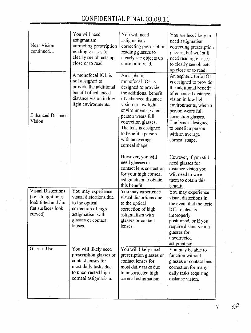
You will need You will need You are less likely toastigmatism astigmatism need astigmatism
Near Vision correcting prescription correcting prescription correcting prescriptioncontinued... reading glasses to reading glasses to glasses, but will still
clearly see objects up clearly see objects up need reading glassesclose or to read. close or to read. to clearly see objects
up close or to read.A monofocal IOL is An aspheric An aspheric toric IOLnot designed to monofocal IOL is is designed to provideprovide the additional designed to provide the additional benefitbenefit of enhanced the additional benefit of enhanced distancedistance vision in low of enhanced distance vision in low lightlight environments. vision in low light environments, when a
environments, when a person wears fullEnhanced Distance person wears full correction glasses.Vision correction glasses. The lens is designed
The lens is designed to benefit a personto benefit a person with an averagewith an average corneal shape.corneal shape.
However, you will However, if you stillneed glasses or need glasses forcontact lens correction distance vision youfor your high comeal will need to wearastigmatism to obtain them to obtain thisthis benefit. benefit.
Visual Distortions You may experience You may experience You may experience(i.e. straight lines visual distortions due visual distortions due visual distortions inlook tilted and / or to the optical to the optical the event that the toricflat surfaces look correction of high correction of high IOL rotates, iscurved) astigmatism with astigmatism with improperly
glasses or contact glasses or contact positioned, or if youlenses. lenses. require distant vision
glasses foruncorrectedastigmatism.
Glasses Use You will likely need You will likely need You may be able toprescription glasses or prescription glasses or function withoutcontact lenses for contact lenses for glasses or contact lensmost daily tasks due most daily tasks due correction for manyto uncorrected high to uncorrected high daily tasks requiringcorneal astigmatism. corneal astigmatism. distance vision.
CONFIDENTIAL FINAL 03.08.11
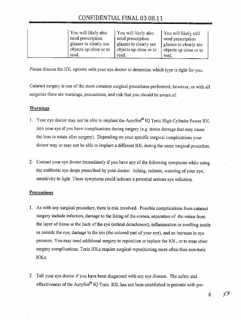
You will likely also You will likely also You will likely stillneed prescription need prescription need prescriptionglasses to clearly see glasses to clearly see glasses to clearly seeobjects up close or to objects up close or to objects up close or toread. read. read.
Please discuss the IOL options with your eye doctor to determine which type is right for you.
Cataract surgery is one of the most common surgical procedures performed; however, as with all
surgeries there are warnings, precautions, and risk that you should be aware of.
Warnings
1. Your eye doctor may not be able to implant the AcrySofe IQ Toric High Cylinder Power IOL
into your eye if you have complications during surgery (e.g. tissue damage that may cause
the lens to rotate after surgery). Depending on your specific surgical complications your
doctor may or may not be able to implant a different 1OL during the same surgical procedure.
2. Contact your eye doctor immediately if you have any of the following symptoms while using
the antibiotic eye drops prescribed by your doctor: itching, redness, watering of your eye,
sensitivity to light. These symptoms could indicate a potential serious eye infection.
Precautions
1. As with any surgical procedure, there is risk involved. Possible complications from cataract
surgery include infection, damage to the lining of the cornea, separation of the retina from
the layer of tissue at the back of the eye (retinal detachment), inflammation or swelling inside
or outside the eye, damage to the iris (the colored part of your eye), and an increase in eye
pressure. You may need additional surgery to reposition or replace the 1OL, or to treat other
surgery complications. Toric IOLs require surgical repositioning more often than non-toric
IOLs.
2. Tell your eye doctor if you have been diagnosed with any eye disease. The safety and
effectiveness of the AcrySofo IQ Toric 1OL has not been established in patients with pre-
8
CONFIDENTIAL FINAL 03.08.11
existing eye conditions and complications during surgery, such as an increase in eye pressure
(glaucoma) or complications of diabetes in the eye (diabetic retinopathy). The outcome of
cataract surgery will depend on the health of your eye before surgery.
3. You will need to wear glasses to benefit from an aspheric 1OL designed to enhance distance
vision under low light conditions, if you have any of the following:
a. Nearsightedness or farsightedness after surgery: These conditions may result from errors
in measurements before surgery, wrong lens power, or changes in the cornea in response
to the surgery;
b. Uncorrected astigmatism after surgery: This condition may result from the same reasons
as stated above. In addition, uncorrected astigmatism could also result from improper
position of the IOL or if your corneal astigmatism is greater than the amount that can be
corrected with the 1OL.
4. A toric IOL corrects astigmatism only when it is placed in the correct position in the eye.
There is a possibility that the toric IOL could be placed incorrectly or could move within the
eye. If the toric lens is not positioned correctly following surgery, the change in your
astigmatism correction by the 1OL, along with any necessary correction with glasses, may
cause visual distortions.
5. Avoid any activity that could harm your eye while you are recovering from surgery.
Potential Risks
There are risks associated with cataract surgery. You may have reactions to medicines, and side
effects include redness, scratchiness of the eye, and sensitivity to light. Possible complications
from cataract surgery include infection, bleeding, inflammation, tissue damage, tissue swelling
of the front or back of the eye, or an increase in eye pressure. If your lens is not in the correct
position, your vision may also be affected and the normal flow of fluid within the eye may be
blocked. Your vision may not improve or may get worse if these complications occur. You may
require additional surgery to treat these side effects.
9

CONFIDENTIAL FINAL 03.08.11
If you have high corneal astigmatism, you may notice that some objects appear tilted or
misshapen or floors appear curved. These visual distortions may be present before cataract
surgery but may remain after surgery if your astigmatism is not fully corrected or if the IOL is
not in the proper position in your eye. It may take some time to adapt to your new IOL(s) and
any changes in your astigmatism. Please discuss with your eye doctor about your vision and any
symptoms after surgery.
Your eye doctor may advise that you have a second surgery if the toric IOL is not properly
positioned in your eye.
The overall risks associated with cataract surgery, compared to other types of surgeries, is
relatively low. Toric IOLs require surgical repositioning more often than non-toric IOLs.
Discuss any questions about the possible risks and benefits of cataract surgery and the AcrySofe
IQ Toric High Cylinder Power IOL with your eye doctor.
What to expect during cataract surgery?
Cataract surgery is a procedure to replace your cloudy natural crystalline lens with an intraocular
lens implant. You should expect the following before, during, and after surgery.
Before Surgery
You will need a thorough eye examination. Be sure to tell your eye doctor about any problems
about your vision or general health. Your eye will be measured after you and your eye doctor
have decided that you will have your cataract removed. This will determine your amount of
corneal astigmatism and the IOL power that will be right for you. You should plan to have
someone else drive you home.
Day of Surgery
Cataract surgery techniques vary widely. However, the eye is always numbed to make the
operation painless. To perform surgery, your doctor will use a microscope to have a magnified
10

CONFIDENTIAL FINAL 03.08.11
view of your eye to properly position the toric 1OL. Your natural lens sits in a bag-like structure
called the capsule. The capsule is located just behind the colored part of your eye (iris). A small
incision is made on the clear front part of your eye (cornea) to reach and remove the cataract. AnIOL is then placed into the capsule to replace your natural lens. The IOL will act in the same
way as your natural lens to restore your distance vision. The eye doctor will usually place a
shield over your eye to protect it after surgery. After a short stay in the outpatient recovery areayou will be ready to go home. Your eye doctor will let you know when your vision is good
enough to drive again.
Below in Figure 1 shows the basic parts of the eye with an implanted 1OL.
Figure 1 - Drawing of Eye with an Implanted IOL
CORNEAIOL
RETINA
CAPSULEIRI S
After Surgery
After surgery, your eye doctor should give you a wallet card that identifies the type of implant in
your eye. Typically, your eye doctor will examine you the following day. Many patients may
see better within 1 to 2 days, most are stable atl0 to 14 days, but some may take 4 to 6 weeks to
fully recover from the surgery. Improvements in vision are different for each individual.
Take all prescribed medicines and apply antibiotic eye drops as instructed by your eye doctor. Be
sure to consult your eye doctor if you have any questions or concerns as a result of cataract
surgery.
11

CONFIDENTIAL FINAL 03.08.11
Key points to remember regarding your choice
1. Both the AcrySofe IQ Monofocal and AcrySofe IQ Toric High Cylinder Power IOLs restore
distance vision following cataract surgery. However, the AcrySofM IQ Toric High Cylinder
Power IOL is designed to also optically correct your high corneal astigmatism.
2. If reduced dependence on glasses or enhanced distance vision is your desired outcome, you
should consider the AcrySofe IQ Toric High Cylinder Power 1OL to correct high corneal
astigmatism during cataract surgery.
3. Discussing your lifestyle or visual needs with your eye doctor can help determine which 1OL
is best for you.
Thank you for considering the AcrySofe IQ Toric High Cylinder Power 1OL.
Alcon Laboratories, Inc.
6201 South Freeway
Fort Worth, Texas 76134-4630
© 2011 Alcon, Inc. X/I 1 RESXXX
12

PROPOSED FINAL P930014/S45
PRODUCT INFORMATIONAlcon Laboratories, Inc.
SINGLE-Piece NATURAL 10L
STERILE UV and Blue Light Filtering Acrylic FoldableToric Optic Single-Piece Posterior Chamber Lenses
CAUTION: Federal (USA) law restricts this device to the sale by or on the order of a physician.DESCRIPTION
The AcrySof@ Toric Posterior Chamber Intraocular Lens (IOL) is a UV-absorbing foldable intraocular lens (lOL). These IOLs have a biconvex toric optic withcylinder axis marks to denote the flat meridian (plus cylinder axis). The single-piece design (see Figure I and Table 1) consists of a high refractive index material withproprietary blue light filtering chromophore which filters light in a manner that approximates the human crystalline lens in the 400-475 nm blue light wavelengthrange (Boettner and Wolter, 1962). In addition to standard UV-light filtering, the blue-light filtering chromophore reduces transmittance of blue light wavelengths (seeTable 2). The biconvex toric optic consists of a high refractive index soft acrylic material capable of being folded prior to insertion, allowing placement through anincision smaller than the optic diameter of the lens. After surgical insertion into the eye, the lens gently unfolds to restore the optical performance. The supportinghaptics provide for proper positioning and fixation of the IOL optic within the eye.
Figure 1: Physical Characteristics of AcrySof@ Toric lOLs(All dimensions in millimeters)
ANTERIOR01 .0 ASYMMETRIC
BICONVEX ORIC OPTICSURFACE
0 .0
AXISMARKS - 43
Table 1:Ph ysical Characteristics of AcrySof®Toric IOLs
Characteristics ModelSN60T6 SN60T7 SN60T8
Optic Type Biconvex Toric OpticOptic / Haptic Material Ultraviolet and blue light filtering Acrylate/Methacrylate Copolymer
UV cutoff at 10% T: 399 nm (+6.0 diopter lens)407 nm (+34.0 diopter lens)
IOL Powers(spherical eauivalent diopters) For available power range see Alcon Product GuideIOL Cylinder Power (Diopters) 3.75 D 4.50 D 5.25 D 6.00 D
Index Of Refraction 1.55Haptic Configuration STABLEFORCE@Optic Diameter (mm) 6.0Overall Length (mm) 13.0
Haptic Angle 00

PROPOSED FINAL P930014/S45
Figure 2SPECTRAL TRANSMITTANCE CURVES
(PERCENTAGE OF ULTRAVIOLET TRANSMITTANCE)
II N60T6 - SN60T940-0 6.0 - 34,0 D range
--- SA60AT 20.0 0
300 40600 700 SooWAVE LENGTH (nm)
NOTE:* Human lens data from Boettner and Wolter (1962).
Table 2Average% Transmittance Comparison
Model 400 nm 425 nm 450 nm 475 nmSA60AT 21 86 88 88SN60T9 6 31 47 67
Transmittance Difference(SA60AT -SN60T9) 15 55 41 21
Transmittance Reductionwith SN60T9 71 64 47 24
(% of SA60AT)
MODE OF ACTIONAcrySof@ Toric IOLs are intended to be positioned in the posterior chamber of the eye, replacing the natural crystalline lens. This position allows the lens to function as
a refractive medium in the correction of aphakia. These lOLs have a biconvex toric optic with cylinder axis marks to denote the flat meridian (plus cylinder axis).Alignment of the toric IOL cylinder axis marks with the post-operative steep corneal meridian allows the lens to correct astigmatism.. The astigmatic correction at thecomeal plane for AcrySof@Toric intraocular lenses is shown in Table 3:
Table 3IOL Cylinder Power Cylinder Power at
Model (diopters) Corneal Plane (diopters)*SN60T6 3.75 2.57SN60T7 4.50 3.08SN60T8 5.25 3.60SN60T9 6.00 4.11
*Based on an average pseudophakic human eye
INDICATIONSThe AcrySof@ Toric posterior chamber intraocular lenses are intended for primary implantation in the capsular bag of the eye for visual correction of aphakia and pre-existing corneal astigmatism secondary to removal of a cataractous lens in adult patients with or without presbyopia, who desire improved uncorrected distance vision,reduction of residual refractive cylinder and increased spectacle independence for distance vision.
WARNINGSI. This lens should not be implanted if the posterior capsule is ruptured, if the zonules are damaged, or ifa primary posterior capsulotomy is planned.2. Rotation of AcrySoftToric IOLs away from their intended axis can reduce their astigmatic correction. Misalignment greater than 30 may increase
postoperative refractive cylinder. If necessary, lens repositioning should occur as early as possible prior to lens encapsulation. Some clinical cases suggestencapsulation is complete within four weeks of implantation.
3. Carefully remove all viscoelastic from both the anterior and posterior sides of the lens. Residual viscoelastic may allow the lens to rotate causing misalignmentof the AcrySof@Toric IOL with the intended axis of placement.
PRECAUTIONS1. Prior to surgery, physicians should provide prospective patients with a copy of the Patient Information Brochure available from Alcon for this product informing
them of possible risks and benefits associated with the AcrySoff@ Toric High Cylinder Power IOLs2. A high level of surgical skill is required for intraocular lens implantation. The surgeon should have observed and/or assisted in numerous implantations and
successfully completed one or more courses on intraocular lens implantation before attempting to implant intraocular lenses.3. As with any surgical procedure, there are risks involved. Potential complications accompanying cataract or implant surgery may include, but are not limited to the
following: comeal endothelial damage, infection (endophthalmitis), retinal detachment, vitritis, cystoid macular edema, comeal edema, pupillary block, cycliticmembrane, iris prolapse, hypopyon, transient or persistent glaucoma and secondary surgical intervention. Secondary surgical interventions include, but are not limitedto: lens repositioning, lens replacement, vitreous aspirations or iridectomy for pupillary block, wound leak repair, and retinal detachment repair.
,2

PROPOSED FINAL P930014/S45
4. The safety and effectiveness of the Toric intraocular lens have not been substantiated in patients with the following preexisting ocular conditions and intraoperativecomplications (see below). Careful preoperative evaluation and sound clinical judgment should be used by the surgeon to decide the benefit/risk ratio beforeimplanting a lens in a patient with one or more of these conditions.
Before Surgery* Choroidal hemorrhage* Chronic severe uveitis* Concomitant severe eye disease* Extremely shallow anterior chamber* Medically uncontrolled glaucoma* Microphthalmos* Non-age-related cataract* Proliferative diabetic retinopathy (severe)* Severe comeal dystrophy* Severe optic nerve atrophy* Irregular corneal astigmatism* Color vision deficiencies
Studies have shown that color vision discrimination is not adversely affected in individuals with the AcrySof& Natural lOL and normal color vision. The effectof the AcrySofV Natural IOL in subjects with hereditary color vision defects and acquired color vision defects secondary to ocular disease (e.g. glaucoma,diabetic retinopathy, chronic uveitis, and other retinal or optical nerve diseases) has not been studied.During Surgery* Excessive vitreous loss* Capsulotomy by any technique other than a circular tear* The presence of radial tears known or suspected at the time of surgery* Situations in which the integrity of the circular tear cannot be confirmed by direct visualization* Cataract extraction by techniques other than phacoemulsification or liquefaction* Situations where the need for a large capsulotomy can be anticipated (e.g., diabetics, retinal detachment in the fellow eye, peripheral retinal pathology, etc.)* Capsular rupture* Significant anterior chamber hyphema* Uncontrollable positive intraocular pressure* Zonular damage
5. Some adverse reactions which have been associated with the implantation of intraocular lenses are: hypopyon, intraocular infection, acute corneal decompensationand secondary surgical intervention. Secondary surgical interventions include, but are not limited to: lens repositioning, lens replacement, vitreous aspiration orirdectomy for pupillary block, wound leak repair and retinal detachment repair.6. Patients with preoperative problems such as corneal endothelial disease, abnormal cornea, macular degeneration, retinal degeneration, glaucoma, and chronic drugmiosis may not achieve the visual acuity of patients without such problems. The physician must determine the benefits to be derived from lens implantation whensuch conditions exist.7. DO NOT store the IOL at temperatures over 450 C (1130 F).8. DO NOT reuse the IOL. This IOL is for single use only.9. DO NOT resterilize the IOL by any method.10. Use only sterile intraocular irrigating solutions such as BSS® or BSS PLUS@ to rinse and/or soak lenses.11. Accurate keratometry and biometry in addition to the use of the Toric Calculator (www.acrysoftoriccalculator.com) are recommended to achieve optimal visualoutcomes.12. Optical theory suggests that high astigmatic patients may experience spatial distortions. Possible toric IOL related factors may include residual cylindrical erroror axis misalignments.
CALCULATION OF LENS POWERAccurate keratometry and biometry is essential to successful visual outcomes. Preoperative calculation of the required spherical equivalent lens power for these posterior
chamber intraocular lenses should be determined by the surgeon's experience, preference, and intended lens placement. The A-constant listed on the outer label ispresented as a guideline and is a starting point for implant power calculations. This provisional A-constant has been theoretically derived. Lens constants must bepersonalized" to compensate for the differences in instrumentation, measurement technique, and IOL power calculation methods. A convenient initial estimate can beobtained by referencing to the personalized lens constant for a similar lens model (e.g. AcrvSof@ Toric IOL Models SN60T3. SN60T4, or SN60T5).AcrySof® Toric IOLs are labeled with the IOL spherical equivalent power. The results obtained from the calculation formulas listed below should not be modified, asthey result in the appropriate power consistent with the labeling of the AcrySof Toric IOL. Lens power calculation methods are described in the following references:Hoffer, K.J. The Hoffer Q formula: A comparison of theoretic and regression formulas. J Cataract Refract. Surg. 19:700-712, 1993.Holladay, J.T., et al. A three-part system for refining intraocular lens power calculations. J Cataract Refract. Surg. 14:17-24, 1988.Holladay, J.T., et al., Standardizing constants for ultrasonic biometry, keratometry, and IOL power calculations, J. Cataract Refract. Surg. 23:1356-1370, 1997.Retzlaff, J.A., Sanders, DR., and Kraff, M. Lens Implant Power Calculation, 3rd ed., Slack, Inc., Thorofare, N.J., 1990.
DIRECTIONS FOR USEI. Examine the label on the unopened package for model, power (spherical equivalent and cylinder), and expiration date.2. After opening the cardboard storage container verify lens case information (model, power, and serial number) is consistent with information on outer package
labeling.3. This device is sterile until the inner pouch is opened. Inspect the pouch carefully for tears, cuts, punctures or other signs that the pouch has been opened or
damaged. DO NOT implant the IOL if the sterility has been compromised. (See RETURNED GOODS POLICY).4. To remove the lens, open the undamaged pouch and transfer the case to a sterile environment. Carefully open the case to expose the lens.5. To minimize the occurrence of marks on the lens due to handling, all instrumentation should be scrupulously clean. Any forceps used for lens handling must
have round edges and smooth surfaces.6. When removing the lens from the case, DO NOT grasp the optical area with forceps. The IOL should only be handled by the haptics. Handle the lenses
carefully to avoid damage to lens surfaces or haptics.'DO NOT attempt to reshape haptics in any way.7. Rinse the lens thoroughly using sterile intraocular irrigating solution such as BSS0 or BSS PLUS®. DO NOT rinse the IOL in solutions other than sterile
intraocular irrigating solution. Prior to insertion, the IOL should be carefully examined to ensure that particles have not adhered during handling.8. Alcon recommends using the MONARCH® III delivery system, or equivalent Alcon approved delivery system.9. There are various surgical procedures that can be utilized, and the surgeon should select a procedure that is appropriate for the patient. Current techniques,appropriate instrumentation, and a list of their equivalents for delivery and implantation are available from Alcon. Surgeons should verify that appropriate
instrumentation is available prior to surgery.
3

PROPOSED FINAL P930014/S45
Selection and Placement of the AcrvSof@ ToricThe astigmatism to be corrected should be determined from keratometry and biometry data rather than refractive data since the presence of lenticular astigmatismin the crystalline lens to be removed may influence results. The size and location of the surgical incision may affect the amount and axis of corneal astigmatism. Inorder to optimize IOL selection and axis placement, Alcon provides a proprietary web-based tool (www.acrysoftoriccalculator.com) for the surgeon. Pre-operative
keratometry and biometry data, incision location, and the surgeon's estimated surgically induced corneal astigmatism are used to determine the appropriate AcrySofNToric IOL model, spherical equivalent lens power, and axis of placement in the eye.
For optimal results, the surgeon must ensure the correct placement and orientation of the lens within the capsular bag. The posterior surface of the IOL is marked withindentations (three at each end) at the haptic/optic junction that identify the flat meridian of the AcrySof& Toric optic. These indentations form an imaginary linerepresenting the plus cylinder axis (note: IOL cylinder steep meridian is 90' away). The AcrySof@Toric IOL cylinder axis marks should be aligned with the post-incisionsteep corneal meridian (intended axis of placement).
Prior to surgery the operative eye should be marked with at least two reference points (e.g. three o'clock and nine o'clock positions) while the patient is sitting upright toprevent cyclotorsion. Using these marks as reference points, an axis marker can be used immediately prior to or during surgery to mark the axis of lens placementfollowing the use of the Toric IOL calculator to determine the optimal axis of placement.
After the lens is inserted, precisely align the axis marking indentations on the AcrySof@ Toric IOL with the marked axis of lens placement. Carefully remove allviscoelastic from both the anterior and posterior sides of the. lens. This may be accomplished by manipulating the IOL optic with the I/A tip and using standardirrigation/aspiration techniques to remove all viscoelastic from the eye. Bimanual techniques may be used, if preferred, to ensure removal of viscoelastic from behind thelens implant. Special care should be taken to ensure proper positioning of the AcrySof@ Toric IOL at the intended axis following viscoelastic removal. Residualviscoelastic may allow the lens to rotate causing misalignment of the AcrySof@Toric IOL with the intended axis of placement.
Misalignment of the axis of the lens with the intended axis of placement may compromise its astigmatic correction. Such misalignment can result from inaccuratekeratometry or marking of the cornea, inaccurate placement of the AcrySof@ Toric IOL axis during surgery, an unanticipated surgically induced change in the cornea, orphysical rotation of the AcrySof@ Toric IOL after implantation. In order to minimize this effect, the surgeon should be careful to ensure that preoperative keratometry andbiometry is accurate and that the 1OL is properly oriented prior to the end of surgery.
PATIENT REGISTRATION AND REPORTINGFDA requirement for US implanting surgeons only: Each patient must be registered with Alcon Laboratories, Inc. immediately following implantation of one of
these lenses. Registration is accomplished by completing the prepaid Implant Registration Card that is enclosed in the lens box and mailing it to Alcon Laboratories,Inc. Patient registration is essential for Alcon Laboratories, Inc. long-term patient follow-up program and will assist us in responding to adverse event reports. ThePatient Identification Card included in the package is to be completed and given to the patient, together with instructions to keep the card as a permanent record to beshown to any eye care practitioner the patient consults in the future.
Adverse events that may reasonably be regarded as lens-related and that were not previously expected in nature, severity, or degree of incidence should be reportedto Alcon Laboratories, Inc. This information is being requested from all surgeons in order to document potential long-term effects of intraocular lens implantation.Surgeons should use the following address and telephone number for reporting adverse events involving these intraocular lenses:
Alcon Laboratories, Inc., Medical Safety (AB2-6)6201 South Freeway, Fort Worth, Texas 76134.
Call Toll-Free (800) 757-9780 or Collect: (817) 551-4445.
Outside the United States, contact local Alcon offices or distributors regarding any reports of adverse events.
OVERVIEW OF AcrySof@ SINGLE-PIECE STUDIESThe following clinical studies were conducted on the AcrySof@ Single-Piece Intraocular Lenses:
1. AcrySof@ NATURAL SINGLE-PIECE IOL CLINICAL STUDYA clinical study was conducted on patients receiving the AcrySof® Natural Single Piece IOL as compared to the AcrySof% UV Single Piece IOL. The results
achieved by the patients successfully followed for a minimum of one year postoperatively provided reasonable assurance of safety and effectiveness for the visualcorrection of aphakia. For information pertaining to the results obtained in this clinical study, please reference the corresponding Physicians Labeling or that providedwith other AcrySof Natural monofocal IOLs.
2. AcrySof@ NATURAL SINGLE-PIECE IOL COLOR PERCEPTIONColor perception testing using the Farnsworth 0-15 Panel Test was conducted at the 120 to 180 day postoperative period. Of the 109 subjects with normal color
vision implanted with the AcrySofM Natural IOL in the first operative eye and examined at the 120-180 day postoperative visit, 107 (98.2%) passed the colorperception test. Of the 102 subjects with normal color vision implanted with a AcrySof& UV IOL in the first operative eye and examined at the 120-180 daypostoperative visit, 97 (95.1%) passed the color perception test. There were no statistically significant differences between AcrySofW Natural IOL and AcrySofe UVIOL for the percent of subjects that passed the color perception test at the 120 to 180 day postoperative visit. Therefore, the addition of the proprietary chromophoredoes not negatively affect color vision in patients with normal color vision.
OVERVIEW OF AcrySof@ TORIC INTRAOCULAR LENS CLNICAL STUDIESThe following clinical studies have been conducted on AcrySofW Toric Intraocular Lenses. In addition to the data from the recent high cylinder power clinical
study, the clinical data from the original study of the AcrySof@ Toric IOLs are also included in order to provide data intended to help you make an informed decisionas to whether or not to implant a Toric IOL:
I. AcrySof® Toric Intraocular Lens Original Study, and2. AcrySof@ Toric High Cylinder Power Intraocular Lens Clinical Study
Summaries of each of the above clinical studies are provided below.
1. AcrySof@ TORIC INTRAOCULAR LENS ORIGINAL CLINICAL STUDYA clinical study was conducted to demonstrate the safety and effectiveness of the AcrySof@ Toric Posterior Chamber Lens Model SA60TT (Models SA60T3,
SA60T4, and SA60T5). This was a randomized clinical study that included the AcrySof@ Model SA60AT as a control lens. Only data from the first operative eyefrom those subjects who received either a Model SA60TT or Model SA60AT intraocular lens are included.
Three different lens models of varying cylinder correction were evaluated in this clinical study. Collectively, the three models are referred to as Model SA60TT.The three different models evaluated and their applicable cylinder powers are listed below.
4
PROPOSED FINAL P930014/S45
Table 4
Toric Web-basedCylinder Power Calculator
Recommendedat IOL plane at corneal plane Corneal Astigmatism
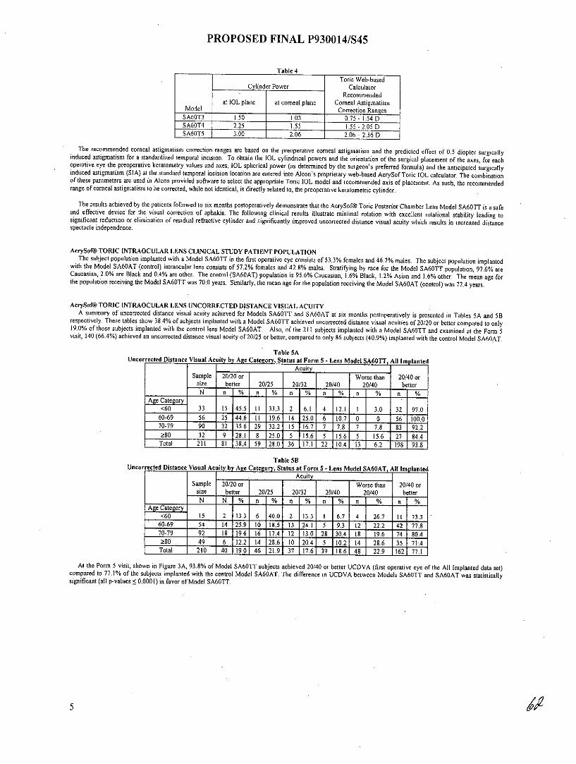
Model Correction RangesSA6OT3 1.50 1.03 0.75 - 1.54 DSA60T4 2.25 1.55 1.55 - 2.05 DSA60T5 . 3.00 2.06 2.06 - 2.56 D
The recommended corneal astigmatism correction ranges are based on the preoperative corneal astigmatism and the predicted effect of 0.5 diopter surgicallyinduced astigmatism for a standardized temporal incision. To obtain the IOL cylindrical powers and the orientation of the surgical placement of the axes, for eachoperative eye the preoperative keratometry values and axes, IOL spherical power (as determined by the surgeon's preferred formula) and the anticipated surgicallyinduced astigmatism (SIA) at the standard temporal incision location are entered into Alcon's proprietary web-based AcrySofToric IOL calculator. The combinationof these parameters are used in Alcon provided software to select the appropriate Toric IOL model and recommended axis of placement. As such, the recommendedrange of corneal astigmatism to be corrected, while not identical, is directly related to, the preoperative keratometric cylinder.
The results achieved by the patients followed to six months postoperatively demonstrate that the AcrySof@ Toric Posterior Chamber Lens Model SA60TT is a safeand effective device for the visual correction of aphakia. The following clinical results illustrate minimal rotation with excellent rotational stability leading tosignificant reduction or elimination of residual refractive cylinder and significantly improved uncorrected distance visual acuity which results in increased distancespectacle independence.
AcrySof@ TORIC INTRAOCULAR LENS CLINICAL STUDY PATIENT POPULATIONThe subject population implanted with a Model SA60TT in the first operative eye consists of 53.3% females and 46.7% males. The subject population implanted
with the Model SA60AT (control) intraocular lens consists of 57.2% females and 42.8% males. Stratifying by race for the Model SA60TT population, 97.6% areCaucasian, 2.0% are Black and 0.4% are other. The control (SA60AT) population is 95.6% Caucasian, 1.6% Black, 1.2% Asian and 1.6% other. The mean age forthe population receiving the Model SA60TT was 70.0 years. Similarly, the mean age for the population receiving the Model SA60AT (control) was 72.4 years.
AcrySof@ TORIC INTRAOCULAR LENS UNCORRECTED DISTANCE VISUAL ACUITYA summary of uncorrected distance visual acuity achieved for Models SA60TT and SA60AT at six months postoperatively is presented in Tables SA and SB
respectively. These tables show 38.4% of subjects implanted with a Model SA60TT achieved uncorrected distance visual acuities of 20/20 or better compared to only19.0% of those subjects implanted with the control lens Model SA60AT. Also, of the 211 subjects implanted with a Model SA60TT and examined at the Form 5visit, 140 (66.4%) achieved an uncorrected distance visual acuity of 20/25 or better, compared to only 86 subjects (40.9%) implanted with the control Model SA60AT.
Table SAUncorrected Distance Visual Acuity by Age Category, Status at Form 5 - Lens Model SA60TT, All Implanted
AcuitySample 20/20 or Worse than 20/40 or
size better 20/25 20/32 20/40 20/40 betterN n % n % n % n % .n % n %
Age Category<60 33 15 45.5 11 33.3 2 6.1 4 12.1 1 3.0 32 97.0
60-69 56 25 44.6 11 19.6 14 25.0 6 10.7 0 0 56 100.070-79 90 32 35.6 29 32.2 I5 16.7 7 7.8 7 7.8 83 92.2280 32 9 28.1 8 25.0 5 15.6 5 15.6 5 15.6 27 84.4
Total 211 81 38.4 59 28.0 36 17.1 22 10.4 13 6.2 198 93.8
Table SBUncorrected Distance Visual Acuity by Age Category, Status at Form 5 - Lens Model SA60AT, All Implanted
AcuitySample 20/20 or Worse than 20/40 or
size better 20/25 20/32 20/40 20/40 betterN N % n % n % n % n % n %
Age Categor<60 15 2 13.3 6 40.0 2 13.3 1 6.7 4 26.7 11 73.3
60-69 54 14 25.9 10 18.5 13 24.1 5 9.3 12 22.2 42 77.870-79 92 18 19.6 16 17.4 12 13.0 28 30.4 18 19.6 74 80.4
80 . 49 6 12.2 14 28.6 10 20.4 5 10.2 14 28.6 35 71.4Total 210 40 19.0 46 21.9 37 17.6 39 18.6 48 22.9 162 77.1
At the Form 5 visit, shown in Figure 3A, 93.8% of Model SA60TT subjects achieved 20/40 or better UCDVA (first operative eye of the All Implanted data set)compared to 77.1% of the subjects implanted with the control Model SA60AT. The difference in UCDVA between Models SA60TT and SA60AT was statisticallysignificant (all p-values < 0.0001) in favor of Model SA60TT.
5

PROPOSED FINAL P930014/S45
Figure 3ACumulative UCDVA, Status at Form 5, Model SA60TT vs. Control
100 938 S A60TTp!0.0001 p!0.0001
90 83.4 7 E. Control
80 -- -p:50.0001
70
60
50 --4p:0.0001 40.9
O 40 - ', n
30 - -o19.0 22.9
S 20
10 e"
10
20/20 or 20/25 or 20/32 or 20/40 or Worse thanbetter better better better 20/40
Cumulative UCVA
Figures 3B - 3D show a summary of cumulative uncorrected distance visual acuities for each Toric 1OL model compared to the control subjects in the samecylinder range. Figure 3B shows that the difference in cumulative UCDVA between Models SA60T3 and SA60AT was statistically significant (all p-values < 0.0115)for each visual acuity category (20/20 or better, 20/25 or better, 20/32 or better and 20/40 or better) in favor of Model SA60T3.
Figure 3B0 Cumulative UCDVA, Model SA60T3 vs. Control, Form 5, All Implanted
100 93. _ ] SA60T3p= 0.0014 p=001
9082.5 82.0 CntO
80p= 0.0011
7061.5
60
2 p= 0.0001Q 50
40
30 -
20 -1.
10 ------
0 -
20/20 or 20/25 or 20/32 or 20/40 or Worse thanbetter better better better 20/40
Cumulative UCVA

PROPOSED FINAL P930014/S45
Figure 3C shows that the difference in cumulative UCDVA between Models SA60T4 and SA60AT was statistically significant (all p-values < 0.0082) for eachvisual acuity category (20/25 or better, 20/32 or better and 20/40 or better) in favor of Model SA60T4 with the exception of the 20/20 or better category.
Figure 3CCumulative UCDVA, Model SA60T4 vs. Control, Form 5, All Implanted
100 .- J SA60T491.1 p= 0.0014
90P= 0.0007 lIS Control
80p= 0.0082
69.6 71.4t~70
60
C 50p= 0.5442 41.1
40
30 28.6
30
20/20 or 20/25 or 20/32 or 20/40 or Worse thanbetter better better better 20/40
Cumulative UCVA
Figure 3D shows that the difference in cumulative UCDVA between Models SA60TS and SA60AT was statistically significant (all p-values < 0.0171) for eachvisual acuity category (20/20 or better, 20/25 or better, 20/32 or better and 20/40 or better) in favor of Model SA60T5.
Figure 3DCumulative UCDVA, Model SA60T5 vs. Control, Form 5, All Implanted
100 LJ SA60T52 p= 0.0171
90 90 0l Controlp= 0.0 171
80 p
70870 -p= 0.0 17 1
58.5it 60
50
40p=9.0 171 1.
10 7:20 - - - -
20/20 or 20/25 or 20/32 or 20/40 or Worse thanbetter better better better 20/40
Cumulative UCVA
7 /
PROPOSED FINAL P930014/S45
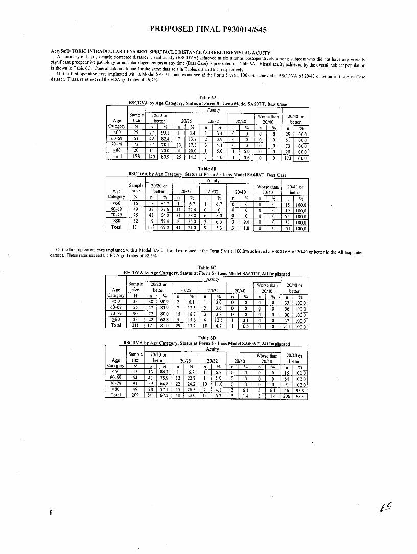
AcrySof@ TORIC INTRAOCULAR LENS BEST SPECTACLE DISTANCE CORRECTED VISUAL ACUITYA summary of best spectacle corrected distance visual acuity (BSCDVA) achieved at six months postoperatively among subjects who did not have any visuallysignificant preoperative pathology or macular degeneration at any time (Best Case) is presented in Table 6A. Visual acuity achieved by the overall subject populationis shown in Table 6C. Control data are found for the same data sets in Tables 6B and 6D, respectively.Of the first operative eyes implanted with a Model SA60TT and examined at the Form 5 visit, 100.0% achieved a BSCDVA of 20/40 or better in the Best Casedataset. These rates exceed the FDA grid rates of 96.7%.
Table 6ABSCDVA by Age Category, Status at Form 5 - Lens Model SA60TT, Best Case
Acuity
Sample 20/20 or Worse than 20/40 orAge size better 20/25 20/32 20/40 20/40 better
Category N n % n % n % n % n % n %<60 29 27 93.1 1. 3.4 1 3.4 0 0 0 0 29 100.0
60-69 51 42 82.4 7 13.7 2 3.9 0 0 0 0 51 100.070-79 73 57 78.1 13 17.8 3 4.1 0 0 0 0 73 100.0
80 20 14 70.0 4 20.0 1 5.0 1 5.0 0 0 20 100.0Total 173 140 809 25 14.5 7 4.0 1 0.6 0 0 173 100.0
Table 6BBSCDVA by Age Category, Status at Form 5 - Lens Model SA6OAT, Best C se
Sample 20/20 or Worse than 20/40 orAge size better 20/25 20/32 20/40 20/40 better
Category N n % n n % n % I n % n %<60 15 13 86.7 6.7 1 6.7 0 0 0 0 15 100.0
60-69 49 38 77.6 11 22.4 0 0 0 0 0 0 49 100.070-79 75 48 64.0 21 28.0 6 8.0 0 0 0 0 75 100.0>80 32 19 59.4 8 25.0 2 6.3 3 9.4 0 0 100.0
Total 171 118 69.0 41 24.0 9 5.3 3 1.8 0 0 171 100.0
Of the first operative eyes implanted with a Model SA60TT and examined at the Form 5 visit, 100.0% achieved a BSCDVA of 20/40 or better in the All Implanteddataset. These rates exceed the FDA grid rates of 92.5%.
Table 6CBSCDVA by Age Category, Status at Form 5 - Lens Model SA60TT, All Implanted
Acuity
Sample 20/20 or Worse than 20/40 orAge size better 20/25 20/32 20/40 20/40 better
Category N n % n % n % n % n % n %<60 33 30 90.9 2 6.1 1 3.0 0 0 0 0 33 100.0
60-69 56 47 83.9 7 12.5 2 3.6 0 0 0 0 56 100.070-79 90 72 80.0 15 16.7 3 3.3 0 0 0 0 90 100.0>80 32 22 68.8 5 15.6 4 12.5 I 3.1 0 0 32 100.0
Total 211 171 81.0 29 13.7 10 4.7 1 0.5 0 0 211 100.0
Table 6DBSCDVA by Age Category, Status at Form 5 - Lens Model SA60AT, All Implanted
AcuitySample 20/20 or Worse than 20/40 or
Age size better 20/25 20/32 20/40 20/40 betterCategory N n % n % n % n % n % n %
<60 15 13 §86.7 I 6.7 I 6.7 0 0 0 0 15 100.060-69 54 41 75.9 12 22.2 1 1.9 0 0 0 0 54 100.070-79 91 59 64.8 22 24.2 10 11.0 0 0 0 0 91 100.0>80 49 28 57.1 13 26.5 2 4.1 3 6.1 3 6.1 46 93.9
Total 209 141 67.5 48 23.0 14 6.7 3 1.4 3 1.4 206 98.6

PROPOSED FINAL P930014/S45
Figures 4A - 4C show a summary of cumulative best corrected visual acuities for each Toric model compared to the control subjects in the same cylinder range forthe All Implanted dataset.
Figure 4ACumulative BSCDVA, Model SA60T3 vs. Control, Form 5, All Implanted
100 100 100
91 97.5 99.290
80
70-
> 60
50
12 40
30
010 - - - .: ]
20/20 or 20/25 or 20/32 or 20/40 or Worse thanbetter better better better 20/40
Cumulative BSCVA
Figure 4BCumulative BSCDVA, Model SA60T4 vs. Control, Form 5, All Implanted
100 8 0100 0 SA60T4.96.4 96.4 1 98.2 98.2
70
6050
99
30-
20
10
0 .0
20/20 or 20/25 or 20/32 or 20/40 or Worse thanbetter better better better 20/40
Cumulative BSCVA

PROPOSED FINAL P930014/S45
Figure 4CCumulative BSCDVA, Model SA60T5 vs. Control, Form 5, All Implanted
100 00 100
90.2 96.99090 M Control
80 -
so -
70
20/20 or 20/25 or 20/32 or 20/40 or Worse thanbetter better better better 20/40
Cumulative BSCVA
AcrySof@ TORIC INTRAOCULAR LENS ABSOLUTE RESIDUAL REFRACTIVE CYLINDERFigures 5A through 5C demonstrate that residual refractive cylinder values were statistically significantly lower among those subjects implanted with either an
AcrySofV Toric Model SA60T3, SA60T4 or SA60T5 IOL when compared to the corresponding subjects implanted with the control Model SA60AT. Subjectsimplanted with an AcrySof@ Toric Model SA60T3 showed a 62.4% mean reduction in refractive cylinder from the preoperative visit (keratometric cylinder) ascompared to the 10.8% mean reduction for subjects implanted with the concurrent control Model SA60AT. Subjects implanted with an AcrySofO Toric ModelSA60T4 or SA60T5 showed similar results with a mean reduction in refractive cylinder of 54.8 % and 67.8%, respectively, as compared to subjects implanted withthe concurrent control model who had a mean reduction in refractive cylinder of 22.1% and 27.7%, respectively. Each of the AcrySofW Toric Lens Models SA60T3,SA60T4 and SA60T5 had at least a 3-fold increase in the likelihood of achieving residual refractive cylinder of 0.5 D or less as compared to the corresponding controlmodel.
Figure 5AAbsolute Residual Refractive Cylinder,
Model SA60T3 vs. Control, Form 5, All Implanted50 O. ISA60T3
U Control
4036.8 3.
32.5 overallV--5 0.0001
26.4
8 228s
20
,E 14.9
10 10.
0
0.00 >0.00 to >0.50 to >1.00 to >1.50 to >2.00050 1.00 1.50 2.00
Cylinder Amount (D)
10

PROPOSED FINAL P930014/S45
Figure 5BAbsolute Residual Refractive Cylinder,
Model SA60T4 vs. Control, Form 5, All Implanted50 USA60T4
41.1 Mil Control
40
32.1 overall301 2 8. p! 0.0001
30
8, . 21.4
o20 -16.1
l1 4.3
8.9 107
180.01
0.00 >0.00 to >0.50 to >1.00 to >1.50 to >2.000.50 1.00 1.50 2.00
Cylinder Amount (D)
Figure SCAbsolute Residual Refractive Cylinder,
Model SA60T5 vs. Control, Form 5, All Implanted50 C SA6 UB
l Control
40
overall30 p0.0001
30 26.8 26.8
22. 21.9 21,920
0'2.4 2.4 >
0 ' L, 1-W -12
0.00 >0.00 to >0.50 to >1.00 to >1.50 to >2.000.50 1.00 1.50 2.00
Cylinder Amount (D)
PROPOSED FINAL P930014/S45
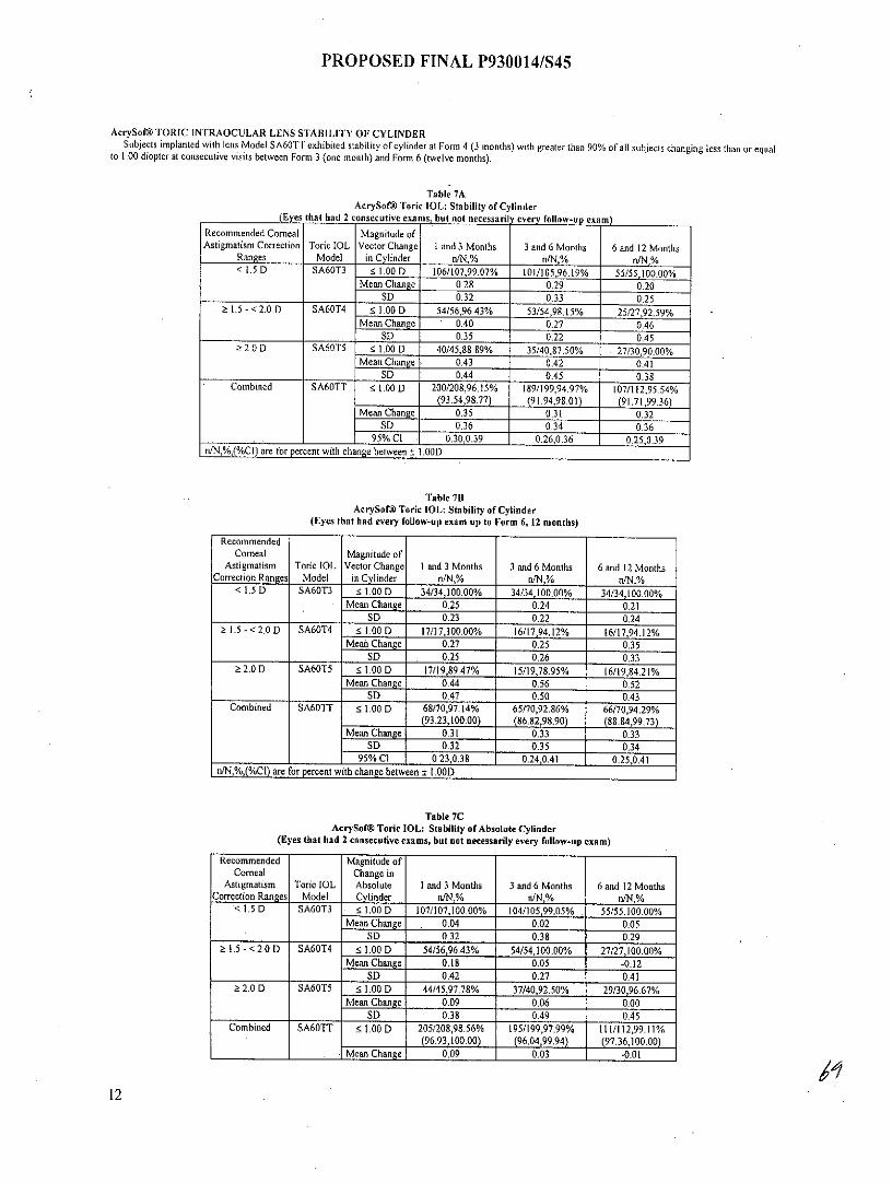
AcrySof@ TORIC INTRAOCULAR LENS STABILITY OF CYLINDERSubjects implanted with lens Model SA60TT exhibited stability of cylinder at Form 4 (3 months) with greater than 90% of all subjects changing less than or equal
to 1.00 diopter at consecutive visits between Form 3 (one month) and Form 6 (twelve months).
Table 7AAcrySof@ Toric IOL: Stability of Cylinder
(Eyes that had 2 consecutive exams, but not necessaril every follow-up exam)Recommended Corneal Magnitude ofAstigmatism Correction Toric IOL Vector Change I and 3 Months 3 and 6 Months 6 and 12 Months
Ranges Model in Cylinder n/N,% nfN,% niN,%< 1.5 D SA60T3 5 1.00 D 106/107,99.07% 101/105,96.19% 55/55,100.00%
Mean Change 0.28 0.29 0.20SD 0.32 0.33 0.25
1.5 - <2.0 D SA60T4 1 1.00 D 54/56,96.43% 53/54,98.15% 25/27,92.59%Mean Change 0.40 0.27 0.46
SD 0.35 0.22 0.452.0 D SA60T5 1.00 D 40/45,88.89% 35/40,87.50% . 27/30,90.00%
Mean Change 0.43 0.42 0.41SD 0.44 0.45 0.38
Combined SA60TT 1.00 D 200/208,96.15% 189/199,94.97% 107/112,95.54%(93.54,98.77) (91.94,98.01) (91.71,99.36)
Mean Change 0.35 0.31 0.32SD 0.36 0.34 0.36
95% Cl 0.30,0.39 0.26,0.36 0.25,0.39n/N,%,(%CI) are for percent with change between ± I .00D
Table 7BAcrySof@ Toric IOL: Stability of Cylinder
(Eyes that had every follow-up exam up to Form 6, 12 months)
RecommendedCorneal Magnitude of
Astigmatism Toric IOL Vector Change I and 3 Months 3 and 6 Months 6 and 12 MonthsCorrection Ranges Model in Cylinder n/N,% n/N,% n/N,%
< 1.5 D SA60T3 ! 1.00 D 34/34,100.00% 34/34,100.00% 34/34,100.00%Mean Change 0.25 0.24 0.21
SD 0.23 0.22 0.241.5 - <2.0 D SA60T4 5 1.00 D 17/17,100.00% 16/17,94.12% 16/17,94.12%
Mea Change 0.27 0.25 . 0.35SD 0.25 0.26 0.33
2.0 D SA60T5 1.00 D 17/19,89.47% 15/19,78.95% 16/19,84.21%Mean Change 0.44 0.56 0.52
SD 0.47 0.50 0.43Combined SA60TT 5 1.00 D 68/70,97.14% 65/70,92.86% 66/70,94.29%
(93.23,100.00) (86.82,98.90) (88.84,99.73)Mean Change 0.31 0.33 0.33
SD 0.32 0.35 0.3495% Cl 0.23,0.38 0.24,0.41 0.25,0.41
n/N,%,(%CI) are for percent with change between ± I.00D
Table 7CAcrySof@ Toric IOL: Stability of Absolute Cylinder
(Eyes that had 2 consecutive exams, but not necessarily every follow-up exam)
Recommended Magnitude ofCorneal Change in
Astigmatism Toric IOL Absolute I and 3 Months 3 and 6 Months 6 and 12 MonthsCorrection Ranges Model Cylinder n/N,% n/N,% n/N,%
< 1.5 D SA60T3 - 1.00 D 107/107,100.00% 104/105,99.05% . 55/55,100.00%Mean Change . 0.04 0.02 0.05
SD 0.32 0.38 0.29S.5 - < 2.0 D SA60T4 1.00 D 54/56,96.43% 54/54,100.00% 27/27,100.00%
Mean Change 0.18 0.05 -0.12SD 0.42 0.27 0.41
2.0 D SA60T5 - 1.00 D 44/45,97.78% 37/40,92.50% 29/30,96.67%Mean Change 0.09 0.06 0.00
SD 0.38 0.49 0.45
Combined SA60TT 1.00 D 205/208,98.56% 195/199,97.99% 111/112,99.11%(96.93,100.00) (96.04,99.94) (97.36,100.00)
Mean Change 0.09 0.03 -0.01
12

PROPOSED FINAL P930014/S45
Recommended Magnitude ofCorneal Change in
Astigmatism Toric IOL Absolute I and 3 Months 3 and 6 Months 6 and 12 MonthsCorrection Ranges Model Cylinder nIN,% n/N,% n/N %
SD 0.37 0.38 0.3795% CI 0.04,0 14 -0.02,0.09 -0,08,0.06
n/N,%,(%CI) are for percent with change between ± 1.00D
Table 7DAcrySof@ Toric IOL: Stability of Absolute Cylinder
(Eyes that had every follow-up exam up to Form 6, 12 months)
Recommended Magnitude ofCorneal Change in
Astigmatism Toric 1OL Absolute I and 3 Months 3 and 6 Months 6 and 12 MonthsCorrection Ranges Model Cylinder n/N,% n/N,% n/N,%
< 1.5 D SA60T3 1.00 D 34/34,100.00% 34/34,100.00% 34/34,100.00%Mean Change 0.01 -0.01 0.07
SD 0.28 0.31 0.281.5 - < 2.0 D SA60T4 1.00 D 17/17,100.00% 17/17,100,00% 17/17,100.00%
Mean Change 0.06 0.19 -0.04SD 0.30 0.21 0.42
2.0 D SA60T5 5 1.00 D 18/19,94.74% 17/19,89.47% 18/19,94.74%Mean Change 0.17 0.05 0.01
SD 0.45 0.54 0.55Combined SA60TT 1.00 D 69/70,98.57% 68/70,97.14% 69/70,98.57%
(95.78,100.00) (93.23,100.00) (95.78,100.00)Mean Change 0.07 0.05 0.03
SD 0.34 0.38 0.4095% CI -0.01,0.15 -0.04,0.14 -0.07,0.12
n/N,%,(%CI) are for percent with change between ± I.00D
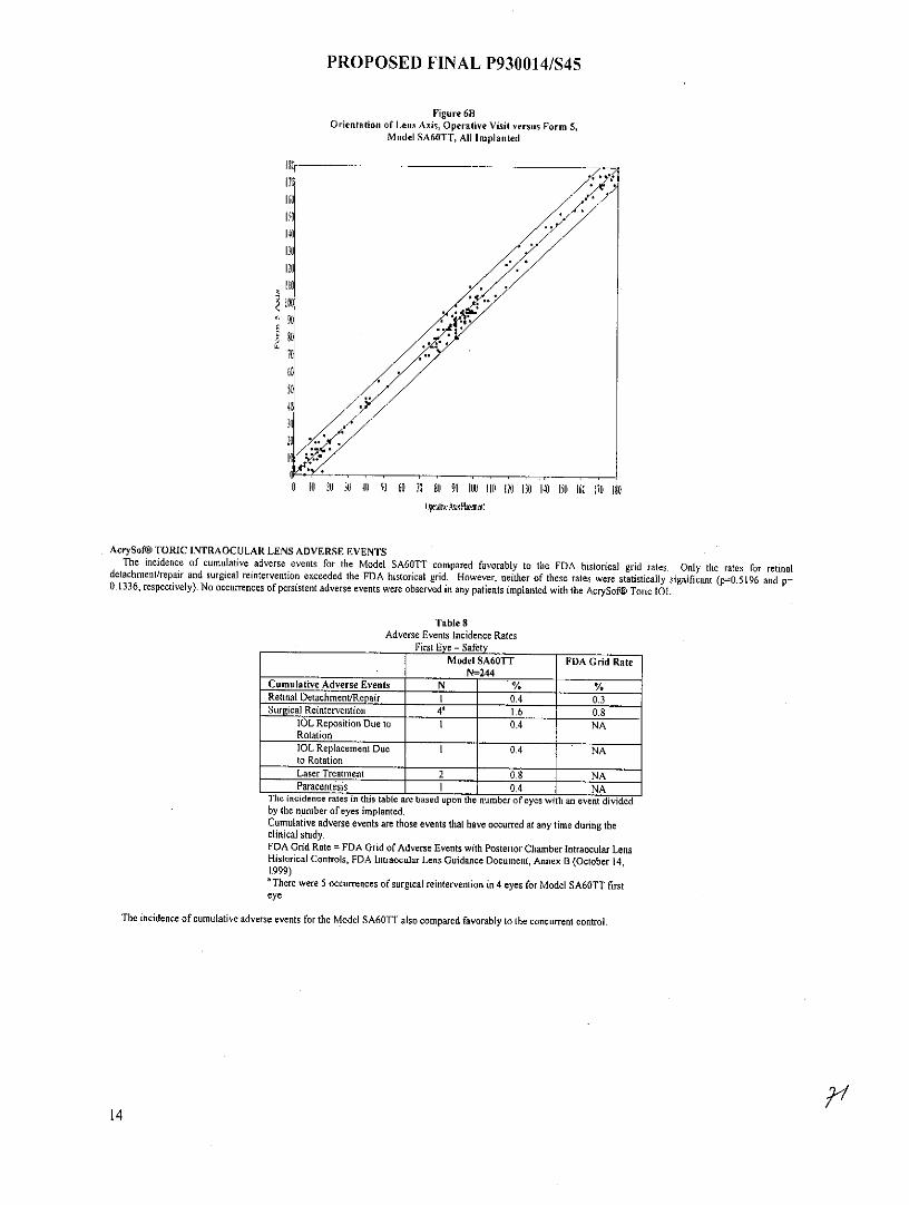
AcrySof@ TORIC INTRAOCULAR LENS ROTATIONAL STABILITYA summary of the change in axis orientation (rotation) from the operative visit to the Form 5 visit (120-180 days postoperative) is presented in Figures 6A and 6B.
The rotational stability of the AcrySof® Toric Model SA60TT is established with the majority of the lenses rotating - 50. Figure 6A also demonstrates that theamount of rotation seen in each AcrySofW Toric IOL model is independent of the amount of cylinder power present on the lens.
Figure 6AChange in Axis Orientation from Operative Visit to Form 5, All Implanted
100
I1SA6OT390
N SA60T480
OSA60T570
S 60-
30
20
10 -
00" to 5" >5 to 10" >10 to 15" >15" to 530" >30*
Amount of Rotation (degrees)
13
PROPOSED FINAL P930014/S45
Figure 6BOrientation of Lens Axis, Operative Visit versus Form 5,
Model SA60TT, All Implanted
176160
140
I0
6 16 26 3 46 56 60 70 86 96 IN) 1161 126 130 46 100 166 170 6609eraukl kj Pbxmail
AcrySof@ TORIC INTRAOCULAR LENS ADVERSE EVENTSThe incidence of cumulative adverse events for the Model SA60TT compared favorably to the FDA historical grid rates. Only the rates for retinaldetachment/repair and surgical reintervention exceeded the FDA historical grid. However, neither of these rates were statistically significant (p=0.5196 and p=0.1336, respectively). No occurrences of persistent adverse events were observed in any patients implanted with the AcrySofo Toric IOL.
Table 8Adverse Events Incidence Rates
First Eye - SafetyModel SA60TT FDA Grid Rate
N=244Cumulative Adverse Events N % %Retinal Detachment/Repair I 0.4 0.3Surgical Reintervention 4' 1.6 0.8
IOL Reposition Due to 1 0.4 NARotationIOL Replacement Due 1 0.4 NAto RotationLaser Treatment 2 0.8 NAParacentesis I 0.4 NA
The incidence rates in this table are based upon the number of eyes with an event dividedby the number of eyes implanted.Cumulative adverse events are those events that have occurred at any time during theclinical study.FDA Grid Rate = FDA Grid of Adverse Events with Posterior Chamber Intraocular LensHistorical Controls, FDA Intraocular Lens Guidance Document, Annex B (October 14,1999)There were 5 occurrences of surgical reintervention in 4 eyes for Model SA60TT first
eye
The incidence of cumulative adverse events for the Model SA60TT also compared favorably to the concurrent control.
14
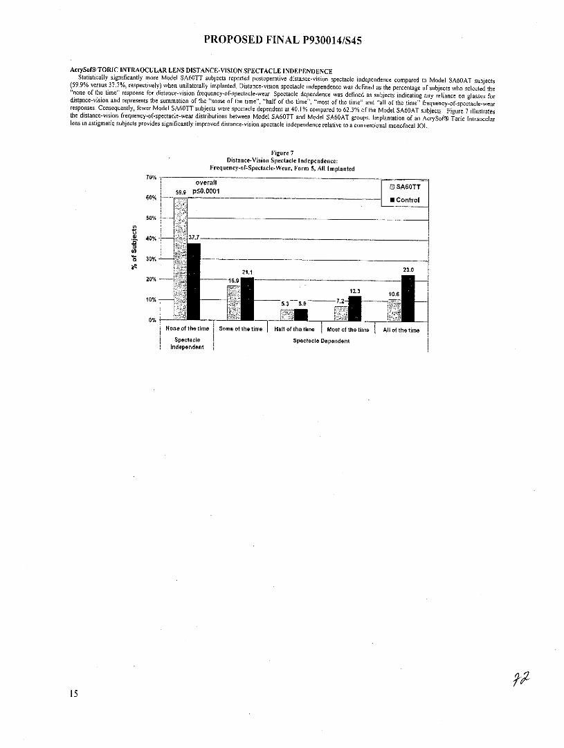
PROPOSED FINAL P930014/S45
AcrySof@ TORIC INTRAOCULAR LENS DISTANCE-VISION SPECTACLE INDEPENDENCEStatistically significantly more Model SA60TT subjects reported postoperative distance-vision spectacle independence compared to Model SA60AT subjects(59.9% versus 37.7%, respectively) when unilaterally implanted. Distance-vision spectacle independence was defined as the percentage of subjects who selected the
"none of the time" response for distance-vision frequency-of-spectacle-wear. Spectacle dependence was defined as subjects indicating any reliance on glasses fordistance-vision and represents the summation of the "some of the time", "half of the time", "most of the time" and "all of the time" frequency-of-spectacle-wearresponses. Consequently, fewer Model SA60TT subjects were spectacle dependent at 40.1% compared to 62.3% of the Model SA60AT subjects. Figure 7 illustratesthe distance-vision frequency-of-spectacle-wear distributions between Model SA60TT and Model SA60AT groups. Implantation of an AcrySofG Toric Intraocularlens in astigmatic subjects provides significantly improved distance-vision spectacle independence relative to a conventional monofocal 1OL.
Figure 7Distance-Vision Spectacle Independence:
Frequency-of-Spectacle-Wear, Form 5, All Implanted70% -
overall 0 SA60TT59.9 p50.0001
60% - 0 - - Control
50%
,1 40% ' 37.7
S 30%-
23.021.120% -- 1 16.9
12.3 1 .610% 53 59
None of the time Some of the time Hat of the time Most of the time A I of the time
Spectacle Spectacle Dependentindependent
15

PROPOSED FINAL P930014/S45
2. AcrySof@ TORIC HIGH CYLINDER POWER INTRAOCULAR LENS CLINICAL STUDYA clinical study was conducted to investigate the rates of spatial distortions related to axial misalignment of the AcrySof@ Toric Posterior Chamber High Cylinder
Power Intraocular Lenses shown in Table 9.
Table 9
Toric CalculatorCylinder Power Recommended
Corneal AstigmatismModel at IOL plane at corneal plane Correction Ranges
SN60T6 3.75 2.57 2.57 - 3.07 DSN60T7 4.50 3.08 3.08-3.59 DSN60T8 5.25 3.60 3.60-4.10 DSN60T9 6.00 4.11 4.11 D and up
These recommended corneal astigmatism correction ranges are based on the preoperative corneal astigmatism and the predicted effect of surgically inducedastigmatism. To obtain the IOL cylindrical powers and the orientation of the surgical placement of the axes, for each operative eye the preoperative keratometryvalues and axes, IOL spherical power (as determined by the surgeon's preferred formula) and the surgeon's estimated surgically induced astigmatism (SIA) at thestandard temporal incision location are entered into Alcon's proprietary web-based AcrySofToric IOL calculator. The combination of these parameters are used in theAlcon provided software to select the appropriate Toric IOL model and recommended axis of placement. As such, the recommended range of corneal astigmatism tobe corrected while not identical, is directly related to, the preoperative keratometric cylinder,
The results achieved by the patients followed to six months (Visit SA) postoperatively demonstrate that the AcrySof@ Toric high cylinder intraocular lens modelsare safe and effective for the visual correction of aphakia. The following clinical results illustrate the AcrySofT Toric high cylinder power lOL's effectiveness insignificantly reducing pre-existing corneal astigmatism and the IOL's excellent rotational stability following implantation in the capsular bag
AcrySof@ TORIC HIGH CYLINDER POWER INTROCULAR LENS CLINICAL STUDY PATIENT POPULATIONThis study focused on the highest cylinder power IOL Model SN60T9; however, due to the rarity of this level of astigmatism IOL Model SN60T8 was included to
expand the inclusion criteria for the second eye. The subject population implanted with an IOL Model SN60T9 in the first operative eye consists of 80% (12/15)females and 20% (3/15) males. For the fellow eye, 3 subjects were implanted with IOL Model SN60T9, while 12 were implanted with lOL Model SN60T8. All 15(100%) of the implanted subjects were white. The mean age for the population was 67 years old (range of 43 to 82 years) at the time of surgery.
AcrySof@ TORIC HIGH CYLINDER POWER INTRAOCULAR LENS ABSOLUTE RESIDUAL REFRACTIVE CYLINDERRefractive cylinder six months postoperatively \was reduced for all subjects implanted with either an AcrySofM Toric IOL Model SN60T8 or SN60T9 compared to
preoperative baseline. Results show a statistically significant reduction (p-value <.0001) in residual refractive cylinder in eyes implanted with IOL Model SN60T9[85.7% in first eyes (n=15), 87.8% in second eyes (n=3) and IOL Model SN60T8 [87.3% (n=12)1..
AcrySof@ TORIC HIGH CYLINDER POWER INTRAOCULAR LENS ROTATIONAL STABILITYA summary of the change in axis orientation (rotation) from the operative visit to Visit SA (six months postoperative) is presented in Figure 8A and 8B. The
AcrySof@ Toric high cylinder power IOLs demonstrated rotational stability with the majority of the lenses rotating 5*.
Figure 8AChange in Lens Axis Orientation - Visit SA (Final Visit) versus Target
100%
90% 1..
70%
t0 40%
30%
. ........
00 to 50 >5to, 100 >101too 0 IS0to 200 >20 0 to300 >300
Amount of Rotation
16
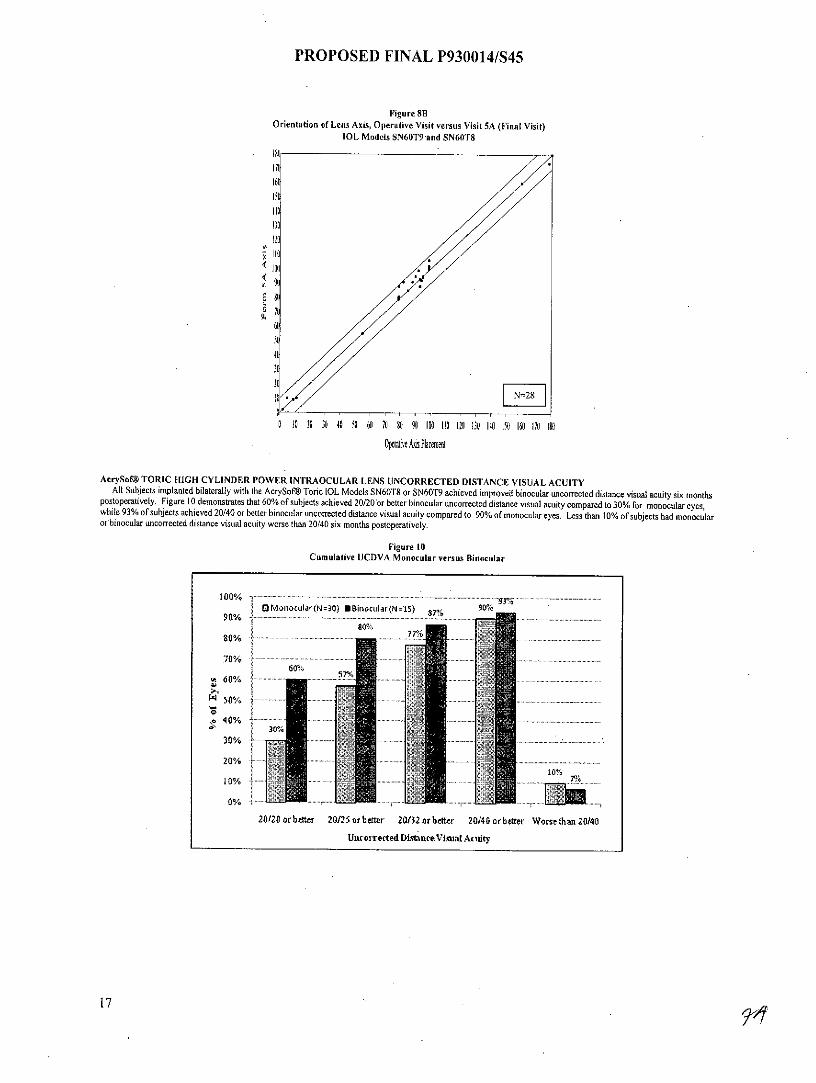
PROPOSED FINAL P930014/S45
Figure 8BOrientation of Lens Axis, Operative Visit versus Visit SA (Final Visit)
IOL Models SN60T9 and SN60T8
15
130120
IIH
0 10 20 30 40 50 60 70 8U %1 100 110 120 130 140 [50 6 170 180
Oplire A2s Placemnt
AcrySof@ TORIC HIGH CYLINDER POWER INTRAOCULAR LENS UNCORRECTED DISTANCE VISUAL ACUITYAll Subjects implanted bilaterally with the AcrySof Toric [OL Models SN60T8 or SN60T9 achieved improved binocular uncorrected distance visual acuity six months
postoperatively. Figure 10 demonstrates that 60% of subjects achieved 20/20 or better binocular uncorrected distance visual acuity compared to 30% for monocular eyes,while 93% of subjects achieved 20/40 or better binocular uncorrected distance visual acuity compared to 90% of monocular eyes. Less than 10% of subjects had monocularor binocular uncorrected distance visual acuity worse than 20/40 six months postoperatively.
Figure 10Cumulative UCDVA Monocular versus Binocular
100%............. ................................... .....90 MonocularlN=30) U8inocular{N=15) 8 7%
90%w50%%
30%
60%60% 57%
40% j 30%30%
20/o ........ -. .n
.2..........0%
20/20 orbetter 20125 orbetter 20/32 orbetter 20/40 or better Worse than 20140
UncorectedDistatreVisitmlAcuity
17
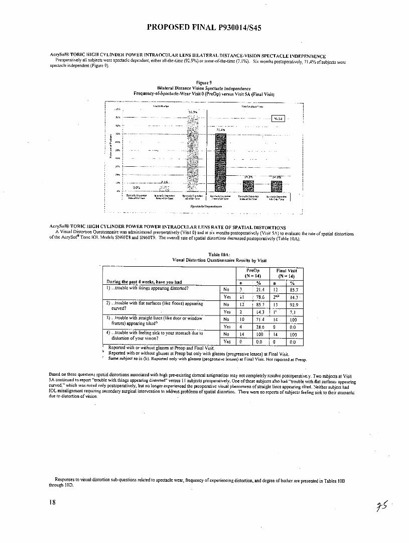
PROPOSED FINAL P930014/S45
AcrySof@TORIC HIGH CYLINDER POWER INTRAOCULAR LENS BILATERAL DISTANCE-VISION SPECTACLE INDEPENDENCEPreoperatively all subjects were spectacle dependent, either all-the-time (92.9%) or some-of-the-time (7.1%). Six months postoperatively, 71.4% of subjects were
spectacle independent (Figure 9).
Figure 9Bilateral Distance Vision Spectacle Independence
Frequency-of-Spectacle-Wear Visit 0 (PreOp) versus Visit 5A (Final Visit)
? I A%
AcrySof@ TORIC HIGH CYLINDER POWER POWER INTRAOCULAR LENS RATE OF SPATIAL DISTORTIONSA Visual Distortion Questionnaire was administered preoperatively (Visit 0) and at six months postoperatively (Visit SA) to evaluate the rate of spatial distortions
of the AcrySof' Toric IOL Models SN60T8 and SN60T9. The overall rate of spatial distortions decreased postoperatively (Table 10A).
Table 10A:Visual Distortion Questionnaire Results by Visit
PreOp Final Visit(N =14) (N "14)
During the past 4 weeks, have you had n % n %1) trouble with things appearing distorted? No 3 21.4 12 85.7
Yes 11 78.6 2a-b 14.32) ...trouble with flat surfaces (like floors) appearing No 12 85.7 13 92.9
curved?Yes 2 14.3 IV 7,1
3) ...trouble with straight lines (like door or window No 10 71.4 14 100frames) appearing tilted?
Yes 4 28.6 0 0.04) ...trouble with feeling sick to your stomach due to No 14 100 14 100
distortion of your vision? Yes 0 0.0 0 0.0
Reported with or without glasses at Preop and Final Visit.Reported with or without glasses at Preop but only with glasses (progressive lenses) at Final Visit.Same subject as in (b). Reported only with glasses (progressive lenses) at Final Visit. Not reported at Preop.
Based on these questions spatial distortions associated with high pre-existing corneal astigmatism may not completely resolve postoperatively. Two subjects at Visit5A continued to report "trouble with things appearing distorted" versus 11 subjects preoperatively. One of these subjects also had "trouble with flat surfaces appearingcurved," which was noted only postoperatively, but no longer experienced the preoperative visual phenomena of straight lines appearing tilted. Neither subject hadIOL misalignment requiring secondary surgical intervention to address problems of spatial distortion. There were no reportns of subjects feeling sick to their stomachsdue to distortion of vision.
Responses to visual distortion sub-questions related to spectacle wear, frequency of experiencing distortion, and degree of bother are presented in Tables 10Bthrough 10D.
18
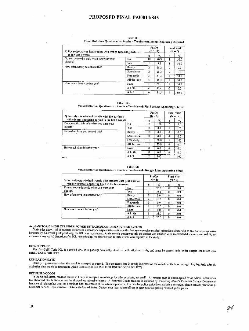
PROPOSED FINAL P930014/S45
Table 10B:Visual Distortion Questionnaire Results - Trouble with Things Appearing Distorted
PreOp Final Visit1) For subjects who had trouble with things appearing distorted (N= 11) - (N = 2)
in the last 4 weeks: n % n /Do you notice this only when you wear your No 10 90.9 1 50.0glasses? Yes 1 9.1 1 50.0How often have you noticed this? Rarely 2 18.2 0 0,0
Sometimes 2 18.2 0 0.0Frequently 3 27.3 1 50.0All the time 4 36.4 1 50.0
How much does it bother you? None 1 9.1 I 50.0A Little 4 36.4 . 0 0.0__A Lot 6 54.5 I . 50.0
Table 10C:Visual Distortion Questionnaire Results - Trouble with Flat Surfaces Appearing Curved
PreOp Final Visit2) For subjects who had trouble with flat surfaces (N = 2) (N 1)
(like floors) appearing curved in the last 4 weeks: n % n ,VDo you notice this only when you wear your No 2 100 0 0.0glasses? Yes 0 0.0 1 100How often have you noticed this? Rarely 0 0.0 0 0.0
Sometimes 0 0.0 0 0.0Frequently I 50.0 I 100All the time 1 50.0 0 0.0
How much does it bother you? None 0 0.0 0 0.0A Little 0 0,0 0 0.0_ALot 2 100 1 100
Table 10DVisual Distortion Questionnaire Results - Trouble with Straight Lines Appearing Tilted
PreOp Final Visit3) For subjects who had trouble with straight lines (like door or _(N= 4) (N= 0)
window frames) appearing tilted in the last 4 weeks: n % n %Do you notice this only when you wear your No 3 75.0 0 0.0glasses? Yes 1 25.0 0 0.0How often have you noticed this? Rarely 0 . 0.0 0 0.0
Sometimes 2 50.0 0 0.0Frequently 0 0.0 0 0.0All the time 2 50.0 0 0.0
How much does it bother you? None 0 0.0 0 0.0A Little I 25.0 0 .0.0A Lot 3 75.0 0 0.0
AcrySof@ TORIC HIGH CYLINDER POWER INTRAOCULAR LENS ADVERSE EVENTSDuring the study, I of 15 subjects underwent a secondary surgical intervention in the first eye to resolve residual refractive cylinder due to an error in preoperative
keratometry. One week postoperatively, the IOL was repositioned. At six months postoperatively the subject was satisfied with uncorrected distance vision and did notexperience any spatial distortion after IOL repositioning. No other serious adverse events were reported in the study.
HOW SUPPLIEDThe AcrySof@ Toric IOL is supplied dry, in a package terminally sterilized with ethylene oxide, and must be opened only under aseptic conditions (See
DIRECTIONS FOR USE).
EXPIRATION DATESterility is guaranteed unless the pouch is damaged or opened. The expiration date is clearly indicated on the outside of the lens package. Any lens held after the
expiration date should be returned to Alcon Laboratories, Inc. (See RETURNED GOODS POLICY).
RETURNED GOODSIn the United States, returned lenses will only be accepted in exchange for other products, not credit. All returns must be accompanied by an Alcon Laboratories,
Inc. Returned Goods Number and be shipped via traceable means. A Returned Goods Number is obtained by contacting Alcon's Customer Service Department.Issuance of this number does not constitute final acceptance of the returned products. For detailed policy guidelines including.exchange, please contact your Sales orCustomer Service Representative. Outside the United States, Contact your local Alcon offices or distributors regarding returned goods policy
19

PROPOSED FINAL P930014/S45
REFERENCESBoettner, E.A. and Wolter, J.R. Transmission of the ocular media. Invest. Ophthalmol. 1:776-783, 1962
Symbo s Used on LabelingSYMBOL ENGLISH
IOL Intraocular lensPC Posterior chamber
PCL Posterior chamber lensUV UltravioletD Diopter (spherical equivalent)
CYL Cylinder Power0a Body diameter (Optic diameter)
OT Overall diameter (Overall length)
V Do not reuse
or Use by (YYYY-MM: year-month)
STERILE EQ Sterilized by ethylene oxide
SN or SN Serial Number
Ax Attention: See instructions for use
E Batch Code
Authorized Representative in the EuropeanCommunity
Manufacturer
Alcon Laboratories, Inc.6201 South FreewayFort Worth, Texas, USA76134-2099
U.S. Pat. No's. 5,403,901, 5,470,932, 5,543,504, 5,603,774, and 5,716,403.
C2011 Alcon, Inc.
20

PROPOSED FINAL 01.27.11 P930014/S45
PRODUCT INFORMATIONAlcon Laboratories, Inc.
STERILE UV and Blue Light Filtering Acrylic FoldableToric Aspheric Optic Single-Piece Posterior Chamber Lenses
CAUTION: Federal (USA) law restricts this device to the sale by or on the order of a physician.
DESCRIPTIONThe AcrySof@ IQ Toric Posterior Chamber Intraocular Lens (IOL) is a UV-absorbing foldable intraocular lens (1OL). These IOLs have a biconvex toric asphericoptic with cylinder axis marks to denote the flat meridian (plus cylinder axis). The single-piece design (see Figure I and Table 1) consists of a high refractive index
material with proprietary blue light filtering chromophore which filters light in a manner that approximates the human crystalline lens in the 400-475 nm blue lightwavelength range (Boettner and Wolter, 1962). In addition to standard UV-light filtering, the blue-light filtering chromophore reduces transmittance of blue light (seeTable 2). The biconvex toric aspheric optic consists of a high refractive index soft acrylic material capable of being folded prior to insertion, allowing placementthrough an incision smaller than the optic diameter of the lens. After surgical insertion into the eye, the lens gently unfolds to restore the optical performance. Thesupporting haptics provide for proper positioning and fixation of the IOL optic within the eye. The anterior surface of the AcrySof@ IQ Toric IOL Model SN6ATT isdesigned with negative spherical aberration identical to the aspheric AcrySof@ IQ IOL Model SN60WF to compensate for the positive spherical aberration of thecomea.
Figure 1: Physical Characteristics of AcrySof@ IQ Toric IOLs(All dimensions in millimeters)
01%.0
tANITEIOR A ~LTIECOL-.-
Ltss SUCFACE
* The effects of this aspheric design feature have been clinically assessed on AcrySofV IQ [OL Model SN60WF.
Table 1: Physical Characteristics of AcrySof@ IQ Toric IOLs
Characteristics ModelSN6AT6 I SN6A [ SN6AT _ t SN6AT9
Collectively referred to as Model SN6ATTOptic Type Biconvex Toric Aspheric Optic
Optic / Haptic Material Ultraviolet and blue light filtering Acrylate/Methacrylate CopolymerUV cutoff at 10% T: 399 nm (+6.0 diopter lens)
407 nm (+34.0 diopter lens)IOL Powers
(spherical equivalent diopters) For available power range see Alcon Product GuideIOL Cylinder Power (Diopters) 3.75 D 4.50 D 5.25 D 6.00 D
Index Of Refraction 1.55Haptic Configuration STABLEFORCE® HapticOptic Diameter (mm) 6.0Overall Length (mm) 13.0
Haptic Angle 0.
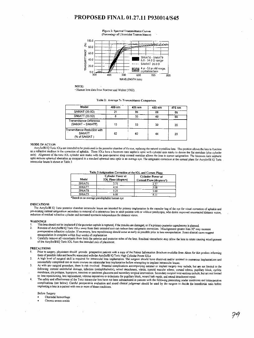
PROPOSED FINAL 01.27.11 P930014/S45
Figure 2: Spectral Transmittance Curves(Percentage of Ultraviolet Transmittance)
100.0
80.0
z 60.040.0 - - SN6AT6-SN6AT9
40,0 ' 6.0 -34.0Drange2.0- SA60AT 20.0 0
<2 0.0S4 yr -53 yr old range,
0.0 crystallIne lens300 400 500 600 700 800
WAVELENGTH (nm)
NOTE:Human lens data from Boettner and Wolter (1962).
Table 2: Average % Transmittance Comparison
Model 400 nm 425 nm 450 nm 475 nmSA60AT (20.0D) 21 86 88 88SN6ATT (20.0D) 8 33 49 68
Transmittance Difference(SA60AT - SN6ATT) 13 53 39 20
Transmittance Reduction withSN6ATT 62 62 44 23
(% of SA60AT)
MODE OF ACTIONAcrySof IQ Toric IOLs are intended to be positioned in the posterior chamber of the eye, replacing the natural crystalline lens. This position allows the lens to function
as a refractive medium in the correction of aphakia. These IOLs have a biconvex toric aspheric optic with cylinder axis marks to denote the flat meridian (plus cylinderaxis). Alignment of the toric IOL cylinder axis marks with the post-operative steep comeal meridian allows the lens to correct astigmatism. The biconvex toric asphericoptic reduces spherical aberration as compared to a standard spherical toric optic in an average eye. The astigmatic correction at the corneal plane for AcrySof@ IQ Toricintraocular lenses is shown in Table 3:
Table 3 Astigmatism Correction at the IOL and Cornea PlaneCylinder Power at Cylinder Power at
Model IOL Plane (diopters) Corneal Plane (diopters*)SN6AT6 3.75 2.57SN6AT7 4.50 3.08SN6AT8 5.25 3.60SN6AT9 6.00 4.11
*Based on an average pseudophakic human eye
INDICATIONSThe AcrySof@ IQ Toric posterior chamber intraocular lenses are intended for primary implantation in the capsular bag of the eye for visual correction of aphakia and
pre-existing comeal astigmatism secondary to removal of a cataractous lens in adult patients with or without presbyopia, who desire improved uncorrected distance vision,reduction of residual refractive cylinder and increased spectacle independence for distance vision.
WARNINGSI. This lens should not be implanted if the posterior capsule is ruptured, if the zonules are damaged, or if a primary posterior capsulotomy is planned.2. Rotation of AcrySof6IQ Toric IOLs away from their intended axis can reduce their astigmatic correction. Misalignment greater than 30 may increase
postoperative refractive cylinder. If necessary, lens repositioning should occur as early as possible prior to lens encapsulation. Some clinical cases suggestencapsulation is complete within four weeks of implantation.
3. Carefully remove all viscoelastic from both the anterior and posterior sides of the lens. Residual viscoelastic may allow the lens to rotate causing misalignmentof the AcrySof0IQ Toric IOL from the intended axis of placement.
PRECAUTIONS1. Prior to surgery, physicians should provide prospective patients with a copy of the Patient Information Brochure available from Alcon for this product informing
them of possible risks and benefits associated with the AcrySof@l IQ Toric High Cylinder Power IOLs2. A high level of surgical skill is required for intraocular lens implantation. The surgeon should have observed and/or assisted in numerous implantations and
successfully completed one or more courses on intraocular lens implantation before attempting to implant intraocular lenses.3. As with any surgical procedure, there is risk involved. Potential complications accompanying cataract or implant surgery may include, but are not limited to the
following: comeal endothelial damage, infection (endophthalmitis), retinal detachment, vitritis, cystoid macular edema, comeal edema, pupillary block, cycliticmembrane, iris prolapse, hypopyon, transient or persistent glaucoma and secondary surgical intervention. Secondary surgical interventions include, but are not limitedto: lens repositioning, lens replacement, vitreous aspirations or iridectomy for pupillary block, wound leak repair, and retinal detachment repair.
4. The safety and effectiveness of the Toric intraocular lens have not been substantiated in patients with the following preexisting ocular conditions and intraoperativecomplications (see below). Careful preoperative evaluation and sound clinical judgement should be used by the surgeon to decide the benefit/risk ratio beforeimplanting a lens in a patient with one or more of these conditions.
Before Surgery* Choroidal hemorrhage* Chronic severe uveitis
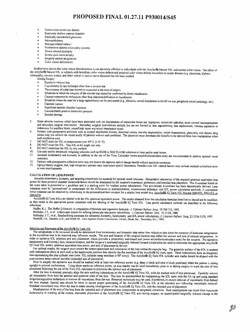
PROPOSED FINAL 01.27.11 P930014/S45
* Concomitant severe eye disease* Extremely shallow anterior chamber* Medically uncontrolled glaucoma* Microphthalmos* Non-age-related cataract* Proliferative diabetic retinopathy (severe)* Severe comeal dystrophy* Severe optic nerve atrophy* Irregular comeal astigmatism* Color vision deficiencies
Studies have shown that color vision discrimination is not adversely affected in individuals with the AcrySofW Natural IOL and normal color vision. The effect ofthe AcrySof@ Natural IOL in subjects with hereditary color vision defects and acquired color vision defects secondary to ocular disease (e.g. glaucoma, diabeticretinopathy, chronic uveitis, and other retinal or optical nerve diseases) has not been studied.
During Surgery* Excessive vitreous loss* Capsulotomy by any technique other than a circular tear* The presence of radial tears known or suspected at the time of surgery* Situations in which the integrity of the circular tear cannot be confirmed by direct visualization* Cataract extraction by techniques other than phacoemulsification or liquefaction* Situations where the need for a large capsulotomy can be anticipated (e.g., diabetics, retinal detachment in the fellow eye, peripheral retinal pathology, etc.)* Capsular rupture* Significant anterior chamber hyphema* Uncontrollable positive intraocular pressure* Zonular damage
5. Some adverse reactions which have been associated with the implantation of intraocular lenses are: hypopyon, intraocular infection, acute comeal decompensationand secondary surgical intervention. Secondary surgical interventions include, but are not limited to: lens repositioning, lens replacement, vitreous aspiration oriridectomy for pupillary block, wound leak repair and retinal detachment repair.
6. Patients with preoperative problems such as comeal endothelial disease, abnormal cornea, macular degeneration, retinal degeneration, glaucoma, and chronic drugmiosis may not achieve the visual acuity of patients without such problems. The physician must determine the benefits to be derived from lens implantation whensuch conditions exist.
7. DO NOT store the IOL at temperatures over 450 C (1130 F).8. DO NOT reuse the IOL. This IOL is for single use only.9. DO NOT resterilize the IOL by any method.10. Use only sterile intraocular irrigating solutions such as BSS® or BSS PLUS@ solutions to rinse and/or soak lenses.I1. Accurate keratometty and biometry in addition to the use of the Toric Calculator (www.acrysofloriccalculator.com) are recommended to achieve optimal visual
outcomes.12. Patients with postoperative refractive error may not receive the aspheric optical design benefit without spectacle correction.13. Optical theory suggest, that, high astigmatic patients may experience spatial distortions. Possible toric IOL related factors may include residual cylindrical error
or axis misalignments.
CALCULATION OF LENS POWERAccurate keratometry, biometry, and targeting emmetropin are essential for optimal visual outcomes. Preoperative calculation of the required spherical equivalent lens
power for these posterior chamber intraocular lenses should be determined by the surgeon's experience, preference, and intended lens placement. The A-constant listed onthe outer label is presented as a guideline and is a starting point for implant power calculations. This provisional A-constant has been theoretically derived. Lensconstants must be "personalized" to compensate for the differences in instrumentation, measurement technique, and IOL power calculation methods. A convenientinitial estimate can be obtained by referencing to the personalized lens constant for a similar lens model (e.g. AcrvSof® 10 Toric IOL Models SN6AT3. SN6AT4, oraN6AT5).
AcrySof IQ Toric IOLs are labeled with the IOL spherical equivalent power. The results obtained from the calculation formulas listed below should not be modified,as they result in the appropriate power consistent with the labeling of the AcrySof® IQ Toric IOL. Lens power calculation methods are described in the followingreferences:
Hoffer, K.J. The Hoffer Q formula: A comparison of theoretic and regression formulas. J Cataract Refract Surg. 19:700-712, 1993.Holladay, J.T., et al. A three-part system for refining intraocular lens power calculations. J. Cataract Refract. Surg 14:17-24, 1988.Holladay, J.T., et al., Standardizing constants for ultrasonic biometry, keratometry, and IOL power calculations, J. Cataract Refract. Surg. 23:1356-1370, 1997.Retzlaff, J.A., Sanders, D.R., and Kraff, M. Lens Implant Power Calculation, 3rd ed., Slack, Inc., Thorofare, NJ., 1990.
Selection and Placement of the AcrvSof@l 10 Toric IOLThe astigmatism to be corrected should be determined from keratometry and biometry data rather than refractive data since the presence of lenticular astigmatism
in the crystalline lens to be removed may influence results. The size and location of the surgical incision may affect the amount and axis of corneal astigmatism. Inorder to optimize IOL selection and axis placement, Alcon provides a proprietary web-based tool (www.acrysofloriccalculator.com) for the surgeon. Pre-operativekeratometry and biometry data, incision location, and the surgeon's estimated surgically induced corneal astigmatism are used to determine the appropriate AcrySofVIQ Toric IOL model, spherical equivalent lens power, and axis of placement in the eye.
For optimal results, the surgeon must ensure the correct placement and orientation of the lens within the capsular bag. The posterior surface of the IOL is markedwith indentations (three at each end) at the haptic/optic junction that identify the flat meridian of the AcrySoft IQ Toric optic. These indentations form an imaginaryline representing the plus cylinder axis (note: IOL cylinder steep meridian is 90* away). The AcrySof@ IQ Toric IOL cylinder axis marks should be aligned with thepost-incision steep corneal meridian (intended axis of placement).
Prior to surgery the operative eye should be marked with at least two reference points (e.g. three o'clock and nine o'clock positions) while the patient is sittingupright to prevent cyclotorsion. Using these marks as reference points, an axis marker can be used immediately prior to or during surgery to mark the axis of lensplacement following the use of the Toric IOL calculator to determine the optimal axis of placement,
After the lens is inserted, precisely align the axis marking indentations on the AcrySof® IQ Toric IOL with the marked axis of lens placement. Carefully removeall viscoelastic from both the anterior and posterior sides of the lens. This may be accomplished by manipulating the IOL optic with the 1/A tip and using standardirrigation/aspiration techniques to remove all viscoelastic from the eye. Bimanual techniques may be used, if preferred, to ensure removal of viscoelastic from behindthe lens implant. Special care should be taken to ensure proper positioning of the AcrySof@ IQ Toric IOL at the intended axis following viscoelastic removal.Residual viscoelastic may allow the lens to rotate causing misalignment of the AcrySof@ IQ Toric IOL with the intended axis of placement.
Misalignment of the axis of the lens from the intended axis of placement may compromise its astigmatic correction. Such misalignment can result from inaccuratekeratometry or marking of the cornea, inaccurate placement of the AcrySoft IQ Toric IOL axis during surgery, an unanticipated surgically induced change in the

PROPOSED FINAL 01.27.11 P930014/S45
cornea, or physical rotation of the AcrySof® IQ Toric IOL after implantation, In order to minimize this effect, the surgeon should be careful to ensure thatpreoperative keratometry and biometry are accurate and that the IOL is properly oriented prior to the end of surgery.
DIRECTIONS FOR USEI. Examine the label on the unopened package for model, power (spherical equivalent and cylinder), and expiration date.2. After opening the cardboard storage container verify lens case information (model, power, and serial number) is consistent with information on outer packagelabeling.3. This device is sterile until the inner pouch is opened. Inspect the pouch carefully for tears, cuts, punctures or other signs that the pouch has been opened ordamaged. DO NOT implant the IOL if the sterility has been compromised. (See RETURNED GOODS POLICY).4. To remove the lens, open the undamaged pouch and transfer the case to a sterile environment. Carefully open the case to expose the lens.5. To minimize the occurrence of marks on the lens due to handling, all instrumentation should be scrupulously clean. Any forceps used for lens handling musthave round edges and smooth surfaces.6. When removing the lens from the case, DO NOT grasp the optical area with forceps. The IOL should only be handled by the haptics. Handle the lensescarefully to avoid damage to lens surfaces or haptics. DO NOT attempt to reshape haptics in any way.7. Rinse the lens thoroughly using sterile intraocular irrigating solution such as BSS® or BSS PLUS®. DO NOT rinse the IOL in solutions other than sterileintraocular irrigating solution. Prior to insertion, the IOL should be carefully examined to ensure that particles have not adhered during handling.8. Alcon recommends using the MONARCH® 11 delivery system, or equivalent Alcon approved delivery system.9. There are various surgical procedures that can be utilized, and the surgeon should select a procedure that is appropriate for the patient. Current techniques,appropriate instrumentation, and a list of their equivalents for delivery and implantation are available from Alcon. Surgeons should verify that appropriatenstrumentation is available prior to surgery.
PATIENT REGISTRATION AND REPORTINGFDA requirement for US implanting surgeons only: Each patient must be registered with Alcon Laboratories, Inc. immediately following implantation of one ofthese lenses. Registration is accomplished by completing the prepaid Implant Registration Card that is enclosed in the lens box and mailing it to Alcon Laboratories,Inc. Patient registration is essential for Alcon Laboratories, Inc. long-term patient follow-up program and will assist us in responding to adverse event reports. ThePatient Identification Card included in the package is to be completed and given to the patient, together with instructions to keep the card as a permanent record to beshown to any eye care practitioner the patient consults in the future.Adverse events that may reasonably be regarded as lens-related and that were not previously expected in nature, severity, or degree of incidence should be reportedto Alcon Laboratories, Inc. This information is being requested from all surgeons in order to document potential long-term effects of intraocular lens implantation.Surgeons should use the following address and telephone number for reporting adverse events involving these intraocular lenses:
Alcon Research, Ltd. Medical Safety (AB 2-6)6201 South Freeway, Fort Worth, Texas 76134.
Call Toll-Free (800) 757-9780 or Call Collect: (817) 551-4445.
Outside the United States, contact local Alcon offices or distributors regarding any reports of adverse events.
OVERVIEW OF AcrySof@ TORIC INTRAOCULAR LENS CLNICAL STUDIESThe following clinical studies have been conducted on AcrySof® Toric Intraocular Lenses. In addition to the data from the recent high cylinder power clinicalstudy, the clinical data from the original study of the AcrySof9 Toric IOLs are also included in order to provide data intended to help you make an informed decisionas to whether or not to implant a Toric IOL:
I. AcrySof® Toric Intraocular Lens Original Study, and2. AcrySof@ Toric High Cylinder Power Intraocular Lens Clinical Study
Summaries of each of the above clinical studies are provided below.
1, AcrySof@ TORIC INTRAOCULAR LENS ORIGINAL CLINICAL STUDIESAcrySof® IQ Toric IOL Models SN6AT6, SN6AT7, SN6AT8, and SN6AT9 are minor design modifications to the clinically studied AcrySofM Toric IOL ModelsSA60T3, SA60T4, and SA60T5.A clinical study was conducted to demonstrate the safety and effectiveness of the AcrySof® Toric Posterior Chamber Lens Model SA60TT (Models SA60T3,
SA60T4, and SA60T5). This was a randomized clinical study that included the AcrySof@ Model SA60AT as a control lens. Only data from the first operative eyefrom those subjects who received either a Model SA60TT or Model SA60AT intraocular lens are included.Three different lens models of varying cylinder correction were evaluated in this clinical study. Collectively, the three models are referred to as Model SA60TT.The three different models evaluated and their applicable cylinder powers are listed below in table 4.
Table 4: Cylinder Powers and Recommended Correction RangesCylinder Power Toric Web-based
at IOL plane at comeal plane CalculatorRecommended
Corneal AstigmatismModel Correction Ranges
SA60T3 1.50 1.03 0.75 - 1.54 DSA60T4 2.25 1.55 1.55 - 2.05 DSA60T5 3.00 2.06 . 2.06 -2.56 D
The recommended corneal astigmatism correction ranges are based on the preoperative corneal astigmatism and the predicted effect of 0.5 diopter surgicallyinduced astigmatism for a standardized temporal incision. To obtain the IOL cylindrical powers and the orientation of the surgical placement of the axes, for eachoperative eye the preoperative keratometry values and axes, IOL spherical power (as determined by the surgeon's preferred formula) and the anticipated surgicallyinduced astigmatism (SIA) at the standard temporal incision location are entered into Alcon's proprietary web-based AcrySof Toric IOL calculator, The combinationof these parameters are used in Alcon provided software to select the appropriate Toric IOL model and recommended axis of placement. As such, the recommendedrange of comeal astigmatism to be corrected, while not identical, is directly related to, the preoperative keratometric cylinder.
The results achieved by the patients followed to six months postoperatively demonstrate that the AcrySofv Toric Posterior Chamber Lens Model SA60TT is a safeand effective device for the visual correction of aphakia. The following clinical results illustrate minimal rotation with excellent rotational stability leading tosignificant reduction or elimination of residual refractive cylinder and significantly improved uncorrected distance visual acuity which results in increased distancespectacle independence.

PROPOSED FINAL 01.27.11 P930014/S45
AcrySof@TORIC INTRAOCULAR LENS CLINICAL STUDY PATIENT.POPULATIONThe subject population implanted with a Model SA60TT in the first operative eye consists of 53.3% females and 46.7% males. The subject population implanted
with (he Model SA60AT (control) intraocular lens consists of 57.2% females and 42.8% males. Stratifying by race for the Model SA60TT population, 97.6% areCaucasian, 2.0% are Black and 0.4% are other. The control (SA60AT) population is 95.6% Caucasian, 1.6% Black, 1.2% Asian and 1.6% other. The mean age forthe population receiving the Model SA60TT was 70.0 years. Similarly, the mean age for the population receiving the Model SA60AT (control) was 72.4 years.
AcrySof@ TORIC INTRAOCULAR LENS UNCORRECTED DISTANCE VISUAL ACUITYA summary of uncorrected distance visual acuity achieved for Models SA60TT and SA60AT at six months postoperatively is presented in Tables SA and SB
respectively. These tables show 38.4% of subjects implanted with a Model SA60TT achieved uncorrected distance visual acuities of 20/20 or better compared to only19.0% of those subjects implanted with the control lens Model SA60AT. Also, of the 211 subjects implanted with a Model SA60TT and examined at the Form 5visit, 140 (66.4%) achieved an uncorrected distance visual acuity of 20/25 or better, compared to only 86 subjects (40.9%) implanted with the control Model SA60AT.
Table SAUncorrected Distance Visual Acuity by Age Category, Status at Form 5- Lens Model SA60TT, All Implanted
Acuity
Sample 20/20 or Worse than 20/40 orsize better 20/25 20/32 20/40 20/40 betterN nI% n % n % n . n % n %
Age Category<60 33 15 45.5 11 33.3 2 6.1 4 12.1 1 3.0 32 97.0
60-69 56 25 44.6 11 19.6 14 25.0 6 10.7 0 0 56 100.070-79 90 32 35.6 29 32.2 15 16.7 7 7.8 7 7.8 83 92.2
80 32 9 28.1 8 25.0 5 15.6 5 15.6 5 15.6 27 84.4Total 211 81l 384 59 28.0 36 17.1 22 10.4 13 6.2 198 93.8
Table 5BUncorrected Distance Visual Acuity by Age Category, Status at Form 5 - Lens Model SA60AT, All Implanted
Acuity
Sample 20/20 or Worse than 20/40 orsize better 20/25 20/32 20/40 20/40 betterN NI% n % n % n % n % n %
Age Category<60 15 2 13.3 6 40.0 2 13.3 1 6.7 4 26.7 11 73.3
60-69 54 14 25.9 10 18.5 13 24.1 5 9.3 12 22.2 42 77.870-79 92 18 19.6 16 17.4 12 13.0 28 30.4 18 19.6 74 80.4
80 49 6 12.2 14 28.6 10 20.4 5 10.2 14 28.6 35 71.4Total 210 40 19.0 46 21.9 37 17.6 39 18.6 48 22.9 162 77.1
At the Form 5 visit, shown in Figure 3A, 93.8% of Model SA60TT subjects achieved 20/40 or better UCDVA (first operative eye of the All Implanted data set)compared to 77.1% of the subjects implanted with the control Model SA60AT. The difference in UCDVA between Models SA60TT and SA60AT was statisticallysignificant (all p-values <0.0001) in favor of Model SA60TT.

PROPOSED FINAL 01.27.11 P930014/S45
Figure 3ACumulative UCDVA, Status at Form 5, Model SA60TT vs. Control
100p..001 93.8 U SA60TT
90 9083.4 Mil Control
80p-0.0001~ 70
.8660
50
P 0.0001
30 - - -
o19.0 22.920 0
10 - - .
20/20 or 20/25 or 20/32 or 20/40 or Worse thanbetter better better better 20/40
Cumulative UCVA
Figures 3B - 3D show a summary of cumulative uncorrected distance visual acuities for each Toric IOL model compared to the control subjects in the same cylinderrange. Figure 3B shows that the difference in cumulative UCDVA between Models SA60T3 and SA60AT was statistically significant (all p-values <0.0115) for eachvisual acuity category (20/20 or better, 20/25 or better, 20/32 or better and 20/40 or better) in favor of Model SA60T3.
Figure 3BCumulative UCDVA, Model SA60T3 vs. Control, Form 5, All Implanted
0
100 0 SANDp= 0.0014 930 P= .0115
80p= 0.00 11
70
60 - -
-V 0.00015043.040 - J -
30
20
20/20 or 20/25 or 20/32 or 20/40 or Worse thanbetter better better better 20/40
Cumulative UCVA
Figure 3C shows that the difference in cumulative UCDVA between Models SA60T4 and SA60AT was statistically significant (all p-values < 0.0082) for each visualacuity category (20/25 or better, 20/32 or better and 20/40 or better) in favor of Model SA6OT4 with the exception of the 20/20 or better category.
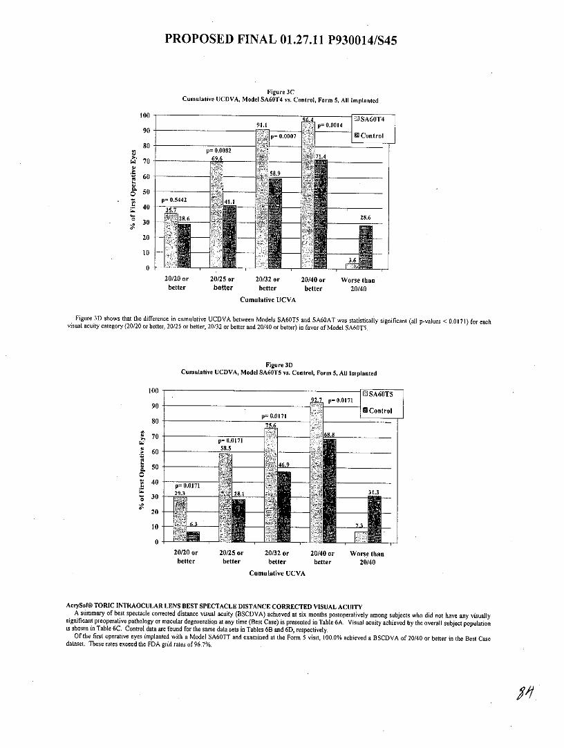
PROPOSED FINAL 01.27.11 P930014/S45
Figure 3CCumulative UCDVA, Model SA60T4 vs. Control, Form 5, All Implanted
100 -4 SA60T491.1 p= 0.0014
90 - -p= 0.0007 10 Control
80 p= 0.0082S70
60 s
650 --
p= 0.5442 41 140
35.30 28.6
20 - - -
10
0
20/20 or 20/25 or 20/32 or 20/40 or Worse thanbetter better better better 20/40
Cumulative UCVA
Figure 3D shows that the difference in cumulative UCDVA between Models SA60T5 and SA60AT was statistically significant (all p-values < 0.0171) for eachvisual acuity category (20/20 or better, 20/25 or better, 20/32 or better and 20/40 or better) in favor of Model SA60T5.
Figure 3DCumulative UCDVA, Model SA60T5 vs. Control, Form 5, All Implanted
100 SA60T597 p= 0.0171
901. Control
p= 0.0 17180 75.6
~,70 6.p= 0.0171
4 6058.5
0.0
p0= 0.0171
30 2331.3
20
20/20 or 20/25 or 20/32 or 20/40 or Worse thanbetter better better better 20/40
Cumulative UCVA
AcrySof@ TORIC INTRAOCULAR LENS BEST SPECTACLE DISTANCE CORRECTED VISUAL ACUITYA summary of best spectacle corrected distance visual acuity (BSCDVA) achieved at six months postoperatively among subjects who did not have any visually
significant preoperative pathology or macular degeneration at any time (Best Case) is presented in Table 6A. Visual acuity achieved by the overall subject populationis shown in Table 6C. Control data are found for the same data sets in Tables 6B and 6D, respectively.
Of the first operative eyes implanted with a Model SA60TT and examined at the Form 5 visit, 100.0% achieved a BSCDVA of 20/40 or better in the Best Casedataset. These rates exceed the FDA grid rates of 96.7%.
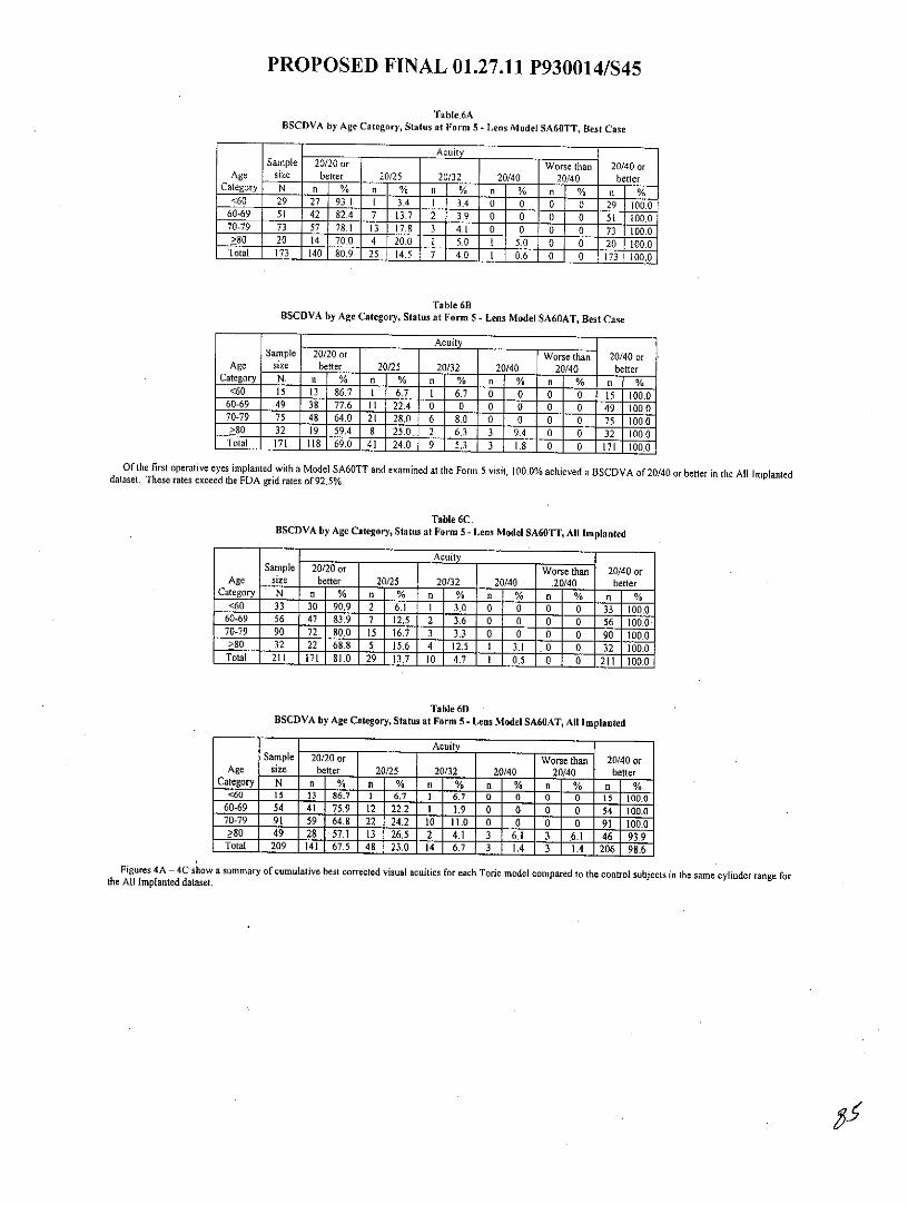
PROPOSED FINAL 01.27.11 P930014/S45
Table.6ABSCDVA by Age Category, Status at Form 5 - Lens Model SA60TT, Best Case
Acuity
Sample 20/20 or Worse than 20/40 orAge size better 20/25 20/32 20/40 20/40 better
Category N % n % n n % n % n %<60 29 27 93.1 1 3.4 1 3.4 0 0 0 0 29 100.0
60-69 51 42 82.4 7 13.7 2 3.9 0 0 0 0 SI 100.070-79 73 57 78.1 13 17. 8 3 4.1 0 0 0 0 73 100.0>80 20 70.0 4 20.0 .0 50 0 0 20 100.0Total 173 4 4 7 4.0 1 06 0 0 173 100.0
Table 6BBSCDVA by Age Category, Status at Form 5 - Lens Model SA60AT, Best Case
Acuity
Sample 20/20 or Worse than 20/40 orAge size better 20/25 20/32 20/40 20/40 better
Category N nnm % n % n % n % n %<60 15 13 86.7 1 6.7 I 6.7 0 0 0 0 15 100.0
60-69 49 38 77.6 11 22.4 0 0 0 0 0 0.49100.070-79 75 48 64.0 21 T280 6 8.0 0 0 0 0 75 1000>80 32 19 59.4 8 25,0. 2 6.3 3 9.4 0 0 32 100.0Total 171 118 69.0 41 1240. 9 5.3 3 1.8 0 0 171 100.0
Of the first operative eyes implanted with a Model SA60TT and examined at the Form 5 visit, 100.0% achieved a BSCDVA of 20/40 or better in the All Implanteddataset. These rates exceed the FDA grid rates of 92.5%.
Table 6C.BSCDVA by Age Category, Status at Form 5 - Lens Model SA60TT, All Implanted
Acuity
Sample 20/20 or . Worse than 20/40 orAge size better 20/25 20/32 20/40 .20/40 better
Category N I % n % n % n [ % n % n %<60 33 30 90.9 2 6.1 1 3.0 0 0 0 0 33 100.0
60-69 56 47 83.9 7 12.5 2 3.6 0 0 0 0 56 100.070-79 90 72 80.0 15 16.7 3 3.3 0 0 0 0 90 100.0>80 32 68.8 5 15.6 4 12.5 1 3.1 0 0 32 100.0
Total 211 171 81.0 29 13.7 10 4.7 l 0.5 0 0 211 100.0
Table 6DBSCDVA by Age Category, Status at Form 5 - Lens Model SA60AT, All Implanted
AcuitySample 20/20 or Worse than 20/40 or
Age size better 20/25 20/32 20/40 20/40 betterCategory N n % n % n % n % n % n %
<60 15 13 86.7 t 6.7 1 6.7 0 0 0 0 15 100.060-69 54 41 5.9 12 22.2 1 1.9 0 0 0 0 54 100.070-79 91 59 64.8 22 24.2 10 11. 0 0 0 91 100.0>80 49 28 571 13 26.5 2 4.1 3 6.1 3 6.1 46 93.9
Total 209 141 67.5 48 23.0 14 6.7 3 IA 3 1.4 206 98.6
Figures 4A - 4C show a summary of cumulative best corrected visual acuities for each Toric model compared to the control subjects in the same cylinder range forthe All Implanted dataset.

PROPOSED FINAL 01.27.11 P930014/S45
Figure 4ACumulative BSCDVA, Model SA60T3 vs. Control, Form 5, All Implanted
1006EDS6T
90 EI ControlI 81.6
0 0
80
70
60 60
50-
30 -- --
20 -
10
20/20 or 20/25 or 20/32 or 20/40 or Worse thanbetter better better better 20/40
Cumulative BSCVAFigure 4B
Cumulative BSCDVA, Model SA60T4 vs. Control, Form 5, All Implanted
100100 100 ID SA60T490 96.4 96.4 982 982
85.7 IED Control
80
6 60 -
50
20
10.
0 .L4.
20/20 or 20/25 or 20/32 or 20/40 or Worse thanbetter better better better 20/40
Cumulative BSCVA
Figure 4CCumulative BSCDVA, Model SA60T5 vs. Control, Form 5, All Implanted
100 o o100 lo - -100 E SA60T5
90390 9. 96.9
!81.3 0 Control
80 -
>,70 -- - -
> 60
50 ----
40 301
20 .
20/20 or 20/25 or 20/32 or 20/40 or Worse thanbetter better better better 20/40
Cumulative BSCVA

PROPOSED FINAL 01.27.11 P930014/S45
AcrySof@ TORIC INTRAOCULAR LENS ABSOLUTE RESIDUAL REFRACTIVE CYLINDERFigures 5A through 5C demonstrate that residual refractive cylinder values were statistically significantly lower among those subjects implanted with either anAcrySof@ Toric Model SA60T3, SA60T4 or SA60T5 IOL when compared to the corresponding subjects implanted with the control Model SA60AT. Subjects
implanted with an AcrySof@ Toric Model SA60T3 showed a 62.4% mean reduction in refractive cylinder from the preoperative visit (keratometric cylinder) ascompared to the 10.8% mean reduction for subjects implanted with the concurrent control Model SA60AT. Subjects implanted with an AcrySof@ Toric ModelSA60T4 or SA60T5 showed similar results with a mean reduction in refractive cylinder of 54.8 % and 67.8%, respectively, as compared to subjects implanted withthe concurrent control model who had a mean reduction in refractive cylinder of 22.1% and 27.7%, respectively. Each of the AcrySof@ Toric Lens Models SA60T3,SA60T4 and SA60T5 had at least a 3-fold increase in the likelihood of achieving residual refractive cylinder of 0.5 D or less as compared to the corresponding controlmodel.
Figure SAAbsolute Residual Refractive Cylinder,
Model SA60T3 vs. Control, Form 5, All Implanted50 03 SA60T3
E Control
4036.8 3.
32.5 overall30 p0.0001S30
26.4
o 20-CL
20
10
0.00 >0.00 to >0.50 to >1.00 to >1.50 to >2.005 0.50 1.00 1.50 2.00
Cylinder Amount (D)
Figure SBAbsolute Residual Refractive Cylinder,
Model SA60T4 vs. Control, Form 5, All Implanted
50 LJ~~'50 -- - SA60T4
41.1 M Control
40
32.1 overall
30 >0 t .50p 0.0001
21.4
16.116.1
14.3
' 10 8.9 10.7
10.
0 711 - 0.0
0.00 >0.00 to >0.50 to >1.00 to >1.50 to >2.00
!5 0.50 !5 1.00 :5 1.50 :5 2.00
Cy linder A mo unt (D)

PROPOSED FINAL 01.27.11 P930014/S45
Figure SCAbsolute Residual Refractive Cylinder,
Model SA60TS vs. Control, Form 5, All Implanted
50 -S NA60T5
I9 Control
40
overall30 p0.0001
30 26.8 26.8
20
10
2 , 4 2.
0.00 >0.00 to >0.50 to >1.00 to >1.50 to >2.00f 0.50 1.00 1.50 2.00
Cylinder Amount (D)
AcrySof@ TORIC INTRAOCULAR LENS STABILITY OF CYLINDERSubjects implanted with lens Model SA60TT exhibited stability of cylinder at Form 4 (3 months) with greater than 90% of all subjects changing less than or equal
to 1.00 diopter at consecutive visits between Form 3 (one month) and Form 6 (twelve months).
Table 7AAcrySof@ Toric IOL: Stability of Cylinder
(Eyes that had 2 consecutive exams, but not necessarily every follow-up exam)
Recommended Corneal Magnitude ofAstigmatism Correction Toric IOL Vector Change I and 3 Months 3 and 6 Months 6 and 12 Months
Ranges Model in Cylinder n/N,% nfN,% n/N,%< 1.5 D SA60T3 !1.00 D 106/107,99.07% 101/105,96.19% 55/55,100.00%
Mean Change 0.28 0.29 0.20SD 0.32 0.33 0.25
S1.5 - < 2.0 D SA60T4 5 1.00 D 54/56,96.43% 53/54,98.15% 25/27,92.59%Mean Change 0.40 0.27 0.46
SD 0.35 0.22 0.452.0 D SA60T5 5 1.00 D 40/4588 .89% 35/40,87.50% 27/30,90.00%
Mean Change 0.43 0.42 0.41SD . 0.44 0.45 0.38
Combined SA60TT 1 1.00 D 200/208,96.15% 189/199,94.97% 107/112,95.54%(93.54,98.77) (91.94,91:01) (91.71,99.36)
Mean Change 0.35 0.31 0.32SD 0.36 0.34 0.36
95% Cl 0.30,0.39 0.26,0.36 0.25,0.39
n/N,%,(%Cl) are for percent with change between ± I.001D

PROPOSED FINAL 01.27.11 P930014/S45
Table 7BAcrySof@ Toric IOL: Stability of Cylinder
(Eyes that had every follow-up exam up to Form 6, 12 months)
RecommendedCorneal Magnitude of
Astigmatism Toric IOL Vector Change I and 3 Months 3 and 6 Months 6 and 12 MonthsCorrection Ranges Model in Cylinder n/N,% n/N,% n/N,%
< 1.5 D SA60T3 1.00 D 34/34,100.00% 34/34,100.00% 34/34,100.00%Mean Change 0.25 0.24 0.21
SD 0.23 0.22 0.241 [.5 - < 2.0 D SA60T4 1 [.00 D 17/17,100.00% 16/17,94.12% 16/1794.12%
Mean Change 0.27 0.25 0.35SD 0.25 0.26 0.33
2.0 D SA60T5 1.00 D 17/19,89.47% 15/19,78.95% 16/19,84.21%Mean Change 0.44 0.56 0.52
SD 0.47 0.50 0.43Combined SA60TT g 1.00 D 68/70,97.14% 65/70,92.86% 66/70,94.29%
(93.23,100.00) (86.82,98.90) (88.84,99.73)Mean Change 0.31 0.33 0.33
SD 0.32 0.35 0.3495%CI 0.23,0.38 0.24,0.41 0.25,0.41
n/N,%,(%CI) are for percent with change between ± 1.00D
Table 7CAcrySof@ Toric 1OL: Stability of Absolute Cylinder
(Eyes that had 2 consecutive exams, but not necessarily every follow-up exam)
Recommended Magnitude ofCorneal Change in
Astigmatism Toric IOL Absolute I and 3 Months 3 and 6 Months 6 and 12 MonthsCorrection Ranges Model Cylinder n/N,% n/N,% n/N,%
< 1.5 D SA60T3 9 1.00 D 107/107,100.00% 104/105,99.05% 55/55,100.00%Mean Change 0.04 0.02 0.05
SD 0.32 0.38 0.29a 1.5 - < 2.0 D SA60T4 ! 1.00 D 54/56,96.43% 54/54,100.00% 27/27,100.00%
Mean Change 0.18 0.05 -0.12SD 0.42 0.27 0.41
2 20 D SA60T5 5 1.00 D 44/45,97.78% 37/40,92,50% 29/30,96.67%Mean Change 0.09 006 0.00
SD 0.38 0.49 0.45Combined SA60TT [.00 D 205/208,98.56% 195/199,97.99% 111/112,99.11%
(96.93,100.00) (96.04,99.94) (97.36,100.00)Mean Change 0.09 0.03 -0.01
SD 0.37 0.38 0.3795% Cl 0.04,0.14 -0.02,0.09 -0.08,0.06
n/N,%,(%CI) are for percent with change between ± 1.00D
Table 7DAcrySof@ Toric IOL: Stability of Absolute Cylinder
(Eyes that had every follow-up exam up to Form 6, 12 months)
Recommended Magnitude ofCorneal Change in
Astigmatism Toric [0L Absolute I and 3 Months 3 and 6 Months 6 and 12 MonthsCorrection Ranges Model Cylinder n/N,% n/N,% n/N,%
< 1.5 D SA60T3 5 1.00 D 34/34,100.00% . 34/34,100.00% 34/34,100.00%Mean Change 0.01 -0.01 0.07
SD 0.28 0.31 0.28a 1.5 - < 2.0 D SA60T4 s [.00 D 17/17,100.00% 17/17,100.00% 17/17,100.00%
Mean Change 0.06 0.19 -0.04SD 0.30 0.21 0.42
2 2.0 D SA60T5 I [.00 D 18/19,94.74% 17/19,89.47% 18/19,94.74%Mean Change 0.17 . 0.05 0.01
SD 0.45 0.54 0.55Combined SA60TT 1.00 D 69/70,98.57% 68/70,97.14% 69/70,98.57%
(95.78,100.00) (93.23,100.00) (95.78,100.00)Mean Change 0.07 0.05 0.03
SD 0.34 0.38 0.4095% Cl -0.01,0.15 -0.04,0.14 -0.07,0.12
n/N,%,(%Cl) are for percent with change between .I .00D
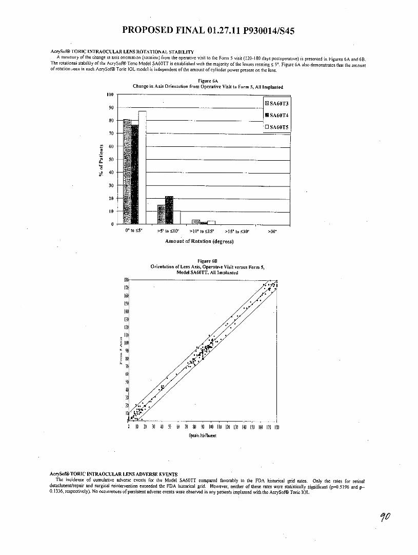
PROPOSED FINAL 01.27.11 P930014/S45
AcrySof@ TORIC INTRAOCULAR LENS ROTATIONAL STABILITYA summary of the change in axis orientation (rotation) from the operative visit to the Form 5 visit (120-180 days postoperative) is presented in Figures 6A and 6B.
The rotational stability of the AcrySof@ Toric Model SA60TT is established with the majority of the lenses rotating s 5'. Figure 6A also demonstrates that the amountof rotation seen in each AcrySof® Toric IOL model is independent of the amount of cylinder power present on the lens.
Figure 6AChange in Axis Orientation from Operative Visit to Form 5, All Implanted
100
0SA60T390
lSA60T480-
OSA60T570 -
60
50
', 40
30-
20
10-
0 to 55' >51 to 10" >10 to 15" >15" to 30" >30"
Amount of Rotation (degrees)
Figure 6BOrientation of Lens Axis, Operative Visit versus Form 5,
Model SA60TT. All Imolanted
160150
13C I**120
1000
6(
0 10 20 30 4 50 o60 70 80 90 100 110 120 130 140 150 160 170 100Opttw Ais Plant
AcrySof@ TORIC INTRAOCULAR LENS ADVERSE EVENTSThe incidence of cumulative adverse events for the Model SA60TT compared favorably to the FDA historical grid rates. Only the rates for retinal
detachment/repair and surgical reintervention exceeded the FDA historical grid. However, neither of these rates were statistically significant (p=0.5196 and p-0.1336, respectively). No occurrences of persistent adverse events were observed in any patients implanted with the AcrySof@ Toric IOL.

PROPOSED FINAL 01.27.11 P930014/S45
Table 8Adverse Events Incidence Rates
First Eye - SafetyModel S60TT FDA Grid Rate
N=244Cumulative Adverse Events N , %Retinal Detachment/Repair 1 0.4 0.3Surgical Reintervention 4' 1.6 0.8
IOL Reposition Due to I 0.4 NARotationIOL Replacement Due 1 0.4 NAto RotationLaser Treatment 2 0.8 NAParacentesis 1 0.4 NA
The incidence rates in this table are based upon the number ofeyes with an event dividedby the number of eyes implanted.Cumulative adverse events are those events that have occurred at any time during theclinical study.FDA Grid Rate = FDA Grid of Adverse Events with Posterior Chamber Intraocular LensHistorical Controls, FDA Intraocular Lens Guidance Document, Annex B (October 14,1999)'There were 5 occurrences of surgical reintervention in 4 eyes for Model SA60TT firsteye
The incidence of cumulative adverse events for the Model SA60TT also compared favorably to the concurrent control.
AcrySof@ TORIC INTRAOCULAR LENS DISTANCE-VISION SPECTACLE INDEPENDENCEStatistically significantly more Model SA60TT subjects reported postoperative distance-vision spectacle independence compared to Model SA60AT subjects(59.9% versus 37.7%, respectively) when unilaterally implanted. Distance-vision spectacle independence was defined as the percentage of subjects who selected thenone of the time" response for distance-vision frequency-of-spectacle-wear. Spectacle dependence was defined as subjects indicating any reliance on glasses fordistance-vision and represents the summation of the "some of the time", "half of the time", "most of the time" and "all of the time" frequency-of-spectacle-wear
responses. Consequently, fewer Model SA60TT subjects were spectacle dependent at 40.1% compared to 62.3% of the Model SA60AT subjects. Figure 7 illustratesthe distance-vision frequency-of-spectacle-wear distributions between Model SA60TT and Model SA60AT groups. Implantation of an AcrySof@ Toric Intraocularlens in astigmatic subjects provides significantly improved distance-vision spectacle independence relative to a conventional monofocal IOL.
Figure 7Distance-Vision Spectacle Independence:
Frequency-of-Spectacle-Wear, Form 5, All Implanted
70%70% oerallElSA60TT
60% N o-o50% --- ~ - --- ------- U Contro
W 40% 1~ 37,7
o 30%
21.1 2.20% 16.9
12.3 10
0% . 3 -
None of the time Some of the time Half of the time ost of the time All of the time
Spectacle Spectacle Dependentindependent
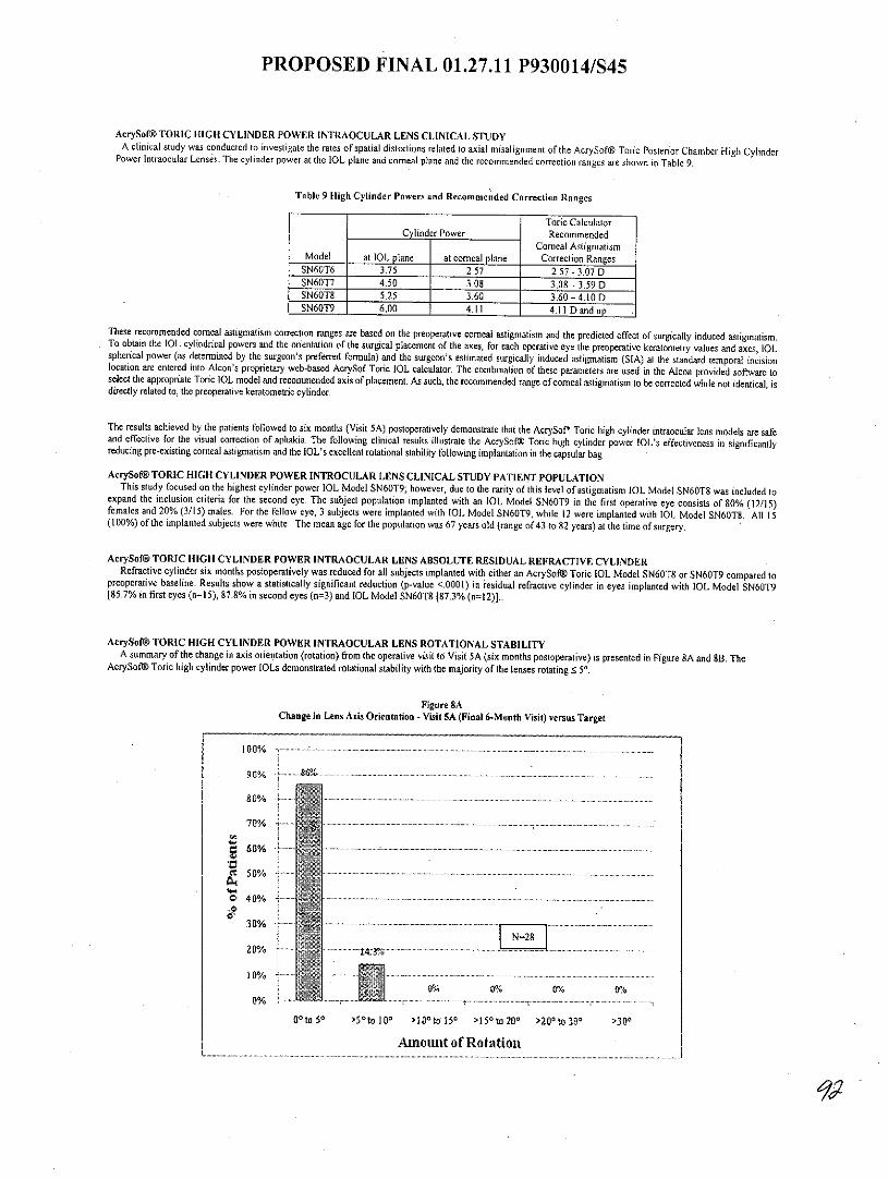
PROPOSED FINAL 01.27.11 P930014/S45
AcrySof@ TORIC HIGH CYLINDER POWER INTRAOCULAR LENS CLINICAL STUDYA clinical study was conducted to investigate the rates of spatial distortions related to axial misalignment of the AcrySof@ Toric Posterior Chamber High Cylinder
Power Intraocular Lenses. The cylinder power at the IOL plane and corneal plane and the recommended correction ranges are shown in Table 9.
Table 9 High Cylinder Powers and Recommended Correction Ranges
Toric CalculatorCylinder Power Recommended
Corneal AstigmatismModel at 1OL plane at comeal plane Correction Ranges
SN60T6 3.75 2.57 2.57 - 3.07 DSN60T7 4.50 3.08 3.08 - 3.59 DSN60T8 5.25 3.60 3.60 - 4.10 DSN60T9 6.00 4.11 4.11 D and up
These recommended corneal astigmatism correction ranges are based on the preoperative corneal astigmatism and the predicted effect of surgically induced astigmatism.To obtain the IOL cylindrical powers and the orientation of the surgical placement of the axes, for each operative eye the preoperative keratometry values and axes, IOLspherical power (as determined by the surgeon's preferred formula) and the surgeon's estimated surgically induced astigmatism (SIA) at the standard temporal incisionlocation are entered into Alcon's proprietary web-based AciySof Toric IOL calculator. The combination of these parameters are used in the Alcon provided software toselect the appropriate Toric IOL model and recommended axis of placement. As such, the recommended range of corneal astigmatism to be corrected while not identical, isdirectly related to, the preoperative keratometric cylinder.
The results achieved by the patients followed to six months (Visit 5A) postoperatively demonstrate that the AcrySof* Toric high cylinder intraocular lens models are safeand effective for the visual correction of aphakia. The following clinical results illustrate the AcrySof@ Toric high cylinder power IOL's effectiveness in significantlyreducing pre-existing corneal astigmatism and the IOL's excellent rotational stability following implantation in the capsular bag
AcrySof@ TORIC HIGH CYLINDER POWER INTROCULAR LENS CLINICAL STUDY PATIENT POPULATIONThis study focused on the highest cylinder power IOL Model SN60T9; however, due to the rarity of this level of astigmatism IOL Model SN60T8 was included to
expand the inclusion criteria for the second eye. The subject population implanted with an 1OL Model SN60T9 in the first operative eye consists of 80% (12/15)females and 20% (3/15) males. For the fellow eye, 3 subjects were implanted with IOL Model SN60T9, while 12 were implanted with IOL Model SN60T8. All 15(100%) of the implanted subjects were white. The mean age for the population was 67 years old (range of43 to 82 years) at the time of surgery.
AcrySof@ TORIC HIGH CYLINDER POWER INTRAOCULAR LENS ABSOLUTE RESIDUAL REFRACTIVE CYLINDERRefractive cylinder six months postoperatively was reduced for all subjects implanted with either an AcrySofW Toric IOL Model SN60T8 or SN60T9 compared to
preoperative baseline. Results show a statistically significant reduction (p-value <.0001) in residual refractive cylinder in eyes implanted with IOL Model SN60T9[85.7% in first eyes (n=15), 87.8% in second eyes (n=3) and 1OL Model SN60T8 [87.3% (n=12)]..
AcrySof@ TORIC HIGH CYLINDER POWER INTRAOCULAR LENS ROTATIONAL STABILITYA summary of the change in axis orientation (rotation) from the operative visit to Visit 5A (six months postoperative) is presented in Figure 8A and 8B. The
AcrySof& Toric high cylinder power lOLs demonstrated rotational stability with the majority of the lenses rotating !c 5*.
Figure 8AChange in Lens Axis Orientation - Visit SA (Final 6-Month Visit) versus Target
100%...............................................
....................80%
70%
O4 0%
30%
10%1]0 0%%
0/%
Ontoo 50 >t10l00 >l0atol150 >150to200 >200to 30s >300
Amount of Rotation

PROPOSED FINAL 01.27.11 P930014/S45
Figure 8BOrientation of Lens Axis, Operative Visit versus Visit SA (Final Visit)
IOL Models SN60T9 and SN60T8
170160
ISO140INPO
110
90
0 7
61
31
0 10 20 30 40 50 60 70 80 90 100 110 120 130 140 0S0 160 170 180
Operaive Ais Placernefti
AcrySof@ TORIC HIGH CYLINDER POWER INTRAOCULAR LENS UNCORRECTED DISTANCE VISUAL ACUITYAll Subjects implanted bilaterally with the AcrySof Toric IOL Models SN60T8 or SN60T9 achieved improved binocular uncorrected distance visual acuity six months
postoperatively. Figure 9 demonstrates that 60% of subjects achieved 20/20 or better binocular uncorrected distance visual acuity compared to 30% for monocular eyes,while 93% of subjects achieved 20/40 or better binocular uncorrected distance visual acuity compared to 90% ofmonocular eyes. Less than 10% of subjects had monocularor binocular uncorrected distance visual acuity worse than 20/40 six months postoperatively.
Figure 9Cumulative UCDVA Monocular versus Binocular
1030%.. ................... ... ........... ..... .
0 Monocular IN:30) 8 hcua tx5 ~80%
SDA
70%
60%o
*40%30%
30%
20%' 'd.. ...
10%73
0% t
20/20 or better 20/25 or better 20/32 or better 20/40 orbetter Worsethan 20/40
Uncorrected Distance'VisualAcuity

PROPOSED FINAL 01.27.11 P930014/S45
AcrySof@ TORIC HIGH CYLINDER POWER INTRAOCULAR LENS BILATERAL DISTANCE-VISION SPECTACLE INDEPENDENCEPreoperatively all subjects were spectacle dependent, either all-the-time (92.9%) or some-of-the-time (7.1%). Six months postoperatively, 7 1.4% of subjects werespectacle independent (Figure 10).
Figure 10Bilateral Distance Vision Spectacle Independence
Frequency-of-Spectacle-Wear Visit 0 (PreOp) versus Visit 5A (Final Visit)
. .. - - ----- ..
- --- --- -- - - - -
AcrySof@ TORIC HIGH CYLINDER POWER INTRAOCULAR LENS RATE OF SPATIAL DISTORTIONSA Visual Distortion Questionnaire was administered preoperatively (Visit 0) and at six months postoperatively (Visit SA) to evaluate the rate of spatial distortions
of the AcrySofo Toric IOL Models SN60T8 and SN60T9. The overall rate of spatial distortions decreased postoperatively (Table 10A).
Table 10A:Visual Distortion Questionnaire Results by Visit
PreOp Final Visit(N =14) (N =14)
During the past 4 weeks, have you had n % n o
1) ...trouble with things appearing distorted? No 3 21.4 12 85.7Yes 11 78.6 2a,b 14.3
2) ...trouble with flat surfaces (like floors) appearing No 12 85.7 13 92.9curved?
Yes 2 14.3 I 7.13) ...trouble with straight lines (like door or window No 10 71.4 14 100
frames) appearing tilted? Ys2. 0 .Yes 4 28.6 0 0.0
4) _trouble with feeling sick to your stomach due to No 14 100 14 100distortion of your vision? Yes 0 0.0 0 0.0
Reported with or without glasses at Preop and Final Visit.Reported with or without glasses at Preop but only with glasses (progressive lenses) at Final Visit.
0 Same subject as in (b). Reported only with glasses (progressive lenses) at Final Visit. Not reported at Preop.
Based on these questions spatial distortions associated with high pre-existing corneal astigmatism may not completely resolve postoperatively. Two subjects at Visit5A continued to report "trouble with things appearing distorted" versus It subjects preoperatively. One of these subjects had "trouble with flat surfaces appearingcurved," which was noted only postoperatively, but no longer experienced the preoperative visual phenomena of straight lines appearing tilted. Neither subject hadIOL misalignment requiring secondary surgical intervention to address problems of spatial distortion. There were no reports of subjects feeling sick to their stomachsdue to distortion of vision.
Responses to visual distortion sub-questions related to spectacle wear, frequency of experiencing distortion, and degree of bother are presented in Tables 103through 10D.

PROPOSED FINAL 01.27.11 P930014/S45
Table 10B:Visual Distortion Questionnaire Results - Trouble with Things Appearing Distorted
PreOp Final Visit1) For subjects who had trouble with things appearing distorted - N= 1 (N= 2)
in the last 4 weeks: n % n %Do you notice this only when you wear your No 10 90.9 I 50.0glasses? Yes T 9.1 I 50.0How often have you noticed this? Rarely 2 18.2 0 0.0
Sometimes 2 18.2 0 0.0Frequently 3 27.3 1 50.0All the time 4 36.4 1 50.0
How much does it bother you? None I 9.1 50.0A Little 4 36.4 0 0.0A Lot 6 54.5 1 50.0
Table 10C:Visual Distortion Questionnaire Results - Trouble with Flat Surfaces Appearing Curved
PreOp Final Visit2) For subjects who had trouble with flat surfaces (N 2) (N = 1)
(like floors) appearing curved in the last 4 weeks: n % n %Do you notice this only when you wear your No 2 100 0 0.0glasses? Yes 0 0.0 1 100How often have you noticed this? Rarely 0 0.0 0 0.0
Sometimes 0 0.0 0 0.0Frequently I 50.0 1 100All the time 1 50.0 0 0.0
How much does it bother you? None 0 0.0 0 0.0A Little 0 0.0 0 0.0A Lot 2 100 1 100
Table 10DVisual Distortion Questionnaire Results - Trouble with Straight Lines Appearing Tilted
PreOp Final Visit3) For subjects who had trouble with straight lines (like door or (N= 4) (N= 0)
window frames) appearing tilted in the last 4 weeks: n % n %Do you notice this only when you wear your No 3 75.0 0 0.0glasses? Yes 1 25.0 0 0.0How often have you noticed this? Rarely 0 0.0 0 0.0
Sometimes 2 50.0 0 0.0Frequently 0 0.0 0 0.0All the time 2 50.0 0 0.0
How much does it bother you? None 0 0.0 0 0.0A Little 1 25.0 0 0.0
__________________________ A Lot 3 75,0 0 0.0
AcrySof@ TORIC HIGH CYLINDER POWER INTRAOCULAR LENS ADVERSE EVENTSDuring the study, I of 15 subjects underwent a secondary surgical intervention in the first eye to resolve residual refractive cylinder due to an error in preoperativekeratometry. One week postoperatively, the IOL was repositioned. At six months postoperatively the subject was satisfied with uncorrected distance vision and did notexperience any spatial distortion after IOL repositioning. No other serious adverse events were reported in the study.
OVERVIEW OF AcrySof@ SINGLE-PIECE STUDIES
AcrySof@ NATURAL SINGLE-PIECE IOL CLINICAL STUDYA clinical study was conducted on patients receiving the AcrySofg Natural Single Piece IOL as compared to the AcrySoft UV Single Piece IOL. The results
achieved by the patients successfully followed for a minimum of one year postoperatively provided reasonable assurance of safety and effectiveness for the visualcorrection of aphakia. For information pertaining to the results obtained in this clinical study, please reference the corresponding Physicians Labeling or that providedwith other AcrySofg Natural monofocal IOLs.
AcrySof@ NATURAL SINGLE-PIECE IOL COLOR PERCEPTIONColor perception testing using the Farnsworth D-15 Panel Test was conducted at the 120 to 180 day postoperative period. Of the 109 subjects with normal color
vision implanted with the AcrySof@ Natural IOL in the first operative eye and examined at the 120-180 day postoperative visit, 107 (98.2%) passed the colorperception test. Of the 102 subjects with normal color vision implanted with a AcrySof& UV IOL in the first operative eye and examined at the 120-180 daypostoperative visit, 97 (95.1%) passed the color perception test. There were no statistically significant differences between AcrySofO Natural IOL and AcrySofW UV

PROPOSED FINAL 01.27.11 P930014/S45
IOL for the percent of subjects that passed the color perception test at the 120 to 180 day postoperative visit. Therefore, the addition of the proprietary chromophoredoes not negatively affect color vision in patients with normal color vision.
AcrySof@ IQ LENS CLINICAL STUDYConsistent with the design of similar previously conducted IOL studies, adult subjects in good general ocular health (e.g. no prior ocular surgery, degenerative
visual disorder which would significantly impact visual acuity, or severe acute or chronic condition that may increase patient risk) having bilateral cataracts wereenrolled ina controlled, randomized, double-masked, multi-center, contralateral implant clinical investigation of the AcrySof@ IQ lens versus a spherical control lens.Ocular spherical aberrations were statistically significantly less with the AcrySof@ IQ lens than the control lens. Contrast sensitivity results demonstrated astatistically significant postoperative (at 3 months) improvement in favor of AcrySof® IQ implanted eyes. Eyes implanted with the AcrySof® IQ lens alsoexperienced statistically and clinically significant improvements in a functional vision measurement, simulated night driving, under several conditions tested -especially glare and fog. These results reflect that the AcrySof® IQ IOL (an aspheric optic on a material platform containing a blue-light filtering chromophore)provides beneficial clinical performance as compared to the monofocal AcrySof@ lOL (without an aspheric optic and blue-light filtering chromophore).
AcrySof@ IQ LENS - SPHERICAL AND TOTAL HIGHER ORDER ABERRATIONSThe mean ocular spherical aberration of the AcrySof@ IQ eyes was approximately 0.1 micrometers. Figure II represents the statistically significant reduction in
spherical and.total higher order aberrations observed in favor of the AcrySof@ IQ lens. Figure 12 provides the mean spherical aberration measurements of all eyeswith wavefront aberrometer measurements by lens and age group. As depicted in this chart, the reduction in spherical aberration of the AcrySof@ IQ eyes wasindependent of age.
Figure IISpherical and Total Higher Order RMS
90-120 Days after 2,d Eye ImplanlAcrySof8 IQ IOL (N=73) a Control (N=73)
o0-
0
100Z
Spherical Aberrations Total HO RMS* Differences favor AcrySof@ IQ IOL overall and at each visit (p<0.0001)
Figure 12Mean Spherical Aberration Overall and by Age Group
90-120 Days after 2,d Eye Implant
M AcrySof6 1QIOL [ Control0.8
0.7 *
0.6
< 0.5
E 0A4
0.3
C 0.2 -
0Total < 60 60 -69 >70N=73 N=6 N=19 N=48
Age Group (Years)Denotes statistical significance between lenses (p<0 0001)
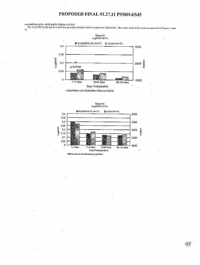
PROPOSED FINAL 01.27.11 P930014/S45
AcrySof@ IQ LENS - DISTANCE VISUAL ACUITYThe AcrySofi IQ lens and the control lens provided clinically similar postoperative visual acuity. Monocular visual acuity results are presented in Figures 13 and
14.
Figure 13LogMAR BCVA
M AcrySof@10 I0L (N=77) M Control (N=75)0.2 20/30
0.15
0.1 20/25 a0 C
p=0.0199
0.05
0 20/207-14 Days 30-60 Days 90-120 Days
Days Postoperative
Quantitative Lens Model Main Effect (p=0.0214)
Figure 14LogMAR UCVA
5AcrySof 1Q IOL (N=77) El Control (N=75)0.4 20/50
0.35
0.3 20/40
0.25
0.2 20/30 .2S0.15
0.1 ~ > 20/250.05
0 J~~.0 L I J L L J .L 4 20/201-2 Days 7-14 Days 30-60 Days 90-120 Days
Days PostoperativeDifferences are not statistically significant
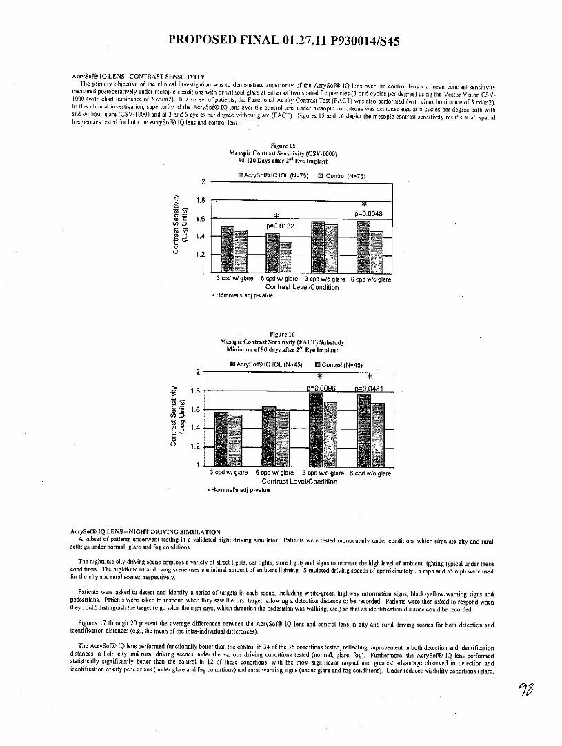
PROPOSED FINAL 01.27.11 P930014/S45
AcrySof@ IQ LENS -CONTRAST SENSITIVITYThe primary objective of the clinical investigation was to demonstrate superiority of the AcrySof@ IQ lens over the control lens via mean contrast sensitivity
measured postoperatively under mesopic conditions with or without glare at either of two spatial frequencies (3 or 6 cycles per degree) using the Vector Vision CSV-1000 (with chart luminance of 3 cd/m2). In a subset of patients, the Functional Acuity Contrast Test (FACT) was also performed (with chart luminance of 3 cd/m2).In this clinical investigation, superiority of the AcrySof® IQ lens over the control lens under mesopic conditions was demonstrated at 6 cycles per degree both withand without glare (CSV-1000) and at 3 and 6 cycles per degree without glare (FACT). Figures 15 and 16 depict the mesopic contrast sensitivity results at all spatialfrequencies tested for both the AcrySof@ IQ lens and control lens.
Figure 15Mesopic Contrast Sensitivity (CSV-1000)
90-120 Days after 2 nd Eye Implant
M AcrySofO IQ IOL (N=75) El Control (N=75)2
p=0.0048' 1.6
p=0.01320
1.2
3 cpd w/ glare 6 cpd w/ glare 3 cpd w/o glare 6 cpd w/o glareContrast Level/Condition
* Hommels adj p-value
Figure 16Mesopic Contrast Sensitivity (FACT) Substudy
Minimum of 90 days after 2 d Eye Implant
*AcrySof@ IQ IOL (N=45) ED Control (N=45)2
1.D=0'0096 p=0.04811.8.5
j 1.405
1.2 - -
13 cpd w/ glare 6 cpd w/ glare 3 cpd wio glare 6 cpd w/o glare
Contrast Level/Condition* Hommel's adj p-value
AcrySof@ IQ LENS - NIGHT DRIVING SIMULATIONA subset of patients underwent testing in a validated night driving simulator. Patients were tested monocularly under conditions which simulate city and rural
settings under normal, glare and fog conditions.
The nighttime city driving scene employs a variety of street lights, car lights, store lights and signs to recreate the high level of ambient lighting typical under theseconditions. The nighttime rural driving scene uses a minimal amount of ambient lighting. Simulated driving speeds of approximately 35 mph and 55 mph were usedfor the city and rural scenes, respectively.
Patients were asked to detect and identify a series of targets in each scene, including white-green highway information signs, black-yellow warning signs andpedestrians. Patients were asked to respond when they saw the first target, allowing a detection distance to be recorded. Patients were then asked to respond whenthey could distinguish the target (e.g., what the sign says, which direction the pedestrian was walking, etc.) so that an identification distance could be recorded.
Figures 17 through 20 present the average differences between the AcrySof IQ lens and control lens in city and rural driving scenes for both detection andidentification distances (e.g., the mean of the intra-individual differences).
The AcrySof@ IQ lens performed functionally better than the control in 34 of the 36 conditions tested, reflecting improvement in both detection and identificationdistances in both city and rural driving scenes under the various driving conditions tested (normal, glare, fog). Furthermore, the AcrySofW IQ lens performedstatistically significantly better than the control in 12 of these conditions, with the most significant impact and greatest advantage observed in detection andidentification of city pedestrians (under glare and fog conditions) and rural warning signs (under glare and fog conditions). Under reduced visibility conditions (glare,

PROPOSED FINAL 01.27.11 P930014/S45
fog) in the city scene, the increased visibility distance at 35 mph provides the AcrySof@ IQ lens greater than 0.5 second additional time to respond to a pedestriantarget, a hazard more commonly encountered in city settings. This 0.5 second increase is functionally significant in allowing for greater time to take appropriateactions such as stopping, avoidance, etc. (Green, 2000; McBride and Matson, 2004). Under all conditions in the rural scene, the increased visibility distance at 55mph provides the AcrySofg IQ lens more than I second additional time to identify warning signs, a situation frequently encountered in rural areas. A 0.5 secondincrease is functionally significant in allowing for greater time to take appropriate action while driving, which becomes critical at night in unfamiliar rural areas whereambient lighting is often absent. There were 6 patients in the substudy who postoperatively experienced macular degeneration or PCO, When these patients wereremoved from the driving analysis, the difference between lOLs for detection and identification of pedestrian targets under glare conditions in the city location fellshort of the 0.5-second threshold for clinical relevance. When the original analyses were adjusted for multiplicity, the difference between lOLs was no longerstatistically significant for city detection of text under glare (Hommel's p-value = 0.0539) or for rural detection of pedestrian under glare (Hotmmel's p-value=0.0507)
These results demonstrate improved functional vision and likely meaningful safety benefits to elderly drivers with the AcrySof@ IQ lens and to other drivers andpedestrians with whom they share the road. The results of this test demonstrate that the AcrySof@i IQ lens improves functional vision, which in turn may improvepatient safety for other life situations under low visibility conditions.
Figure 17Night Driving Simulator
Mean Intra-individual Differences in Detection Sight Distances, CityMinimum of 90 days PostoperativelyAcrySof@ IQ IOL -Control (n=44)
C Differences (feet) - - Distance difference equivalent to 0.5 secondsU 40cu 35
8 30
as25 7720
.c 15
tncs fos r C 4
cs a) (D C
Fog Glare NormalDistances for both lens groups were normalized
Denotes statistical significance (p<0.05) in favor ofAcrySofO IQ IOL.
Fisure 18Night Driving Simulator
Mean Intra-individual Differences in Identification Sight Distances, CityMinimum of 90 days PostoperativelyAcrySof@ IQ IOL-Control (n=44)
M Differences (feet) - Distance difference equivalent to 0.5 secondsu 40
35 *M 30
20c- 15
a 10 1U -
Q. CL a. 0
Fog Glare NormalDistances for both lens groups were normalized.
* Denotes statistical significance (p<0.05) in favor ofAcrySofi IQ 10L.
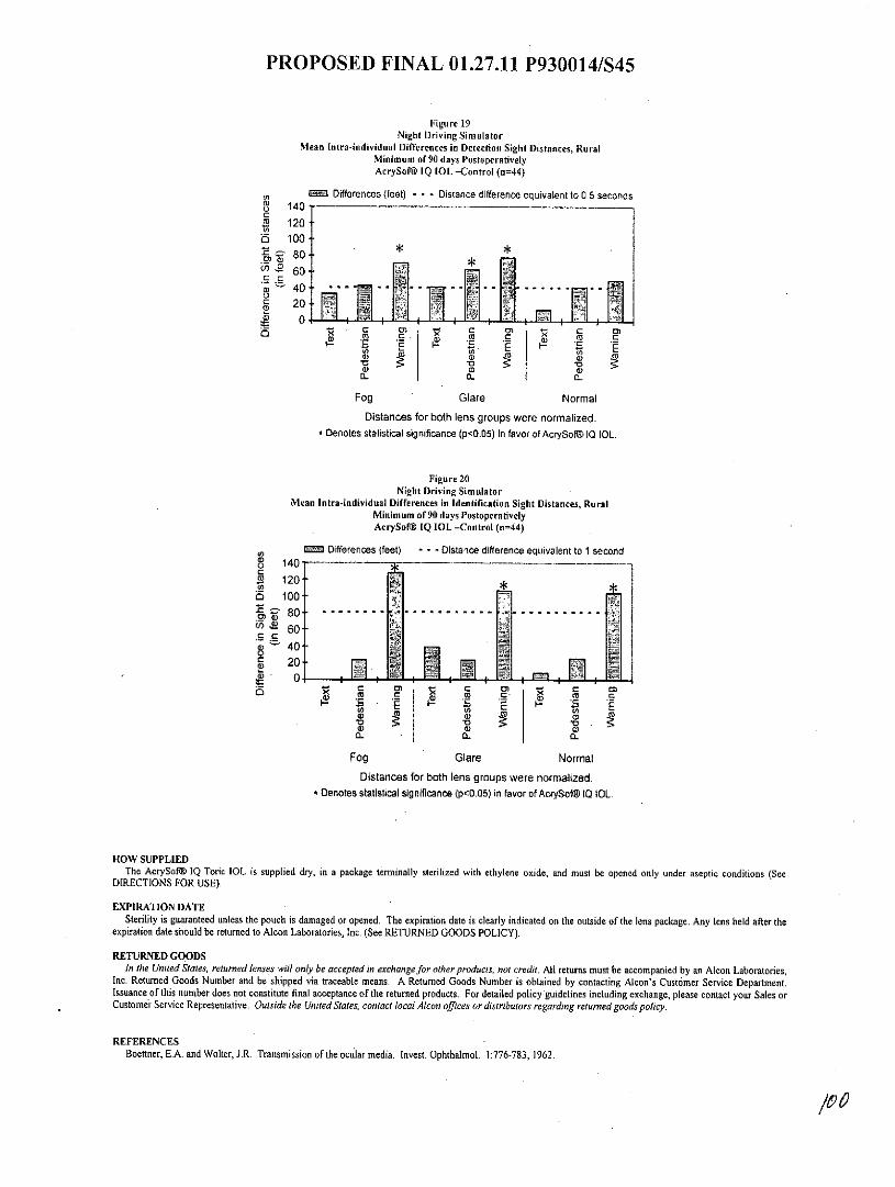
PROPOSED FINAL 01.27.11 P930014/S45
Figure 19Night Driving Simulator
Mean Intra-individual Differences in Detection Sight Distances, RuralMinimum of 90 days PostoperativelyAcrySof@ IQ IOL -Control (n=44)
C Differences (feet) - - - Distance difference equivalent to 0.5 secondsL) 140 - - - - - - -- - - - - -C2 120
6 1008060
C
Cge 20
00
4) a) a)
Fog Glare Normal
Distances for both lens groups were normalized.Denotes statistical significance (p<0.05) in fovor of AcrySofOg 10 10L.
Figure 20Night Driving Simulator
Mean Intra-individual Differences in Identification Sight Distances, RuralMinimum of 90 days PostoperativelyAcrySof@ IQ IOL -Control (n=44)
E= Differences (feet) - * - Distance difference equivalent to 1 secondo 140 - --_ ---- _ -- - -_- -
S 120 **o 100
O 6040
20
s C ) n- C 0
Fog Glare Normal
Distances for both lens groups were normalized.* Denotes statistical significance (p<0.05) in favor of AcrySofi 10 IOL.
HOW SUPPLIEDThe AcrySof@ IQ Toric IOL is supplied dry, in a package terminally sterilized with ethylene oxide, and must be opened only under aseptic conditions (See
DIRECTIONS FOR USE).
EXPIRATION DATESterility is guaranteed unless the pouch is damaged or opened. The expiration date is clearly indicated on the outside of the lens package. Any lens held after the
expiration date should be returned to Alcon Laboratories, Inc. (See RETURNED GOODS POLICY).
RETURNED GOODSIn the United States, returned lenses will only be accepted in exchange for other products, not credit. All returns must be accompanied by an Alcon Laboratories,
Inc. Returned Goods Number and be shipped via traceable means. A Returned Goods Number is obtained by contacting Alcon's Customer Service Department.Issuance of this number does not constitute final acceptance of the returned products. For detailed policy guidelines including exchange, please contact your Sales orCustomer Service Representative. Outside the United States, contact local Alcon offices or distributors regarding returned goods policy.
REFERENCESBoettner, E.A. and Wolter, J.R. Transmission of the ocular media. Invest. Ophthalmol. 1:776-783, 1962.
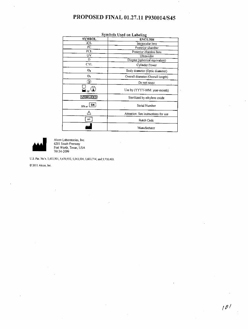
PROPOSED FINAL 01.27.11 P930014/S45
Symbols Used on LabelingSYMBOL ENGLISH
1OL Intraocular. lensPC Posterior chamber
PCL Posterior chamber lensUV UltravioletD Diopter (spherical equivalent)
CYL Cylinder Power
On Body diameter (Optic diameter)
OT Overall diameter (Overall length)
Do not reuse
or Use by (YYYY-MM: year-month)
STERILE EQf Sterilized by ethylene oxide
SN orFSN Serial Number
±S Attention: See instructions for use
Batch Code
Manufacturer
Alcon Laboratories, Inc.6201 South FreewayFort Worth, Texas, USA76134-2099
U.S. Pat. No's. 5,403,901, 5,470,932, 5,543,504, 5,603,774, and 5,716,403.
0 2011 Alcon, Inc.