patient and staff radiological protection in cardiology (icrp) and staff radiological protection...
TRANSCRIPT

DRAFT REPORT FOR CONSULTATION
1
ICRP ref 4818-2733-7736 1 May 20, 2011 2
3
4
Annals of the ICRP 5
6
7
ICRP PUBLICATION XXX 8
9
10
Patient and Staff Radiological 11
Protection in Cardiology 12
13
14
15
16
Full Members: Claire Cousins (Chair) 17
Donald Miller (Co-Chair) 18
Guglielmo Bernardi 19
Madan Rehani 20
Peter Schofield 21
Eliseo Vano 22
23
24
Corresponding Members: Bernhard Geiger 25
Philip Heintz 26
Renato Padovani 27
Kui-Hian Sim 28
Andrew J. Einstein 29
30
31

DRAFT REPORT FOR CONSULTATION
2
Contents 32
33
Abstract 34
35
Preface 36
37
Executive Summary 38
39
Recommendations 40
41
Glossary 42
43
1. Introduction 44
45
2. The Biological Effects of Radiation 46
47
3. Clinical Examples of Deterministic Injury after 48
Fluoroscopically Guided Cardiac Procedures 49
50
4. Principles of Radiological protection for Patients and Staff 51
52
5. Managing Patient Dose in Fluoroscopically Guided 53
Interventions 54
55
6. Radiation Doses and Protection of Staff during Interventional 56
Fluoroscopy 57
58
7. Radiological Protection for Nuclear Cardiology 59
60
8. Radiological Protection for Cardiac CT 61
62
9. Radiological protection Training for Interventional 63
Fluoroscopy 64
65
10. Quality Assurance Programmes 66
67

DRAFT REPORT FOR CONSULTATION
3
68
Patient and Staff Radiological 69
Protection in Cardiology 70
71
ICRP PUBLICATION XXX 72
73
Approved by the Commission in Xxxxxxx 20XX 74
75 Abstract- Cardiac nuclear medicine, cardiac CT, percutaneous coronary 76
interventions and electrophysiology procedures are increasing in number and 77
account for an important share of patient radiation exposure in medicine. Complex 78
percutaneous coronary interventions and cardiac electrophysiology procedures are 79
associated with high radiation doses. These procedures can result in patient skin 80
doses high enough to cause radiation injury and, in children, an increased risk of 81
cancer. Treatment of congenital heart disease in children is of particular concern. 82
Additionally, staff in cardiac catheterization laboratories may receive high radiation 83
doses if radiological protection tools are not used properly. 84
The Commission has provided recommendations for radiological protection 85
during fluoroscopically guided interventions in ICRP Publication 85, for 86
radiological protection in CT in ICRP Publications 87 and 102, and for training in 87
radiological protection in ICRP Publication 113 (ICRP 2000a,b, 2007, 2009). This 88
report is focused specifically on cardiology, and brings together information relevant 89
to cardiology from the Commission‘s published documents. There is emphasis on 90
those imaging procedures and interventions specific to cardiology. The material and 91
recommendations in the current document have been updated to reflect the most 92
recent recommendations of the Commission. 93
This report provides guidance to assist the cardiologist with justification and 94
optimization of cardiac CT studies, cardiac nuclear medicine studies and 95
fluoroscopically guided cardiac interventions. It includes discussions of the 96
biological effects of radiation, principles of radiological protection, protection of 97
staff during fluoroscopically guided interventions, radiological protection training 98
and establishment of a quality assurance programme for cardiac imaging and 99
intervention. 100
Because tissue injury, principally skin injury, is a risk for fluoroscopically 101
guided interventions, particular attention is devoted to clinical examples of 102
radiation-related skin injuries from cardiac interventions, methods to reduce patient 103
radiation dose, training recommendations, and quality assurance programs for 104
interventional fluoroscopy. 105
© 2011 ICRP Published by Elsevier Ltd. All rights reserved. 106
107
Keywords: Cardiology, Computed Tomography, Nuclear Medicine, Cardiac 108
Catheterization, Radiological Protection 109
110
111
DRAFT REPORT FOR CONSULTATION
4
PREFACE 112
113
Over the years, The International Commission on Radiological Protection (ICRP) 114
referred to below as ‗the Commission‘, has issued a number of reports that provide 115
advice on radiological protection and safety in medicine. ICRP Publication 105 is a 116
general overview of this area (ICRP, 2007a). These reports summarize the general 117
principles of radiological protection and provide advice on the application of these 118
principles to the various uses of ionising radiation in medicine. 119
Some previous reports have dealt in part with issues relevant to cardiology and 120
have appeared in print as Publications 85, 87, 102 and 113 (ICRP, 2000a,b, 2007b, 121
2009) and Supporting Guidance 2 (ICRP, 2001). The present report continues this 122
series of concise and focused documents. 123
In cardiology, patient radiation exposure is due to nuclear medicine, CT, 124
percutaneous coronary interventions and electrophysiology procedures. This rapidly 125
expanding field of medicine, both in numbers and complexity, requires guidance for 126
practitioners. 127
At their meeting in Beijing in 2004, the Commission decided that there would be 128
value in developing guidance on radiological protection for cardiologists. Due to a 129
variety of other priorities, work on the document was interrupted for a time and 130
resumed in earnest in 2010. 131
The membership of the Task Group was as follows: 132
133
C. Cousins (Chair) D.L. Miller (Co-Chair) G. Bernardi
M.M. Rehani P. Schofield (1) E. Vañó
134
Corresponding members were: 135
136
(2) B. Geiger P. Heintz R. Padovani
K.-H. Sim A.J. Einstein
137
In addition, Jacques Lochard and John Boice, Main Commission members, made 138
important contributions as critical reviewers. 139
The membership of Committee 3 during the period of final preparation of this 140
report was: 141
142
E. Vañó (Chair) M. M. Rehani (Secretary) M.R. Baeza
J.M. Cosset L.T. Dauer I. Gusev
J.W. Hopewell P.-L. Khong P. Ortiz López
S. Mattson D.L. Miller (3) K. Åhlström
Riklund
H. Ringertz M. Rosenstein Y. Yonekura
B. Yue 143
144
References 145
146 ICRP, 2000a. Avoidance of radiation injuries from medical interventional 147
procedures. ICRP Publication 85. Ann. ICRP 30 (2). 148

DRAFT REPORT FOR CONSULTATION
5
ICRP, 2000b. Managing patient dose in computed tomography. ICRP Publication 149
87. Ann. ICRP 30 (4). 150
ICRP, 2007a. Managing patient dose in multi-detector computed tomography. ICRP 151
Publication 102. Ann. ICRP 37 (1). 152
ICRP, 2007b. Radiological protection in medicine. ICRP Publication 105. Ann. 153
ICRP 37 (6). 154
ICRP, 2009. Recommendations of the International Commission on Radiological 155
Protection. ICRP Publication 113, Ann. ICRP 39 (5). 156
ICRP, 2001. Radiation and your patient – a guide for medical practitioners. ICRP 157
Supporting Guidance 2. Ann. ICRP 31 (4). 158
159
160
161
DRAFT REPORT FOR CONSULTATION
6
EXECUTIVE SUMMARY 162 163
In cardiology, patient radiation exposure is due to nuclear medicine, CT, 164
percutaneous coronary interventions and electrophysiology procedures. Cardiac 165
nuclear medicine, cardiac CT, percutaneous coronary interventions and 166
electrophysiology procedures are increasing in number and account for an important 167
share of patient radiation exposure in medicine. Complex percutaneous coronary 168
interventions and cardiac electrophysiology procedures are associated with high 169
radiation doses. These procedures can result in patient skin doses high enough to 170
cause radiation injury and, in children, an increased risk of cancer. Treatment of 171
congenital heart disease in children is of particular concern. Additionally, staff in 172
cardiac catheterization laboratories may receive high radiation doses if radiological 173
protection tools are not used properly. 174
175
1. The Biological Effects of Radiation 176 177
Stochastic effects are malignant disease and heritable effects for which the 178
probability of an effect occurring, but not its severity, is regarded as a function of 179
dose without threshold. The likelihood of inducing a stochastic effect increases with 180
dose, but the exact relationship between dose and effect is not known. Children are 181
approximately 2-3 times more sensitive to the stochastic effects of radiation than 182
adults. They also have a longer potential lifespan than do adults, so they have more 183
time to develop possible radiation related sequelae. 184
Deterministic effects (e.g., skin injury) are due to injury in populations of 185
cells, characterised by a threshold dose and an increase in the incidence and severity 186
of the reaction as the dose is increased further. Deterministic effects are also termed 187
tissue reactions. Radiation-induced skin injuries may not become fully manifest until 188
months after the radiation dose was administered. The diagnosis of a radiation-189
induced skin injury is often delayed. Deterministic injuries may extend into deeper 190
tissues and can cause symptoms that persist for years. Deterministic injuries may be 191
accompanied by an increase in stochastic risk. 192
The mechanisms of heart radiation damage include inflammatory processes, in 193
particular after low doses, and after higher doses there is a progressive reduction in 194
the number of patent capillaries eventually leading to ischemia, myocardial cell 195
death and fibrosis, accelerated atherosclerosis in major blood vessels, decreased 196
cardiac function, and fatal congestive heart failure. Cardiovascular radiation effects 197
have been reported to occur at doses > 0.5 Gy. Organ doses may reach this level in 198
some complex fluoroscopically guided cardiac procedures. 199
The lens of the eye is a radiosensitive tissue. Ionizing radiation typically 200
causes posterior subcapsular cataract formation in the lens of the eye. Surveys of 201
cardiologists and support staff working in catheterization laboratories have found a 202
high percentage of lens opacities attributable to occupational radiation exposure 203
when radiological protection tools have not been used properly. 204
205
2. Principles of Radiological Protection for Patients and Staff 206 207
The Commission recommends three principles of radiological protection: 208
justification, optimization of protection, and application of dose limits (ICRP, 2007). 209
The first two are source related and apply to all radiation exposure situations. The 210
DRAFT REPORT FOR CONSULTATION
7
third applies to staff, but does not apply to medical exposures of patients or to carers 211
and comforters. 212
Justification means that a medical procedure should only be performed when it 213
is appropriate for a particular patient— the anticipated clinical benefits should 214
exceed all anticipated procedural risks, including radiation risk. For CT and nuclear 215
medicine studies, justification is a responsibility shared between the referring 216
clinician and the cardiac imager. For fluoroscopically guided interventions, the 217
responsibility rests with the interventionalist. 218
Optimization means that the radiation dose to the patient is suitable for the 219
medical purpose, and radiation that is clinically unnecessary or unproductive is 220
avoided. Patient radiation dose is optimized when imaging is performed with the 221
least amount of radiation required to provide adequate image quality, diagnostic 222
information, and for fluoroscopy, adequate imaging guidance. 223
224
3. Managing patient dose in fluoroscopically guided interventions 225 226
The informed consent process should include information on radiation risk if 227
the risk of radiation injury is thought to be significant. Important aspects of the 228
patient‘s medical history that should be considered when estimating radiation risk 229
are genetic factors, co-existing diseases, medication use, radiation history, and 230
pregnancy. 231
Some of the factors that affect the patient‘s radiation dose depend on the x-ray 232
system, but many others depend on how the operator uses the x-ray system. During 233
the procedure, the cardiologist should be kept aware of the fluoroscopy time, the 234
number of cine series and cine frames, and the total patient dose. As patient 235
radiation dose increases, the operator should consider the radiation dose already 236
delivered to the patient and the additional radiation necessary to complete the 237
procedure. 238
Patient radiation dose reports should be produced at the end of the procedure, 239
and archived. Radiation dose data should be recorded in the patient‘s medical 240
record after the procedure. When the patient‘s radiation dose from the procedure is 241
high, clinical follow-up is essential for early detection and management of skin 242
injuries. Patients who have received a substantial radiation dose should have follow-243
up at 10-14 days and at one month after the procedure for potential radiation 244
injuries. 245
246
4. Protection of staff during interventional fluoroscopy 247
248
The basic tools of occupational radiological protection are time, distance and 249
shielding. The use of personal protective shielding is necessary in the cardiac 250
catheterization laboratory. Occupational doses can be reduced to very low levels if 251
ceiling suspended lead screens and protective lead curtains suspended from the side 252
of the procedure table are used properly. In general, reducing patient dose will also 253
reduce operator dose. With proper use of radiological protection tools and 254
techniques, the effective dose (E) for an interventionalist is typically 2–4 mSv/year, 255
and is well below the 20 mSv/year limit recommended by the Commission. 256
Radiation exposure to the operator is neither uniform nor symmetric. 257
Radiological protection for the eyes is necessary for interventionalists. Proper use of 258
DRAFT REPORT FOR CONSULTATION
8
personal monitoring badges is necessary in cardiac catheterization laboratories in 259
order to monitor and audit occupational radiation dose. 260
261
5. Radiological protection for nuclear cardiology 262 263
Appropriate use criteria and guidelines that help to set standards for 264
justification of nuclear cardiology procedures have been developed through 265
consensus efforts of professional societies. Justification needs to be performed on 266
an individualized, patient-by-patient basis. Optimization of nuclear cardiology 267
procedures involves the judicious selection of radiopharmaceuticals and 268
administered activities to ensure diagnostic image quality while minimizing patient 269
dose. Administered activities should be within pre-specified ranges, as provided in 270
international and national guidelines, and should reflect patient habitus. If stress 271
imaging is normal, rest imaging can be omitted to minimize total dose. For SPECT 272
protocols, Tc-99m-based agents yield lower effective doses than Tl-201, and are 273
preferred on dosimetric grounds. Practitioners need good quality dosimetry data to 274
perform proper benefit-risk analyses for their patients. 275
276
6. Radiological protection for cardiac CT 277 278
Appropriate use criteria and guidelines for justification of cardiac CT have 279
been developed through consensus efforts of professional societies. Justification 280
needs to be performed on an individualized, patient-by-patient basis, weighing the 281
benefits and risks of each imaging test under consideration as well as of doing no 282
test. Assessment of radiation risk is one part of this process. 283
Dose from cardiac CT is strongly dependent on scanner mode, tube current, 284
and tube voltage. For patients with a heart rate less than 65-70 bpm and a regular 285
rhythm, diagnostic image quality can generally be maintained while using dose-286
reduction methods such as ECG-controlled tube current modulation and axial 287
imaging. The maximum tube current should be appropriate for the patient‘s habitus. 288
Further research is needed to develop and validate methods, such as newer scan 289
modes and low-voltage scanning, to minimize radiation dose to patients and 290
practitioners. 291
292
7. Radiological protection training for interventional fluoroscopy 293 294
Legislation in most countries requires that individuals who take responsibility 295
for medical exposures must be properly trained in radiological protection (RP). 296
Interventional cardiologists worldwide typically have little or no training in RP. The 297
Commission recommends that, in addition to the training recommended for other 298
physicians who use X-rays, interventionalists, including interventional cardiologists, 299
should receive a second, higher level of RP training. 300
Training programmes should include both initial training for all incoming staff 301
and regular updating and retraining. Scientific congresses should include refresher 302
courses on RP, attendance at which could be a requirement for continuing 303
professional development. 304
Training activities in RP should be followed by an evaluation of the 305
knowledge acquired from the training programme (a formal examination system). 306
Physicians who have completed training should be able to demonstrate that they 307
DRAFT REPORT FOR CONSULTATION
9
possess the knowledge specified by the curriculum by passing an appropriate 308
certifying examination. 309
The Commission recommends that nurses and other healthcare professionals 310
who assist during fluoroscopic procedures should be familiar with radiation risks 311
and radiological protection principles, in order to minimise their own exposure and 312
that of others. 313
314
8. Quality assurance programmes 315 316
Two basic objectives of the radiological protection quality assurance 317
programme (QAP) are to evaluate patient radiation dose on a periodic basis and to 318
monitor occupational radiation dose for workers in cardiology facilities where 319
radiation is used. A cardiologist should be in charge of the QAP aspects of RP for 320
cardiology procedures, and should be assisted by a medical physicist. A senior 321
interventionalist and a medical physicist should be included in the planning for a 322
new interventional fluoroscopy laboratory, installation of a new x-ray or nuclear 323
medicine system and the upgrade of existing equipment. 324
Periodic evaluation of image quality and procedure protocols should be 325
included in the QAP. The QAP should establish a trigger level for individual clinical 326
follow-up when there is a risk of radiation-induced skin injuries. The QAP should 327
ensure the regular use of personal dosimeters and include a review of all abnormal 328
dose values. 329
Patient dose reports should be produced at the end of procedures, archived and 330
recorded in the patient‘s medical record. If dose reports are not available, dose 331
values should be recorded in the patient‘s medical record together with procedure 332
and patient identification. Patient dose audits (including comparison with Diagnostic 333
Reference Levels) and reporting are important components of the QAP. 334
335
9. Reference 336 337
ICRP, 2007. The 2007 Recommendations of the International Commission on 338
Radiological Protection. ICRP publication 103. Ann. ICRP 37, 1-332. 339 340

DRAFT REPORT FOR CONSULTATION
10
Recommendations 341
342
Individuals who request, perform or interpret cardiology imaging 343
procedures should be aware of the radiation risks of the procedure. 344
Appropriate use criteria and guidelines for justification have been 345
developed and should be used in clinical practice. 346
Nuclear cardiology examinations and cardiac CT examinations should be 347
optimized and dose reduction techniques used whenever applicable. 348
The informed consent process should include information on radiation risk 349
if a risk of radiation injury is thought to exist. 350
Radiation dose data should be recorded in the patient’s medical record 351
after the procedure; patient dose reports should be archived for quality 352
assurance purposes. 353
When the patient’s radiation dose from an interventional procedure 354
exceeds the institution’s trigger level, clinical follow-up should be 355
performed for early detection and management of skin injuries. 356
Suggested values for the trigger level are a skin dose of 3 Gy, a kerma-area 357
product of 500 Gy·cm2, or an air kerma at the patient entrance reference 358
point of 5 Gy. 359
Individuals who perform cardiology procedures where there is a risk of 360
deterministic injury to patients should be able to recognize these skin 361
injuries. 362
Individuals who perform interventional cardiology procedures should be 363
familiar with methods to reduce radiation dose to patients and staff. 364
Nurses and other healthcare professionals who assist during fluoroscopic 365
procedures should be familiar with radiation risks and radiological 366
protection principles, in order to minimise their own exposure and that of 367
others. 368
Whenever there is a possibility of occupational radiation exposure, staff 369
should use personal protective shielding. 370
Training programmes in radiological protection should include both initial 371
training for all incoming staff and regular updating and retraining. 372
In addition to the training recommended for other physicians who use X-373
rays, interventionalists, including interventional cardiologists, should 374
receive a second, higher level of radiological protection training. 375
A cardiologist should be in charge of the quality assurance programme 376
aspects of radiological protection for cardiology procedures, and should be 377
assisted by a medical physicist. 378
Quality assurance programmes in cardiology should include patient dose 379
audits. 380
Quality assurance programmes should ensure the regular use of personal 381
dosimeters and should include a review of all abnormal dose values. 382 383
384

DRAFT REPORT FOR CONSULTATION
11
385
386
GLOSSARY 387
388
1. Definitions 389 390
391
Absorbed dose, D 392
The fundamental dose quantity given by 393
394
395 396
Where d is the mean energy imparted to matter of mass dm by ionising 397
radiation. The SI unit for absorbed dose is joule per kilogram (J kg-1
). Its 398
special name is gray (Gy) (ICRP, 2007). In layman‘s terms, absorbed dose is 399
the measure of energy absorbed by tissue from ionizing radiation. 400
401
Acceptance test 402
A test carried out after new equipment has been installed or major 403
modifications have been made to existing equipment, in order to verify 404
compliance with the manufacturer‘s specifications, contractual specifications 405
and applicable local regulations. 406
407
ALARA 408
An acronym for As Low As Reasonably Achievable. See Optimisation of 409
protection. 410
411
Becquerel (Bq) 412
The special name for the SI unit of activity. 1 Bq = 1 s-1
(≈2.7 10-11
Ci). 413
414
Brachytherapy 415
Radiation treatment of a patient using sealed or unsealed sources of radiation 416
placed within the patient‘s body. 417
418
Bradycardia 419
An abnormally slow heart rhythm. Depending on the heart rate and the 420
underlying abnormality, bradycardias may or may not require treatment. 421
422
423
Cardiomyopathy 424
Any condition that results in weakening of the pumping strength of the 425
cardiac ventricles, or that causes areas of scar tissue to develop in the 426
ventricles. 427
428
Cardiovertor-defibrillator 429
Devices, usually implanted in the same way as pacemakers, that 430
continuously monitor the heart rhythm, automatically function as pacemakers 431

DRAFT REPORT FOR CONSULTATION
12
for bradycardia, and deliver life-saving shocks if a dangerous tachycardia is 432
detected. 433
434
Carers and comforters 435
Individuals, other than staff, who care for and comfort patients. These 436
individuals include parents and others, normally family or close friends, who 437
hold children during diagnostic procedures or may come close to patients 438
following the administration of radiopharmaceuticals or during 439
brachytherapy (ICRP, 2007). 440
441
Commissioning 442
Testing carried out after new equipment has been installed, in order to verify 443
that the equipment is properly configured for its clinical application at the 444
centre (NCRP, 2010). 445
446
Constancy test 447
Each of a series of tests, carried out to ensure that the functional performance 448
of equipment meets established criteria, or to enable the early recognition of 449
changes in the properties of components of the equipment (IEC, 1993). 450
451
Deterministic effect 452
Injury in populations of cells, characterised by a threshold dose and an 453
increase in the severity of the reaction as the dose is increased further. 454
Deterministic effects are also termed tissue reactions. In some cases, 455
deterministic effects are modifiable by post-irradiation procedures including 456
biological response modifiers (ICRP, 2007). 457
458
Diagnostic reference level 459
Used in medical imaging with ionizing radiation to indicate whether, in 460
routine conditions, the patient dose or administered activity (amount of 461
radioactive material) from a specified procedure is unusually high or low for 462
that procedure (ICRP, 2007). 463
464
Diastasis 465
The midportion of diastole, when the blood enters the ventricle slowly or 466
ceases to enter. Diastasis duration is in inverse proportion to heart rate and is 467
absent at very high heart rates. 468
469
Dose coefficient 470
Used as a synonym for dose per unit intake of a radioactive substance, but 471
sometimes also used to describe other coefficients linking quantities or 472
concentrations of activity to doses or dose rates, such as the external dose 473
rate at a specified distance above a surface with a deposit of a specified 474
activity per unit area of a specified radionuclide (ICRP, 2007). 475
476
Dose limit 477
The value of the effective dose or the equivalent dose to individuals from 478
planned exposure situations that shall not be exceeded (ICRP, 2007). 479
480

DRAFT REPORT FOR CONSULTATION
13
Dysrhythmia 481
A disorder of heart rhythm, also called arrhythmia. Dysrhythmias may be 482
due to electrical, circulatory or structural diseases or disorders. Some 483
dysrhythmias are harmless, and some are life-threatening. 484
485
Effective dose, E 486
The tissue-weighted sum of the equivalent doses in all specified tissues and 487
organs of the body, given by the expression: 488
489
490 491
where HT or wRDT,R is the equivalent dose in a tissue or organ, T, and wT is 492
the tissue weighting factor. The unit for the effective dose is the same as for 493
absorbed dose, J kg-1
. Its special name is sievert (Sv) (ICRP, 2007). 494
Effective dose was developed as a practical quantity for use in the general 495
system of radiation protection, particularly with regard to applying the 496
principles of optimization of radiation protection and dose limitation for 497
stochastic effects. 498
499
Electrophysiology 500
Cardiac electrophysiology is directed at evaluation and treating abnormalities 501
of the electrical conduction system of the heart. Cardiac electrophysiology 502
procedures involve the recording of intracardiac electrical signals and 503
programmed electrical stimulation of the heart. The procedure may be 504
performed for diagnostic purposes only or may be part of a combined 505
diagnostic and therapeutic (e.g., ablation) procedure. Catheters for pacing 506
and recording are advanced through blood vessels into multiple cardiac 507
chambers. The designs of the catheters and the sites appropriate for their 508
placement are determined according to the nature of the arrhythmia under 509
investigation. 510
511
Employer 512
An organisation, corporation, partnership, firm, association, trust, estate, 513
public or private institution, group, political or administrative entity, or other 514
persons designated in accordance with national legislation, with recognized 515
responsibility, commitment, and duties towards a worker in her or his 516
employment by virtue of a mutually agreed relationship. A self-employed 517
person is regarded as being both an employer and a worker (ICRP, 2007). 518
519
Equivalent dose, HT 520
The dose in a tissue or organ T given by: 521
522
523 524
where DT,R is the mean absorbed dose from radiation R in a tissue or organ 525
T, and wR is the radiation weighting factor. Since wR is dimensionless, the 526
DRAFT REPORT FOR CONSULTATION
14
unit for the equivalent dose is the same as for absorbed dose, J kg-1
. This 527
unit‘s special name is sievert (Sv) (ICRP, 2007). For x-rays used in 528
fluoroscopy, wR = 1, so the equivalent dose is numerically equal to the mean 529
absorbed dose in mGy. 530
531
Fluoroscopically guided interventions 532
Procedures comprising guided therapeutic and diagnostic interventions, by 533
percutaneous or other access, usually performed under local anaesthesia 534
and/or sedation, with fluoroscopic imaging used to localise the 535
lesion/treatment site, monitor the procedure, and control and document the 536
therapy (ICRP, 2000). 537
538
Gray (Gy) 539
The special name for the SI unit of absorbed dose: 1 Gy = 1 J kg-1
. 540
541
Justification 542
The process of determining whether either (1) a planned activity involving 543
radiation is, overall, beneficial, i.e. whether the benefits to individuals and to 544
society from introducing or continuing the activity outweigh the harm 545
(including radiation detriment) resulting from the activity; or (2) a proposed 546
remedial action in an emergency or existing exposure situation is likely, 547
overall, to be beneficial, i.e., whether the benefits to individuals and to 548
society (including the reduction in radiation detriment) from introducing or 549
continuing the remedial action outweigh its cost and any harm or damage it 550
causes (ICRP, 2007). 551
552
Interventional Reference Point, see Patient Entrance Reference Point 553
554
KAP, see Kerma-area product 555
556
Kerma, K 557
The quotient of the sum of the kinetic energies, dEtr, of all charged particles 558
liberated by uncharged particles in a mass dm of material, and the mass dm 559
of that material. 560
561
562 563
Kerma is defined as a non-stochastic quantity and dEtr is the expectation 564
value of the sum of the kinetic energies. The unit for kerma is joule per 565
kilogram (J kg-1
). This unit‘s special name is gray (Gy) (ICRP, 2007). 566
―Kerma‖ is an acronym for Kinetic Energy Released in a Mass. 567
568
Kerma-area product, KAP 569
The integral of air kerma across the entire x-ray beam emitted from the x-ray 570
tube. Kerma-area product is a surrogate measurement for the entire amount 571
of energy delivered to the patient by the beam. Kerma-area product is 572
measured in units of Gy·cm2. This quantity was previously called dose-area 573

DRAFT REPORT FOR CONSULTATION
15
product. Earlier publications used the abbreviation ‗DAP‘ for this quantity 574
(Stecker et al, 2009). 575
576
Mean absorbed dose in a tissue or organ (T), DT 577
The absorbed dose DT, averaged over the tissue or organ T, which is given 578
by 579
580
581 582
where εT is the mean total energy imparted in a tissue or organ T, and mT is 583
the mass of that tissue or organ (ICRP, 2007). 584
585
Medical exposure 586
Exposure incurred by patients as part of their own medical or dental 587
diagnosis or treatment; by persons, other than those occupationally exposed, 588
knowingly, while voluntarily helping in the support and comfort of patients; 589
and by volunteers in a programme of biomedical research involving their 590
exposure (ICRP, 2007). 591
592
Myocardial perfusion 593
Blood flow to the heart muscle. 594
595
Occupational exposure 596
This refers to all exposure incurred by workers in the course of their work, 597
with the exception of 1) excluded exposures and exposures from exempt 598
activities involving radiation or exempt sources; 2) any medical exposure; 599
and 3) the normal local natural background radiation (ICRP, 2007). 600
601
Optimisation of protection (and safety) 602
The process of determining what level of protection and safety makes 603
exposures, and the probability and magnitude of potential exposures, as low 604
as reasonably achievable, economic and societal factors being taken into 605
account (ICRP, 2007). 606
607
Patient Entrance Reference Point 608
For isocentric fluoroscopic systems such as C-arm fluoroscopes, the Patient 609
Entrance Reference Point is located along the central x-ray beam at a 610
distance of 15 cm from the isocenter in the direction of the focal spot (IEC, 611
2010). The earlier version of this standard refers to this point as the 612
Interventional Reference Point. (IEC, 2000). The Patient Entrance Reference 613
Point is close to the patient‘s entrance skin surface when the heart is at the 614
isocenter of the gantry. 615
616
DRAFT REPORT FOR CONSULTATION
16
Peak Skin Dose, PSD 617
The maximum absorbed dose to the most heavily irradiated localized region 618
of skin (i.e., the localized region of skin that lies within the primary x-ray 619
beam for the longest period of time during an FGI procedure). Peak skin 620
dose is measured in units of Gy (NCRP, 168). 621
622
Percutaneous coronary intervention (PCI) 623
PCI encompasses a variety of procedures used to treat patients with diseased 624
coronary arteries. A catheter is advanced into the diseased artery, and a 625
balloon is inflated within the stenotic portion of the artery, often 626
accompanied by placement of a stent (a wire mesh tube) to act as a 627
permanent scaffold. The procedure is commonly known as coronary 628
angioplasty. 629
630
Principles of protection 631
A set of principles that apply equally to all controllable exposure situations: 632
the principle of justification, the principle of optimisation of protection, and 633
the principle of application of limits on maximum doses in planned situations 634
(ICRP, 2007). 635
636
PSD, see Peak Skin Dose 637
638
Radiation weighting factor, wR 639
A dimensionless factor by which the organ or tissue absorbed dose is 640
multiplied to reflect the higher biological effectiveness of high-LET 641
radiations compared with low-LET radiations. It is used to derive the 642
equivalent dose from the absorbed dose averaged over a tissue or organ 643
(ICRP, 2007). 644
645
Radiofrequency ablation 646
In cardiology, a procedure where one or more catheters are guided via 647
fluoroscopy into the blood vessels and directed to the heart muscle. A burst 648
of radiofrequency energy destroys very small areas of tissue that give rise to 649
abnormal electrical signals. 650
651
Reference Air Kerma (RAK) 652
Air kerma of the primary X-ray beam measured under specific conditions 653
and expressed as the equivalent value at the Patient Entrance Reference Point 654
(IEC, 2004, IEC, 2010). It is the air kerma accumulated at a specific point in 655
space relative to the fluoroscopic gantry (see Patient Entrance Reference 656
Point, above) during a procedure. Reference air kerma does not include 657
backscatter and is measured in units of Gy. Reference air kerma is sometimes 658
referred to as reference dose or cumulative air kerma. Earlier publications 659
used the term ‗cumulative dose‘ and the abbreviation ‗CD‘ for this quantity 660
(Stecker, 2009). 661
662
Sievert (Sv) 663
The special name for the SI unit of equivalent dose, effective dose, and 664
operational dose quantities. The unit is joule per kilogram (J kg-1
). 665

DRAFT REPORT FOR CONSULTATION
17
666
SRDL, see Substantial Radiation Dose Level 667
668
Stochastic effects of radiation 669
Malignant disease and heritable effects for which the probability of an effect 670
occurring, but not its severity, is regarded as a function of dose without 671
threshold (ICRP, 2007). 672
673
Stenosis 674
Narrowing of a hollow structure. With respect to percutaneous coronary 675
interventions, narrowing of the inner diameter of a coronary artery. 676
677
Stress test 678
A standardized procedure for assessing the effect of stress on heart function 679
and myocardial perfusion. Stress may be induced by exercise or simulated by 680
administration of drugs. A normal stress test implies that blood flow through 681
the coronary arteries is normal. 682
683
Substantial Radiation Dose Level (SRDL) 684
An appropriately selected reference value used to trigger additional dose 685
management actions during a procedure and medical follow-up for a 686
radiation level that might produce a clinically relevant injury in an average 687
patient. There is no implication that radiation levels above the SRDL will 688
always cause an injury or that radiation levels below the SRDL will never 689
cause an injury (NCRP 168, 2010). 690
691
Tachycardia 692
An abnormally fast heart rhythm. Depending on the heart rate and the 693
underlying abnormality, tachycardias may or may not require treatment. 694
695
Threshold dose for tissue reactions 696
Dose estimated to result in only 1% incidence of tissue reactions (ICRP, 697
2007). 698
699
Tissue reaction 700
See ‗Deterministic effect‘. 701
702
Tissue weighting factor, wT 703
The factor by which the equivalent dose in a tissue or organ T is weighted to 704
represent the relative contribution of that tissue or organ to the total health 705
detriment resulting from uniform irradiation of the body (ICRP 1991). It is 706
weighted such that: 707
708
709 710
(ICRP, 2007). 711
712
Valvular heart disease 713
DRAFT REPORT FOR CONSULTATION
18
Heart disease due to one or more abnormal heart valves. Abnormally 714
narrowed or leaky heart valves can interfere with the heart‘s ability to push 715
blood forward from chamber to chamber, and then out to the lungs and body. 716
717
Worker 718
Any person who is employed, whether full time, part time or temporarily, by 719
an employer, and who has recognised rights and duties in relation to 720
occupational radiological protection (ICRP, 2007). 721
722
2. References 723
724 IEC, 1993. Medical electrical equipment - Part 1-61223: Evaluation and routine 725
testing in medical imaging departments. 1st ed. Geneva, Switzerland: 726
International Electrotechnical Commission. 727
IEC, 2000. Medical electrical equipment. Part 2-43: Particular requirements for the 728
safety of X-ray equipment for interventional procedures. IEC 60601-2-43. 729
Geneva, Switzerland: International Electrotechnical Commission. 730
IEC, 2004. Medical electrical equipment - Technical Report 60788: Glossary of 731
defined terms. IEC TR 60788. 2nd
edition. Geneva, Switzerland: International 732
Electrotechnical Commission. 733
IEC, 2010. Medical electrical equipment. Part 2-43: Particular requirements for the 734
safety of X-ray equipment for interventional procedures. IEC 60601-2-43. 2nd
735
edition. Geneva, Switzerland: International Electrotechnical Commission. 736
ICRP, 1991. The 1990 Recommendations of the International Commission on 737
Radiological Protection. ICRP Publication 60. Ann ICRP 21(1-3):1-201. 738
ICRP, 2000. Avoidance of radiation injuries from medical interventional 739
procedures. ICRP Publication 85. Ann ICRP 30:7-67. 740
ICRP, 2007. The 2007 Recommendations of the International Commission on 741
Radiological Protection. ICRP Publication 103. Ann ICRP 37(2-4):1-332. 742
NCRP, 2010. Radiation dose management for fluoroscopically guided 743
interventional medical procedures. NCRP Report No. 168. Bethesda, MD: 744
National Council on Radiation Protection and Measurements. 745
Stecker MS, Balter S, Towbin RB, Miller DL, Vañó E, Bartal G, Angle JF, Chao 746
CP, Cohen AM, Dixon RG, Gross K, Hartnell GG, Schueler B, Statler JD, de 747
Baère T, and Cardella JF. Guidelines for patient radiation dose management. J 748
Vasc Interv Radiol 2009; 20: S263-S273. 749
750

DRAFT REPORT FOR CONSULTATION
19
751
752
753
1. INTRODUCTION 754
755
Main Points 756
757
In cardiology, patient radiation exposure is due to nuclear medicine, CT, 758
percutaneous coronary interventions, electrophysiology procedures, 759
procedures for the correction of congenital heart disease or acquired 760
valvular disease, and other vascular interventional procedures. 761
Cardiac nuclear medicine, CT, percutaneous coronary interventions and 762
electrophysiology procedures are increasing in number and account for a 763
disproportionate share of patient radiation exposure. 764
Interventional cardiology procedures can result in patient skin doses high 765
enough to cause radiation injury and an increased risk of cancer in 766
children. 767
Complex percutaneous coronary interventions and cardiac 768
electrophysiology procedures are associated with higher radiation doses 769
Treatment of congenital heart disease in children is of particular concern, 770
due to their greater sensitivity to radiation. 771
Staff in cardiac catheterization laboratories may receive high radiation 772
doses if radiological protection tools are not used properly. 773 774
1.0 Introduction 775 776
(1) In cardiology, patients are exposed to ionizing radiation from three 777
different modalities: fluoroscopy (including cineangiography), computed 778
tomography (CT) and nuclear medicine. These three modalities differ considerably 779
in the frequency with which they are performed, in patient radiation doses, in the 780
way radiation is administered to the patient, and in radiation dose to operators and 781
staff. 782
783
1.1 Fluoroscopically guided procedures 784
785 (2) Cardiologists perform a variety of fluoroscopically guided procedures. 786
These include procedures to diagnose and treat abnormal coronary arteries, 787
procedures to diagnose and treat cardiac dysrhythmias, procedures to diagnose and 788
treat congenital and valvular heart disease and other vascular interventions. These 789
procedures may be performed on patients of all ages, from newborns to the elderly. 790
The Commission has addressed avoidance of radiation injury from fluoroscopically 791
guided procedures in the past (ICRP 2000), but advances in technology and in our 792
understanding of radiation effects have occurred in the past decade. 793
794
1.1.1 Percutaneous coronary interventions (PCI) 795
796 (3) Despite the continuing development of non-invasive cardiac imaging 797
techniques over the past decade, including echocardiography, cardiac CT scanning 798
and cardiac MRI, an increasing number of patients undergo fluoroscopically guided 799
DRAFT REPORT FOR CONSULTATION
20
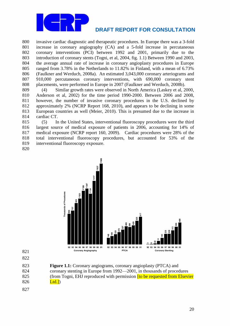
invasive cardiac diagnostic and therapeutic procedures. In Europe there was a 3-fold 800
increase in coronary angiography (CA) and a 5-fold increase in percutaneous 801
coronary interventions (PCI) between 1992 and 2001, primarily due to the 802
introduction of coronary stents (Togni, et al, 2004, fig. 1.1) Between 1990 and 2003, 803
the average annual rate of increase in coronary angioplasty procedures in Europe 804
ranged from 3.78% in the Netherlands to 11.82% in Finland, with a mean of 6.73% 805
(Faulkner and Werduch, 2008a). An estimated 3,043,000 coronary arteriograms and 806
910,000 percutaneous coronary interventions, with 690,000 coronary stent 807
placements, were performed in Europe in 2007 (Faulkner and Werduch, 2008b). 808
(4) Similar growth rates were observed in North America (Laskey et al, 2000, 809
Anderson et al, 2002) for the time period 1990-2000. Between 2006 and 2008, 810
however, the number of invasive coronary procedures in the U.S. declined by 811
approximately 2% (NCRP Report 168, 2010), and appears to be declining in some 812
European countries as well (Meier, 2010). This is presumed due to the increase in 813
cardiac CT. 814
(5) In the United States, interventional fluoroscopy procedures were the third 815
largest source of medical exposure of patients in 2006, accounting for 14% of 816
medical exposure (NCRP report 160, 2009). Cardiac procedures were 28% of the 817
total interventional fluoroscopy procedures, but accounted for 53% of the 818
interventional fluoroscopy exposure. 819
820
821
822
Figure 1.1: Coronary angiograms, coronary angioplasty (PTCA) and 823
coronary stenting in Europe from 1992—2001, in thousands of procedures 824
(from Togni, EHJ reproduced with permission [to be requested from Elsevier 825
Ltd.]) 826
827

DRAFT REPORT FOR CONSULTATION
21
(6) This growth has involved mainly the Western world, but a similar trend is 828
seen in other countries: in China the annual increment rate for PCI is around 40% 829
(Cheng et al, 2004). This number is relatively small and may reflect the lower 830
prevalence of coronary artery disease in the Chinese population (3-7%, about one 831
quarter of that of Western Caucasians), but is expected to grow as a consequence of 832
changing dietary habits, life-style and cigarette smoking (Cheng et al, 2004, Moran 833
2010). 834
(7) A survey of developing countries conducted by the IAEA revealed that 835
about 30% of the 20 participating countries demonstrated a 100% increase in 836
workload in the 3-year period from 2004 to 2007 (Tsapaki, 2009). The same study 837
indicated that the numbers of paediatric interventional procedures can reach the 838
levels of adult interventional procedures, even in developing countries. 839
840
1.1.2 Skin injuries 841
(8) Both PCI and interventional electrophysiology procedures can result in 842
patient skin doses high enough to cause deterministic skin injuries (see Chapters 2 843
and 3) (Miller 2008). At one centre, the frequency of skin injuries was estimated at 844
3 x 10⁻⁴
(Padovani 2005). Although the number of radiation injuries due to cardiac 845
procedures remains small, these injuries have a major impact on the patients who are 846
affected. Therefore, it is important to inform and continue to remind practicing 847
clinicians of the potential risks involved with these procedures. 848
(9) The number of patients undergoing multiple procedures continues to 849
increase (Laskey et al, 2001). Complex cases may be treated in more than one 850
session (staged procedures). Restenosis and disease progression may also prompt 851
repeated interventions. In a recent series of 3332 patients (Padovani et al, 2005) 852
almost one third underwent at least two procedures. Vano et al. (Vano 2001) 853
observed a much greater rate of skin effects in patients who had undergone multiple 854
fluoroscopically guided coronary procedures. Repeated procedures, especially when 855
performed within a short period of time, increase the risk of skin injury (Balter, 856
2010). Multiple cardiac fluoroscopic procedures should be a cause of concern with 857
regard to radiological protection. The risk of skin injuries should not be 858
underestimated. 859
(10) Patient radiation dose is related to procedure complexity (Bernardi et al, 860
2000, Peterzol et al, 2002, Balter et al, 2009, IAEA 2009). Multi-vessel PCI is 861
considered a complexity factor, but this may not be always the case (Bernardi et al, 862
2000). Other factors that appear to affect complexity for PCI include the type of 863
lesion, the chronicity of the occlusion, the degree of vessel tortuosity and the 864
involvement of vessel bifurcations (Balter et al, 2009, IAEA 2009). 865
866
1.1.3 Cardiac electrophysiology procedures 867
(11) A second field where there has been an increase in both the number and 868
complexity of procedures is interventional electrophysiology. Permanent pacemaker 869
implantation for bradycardia is carried out in large numbers of patients. From 1997 870
to 2001, the number of new pacemaker implants increased about 50% worldwide 871
(Mond et al, 2004). More recently, bi-ventricular pacemakers (cardiac 872
resynchronisation therapy) have been introduced for the treatment of patients with 873
cardiac failure and cardiomyopathies (Salukhe et al, 2004). The use of cardioverter-874
defibrillators has also increased, as a result of studies (Moss et al, 2002, Salukhe et 875

DRAFT REPORT FOR CONSULTATION
22
al, 2004) that demonstrated their life-saving role in patients at risk of sudden cardiac 876
death. An estimated 554,000 pacemaker implantations were performed in Europe in 877
2007 (Faulkner and Werduch, 2008b) and an estimated 189,000 electrophysiology 878
procedures and 361,000 cardiac device implantations were performed in the U.S. in 879
2008 (NCRP Report No. 168, 2010). 880
(12) Cardiac electrophysiology procedures also include treatment of patients 881
with re-entrant tachycardias. These patients are often much younger than patients 882
with coronary heart disease, and require both diagnostic procedures and treatment by 883
radiofrequency ablation. Due to the long fluoroscopy times required for these 884
procedures, these patients can be exposed to very high radiation doses and a 885
substantial risk of deterministic effects if technique is not optimized (Rosenthal, 886
1998, McFadden, 2002). 887
888
1.1.4 Congenital and valvular heart disease 889
(13) Two other groups of cardiac disease where catheter techniques are used 890
and are likely to expand in the near future are congenital and valvular heart disease. 891
These groups represent a small percentage of patients undergoing percutaneous 892
interventions, but these diseases are seen in both children and adults. Children are at 893
greater risk for the development of stochastic radiation effects, principally cancer, 894
due to their longer expected life span and their increased sensitivity to radiation as 895
compared to adults (Hall, 2009). It has been estimated that approximately 7% of all 896
cardiac angiography procedures are carried out in children aged 0 to 15 years 897
(UNSCEAR 2000). The most widely performed procedures are balloon 898
valvuloplasty, device closure of atrial septal defect, patent foramen ovale or ductus 899
arteriosus, stenting of pulmonary artery stenosis or coarctation of the aorta and 900
electrophysiology studies. These procedures may involve long fluoroscopy times. In 901
addition to these well-established procedures, new procedures have been introduced, 902
including percutaneous pulmonary and aortic valve replacement, ventricular septal 903
defect closure, implantation of banding devices to limit pulmonary blood flow, and 904
radiofrequency perforation to create continuity between cardiac chambers and 905
vessels (Levi et al, 2003). (Percutaneous aortic valve replacement is performed 906
primarily in elderly patients unfit for surgery). A percutaneous or combined 907
percutaneous/surgical approach has been proposed to treat complex diseases such as 908
hypoplastic left heart syndrome. Fetal interventions are also possible. 909
(14) These techniques to treat congenital and valvular heart disease are largely 910
justified as they may replace very high-risk surgical procedures. Although 911
transesophageal and intracardiac ultrasound may partially replace fluoroscopy (Rice 912
et al, 2002, Zanchetta et al, 2004), radiation risk still remains a problem and is often 913
underestimated. Fluoroscopy times as high as 129 minutes may be required to 914
implant a pulmonary valve (Bonhoeffer et al, 2002). There is little literature 915
concerning the safety issues of these new devices to be used in infants and children 916
(Levi et al, 2003). 917
918
1.1.5 Paediatric patients 919
(15) A survey of patient doses in 137 children, aged from < 1 year to 16 years, 920
undergoing cardiac procedures performed using a biplane flat panel detector X-ray 921
system, demonstrated mean values of 1.9 to 8.6 Gy·cm2 for diagnostic procedures. 922
Mean dose values for therapeutic procedures, in both extremes of the paediatric age 923

DRAFT REPORT FOR CONSULTATION
23
group, ranged from 2.4 to 17.8 Gy·cm2 (Martinez et al, 2007). In a series of 205 924
children (mean age 4.1 y) who underwent diagnostic cardiac catheterization, the 925
mean dose was 17 Gy·cm2 (Chida et al, 2010). In comparison to proposed diagnostic 926
reference levels for fluoroscopically guided cardiac interventions in adults of 50 927
Gy·cm2 for diagnostic procedures and 125 Gy·cm
2 for therapeutic procedures 928
(Balter et al, 2008), paediatric patients have typically received less than 20% of the 929
dose received by adult patients. Nonetheless, radiation doses from paediatric cardiac 930
catheterization procedures are of concern (Andreassi, 2006, Andreassi, 2009). 931
932
1.2 Cardiac CT 933 934
(16) Cardiac CT technology has evolved rapidly in recent years, and these 935
advancements have enabled a variety of types of cardiac CT studies to be performed 936
that go well beyond detection of the coronary arteries. Today, cardiac CT 937
encompasses several distinct procedures, including coronary artery calcium (CAC) 938
scoring, CT coronary angiography (CTCA), pulmonary vein CT angiography, and 939
CT attenuation correction of nuclear cardiology image data. Recent technological 940
advances have been associated with an increase in the number of procedures 941
performed, although reliable statistics on worldwide numbers are not presently 942
available. In the United States, CT was the largest source of medical exposures to 943
patients in 2006, accounting for 49% of the medical exposure of patients (NCRP 944
report 160, 2009). Cardiac CT (including CTCA and CAC) accounted for 4.7% of 945
CT examinations, but 12.1% of patient exposure from CT. 946
947
1.3 Nuclear cardiology 948
949 (17) An estimated 32.7 million diagnostic nuclear medicine procedures are 950
performed annually worldwide (UNSCEAR 2008). Of these, approximately 14 951
million are nuclear cardiology procedures, and this number has increased rapidly 952
(Davis, 2006). More than 90% of nuclear cardiology studies are myocardial 953
perfusion scintigraphy studies for the assessment of myocardial perfusion and/or 954
viability. The vast majority of nuclear cardiology procedures performed employ 955
single photon emission computed tomography (SPECT), although a small but 956
growing number of laboratories perform positron emission tomography (PET) 957
studies. 958
(18) In the U.S., nuclear medicine procedures accounted for 26% of the 959
medical exposure of patients in 2006, and cardiac studies accounted for 85% of the 960
nuclear medicine exposure (NCRP report 160, 2009). Nuclear medicine procedures 961
were the second largest source of medical exposures, after CT. 962
(19) More nuclear cardiology procedures are performed in the United States 963
than in the rest of the world combined. Reasons suggested for this disparity include 964
better access to testing, a more litigious medicolegal climate, and profit motives for 965
testing. However, multiple U.S. series have demonstrated that for those procedures 966
where sufficient data are available to permit a determination of appropriateness, only 967
~15% are performed for inappropriate indications (Gibbons, 2008; Hendel, 2010). 968
Nonetheless, cardiologists should consider using alternative methodologies that do 969
not require ionizing radiation, such as stress echocardiography, whenever possible. 970
971
972
DRAFT REPORT FOR CONSULTATION
24
1.4 Occupational radiation risk 973
(20) Radiation risk is not limited to patients. Operators and staff receive 974
radiation exposure during fluoroscopically guided procedures. The increased 975
complexity of interventional cardiology procedures appears to have offset dose 976
reductions due to improvements in technology (Kim, 2008). There is considerable 977
variation in operator doses observed for the same type of procedure, indicating that 978
radiological protection practices can be improved (Kim, 2009). Recent studies have 979
shown that there is an increased incidence of radiation-related cataracts in 980
interventional cardiologists when radiological protection tools are not used properly 981
(Vano, 2010, Ciraj-Bjelac, 2010) Unfortunately, there is lack of proper monitoring 982
of radiation doses to staff and lack of reliable data on occupational doses (Padovani, 983
2011). 984
985
1.5 Summary 986
(21) In summary, fluoroscopically guided cardiology procedures are increasing 987
in number and complexity. The benefits for patients are clear, but radiation doses for 988
both patients and staff are important and must be managed appropriately. For young 989
patients, the increased risk of cancer should be considered in the optimisation of 990
these procedures. For older patients cancer risk is not as important, but avoidance of 991
deterministic effects (skin injuries) should be taken into account. Interventional 992
cardiologists are among the radiation workers with the highest occupational 993
radiation risk, and should know how to protect both patients and themselves. This 994
ICRP report is intended to help achieve this goal. 995
996
1.6 References 997
Anderson HV, Shaw RE, Brindis RG, Hewitt K, Krone RJ, Block PC, McKay CR, 998
Weintraub WS. A contemporary overview of percutaneous coronary 999
interventions. The American College of Cardiology-National Cardiovascular 1000
Data Registry (ACC-NCDR). J Am Coll Cardiol 2002; 39:1096-103. 1001
Andreassi MG, Ait-Ali L, Botto N, Manfredi S, Mottola G, Picano E. Cardiac 1002
catheterization and long-term chromosomal damage in children with congenital 1003
heart disease. Eur Heart J 2006; 27:2703–2708. 1004
Andreassi MG. Radiation risk from pediatric cardiac catheterization: friendly fire 1005
on children with congenital heart disease. Circulation 2009;120(19):1847-1849. 1006
Balter S, Miller DL, Vano E, Ortiz Lopez P, Bernardi G, Cotelo E, Faulkner K, 1007
Nowotny R, Padovani R, Ramirez A. A pilot study exploring the possibility of 1008
establishing guidance levels in x-ray directed interventional procedures. Med 1009
Phys 2008;35:673-80. 1010
Balter S, Hopewell JW, Miller DL, Wagner LK, Zelefsky MJ. Fluoroscopically 1011
guided interventional procedures: A review of radiation effects on patients' skin 1012
and hair. Radiology 2010;254:326-341. 1013
Bernardi G, Padovani R, Morocutti G, Vano E, Malisan MR, Rinuncini M, 1014
Spedicato L, Fioretti PM. Clinical and technical determinants of the complexity 1015
of PTCA procedures. Analysis in relation to radiation exposure parameters. 1016
Cathet Cardiovasc Interv 2000;51:1-9. 1017
Bonhoeffer P, Boudjemline Y, Qureshi SA, Le Bidois J, Iserin L, Acar P, Merckx J, 1018
Kachaner J, Sidi D. Percutaneous insertion of the pulmonary valve. J Am Coll 1019
Cardiol 2002;39:1664-9. 1020

DRAFT REPORT FOR CONSULTATION
25
1021
Ciraj-Bjelac O, Rehani MM, Sim KH, Liew HB, Vano E, Kleiman NJ. Risk for 1022
radiation induced cataract for staff in interventional cardiology: Is there reason 1023
for concern? Catheter Cardiovasc Interv 2010;76(6):826-834. 1024
Cheng TO. The current state of cardiology in China. Int J Cardiol. 2004 1025
Sep;96(3):425-39. 1026
Chida K, Ohno T, Kakizaki S, Takegawa M, Yuuki H, Nakada M, Takahashi S, 1027
Zuguchi M. Radiation dose to the pediatric cardiac catheterization and 1028
intervention patient. AJR American Journal of Roentgenology 2010; 1029
195(5):1175-1179. 1030
Davis W. Mapping improvements in molecular imaging. Medical Imaging. 1031
2006;May 2006. Available at: 1032
http://www.imagingeconomics.com/issues/articles/MI_2006-05_01.asp 1033
Accessed December 31, 2010. 1034
Fajadet J, Morice MC, Bode C, Barragan P, Serruys PW, Wijns W, Constantini CR, 1035
Guermonprez JL, Eltchaninoff H, Blanchard D, Bartorelli A, Laarman GJ, Perin 1036
M, Sousa JE, Schuler G, Molnar F, Guagliumi G, Colombo A, Ban Hayashi E, 1037
Wulfert E. Maintenance of long-term clinical benefit with sirolimus-eluting 1038
coronary stents: three-year results of the RAVEL trial. Circulation 1039
2005;111:1040-4. 1040
Faulkner K, Werduch A. An estimate of the collective dose to the European 1041
population from cardiac x-ray procedures. Br J Radiol 2008a; 81:955-962. 1042
Faulkner K, Werduch A. Analysis of the frequency of interventional cardiology in 1043
various European countries. Rad Prot Dosim 2008b; 129(1-3):74-76. 1044
Hall EJ. Radiation biology for pediatric radiologists. Pediatr Radiol 2009;39 Suppl 1045
1:S57-64. 1046
Gibbons RJ, Miller TD, Hodge D, et al. Application of appropriateness criteria to 1047
stress single-photon emission computed tomography sestamibi studies and 1048
stress echocardiograms in an academic medical center. J Am Coll Cardiol. 1049
2008;51(13):1283-1289.International Atomic Energy Agency. Establishing 1050
guidance levels in x ray guided medical interventional procedures: A pilot 1051
study. Safety Reports Series No. 59. Vienna: International Atomic Energy 1052
Agency; 2009. 1053
Hendel RC, Cerqueira M, Douglas PS, et al. A multicenter assessment of the use of 1054
single-photon emission computed tomography myocardial perfusion imaging 1055
with appropriateness criteria. J Am Coll Cardiol. 2010;55(2):156-162. 1056
International Commission on Radiological Protection. Avoidance of radiation 1057
injuries from medical interventional procedures. ICRP Publication 85. Ann 1058
ICRP 2000;30:7-67. 1059
Kim KP, Miller DL, Balter S, Kleinerman RA, Linet MS, Kwon D, Simon SL. 1060
Occupational radiation doses to operators performing cardiac catheterization 1061
procedures. Health Phys 2008;94:211-227. 1062
Kim KP, Miller DL. Minimising radiation exposure to physicians performing 1063
fluoroscopically guided cardiac catheterisation procedures: a review. Radiat 1064
Prot Dosimetry 2009;133:227-33. 1065
Laskey WK, Kimmel S, MD, Krone RJ. Contemporary Trends in Coronary 1066
Intervention: A Report From the Registry of the Society for Cardiac 1067
Angiography and Interventions Cathet. Cardiovasc. Intervent. 49:19–22, 2000. 1068
DRAFT REPORT FOR CONSULTATION
26
Laskey WK, Williams DO, Vlachos HA, MS, H, Holmes DR, King SB, Kelsey SF, 1069
Slater J, Faxon D, Al-Bassam M, Block E, Detre KM for the Dynamic Registry 1070
Investigators. Changes in the Practice of Percutaneous Coronary Intervention: A 1071
Comparison of Enrollment Waves in the National Heart, Lung, and Blood 1072
Institute (NHLBI) Dynamic Registry. Am J Cardiol 2001;87:964–969. 1073
Lemos PA, Serruys PW, van Domburg RT, Saia F, Arampatzis CA, Hoye A, 1074
Degertekin M, Tanabe K, Daemen J, Liu TK, McFadden E, Sianos G, Hofma 1075
SH, Smits PC, van der Giessen WJ, de Feyter PJ. Unrestricted utilization of 1076
sirolimus-eluting stents compared with conventional bare stent implantation in 1077
the "real world": the Rapamycin-Eluting Stent Evaluated At Rotterdam 1078
Cardiology Hospital (RESEARCH) registry. Circulation 2004;109:190-5. 1079
Levi DS, Alejos JC, Moore JW. Future of interventional cardiology in pediatrics. 1080
Curr Opin Cardiol. 2003;18:79-90. 1081
Martinez LC, Vano E, Gutierrez F, Rodriguez C, Gilarranz R, Manzanas MJ. 1082
Patient doses from fluoroscopically guided cardiac procedures in pediatrics. 1083
Phys Med Biol. 2007;52(16):4749-59. 1084
McFadden SL, Mooney RB, Shepherd PH. X-ray dose and associated risks from 1085
radiofrequency catheter ablation procedures. Br J Radiol 2002;75:253-65. 1086
Meier, B. Cardiac catheterization interventions: European statistics 2007/2008. 1087
Presented at Euro PCR, May, 2010. Available at: 1088
http://www.escardio.org/communities/EAPCI/Documents/Cardiac-1089
catheterization-Interventions-stats07-08-EAPCI-Assembly-may2010.pdf 1090
Accessed February 22, 2011. 1091
Miller DL. Overview of contemporary interventional fluoroscopy procedures. 1092
Health Phys 2008;95:638-44. 1093
Mond HG, Irwin M, Morillo C, Ector H. The world survey of cardiac pacing and 1094
cardioverter defibrillators: calendar year 2001. Pacing Clin Electrophysiol 1095
2004;27:955-64 1096
Moran A, Gu D, Zhao D, Coxson P, Wang YC, Chen CS, Liu J, Cheng J, Bibbins-1097
Domingo K, Shen YM, He J, Goldman L. Future cardiovascular disease in 1098
china: markov model and risk factor scenario projections from the coronary 1099
heart disease policy model-china. Circ Cardiovasc Qual Outcomes. 1100
2010;3(3):243-52. 1101
Morice MC, Serruys PW, Sousa JE, Fajadet J, Ban Hayashi E, Perin M, Colombo 1102
A, Schuler G, Barragan P, Guagliumi G, Molnar F, Falotico R; RAVEL Study 1103
Group. Randomized Study with the Sirolimus-Coated Bx Velocity Balloon-1104
Expandable Stent in the Treatment of Patients with de Novo Native Coronary 1105
Artery Lesions. A randomized comparison of a sirolimus-eluting stent with a 1106
standard stent for coronary revascularization. N Engl J Med 2002;346:1773-80. 1107
Moss AJ, Zareba W, Hall WJ, Klein H, Wilber DJ, Cannom DS, Daubert JP, 1108
Higgins SL, Brown MW, Andrews ML; Multicenter Automatic Defibrillator 1109
Implantation Trial II Investigators. Prophylactic implantation of a defibrillator 1110
in patients with myocardial infarction and reduced ejection fraction. N Engl J 1111
Med 2002;346:877-83. 1112
National Council on Radiation Protection and Measurements. Ionizing radiation 1113
exposure of the population of the United States. NCRP Report No. 160. 1114
Bethesda, MD: National Council on Radiation Protection and Measurements; 1115
2009. 1116

DRAFT REPORT FOR CONSULTATION
27
National Council on Radiation Protection and Measurements. Radiation dose 1117
management for fluoroscopically guided interventional medical procedures. 1118
NCRP Report No. 168. Bethesda, MD: National Council on Radiation 1119
Protection and Measurements; 2010. 1120
Padovani R, Bernardi G, Quai E, Toh HS, Signor M, Morocutti G, Spedicato L. 1121
Retrospective evaluation of occurrence of skin injuries in interventional cardiac 1122
procedures. Radiat Prot Dosimetry 2005; 117 (1-3):247-250. 1123
Padovani, R., Le Heron, J., Cruz-Suarez, R., Duran, A., Lefaure, C., Miller, D.L., 1124
Sim, H.K., Vano, E., Rehani, M., Czarwinski, R., 2010. International project on 1125
individual monitoring and radiation exposure levels in interventional 1126
cardiology. Radiat Prot Dosimetry 2011; in press. 1127
Peterzol A, Quai E, Padovani R, Bernardi G, Kotre J, Dowling A. Reference levels 1128
in PTCA as a function of procedure complexity. Radiat Prot Dosimetry 1129
2005;117 (1-3): 54-58. 1130
Prieto C, Vano E, Fernández JM, Galvan C, Sabate M, Gonzalez L, Martinez D. Six 1131
years experience in intracoronary brachytherapy procedures: patient doses from 1132
fluoroscopy. Br J Radiol. 2006 Sep; 79(945): 730-733. 1133
Rice MJ, McDonald RW, Li X, Shen I, Ungerleider RM, Sahn DJ. New 1134
Technology and Methodologies for Intraoperative, Perioperative, and 1135
Intraprocedural Monitoring of Surgical and Catheter Interventions for 1136
Congenital Heart Disease Echocardiography 2002; 19:725-734. 1137
Rosenthal LS, Mahesh M, Beck TJ, Saul JP, Miller JM, Kay N, Klein LS, Huang S, 1138
Gillette P, Prystowsky E, Carlson M, Berger RD, Lawrence JH, Yong P, 1139
Calkins H. Predictors of fluoroscopy time and estimated radiation exposure 1140
during radiofrequency catheter ablation procedures. Am J Cardiol 1998;82:451-1141
458. 1142
Salukhe TV, Dimopoulos K, Francis D. Cardiac resynchronisation may reduce all-1143
cause mortality: meta-analysis of preliminary COMPANION data with 1144
CONTAK-CD, InSync ICD, MIRACLE and MUSTIC. Int J Cardiol 1145
2004;93:101-3. 1146
The SoS Investigators. Coronary artery bypass surgery versus percutaneous 1147
coronary intervention with stent implantation in patients with multivessel 1148
coronary artery disease (the Stent or Surgery trial): a randomized controlled 1149
trial. Lancet 2002;360:965–70. 1150
Togni M, Balmer F, Pfiffner D, Maier W, Zeiher AM, Meier B, on behalf of the 1151
Working Group Interventional Cardiology and Coronary Pathophysiology of the 1152
European Society of Cardiology. Percutaneous coronary interventions in Europe 1153
1992–2001. Eur Heart J 2004;25:1208–1213. 1154
Tsapaki, V., Ahmed, N.A., AlSuwaidi, J.S., Beganovic, A., Benider, A., 1155
BenOmrane, L., Borisova, R., Economides, S., El-Nachef, L., Faj, D., 1156
Hovhannesyan, A., Kharita, M.H., Khelassi-Toutaoui, N., Manatrakul, N., 1157
Mirsaidov, I., Shaaban, M., Ursulean, I., Wambani, J.S., Zaman, A., Ziliukas, J., 1158
Zontar, D., Rehani, M.M., 2009. Radiation exposure to patients during 1159
interventional procedures in 20 countries: initial IAEA project results. AJR. 1160
Am. J. Roentgenol. 193, 559-569. 1161
UNSCEAR. Sources and effects of ionizing radiation: volume I, sources: annex D, 1162
medical radiation exposure. Report to the General Assembly of the United 1163
Nations; 2000; New York, NY Available at: 1164

DRAFT REPORT FOR CONSULTATION
28
http://www.unscear.org/unscear/en/publications/2000_1.html Accessed 1165
December 31, 2010. 1166
UNSCEAR. Sources of ionizing radiation: volume I. Report to the General 1167
Assembly, with scientific annexes; 2010; New York, NY. Available at: 1168
http://www.unscear.org/unscear/en/publications/2008_1.html Accessed 1169
December 31, 2010. 1170
Vano E, Goicolea J, Galvan C, Gonzalez L, Meiggs L, Ten JI, Macaya C. Skin 1171
radiation injuries in patients following repeated coronary angioplasty 1172
procedures. Br J Radiol. 2001 Nov;74(887):1023-31. 1173
Vano E, Kleiman NJ, Duran A, Rehani MM, Echeverri D, Cabrera. M. Radiation 1174
cataract risk in interventional cardiology personnel. Radiat Res, 2010; 1175
174(4):490-495 1176
Verin V, Popowski Y, de Bruyne B, Baumgart D, Sauerwein W, Lins M, Kovacs G, 1177
Thomas M, Calman F, Disco C, Serruys PW, Wijns W, Piessens M, Kurtz J, 1178
Simon R, Delafontaine P, Erbel R. Endoluminal Beta-Radiation Therapy for the 1179
Prevention of Coronary Restenosis after Balloon Angioplasty. N Engl J Med 1180
2001;344:243-249. 1181
Waksman R, Bhargava B, White L, Chan RC, Mehran R, Lansky AJ, Mintz GS, 1182
Satler LF, Pichard AD, Leon MB, Kent KK. Intracoronary beta-radiation 1183
therapy inhibits recurrence of in-stent restenosis. Circulation 2000;101:1895-8. 1184
Williams DO, Holubkov R, Yeh W, Bourassa MG, Al-Bassam M, Block PC, Coady 1185
P, Cohen H, Cowley M, Dorros G, Faxon D, Holmes DR, Jacobs A, Kelsey SF, 1186
King SB, Myler R, Slater J, Stanek V, Vlachos HA, Detre KM. Percutaneous 1187
Coronary Intervention in the Current Era Compared With 1985–1986 The 1188
National Heart, Lung, and Blood Institute Registries. Circulation 1189
2000;102:2945-2951. 1190
Zanchetta M, Maiolino P. Intracardiac echocardiography. Do we need a new 1191
ultrasonographic window? Ital Heart J 2004;5:173-177. 1192
1193

DRAFT REPORT FOR CONSULTATION
29
1194
2. THE BIOLOGICAL EFFECTS OF RADIATION 1195
1196
Main Points 1197
1198
Deterministic effects are due to injury in populations of cells, 1199
characterised by a threshold dose and an increase in the incidence and 1200
severity of the reaction as the dose is increased further. Deterministic 1201
effects are also termed tissue reactions. 1202
Stochastic effects are malignant disease and heritable effects for which 1203
the probability of an effect occurring, but not its severity, is regarded as 1204
a function of dose without threshold. 1205
Radiation-induced skin injuries may not become fully manifest until 1206
months after the radiation dose was administered. 1207
The diagnosis of a radiation induced skin injury is often delayed. 1208
The lens of the eye is a radiosensitive tissue. 1209
In the lens of the eye, ionizing radiation typically causes posterior 1210
subcapsular cataract formation. 1211
Surveys of cardiologists and support staff working in catheterization 1212
laboratories have found a high percentage of lens opacities attributable 1213
to occupational radiation exposure when radiological protection tools 1214
have not been used properly. 1215
1216
2.1 Types of radiation effects 1217 1218
(22) The effects of radiation can be classified into two groups: deterministic 1219
effects (harmful tissue reactions) and stochastic effects (cancer and heritable 1220
effects). 1221
(23) Deterministic effects (e.g. skin injury) are largely caused by the 1222
reproductive sterilisation of cells following high radiation doses. The induction of 1223
tissue reactions is generally characterised by a threshold dose. The reason for the 1224
presence of this threshold dose is that radiation-induced reproductive survival of a 1225
critical population of cells in a given tissue needs to be sustained before injury is 1226
expressed in a clinically relevant form. Above the threshold dose the incidence and 1227
severity of the injury, including impairment of the capacity for tissue recovery, 1228
increases with dose (ICRP 103). The threshold is variable, depending on the nature 1229
and condition of the exposed tissue (Balter, 2010). 1230
(24) The injury is not expressed clinically until the cells die as a result of an 1231
unsuccessfully attempt at cell division or differentiation and are lost as part of the 1232
normal process of tissue turnover (Balter, 2010). The incidence as well as the 1233
severity of the injury, including impairment of the capacity for tissue recovery, 1234
increases with dose. After a high radiation dose, the outcome for the affected 1235
individual can be devastating (Balter, 2010). 1236
(25) Eighty percent of reported radiation-induced skin injuries in one large 1237
series were from cardiac procedures (Koenig et al 2001). Nonetheless, cardiologists 1238
often do not recognise that a radiation injury is related to a cardiac procedure, either 1239
because they are unaware of the magnitude of radiation dose delivered or they do 1240
not know that radiation can cause skin injuries. 1241

DRAFT REPORT FOR CONSULTATION
30
(26) The dose of radiation received by some patients is high and the number of 1242
radiation injury cases is increasing (NCI, 2005). However, most currently practising 1243
interventional cardiologists have no personal experience of a case of radiation 1244
injury. The number of radiation injuries is small compared with the number of 1245
fluoroscopically guided cardiology procedures performed worldwide. 1246
(27) Stochastic effects The accumulation of cellular and animal data relevant to 1247
radiation tumourigenesis has, since 1990, strengthened the view that DNA damage 1248
response processes in single cells are of critical importance to the development of 1249
cancer after radiation exposure. Epidemiological and experimental studies provide 1250
evidence of radiation risk, albeit with uncertainties at doses about 100 mSv or less 1251
(ICRP 103). 1252
(28) These effects are probabilistic—there is no identifiable threshold for 1253
producing the effect. The likelihood of inducing a stochastic effect increases with 1254
dose, but the exact relationship between dose and effect is not known. In the low 1255
dose range, below about 100 mSv, it is scientifically plausible to assume that the 1256
incidence of cancer or heritable effects will rise in direct proportion to an increase in 1257
the equivalent dose in the relevant organs and tissues (the ―linear-non-threshold‖ or 1258
LNT model) (ICRP 103). Dose has no relationship to the severity of the effect. 1259
(29) Children are approximately 2-3 times more sensitive to the stochastic 1260
effects of radiation than adults (ICRP 1991). They also have a longer potential 1261
lifespan than do adults, so they have more time to develop possible radiation related 1262
sequelae. In children, the probability of a fatal cancer per fluoroscopically guided 1263
procedure is estimated at approximately 0.07-0.08%, but this risk may vary widely 1264
depending on patient age, underlying life expectancy and how the procedure is 1265
performed (Martinez et al, 2007, Bacher et al, 2005). 1266
(30) While there is compelling evidence that radiation causes heritable effects 1267
in experimental animals, there continues to be no direct evidence that exposure of 1268
humans to radiation leads to excess heritable disease in offspring (ICRP 103). 1269
1270
2.2 Background 1271 1272
(31) Some months after the discovery of x-rays in 1895, radiation-induced skin 1273
changes were observed (Daniel 1896, Codman 1896). Some early radiologists 1274
suffered severe dermatitis, radiation cancer and amputation of digits. There was a 1275
delay in recognising that x-rays were the cause because they are invisible and do not 1276
cause any sensation during exposure. As noted in ICRP Publication 103, the goal of 1277
preventing these radiation injuries was the impetus for the formation of what is now 1278
the Commission (ICRP 2007). 1279
(32) Following the dramatic rise in the number of percutaneous coronary 1280
interventional procedures, cases of patients with deep skin ulceration and necrosis 1281
were reported in the 1990s (Shope, 1996). In 1994 the U.S. Food and Drug 1282
Administration issued an advisory regarding skin injury from fluoroscopically 1283
guided procedures (FDA 1994). Radiation skin injury has also been reported 1284
following radiofrequency catheter ablations (Vano, 1998). This is of particular 1285
concern because many of these patients are young adults, and some are children. 1286
The Commission drew attention to prevention of skin injuries from interventional 1287
fluoroscopy procedures in Publication 85 (ICRP 2000), and reiterated the 1288
importance of preventing skin injuries in Publication 105 (ICRP 2007). 1289
1290

DRAFT REPORT FOR CONSULTATION
31
2.3 Radiation Effects and the Skin 1291
1292 (33) The response of the skin to radiation is dose-related and occurs when this 1293
dose is concentrated on one area, usually the site where the x-rays enter the patient. 1294
The term ―absorbed dose‖ is used to assess the amount of radiation to which a tissue 1295
is exposed (see the Glossary). The skin response follows a characteristic pattern, 1296
although the time course is variable (Balter et al, 2010). The threshold doses and 1297
time of appearance for various types of skin injury are summarised in Table 2.1. 1298
(34) Defects in DNA repair genes may predispose individuals to radiogenic 1299
cancer or lower the threshold for the development of deterministic effects. Some 1300
patients with serious and unanticipated radiation injuries may be among the 1% of 1301
the population heterozygous for the ATM gene, an autosomal recessive gene 1302
responsible for ataxia telangiectasia, or may harbour some other ATM abnormality. 1303
(Hymes, 2006, Allan, 2008) Other disorders with a genetic component affecting 1304
DNA breakage or repair also increase radiation sensitivity, including Fanconi 1305
anaemia, Bloom syndrome and xeroderma pigmentosum. Familial polyposis, 1306
Gardner syndrome, hereditary malignant melanoma and dysplastic nevus syndrome 1307
also increase radiation sensitivity (Hymes, 2006). Certain familial cancer syndromes 1308
may increase susceptibility to radiogenic cancer, including neurofibromatosis, Li-1309
Fraumeni syndrome and hereditary retinoblastoma (Allan, 2008). 1310
(35) Autoimmune and connective tissue disorders predispose patients to the 1311
development of severe cutaneous radiation effects in an unpredictable fashion. 1312
These typically occur in association with the high radiation doses administered 1313
during radiation therapy. The aetiology is not known. These disorders include 1314
scleroderma, systemic lupus erythematosus and possibly rheumatoid arthritis.( 1315
(Wagner et al, 1999, Hymes, 2006) Hyperthyroidism and diabetes mellitus are also 1316
associated with increased radiation sensitivity (Koenig Part 1, 2001) Diabetes is 1317
believed to predispose to radiation injury secondary to small vessel vascular disease 1318
and consequent decreased healing capacity (Herold, 1999). A number of drugs 1319
increase radiation sensitivity, including actinomycin D, doxorubicin, bleomycin, 5-1320
fluorouracil and methotrexate (Koenig Part 1, 2001) Again, this effect is usually 1321
seen only with the high radiation doses delivered during radiation therapy. 1322
(36) It is apparent from the foregoing and from Table 2.1 that there are no 1323
rigid thresholds for dose or time of appearance of radiation-induced skin changes, 1324
because individuals vary in their radio-sensitivity and radio-responsiveness (Balter 1325
et al, 2010). These ranges are shown graphically in Figure 2.1. In the discussion 1326
below, threshold doses are given for an average person, but it should be understood 1327
that these will vary from individual to individual. For most patients, clinically 1328
important skin reactions occur only when the absorbed skin dose is greater than 1329
5 Gy (Balter et al, 2010; ICRP Tissue Reactions, 2011a). 1330
1331

DRAFT REPORT FOR CONSULTATION
32
Table 2.1: Tissue reactions from a single-delivery radiation dose to the skin of the neck, torso, pelvis, buttocks or 1332 arms. (from Balter et al, 2010) 1333
- This table is applicable to the normal range of patient radiosensitivities in the absence of mitigating or aggravating physical or clinical factors. - Skin dose refers to absorbed skin dose (including backscatter). This quantity is not the reference air kerma (Ka,r) described by the Food and Drug Administration (21 CFR § 1020.32 (2008)) or the International Electrotechnical Commission.(IEC, 2010) - This table does not apply to the skin of the scalp. - Abrasion or infection of the irradiated area is likely to exacerbate radiation effects. - The dose and time bands are not rigid boundaries. Signs and symptoms are expected to appear earlier as the skin dose increases.
Band
Single-site Acute Skin-Dose Range
(Gy)1
NCI Skin
Reaction Grade*
Approximate time of onset of effects
(4) Prompt < 2 weeks
(5) Early 2 – 8 weeks
Mid term 6 – 52 weeks
Long term > 40 weeks
A1 0-2 N/A No observable effects expected
A2 2-5 1 - Transient erythema - Epilation - Recovery from hair loss (6) - None expected
B 5-10 1
- Transient erythema - Erythema, epilation - Recovery. - At higher doses;
prolonged erythema, permanent partial epilation
(7) - Recovery. - At higher doses dermal
atrophy/induration.
C 10-15 1-2
- Transient erythema
- Erythema, epilation. - Possible dry or moist
desquamation
(8) - Recovery from desquamation
(9) - Prolonged erythema. - Permanent epilation.
(10) - Telangiectasia2
- Dermal atrophy/induration. - Skin likely to be weak.
D > 15 3-4
- Transient erythema - After very high doses,
edema and acute ulceration; long-term surgical intervention likely to be required.
- Erythema, epilation.
(11) - Moist desquamation
(12) - Dermal atrophy, - Secondary ulceration due
to failure of moist desquamation to heal; surgical intervention likely to be required.
- At higher doses, dermal necrosis; surgical intervention likely to be required..
(13) - Telangiectasia2.
- Dermal atrophy/induration, - Possible late skin breakdown. - Wound might be persistent and
progress into a deeper lesion. - Surgical intervention likely to be
required.
1 Skin dosimetry is unlikely to be more accurate than ± 50%
2 Refers to radiation-induced telangiectasia. Telangiectasia associated with an area of initial moist desquamation or the healing of
ulceration may be present earlier. *NCI = U.S. National Cancer Institute

DRAFT REPORT FOR CONSULTATION
33
1334
1335
1336 1337
1338
Figure 2.1: Graphical representation of data in Table 2.1 showing overlap in the skin 1339
effects with both dose and time. 1340
1341
1342
(37) The lowest dose that may produce a noticeable skin change in individuals with 1343
average radiation sensitivity is conventionally considered to be 2 Gy. Histamine-like 1344
substances are activated and dilate capillaries, resulting in reddening (transient erythema). 1345
This usually occurs within hours of exposure and fades after 24 hours. This effect is 1346
likely to be under-reported due to its short duration. 1347
(38) After a dose of 6 Gy, a second hyperaemic phase (main erythema) commences 1348
at approximately 10 days. This phase may be apparent earlier after doses > 6 Gy. It 1349
results from the destruction of proliferating basal cells in the epidermis. The patient may 1350
complain of burning, tenderness and itching, and the skin becomes warm and 1351
oedematous. The erythema usually peaks at 2 weeks and fades by 4 weeks (Koenig et al, 1352
2001). 1353
(39) If doses exceed 10 Gy, the erythema may be more prolonged, with 1354
hyperpigmentation. At skin doses > 14 Gy the inflammation can progress to dry 1355
desquamation—the erythematous skin is covered with scales and flakes of corneum, with 1356
an appearance resembling sunburn. Moist desquamation occurs at doses of about 18 Gy. 1357
The skin blisters and sloughs with weeping of serum from the deep cutaneous layers. 1358

DRAFT REPORT FOR CONSULTATION
34
This is associated with considerable pain and the skin becomes susceptible to infection. 1359
Topical antibiotics are often required (Shack et al, 1987). The proliferative cells in the 1360
basal layer of the epidermis are damaged and reduced in number. Desquamation usually 1361
appears 4 weeks after exposure and can last many weeks, particularly if secondary 1362
infection occurs. 1363
(40) A late phase of erythema can develop 8-10 weeks after radiation exposure of 1364
approximately 15 Gy. The skin has a mauve or dusky appearance. A skin dose of about 1365
18 Gy may result in vascular insufficiency of the dermis, leading to ischemic dermal 1366
necrosis 10-16 weeks following exposure. The damage is greater at higher doses (Koenig 1367
et al, 2001). 1368
(41) Dermal atrophy occurs after prolonged erythema, particularly when associated 1369
with moist desquamation. This is typically seen in two phases, initially at 3 months and 1370
then at 1 year. At doses above 10 Gy, telangiectasia may also develop because of dilation 1371
of the dermal capillaries. This is often a late phenomenon, occurring more than a year 1372
after exposure, but has been noted earlier and can increase over time (Turreson et al, 1373
1986). Trauma may precipitate late necrosis in skin that shows these late changes. The 1374
threshold for this is approximately12 Gy, so it may be seen in the absence of earlier skin 1375
desquamation. 1376
(42) The diagnosis of a radiation-induced skin injury is often delayed because these 1377
lesions are relatively rare and the cause may not be recognized. Also, there is often a 1378
latent period of many months before the lesion is fully apparent (Balter et al, 2010). 1379
Patients often seek care from a dermatologist, rather than the physician who performed 1380
the interventional procedure. As a result, the history of fluoroscopy may be overlooked 1381
or considered irrelevant (Frazier et al 2007). Skin biopsy is frequently performed, 1382
although the results are not specific for radiation injury and can lead to a non-healing 1383
ulcer, as can other forms of trauma. Misdiagnoses are often made, including contact 1384
dermatitis from an electrode pad, allergy to adhesive tape or skin disinfectant, drug 1385
eruption, viral or bacterial infection and even insect bite. The deep pain associated with 1386
an injury may lead to extensive chest and abdominal evaluation (Vlietstra et al 2004). 1387
Severe injuries may extend into muscle (Monaco et al, 2003). 1388
(43) Skin cancer directly related to radiation from an interventional procedure has 1389
not been reported. Cases of basal cell carcinoma have been documented following x-ray 1390
treatment for scalp ringworm (Shore 2002) with a relative risk of 3.6 after a scalp dose of 1391
4.8 Gy. The relative risk of skin cancer in Chinese medical x-ray workers has been 1392
estimated at 4.1 in a cohort studied from 1950 – 1995. (Wang 2002) 1393
1394
2.4 The Lens of the Eye and Radiation 1395
1396 (44) The prevalence of cataract is difficult to estimate, as it depends in part on the 1397
definition of cataract. The Framingham Eye Study (Kahn et al, 1977) found a 91% 1398
prevalence in 75-85 year olds, although this figure was reduced to 46% if ‗modest visual 1399
deficit‘ is added to the definition. A more recent Spanish study gave a prevalence of 1400
cataract and decreased visual acuity of more than 60% of 75 year olds. (Acosta et al, 1401
2006) 1402
DRAFT REPORT FOR CONSULTATION
35
(45) The majority of lens opacities that are not due to radiation are associated with 1403
cortical changes in the superficial substance of the lens. The lens is a radiosensitive 1404
tissue. Ionizing radiation typically causes posterior subcapsular (PSC) cataract formation 1405
(Figure 2.2). Unlike an age-related cataract, which usually interferes initially with visual 1406
acuity, a PSC cataract reduces contrast sensitivity before reducing visual acuity. 1407
1408
1409
1410
1411
1412
1413
1414
1415
1416
1417
1418
1419
1420
1421
1422
1423
1424
1425
1426
1427
Figure 2.2: a) A radiation-induced posterior subcapsular (PSC) cataract is shown as a 1428
central black shadow at the posterior aspect of the lens. b) Retroillumination photograph 1429
of a PSC cataract at the posterior aspect of the lens. This causes glare and poor vision in 1430
bright light conditions as well as poor reading vision. (From RSNA News, June 2004 1431
(http://www.rsna.org/Publications/rsnanews/upload/jun2004.pdf)) [Permission to be 1432
requested from RSNA] 1433
1434
(46) The response of the lens to radiation has traditionally been considered a 1435
deterministic effect. The threshold dose for detectable human lens opacities has been 1436
considered to be 2 Sv for a single acute exposure and 5 Sv for protracted exposure. For 1437
cataract with visual impairment, the thresholds have been considered to be 5 Sv and 8 Sv 1438
respectively. (ICRP 1991, NCRP 1993). More recent data in populations exposed to 1439
lower doses of radiation suggest that dose related lens opacification occurs at exposures 1440
significantly lower than 2 Sv, and that there may be no dose threshold. (Worgul et al, 1441
2007, Kleiman 2007, NCRP 168, 2010, Shore 2010, ICRP XXX [Tissue Reactions], 1442
2011a) 1443
(47) There have been reports of radiation-induced cataracts in interventionalists 1444
who have performed procedures for a number of years, and of doses to the lens 1445
approaching the annual limit of 150 mSv during angiographic procedures (Figure 2.3) 1446

DRAFT REPORT FOR CONSULTATION
36
(Vano et al, 1998, Pages 2000, Hidajat 2006, Vano et al 2010). Recent studies have 1447
shown that with typical reported interventional workloads the radiation dose to the lens 1448
may exceed the current threshold for deterministic effects after several years of work, if 1449
radiological protection tools are not used (Vano et al, 2008, Kim et al, 2008) Several 1450
surveys of cardiologists and support staff working in catheterization laboratories, 1451
conducted with coordination provided by the International Atomic Energy Agency 1452
(IAEA) in Latin America and Asia, have found a high percentage of lens opacities 1453
attributable to occupational radiation exposure (Vano et al, 2010, Ciraj-Bjelac et al, 1454
2010). 1455
(48) These recent data and the mechanistic uncertainties regarding cataract 1456
development have highlighted the need for a detailed reappraisal of the radiosensitivity of 1457
the lens of the eye. This issue is addressed in ICRP Publication XXX, on Tissue 1458
Reactions and Other Non-Cancer Effects of Radiation and its Statement on Tissue 1459
Reactions (ICRP, 2011a, 2011b). The previous Commission recommendation (ICRP, 1460
1991) of a dose limit of 150 mSv per year for occupational exposure in a planned 1461
exposure situation (e.g., occupational exposure of interventionalists) has been changed. 1462
The Commission now recommends that the lens dose limit for chronic occupational 1463
exposure should be 20 mSv in a year, averaged over defined periods of 5 years, with no 1464
single year exceeding 50 mSv, i.e. the same as the annual whole body limit for workers 1465
(ICRP, 2011a, 2011b). 1466
(49) The Commission now considers the threshold in absorbed dose to the lens of 1467
the eye to be 0.5 Gy (ICRP, 2011b). The Commission judges, based on existing 1468
evidence, that an acute dose of up to around 100 mGy produces no functional impairment 1469
of tissues, including the lens of the eye with respect to cataract, although the use of a 1470
threshold model remains uncertain for this tissue (ICRP, 2011a). 1471
1472
1473
1474 1475
Figure 2.3: PSC cataract in the eye of an interventionist using an old x-ray system and 1476
high scatter radiation from improper working conditions (E. Vano BJR 1998) 1477

DRAFT REPORT FOR CONSULTATION
37
1478
1479
2.5 Cardiovascular effects of radiation exposure 1480 1481
(50) The mechanisms of heart radiation damage include inflammatory processes, in 1482
particular after low doses, and after higher doses there is a progressive reduction in the 1483
number of patent capillaries eventually leading to ischemia, myocardial cell death and 1484
fibrosis, accelerated atherosclerosis in major blood vessels, decreased cardiac function, 1485
and fatal congestive heart failure. There are no known mitigators of radiation-induced 1486
cardiovascular disease (ICRP, 2011). 1487
(51) Analyses of the atomic bomb survivors have shown that radiation doses above 1488
0.5 Gy are associated with an elevated risk of both stroke and heart disease (Shimizu et 1489
al, 2010). These findings are consistent with other studies that demonstrated an increased 1490
risk of heart disease after radiation therapy to the chest (Bhatti et al, 2008). There is 1491
compelling evidence that ionizing radiation in the doses using for radiation therapy can 1492
increase the risk of heart disease (McGale and Darby, 2008). 1493
(52) Radiation induced heart disease can occur as a result of both microvascular 1494
damage to the myocardium, leading to focal myocardial degeneration and fibrosis, and 1495
accelerated atherosclerosis in major blood vessels. Cardiovascular radiation effects have 1496
been reported to occur at doses > 0.5 Gy (ICRP, 2011). Although uncertainty remains, 1497
medical practitioners should be aware that the absorbed dose threshold for circulatory 1498
disease may be as low as 0.5 Gy to the heart (ICRP, 2011b). In some complex 1499
fluoroscopically guided cardiac procedures, organ doses may be > 0.5 Gy. These 1500
radiation effects need to be considered during the optimization process. 1501
(53) At lower doses (below 0.5 Gy) the relationship between radiation dose and 1502
increased cardiovascular risk is unclear (Shimizu et al, 2010). McGeoghegan and 1503
colleagues (2008) observed an association between mortality from non-cancer causes of 1504
death, particularly circulatory system disease, and exposure to ionizing radiation in their 1505
analysis of 42,000 radiation workers with low-dose, long-term radiation exposure. Other 1506
studies have shown mixed results (McGale and Darby, 2008). Recent reviews of 1507
epidemiological studies of populations medically, occupationally or environmentally 1508
exposed to relatively low-dose radiation showed that there was substantial heterogeneity 1509
in the association between radiation exposure and circulatory disease, with respect to the 1510
risk per unit radiation dose, possibly resulting from confounding factors or bias (ICRP, 1511
2011). As there is no clear understanding of the underlying biological mechanisms, it is 1512
difficult to interpret these mixed results (Dauer et al, 2010). 1513
1514
2.6 Occupational radiation exposure and intracranial neoplasms 1515 1516
(54) Ionizing radiation is one of the few established causes of neural tumours 1517
(Yonehara et al., 2004). Preston and colleagues studied the incidence of nervous system 1518
tumours in atomic bomb survivors (Preston et al., 2007; Preston et al., 2002). They found 1519
a significant dose-related excess of nervous system tumours. They concluded that 1520
exposure to doses of radiation as low as < 1 Sv is associated with an elevated incidence 1521
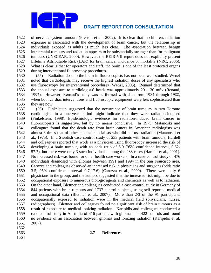
DRAFT REPORT FOR CONSULTATION
38
of nervous system tumours (Preston et al., 2002). It is clear that in children, radiation 1522
exposure is associated with the development of brain cancer, but the relationship in 1523
individuals exposed as adults is much less clear. The association between benign 1524
intracranial tumours and radiation appears to be substantially stronger than for malignant 1525
tumours (UNSCEAR, 2000). However, the BEIR-VII report does not explicitly present 1526
Lifetime Attributable Risk (LAR) for brain cancer incidence or mortality (NRC, 2006). 1527
What is clear is that for operators and staff, the brain is one of the least protected organs 1528
during interventional fluoroscopy procedures. 1529
(55) Radiation dose to the brain in fluoroscopists has not been well studied. Wenzl 1530
noted that cardiologists may receive the highest radiation doses of any specialists who 1531
use fluoroscopy for interventional procedures (Wenzl, 2005). Renaud determined that 1532
the annual exposure to cardiologists‘ heads was approximately 20 – 30 mSv (Renaud, 1533
1992). However, Renaud‘s study was performed with data from 1984 through 1988, 1534
when both cardiac interventions and fluoroscopic equipment were less sophisticated than 1535
they are now. 1536
(56) Finkelstein suggested that the occurrence of brain tumours in two Toronto 1537
cardiologists in a one-year period might indicate that they were radiation-induced 1538
(Finkelstein, 1998). Epidemiologic evidence for radiation-induced brain cancer in 1539
fluoroscopists is suggestive, but by no means conclusive. In 1975, Matanoski and 1540
colleagues found that the death rate from brain cancer in American radiologists was 1541
almost 3 times that of other medical specialists who did not use radiation (Matanoski et 1542
al., 1975). In a Swedish case-control study of 233 patients with brain tumours, Hardell 1543
and colleagues reported that work as a physician using fluoroscopy increased the risk of 1544
developing a brain tumour, with an odds ratio of 6.0 (95% confidence interval, 0.62-1545
57.7), but there were only 3 such individuals among the 233 cases (Hardell et al., 2001). 1546
No increased risk was found for other health care workers. In a case-control study of 476 1547
individuals diagnosed with gliomas between 1991 and 1994 in the San Francisco area, 1548
Carozza and colleagues observed an increased risk in physicians and surgeons (odds ratio 1549
3.5, 95% confidence interval 0.7-17.6) (Carozza et al., 2000). There were only 6 1550
physicians in the group, and the authors suggested that the increased risk might be due to 1551
occupational exposure to numerous biologic agents and chemicals as well as to radiation. 1552
On the other hand, Blettner and colleagues conducted a case-control study in Germany of 1553
844 patients with brain tumours and 1737 control subjects, using self-reported medical 1554
and occupational data (Blettner et al., 2007). More than 2/3 of the 91 participants 1555
occupationally exposed to radiation were in the medical field (physicians, nurses, 1556
radiographers). Blettner and colleagues found no significant risk of brain tumours as a 1557
result of exposure to medical ionizing radiation. Karipidis and colleagues conducted a 1558
case-control study in Australia of 416 patients with gliomas and 422 controls and found 1559
no evidence of an association between gliomas and ionizing radiation (Karipidis et al. 1560
2007). 1561
1562
2.7 References 1563 1564

DRAFT REPORT FOR CONSULTATION
39
Acosta R, Hoffmeister L, Roman R, Comas M, Castilla M, Castells X. Systematic 1565
review of population-based studies of the prevalence of cataracts. Arch Soc Esp 1566
Oftalmol 2006; 81: 509-516 1567
Allan JM. Genetic susceptibility to radiogenic cancer in humans. Health Phys 2008; 1568
95:677-686. 1569
Bacher K, Bogaert E, Lapere R, De Wolf D, Thierens H. Patient-specific dose and 1570
radiation risk estimation in pediatric cardiac catheterization. Circulation. 2005 Jan 1571
4;111(1):83-9. 1572
Balter S, Hopewell JW, Miller DL, Wagner LK, Zelefsky MJ. Fluoroscopically guided 1573
interventional procedures: A review of radiation effects on patients' skin and hair. 1574
Radiology 2010; 254:326-41. 1575
Bhatti P, Sigurdson AJ, Mabuchi K. Cam low-dose radiation increase risk of 1576
cardiovascular disease? Lancet 2008; 372:697-699. 1577
Blettner M., Schlehofer B., Samkange-Zeeb F., Berg G., Schlaefer K., Schüz J. (2007) 1578
Medical exposure to ionising radiation and the risk of brain tumours: Interphone 1579
study group, Germany. Eur J Cancer 43:1990-8. 1580
Carozza S.E., Wrensch M., Miike R., Newman B., Olshan A.F., Savitz D.A., Yost M., 1581
Lee M. (2000) Occupation and adult gliomas. Am J Epidemiol 152:838-46. 1582
Finkelstein M.M. (1998) Is brain cancer an occupational disease of cardiologists? Can J 1583
Cardiol 14:1385-8. 1584
Ciraj-Bjelac O, Rehani MM, Sim KH, Liew HB, Vano E, Kleiman NJ. Risk for radiation 1585
induced cataract for staff in interventional cardiology: Is there reason for concern? 1586
Catheter Cardiovasc Interv 2010;76(6):826-834. 1587
Codman EA. The cause of burns from x-rays. (letter) Bos Med Surg J 1896; 136:610-1588
611 1589
Daniel J. The x-rays. (letter) Science 1896; 3:562 1590
Dauer LT, Brooks AL, Hoel DG, Morgan WF, Stram D, Tran P. Review and evaluation 1591
of updated research on the health effects associated with low-dose ionising radiation. 1592
Radiat Prot Dosim 2010; 140(2):103-136 1593
Frazier TH, Richardson JB, Fabre VC, Callen JP. Fluoroscopy-induced chronic 1594
radiation skin injury: a disease perhaps often overlooked. Arch Dermatol 2007; 1595
143(5): 637-640 1596
Hardell L., Hansson Mild K., Påhlson A., Hallquist A. (2001) Ionizing radiation, cellular 1597
telephones and the risk for brain tumours. Eur J Cancer Prev 10:523-9. 1598
Herold DM, Hanlon AL, Hanks GE. Diabetes mellitus: a predictor for late radiation 1599
morbidity. Int J Radiat Oncol Biol Phys 1999; 43: 475-479 1600
Hidajat N, Wust P, Felix R, Schroder RJ. Radiation exposure to patient and staff in 1601
hepatic chemoembolization: risk estimation of cancer and deterministic effects. 1602
Cardiovasc Intervent Radiol 2006; 29(5): 791-796 1603
Hymes SR, Strom EA, Fife C. Radiation dermatitis: clinical presentation, 1604
pathophysiology, and treatment 2006. J Am Acad Dermatol 2006;54:28-46 1605
IEC. Report 60601 Medical electrical equipment - Part 2-43: Particular requirements for 1606
the safety of X-ray equipment for interventional procedures. 2nd ed. Geneva, 1607
Switzerland: International Electrotechnical Commission; 2010. 1608
DRAFT REPORT FOR CONSULTATION
40
ICRP, 1990. The 1990 Recommendations of the International Commission on 1609
Radiological Protection. ICRP Publication 60. Ann ICRP 1991; 21(1-3):1-201 1610
ICRP, 2000. Avoidance of radiation injuries from medical interventional procedures. 1611
ICRP Publication 85. Ann ICRP 30:7-67. 1612
ICRP, 2007a. The 2007 Recommendations of the International Commission on 1613
Radiological Protection. ICRP Publication 103. Ann ICRP 37(2-4):1-332 1614
ICRP, 2007b.. Radiological protection in medicine. ICRP Publication 105. Ann ICRP 1615
37:1-63 1616
ICRP Protection 2011a. Tissue Reactions and Other Non-Cancer Effects of Radiation. 1617
ICRP Publication XXX. Ann ICRP 2011; in press 1618
ICRP 2011b. Statement on Tissue reactions. Approved by the Commission on April 1619
21, 2011. Available at 1620
http://www.icrp.org/docs/ICRP%20Statement%20on%20Tissue%20Reactions.pdf 1621
Accessed May 9, 2011. (in press) 1622
Kahn, HA, Liebowitz HM, Ganley JP, Kini MM, Colton T, Nickerson RS, Dawber TR. 1623
The Framingham Eye Study. I. Outline and major prevalence findings. Am J 1624
Epidemiol 1977; 106(1): 17-32 1625
Karipidis KK, Benke G, Sim MR, Kauppinen T, Giles G. (2007) Occupational exposure 1626
to ionizing and non-ionizing radiation and the risk of glioma. Occup Med (Lond) 1627
57:518-524. 1628
Kim KP, Miller DL, Balter S, Kleinerman RA, Linet MS, Kwon D, Simon SL. 1629
Occupational radiation doses to operators performing cardiac catheterization 1630
procedures. Health Phys 2008;94:211-227. 1631
Kleiman NJ. Radiation cataract. In: Working Party on Research Implications on Health 1632
and Safety Standards of the Article 31 Group of Experts, ed. Radiation Protection 1633
145 EU Scientific Seminar 2006 New insights in radiation risk and basic safety 1634
standards. Brussels: European Commission; 2007:81-95. Available at 1635
http://ec.europa.eu/energy/nuclear/radioprotection/publication/doc/145_en.pdf 1636
Accessed July 28, 2010 1637
Koenig TR, Wolff D, Mettler FA, Wagner LK. Skin injuries from fluoroscopically 1638
guided procedures: Part 1, Characteristics of radiation injury. AJR 2001; 177: 3-11 1639
Martinez LC, Vano E, Gutierrez F, Rodriguez C, Gilarranz R, Manzanas MJ. Patient 1640
doses from fluoroscopically guided cardiac procedures in pediatrics. Phys Med Biol 1641
2007; 52: 4749-4759. 1642
Matanoski G.M., Seltser R., Sartwell P.E., Diamond E.L., Elliott E.A. (1975) The 1643
current mortality rates of radiologists and other physician specialists: specific causes 1644
of death. Am J Epidemiol 101:199-210. 1645
McGale P, Darby SC. Commentary: A dose-response relationship for radiation-induced 1646
heart disease—current issues and future prospects. Int J Epidemiol 2008; 37:518-1647
523 1648
McGeoghegan D, Binks K, Gillies M, Jones S, Whaley S. The non-cancer mortality 1649
experience of male workers at British Nuclear Fuels plc, 1946-2005. Int J Epidemiol 1650
2008; 37:506-518 1651

DRAFT REPORT FOR CONSULTATION
41
Monaco JL, Bowen K, Tadros PN, Witt PD. Iatrogenic deep musculocutaneous radiation 1652
injury following percutaneous coronary intervention. J Invasive Cardiol 1653
2003;15:451-3. 1654
National Cancer Institute, 2005. Interventional fluoroscopy: reducing radiation risks for 1655
patients and staff. NIH Publication No. 05-5286. National Cancer Institute, 1656
Bethesda, MD. Available at: http://www.cancer.gov/images/Documents/45bae965-1657
697a-4de8-9dae-b77222e0e79d/InterventionalFluor.pdf Accessed December 31, 1658
2010. 1659
National Council on Radiation Protection and Measurements. Limitation of exposure to 1660
ionizing radiation. NCRP Report 116. Bethesda, MD: National Council on Radiation 1661
Protection and Measurements; 1993. 1662
National Council on Radiation Protection and Measurements. Radiation dose 1663
management for fluoroscopically guided interventional medical procedures. NCRP 1664
Report No. 168. Bethesda, MD: National Council on Radiation Protection and 1665
Measurements; 2010. 1666
NRC. (2006) Health risks from exposure to low levels of ionizing radiation: BEIR-VII 1667
Phase 2 National Academy Press, Washington, DC. 1668
Pages J. Effective dose and dose to the crystalline lens during angiographic procedures. 1669
JBR-BTR 2000; 83:108-110 1670
Preston D.L., Ron E., Tokuoka S., Funamoto S., Nishi N., Soda M., Mabuchi K., 1671
Kodama K. (2007) Solid cancer incidence in atomic bomb survivors: 1958-1998. 1672
Radiat Res 168:1-64. 1673
Preston D.L., Ron E., Yonehara S., Kobuke T., Fujii H., Kishikawa M., Tokunaga M., 1674
Tokuoka S., Mabuchi K. (2002) Tumors of the nervous system and pituitary gland 1675
associated with atomic bomb radiation exposure. J Natl Cancer Inst 94:1555-63. 1676
Renaud L. (1992) A 5-y follow-up of the radiation exposure to in-room personnel during 1677
cardiac catheterization. Health Phys 62:10-15. 1678
Shack RB, Lynch JB. Radiation dermatitis. Clin Plast Surg 1987; 14: 391-401 1679
Shimizu Y, Kodama K, Nishi N, Kasagi F, Suyama A, Soda M, Grant EJ, Sugiyama H, 1680
Sakata R, Moriwaki H, Hayashi M, Konda M, Shore RE. Radiation exposure and 1681
circulatory disease risk: Hiroshima and Nagaski atomic bomb survivor data, 1950-1682
2003. BMJ 2010; 340:b5349 1683
Shope, T.B., 1996. Radiation-induced skin injuries from fluoroscopy. Radiographics 16, 1684
1195-1199. 1685
Shore RE, Moseson M, Xue X, Tse Y, Harley N, Pasternack BS. Skin cancer after x-ray 1686
treatment for scalp ringworm. Radiat Res 2002: 157 (4): 410-418 1687
Shore, R.E., Neriishi, K., Nakashima, E., 2010. Epidemiological studies of cataract risk 1688
at low to moderate radiation doses: (not) seeing is believing. Radiat. Res. 174, 889-1689
894. 1690
Turreson I, Notter G. The predictive value of skin telangiectasia for late radiation effect 1691
in different normal tissues. Int J Radiat Oncol Biol Phys 1986; 12 603-609 1692
UNSCEAR. (2000) Sources and effects of ionizing radiation. United Nations, New 1693
York. 1694
DRAFT REPORT FOR CONSULTATION
42
United States Food and Drug Administration . Public Health Advisory. Avoidance of 1695
serious x-ray induced skin injuries to patients during fluoroscopically guided 1696
procedures. Rockville, MD: Center for Devices and Radiological Health, United 1697
States Food and Drug Administration, 1994 Available at 1698
http://www.fda.gov/MedicalDevices/Safety/AlertsandNotices/PublicHealthNotificati1699
ons/ucm063084.htm Accessed July 29, 2010. 1700
Vano E, Arranz L, Sastre JM, Moro C, Ledo A, Garate MT, Minguez I. Dosimetric and 1701
radiation protection considerations based on some cases of patient skin injuries in 1702
interventional cardiology. Br J Radiol 1998; 71: 510-516 1703
Vano E, Gonzalez L, Beneytez F, Moreno F. Lens injuries induced by occupational 1704
exposure to non-optimized interventional radiology laboratories. Br J Radiol 1998; 1705
71: 728-733 1706
Vano E, Gonzalez L, Fernandez M, Haskal ZJ. Eye lens exposure to radiation in 1707
interventional suites: caution is warranted. Radiology 2008; 248: 945-953 1708
Vano E, Kleiman NJ, Duran A, Rehani MM, Echeverri D, Cabrera. M. Radiation 1709
cataract risk in interventional cardiology personnel. Radiat Res, 2010; 174(4):490-1710
495 1711
Vlietstra RE, Wagner LK, Koenig T, Mettler F. Radiation burns as a severe complication 1712
of fluoroscopically guided cardiological interventions. J Int Cardiol 2004; 17(3): 1-1713
12 1714
Wagner LK, McNesse MD, Marx MV, Siegel EL. Severe skin reactions from 1715
interventional fluoroscopy: case report and review of literature. Radiology 1999; 1716
213: 773-776 1717
Wang JX, Zhang LA, Li BX, Zhao YC, Wang ZQ, Zhang JY, Aoyama T. Cancer 1718
incidence and risk estimation among medical x-ray workers in China, 1950-1955. 1719
Health Phys 2002; 82(4): 455-466 1720
Wenzl T.B. (2005) Increased brain cancer risk in physicians with high radiation 1721
exposure. Radiology 235:709-10; author reply 710-1. 1722
Worgul BV, Kundiyev YI, Sergiyenko NM, Chumak VV, Vitte PM, Medvedovsky C, 1723
Bakhanova EV, Junk AK, Kryrychenko OY, Musijachenko NV, Shylo SA, Vitte OP, 1724
Xu S, Xue X, Shore RE. Cataracts among Chernobyl clean-up workers: implications 1725
regarding permissible eye exposures. Radiat Res 2007; 167(2): 233-243 1726
Yonehara S., Brenner A.V., Kishikawa M., Inskip P.D., Preston D.L., Ron E., Mabuchi 1727
K., Tokuoka S. (2004) Clinical and epidemiologic characteristics of first primary 1728
tumors of the central nervous system and related organs among atomic bomb 1729
survivors in Hiroshima and Nagasaki, 1958-1995. Cancer 101:1644-54. 1730
1731
1732
1733

DRAFT REPORT FOR CONSULTATION
43
3. CLINICAL EXAMPLES OF DETERMINISTIC INJURY AFTER 1734
FLUOROSCOPICALLY GUIDED CARDIAC PROCEDURES 1735
1736
Main Points 1737
1738
There is increasing concern about skin radiation dose levels in cardiology. 1739
The cases presented in this chapter provide a clinical context and illustrate 1740
skin changes due to radiation injury. 1741
Deterministic injuries may extend into deeper tissues and can cause symptoms 1742
that persist for years. 1743
Deterministic injuries may be accompanied by an increase in stochastic risk. 1744
1745
3.1 Introduction 1746
1747 (57) There is increasing concern about skin radiation dose levels in cardiology. 1748
This is because of the discovery of deterministic injuries in patients who have undergone 1749
long procedures using suboptimal equipment, performed by individuals inadequately 1750
trained in radiological protection (UNSCEAR, 2010). However, high skin doses can 1751
occur in obese patients, or patients undergoing complex interventions, even when the 1752
procedure is performed by an experienced, well-trained operator using modern, well-1753
maintained equipment (Suzuki, 2008; Bryk, 2006). 1754
(58) The information presented in Chapter 2 (section 2.3) on the radiobiology of the 1755
skin can be difficult to interpret without a clinical context. The cases presented in this 1756
chapter provide that clinical context and illustrate the skin changes discussed in Chapter 1757
2. It should be apparent that these injuries can be severe and debilitating. Some patients 1758
will require life-long therapy and observation. Treatment often requires a 1759
multidisciplinary team working in a specialized centre. Pain management and 1760
psychological support are important components of treatment. 1761
(59) Methods to optimize patient radiation dose and minimize skin dose are 1762
described in Chapter 5 and listed in Table 5.1, but are repeated here because of their 1763
importance. Limit fluoroscopy time and the number of cine frames to the least number 1764
possible for successful completion of the procedure. Monitor patient radiation dose 1765
during the procedure. Use fluoroscopy equipment with pulsed fluoroscopy and use the 1766
lowest pulse rate that provides adequate fluoroscopic guidance. Use the lowest 1767
fluoroscopic and cine dose rates necessary for each stage of the procedure. When 1768
possible, slightly rotate the gantry so that the entrance beam is periodically directed at a 1769
different entrance skin site. Keep the image receptor (image intensifier or flat panel 1770
detector) as close as possible to the patient, and keep the x-ray tube as far away as 1771
possible from the entrance skin site. 1772
1773
1774
1775
1776
1777
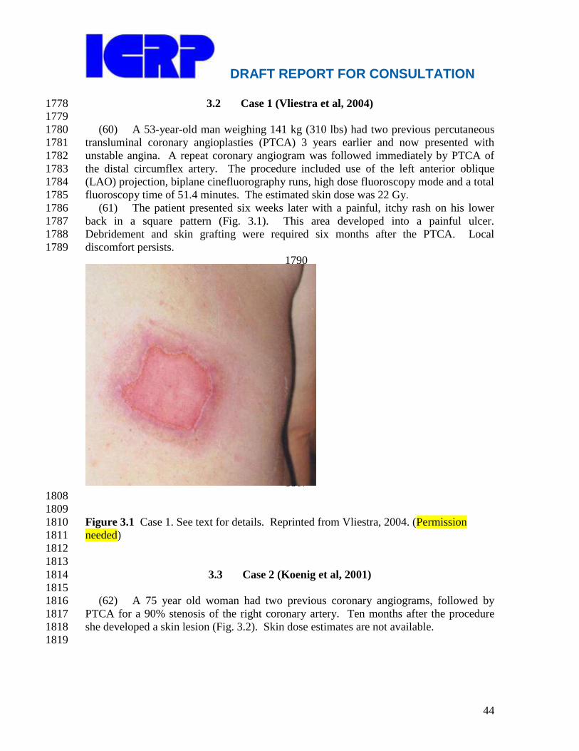
DRAFT REPORT FOR CONSULTATION
44
3.2 Case 1 (Vliestra et al, 2004) 1778 1779
(60) A 53-year-old man weighing 141 kg (310 lbs) had two previous percutaneous 1780
transluminal coronary angioplasties (PTCA) 3 years earlier and now presented with 1781
unstable angina. A repeat coronary angiogram was followed immediately by PTCA of 1782
the distal circumflex artery. The procedure included use of the left anterior oblique 1783
(LAO) projection, biplane cinefluorography runs, high dose fluoroscopy mode and a total 1784
fluoroscopy time of 51.4 minutes. The estimated skin dose was 22 Gy. 1785
(61) The patient presented six weeks later with a painful, itchy rash on his lower 1786
back in a square pattern (Fig. 3.1). This area developed into a painful ulcer. 1787
Debridement and skin grafting were required six months after the PTCA. Local 1788
discomfort persists. 1789
1790
1791
1792
1793
1794
1795
1796
1797
1798
1799
1800
1801
1802
1803
1804
1805
1806
1807
1808
1809
Figure 3.1 Case 1. See text for details. Reprinted from Vliestra, 2004. (Permission 1810
needed) 1811
1812
1813
3.3 Case 2 (Koenig et al, 2001) 1814
1815 (62) A 75 year old woman had two previous coronary angiograms, followed by 1816
PTCA for a 90% stenosis of the right coronary artery. Ten months after the procedure 1817
she developed a skin lesion (Fig. 3.2). Skin dose estimates are not available. 1818
1819
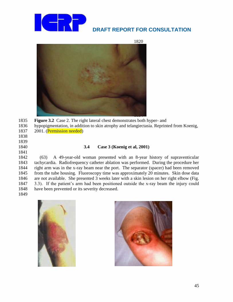
DRAFT REPORT FOR CONSULTATION
45
1820
1821
1822
1823
1824
1825
1826
1827
1828
1829
1830
1831
1832
1833
1834 Figure 3.2 Case 2. The right lateral chest demonstrates both hyper- and 1835
hypopigmentation, in addition to skin atrophy and telangiectasia. Reprinted from Koenig, 1836
2001. (Permission needed) 1837
1838
1839
3.4 Case 3 (Koenig et al, 2001) 1840
1841 (63) A 49-year-old woman presented with an 8-year history of supraventicular 1842
tachycardia. Radiofrequency catheter ablation was performed. During the procedure her 1843
right arm was in the x-ray beam near the port. The separator (spacer) had been removed 1844
from the tube housing. Fluoroscopy time was approximately 20 minutes. Skin dose data 1845
are not available. She presented 3 weeks later with a skin lesion on her right elbow (Fig. 1846
3.3). If the patient‘s arm had been positioned outside the x-ray beam the injury could 1847
have been prevented or its severity decreased. 1848
1849

DRAFT REPORT FOR CONSULTATION
46
1864
1865
1866
1867
1868
1869
1870
1871
1872
1873
1874
1875
1876
1877
Figure 3.3 Case 3. See text for details. A) 3 weeks: Area of sharply demarcated 1878
erythema. B) 5 months: Tissue necrosis. C) 6½ months: Deep ulceration with exposure of 1879
the bone. D) Following surgical flap. Reprinted from Koenig, 2001. (Permission 1880
needed) 1881
1882
3.5 Case 4 (Vliestra et al, 2004) 1883
1884 (64) A 38-year-old man weighing 114 kg (250 lbs) was diagnosed with Wolff-1885
Parkinson-White syndrome. An attempt at radiofrequency ablation using biplane 1886
fluoroscopy was unsuccessful. A few weeks after the procedure, the patient developed 1887
areas of brownish-red discolouration on his back, which resolved. A second unsuccessful 1888
ablation procedure was performed 2½ months later, with reappearance of the skin 1889
discolouration after 1 week. The physician thought the skin lesion was due to the 1890
grounding pad used for radiofrequency ablation rather than to radiation. A third 1891
unsuccessful ablation procedure was performed; skin lesions appeared 8 days later (Fig 1892
3.4). Each of the three procedures used more than 100 min of fluoroscopy time. Skin 1893
dose estimates are not available. The severe injury to the right arm was due to its 1894
position. If the arm had been positioned away from the entrance x-ray beam, the injury 1895
might have been avoided. 1896
1897
1898
1899
1900
1901
1902
1903
1904
1905
1906
1907

DRAFT REPORT FOR CONSULTATION
47
1908
1909 Figure 3.4 Case 4. The right-sided lesions show desquamation. The erythema on the 1910
back healed into discoloured scars. The right arm lesion, closer to the x-ray beam, 1911
developed necrosis and required a skin graft. Reprinted from Vliestra, 2004. (Permission 1912
needed) 1913
1914
3.6 Case 5 (Vañó et al, 1998) 1915 1916
(65) A 17-year-old female underwent an electrophysiology ablation procedure for 1917
posterior pathway pre-excitation that lasted 5 hours. Eleven months later she underwent 1918
a second procedure that also lasted 5 hours. Both procedures were performed with 1919
biplane fluoroscopy. Fluoroscopy time for the lateral plane was estimated at 90-120 1920
minutes. Skin dose estimates are not available. Twelve hours after the second procedure 1921
she developed an erythematous plaque in the right axilla. One month later she consulted 1922
a dermatologist for red macular and blister lesions on her right side. Twenty-six months 1923
after the second procedure an indurated, atrophic plaque with linear edges, 10 x 5 cm2, 1924
was observed (Fig. 3.5). The diagnosis was chronic radiodermatitis. The muscles in her 1925
right arm have also been affected, with resultant limitation in the range of motion. 1926
Because of the patient‘s age and the region irradiated, her risk of subsequent breast 1927
cancer is also increased. 1928
1929
1930
1931 1932
Figure 3.5 Case 5. Indurated, atrophic plaque with linear edges, with areas of hyper- and 1933
hypopigmentation. Reprinted from Vañó, 1998. (Permission needed) 1934
1935
1936

DRAFT REPORT FOR CONSULTATION
48
3.7 Case 6 (Courtesy of Dr. M. Portas, Buenos Aires, Argentina) 1937
1938 (66) An obese 57-year-old female, a heavy smoker, underwent PTCA. The 1939
procedure time was approximately 6 hours. No data on radiation dose are available. 1940
Early manifestations were blisters on the skin of the back in the lumbar region. This was 1941
diagnosed by a dermatologist as a herpes zoster infection. Two months later, a deep ulcer 1942
(Radiation Therapy Oncology Group/European Organisation for Research and Treatment 1943
of Cancer [RTOG/EORTC] cutaneous radiotoxicity grade 4) appeared at the same site. 1944
(No photographs of the injury at this stage are available.) It was extremely painful. The 1945
following year the patient underwent a plastic surgery procedure, with two rotation flaps 1946
to close the wound. The rotation flaps subsequently underwent necrosis, leaving an ulcer 1947
approximately 20 x 20 cm (Fig. 3.6). During the next several years, conservative 1948
treatment was performed at a specialized burn centre. Wound coverage was performed 1949
with porcine dermis, skin allografts and autografts, in conjunction with anti-inflammatory 1950
and antibacterial therapy and hyperbaric oxygen treatments. This treatment led to 1951
progressive wound closure. After 3 years of treatment (5 years after the PTCA), the 1952
dimensions of the ulcer were reduced to 3 x 1.5 cm (Fig 3.7). In vitro radiosensitivity 1953
testing demonstrated that the patient had normal radiosensitivity. The injury and 1954
prolonged recovery were attributed to radiation exposure, obesity and heavy smoking. 1955
1956
1957
1958 1959
1960
Figure 3.6 Case 6. Appearance of the patient‘s back following the initial surgery and 1961
necrosis of the rotation flaps. The ulcer is approximately 20 x 20 cm. 1962
1963

DRAFT REPORT FOR CONSULTATION
49
1964
1965 1966
Figure 3.7 Case 6. Appearance of the patient‘s back 5 years after the PTCA. After 3 1967
years of treatment, the ulcer is reduced in size to 3 x 1.5 cm. The patient‘s quality of life 1968
is much improved. 1969
1970
1971
3.8 References 1972 1973
Bryk, S.G., Censullo, M.L., Wagner, L.K., Rossman, L.L., Cohen, A.M., 2006. 1974
Endovascular and interventional procedures in obese patients: a review of procedural 1975
technique modifications and radiation management. J. Vasc. Interv. Radiol. 17, 27-1976
33. 1977
Koenig, T.R., Wolff, D., Mettler, F.A., Wagner, L.K., 2001. Skin injuries from 1978
fluoroscopically guided procedures: Part 1, Characteristics of radiation injury. AJR. 1979
Am. J. Roentgenol. 177, 3-11. 1980
Suzuki, S., Furui, S., Isshiki, T., Kozuma, K., Koyama, Y., Yamamoto, H., Ochiai, M., 1981
Asakura, Y., Ikari, Y., 2008. Patients' skin dose during percutaneous coronary 1982
intervention for chronic total occlusion. Catheter. Cardiovasc. Interv. 71, 160-164. 1983
UNSCEAR. Sources of ionizing radiation: volume I. Report to the General Assembly, 1984
with scientific annexes; 2010; New York, NY. Available at: 1985
http://www.unscear.org/unscear/en/publications/2008_1.html Accessed December 1986
31, 2010. 1987

DRAFT REPORT FOR CONSULTATION
50
Vañó, E., Arranz, L., Sastre, J.M., Moro, C., Ledo, A., Gárate, M.T., Minguez, I., 1998. 1988
Dosimetric and radiation protection considerations based on some cases of patient 1989
skin injuries in interventional cardiology. Br. J. Radiol. 71, 510-516. 1990
Vlietstra, R.E., Wagner, L.K., Koenig, T., Mettler, F., 2004. Radiation burns as a severe 1991
complication of fluoroscopically guided cardiological interventions. J Interv Cardiol 1992
17, 1-12. 1993
1994
1995
1996
DRAFT REPORT FOR CONSULTATION
51
1997
4. PRINCIPLES OF RADIOLOGICAL PROTECTION FOR 1998
PATIENTS AND STAFF 1999
2000
Main Points 2001
2002
Justification means that a medical procedure should only be performed when it 2003
is appropriate for a particular patient— the anticipated clinical benefits should 2004
exceed all anticipated procedural risks, including radiation risk. 2005
For CT and nuclear medicine studies, justification is a responsibility shared 2006
between the referring clinician and the cardiac imager. For fluoroscopically 2007
guided interventions, the responsibility rests with the interventionalist. 2008
Optimization means that the radiation dose to the patient is suitable for the 2009
medical purpose, and radiation that is clinically unnecessary or unproductive is 2010
avoided. 2011
Patient radiation dose is optimized when imaging is performed with the least 2012
amount of radiation required to provide adequate image quality, diagnostic 2013
information, and for fluoroscopy, adequate imaging guidance. 2014
Dose limits apply to occupational exposure of cardiologists and staff. 2015
Dose limits do not apply to medical exposures of patients or to carers and 2016
comforters. 2017 2018
4.1 Introduction 2019
(67) The Commission recommends three fundamental principles of radiological 2020
protection: justification, optimization of protection, and application of dose limits (ICRP 2021
103, ICRP 105). The first two are source related and apply to all radiation exposure 2022
situations. The third applies to staff, but does not apply to medical exposures of patients 2023
or to carers and comforters. 2024
4.2 Justification 2025
(68) The principle of justification is that, in general, ―any decision that alters the 2026
radiation exposure situation should do more good than harm. This means that by 2027
introducing a new radiation source, by reducing existing exposure, or by reducing the risk 2028
of potential exposure, one should achieve sufficient individual or societal benefit to offset 2029
the detriment it causes.‖ (ICRP 103, ICRP 105). The principal aim of medical exposures 2030
is to do more good than harm to the patient, subsidiary account being taken of the 2031
radiation detriment from the exposure of the radio- logical staff and of other individuals 2032
(ICRP 103). 2033
(69) A medical procedure should only be performed when it is appropriate for a 2034
particular patient. The RAND Corporation has developed a definition of ―appropriate‖ 2035
that is widely used: the expected health benefit (i.e., increased life expectancy, relief of 2036
pain, reduction in anxiety, improved functional capacity) exceeds the expected negative 2037
consequences (i.e., mortality, morbidity, anxiety of anticipating the procedure, pain 2038
produced by the procedure, misleading or false diagnoses, time lost from work) by a 2039

DRAFT REPORT FOR CONSULTATION
52
sufficiently wide margin that the procedure is worth doing (Sistrom, 2008, NHS, 1993). 2040
In other words, the anticipated clinical benefits should exceed all anticipated procedural 2041
risks, including radiation risk. 2042
(70) In the United States, appropriateness criteria have been developed for many 2043
clinical scenarios (Brindis et al, 2005, Douglas, 2008, Patel, 2009, Hendel 2009, ACR, 2044
2010, Taylor 2010). Similar guidelines have been developed in the United Kingdom, 2045
though they are less readily available (RCR, 2007). European guidelines are also 2046
available (Hesse, 2005, Schroeder 2008). These recommendations are typically based on 2047
a standardized literature review and compilation of evidence tables, followed by rating of 2048
each indication by an expert panel with varied composition (Patel et al, 2005). 2049
Appropriateness may vary based on national and local norms and practice patterns, as 2050
well as well as patient and family values and preferences (Wolk et al, 2004). 2051
(71) The responsibility for the justification of the use of a particular procedure falls 2052
on the relevant medical practitioners (ICRP 103). For CT and nuclear medicine studies, 2053
justification is a responsibility shared between the referring clinician and the cardiac 2054
imager. For the referring clinician, this entails weighing the benefits of a test against its 2055
risks, including radiation exposure, and considering such an analysis for all possible 2056
alternatives including performing no test. For the cardiac imager, justification entails 2057
ensuring that the test has a reasonable indication, given the available information, and 2058
discussing the indication with the referring clinician if there is concern in this respect. 2059
For fluoroscopically guided interventions, the responsibility rests with the 2060
interventionalist. 2061
2062
4.3 Optimization 2063
(72) The principle of optimization of protection is that ―the likelihood of incurring 2064
exposures, the number of people exposed, and the magnitude of their individual doses 2065
should all be kept as low as reasonably achievable, taking into account economic and 2066
societal factors. This means that the level of protection should be the best under the 2067
prevailing circumstances, maximizing the margin of benefit over harm‖ (ICRP 103, ICRP 2068
105, NCRP 1993). This is often summarized using the acronym ALARA, which stands 2069
for As Low As Reasonably Achievable. 2070
(73) For cardiology procedures, this principle is applied in the design of cardiac 2071
facilities that use ionizing radiation, appropriate selection and use of equipment, and in 2072
day-to-day working procedures. Optimization is best described as a radiation dose to the 2073
patient that is suitable for the medical purpose, and avoidance of radiation that is 2074
clinically unnecessary or unproductive. 2075
(74) Dose optimization means delivering a radiation dose to the organs and tissues 2076
of clinical interest no greater than that required for adequate imaging and minimizing 2077
dose to other structures (e.g., the skin). Patient radiation dose is considered to be 2078
optimized when imaging is performed with the least amount of radiation required to 2079
provide adequate image quality and, for fluoroscopy, adequate imaging guidance (NCI, 2080
2005). The goal of every imaging procedure is to provide images adequate for the 2081
clinical purpose. Imaging requirements depend on the specific patient and the specific 2082
procedure. Reducing patient radiation dose to the point where images are inadequate is 2083

DRAFT REPORT FOR CONSULTATION
53
counterproductive; it results in radiation dose to the patient without answering the clinical 2084
question. Improving image quality beyond what is clinically needed subjects the patient 2085
to additional radiation dose without additional clinical benefit. The goal of radiation 2086
management is to keep patient radiation dose as low as possible consistent with the use of 2087
appropriate equipment and the imaging requirements for a specific patient and a specific 2088
procedure. 2089
4.4 Dose limits 2090
(75) The principle of application of dose limits states that ―the total dose to any 2091
individual from regulated sources in planned exposure situations other than medical 2092
exposure of patients should not exceed the appropriate limits recommended by the 2093
Commission‖ (ICRP 103, ICRP 105). This principle does not apply to medical exposure 2094
of patients. As noted in ICRP Publication 105, ―Provided that the medical exposures of 2095
patients have been properly justified and that the associated doses are commensurate with 2096
the medical purpose, it is not appropriate to apply dose limits or dose constraints to the 2097
medical exposure of patients, because such limits or constraints would often do more 2098
harm than good.‖(ICRP 105) For interventional procedures, the medical condition being 2099
treated and the non-radiation risks of the procedure typically present substantially greater 2100
morbidity and mortality than do the radiation risks (Miller, 2008, NCRP 168, 2010). 2101
2102
4.5 References 2103 2104
What do we mean by appropriate health care? Report of a working group prepared for 2105
the Director of Research and Development of the NHS Management Executive. Qual 2106
Health Care 1993;2:117-23. 2107
American College of Radiology. ACR Appropriateness Criteria®. Available at: 2108
http://www.acr.org/SecondaryMainMenuCategories/quality_safety/app_criteria.aspx 2109
Accessed August 3, 2010. 2110
Brindis RG, Douglas PS, Hendel RC, Peterson ED, Wolk MJ, Allen JM, Patel MR, 2111
Raskin IE, Bateman TM, Cerqueira MD, Gibbons RJ, Gillam LD, Gillespie JA, 2112
Iskandrian AE, Jerome SD, Krumholz HM, Messer JV, Spertus JA, Stowers SA. 2113
ACCF/ASNC appropriateness criteria for single-photon emission computed 2114
tomography myocardial perfusion imaging (SPECT MPI): a report of the American 2115
College of Cardiology Foundation Quality Strategic Directions Committee 2116
Appropriateness Criteria Working Group and the American Society of Nuclear 2117
Cardiology endorsed by the American Heart Association. J Am Coll Cardiol 2118
2005;46:1587-1605. 2119
Douglas, P.S., Khandheria, B., Stainback, R.F., Weissman, N.J., Peterson, E.D., Hendel, 2120
R.C., Blaivas, M., Des Prez, R.D., Gillam, L.D., Golash, T., Hiratzka, L.F., 2121
Kussmaul, W.G., Labovitz, A.J., Lindenfeld, J., Masoudi, F.A., Mayo, P.H., 2122
Porembka, D., Spertus, J.A., Wann, L.S., Wiegers, S.E., Brindis, R.G., Patel, M.R., 2123
Wolk, M.J., Allen, J.M., 2008. ACCF/ASE/ACEP/AHA/ASNC/SCAI/SCCT/SCMR 2124
2008 appropriateness criteria for stress echocardiography: a report of the American 2125
College of Cardiology Foundation Appropriateness Criteria Task Force, American 2126
Society of Echocardiography, American College of Emergency Physicians, 2127

DRAFT REPORT FOR CONSULTATION
54
American Heart Association, American Society of Nuclear Cardiology, Society for 2128
Cardiovascular Angiography and Interventions, Society of Cardiovascular Computed 2129
Tomography, and Society for Cardiovascular Magnetic Resonance endorsed by the 2130
Heart Rhythm Society and the Society of Critical Care Medicine. J. Am. Coll. 2131
Cardiol. 51, 1127-1147. Available at: 2132
http://content.onlinejacc.org/cgi/content/full/51/11/1127 Accessed January 1, 2011. 2133
Hendel RC, Berman DS, Di Carli MF, et al. 2134
ACCF/ASNC/ACR/AHA/ASE/SCCT/SCMR/SNM 2009 appropriate use criteria for 2135
cardiac radionuclide imaging: a report of the American College of Cardiology 2136
Foundation Appropriate Use Criteria Task Force, the American Society of Nuclear 2137
Cardiology, the American College of Radiology, the American Heart Association, 2138
the American Society of Echocardiography, the Society of Cardiovascular Computed 2139
Tomography, the Society for Cardiovascular Magnetic Resonance, and the Society of 2140
Nuclear Medicine. Circulation. 2009;119(22):e561-587. 2141
Hesse, B., Tagil, K., Cuocolo, A., Anagnostopoulos, C., Bardies, M., Bax, J., Bengel, F., 2142
Busemann Sokole, E., Davies, G., Dondi, M., Edenbrandt, L., Franken, P., Kjaer, A., 2143
Knuuti, J., Lassmann, M., Ljungberg, M., Marcassa, C., Marie, P.Y., McKiddie, F., 2144
O'Connor, M., Prvulovich, E., Underwood, R., van Eck-Smit, B., 2005. EANM/ESC 2145
procedural guidelines for myocardial perfusion imaging in nuclear cardiology. Eur J 2146
Nucl Med Mol Imaging 32, 855-897. Available at: 2147
http://www.eanm.org/scientific_info/guidelines/gl_cardio_myocard_perf.pdf 2148
Accessed January 1, 2011. 2149
International Commission on Radiological Protection. The 2007 Recommendations of 2150
the International Commission on Radiological Protection. ICRP Publication 103. 2151
Ann ICRP 2007; 37(2-4):1-332 2152
International Commission on Radiological Protection. Radiological protection in 2153
medicine. ICRP Publication 105. Ann ICRP 2007;37:1-63. 2154
Miller DL. Overview of contemporary interventional fluoroscopy procedures. Health 2155
Phys 2008; 95:638-644. 2156
NCI. Interventional fluoroscopy: reducing radiation risks for patients and staff. NIH 2157
Publication No. 05-5286. Bethesda:National Cancer Institute, 2005. Available at: 2158
http://www.cancer.gov/images/Documents/45bae965-697a-4de8-9dae-2159
b77222e0e79d/InterventionalFluor.pdf Accessed January 1, 2011. 2160
NCRP. Limitation of exposure to ionizing radiation. NCRP Report No. 116. Bethesda, 2161
MD: National Council on Radiation Protection and Measurements; 1993. 2162
NCRP. Radiation dose management for fluoroscopically guided interventional medical 2163
procedures. NCRP Report No. 168. Bethesda, MD: National Council on Radiation 2164
Protection and Measurements; 2010. 2165
Patel MR, Spertus JA, Brindis RG, Hendel RC, Douglas PS, Peterson ED, Wolk MJ, 2166
Allen JM, Raskin IE. ACCF proposed method for evaluating the appropriateness of 2167
cardiovascular imaging. J Am Coll Cardiol 2005;46:1606-1613. 2168
Patel, M.R., Dehmer, G.J., Hirshfeld, J.W., Smith, P.K., Spertus, J.A., 2009. 2169
ACCF/SCAI/STS/AATS/AHA/ASNC 2009 Appropriateness Criteria for Coronary 2170
Revascularization: a report by the American College of Cardiology Foundation 2171
DRAFT REPORT FOR CONSULTATION
55
Appropriateness Criteria Task Force, Society for Cardiovascular Angiography and 2172
Interventions, Society of Thoracic Surgeons, American Association for Thoracic 2173
Surgery, American Heart Association, and the American Society of Nuclear 2174
Cardiology Endorsed by the American Society of Echocardiography, the Heart 2175
Failure Society of America, and the Society of Cardiovascular Computed 2176
Tomography. J. Am. Coll. Cardiol. 53, 530-553. Available at: 2177
http://content.onlinejacc.org/cgi/content/full/53/6/530 Accessed January 1, 2011. 2178
The Royal College of Radiologists. Making the best use of clinical radiology services: 2179
referral guidelines. 6th
ed. London:The Royal College of Radiologists, 2007. 2180
Schroeder S, Achenbach S, Bengel F, et al. Cardiac computed tomography: indications, 2181
applications, limitations, and training requirements: report of a Writing Group 2182
deployed by the Working Group Nuclear Cardiology and Cardiac CT of the 2183
European Society of Cardiology and the European Council of Nuclear Cardiology. 2184
Eur Heart J 2008;29(4):531-556. 2185
Sistrom CL. In support of the ACR Appropriateness Criteria®. J Am Coll Radiol 2186
2008;5:630-635. 2187
Taylor, A.J., Cerqueira, M., Hodgson, J.M., Mark, D., Min, J., O'Gara, P., Rubin, G.D., 2188
Kramer, C.M., Berman, D., Brown, A., Chaudhry, F.A., Cury, R.C., Desai, M.Y., 2189
Einstein, A.J., Gomes, A.S., Harrington, R., Hoffmann, U., Khare, R., Lesser, J., 2190
McGann, C., Rosenberg, A., Schwartz, R., Shelton, M., Smetana, G.W., Smith, S.C., 2191
Jr., 2010. ACCF/SCCT/ACR/AHA/ASE/ASNC/NASCI/SCAI/SCMR 2010 2192
appropriate use criteria for cardiac computed tomography: a report of the American 2193
College of Cardiology Foundation Appropriate Use Criteria Task Force, the Society 2194
of Cardiovascular Computed Tomography, the American College of Radiology, the 2195
American Heart Association, the American Society of Echocardiography, the 2196
American Society of Nuclear Cardiology, the North American Society for 2197
Cardiovascular Imaging, the Society for Cardiovascular Angiography and 2198
Interventions, and the Society for Cardiovascular Magnetic Resonance. J. Am. Coll. 2199
Cardiol. 56, 1864-1894. Available at: 2200
http://content.onlinejacc.org/cgi/content/full/56/22/1864 Accessed January 1, 2011.in 2201
press. 2202
Wolk MJ, Peterson E, Brindis R, Eagle K. President's page: the appropriate cardiologist: 2203
responsible stewardship in a golden era of cardiology. J Am Coll Cardiol 2204
2004;44:933-935. 2205
2206

DRAFT REPORT FOR CONSULTATION
56
5. MANAGING PATIENT DOSE IN FLUOROSCOPICALLY 2207
GUIDED INTERVENTIONS 2208
2209
Main Points 2210
2211
The informed consent process should include information on radiation risk if 2212
the risk of radiation injury is thought to be significant. 2213
Important aspects of the patient’s medical history that should be considered 2214
when estimating radiation risk are genetic factors, co-existing diseases, 2215
medication use, radiation history, and pregnancy. 2216
Some of the factors that affect the patient’s radiation dose depend on the x-ray 2217
system, but many others depend on how the operator uses the x-ray system. 2218
During the procedure, the cardiologist should be kept aware of the fluoroscopy 2219
time, the number of cine series and cine frames, and the total patient dose. 2220
As patient radiation dose increases, the operator should consider the radiation 2221
dose already delivered to the patient and the additional radiation necessary to 2222
complete the procedure. 2223
Patient radiation dose reports should be produced at the end of the procedure, 2224
and archived. 2225
Radiation dose data should be recorded in the patient’s medical record after the 2226
procedure. 2227
When the patient’s radiation dose from the procedure is high, clinical follow-up 2228
is essential for early detection and management of skin injuries. 2229
Patients who have received a substantial radiation dose should have follow-up 2230
at 10-14 days and at one month after the procedure for possible deterministic 2231
effects. 2232 2233
5.1 Introduction 2234 2235
(76) Fluoroscopically guided interventions (FGI) comprise guided therapeutic and 2236
diagnostic interventions, by percutaneous or other access, usually performed under local 2237
anaesthesia and/or sedation, with fluoroscopic imaging used to localise the 2238
lesion/treatment site, monitor the procedure, and control and document the therapy 2239
(ICRP, 2000). This chapter deals with clinical radiation management before, during and 2240
after FGI. 2241
(77) The doses received by patients during fluoroscopically guided cardiac 2242
procedures can be high, and some patients may have several procedures carried out in a 2243
relatively short period of time. Hence, it is essential that the cardiologist optimises 2244
patient radiation dose (Chambers, 2011). If a certain dose threshold is exceeded (see 2245
Chapter 2), the procedure could result in deterministic effects (harmful tissue reactions). 2246
High radiation doses also increase stochastic risk (cancer and heritable effects). 2247
2248
DRAFT REPORT FOR CONSULTATION
57
It is important for medical practitioners to be aware that although uncertainty remains, the 2249
absorbed dose threshold for circulatory disease may be as low as 0.5 Gy to the heart and 2250
brain (ICRP, 2011a). In some complex fluoroscopically guided cardiac procedures, 2251
organ doses may be > 0.5 Gy. Cardiovascular radiation effects have been reported to 2252
occur at these doses, including focal myocardial degeneration and fibrosis, and 2253
accelerated atherosclerosis in major blood vessels. (ICRP XXX Tissue Reactions, 2254
2011b). 2255
(78) The mean age of patients undergoing cardiac procedures is relatively high. 2256
Stochastic risk is not a great concern for older patients because of the latency period for 2257
the development of cancer and these patients‘ relatively shorter life expectancies. 2258
Stochastic risk is of greater concern when fluoroscopically guided procedures are 2259
performed on children. Children have longer life expectancies and are also more 2260
sensitive to the effects of radiation. 2261
(79) Initial and continuous training in dose management and radiological protection 2262
has a definitive influence on patient doses, and is essential for interventionalists 2263
(Hirshfeld, 2005, Rehani, 2007, ICRP 2009). Several recent publications have 2264
demonstrated that this training helps to optimise patient dose and reduce operator dose 2265
(Whitby, 2005, Vano, 2006, Bor, 2008, Bernardi, 2008, Kim, 2010, IAEA TECDOC 2266
1641, 2010). Training is discussed further in Chapter 9. 2267
2268
5.2 Before the Procedure 2269 2270
(80) A discussion of radiation risk is an appropriate part of the informed consent 2271
process if radiation risk factors are present or a substantial radiation dose is anticipated. 2272
ICRP recommends that patients should be counselled before the procedure if the risk of 2273
radiation injury is thought to be significant (ICRP Publication 85). Important aspects of 2274
the patient‘s medical history that should be considered when estimating radiation risk are 2275
genetic factors, co-existing diseases, medication use, radiation history, and pregnancy 2276
(Miller et al, 2010). 2277
(81) Obese patients are at a higher risk of radiation-induced skin injury because of 2278
poor radiation penetration and the accompanying closer proximity of the x-ray source to 2279
the patient (Bryk, 2006). Absorbed dose at the entrance skin site in obese patients can be 2280
as much as 10 times higher than in non-obese patients (Wagner, JVIR 2000). Many of the 2281
documented injuries associated with fluoroscopic procedures have been seen in larger 2282
patients (Koenig Part 2, 2001). 2283
(82) For some complex procedures, and especially when procedures are repeated in 2284
large or obese patients, a medical physicist can provide useful advice to help optimise the 2285
procedure. If a previous procedure has resulted in a high peak skin dose, the strategy for 2286
further possible procedures in the same patient should include modifying subsequent 2287
procedures to reduce skin dose, if possible. Other procedure modifications are often 2288
necessary in obese patients (Bryk, 2006). 2289
(83) Except for time-critical emergency procedures, pregnancy status should be 2290
determined prior to a fluoroscopically guided intervention (ICRP 105). If possible, 2291
elective procedures on pregnant patients should be deferred until the patient is no longer 2292
DRAFT REPORT FOR CONSULTATION
58
pregnant. When medically indicated FGI procedures must be performed on pregnant 2293
patients, and except for time-critical emergency procedures, the Commission 2294
recommends that procedure planning include feasible modifications to minimize 2295
conceptus dose, estimation of expected radiation dose to the conceptus, evaluation of the 2296
radiogenic risk to the conceptus, and inclusion in the informed consent process of the 2297
expected benefits and potential risks of the procedure to both the patient and the 2298
conceptus (ICRP 84). Whenever possible, and if time permits, the pre-procedure 2299
planning process should involve a qualified physicist. 2300
(84) The Commission has stated that in general, termination of pregnancy at foetal 2301
doses of less than 100 mGy is not justified based upon radiation risk (ICRP Publication 2302
84). For comparison, a typical fetal dose from CTA of the coronary arteries is 2303
approximately 0.1 mGy (McCollough, 2007). 2304
2305
5.3 During the Procedure 2306 2307
(85) When optimizing patient radiation dose, the first priority must be to obtain a 2308
sufficient number of images of a high enough quality to permit diagnosis and guide 2309
interventions. This will require a certain minimum amount of fluoroscopy time and 2310
number and length of cine series. Optimal management of patient dose requires 2311
knowledge and control of the typical fluoroscopic dose rates and values of dose per cine 2312
frame for the most common operational modes. 2313
(86) Typical values of skin dose rate (surface entrance air kerma rate) during 2314
cardiology procedures for a medium size patient are 15-45 mGy/min for ―medium‖ 2315
fluoroscopy mode and 50-150 mGy/min for ―high‖ fluoroscopy mode. Skin dose per cine 2316
frame is typically between 0.1 and 1.0 mGy. Skin doses in cardiac procedures can reach 2317
several Gy, especially for complex procedures and when several projections with similar 2318
C-arm angulations are required (Miller, 2008). Organ doses may reach 100 Gy and 2319
effective doses may reach 50 mSv. Variation in patient doses between centres may be 2320
substantial. Some of this variation is likely to be due to the settings of the x-ray systems. 2321
A study carried out by the IAEA comparing x-ray systems from different countries 2322
demonstrated 10-fold differences for dose values when phantoms of the same thickness 2323
were imaged (Ortiz at al, 2004). 2324
(87) Several operational factors can substantially modify the radiation dose received 2325
by the patients and affect the kerma-area product (KAP) and the patient‘s skin dose 2326
(Publication 85). These are also discussed and illustrated in an ICRP publication devoted 2327
to radiological protection outside the imaging department (reference ICRP TG 78). Some 2328
of these factors depend on the x-ray system (e.g. availability of pulsed fluoroscopy, 2329
virtual collimation, stored fluoroscopy loops, extra filtration, wedge filters, rotational and 2330
cone beam CT acquisition modes, etc.), but others depend on how the operator uses the x-2331
ray system (e.g. collimation to the area of interest, use of low fluoroscopy modes when 2332
possible, acquiring cine series at 12.5-15 frames per second when possible, keeping the 2333
image detector as close as possible to the patient, avoiding steeply angulated projections, 2334
reducing the number of frames per cine series) (NCRP Report 168, 2010). 2335
Recommendations for dose optimization in the radiology literature apply equally to 2336

DRAFT REPORT FOR CONSULTATION
59
interventional cardiology procedures (Miller, 2002, Miller, 2010, Wagner, JVIR 2000, 2337
Wagner 2007). Table 5.1 provides some practical advice. 2338
(88) During the procedure, the cardiologist should be aware of the fluoroscopy time, 2339
the number of cine series and cine frames, and the total patient dose, either as KAP or as 2340
Reference Air Kerma (RAK) the cumulative air kerma at the Interventional Reference 2341
Point (see Glossary). (The Interventional Reference Point is also known as the Patient 2342
Entrance Reference Point.) The need here is to monitor, in real time, whether the 2343
threshold doses for deterministic effects are being approached or exceeded (ICRP 105, 2344
ICRP XXX 2011b). Modern fluoroscopy systems that are compliant with the 2345
international standard for interventional fluoroscopy systems display radiation data to the 2346
operator during the procedure (IEC, 2010). The responsibility for monitoring radiation 2347
dose may be delegated to a technologist, nurse or other person depending on national or 2348
local regulations and the institution‘s policy and needs (NCRP 168, 2010). A specific 2349
individual should be tasked with this responsibility. The purpose of dose monitoring is to 2350
ensure that the operator is aware of how much radiation is being administered. 2351
(89) As patient radiation dose increases, the operator should consider the radiation 2352
dose already delivered to the patient and the additional radiation necessary to complete 2353
the procedure. It may be possible to reduce further radiation usage and control skin dose 2354
by limiting the number and length of cine series, decreasing the dose rate for cine or 2355
fluoroscopy, using collimation or changing the gantry angle slightly. 2356
(90) Knowledge of the patient‘s skin dose distribution could help to avoid the risk 2357
of skin injuries, but measurement of skin dose distribution is not an easy task in 2358
fluoroscopically guided procedures. This is especially true in cardiology, where very 2359
different C-arm angulations are used during the procedures and the regions of the 2360
irradiated skin can also be very different. However, using different C-arm angulations 2361
can help reduce peak skin dose, especially when collimation is also used (Miller, 2002). 2362
Figure 5.1 shows an example of skin dose distribution measured with slow film (Vano et 2363
al. 1997) and how overlap of radiation fields can increase the dose to a certain area of the 2364
skin. 2365
2366
5.4 After the procedure 2367 2368
(91) Modern fluoroscopy systems that are compliant with the international standard 2369
for interventional fluoroscopy systems provide a dose report at the conclusion of the 2370
procedure (IEC, 2010). An example of a typical dose report is shown in Fig 5.2. Several 2371
companies offer dose reports for cardiology procedures that include information on skin 2372
dose distribution. Patient radiation dose reports should be produced at the end of the 2373
procedure, and archived. Radiation dose data should be recorded in the patient‘s medical 2374
record after the procedure (Chambers, 2011). 2375
(92) Patient doses for cardiac procedures are often reported as kerma-area product 2376
(KAP). Skin dose distribution, and especially RAK and peak skin dose (PSD) (defined in 2377
the glossary), are sometimes more important, particularly when repeated procedures are 2378
performed on the same patient (Miller, 2002). Fluoroscopy time does not include the 2379
effect of fluoroscopy dose rate and does not indicate the radiation dose from cine. It is 2380
DRAFT REPORT FOR CONSULTATION
60
not a useful descriptor of patient radiation dose (Chida, 2006, Fletcher, 2002). 2381
Fluoroscopy time should not be the only dose measurement recorded or audited 2382
(Chambers, 2011, NCRP Report 168, 2010). 2383
(93) The management and follow-up of patients who have received a high dose of 2384
radiation is also important. The operator should be notified promptly if the substantial 2385
radiation dose level (SRDL) was exceeded. (SRDL is defined in the Glossary and 2386
discussed further in Section 10.6.) The operator should write an appropriate note in the 2387
patient‘s medical record, stating that a substantial radiation dose has been administered, 2388
and indicating the reason (Hirshfeld, 2005). This information may be included in the 2389
post-procedure note. 2390
(94) When the SRDL has been exceeded, clinical follow-up is essential for early 2391
detection and management of skin injuries (NCRP Report 168, 2010, Chambers, 2011). 2392
The patient should be advised of the possibility of a deterministic skin injury, and should 2393
be told to examine the beam entrance site at 2 – 4 weeks after the procedure. The 2394
operator should be notified if any skin changes are seen. Patients should also be 2395
contacted by telephone at approximately 30 days after the procedure. If a skin injury is 2396
suspected, the interventionalist should see the patient at an office visit, and should 2397
arrange for appropriate follow-up care (NCRP Report 168, 2010, Chambers, 2011). The 2398
physician responsible for the patient‘s care should be informed of the possibility of 2399
radiation effects. Ideally, a system should be established to identify and monitor repeated 2400
procedures (ICRP 85, 2000). 2401
2402
5.5 Paediatric Patients 2403 2404
(95) Paediatric cardiology procedures require special consideration. These 2405
interventions are often challenging, time-consuming and may require multi-stage 2406
procedures, leading to high radiation exposure. Contributing factors include the higher 2407
heart rates, smaller cardiovascular structures, small body size and wider variety of 2408
unusual anatomic variants seen in children (Justino 2006). 2409
(96) Patient radiation dose from paediatric interventional cardiology procedures can 2410
be reduced by the use of dedicated radiographic protocols that include tighter collimation, 2411
pulsed fluoroscopy frame rates of 25-30 frames/sec and cine frame rates of 25-50 2412
frames/sec. As part of the Step Lightly initiative, the Alliance for Radiation Safety in 2413
Pediatric Imaging has published a checklist for use during paediatric interventional 2414
fluoroscopy to help reduce patient doses (Sidhu, 2009). 2415
2416
5.6 References 2417 2418
American College of Radiology. ACR practice guideline for imaging pregnant or 2419
potentially pregnant adolescents and women with ionizing radiation. Practice 2420
Guidelines and Technical Standards 2008. Reston, VA: American College of 2421
Radiology, 2008; p. 23-37. Available at: 2422
http://www.acr.org/SecondaryMainMenuCategories/quality_safety/guidelines/dx/Pre2423
gnancy.aspx Accessed August 11, 2010. 2424

DRAFT REPORT FOR CONSULTATION
61
Balter S. Interventional fluoroscopy. Physics, technology and safety. Wiley-Liss, New 2425
York, 2001. 2426
Bernardi G, Padovani R, Morocutti G, Vano E, Malisan MR, Rinuncini M, Spedicato L, 2427
Fioretti PM. Clinical and technical determinants of the complexity of percutaneous 2428
transluminal coronary angioplasty procedures: analysis in relation to radiation 2429
exposure parameters. Catheter Cardiovasc Interv 2000 Sep;51(1):1-9; discussion 10. 2430
Bernardi G, Padovani R, Trianni A, Morocutti G, Spedicato L, Zanuttini D, Werren M, 2431
Wagner LK. The effect of fellows‘ training in invasive cardiology on radiological 2432
exposure of patients. Radiat Prot Dosimetry 2008, 128(1):72–76. 2433
Bor D, Sancak T, Toklu T, Olgar T, Ener S. Effects of radiologists' skill and experience 2434
on patient doses in interventional examinations. Radiat Prot Dosimetry 2008;129:32-2435
5. 2436
Bryk SG, Censullo ML, Wagner LK, Rossman LL, Cohen AM. Endovascular and 2437
interventional procedures in obese patients: a review of procedural technique 2438
modifications and radiation management. J Vasc Interv Radiol 2006;17:27-33. 2439
Chambers CE, Fetterly KA, Holzer R, Lin P-JP, Blankenship JC, Balter S, Laskey WK. 2440
Radiation safety program for the cardiac catheterization laboratory. Catheter 2441
Cardiovasc Interv 2011, in press. 2442
Chida K, Saito H, Otani H, Kohzuki M, Takahashi S, Yamada S, Shirato K, Zuguchi M. 2443
Relationship between fluoroscopic time, dose-area product, body weight, and 2444
maximum radiation skin dose in cardiac interventional procedures. Am J Roentgenol. 2445
2006;186:774-8. 2446
Den Boer A, de Feyter PJ, Hummel WA, Keane D, Roelandt JR. Reduction of radiation 2447
exposure while maintaining high-quality fluoroscopic images during interventional 2448
cardiology using novel x-ray tube technology with extra beam filtering. Circulation. 2449
1994 Jun;89(6):2710-4. 2450
Faulkner K, Vano E. Deterministic effects in interventional radiology. Radiat Prot 2451
Dosimetry. 2001;94(1-2):95-8. 2452
Fletcher DW, Miller Dl, Balter S, Taylor MA. Comparison of four techniques to 2453
estimate radiation dose to skin during angiographic and interventional radiology 2454
procedures. J Vasc Interv Radiol 2002;13:391-97. 2455
Guibelalde E, Vano E, Gonzalez L, Prieto C, Fernandez JM, Ten JI. Practical aspects for 2456
the evaluation of skin doses in interventional cardiology using a new slow film. Br J 2457
Radiol. 2003 May;76(905):332-6. 2458
Hirshfeld JW Jr, Balter S, Brinker JA, Kern MJ, Klein LW, Lindsay BD, Tommaso CL, 2459
Tracy CM, Wagner LK, Creager MA, Elnicki M, Lorell BH, Rodgers GP, Weitz 2460
HH; American College of Cardiology Foundation; American Heart Association; 2461
HRS; SCAI; American College of Physicians Task Force on Clinical Competence 2462
and Training. ACCF/AHA/HRS/SCAI clinical competence statement on physician 2463
knowledge to optimize patient safety and image quality in fluoroscopically guided 2464
invasive cardiovascular procedures: a report of the American College of Cardiology 2465
Foundation/American Heart Association/American College of Physicians Task Force 2466
on Clinical Competence and Training. Circulation. 2005;111(4):511-32. 2467

DRAFT REPORT FOR CONSULTATION
62
IAEA, 2010. Patient dose optimization in fluoroscopically guided interventional 2468
procedures. IAEA-TECDOC-1641. Vienna: International Atomic Energy Agency, 2469
2010. 2470
International Commission on Radiological Protection. Radiological protection and safety 2471
in medicine. ICRP Publication 73. Ann ICRP 1996; 26(2):1-47 2472
International Commission on Radiological Protection. Pregnancy and medical radiation. 2473
ICRP Publication 84. Ann ICRP 2000; 30:1-43. 2474
International Commission on Radiological Protection. Avoidance of radiation injuries 2475
from medical interventional procedures. ICRP Publication 85. Ann ICRP 2000; 2476
30(2):1-67. 2477
International Commission on Radiological Protection. The 2007 Recommendations of 2478
the International Commission on Radiological Protection. ICRP publication 103. 2479
Ann ICRP 2007;37:1-332. 2480
International Commission on Radiological Protection. Radiological protection in 2481
medicine. ICRP Publication 105. Ann ICRP 2007; 37:1-63. 2482
ICRP, 2009. Recommendations of the International Commission on Radiological 2483
Protection. ICRP Publication 113, Ann. ICRP 39 (5). 2484
International Commission on Radiological Protection 2011a. Statement on Tissue 2485
reactions. Approved by the Commission on April 21, 2011. ICRP ref 4825-3093-2486
1464. Available at 2487
http://www.icrp.org/docs/ICRP%20Statement%20on%20Tissue%20Reactions.pdf 2488
Accessed May 9, 2011. 2489
International Commission on Radiological Protection 2011b. Tissue Reactions and Other 2490
Non-Cancer Effects of Radiation. ICRP Publication XXX. Ann ICRP 2011; in press 2491
IEC. Medical electrical equipment. Part 2-43: Particular requirements for the safety of 2492
X-ray equipment for interventional procedures. IEC 60601-2-43. 2nd edition, 2493
Geneva, Switzerland: International Electrotechnical Commission; 2010. 2494
Justino H. The ALARA concept in pediatric cardiac catheterization: techniques and 2495
tactics for managing radiation dose. Pediatr Radiol 2006; 36:146-153 2496
Kim C, Vasaiwala S, Haque F, Pratap K, Vidovich MI. Radiation safety among 2497
cardiology fellows. Am J Cardiol 2010;106(1):125-128. 2498
Koenig TR, Mettler FA, Wagner LK. Skin injuries from fluoroscopically guided 2499
procedures: part 2, review of 73 cases and recommendations for minimizing dose 2500
delivered to patient. AJR Am J Roentgenol. 2001 Jul;177(1):13-20. 2501
McCollough CH, Schueler BA, Atwell TD, Braun NN, Regner DM, Brown DL, LeRoy 2502
AJ. Radiation exposure and pregnancy: when should we be concerned? 2503
RadioGraphics 2007; 27:9090-918. 2504
Miller DL, Balter S, Noonan PT, Georgia JD. Minimizing radiation-induced skin injury 2505
in interventional radiology procedures. Radiology 2002;225:329-36. 2506
Miller DL. Overview of contemporary interventional fluoroscopy procedures. Health 2507
Phys 2008, 95:638-644. 2508
Miller DL, Balter S, Schueler BA, Wagner LK, Strauss KJ, Vañó E. Clinical radiation 2509
management for fluoroscopically guided interventional procedures. Radiology 2010, 2510
in press. 2511
DRAFT REPORT FOR CONSULTATION
63
Neofotistou V, Vano E, Padovani R, Kotre J, Dowling A, Toivonen M, Kottou S, 2512
Tsapaki V, Willis S, Bernardi G, Faulkner K. Preliminary reference levels in 2513
interventional cardiology. Eur Radiol. 2003 Oct;13(10):2259-63. 2514
National Council on Radiation Protection and Measurements. Radiation dose 2515
management for fluoroscopically guided interventional medical procedures. NCRP 2516
Report No. 168. Bethesda, MD: National Council on Radiation Protection and 2517
Measurements; 2010. 2518
Neofotistou V. Review of patient dosimetry in cardiology. Radiat Prot Dosimetry. 2519
2001;94(1-2):177-82. 2520
Ortiz P, Vano E, Padovani R, Cotelo E, Ramirez A, Mota H, Faulkner K, Balter S. 2521
Influence of the interventional equipment performance on patient dose and image 2522
quality. Results of a survey under the IAEA International Action Plan for the 2523
Radiological Protection of Patients. ECR 2004. Vienna. Austria. 2524
Rehani MM. Training of interventional cardiologists in radiation protection—the 2525
IAEA‘s initiatives. Int J Cardiol 2007;114:256-260. 2526
Sidhu MK, Goske MJ, Coley BJ, Connolly B, Racadio J, Yoshizumi TT, Utley T, 2527
Strauss KJ. Image gently, step lightly: increasing radiation dose awareness in 2528
pediatric interventions through an international social marketing campaign. J Vasc 2529
Interv Radiol 2009; 20:1115-1119. 2530
Tsapaki V, Kottou S, Kollaros N, Dafnomili P, Kyriakidis Z, Neofotistou V. Dose 2531
performance evaluation of a charge coupled device and a flat-panel digital 2532
fluoroscopy system recently installed in an interventional cardiology laboratory. 2533
Radiat Prot Dosimetry. 2004;111(3):297-304. 2534
Tsapaki V, Kottou S, Vano E, Faulkner K, Giannouleas J, Padovani R, Kyrozi E, 2535
Koutelou M, Vardalaki E, Neofotistou V. Patient dose values in a dedicated Greek 2536
cardiac centre. Br J Radiol. 2003 Oct;76(910):726-30. 2537
Vano E, Arranz L, Sastre JM, Moro C, Ledo A, Garate MT, Minguez I. Dosimetric and 2538
radiation protection considerations based on some cases of patient skin injuries in 2539
interventional cardiology. Br J Radiol. 1998 May;71(845):510-6. 2540
Vano E, Goicolea J, Galvan C, Gonzalez L, Meiggs L, Ten JI, Macaya C. Skin radiation 2541
injuries in patients following repeated coronary angioplasty procedures. Br J Radiol. 2542
2001 Nov;74(887):1023-31. 2543
Vano E, Gonzalez L, Ten JI, Fernandez JM, Guibelalde E, Macaya C. Skin dose and 2544
dose-area product values for interventional cardiology procedures. Br J Radiol. 2001 2545
Jan;74(877):48-55. 2546
Vano E, Gonzalez L. Approaches to establishing reference levels in interventional 2547
radiology. Radiat Prot Dosimetry. 2001;94(1-2):109-12. 2548
Vano E, Guibelalde E, Fernandez JM, Gonzalez L, Ten JI. Patient dosimetry in 2549
interventional radiology using slow films. Br J Radiol. 1997 Feb;70:195-200. 2550
Vano E, Prieto C, Fernandez JM, Gonzalez L, Sabate M, Galvan C. Skin dose and dose-2551
area product values in patients undergoing intracoronary brachytherapy. Br J Radiol. 2552
2003 Jan;76(901):32-8. 2553
Vano E, Gonzalez L, Fernandez JM, Alfonso F, Macaya C.Occupational radiation doses 2554
in interventional cardiology: a 15-year follow-up. Br J Radiol. 2006;79(941):383-8. 2555

DRAFT REPORT FOR CONSULTATION
64
Vlietstra RE, Wagner LK, Koenig T, Mettler F. Radiation burns as a severe complication 2556
of fluoroscopically guided cardiological interventions. J Interv Cardiol. 2004 2557
Jun;17(3):131-42. 2558
Wagner LK and Archer BR. Minimising risks from fluoroscopic x rays. Third Edition. 2559
Partners in Radiation Management (R.M. Partnership). The Woodlands, TX 77381. 2560
USA 2000. 2561
Wagner LK, Archer BR, Cohen AM. Management of patient skin dose in 2562
fluoroscopically guided interventional procedures. J Vasc Interv Radiol 2000;11:23-2563
33. 2564
Wagner LK. Radiation injury is a potentially serious complication to fluoroscopically-2565
guided complex interventions. In: Biomedical Imaging and Intervention Journal. 2566
Lumpur, Malaysia: Dept. of Biomedical Imaging, University of Malaya; 2007:e22. 2567
Available at http://www.biij.org/2007/2/e22/ Accessed Aug 9, 2010 2568
Whitby M, Martin CJ. A study of the distribution of dose across the hands of 2569
interventional radiologists and cardiologists. Br J Radiol 2005;78:219-29. 2570
2571
2572

DRAFT REPORT FOR CONSULTATION
65
Figure 5.1 2573 Example of skin dose distribution in cardiology procedures (measured with slow film at 2574
the San Carlos University Hospital in Madrid). Skin dose distribution measured during a 2575
conventional PTCA. In this case the peak skin dose was 0.4 Gy. 2576
2577
2578
2579
2580
2581
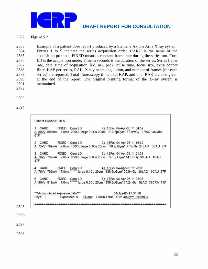
DRAFT REPORT FOR CONSULTATION
66
Figure 5.2 2582
Example of a patient dose report produced by a Siemens Axiom Artis X ray system. 2583
Entries 1 to 5 indicate the series acquisition order. CARD is the name of the 2584
acquisition protocol. FIXED means a constant frame rate during the series run. Coro 2585
LD is the acquisition mode. Time in seconds is the duration of the series. Series frame 2586
rate, date, time of acquisition, kV, mA peak, pulse time, focus size, extra copper 2587
filter, KAP per series, RAK, X-ray beam angulation, and number of frames (for each 2588
series) are reported. Total fluoroscopy time, total KAP, and total RAK are also given 2589
at the end of the report. The original printing format of the X-ray system is 2590
maintained. 2591
2592
2593
2594
2595
2596
2597
2598
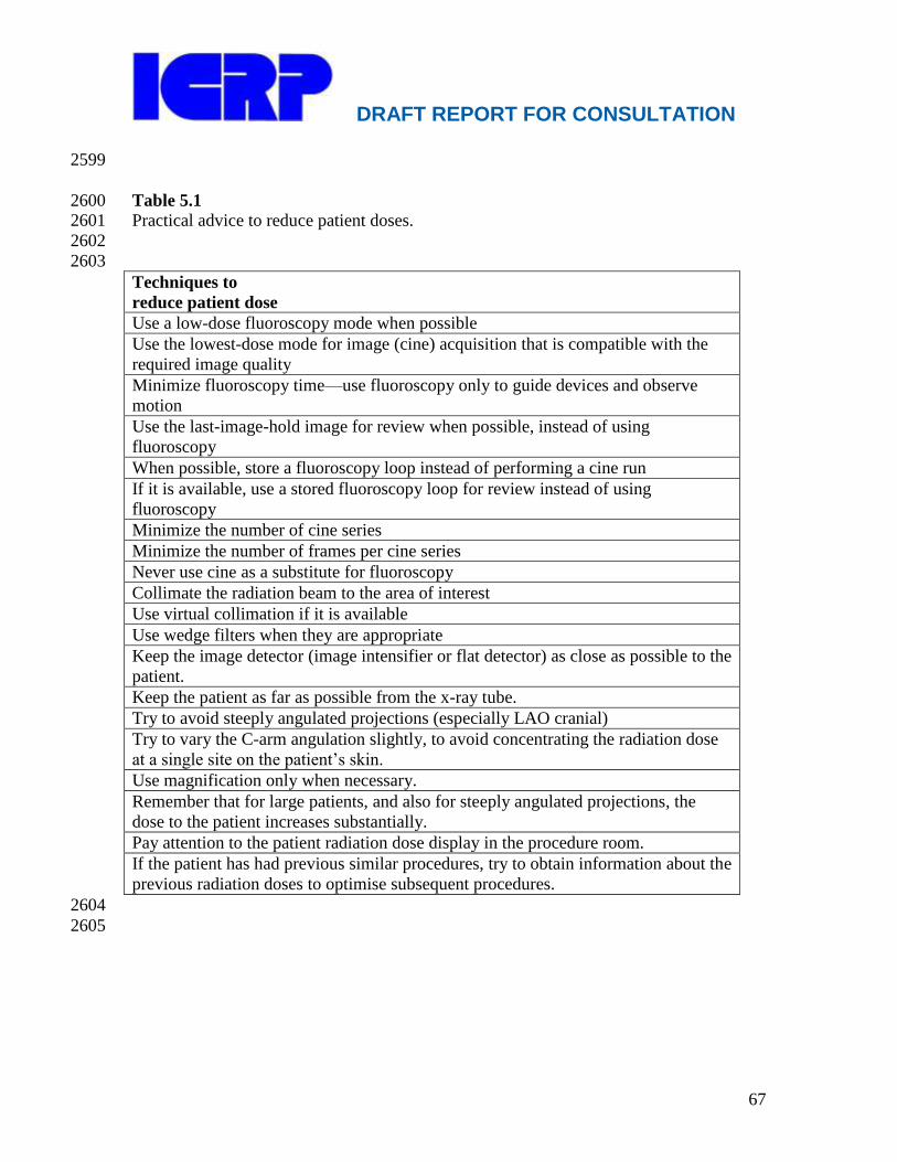
DRAFT REPORT FOR CONSULTATION
67
2599
Table 5.1 2600 Practical advice to reduce patient doses. 2601
2602
2603
Techniques to
reduce patient dose
Use a low-dose fluoroscopy mode when possible
Use the lowest-dose mode for image (cine) acquisition that is compatible with the
required image quality
Minimize fluoroscopy time—use fluoroscopy only to guide devices and observe
motion
Use the last-image-hold image for review when possible, instead of using
fluoroscopy
When possible, store a fluoroscopy loop instead of performing a cine run
If it is available, use a stored fluoroscopy loop for review instead of using
fluoroscopy
Minimize the number of cine series
Minimize the number of frames per cine series
Never use cine as a substitute for fluoroscopy
Collimate the radiation beam to the area of interest
Use virtual collimation if it is available
Use wedge filters when they are appropriate
Keep the image detector (image intensifier or flat detector) as close as possible to the
patient.
Keep the patient as far as possible from the x-ray tube.
Try to avoid steeply angulated projections (especially LAO cranial)
Try to vary the C-arm angulation slightly, to avoid concentrating the radiation dose
at a single site on the patient‘s skin.
Use magnification only when necessary.
Remember that for large patients, and also for steeply angulated projections, the
dose to the patient increases substantially.
Pay attention to the patient radiation dose display in the procedure room.
If the patient has had previous similar procedures, try to obtain information about the
previous radiation doses to optimise subsequent procedures.
2604
2605
DRAFT REPORT FOR CONSULTATION
68
6. RADIATION DOSES AND PROTECTION OF STAFF DURING 2606
INTERVENTIONAL FLUOROSCOPY 2607
2608
Main Points 2609
2610
In general, reducing patient dose will also reduce operator dose. 2611
The basic tools of occupational radiological protection are time, distance and 2612
shielding. 2613
The use of personal protective shielding is necessary in the cardiac 2614
catheterization laboratory. 2615
Radiological protection for the eyes is necessary for interventionalists. 2616
Occupational doses can be reduced to very low levels if ceiling suspended lead 2617
screens and protective lead curtains suspended from the side of the procedure 2618
table are used properly. 2619
Radiation exposure to the operator is neither uniform nor symmetric. 2620
Proper use of personal monitoring badges is necessary in cardiac 2621
catheterization laboratories in order to monitor and audit occupational 2622
radiation dose. 2623 2624
6.1 Introduction 2625
(97) Despite regulatory limits on occupational dose, there have been reports of 2626
cataracts and of fairly high radiation doses to the hands and legs of staff and hair loss in 2627
the portions of the legs not shielded by a protective device (Balter, 2001a). The 2628
occurrence of radiation-induced cataracts in operators (Vano et al. 1998a, Vano et al, 2629
2010, ICRP 2000, Ciraj-Bjelac, 2010) and the debate regarding the incidence of brain 2630
cancer in interventional cardiologists (Finkelstein, 1998, Klein et al, 2009) highlight the 2631
importance of occupational radiological protection for interventionalists, especially for 2632
parts of the body not protected by the lead apron. 2633
(98) The operator is not normally exposed to the x-ray beam directly, but is exposed 2634
to a considerable amount of scatter radiation. There are a number of techniques, 2635
described in Chapter 5, and protective devices, discussed in this Chapter, that, if used 2636
appropriately, should result in the operator‘s annual effective dose being well within 2637
regulatory limits. With proper use of radiological protection tools and techniques, the 2638
effective dose (E) for an interventionalist is typically 2–4 mSv/year, and is well below the 2639
20 mSv/year limit recommended by the Commission (Dendy, 2008, Tsapaki, 2004, 2640
Miller, 2010, ICRP 2007). Proper use of personal monitoring badges is essential in 2641
cardiac catheterization laboratories in order to monitor and audit occupational radiation 2642
dose. Too often, personal monitoring badges are not worn, or are worn improperly 2643
(Padovani, 2011). Training in radiation management and radiological protection, as 2644
discussed in Chapter 9, is essential (ICRP 2000). 2645

DRAFT REPORT FOR CONSULTATION
69
6.2 Comparison of radiation exposure with that of other staff 2646
(99) The interventionalist encounters much more radiation than most other medical 2647
and paramedical staff in a hospital. The radiation intensity from radioisotopes used in 2648
nuclear medicine is smaller by a factor of a few tens or even hundred. Nuclear medicine 2649
staff are likely to be exposed to much less radiation, whether it emanates from the patient 2650
or from external sources (normally in shielded containers). Similarly, while the radiation 2651
sources used in radiotherapy are of very high strength (GBq or TBq of radioactivity), 2652
staff are exposed only to remnant radiation leaking through the shielding material and 2653
scattered through a large distance. Staff in the interventional laboratory who are 2654
positioned in the control room are protected by both shielding and distance from the x-ray 2655
beam. Typically, in a properly designed facility, the radiation intensity in the control 2656
room may be tens of thousands of times less than at the operator‘s position (Rehani and 2657
Ortiz-Lopez, 2005). Exposure factors for the interventionalist are a thousand times higher 2658
than for staff working in the control room. 2659
(100) The major protection in nuclear medicine accrues from the lower radiation 2660
intensity and in radiotherapy from shielding and distance. The situation in interventional 2661
fluoroscopy is very different. First, the operator‘s working position is quite close to the x-2662
ray source and the source of scatter radiation (the patient). Second, the intensity of the x-2663
ray beam lies in between the radiation intensities observed in nuclear medicine and 2664
radiotherapy. Also, beam intensity is 10-fold or 20-fold higher in cine mode than in 2665
fluoroscopy mode (NCRP Report 168, 2010). Shielding plays a major role in radiological 2666
protection in interventional fluoroscopy, due to variability in the operator‘s distance from 2667
the x-ray source, the relative position of the operator, patient, and x-ray source and the 2668
duration of the procedure. 2669
6.3 The essentials of occupational radiological protection 2670
(101) The essentials of occupational radiological protection are time, distance and 2671
shielding. Staff radiological protection cannot be handled independently from patient 2672
protection, since they correlate in many ways. Both patient and occupational radiological 2673
protection are also discussed in an ICRP publication devoted to radiological protection 2674
outside the imaging department (reference ICRP TG 78). In general, reducing patient 2675
dose will also reduce operator dose. 2676
2677
(102) Time, one essential component of radiological protection, is controlled by 2678
reducing the time the x-ray beam is on, both for fluoroscopy and for cine. Reducing 2679
fluoroscopy time and fluoroscopy dose rate reduces patient dose. Reduced patient dose 2680
results in reduced scatter, and therefore in reduced operator dose. Readers are advised to 2681
remember all of the factors discussed in Chapter 4. 2682
(103) Distance is a valuable tool for radiological protection. Radiation dose 2683
decreases as the square of the distance between the radiation source and the operator (the 2684
inverse square law). A person who moves away from the x-ray source to three times the 2685
original distance will receive only one-ninth of the original dose. During a procedure, the 2686
operator cannot normally move further away from the patient than arm‘s length. This can 2687

DRAFT REPORT FOR CONSULTATION
70
result in high operator radiation doses, especially if contrast medium is injected manually 2688
for angiographic runs. However, if a mechanical injector is used for contrast medium 2689
injection, the operator can move back away from the patient, and ideally behind a shield. 2690
(104) In general, scattered radiation is most intense on the entrance beam side of the 2691
patient (Balter, 2001b, Schueler et al, 2006, Stratakis et al, 2006). When using a C-arm 2692
in a lateral projection, the operator should be positioned on the image receptor side of the 2693
patient, if possible. When using a C-arm in a frontal projection, positioning the x-ray 2694
tube below the table will place the area of higher radiation scatter towards the floor, so 2695
that the operator‘s head and neck receive less radiation. 2696
(105) Shielding is of three types: architectural shielding, equipment mounted shields, 2697
and personal protective devices (Miller et al, 2010). Architectural shielding is built into 2698
the walls of the procedure room and is not discussed further here. Rolling and stationary 2699
shields that are constructed of transparent leaded plastic and rest on the floor are useful 2700
for providing additional shielding for both operators and staff. They are particularly well 2701
suited for use by nurses and anaesthesia personnel. The interventionalist is protected by 2702
equipment-mounted shields suspended from the ceiling and the procedure table, and by 2703
personal protective devices such as a lead apron, leaded glasses and a thyroid shield. 2704
(106) Simple measures, such as standing a little away from the table and patient, 2705
limiting the field size (collimation) and carrying out procedures quickly consistent with 2706
case complexity can be very effective in reducing occupational radiation dose. Table 6.1 2707
presents some practical advice to improve occupational protection in the catheterization 2708
laboratory and Table 6.2 presents the relative change in scatter dose rates measured in a 2709
typical catheterization laboratory for different changes in technique. The values in Table 2710
6.2 highlight the large changes in scatter dose associated with changes in technique and 2711
patient body size. 2712
6.4 Personal protective devices 2713 2714
(107) The use of personal protective shielding is essential in the cardiac 2715
catheterization laboratory. In the past, there has been a trend to use lead aprons of higher 2716
lead equivalence (0.5 mm rather than 0.25, 0.3 or 0.35 mm), even though physical 2717
measurements did not demonstrate a great difference in attenuation (Table 6.3). The 2718
inherently conservative safety factor has always influenced practice in radiation, both for 2719
interventionalists and for regulators. 2720
(108) When procedures are performed on thinner patients, and in particular on 2721
children, a lead apron of 0.25 mm lead equivalence will suffice for staff protection, but 2722
for procedures performed on thicker patients, and for procedures performed by physicians 2723
with heavy workload, a 0.5 mm lead apron may be more suitable. Lead is very effective 2724
for protecting against radiation, but is heavy. The weight can cause problems for staff 2725
who have to wear these aprons for long spans of time (Goldstein, 2004). There are reports 2726
of back injuries due to lead aprons among staff who wear these aprons for many years 2727
(NCRP, 2010). Some newer aprons are lighter weight while maintaining approximately 2728
the same lead equivalence. Newer apron designs distribute weight using a variety of 2729
different methods. Two-piece (skirt and vest) wraparound aprons distribute the apron‘s 2730
weight and also provide protection for the wearer‘s back. 2731

DRAFT REPORT FOR CONSULTATION
71
(109) Lead aprons should be properly placed on designated hangers and should not 2732
be folded, creased, or crumpled in any way. Sitting on them, folding them or improperly 2733
hanging them may result in damage that reduces their effectiveness. Lead aprons, gloves 2734
and other leaded protective clothing should be inspected before they are put into service 2735
and then periodically re-inspected to determine that they provide the shielding benefit for 2736
which they were designed. A combination of visual, physical and fluoroscopic inspection 2737
can be employed to ensure the integrity of the garments. Consideration should be given to 2738
minimizing the irradiation of inspectors by minimizing unnecessary fluoroscopy (NCRP 2739
168, 2010). 2740
(110) A lead apron does not protect the eyes, the hands, the lower legs or the back 2741
(unless the apron is the wrap-around type). Radiation exposure of these parts of the 2742
body has become a concern. 2743
(111) Radiological protection for the eyes is essential for interventionalists (Dauer et 2744
al, 2010). Preferably, this protection is provided by ceiling-suspended shields (section 2745
6.3), as these devices protect the entire head, and not just the eyes. However, there are 2746
many procedures where it is not practical to use ceiling-suspended shields, as they 2747
interfere with the operator‘s ability to perform the procedure (Miller et al., 2010). In 2748
these situations, leaded eyeglasses should be worn. Wearing these eyeglasses has been 2749
shown to significantly reduce radiation dose to the operator‘s eyes (Vano et al, 2008; 2750
Thornton et al, 2010). 2751
(112) While the dose reduction factor for 0.5 mm lead equivalent protective glasses 2752
is approximately 0.03 (i.e., 97% of the radiation is attenuated) the extent of radiation 2753
attenuation by the eyeglass lenses is not an adequate descriptor, by itself, of the 2754
effectiveness of the eyewear (NCRP report 168, 2010). For maximum effectiveness, 2755
radiation protective eyewear should intercept as much as possible of the scattered 2756
radiation that is directed at the interventionalist‘s eyes. During interventional procedures, 2757
interventionalists normally turn their heads away from the primary beam to view the 2758
fluoroscopy monitor. This results in exposure of the eyes to scattered radiation from the 2759
side. Protective eyewear should provide shielding for side exposure, using either side 2760
shields or a wrap-around design (NCRP report 168, 2010). Proper fit is necessary to 2761
ensure that the lenses and side shields adequately protect the eye and minimize exposure, 2762
and is also important to minimize discomfort from the weight of the eyewear (Schueler et 2763
al., 2009). Even properly designed and fitted leaded eyewear attenuates scattered 2764
radiation by only a factor of 2 or 3 (Moore et al., 1980; Thornton et al, 2010). The net 2765
effect of protective eyeglasses is dependent on the design of the glasses, the nature of the 2766
clinical procedure, and the wearer‘s work habits. 2767
(113) In younger individuals, the thyroid gland is relatively sensitive to radiation-2768
induced cancer. However, the cancer incidence risk is strongly dependent on age at 2769
exposure, with very little risk after age 30 for males and age 40 for females (NRC, 2006). 2770
For younger workers, wearing a thyroid collar and a protective apron reduces effective 2771
dose to ~50 % of the effective dose achieved by wearing a protective apron alone 2772
(Martin, 2009; von Boetticher et al., 2009). Use of a thyroid collar (or a protective apron 2773
with thyroid coverage) is recommended for younger interventionalists and for all 2774
DRAFT REPORT FOR CONSULTATION
72
personnel whose personal monitor readings at the collar level (unshielded) exceed 4 mSv 2775
(E) in a month (Wagner, 2004). 2776
(114) Flexible, sterile, radiation-attenuating surgical gloves are available to reduce 2777
interventionalist hand exposure. A previous recommendation that protective gloves be 2778
worn in high exposure situations has been reconsidered (NCRP report 133, 2000, NCRP 2779
report 168, 2010). Attenuating surgical gloves may be used to provide a small degree of 2780
protection when hands are exposed only to scattered radiation, but the use of these gloves 2781
does not permit interventionalists to place their hands safely in the primary beam (NCRP 2782
168, 2010). 2783
(115) There are several factors that could lead to higher hand doses for 2784
interventionalists when these gloves are used (Miller et al, 2010). Just as with special 2785
tools that allow for increased distance between the hands of the interventionalist and the 2786
primary x-ray beam, the reduction in tactile feedback from radiation-attenuating surgical 2787
gloves may lead to an increase in fluoroscopy time or CT exposure time for delicate 2788
procedures. Because of the increased dose when any shielding is placed in the primary 2789
beam, and the false sense of security that these gloves provide, protective gloves can 2790
result in increased radiation dose to the hand when the gloved hand is in the primary 2791
beam (Wagner, 1996). With or without added protection, the hands should not be placed 2792
in the primary x-ray beam, except for those rare occasions when it is essential for the 2793
safety and care of the patient. This should be done for the shortest possible time. As a 2794
rule, if an operator‘s hands are visible on the monitor, then practices should be altered 2795
(Limacher et al. 1998). 2796
6. 5 Equipment-mounted shields 2797
(116) The standard equipment-mounted shields used in catheterization laboratories at 2798
present are ceiling suspended lead screens and protective lead curtains suspended from 2799
the side of the procedure table. If these tools are used properly, occupational doses can be 2800
reduced to very low levels. 2801
(117) A leaded glass or plastic screen placed between the patient and the operator 2802
protects the operator‘s eyes, head and neck. Properly placed shields have been shown to 2803
dramatically reduce operator eye dose (Maeder et al., 2006, Thornton et al, 2010). These 2804
screens can effectively replace both leaded eyewear and a thyroid shield. The screens 2805
add no weight to the operator, eliminating the ergonomic consequences of the protective 2806
equipment they replace. 2807
(118) When a frontal (posteroanterior) projection is used and the x-ray tube is below 2808
the procedure table, scatter dose rates under the table are 3-4 times higher than the values 2809
over the table (Schueler et al, 2006). Leaded curtains suspended from the procedure table 2810
should be used to protect the interventionalist‘s lower legs. At present, these shields are 2811
available in almost all interventional suites. 2812
(119) Disposable, lightweight, sterile, lead-free radiological protection drape or pad 2813
shields can be positioned on the patient outside of the beam path to significantly reduce 2814
scattered radiation during cardiac interventional procedures (Sawdy et al, 2009, Germano 2815
et al, 2005). These contain metallic elements (typically bismuth or tungsten-antimony) 2816
and are placed on the patient after the operative site has been prepared and draped. They 2817

DRAFT REPORT FOR CONSULTATION
73
have been shown to reduce operator dose substantially, with reported reductions of 12-2818
fold for the eyes, 26-fold for the thyroid and 29-fold for the hands (King et al, 2002, 2819
Dromi et al, 2006). While their use adds some cost to the procedure, disposable 2820
protective drapes should be considered for complex procedures and procedures where the 2821
operator‘s hands must be near the radiation field (e.g., pacemaker placement) (Miller et 2822
al., 2010). In some institutions they are used routinely (Kim et al, 2010). These drapes 2823
should not be visible in the fluoroscopic image. If they are, the result will be an increase 2824
in patient dose. 2825
6.6 Overall impact of protective devices 2826
(120) The effective dose (E) to the cardiologist per procedure has been reported to 2827
range from 0.2 to 18.8 Sv (Padovani and Rodella, 2001). A more recent review 2828
demonstrated a range of 0.02 to 38.0 Sv (Kim et al, 2008). The wide dose ranges are 2829
most likely due to both the wide variation in procedure complexity and the inconsistent 2830
use of shields and personal protective devices. Modest operator dose reductions over 2831
time were observed for both diagnostic catheterizations and ablation procedures, due to 2832
technological improvements, but doses were not reduced over time for percutaneous 2833
coronary interventions. This was believed to be due mainly to the increased complexity 2834
of interventions. 2835
(121) Even if one assumes a rather high workload of 1000 angiographic procedures 2836
per year, the annual threshold level of 20 mSv will rarely be exceeded. One study 2837
reported an estimate of E for the operator of only 0.04–0.05 mSv/year (Efstathopolous et 2838
al. 2003), although other studies have reported 2–4 mSv/year (Dendy, 2008, Tsapaki, 2839
2004). The extensive studies by Kuon et al. establish that with proper choice of technique 2840
and shielding devices, the operator may be exposed to only 0.8% of typical radiation 2841
levels in advanced cardiac catheterization laboratories (Kuon et al. 2002). 2842
(122) When a lateral projection or steep gantry angulation is used, standing on the x-2843
ray tube side of the C-arm increases operator dose. Kuon et al. have estimated the 2844
influence of angulation of the X-ray tube on the amount of scatter radiation to the 2845
operator (Kuon et al. 2004). Radiation levels have been found to be highest for the left 2846
anterior oblique (LAO) position, whereas in posteroanterior (PA) and right anterior 2847
oblique (RAO) angulations, levels are much lower (Kuon et al. 2002, 2003, 2004). 2848
Simultaneous craniocaudal angulation further increases the dose. The group has shown 2849
that the standard view for the left main stem coronary artery (LAO 60°/20°–) is 2850
associated with a 7.6-fold increase in dose to the operator and a 2.6-fold increase in dose 2851
for the patient as compared to an alternative less frequently used angulation (caudal 2852
PA0°/30°–). 2853
(123) Effective dose does not reflect the doses to susceptible, unprotected parts of the 2854
body—the hands and the eyes. Radiation exposure to the operator is neither uniform nor 2855
symmetric. A right-handed operator performing the procedure via the right femoral 2856
artery has his or her left side turned towards the patient. Therefore the left side of the 2857
body is exposed to the highest level of scatter radiation (Maeder et al. 2005). This is 2858
especially true for the hands, which are at the level where the X-ray beam enters the 2859
DRAFT REPORT FOR CONSULTATION
74
patient. During cardiac catheterization, the left hand has been reported to receive twice 2860
the dose as compared with the right hand (Vaño et al. 1998b). The left eye also receives 2861
higher doses than the right eye. Not surprisingly, a tall operator will receive a lower eye 2862
dose than a short operator, because of the greater distance from the tall operator‘s eyes to 2863
the patient. 2864
(124) Unless personal monitoring devices are always worn, and worn properly, it is 2865
not possible to estimate occupational dose accurately. Failure to wear personal 2866
monitoring devices may lead to the false belief that an individual‘s occupational dose is 2867
low when it is not. 2868
6.7 Personal dosimetry 2869
(125) The Commission recommends the use of two personal dosimeters for 2870
occupational dosimetry cardiac catheterization laboratories: one worn on the trunk of the 2871
body inside the apron and the other worn outside the apron at the level of the collar or the 2872
left shoulder (ICRP 2000). The dosimeter under the apron provides an estimate of the 2873
dose to the organs of the shielded region. The dosimeter worn outside the apron supplies 2874
an estimate of the dose to the organs of the head and neck, including the thyroid and lens 2875
of the eyes (if unshielded), but greatly overestimates the doses to organs of the trunk. 2876
Results obtained from both dosimeters can be used to estimate the occupational effective 2877
dose as recommended by the NCRP (NCRP, 1995) and ICRP (ICRP, 2000). A dosimeter 2878
for the hands may also be useful. 2879
(126) The effective dose, E, can be estimated from the dosimeter values for Hw 2880
(under the apron at the waist, although this position is not critical) and Hn (above the 2881
apron at the neck) from the equation: 2882
2883
E = 0.5 Hw + 0.025 Hn 2884
2885
(127) NCRP report 122 (NCRP 1995) contains specific recommendations for 2886
calculating the effective dose when protective aprons are worn during diagnostic and 2887
interventional medical procedures involving fluoroscopy. In addition to the above 2888
formula, it states that the effective dose can be estimated as Hn/21 if only one dosimeter 2889
is worn on the neck outside the apron. 2890
(128) The European Commission DIMOND project addressed the issues regarding 2891
optimization of staff doses with an attempt to propose preliminary occupational dose 2892
constraints (Tsapaki at al. 2004). The proposed value for cardiologists' annual effective 2893
dose was 0.6 mSv. UNSCEAR (UNSCEAR 2000, paragraph 166) reported that 2894
cardiologists tend to be the most exposed staff in medicine; their average annual dose was 2895
0.4mSv, and an appreciable proportion received more than 1 mSv. A recent review of 2896
radiation exposures to operators from cardiac procedures over a 30 year period 2897
highlighted the difficulty in comparing reported dosimetry results because of significant 2898
differences in dosimetric methods in each study (Kim et al, Health Physics, 2008). Better 2899
standardization of dosimetric methods is recommended. 2900
(129) Many operators not only do not use protective equipment properly, but also do 2901
not regularly wear their dosimeters. Failure to wear dosimeters is a problem throughout 2902
DRAFT REPORT FOR CONSULTATION
75
the world (Vaño et al. 1998b, McCormick, 2002, Padovani, 2011). In addition to 2903
monitoring personal exposure, dosimeter use helps to increase awareness about 2904
radiological protection. In the absence of formal training in radiological protection for 2905
cardiologists in such countries, physicians in training adopt the practices of their seniors 2906
(Rehani and Ortiz-Lopez, 2005). 2907
(130) Compliance with the radiation badge policies is one of the main problems in 2908
many interventional cardiology services (Vano 2005). Reported occupational dose values 2909
are often surprisingly low, and the reason is likely not a high level of radiological 2910
protection, but rather failure to wear personal dosimeters. McCormick et al. (McCormick 2911
2002) reported that before a mandatory radiological protection training programme, 2912
compliance with the radiation badge policy for physicians and nurse clinicians was only 2913
36% in 1999, and afterwards reached a maximum of only 77%. A strict policy on the 2914
regular use of personal dosimeters should be part of any quality programme in cardiology 2915
laboratories. 2916
6.8 References 2917
2918
Balter, S, 2001a. Interventional fluoroscopy – physics, technology, safety. Wiley-Liss, 2919
New York. 2920
Balter S., 2001b. Stray radiation in the cardiac catheterization laboratory. Radiat Prot 2921
Dosim 94(1-2), 183-188. 2922
Ciraj-Bjelac O, Rehani MM, Sim KH, Liew HB, Vano E, Kleiman NJ. Risk for radiation 2923
induced cataract for staff in interventional cardiology: Is there reason for concern? 2924
Catheter Cardiovasc Interv 2010;76(6):826-834. 2925
Dauer, L.T., Thornton, R.H., Solomon, S.B., St Germain, J., 2010. Unprotected operator 2926
eye lens doses in oncologic interventional radiology are clinically significant: 2927
estimation from patient kerma-area-product data. J. Vasc. Interv. Radiol. 21, 1859-2928
1861. 2929
Dendy PP. Radiation risks in interventional radiology. Br J Radiol 2008; 81:1-7. 2930
Dromi, S., Wood, B.J., Oberoi, J., Neeman, Z., 2006. Heavy metal pad shielding during 2931
fluoroscopic interventions. J. Vasc. Interv. Radiol. 17, 1201-1206. 2932
Efstathopoulos E, Makrygiannis SS, Kottou S, et al. Medical personnel and patient 2933
dosimetry during coronary angiography and intervention. Phys Med Biol 2934
2003;48:3059-68. 2935
Finkelstein MM. Is brain cancer an occupational disease of cardiologists? Can J Cardiol 2936
1998;14,1385-8. 2937
Germano, J.J., Day, G., Gregorious, D., Natarajan, V., Cohen, T., 2005. 2938
A novel radiation protection drape reduces radiation exposure during fluoroscopy guided 2939
electrophysiology procedures. J. Invasive Cardiol. 17, 469-72. 2940
Goldstein JA, Balter S, Cowley M, Hodgson J, Klein LW; Occupational hazards of 2941
interventional cardiologists: prevalence of orthopedic health problems in 2942
contemporary practice. Catheter Cardiovasc Interv. 2004 Dec;63(4):407-11. 2943
ICRP. Avoidance of radiation injuries from medical interventional procedures. ICRP 2944
Publication 85. Ann ICRP 2000;30(2). 2945

DRAFT REPORT FOR CONSULTATION
76
ICRP. The 2007 Recommendations of the International Commission on Radiological 2946
Protection. ICRP Publication 103. Ann ICRP 2007; 37(2-4):1-332 2947
Kim KP, Miller DL, Balter S, Kleinerman RA, Linet MS, Kwon D, Simon SL. 2948
Occupational radiation doses to operators performing cardiac catheterization 2949
procedures. Health Phys 94: 211-227, 2008. 2950
Kim, C., Vasaiwala, S., Haque, F., Pratap, K., Vidovich, M.I., 2010. Radiation safety 2951
among cardiology fellows. Am. J. Cardiol. 106, 125-128. 2952
King JN, Champlin AM, Kelsey CA, Tripp DA. Using a sterile disposable protective 2953
surgical drape for reduction of radiation exposure to interventionalists. Am J 2954
Roentgenol 2002; 178:153-157. 2955
Klein LW, Miller DL, Balter S, Laskey W, Haines D, Norbash A, Mauro MA, Goldstein 2956
JA. Occupational health hazards in the interventional laboratory: time for a safer 2957
environment. Heart Rhythm 2009, 6:439-444. 2958
Kuon E, Schmitt M, Dahm JB. Significant reduction of radiation exposure to operator 2959
and staff during cardiac interventions by analysis of radiation leakage and improved 2960
lead shielding. Am J Cardiol 2002; 89:44-9. 2961
Kuon E, Dahm J, Empen K, et al. Identification of less-irradiating tube angulations in 2962
invasive cardiology. J Am Coll Cardiol 2004;44:1420-8. 2963
Limacher MC, Douglas PS, Germano G, et al. ACC expert consensus document. 2964
Radiation safety in the practice of cardiology. J Am Coll Cardiol. 1998;31:892-913. 2965
Maeder M. Verdun FR, Stauffer JC et al. Radiation exposure and radiation protection in 2966
interventional cardiology. Kardiovaskuläre Medizin 2005;8:124-132. 2967
Maeder M, Brunner-La Rocca HP, Wolber T, Ammann P, Roelli H, Rohner F, Rickli H. 2968
Impact of a lead glass screen on scatter radiation to eyes and hands in interventional 2969
cardiologists. Catheter Cardiovasc Interv 2006; 67:18-23. 2970
Marshall NW, Faulkner K, Clarke P. An investigation into the effect of protective 2971
devices on the dose to radiosensitive organs in the head and neck. Br J Radiol. 1992 2972
Sep;65(777):799-802. 2973
Martin, CJ. A review of radiology staff doses and dose monitoring requirements. Radiat 2974
Prot Dosim 2009; 136(3), 140-157. 2975
McCormick VA, Schultz CC, Hollingsworth-Schuler V, Campbell JM, O‘Neill WW, 2976
Ramos R. Reducing radiation dose in the cardiac catheterization laboratory by 2977
design alterations and staff education. Am J Cardiol 2002; 90:903-905. 2978
Miller DL, Vañó E, Bartal G, Balter S, Dixon R, Padovani R, Schueler B, Cardella JF, 2979
de Baère T. Occupational radiation protection in interventional radiology: A joint 2980
guideline of the Cardiovascular and Interventional Radiology Society of Europe and 2981
the Society of Interventional Radiology. Cardiovasc Interv Radiol 2010; 33:230-2982
239. 2983
Moore WE, Ferguson G, Rohrmann C. Physical factors determining the utility of 2984
radiation safety glasses. Med Phys 1980; 7(1), 8-12. 2985
NRC. Health risks from exposure to low levels of ionizing radiation: Phase 2, BEIR VII. 2986
Washington, DC: The National Academies Press, 2006. 2987
NCRP. Use of personal monitors to estimate effective dose equivalent and effective dose 2988
to workers for external exposure to low-LET radiation. NCRP report No. 122. 2989
DRAFT REPORT FOR CONSULTATION
77
Bethesda, Maryland: National Council on Radiation Protection and Measurements; 2990
1995. 2991
NCRP. Radiation protection for procedures performed outside the radiology department. 2992
NCRP Report No. 133. Bethesda, Maryland: National Council on Radiation 2993
Protection and Measurements; 2000. 2994
NCRP. Radiation dose management for fluoroscopically guided interventional medical 2995
procedures. NCRP Report No. 168. Bethesda, MD: National Council on Radiation 2996
Protection and Measurements; 2010. 2997
Padovani R, Rodella CA. Staff dosimetry in interventional cardiology. Radiat Prot 2998
Dosimetry 2001;94:99-103. 2999
Padovani, R., Le Heron, J., Cruz-Suarez, R., Duran, A., Lefaure, C., Miller, D.L., Sim, 3000
H.K., Vano, E., Rehani, M., Czarwinski, R., 2010. International project on individual 3001
monitoring and radiation exposure levels in interventional cardiology. Radiat Prot 3002
Dosimetry 2011; in press. 3003
Rehani MM, Ortiz-Lopez P. Radiation effects in fluoroscopically guided cardiac 3004
interventions – keeping them under control.(Editorial). Int J Cardiol 2006; 109(2): 3005
147-151. 3006
Sawdy, J.M., Gocha, M.D., Olshove, V., Chisolm, J.L., Hill, S.L., Phillips, A., 3007
Galantowicz, M., Cheatham, J.P., Holzer, R.J., 2009. Radiation protection during 3008
hybrid procedures: innovation creates new challenges. J. Invasive Cardiol. 21, 437-3009
40. 3010
Schueler BA, Vrieze TJ, Bjarnason H, Stanson AW. An investigation of operator 3011
exposure in interventional radiology. RadioGraphics 2006; 26(5), 1533-1541. 3012
Schueler BA, Sturchio G, Landsworth R, Hindal M, Magnuson D. (2009) Does new 3013
lightweight leaded eyewear provide adequate radiation protection for fluoroscopists? 3014
(abstract) Med Phys 2009; 36(6), 2747-2748. 3015
Stratakis J, Damilakis J, Hatzidakis A, Theocharopoulos N, Gourtsoyiannis N. 3016
Occupational radiation exposure from fluoroscopically guided percutaneous 3017
transhepatic biliary procedures. J Vasc Interv Radiol 2006; 17(5), 863–871. 3018
Thornton, R.H., Dauer, L.T., Altamirano, J.P., Alvarado, K.J., St. Germain, J., Solomon, 3019
S.B., 2010. Comparing strategies for operator eye protection in the interventional 3020
radiology suite. J. Vasc. Interv. Radiol. 21, 1703-1707. 3021
Tsapaki V, Kottou S, Vano E, Komppa T, Padovani R, Dowling A, Molfetas M, 3022
Neofotistou V. Occupational dose constraints in interventional cardiology 3023
procedures: the DIMOND approach. Phys Med Biol. 2004 Mar 21;49(6):997-1005. 3024
United Nations Scientific Committee on the Effects of Atomic Radiation. Sources and 3025
effects of ionizing radiation. UNSCEAR 2000 report to the General Assembly, with 3026
scientific annexes. Annex E. Occupational radiation exposures. (Available at 3027
www.unscear.org). New York: United Nations; 2000. 3028
Vano E, González L, Beneytez F, et al. Lens injuries induced by occupational exposure 3029
in non-optimised interventional radiology laboratories. Br J Radiol 1998a;71:728-33. 3030
Vano E, Gonzalez L, Guibelalde E, et al. Radiation exposure to medical staff in 3031
interventional and cardiac radiology. Br J Radiol 1998b;71:954-60. 3032

DRAFT REPORT FOR CONSULTATION
78
Vano E. Radiation exposure to cardiologists: how it could be reduced. Heart. 2003; 3033
89(10):1123-4. 3034
Vano E, Gonzalez L, Fernandez JM, Alfonso F, Macaya C. Occupational radiation 3035
doses in interventional cardiology: a 15 year follow-up. Br J Radiol 3036
2006;79(941):383-388. 3037
Vano E, Gonzalez L, Fernandez JM, Prieto C, Guibelalde E. Influence of patient 3038
thickness and operation modes on occupational and patient doses in interventional 3039
cardiology. Radiat Prot Dosim 2006; 118(3): 325-330. 3040
Vano E, Gonzalez L, Fernandez M, Haskal ZJ. Eye lens exposure to radiation in 3041
interventional suites: caution is warranted. Radiology 2008; 248: 945-953. 3042
Vano E, Kleiman NJ, Duran A, Rehani MM, Echeverri D, Cabrera. M. Radiation 3043
cataract risk in interventional cardiology personnel. Radiat Res, 2010; 174(4):490-3044
495 3045
von Boetticher H, Lachmund J, Hoffmann W. Cardiac catheterization: impact of face 3046
and neck shielding on new estimates of effective dose. Health Phys 2009; 97(6), 3047
622– 627. 3048
Wagner LK, Mulhern OR. Radiation-attenuating surgical gloves: effects of scatter and 3049
secondary electron production. Radiology 1996; 200:45-48. 3050
Wagner LK, Archer BR. Minimizing Risks from Fluoroscopic X-Rays: Bioeffects, 3051
Instrumentation, and Examination. 4th Edition. The Woodlands, Texas:(Partners in 3052
Radiation Management, 2004. 3053
3054

DRAFT REPORT FOR CONSULTATION
79
3055
Table 6.1. 3056
3057
Practical advice for interventionalists to improve staff radiation protection (from Vano et 3058
al, 2003 and Miller et al, 2010). 3059
3060
Increase your distance from the patient (the scatter radiation source) whenever 3061
possible. This is obviously only possible when angiographic runs are not performed 3062
by hand. Working at 80 cm from the isocenter instead of 40 cm can decrease scattered 3063
dose to approximately a quarter of the original dose. 3064
Try to position yourself in a low scatter area. Scattered radiation is higher at the x-3065
ray tube side of the gantry and lower on the side of the image receptor. 3066
Use a ceiling suspended screen, a table-suspended screen and other protective 3067
shielding, such as a lead apron, thyroid collar and lead glasses, when possible. 3068
When appropriate, use a dose reduction pad or drape at the catheter entrance site to 3069
reduce your hand dose. 3070
Minimise the use of fluoroscopy and use low-dose fluoroscopy modes (for example, 3071
pulsed fluoroscopy) when possible. 3072
Minimize the number of cine series and the number of frames per cine series. 3073
Use magnification as little as possible. 3074
Collimate the x-ray beam as tightly as possible. 3075
Obtain appropriate training in radiation management and radiation protection. 3076
Wear your dosimeters and know your own dose. 3077
In addition, a final general concept: reduce the patient‘s radiation and you will also be 3078
reducing your own dose. 3079
3080
3081
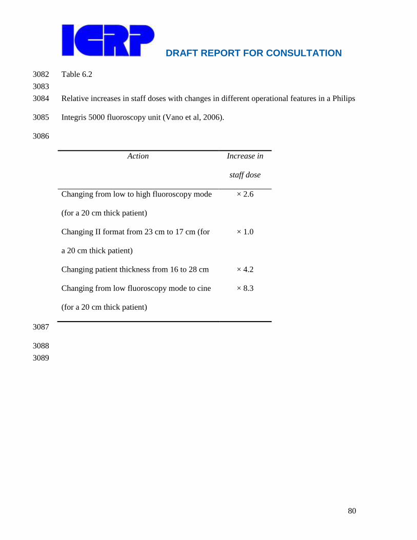
DRAFT REPORT FOR CONSULTATION
80
Table 6.2 3082
3083
Relative increases in staff doses with changes in different operational features in a Philips 3084
Integris 5000 fluoroscopy unit (Vano et al, 2006). 3085
3086
Action Increase in
staff dose
Changing from low to high fluoroscopy mode
(for a 20 cm thick patient)
× 2.6
Changing II format from 23 cm to 17 cm (for
a 20 cm thick patient)
× 1.0
Changing patient thickness from 16 to 28 cm × 4.2
Changing from low fluoroscopy mode to cine
(for a 20 cm thick patient)
× 8.3
3087
3088
3089
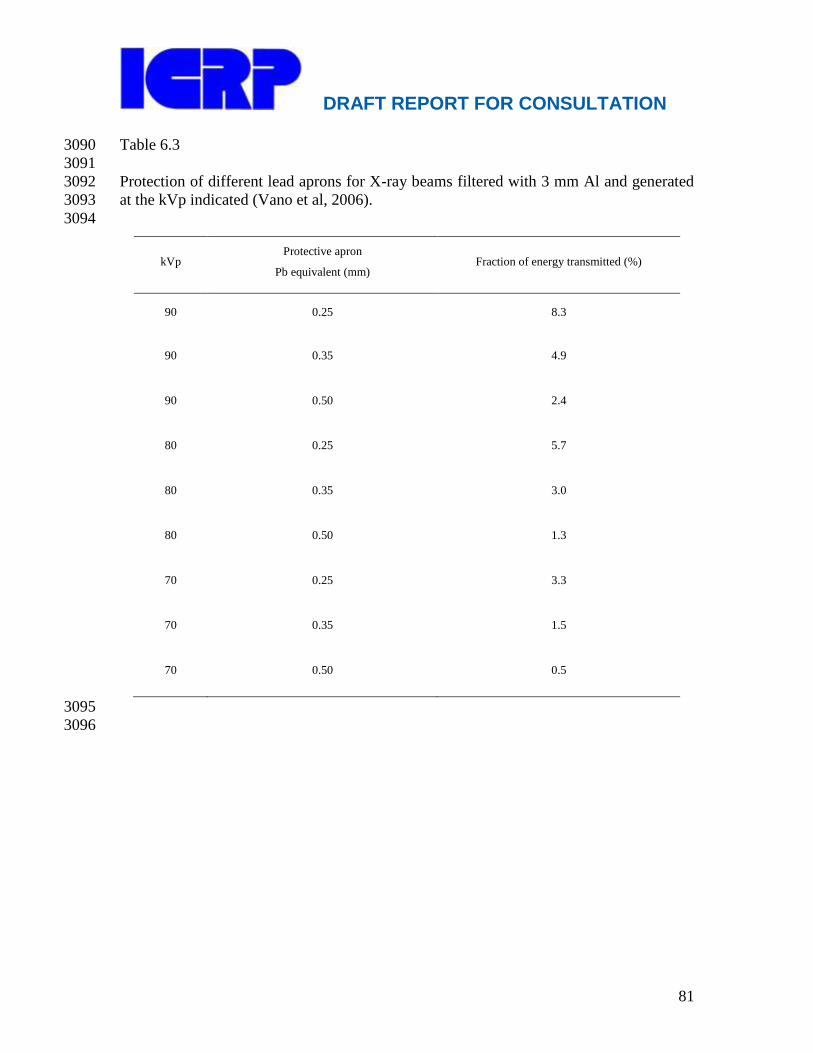
DRAFT REPORT FOR CONSULTATION
81
Table 6.3 3090
3091
Protection of different lead aprons for X-ray beams filtered with 3 mm Al and generated 3092
at the kVp indicated (Vano et al, 2006). 3093
3094
kVp Protective apron
Pb equivalent (mm) Fraction of energy transmitted (%)
90 0.25 8.3
90 0.35 4.9
90 0.50 2.4
80 0.25 5.7
80 0.35 3.0
80 0.50 1.3
70 0.25 3.3
70 0.35 1.5
70 0.50 0.5
3095
3096

DRAFT REPORT FOR CONSULTATION
82
3097
7. RADIOLOGICAL PROTECTION FOR NUCLEAR CARDIOLOGY 3098
3099
Main Points 3100
Appropriate use criteria and guidelines that help to set standards for 3101
justification have been developed through consensus efforts of professional 3102
societies. 3103
Optimization of nuclear cardiology procedures involves the judicious 3104
selection of radiopharmaceuticals and administered activities to ensure 3105
diagnostic image quality while minimizing patient dose. 3106
For SPECT protocols, Tc-99m-based agents yield lower effective doses than 3107
Tl-201, and are preferred on dosimetric grounds. 3108
Administered activities should be within pre-specified ranges, as provided in 3109
international and national guidelines, and should reflect patient habitus. 3110
If stress imaging is normal, rest imaging can be omitted to minimize total 3111
dose. 3112
Practitioners need good quality dosimetry data to perform proper benefit-3113
risk analyses for their patients. 3114 3115
3116
7.1 Introduction 3117 3118
(131) More than 90% of nuclear cardiology studies are myocardial perfusion 3119
scintigraphy studies for the assessment of myocardial perfusion and/or viability. The vast 3120
majority of nuclear cardiology procedures are performed with single photon emission 3121
computed tomography (SPECT). A small but growing number of laboratories perform 3122
positron emission tomography (PET) studies. 3123
(132) An estimated 32.7 million diagnostic nuclear medicine procedures are 3124
performed annually worldwide (UNSCEAR 2008). Of these, approximately 14 million 3125
are nuclear cardiology procedures, and this number has increased rapidly (Davis, 2006). 3126
More nuclear cardiology procedures are performed in the United States than in the rest of 3127
the world combined. In the U.S., nuclear medicine procedures accounted for 26% of the 3128
medical exposure of patients in 2006, and cardiac studies accounted for 85% of the 3129
nuclear medicine exposure (NCRP report 160, 2009). 3130
3131
7.2 Radiopharmaceuticals 3132
3133 (133) The radiopharmaceuticals used most commonly for nuclear cardiology studies 3134
are summarized in Table 7.1. In Europe, most studies are performed using Tc-99m-3135
based agents, while in the United States, a sizable minority of studies are performed using 3136
Tl-201, usually in the context of a dual isotope study with rest Tl-201 imaging followed 3137
by stress Tc-99m imaging. The use of thallium results in a higher dose to the patient 3138
(Einstein et al, 2007). 3139
3140

DRAFT REPORT FOR CONSULTATION
83
Table 7.1. Commonly Used Radiopharmaceuticals for Nuclear Cardiology 3141
Agent Modality
Role
Physical
Half-Life
Effective Dose
(10-3 mSv/MBq)
ICRP
Publication (14) P
erfusion
(15) F
unctio
n
(16) V
iabilit
y
Tc-99m sestamibi SPECT +++ ++ + 6h 9.0 rest/7.9
stress 80(1998)
Tc-99m tetrofosmin SPECT +++ ++ + 6h 7.6 rest/7.0
stress 80(1998)
Tl-201 SPECT +++ + ++ 73h 140 106(2008)
Tc-99m red blood
cells
Planar or
SPECT
MUGA
- +++ - 6h 7.0 80(1998)
Rb-82 PET +++ ++ - 75s 3.4* (17) 8
0(1998)*
N-13 ammonia PET +++ ++ - 10m 2.0 80(1998)
F-18
fluorodeoxyglucose PET - - +++ 110m 19 (18) 8
0(1998)
SPECT: single photon emission computed tomography, PET: positron emission tomography: MUGA: multiple gated 3142 acquisition 3143 * ICRP‘s dose coefficients for Rb-82, dating to Publication 53 (1987) and reiterated in Publication 80 (1998), reflect 3144 for some organs ―worst case‖ conditions, as was stated in Publication 53, and thus dose estimates deriving therefrom 3145 might be overly conservative. Three groups have recently suggested lower dose coefficients (Senthamizhchelvan et al 3146 2010, 1.11 μSv/MBq; Hunter 2010, 0.74 μSv/MBq; and Stabin 2010, 1.7 μSv/MBq); the Commission is currently 3147 revisiting the issue of Rb-82 dosimetry. 3148 3149
(134) Recommended administered activities for nuclear cardiology procedures vary 3150
markedly among the professional societies and accrediting bodies in various countries 3151
(Hesse et al., 2005). Guidelines have been published by both the American Society of 3152
Nuclear Cardiology (ASNC) (DePuey, 2006; Henzlova, 2009) and the European Council 3153
on Nuclear Cardiology (ECNC) (Hesse et al., 2005), a joint group of the European 3154
Association of Nuclear Medicine (EANM) and the European Society of Cardiology 3155
(ESC). Injected activity from these guidelines is summarized in Table 7.2. 3156
3157
3158
3159
3160
3161
3162
3163
3164
3165
3166
3167
3168
3169
3170
3171
3172

DRAFT REPORT FOR CONSULTATION
84
Table 7.2. Recommended Injected Activity (MBq) for Standard Cardiac SPECT and PET 3173
Protocols 3174
3175
3176
3177 *740 to 925 for planar imaging 3178
**for 2 dimensional acquisition using camera with bismuth germanate or lutetium 3179
oxyorthosilicate crystals 3180
3181
7.3 Dosimetry for nuclear cardiology 3182
3183 (135) Two types of dose coefficients can be determined: 1) tissue dose coefficients, 3184
which can be used to estimate the dose to a particular tissue or organ, and 2) effective 3185
dose coefficients, which can be used to estimate effective dose to the individual. Note 3186
however that effective dose is intended for use as a radiological protection quantity. 3187
Effective dose is not recommended for epidemiological evaluations, nor should it be used 3188
for detailed specific retrospective investigations of individual exposure and risk (ICRP, 3189
2007a). 3190
(136) Estimates of organ dose and estimates of effective dose to patients are 3191
generally obtained by using mathematical biokinetic models that quantify the distribution 3192
and metabolism of a radiopharmaceutical in the body. These models incorporate 3193
biokinetic data from humans and/or animals and enable the determination of dose 3194
coefficients. 3195
(137) Tissue dose coefficients quantify absorbed doses to a specific organ in a typical 3196
patient, per unit activity administered. For example, ICRP‘s current liver dose coefficient 3197

DRAFT REPORT FOR CONSULTATION
85
in an adult for the PET tracer F-18 fluorodeoxyglucose is 1.110-2
mGy per MBq (ICRP, 3198
1998). Thus, a 200 MBq injection of F-18 fluorodeoxyglucose is associated with an 3199
estimated dose to the liver of 2.2 mGy. 3200
(138) Effective dose coefficients quantify effective dose per unit activity 3201
administered. ICRP‘s current effective dose coefficient in an adult for F-18 3202
fluorodeoxyglucose is 1.910-2
mSv per MBq (ICRP, 1998), and therefore the same 200 3203
MBq injection of F-18 fluorodeoxyglucose would be associated with an estimated 3204
effective dose of 3.8 mSv. 3205
(139) Several systems provide mathematical frameworks for estimating dose 3206
coefficients, including those of ICRP Publication 30 (ICRP, 1979) and those of the 3207
Society of Nuclear Medicine‘s Medical Internal Radiation Dose committee (Loevinger et 3208
al., 1988) and Radiation Dose Assessment Resource task group (Stabin et al., 2001). 3209
These approaches are essentially equivalent (Stabin, 2006). They estimate radiation dose 3210
as energy per unit mass. Energy is generally determined from biokinetic models of the 3211
radiopharmaceutical‘s time-activity curve, from tables of the mean energy per nuclear 3212
transition, and from Monte Carlo computer models. Organ masses are determined from a 3213
model of a representative person. 3214
(140) There are numerous collections of dose coefficients for specific 3215
radiopharmaceuticals. The most extensive compilations are those of the Commission, for 3216
which current estimates can be found in Publications 53 (ICRP, 1987), 80 (ICRP, 1998), 3217
and 106 (ICRP, 2008). Effective doses for commonly used radiopharmaceuticals for 3218
nuclear cardiology, based on the most recent ICRP effective dose coefficients for these 3219
radiopharmaceuticals, are listed in Table 7.1. These effective doses reflect ICRP 3220
Publication 60 tissue weighting factors; updated effective dose coefficients reflecting 3221
Publication 103 tissue weighting factors will be included in a forthcoming ICRP 3222
publication. In many countries there is a regulatory requirement that dose coefficients be 3223
provided in manufacturers‘ package inserts/product information (PI) sheets for 3224
radiopharmaceuticals. 3225
3226
7.4 Current dosimetry estimates 3227
3228 (141) The dose to a typical patient from a nuclear cardiology study can be estimated 3229
by multiplying dose coefficients by the administered activity. These estimates are 3230
illustrated in Figure 7.1, using the most recent ICRP dose coefficients for each agent and 3231
administered activities in the middle of the range specified in Table 7.2. 3232
3233
3234
3235

DRAFT REPORT FOR CONSULTATION
86
3236
3237 3238 Figure 7.1. Effective doses from standard nuclear cardiology procedures, estimated using the most recent 3239 ICRP dose coefficients and Publication 103 tissue weighting factors (ICRP, 2007a). Stacked bars represent 3240 organ weighted equivalent doses contributing to effective dose. Doses for Tc-99m represent the average of 3241 Tc-99m sestamibi and tetrofosmin. Top: Using average recommended administered activities from 3242 American Society of Nuclear Cardiology guidelines (Henzlova, 2009; DePuey, 2006). Bottom: Using 3243 average recommended administered activities from European Council on Nuclear Cardiology guidelines 3244 (Hesse et al., 2005). 3245 3246 *Note that ICRP‘s dose coefficients for Rb-82, dating to Publication 53 (1987) and reiterated in Publication 3247 80 (1998), reflect for some organs ―worst case‖ conditions, as was stated in Publication 53, and thus dose 3248 estimates derived therefrom might be overly conservative. Three groups have recently suggested lower 3249 dose coefficients (Senthamizhchelvan et al 2010, 1.11 μSv/MBq; Hunter 2010, 0.74 μSv/MBq; and Stabin 3250 2010, 1.7 μSv/MBq); the Commission is currently revisiting the issue of Rb-82 dosimetry. 3251 3252
DRAFT REPORT FOR CONSULTATION
87
3253
3254
7.5 Uncertainty in dosimetry 3255
3256 (142) Because many terms are estimated and multiplied together to determine dose 3257
coefficients, there are numerous potential sources of uncertainty in these dose estimates. 3258
Differences between planned and actual administered activity are considered to be minor 3259
contributors to the total uncertainty, if regular quality control is performed (ICRP, 1987). 3260
The three most sizable contributors to uncertainty are inter-individual variability in organ 3261
masses, absorbed fractions, and total activity in each organ. Uncertainties in organ 3262
activity reflect differences in biokinetics. (Stabin, 2008b) Experimental validation of 3263
calculated absorbed doses has indicated agreement within 20% to 60%, with the larger 3264
value applicable to patients who differed considerably from the body size and shape 3265
assumed in the calculations (Roedler, 1981). More recent publications contend that the 3266
combined uncertainties for any given dose estimate of a radiopharmaceutical are 3267
generally at least a factor of 2 (Stabin, 2008b). 3268
3269
7.6 Discrepancies between ICRP dosimetry and information from 3270
manufacturers 3271
3272 (143) The most readily available source of dosimetric data about a 3273
radiopharmaceutical is typically the information provided by the manufacturer. In 3274
several cases, dose coefficients vary considerably between those given in ICRP 3275
publications and those provided by manufacturers. These discrepancies may affect the 3276
choice of diagnostic tests and the choice of radiopharmaceuticals, since radiation risk is 3277
one factor that should be incorporated into benefit-risk analyses. 3278
(144) One recent report evaluating package inserts in the United States found that 3279
effective doses for Tl-201 estimated from a single manufacturer‘s information were less 3280
than half of those estimated from ICRP tables, while doses estimated from package 3281
inserts from two other manufacturers were greater than or similar to ICRP effective 3282
doses.(Einstein et al., 2007) These discrepancies are due, in part, to the numerous 3283
sources of uncertainty incorporated into dose coefficients. However, they may also be 3284
due to the use of limited and older data by manufacturers (Gerber et al., 2009; Stabin, 3285
2008a). 3286
(145) The Commission recommends that national regulatory authorities implement 3287
programs to ensure the quality of dosimetric data in package inserts and product 3288
information. Aspects of quality include inclusion of effective dose coefficients (as 3289
opposed to total body dose coefficients), periodic post-approval updates to reflect the 3290
available dosimetric data, and transparency in the data sources and sample sizes used to 3291
obtain dose coefficients. 3292
3293
7.7 Radiological protection of patients in nuclear cardiology 3294
3295
DRAFT REPORT FOR CONSULTATION
88
(146) The general principles of radiological protection (chapter 4), i.e. justification 3296
and optimisation, can be applied to the protection of patients in nuclear cardiology. Dose 3297
limitation is not appropriate, but diagnostic reference levels should be used to help 3298
manage the radiation dose so that the dose is commensurate with the clinical purpose 3299
(ICRP, 1977, ICRP, 2007a, ICRP, 2007b). 3300
3301
7.7.1 Justification 3302
3303 (147) Nuclear cardiology studies should always be justified on clinical grounds 3304
(Gerber et al., 2009). Even in highly expert institutions, sizable percentages of nuclear 3305
cardiology studies performed may not meet standardized criteria for appropriateness. To 3306
a certain degree this may reflect limitations with appropriateness criteria, which may not 3307
incorporate all the information included in decision making for a particular patient. 3308
However, in a recent retrospective analysis of 284 patients undergoing nuclear stress 3309
testing at the Mayo Clinic, 25% had inappropriate or uncertain indications (Gibbons et 3310
al., 2008). Four inappropriate indications accounted for 88% of inappropriate studies. 3311
The most common inappropriate indication was stress testing in an asymptomatic low-3312
risk patient. 3313
(148) Pre-test classification of patients by indication, with a requirement for specific 3314
justification for patients with no identified appropriate indication, offers an approach to 3315
decrease the number of nuclear stress tests performed that are not justified. The 3316
Commission encourages the development and validation of national and regional 3317
appropriateness criteria for utilization of cardiac imaging. For clinical scenarios in which 3318
more than one imaging modality might be used, appropriateness criteria should 3319
simultaneously address these multiple modalities. (ACR, 2010). Alternative techniques 3320
(such as stress-echocardiography) are available, and should be considered whenever 3321
possible. 3322
3323
7.7.2 Optimization 3324
3325 (149) Several methods can be used to control patient dose in nuclear cardiology. 3326
These include choosing the most appropriate radiopharmaceutical(s), optimizing injected 3327
activity, avoiding rest imaging when stress imaging is normal and encouraging hydration 3328
and early micturition after radiopharmaceutical administration. Hydration and early 3329
micturition may halve the dose to the bladder wall (Einstein et al., 2007). 3330
(150) The choice of protocols is particularly critical. As illustrated in Table 2 and 3331
Figure 1, a variety of standard protocols are available for the performance of myocardial 3332
perfusion imaging. Their effective doses can range from 2 mSv to nearly 30 mSv. The 3333
lowest dose myocardial perfusion imaging protocols use N-13 ammonia. N-13 ammonia 3334
is a PET tracer that requires an on-site cyclotron due to its 10-minute half-life. This 3335
limits its availability. 3336
(151) SPECT protocols may require one or two injections of a radiopharmaceutical. 3337
The radiopharmaceutical may be Tl-201, a Tc-99m-based agent (sestamibi or 3338
tetrofosmin), or both. The effective dose depends on the radiopharmaceutical(s) and 3339
DRAFT REPORT FOR CONSULTATION
89
injected activities selected. In general, Tc-99m is preferable to Tl-201 on dosimetric 3340
grounds. Effective doses are typically considerably higher for protocols using Tl-201, 3341
and lowest for stress-only Tc-99m protocols. A protocol employing Tl-201 may be 3342
optimal for some patients, e.g. those with a history of Tc-99m images obscured by 3343
increased sub-diaphragmatic tracer uptake, if an alternative imaging modality is not used. 3344
For patients with a low- or low-intermediate pre-test probability of a perfusion defect, in 3345
whom it is expected that stress imaging will be normal, a stress-first/stress-only protocol 3346
is recommended, since rest imaging can be omitted if stress images are normal (Hesse et 3347
al., 2005; Mahmarian, 2010). This approach may be especially useful in conjunction with 3348
attenuation correction, which decreases the percentage of studies with perfusion defects 3349
due to artefact (Gibson et al., 2002). 3350
(152) The Commission recommends formal training in radiological protection, and in 3351
particular in the application of methods to minimize patient dose in accordance with 3352
ALARA principles, for all physicians involved in nuclear cardiology studies, regardless 3353
of their medical specialty. The recommended training is described in ICRP Publication 3354
113 (ICRP, 2009). Additional recommendations are available from the IAEA (IAEA, 3355
2001). 3356
3357
7.7.3 Diagnostic Reference Levels in Nuclear Cardiology 3358
3359 (153) Diagnostic reference levels are used in medical imaging to indicate whether, in 3360
routine conditions, the levels of patient dose from, or administered activity for, a 3361
specified imaging procedure are unusually high or low for that procedure (ICRP, 2007a). 3362
They are discussed further in Chapter 10. If so, a local review should be initiated to 3363
determine whether protection has been adequately optimised or whether corrective action 3364
is required. 3365
(154) Professional medical bodies (in conjunction with national health and 3366
radiological protection authorities) are encouraged to set diagnostic reference levels that 3367
best meet their specific needs and that are consistent for the regional, national, or local 3368
area to which they apply (ICRP, 2007b). In nuclear medicine, reference levels usually 3369
have been derived from pragmatic values of administered activity based on accepted 3370
custom and practice (ICRP, 2007b). Sources of diagnostic reference levels for nuclear 3371
cardiology include ASNC, ECNC, and national guidelines, which provide a range of 3372
administered activities for each protocol. The activity administered to a given patient can 3373
be adjusted within these ranges to reflect patient habitus. For example, while up to 1332 3374
MBq of technetium-99m is recommended per injection in a two-day protocol, this upper 3375
limit should be restricted to larger patients. 3376
3377
7.8 Advice to patients 3378
3379 (155) In recent years, the threat of nuclear terrorism has led to the widespread use of 3380
radiation detectors for security screening at airports and other public facilities. Patients 3381
who have received radiopharmaceuticals for nuclear cardiology studies may retain 3382
sufficient activity to trigger these detectors (Dauer, 2007b). In particular, patients who 3383

DRAFT REPORT FOR CONSULTATION
90
have received Tl-201 may trigger these detectors for up to 51 days following the 3384
procedure (Dauer, 2007a). Patients should be advised of this possibility and should be 3385
given information cards that indicate the potential time for triggering security radiation 3386
detectors after diagnostic cardiac procedures involving the use of Tl-201 or other 3387
radiopharmaceuticals (Dauer, 2007a) 3388
3389
7.9 Current research areas 3390
3391 (156) Recent technological developments in nuclear cardiology, such as more 3392
sophisticated noise-reducing image reconstruction algorithms and new camera designs 3393
that employ arrays of solid-state detectors, offer the possibility to improve camera 3394
efficiency. Research efforts using these technologies have largely focused on decreasing 3395
acquisition time and improving image quality. These technologies also offer the potential 3396
to markedly decrease administered activity and thereby patient dose, while maintaining 3397
comparable diagnostic performance in comparison to conventional scanners. Further 3398
investigation and clinical validation is required (Patton et al., 2007). 3399
3400
7.10 References 3401
3402
ACR, 2010. American College of Radiology ACR Appropriateness Criteria®. Clinical 3403
Condition: Chronic Chest Pain — High Probability of Coronary Artery Disease. 3404
Available at: 3405
http://www.acr.org/SecondaryMainMenuCategories/quality_safety/app_criteria.aspx3406
Accessed January 2, 2011. 3407
Dauer LT, Williamson MJ, St. Germain J, Strauss HW, 2007a. Tl-201 stress tests and 3408
homeland security. J Nucl Cardiol 14(4):582-588. 3409
Dauer LT, Williamson MJ, St. Germain J, Strauss HW, 2007b. Nuclear granny—reply. 3410
J Nucl Cardiol 14(6):904-905. 3411
Davis W. Mapping improvements in molecular imaging. Medical Imaging. 2006;May 3412
2006. Available at: http://www.imagingeconomics.com/issues/articles/MI_2006-3413
05_01.asp Accessed December 31, 2010. 3414
DePuey, E.G., 2006. Imaging guidelines for nuclear cardiology procedures. 13, e21. 3415
Earls, J.P., Berman, E.L., Urban, B.A., Curry, C.A., Lane, J.L., Jennings, R.S., 3416
McCulloch, C.C., Hsieh, J., Londt, J.H., 2008. Prospectively gated transverse 3417
coronary CT angiography versus retrospectively gated helical technique: improved 3418
image quality and reduced radiation dose. Radiology 246, 742-753. 3419
Earls, J.P., Schrack, E.C., 2008. Prospectively gated low-dose CCTA: 24 months 3420
experience in more than 2,000 clinical cases. Int J Cardiovasc Imaging. 3421
Einstein, A.J., Moser, K.W., Thompson, R.C., Cerqueira, M.D., Henzlova, M.J., 2007. 3422
Radiation dose to patients from cardiac diagnostic imaging. Circulation 116, 1290-3423
1305. 3424
Gerber, T.C., Carr, J.J., Arai, A.E., Dixon, R.L., Ferrari, V.A., Gomes, A.S., Heller, 3425
G.V., McCollough, C.H., McNitt-Gray, M.F., Mettler, F.A., Mieres, J.H., Morin, 3426
R.L., Yester, M.V., 2009. Ionizing Radiation in Cardiac Imaging. A Science 3427
DRAFT REPORT FOR CONSULTATION
91
Advisory from the American Heart Association Committee on Cardiac Imaging of 3428
the Council on Clinical Cardiology and Committee on Cardiovascular Imaging and 3429
Intervention of the Council on Cardiovascular Radiology and Intervention. 3430
Circulation 119, 1056-1065. 3431
Gibbons, R.J., Miller, T.D., Hodge, D., Urban, L., Araoz, P.A., Pellikka, P., McCully, 3432
R.B., 2008. Application of appropriateness criteria to stress single-photon emission 3433
computed tomography sestamibi studies and stress echocardiograms in an academic 3434
medical center. J Am Coll Cardiol 51, 1283-1289. 3435
Gibson, P.B., Demus, D., Noto, R., Hudson, W., Johnson, L.L., 2002. Low event rate for 3436
stress-only perfusion imaging in patients evaluated for chest pain. J Am Coll Cardiol 3437
39, 999-1004. 3438
Gutstein, A., Wolak, A., Lee, C., Dey, D., Ohba, M., Suzuki, Y., Cheng, V., Gransar, H., 3439
Suzuki, S., Friedman, J., Thomson, L.E., Hayes, S., Pimentel, R., Paz, W., Slomka, 3440
P., Berman, D.S., 2008. Predicting success of prospective and retrospective gating 3441
with dual-source coronary computed tomography angiography: development of 3442
selection criteria and initial experience. Journal of Cardiovascular Computed 3443
Tomography 2, 81-90. 3444
Henzlova, M.J., Cerqueira, M.D., Hansen, C.L., Taillefer, R., Yao, S.-S., 2009. ASNC 3445
imaging guidelines for nuclear cardiology procedures. Stress prorocols and tracers. 3446
Available at: 3447
http://www.asnc.org/imageuploads/ImagingGuidelinesStressProtocols021109.pdf 3448
Accessed January 2, 2011, 3449
Hesse, B., Tagil, K., Cuocolo, A., Anagnostopoulos, C., Bardies, M., Bax, J., Bengel, F., 3450
Busemann Sokole, E., Davies, G., Dondi, M., Edenbrandt, L., Franken, P., Kjaer, A., 3451
Knuuti, J., Lassmann, M., Ljungberg, M., Marcassa, C., Marie, P.Y., McKiddie, F., 3452
O'Connor, M., Prvulovich, E., Underwood, R., van Eck-Smit, B., 2005. EANM/ESC 3453
procedural guidelines for myocardial perfusion imaging in nuclear cardiology. Eur J 3454
Nucl Med Mol Imaging 32, 855-897. 3455
Hunter, C., Ziadi, M., Etele, J., Hill, J., Beanlands, R., deKemp, R., 2010. New effective 3456
dose estimates for Rubidium-82 based on dynamic PET/CT imaging in humans. J 3457
Nucl Med. 2010; 51(Suppl 2):1429. Available at: 3458
http://jnumedmtg.snmjournals.org/cgi/content/meeting_abstract/51/2_MeetingAbstra3459
cts/1429 Accessed January 2, 2011. 3460
Husmann, L., Valenta, I., Gaemperli, O., Adda, O., Treyer, V., Wyss, C.A., Veit-3461
Haibach, P., Tatsugami, F., von Schulthess, G.K., Kaufmann, P.A., 2008. Feasibility 3462
of low-dose coronary CT angiography: first experience with prospective ECG-3463
gating. Eur Heart J 29, 191-197. 3464
IAEA, 2001. Training in radiation protection and the safe use of radiation sources. 3465
Safety Report Series No. 20. Vienna: International Atomic Energy Agency, 2001. 3466
ICRP, 1977. Recommendations of the International Commission on Radiological 3467
Protection. ICRP publication 26. Ann ICRP 1(3), 1-53. 3468
ICRP, 1979. ICRP publication 30: limits for intakes of radionuclides by workers. Ann 3469
ICRP 2, 1-116. 3470

DRAFT REPORT FOR CONSULTATION
92
ICRP, 1987. Radiation dose to patients from radiopharmaceuticals. A report of a Task 3471
Group of Committee 2 of the International Commission on Radiological Protection. 3472
ICRP Publication 53. Ann.ICRP 18, 1-29. 3473
ICRP, 1998. Radiation dose to patients from radiopharmaceuticals (addendum 2 to ICRP 3474
publication 53). ICRP Publication 80. Ann.ICRP 28, 1-123. 3475
ICRP, 2001. Radiation dose to patients from radiopharmaceuticals. A report of a Task 3476
Group of Committees 2 and 3 of the International Commission on Radiological 3477
Protection. Addendum 5 to ICRP Publication 53. 3478
ICRP, 2007a. The 2007 recommendations of the International Commission on 3479
Radiological Protection. ICRP Publication 103. Ann.ICRP 37, 1-332. 3480
ICRP, 2007b. Radiological Protection in Medicine. ICRP Publication 105. Ann.ICRP 3481
37(6), 1-64. 3482
ICRP, 2008. Radiation dose to patients from radiopharmaceuticals. Addendum 3 to 3483
ICRP Publication 53. ICRP Publication 106. Approved by the Commission in 3484
October 2007. Ann ICRP 38, 1-197. 3485
ICRP, 2009. Radiological protection education and training for healthcare staff and 3486
students (diagnostic imaging and interventional procedures). ICRP Publication 113. 3487
Ann ICRP 39 (5). 3488
Loevinger, R., Budinger, T., Watson, E., 1988. MIRD primer for absorbed dose 3489
calculations. Society of Nuclear Medicine, New York. 3490
Mahmarian, J.J., 2010. Stress only myocardial perfusion imaging: Is it time for a 3491
change? J Nucl Cardiol 17, 529-535. 3492
National Council on Radiation Protection and Measurements. Ionizing radiation 3493
exposure of the population of the United States. NCRP Report No. 160. Bethesda, 3494
MD: National Council on Radiation Protection and Measurements; 2009. 3495
Patton, J.A., Slomka, P.J., Germano, G., Berman, D.S., 2007. Recent technologic 3496
advances in nuclear cardiology. J Nucl Cardiol 14, 501-513. 3497
Roedler, H.D., 1981. Accuracy of internal dose calculations with special consideration of 3498
radiopharmaceutical biokinetics, Third International Radiopharmaceutical Dosimetry 3499
Symposium, HHS Publication FDA 81-8166. Department of Health and Human 3500
Welfare, Bureau of Radiological Health, Rockville, MD. 3501
Rybicki, F.J., Otero, H.J., Steigner, M.L., Vorobiof, G., Nallamshetty, L., Mitsouras, D., 3502
Ersoy, H., Mather, R.T., Judy, P.F., Cai, T., Coyner, K., Schultz, K., Whitmore, 3503
A.G., Di Carli, M.F., 2008. Initial evaluation of coronary images from 320-detector 3504
row computed tomography. Int J Cardiovasc Imaging 24, 535-546. 3505
Senthamizhchelvan, S., Bravo, P.E., Esaias, C., Lodge, M.A., Merrill, J., Hobbs, R.F., 3506
Sgouros, G., Bengel, F.M., 2010. Human biodistribution and radiation dosimetry of 3507 82
Rb. J. Nucl. Med. 51, 1592-1599. 3508
Stabin, M., 2006. Nuclear medicine dosimetry. Phys Med Biol 51, R187-202. 3509
Stabin, M., Siegel, J., Hunt, J., Sparks, R., Lipsztein, J., Eckerman, K., 2001. RADAR: 3510
the radiation dose assessment resource—an online source of dose information for 3511
nuclear medicine and occupational radiation safety [abstract]. J Nucl Med 42(suppl), 3512
243P. 3513

DRAFT REPORT FOR CONSULTATION
93
Stabin, M.G., 2008a. Radiopharmaceuticals for nuclear cardiology: radiation dosimetry, 3514
uncertainties, and risk. J Nucl Med 49, 1555-1563. 3515
Stabin, M.G., 2008b. Uncertainties in internal dose calculations for 3516
radiopharmaceuticals. J Nucl Med 49, 853-860. 3517
Stabin, M.G., 2010. Proposed revision to the radiation dosimetry of 82Rb. Health Phys. 3518
99, 811-813. 3519
UNSCEAR. Sources and effects of ionizing radiation: volume I, sources: annex D, 3520
medical radiation exposure. Report to the General Assembly of the United Nations; 3521
2000; New York, NY Available at: 3522
http://www.unscear.org/unscear/en/publications/2000_1.html Accessed December 3523
31, 2010. 3524
3525
3526
DRAFT REPORT FOR CONSULTATION
94
8. RADIOLOGICAL PROTECTION FOR CARDIAC CT 3527
3528
Main Points 3529
Appropriate use criteria and guidelines for justification have been developed 3530
through consensus efforts of professional societies. 3531
Justification needs to be performed on an individualized, patient-by-patient 3532
basis, weighing the benefits and risks of each imaging test under 3533
consideration as well as of doing no test. Assessment of radiation risk is one 3534
part of this process. 3535
Dose from cardiac CT is strongly dependent on scanner mode, tube current, 3536
and tube voltage. 3537
For patients with a heart rate less than 65-70 bpm and a regular rhythm, 3538
diagnostic image quality can generally be maintained while using dose-3539
reduction methods such as ECG-controlled tube current modulation and 3540
axial imaging. The maximum tube current should be appropriate for the 3541
patient’s habitus. 3542
Further research is needed to develop and validate methods, such as newer 3543
scan modes and low-voltage scanning, to minimize radiation dose to patients 3544
and practitioners. 3545 3546
8.1 Introduction 3547
3548 (157) The possibility of CT of the coronary arteries was suggested by Sir Godfrey 3549
Hounsfield, inventor of the CT scanner, in his 1979 Nobel Lecture when he stated ―A 3550
further promising field may be the detection of the coronary arteries. It may be possible 3551
to detect these under special conditions of scanning.‖ (Hounsfield, 1979). Unlike nuclear 3552
cardiology technology, which has remained largely static, cardiac CT technology has 3553
evolved rapidly in recent years. These advancements have enabled a variety of types of 3554
cardiac CT studies to be performed. Today, cardiac CT encompasses several distinct 3555
procedures, including coronary artery calcium (CAC) scoring, CT coronary angiography 3556
(CTCA), pulmonary vein CT angiography, and CT attenuation correction of nuclear 3557
cardiology image data. Recent technological advances have been associated with an 3558
increase in the number of procedures performed, although reliable statistics on worldwide 3559
numbers are not available at present. 3560
3561
8.2 Types of CT scanners 3562
3563 (158) Each new generation of CT scanners has varied from its predecessors in terms 3564
of technical parameters (e.g., temporal resolution, spatial resolution, craniocaudal 3565
coverage) and also in patent radiation dose. The first scanner capable of performing 3566
cardiac studies, the dynamic spatial reconstructor, used 14 x-ray sources that rotated 3567
around the patient, resulting in patient doses approaching 100 Gy (Block et al., 1984). 3568
The electron beam CT scanner, also called ―ultrafast‖ CT due to its excellent temporal 3569
resolution, superseded this machine. Patient dose from electron beam CT was markedly 3570

DRAFT REPORT FOR CONSULTATION
95
lower, with typical effective doses of approximately 1 mSv for both CAC scoring and 3571
CTCA (Morin et al., 2003). Electron beam CT scanners had low spatial resolution, and 3572
have been supplanted by multiple-detector-row CT (MDCT) scanners. The improved 3573
spatial resolution of MDCT scanners enables a more accurate assessment of coronary 3574
stenosis and plaque visualization. Initial efforts at CTCA were performed with 4-slice 3575
scanners. The technology gained popularity with subsequent generations of faster 16- 3576
and 64-slice scanners and became even more widespread with the advent of 128- and 3577
256-slice scanners. MDCT is the focus of ICRP Publication 102 (ICRP, 2007a). 3578
3579
8.3 Dosimetric Quantities 3580
3581 (159) Currently, three types of dosimetric quantities are utilized for CT. These are: i) 3582
weighted CT dose index (CTDIw) and volume CT dose index (CTDIvol), ii) dose-length 3583
product (DLP), and iii) effective dose. CTDIw and CTDIvol are estimates of the average 3584
dose within the central portion of the scan volume. DLP integrates the CTDIvol over the 3585
length of the anatomy scanned, and reflects the increased patient dose when a longer 3586
portion of the patient is scanned (e.g., chest vs. heart). Effective dose is a calculated 3587
quantity used to reflect the risk of a radiation exposure to a portion of the body in terms 3588
of a uniform whole-body exposure. Effective dose was developed as a radiological 3589
protection quantity, and is used to compare radiation risk among different diagnostic 3590
examinations (ICRP, 2007b; McCollough, 2008) 3591
(160) Current MDCT scanners typically report CTDIvol and DLP for each study. 3592
Effective dose can be estimated by multiplying DLP by a body-region-specific 3593
conversion factor (k factor). For cardiac studies, the most commonly used conversion 3594
factor is of 0.017 mSv·mGy-1
·cm-1
, the European Guidelines on Quality Criteria for 3595
Computed Tomography chest factor (i.e., effective dose is estimated as 0.017·DLP) 3596
(Bongartz et al., 2000). This conversion factor does not reflect the more recent ICRP 3597
Publication 103 tissue weighting factors, is derived from data from single-slice scanners, 3598
and was developed for chest scans rather than cardiac scans (Christner et al., 2010; 3599
Einstein et al., 2010). This method provides a useful approximation of effective dose 3600
from cardiac CT based on easily available data, but it typically underestimates effective 3601
dose. Alternative, more complex approaches for determining effective dose are Monte 3602
Carlo simulations and determination of organ doses in physical anthropomorphic 3603
phantoms. These are discussed in more detail in ICRP Publication 102 (ICRP, 2007a). 3604
3605
8.4 Factors affecting patient dose 3606
3607 (161) Factors affecting patient dose in cardiac CT include both those intrinsic to the 3608
scanner, such as scanner generation, model and manufacturer, and parameters selected by 3609
the operator. Hausleiter et al, in an observational study of 50 sites performing CTCA, 3610
observed a marked difference between scanner manufacturers in effective dose 3611
(Hausleiter et al., 2009). Reported doses from CTCA vary depending on which 3612
generation of MDCT scanners was used (Einstein et al., 2007). The most recent 3613
generation of scanners incorporates technology with the potential to decrease patient 3614

DRAFT REPORT FOR CONSULTATION
96
doses considerably. Operator-selectable parameters that affect dose include x-ray tube 3615
current (mA) or tube current-time product (mAs), tube peak voltage (kVp), pitch (IEC, 3616
2009), scan length (craniocaudal coverage), scan mode, and in some cases the number of 3617
x-ray tubes employed. 3618
3619
8.4.1 Tube Current 3620
3621 (162) The choice of an appropriate mA and kVp for a given study reflects a trade-off 3622
between image noise and radiation dose. Increasing the tube current results in both a 3623
decrease in image noise and an increase in radiation dose. Dose increases in a roughly 3624
linear fashion with increased tube current (Gerber et al., 2005). Baseline tube current 3625
should be adjusted to reflect patient habitus, as larger patients will require a higher tube 3626
current to obtain images with standard levels of noise. For the same tube current, 3627
different scanners will produce images with different amounts of noise, so protocols must 3628
be tailored to each scanner. A sensible balance is required—overly aggressive reductions 3629
in radiation dose may render the scan non-diagnostic. New image reconstruction 3630
algorithms incorporating an iterative noise-reduction methodology may maintain image 3631
quality while decreasing tube current. 3632
3633
8.4.2 Tube Voltage 3634
3635 (163) For cardiac MDCT applications, a tube voltage of 120 kVp is common. For 3636
smaller patients, a lower voltage, e.g. 100 kVp, is used in some centres. Dose varies 3637
approximately with voltage to the 2.5 power, so a 37% dose reduction would be expected 3638
with this decrease in tube voltage. The evidence supporting low-voltage CTCA (Abada 3639
et al., 2006; Bischoff et al., 2009; Hausleiter et al., 2010) is not as robust as that 3640
supporting 120 kVp CTCA (Abdulla et al., 2007). However, many sites have obtained 3641
excellent image quality using reduced voltage (Figure 8.1). 3642
3643
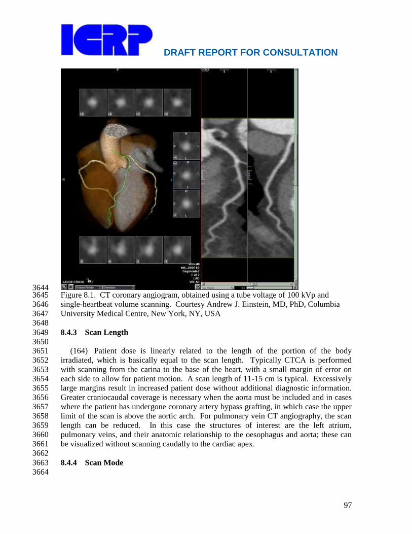
DRAFT REPORT FOR CONSULTATION
97
3644 Figure 8.1. CT coronary angiogram, obtained using a tube voltage of 100 kVp and 3645
single-heartbeat volume scanning. Courtesy Andrew J. Einstein, MD, PhD, Columbia 3646
University Medical Centre, New York, NY, USA 3647
3648
8.4.3 Scan Length 3649
3650 (164) Patient dose is linearly related to the length of the portion of the body 3651
irradiated, which is basically equal to the scan length. Typically CTCA is performed 3652
with scanning from the carina to the base of the heart, with a small margin of error on 3653
each side to allow for patient motion. A scan length of 11-15 cm is typical. Excessively 3654
large margins result in increased patient dose without additional diagnostic information. 3655
Greater craniocaudal coverage is necessary when the aorta must be included and in cases 3656
where the patient has undergone coronary artery bypass grafting, in which case the upper 3657
limit of the scan is above the aortic arch. For pulmonary vein CT angiography, the scan 3658
length can be reduced. In this case the structures of interest are the left atrium, 3659
pulmonary veins, and their anatomic relationship to the oesophagus and aorta; these can 3660
be visualized without scanning caudally to the cardiac apex. 3661
3662
8.4.4 Scan Mode 3663
3664

DRAFT REPORT FOR CONSULTATION
98
(165) Scan modes include conventional helical (spiral) imaging with constant tube 3665
current, conventional helical imaging with ECG-gated tube current modulation 3666
(EGTCM), high-pitch helical imaging and axial imaging, including both step-and-shoot 3667
and volume imaging (Figure 8.2). CTCA using MDCT was first performed using helical 3668
mode and a constant tube current, with a typical pitch of 0.2 for 64-slice scanners 3669
(Figure. 8.2 (a)). All current cardiac scanners offer EGTCM, which keeps tube current 3670
at its maximum during diastasis, when coronary movement is generally minimized, and 3671
decreases tube current during the remainder of the cardiac cycle (Figure 8.2 (b)). This 3672
limits the number of phases of the cardiac cycle in which image reconstructions can be 3673
performed without excessive noise, but for patients with low heart rates (<65 bpm) and 3674
regular heart rhythms, this generally does not pose a problem. Generally, patients should 3675
receive beta blockers or calcium channel blockers to lower heart rate and improve the 3676
efficacy of EGTCM. For patients who do not meet these conditions, reconstructions at 3677
end-systole are often quite useful for visualizing the proximal- and mid-right coronary 3678
artery (Sanz et al., 2005). If EGTCM is applied in these patients, it may be advisable to 3679
widen the period of time during which tube current is maintained at its maximal value. 3680
EGTCM typically decreases effective dose by about one-third. For single-source 3681
scanners, this decrease in dose is more pronounced with lower heart rates (Jakobs et al., 3682
2002). 3683
(166) More recently, axial CTCA protocols have been incorporated into some MDCT 3684
scanners. This approach to scanning acquires image data only during a pre-specified 3685
phase of the cardiac cycle, and the x-ray beam is off during the remainder of the cardiac 3686
cycle. In step-and-shoot (sequential) scanning, x-rays are delivered in one cardiac cycle, 3687
the patient couch is advanced with the beam off during the next cardiac cycle, and the 3688
process is repeated until the entire craniocaudal volume of interest has been scanned. For 3689
64-detector-row scanners, this generally requires 3 or 4 iterations, i.e. 5 or 7 heartbeats (5 3690
heartbeats illustrated in Figure 8.2 (c)). For step-and-shoot imaging to generate 3691
interpretable cardiac images, it is generally thought that heart rate should be less than 70 3692
beats per minute and heart rhythm should be regular, although this has not been well 3693
studied. An advantage of step-and-shoot imaging is reduced dose due to the elimination 3694
of radiation exposure during much of the cardiac cycle and the absence of the overlap of 3695
irradiated areas characteristic of helical CTCA. Disadvantages include the inability to 3696
retrospectively perform image reconstruction at additional phases of the cardiac cycle and 3697
the attendant inability to assess cardiac function and wall motion. 3698
(167) One modification of axial imaging is to increase the length of time that the x-3699
ray tube is on, thus increasing dose but enabling reconstructions within a range of phases 3700
of the cardiac cycle (Figure 8.2 (d)). Thus, rather than obtaining only images in a single 3701
portion of diastasis, a variety of strategies can be employed, such as obtaining images in a 3702
range of diastolic phases, or covering from end-systole through diastasis. Dose is 3703
proportional to exposure time. The optimal strategy for implementation of axial imaging 3704
has not yet been determined. 3705
(168) Two recently-introduced scan modes offer the potential for significant dose 3706
reductions. Both cover the entire heart with x-rays delivered for only a fraction of a 3707
single heartbeat (Figure 8.2 (e)). The extreme case of axial imaging is volume scanning, 3708
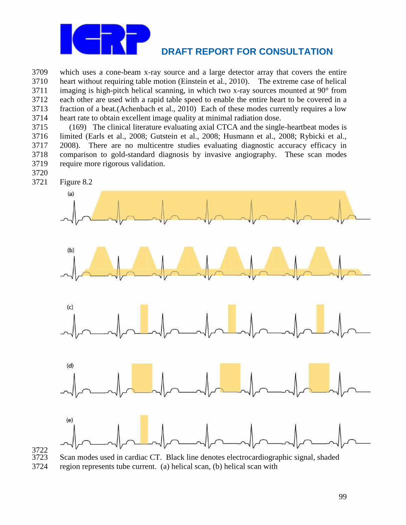
DRAFT REPORT FOR CONSULTATION
99
which uses a cone-beam x-ray source and a large detector array that covers the entire 3709
heart without requiring table motion (Einstein et al., 2010). The extreme case of helical 3710
imaging is high-pitch helical scanning, in which two x-ray sources mounted at 90° from 3711
each other are used with a rapid table speed to enable the entire heart to be covered in a 3712
fraction of a beat.(Achenbach et al., 2010) Each of these modes currently requires a low 3713
heart rate to obtain excellent image quality at minimal radiation dose. 3714
(169) The clinical literature evaluating axial CTCA and the single-heartbeat modes is 3715
limited (Earls et al., 2008; Gutstein et al., 2008; Husmann et al., 2008; Rybicki et al., 3716
2008). There are no multicentre studies evaluating diagnostic accuracy efficacy in 3717
comparison to gold-standard diagnosis by invasive angiography. These scan modes 3718
require more rigorous validation. 3719
3720
Figure 8.2 3721
3722 Scan modes used in cardiac CT. Black line denotes electrocardiographic signal, shaded 3723
region represents tube current. (a) helical scan, (b) helical scan with 3724
DRAFT REPORT FOR CONSULTATION
100
electrocardiographically-gated tube current modulation, (c) axial step-and-shoot scan, (d) 3725
axial step-and-shoot scan, with extending exposure time (―padding‖) to permit 3726
reconstruction of multiple cardiac phases, (e) axial single heartbeat scan (volume and 3727
high-pitch helical scans, illustrated here with no padding). Not all modes are available on 3728
all MDCT scanners. 3729
3730
8.5 Current Dosimetry Estimates 3731
3732 (170) Dosimetry from CTCA depends on many factors, and thus varies markedly 3733
between protocols. Einstein et al reviewed the published literature on effective dose from 3734
cardiac CT in 2007(Einstein et al., 2007). Effective doses from calcium scoring ranged 3735
from 1.0 to 6.2 mSv using helical technique and from 0.5 to 1.8 mSv using axial 3736
technique. For helical 64-slice CTCA, effective dose ranged from 8 to 21.4 mSv without 3737
and from 6.4 to 14 mSv with EGTCM. In a 15 centre study performed in the U.S., 3738
median effective dose, estimated using a k factor of 0.014 mSv·mGy-1
·cm-1
, was 21 mSv 3739
prior to a best-practice dose reduction educational intervention (Raff et al., 2009). In a 3740
50-centre worldwide study, median effective dose was 12 mSv (Hausleiter et al., 2009). 3741
In Hausleiter et al‘s study, there was a 6-fold range in median doses among sites 3742
performing CTCA. EGTCM was associated with a reduction in dose-length product and 3743
effective dose of 25% (95% confidence interval 23-28%), use of an x-ray tube voltage of 3744
100 kV was associated with a reduction of 46% (95% confidence interval 42-51%), and 3745
use of axial step-and-shoot scanning was associated with a reduction of 78% (95% 3746
confidence interval 77-79%) (Hausleiter et al., 2009). Other single-centre studies have 3747
evaluated axial step-and-shoot scanning, and typically report effective doses in the 2-4 3748
mSv range (Earls and Schrack, 2008). In comparison to conventional helical scanning, 3749
volume scanning has been associated with a dose reduction of 84%, (Einstein et al., 3750
2010), and high-pitch helical scanning has been associated with effective dose of <1 mSv 3751
for patients with a slow (≤60 bpm) heart rate who weigh ≤100 kg (Achenbach et al., 3752
2010), using a k factor of 0.014 mSv·mGy-1
·cm-1
. 3753
(171) The wide range of values for effective dose seen in clinical practice makes it 3754
impossible to provide ―typical‖ values for cardiac CT. Effective dose is dependent on 3755
both the CT scanner and the protocol used. Estimates of approximate average values are 3756
presented in Table 8.1, but it must be appreciated that these values should not be 3757
considered as typical values, target values, or representative of clinical practice at any 3758
one institution. 3759
3760
3761
3762
3763
3764
3765
3766
3767
3768

DRAFT REPORT FOR CONSULTATION
101
Table 8.1 Estimated Approximate Average Effective Dose for Various Types of Cardiac 3769
CT Examinations 3770
3771
Examination Effective Dose (mSv)* 3772
3773
CT coronary angiography (CTCA) (helical) 19 3774
3775
CT coronary angiography (CTCA) (tube current modulation) 13 3776
3777
CT coronary angiography (CTCA) (prospectively gated) 4 3778
3779
Coronary artery calcium scoring (CAC) 2 3780
3781
*The data in the Table are reproduced from Einstein, 2009. For other estimates of 3782
effective dose, see, e.g., Einstein et al, 2007; Hausleiter et al, 2009; Kim et al, 2009; 3783
Smith-Bindman et al, 2009; Earls and Schrack, 2009; Raff et al, 2009. 3784
3785
3786
3787
(172) Effective doses reported in many of the studies assessing CT protocols are 3788
determined on a patient-by-patient basis. The existence of conversion factors, such as 3789
those in the European Guidelines on Quality Criteria for CT (Bongartz et al., 2000; 3790
Bongartz et al.), make it easy for an investigator to estimate an ―effective dose‖ for a 3791
single study from the DLP reported on the scanner, but this is not the intended use of 3792
effective dose (Einstein et al., 2008; Gerber et al., 2009; ICRP 2007b). Citation of these 3793
studies is not an endorsement of this approach by the Commission. When the 3794
Commission introduced effective dose in 1990 (ICRP, 1991), it was defined for 3795
populations, not for specific individuals. This has not changed. 3796
3797
8.6 Radiological Protection of Patients in Cardiac CT 3798
3799 (173) The general principles of radiological protection (chapter 4), i.e., justification 3800
and optimisation, can be applied to the protection of patients in cardiac CT. Dose 3801
limitation is not appropriate, but diagnostic reference levels should be used to help 3802
manage the radiation dose so that the dose is commensurate with the clinical purpose 3803
(ICRP, 2007b, ICRP, 2007c). 3804
3805
8.6.1 Justification 3806
3807 (174) The Commission recommends the development and application of appropriate 3808
use criteria for cardiac CT. Appropriate indications for cardiac CT are available from 3809
professional organizations and should be used (Taylor et al, 2010; Schroeder et al., 2008). 3810
3811

DRAFT REPORT FOR CONSULTATION
102
(175) In reports from one institution, 46% of CTCA studies but only 11% of stress 3812
SPECT studies were unclassifiable in terms of appropriateness, and of the remaining 3813
classifiable studies, 51% of CTCA studies and 72% of stress SPECT studies were 3814
appropriate.(Gibbons et al., 2008; Miller et al.) It is unclear from these data whether the 3815
difference between modalities primarily reflects a limitation with the first version of the 3816
U.S. CTCA appropriateness criteria, which left many studies unclassifiable, or whether 3817
CTCA studies are less likely to be performed for appropriate indications that SPECT 3818
studies. Further investigation is required, and programs to ensure maximal adherence to 3819
appropriate use criteria are also encouraged. 3820
3821
8.6.2 Optimization 3822
3823 (176) As discussed in section 8.3, the operator controls numerous scan parameters 3824
that affect patient dose. The operator should be provided with appropriate guidelines for 3825
mAs and kVp selection as a function of patient body habitus. Special consideration 3826
should be given to reducing mAs and/or kVp when evaluation of coronary plaques and 3827
stenoses is not the primary aim, e.g. for evaluation of possible anomalous coronaries, or 3828
prior to repeat cardiac surgery to assess the course of bypass grafts in relation to the 3829
sternum. Scan length should be limited to that needed to reliably image the volume of 3830
interest. 3831
(177) The operator should be provided with appropriate guidelines for selection of 3832
the scan mode. Scan modes that reduce dose should be employed as appropriate (Gerber 3833
et al., 2009). Scans performed for calcium scoring should be performed using axial 3834
imaging, and in combined studies should be reviewed prior to performance of CTCA. 3835
The presence of widespread, heavy coronary calcification may suggest that CTCA should 3836
not be performed, due to the high likelihood of unevaluable coronary segments. For all 3837
patients, with the possible exception of patients scanned on a multiple-source scanner 3838
with variable pitch, rate-control agents should be given as needed with the goal of 3839
decreasing heart rate to approximately 60 beats per minute. 3840
(178) The Commission recommends formal training in radiological protection, and in 3841
particular in the application of the principles of justification and optimization, for all 3842
physicians who refer patients for, or perform, cardiac CT studies (ICRP 113, 2011). This 3843
includes cardiologists, radiologists, nuclear medicine specialists, and internists. 3844
(179) Quality improvement programs have been shown to decrease radiation dose 3845
substantially for CTCA (Raff et al., 2009), and thus their implementation is encouraged. 3846
3847
8.6.3 Diagnostic Reference Levels 3848
3849 (180) Diagnostic reference levels are used in medical imaging to indicate whether, in 3850
routine conditions, the levels of patient dose from, or administered activity for, a 3851
specified imaging procedure are unusually high or low for that procedure (ICRP, 2007b). 3852
They are discussed further in Chapter 10. If so, a local review should be initiated to 3853
determine whether protection has been adequately optimised or whether corrective action 3854
is required. 3855
DRAFT REPORT FOR CONSULTATION
103
(181) Professional medical bodies (in conjunction with national health and 3856
radiological protection authorities) are encouraged to set diagnostic reference levels that 3857
best meet their specific needs and that are consistent for the regional, national, or local 3858
area to which they apply (ICRP, 2007c). At present, no diagnostic reference levels exist 3859
for cardiac CT. 3860
3861
8.7 References 3862 3863
Abada, H.T., Larchez, C., Daoud, B., Sigal-Cinqualbre, A., Paul, J.F., 2006. MDCT of 3864
the coronary arteries: feasibility of low-dose CT with ECG-pulsed tube current 3865
modulation to reduce radiation dose. AJR Am J Roentgenol 186, S387-390. 3866
Abdulla, J., Abildstrom, S.Z., Gotzsche, O., Christensen, E., Kober, L., Torp-Pedersen, 3867
C., 2007. 64-multislice detector computed tomography coronary angiography as 3868
potential alternative to conventional coronary angiography: a systematic review and 3869
meta-analysis. Eur Heart J 28, 3042-3050. 3870
Achenbach, S., Marwan, M., Ropers, D., Schepis, T., Pflederer, T., Anders, K., Kuettner, 3871
A., Daniel, W.G., Uder, M., Lell, M.M., 2010. Coronary computed tomography 3872
angiography with a consistent dose below 1 mSv using prospectively 3873
electrocardiogram-triggered high-pitch spiral acquisition. Eur Heart J 31, 340-346. 3874
Bischoff, B., Hein, F., Meyer, T., Hadamitzky, M., Martinoff, S., Schomig, A., 3875
Hausleiter, J., 2009. Impact of a reduced tube voltage on CT angiography and 3876
radiation dose: results of the PROTECTION I study. JACC Cardiovasc Imaging 2, 3877
940-946. 3878
Block, M., Bove, A.A., Ritman, E.L., 1984. Coronary angiographic examination with the 3879
dynamic spatial reconstructor. Circulation 70, 209-216. 3880
Bongartz, G., Golding, S.J., Jurik, A.G., Leonardi, M., van Meerten, E.v.P., Geleijns, J., 3881
Jessen, K.A., Panzer, W., Shrimpton, P.C., Tosi, G., 2000. European Guidelines on 3882
Quality Criteria for Computed Tomography. EUR 16262. The European 3883
Commission's Study Group on Development of Quality Criteria for Computed 3884
Tomography. 3885
Bongartz, G., Golding, S.J., Jurik, A.G., Leonardi, M., van Meerten, E.v.P., Rodríguez, 3886
R., Schneider, K., Calzado, A., Geleijns, J., Jessen, K.A., Panzer, W., Shrimpton, 3887
P.C., Tosi, G., 2004 CT Quality Criteria, European Commission. 3888
Christner, J.A., Kofler, J.M., McCollough, C.H., 2010. Estimating effective dose for CT 3889
using dose-length product compared with using organ doses: consequences of 3890
adopting International Commission on Radiological Protection publication 103 or 3891
dual-energy scanning. AJR Am J Roentgenol 194, 881-889. 3892
Earls, J.P., Schrack, E.C., 2009. Prospectively gated low-dose CCTA: 24 months 3893
experience in more than 2,000 clinical cases. Int. J. Cardiovasc. Imaging 25, 177-3894
187. 3895
Einstein, A.J., Elliston, C.D., Arai, A.E., Chen, M.Y., Mather, R., Pearson, G.D.N., 3896
DeLaPaz, R.L., Nickoloff, E., Dutta, A., Brenner, D.J., 2010. Radiation dose from 3897
single heartbeat coronary CT angiography performed using a 320 detector-row 3898
volume scanner. Radiology 254, 698-704. 3899
DRAFT REPORT FOR CONSULTATION
104
Einstein, A.J., 2009. Medical imaging: the radiation issue. Nat. Rev. Cardiol. 6, 436-438. 3900
Einstein, A.J., Moser, K.W., Thompson, R.C., Cerqueira, M.D., Henzlova, M.J., 2007. 3901
Radiation dose to patients from cardiac diagnostic imaging. Circulation 116, 1290-3902
1305. 3903
Einstein, A.J., Sanz, J., Dellegrottaglie, S., Milite, M., Sirol, M., Henzlova, M., 3904
Rajagopalan, S., 2008. Radiation dose and cancer risk estimates in 16-slice computed 3905
tomography coronary angiography. J Nucl Cardiol 15, 232-240. 3906
Gerber, T.C., Carr, J.J., Arai, A.E., Dixon, R.L., Ferrari, V.A., Gomes, A.S., Heller, 3907
G.V., McCollough, C.H., McNitt-Gray, M.F., Mettler, F.A., Mieres, J.H., Morin, 3908
R.L., Yester, M.V., 2009. Ionizing Radiation in Cardiac Imaging. A Science 3909
Advisory From the American Heart Association Committee on Cardiac Imaging of 3910
the Council on Clinical Cardiology and Committee on Cardiovascular Imaging and 3911
Intervention of the Council on Cardiovascular Radiology and Intervention. 3912
Circulation 119, 1056-1065. 3913
Gerber, T.C., Stratmann, B.P., Kuzo, R.S., Kantor, B., Morin, R.L., 2005. Effect of 3914
acquisition technique on radiation dose and image quality in multidetector row 3915
computed tomography coronary angiography with submillimeter collimation. 3916
Invest.Radiol. 40, 556. 3917
Gibbons, R.J., Miller, T.D., Hodge, D., Urban, L., Araoz, P.A., Pellikka, P., McCully, 3918
R.B., 2008. Application of appropriateness criteria to stress single-photon emission 3919
computed tomography sestamibi studies and stress echocardiograms in an academic 3920
medical center. J Am Coll Cardiol 51, 1283-1289. 3921
Hausleiter, J., Meyer, T., Hadamitzky, M., Huber, E., Zankl, M., Martinoff, S., Kastrati, 3922
A., Schomig, A., 2006. Radiation dose estimates from cardiac multislice computed 3923
tomography in daily practice: impact of different scanning protocols on effective 3924
dose estimates. Circulation 113, 1305. 3925
Hausleiter, J., Meyer, T., Hermann, F., Hadamitzky, M., Krebs, M., Gerber, T.C., 3926
McCollough, C., Martinoff, S., Kastrati, A., Schömig, A., Achenbach, S., 2009. 3927
Estimated radiation dose associated with cardiac CT angiography. JAMA 301, 500-3928
507. 3929
Hausleiter, J., Martinoff, S., Hadamitzky, M., Martuscelli, E., Pschierer, I., Feuchtner, 3930
G.M., Catalan-Sanz, P., Czermak, B., Meyer, T.S., Hein, F., Bischoff, B., Kuse, M., 3931
Schomig, A., Achenbach, S., 2010. Image quality and radiation exposure with a low 3932
tube voltage protocol for coronary CT angiography. Results of the PROTECTION II 3933
Trial. JACC Cardiovasc Imaging 3, 1113-1123. 3934
Hounsfield, G.N., 1979. Computed medical imaging. Nobel lecture, 8 December 1979. 3935
ICRP, 1991. 1990 Recommendations of the International Commission on Radiological 3936
Protection. ICRP Publication 60. Ann. ICRP 21, 1-201. 3937
ICRP, 2007a. Managing patient dose in multi-detector computed tomography (MDCT). 3938
ICRP Publication 102. Ann. ICRP 37(1), 1-79. 3939
ICRP, 2007b. The 2007 recommendations of the International Commission on 3940
Radiological Protection. ICRP Publication 103. Ann. ICRP 37, 1-332. 3941
ICRP, 2007c. Radiological Protection in Medicine. ICRP Publication 105. Ann. ICRP 3942
37(6), 1-64. 3943

DRAFT REPORT FOR CONSULTATION
105
IEC, 2009. Medical Electrical Equipment - Part 2-44: Particular Requirements for the 3944
Safety of X-ray Equipment for Computed Tomography. International Standard IEC 3945
60601-2-44. 3rd Edition. International Electrotechnical Commission, Geneva. 3946
Jakobs, T.F., Becker, C.R., Ohnesorge, B., Flohr, T., Suess, C., Schoepf, U.J., Reiser, 3947
M.F., 2002. Multislice helical CT of the heart with retrospective ECG gating: 3948
reduction of radiation exposure by ECG-controlled tube current modulation. 3949
Eur.Radiol. 12, 1081. 3950
Kim, K.P., Einstein, A.J., Berrington de Gonzalez, A., 2009. Coronary artery 3951
calcification screening: estimated radiation dose and cancer risk. Arch. Intern. Med. 3952
169, 1188-1194. 3953
McCollough, C.H., 2008. CT dose: how to measure, how to reduce. Health Physics 95, 3954
508-517. 3955
Miller, J.A., Raichlin, E., Williamson, E.E., McCully, R.B., Pellikka, P.A., Hodge, D.O., 3956
Miller, T.D., Gibbons, R.J., Araoz, P.A., 2010. Evaluation of coronary CTA 3957
Appropriateness Criteria in an academic medical center. J Am Coll Radiol 7, 125-3958
131. 3959
Morin, R.L., Gerber, T.C., McCollough, C.H., 2003. Radiation dose in computed 3960
tomography of the heart. Circulation 107, 917. 3961
Raff, G.L., Chinnaiyan, K.M., Share, D.A., Goraya, T.Y., Kazerooni, E.A., Moscucci, 3962
M., Gentry, R.E., Abidov, A., 2009. Radiation dose from cardiac computed 3963
tomography before and after implementation of radiation dose-reduction techniques. 3964
JAMA 301, 2340-2348. 3965
Sanz, J., Rius, T., Kuschnir, P., Fuster, V., Goldberg, J., Ye, X.Y., Wisdom, P., Poon, 3966
M., 2005. The importance of end-systole for optimal reconstruction protocol of 3967
coronary angiography with 16-slice multidetector computed tomography. 3968
Invest.Radiol. 40, 155. 3969
Schroeder, S., Achenbach, S., Bengel, F., Burgstahler, C., Cademartiri, F., de Feyter, P., 3970
George, R., Kaufmann, P., Kopp, A.F., Knuuti, J., Ropers, D., Schuijf, J., Tops, L.F., 3971
Bax, J.J., 2008. Cardiac computed tomography: indications, applications, limitations, 3972
and training requirements: report of a Writing Group deployed by the Working 3973
Group Nuclear Cardiology and Cardiac CT of the European Society of Cardiology 3974
and the European Council of Nuclear Cardiology. Eur Heart J 29, 531-556. 3975
Smith-Bindman, R., Lipson, J., Marcus, R., Kim, K.P., Mahesh, M., Gould, R., 3976
Berrington de Gonzalez, A., Miglioretti, D.L., 2009. Radiation dose associated with 3977
common computed tomography examinations and the associated lifetime attributable 3978
risk of cancer. Arch. Intern. Med. 169, 2078-2086. 3979
Taylor, A.J., Cerqueira, M., Hodgson, J.M., Mark, D., Min, J., O'Gara, P., Rubin, G.D., 3980
Kramer, C.M., Berman, D., Brown, A., Chaudhry, F.A., Cury, R.C., Desai, M.Y., 3981
Einstein, A.J., Gomes, A.S., Harrington, R., Hoffmann, U., Khare, R., Lesser, J., 3982
McGann, C., Rosenberg, A., Schwartz, R., Shelton, M., Smetana, G.W., Smith, S.C., 3983
Jr., 2010. ACCF/SCCT/ACR/AHA/ASE/ASNC/NASCI/SCAI/SCMR 2010 3984
appropriate use criteria for cardiac computed tomography: a report of the American 3985
College of Cardiology Foundation Appropriate Use Criteria Task Force, the Society 3986
of Cardiovascular Computed Tomography, the American College of Radiology, the 3987

DRAFT REPORT FOR CONSULTATION
106
American Heart Association, the American Society of Echocardiography, the 3988
American Society of Nuclear Cardiology, the North American Society for 3989
Cardiovascular Imaging, the Society for Cardiovascular Angiography and 3990
Interventions, and the Society for Cardiovascular Magnetic Resonance. J. Am. Coll. 3991
Cardiol. 56, 1864-1894. 3992
3993
3994
3995
3996

DRAFT REPORT FOR CONSULTATION
107
9. RADIOLOGICAL PROTECTION TRAINING FOR 3997
INTERVENTIONAL FLUOROSCOPY 3998
3999
Main Points 4000
Interventional cardiologists worldwide typically have little or no training in 4001
radiological protection (RP). 4002
Legislation in most countries requires that individuals who take 4003
responsibility for medical exposures must be properly trained in RP. 4004
Training activities in RP should be followed by an evaluation of the 4005
knowledge acquired from the training programme (a formal examination 4006
system). 4007
Physicians who have completed training should be able to demonstrate that 4008
they possess the knowledge specified by the curriculum by passing an 4009
appropriate certifying examination. 4010
In addition to the training recommended for other physicians who use X-4011
rays, interventionalists, including interventional cardiologists, should receive 4012
a second, higher level of RP training. 4013
Nurses and other healthcare professionals who assist during fluoroscopic 4014
procedures should be familiar with radiation risks and radiological 4015
protection principles, in order to minimise their own exposure and that of 4016
others. 4017
Training programmes should include both initial training for all incoming 4018
staff and regular updating and retraining. 4019
Scientific congresses should include refresher courses on RP, attendance at 4020
which could be a requirement for continuing professional development 4021
4022
9.1 Introduction 4023
4024
(182) Despite the extensive and routine use of x-rays in their clinical practice, 4025
interventional cardiologists (IC) worldwide typically have little or no training in 4026
radiological protection (RP). Traditionally, medical students do not receive training in RP 4027
during medical school. Medical professionals who subsequently specialise in radiological 4028
specialties, such as diagnostic radiology, nuclear medicine and radiotherapy, are taught 4029
radiological physics and RP as part of their specialty training. In many countries, there is 4030
no teaching of RP during training in other specialties, such as medicine and cardiology. 4031
(183) In the past, training in radiological physics and RP was not necessary for non-4032
radiologists, as x-rays and other radiation sources were employed only in radiology 4033
departments, by staff with reasonable training in RP. Although x-ray fluoroscopy has 4034
been in use for more than a century now, its early application involved visualization of 4035
body anatomy, movement of structures or passage of contrast media through the body. 4036
Radiologists normally performed these procedures. When fluoroscopically guided 4037
interventions were introduced, other specialists (cardiologists and an increasing number 4038

DRAFT REPORT FOR CONSULTATION
108
of clinicians in other medical specialties) began performing these procedures. Initially, 4039
they did so jointly with radiologists, in radiology departments. Over the years, x-ray 4040
equipment was installed in other clinical departments and used by non-radiologists 4041
without radiologist participation. These non-radiologists were not subject to the training 4042
requirements of radiological physics and RP that were mandatory for radiologists. It is 4043
now clear that this training is essential; hence the need for specific guidance for 4044
cardiology. 4045
(184) The Commission has addressed the specifics of training for interventionalists 4046
and nuclear medicine specialists, among others, in ICRP Publication 113 (ICRP 113, 4047
2009). Further information on training in nuclear medicine is presented in Section 7.7.2. 4048
9.2 Requirements on Radiological protection 4049
4050
(185) In its Publications 85 and 113, the Commission recommends a second level of 4051
RP training for interventional radiologists and cardiologists, in addition to the training 4052
recommended for other physicians who use X-rays (ICRP 85, 2000, ICRP 113, 2009). 4053
The Commission also recommends that nurses and other healthcare professionals who 4054
assist during fluoroscopic procedures should be familiar with radiation risks and 4055
precautions, in order to minimise their own exposure and that of others. 4056
(186) Training activities in RP should be followed by an evaluation of the knowledge 4057
acquired from the training programme. Education and training in RP should be 4058
complemented by formal examination systems to test competency before the person is 4059
awarded certification. If certification in RP is required for some medical specialties (e.g. 4060
interventional cardiology), certification should be obtained before the professional 4061
practices the specialty.. Training programmes should include both initial training for all 4062
incoming staff and regular updating and retraining. Scientific and professional societies 4063
should contribute to the development of the training syllabuses to ensure a consistent 4064
approach, and to promote and support the education and training. Scientific congresses 4065
should include refresher courses on RP, attendance at which could be a requirement for 4066
continuing professional development for professionals using ionising radiation. (ICRP 4067
113, 2009). 4068
(187) The International Basic Safety Standards for Protection against Ionising 4069
Radiation and for the Safety of Radiation Sources (BSS), published by the International 4070
Atomic Energy Agency (IAEA) and jointly sponsored by the Food and Agriculture 4071
Organization (FAO), the International Labour Organization (ILO), the Pan American 4072
Health Organization (PAHO) and the World Health Organization (WHO) (IAEA, 1996), 4073
require appropriate training that is sufficient to perform assigned tasks in the conduct of 4074
diagnostic or therapeutic procedures involving radiation. 4075
(188) The Medical Exposure Directive of EC 97/43/Euratom considers interventional 4076
radiology (Article 9) as a special practice involving high doses to patients (EU, 1997). 4077
According to Article 7, Member States shall ensure that the practitioner has adequate 4078
theoretical and practical training for the purpose of radiological practice as well as 4079
relevant competence in radiological protection. No special mention is made of 4080
interventional cardiology. 4081

DRAFT REPORT FOR CONSULTATION
109
4082
(189) Legislation in most countries requires that individuals who take responsibilities 4083
for medical exposure must be properly trained in RP. However, a training system and 4084
accreditation mechanism is still lacking in many countries. 4085
9.3 Training guidelines, curricula and materials 4086
4087
(190) The Commission, in Publication 85 (ICRP, 2000), states that interventional 4088
procedures are complex and demanding and that radiation dose tends to be operator 4089
dependent. It is particularly important that individuals performing these procedures are 4090
adequately trained both in clinical techniques and in radiological protection. It further 4091
states that special additional training should be planned when new x-ray systems or 4092
techniques are implemented in a centre. Basic and continuing training in radiological 4093
protection should be an integral part of this education. Training requirements are 4094
addressed in Publication 113 (ICRP 113, 2009). 4095
(191) In view of the number of radiation-induced injuries reported in recent years 4096
among patients undergoing interventional procedures (Rehani and Ortiz-Lopez, 2005, 4097
Vano and Gonzalez, 2005, ICRP, 2000, Koenig et al, 2001), a number of organizations 4098
have begun to provide recommendations for training requirements. Published guidelines 4099
were initially for interventional radiologists, but they are gradually becoming available 4100
from cardiology societies. 4101
4102
9.3.1 USA 4103
4104 (192) The Food and Drug Administration (FDA) advisory of 1994 (FDA, 1994) 4105
alerted facilities to ensure proper training. FDA‘s specific recommendations for facilities 4106
in which invasive procedures are performed included the following: 4107
4108
Assure appropriate credentials and training for physicians performing 4109
fluoroscopy. 4110
All operators of the system must be trained and understand the operation of the 4111
fluoroscopic system, including the implications for radiation exposure from each 4112
mode of operation. 4113
Facilities should ensure that physicians performing fluoroscopic procedures are 4114
educated so that they may, on a case-by-case basis, assess risks and benefits for 4115
individual patients, considering variables such as age, beam location and 4116
direction, tissues in the beam and previous fluoroscopic procedures or radiation 4117
therapy. 4118
4119
(193) In 1995, the American College of Cardiology Cardiac Catheterization 4120
Committee published a Position Statement indicating that appropriate training of staff is 4121
imperative, and that ‗‗Proper instruction in the principles of radiation physics and safety 4122
should be a part of every cardiologist‘s education‖ (Brinker et al., 1995). The American 4123

DRAFT REPORT FOR CONSULTATION
110
College of Cardiology Consensus Document further clearly delineated the need for a 4124
radiation safety knowledge base for cardiology staff (Limacher et al., 1998). 4125
(194) In 2004, an American College of Cardiology/American Heart Association/ 4126
American College of Physicians (ACC/AHA/ACP) Task Force published a further report 4127
on clinical competence and training as a companion to the ACC‘s 1998 report (Hirshfeld 4128
et al, 2004; Limacher et al, 1998). The proposed curriculum in the 2004 document 4129
specifies the knowledge that a qualified physician should possess in order to be 4130
credentialed to use x-ray fluoroscopic machines, but does not specify a minimum number 4131
of hours of training. Physicians who have completed training should be able to 4132
demonstrate that they possess the knowledge specified by the curriculum by passing an 4133
appropriate certifying examination. 4134
(195) The necessary knowledge depth varies, depending upon the types of 4135
fluoroscopically guided procedures a particular physician performs. The ACC/AHA/ACP 4136
document outlines two different curricula—basic and advanced. The basic curriculum is 4137
appropriate for physicians who perform simpler fluoroscopically guided critical-care unit 4138
procedures such as right heart catheterization, temporary pacemaker placement, and intra-4139
aortic balloon pump placement. The advanced curriculum is appropriate for physicians 4140
who perform angiographic, interventional, and electrophysiological procedures that 4141
employ greater amounts of radiation in more complex circumstances with different 4142
purposes and a greater attendant risk of patient and personnel injury. 4143
(196) The National Council on Radiation Protection and Measurements (NCRP) in 4144
the U.S. recently published a report on radiation dose management for fluoroscopically 4145
guided interventional medical procedures (NCRP, 2010). This report makes a number of 4146
specific recommendations, including: 4147
4148
Each individual present in a fluoroscopically guided interventional (FGI) 4149
procedure room shall have appropriate radiological protection training. 4150
Every person who operates or supervises the use of FGI equipment shall have 4151
current training in the safe use of that specific equipment. 4152
Interventionalists who perform FGI procedures or other procedures with the 4153
potential for high patient doses require additional knowledge and training beyond 4154
that necessary for interventionalists whose practice is limited to low-dose FGI 4155
procedures. 4156
Clinical training and experience is not an acceptable substitute for formal training 4157
in radiation management. 4158
9.3.2 European Commission 4159
(197) In compliance with European Commission requirements, an outline for specific 4160
training in radiological protection for interventional radiology has been developed (EC, 4161
2000; Vañó et al. 1997). Although there is no special mention of interventional 4162
cardiology in the group of professionals, the table giving suggested number of training 4163
hours has a column for interventional cardiology specialists; 20-30 hours of training are 4164
suggested. The initial Spanish experience, based on these guidelines, has been reported 4165
(Vañó, 2003). This included development of a training CD (MARTIR, 2002). 4166

DRAFT REPORT FOR CONSULTATION
111
9.3.3 International Atomic Energy Agency 4167
4168
(198) The International Atomic Energy Agency (IAEA) has developed a curriculum 4169
with educational objectives specifically for interventional cardiologists. It is directed 4170
primarily at developing countries where the cardiology professional societies are not yet 4171
sufficiently robust to develop their own separate modules for basic and advanced 4172
curricula in the field of radiological protection. For these countries a ―sandwich‖ module 4173
is ideal, particularly in view of the lack of individuals with sufficient expertise in 4174
radiological protection in diagnostic imaging to teach the subject. IAEA has also 4175
prepared educational material in the form of an electronic presentation on CD. This 4176
IAEA training material on Radiation Protection in Cardiology is available without cost 4177
and can be obtained by writing to [email protected] or downloaded from the 4178
website http://rpop.iaea.org. 4179
9.3.4 WHO 4180
4181
(199) The World Health Organization (WHO) has stated that specific training in 4182
interventional radiology is required in addition to basic training and has provided training 4183
requirements (WHO 2000). WHO further stated that the training process must be 4184
continued when new techniques are introduced, when new radiological systems are 4185
installed and when new staff are appointed. It also recommended continuous training and 4186
refresher courses at regular intervals. However, interventional cardiology was outside the 4187
scope of this document. 4188
9.4 Credentialing 4189
4190
(200) There is a distinction between the credentialing of a physician as technically 4191
competent to perform a procedure versus the credentialing of the same physician as 4192
competent to safely use a fluoroscope. Since the amount of radiation employed by the 4193
interventional cardiologist both per patient and annually is no less than that used by an 4194
interventional radiologist, the training standards of radiation physics and radiological 4195
protection in interventional cardiology should be the same as for other interventionalists 4196
(ICRP 113, 2009). 4197
9.5 References 4198
4199
Brinker, J.A. et al, 1995. Position paper of the American College of Cardiology Cardiac 4200
Catheterization Committee. JACC 26, 319-325. 4201
EU, 1997. Council Directive 97/43 Euratom, on health protection of individuals against the 4202
dangers ionizing radiation in relation to medical exposure, and repealing Directive 4203
84/466 Euratom. Official Journal of the European Communities No L 180, 9th July 4204
1997:22-7. 4205
http://europa.eu.int/comm/energy/nuclear/radioprotection/legislation_en.htm 4206

DRAFT REPORT FOR CONSULTATION
112
EC, 2000. Guidelines on education and training in radiation protection for medical 4207
exposures. Radiation Protection 116, European Commission. Directorate General 4208
Environment, Nuclear Safety and Civil Protection. Luxembourg, 2000. Available at: 4209
http://ec.europa.eu/energy/nuclear/radiation_protection/doc/publication/116.pdf 4210
Accessed January 2, 2011. 4211
FDA, 1994. Avoidance of serious x-ray-induced skin injuries to patients during 4212
fluoroscopically-guided procedures. Statement 9 Sept. 1994, Food and Drug 4213
Administration, Rockville, MD, USA. 4214
Hirshfeld, J.W., Jr., Balter, S., Brinker, J.A., Kern, M.J., Klein, L.W., Lindsay, B.D., 4215
Tommaso, C.L., Tracy, C.M., Wagner, L.K., 2004. ACCF/AHA/HRS/SCAI clinical 4216
competence statement on physician knowledge to optimize patient safety and image 4217
quality in fluoroscopically guided invasive cardiovascular procedures: a report of the 4218
American College of Cardiology Foundation/American Heart Association/American 4219
College of Physicians Task Force on Clinical Competence and Training. J. Am. Coll. 4220
Cardiol. 44, 2259-2282. 4221
IAEA, 1996. International Basic Safety Standards against Ionizing Radiation and for the 4222
Safety of Radiation Sources, Safety Series No. 115, Vienna: IAEA. 4223
ICRP, 2000. Avoidance of radiation injuries from medical interventional procedures. ICRP 4224
Publication 85, Annals of the ICRP 30 (2). 4225
ICRP, 2009. Radiological protection education and training for healthcare staff and 4226
students (diagnostic imaging and interventional procedures). ICRP Publication 113. 4227
Ann ICRP 39 (5). 4228
Koenig TR, Wolff D, Mettler FA, Wagner LK. Skin injuries from fluoroscopically guided 4229
procedures: Part 1, Characteristics of radiation injury. AJR 2001; 177: 3-11 4230
Limacher, M.C. et al, 1998. Radiation safety in the practice of cardiology. JACC 31, 892-4231
913. 4232
MARTIR (Multimedia and Audiovisual Radiation Protection Training in Interventional 4233
Radiology), 2002. CD-ROM. Radiation Protection 119, European Commission. 4234
Directorate General Environment, Nuclear Safety and Civil Protection. Luxembourg 4235
(free availability at 4236
http://ec.europa.eu/energy/nuclear/radiation_protection/publications_en.htm; go to 4237
publication 119). 4238
NCRP, 2010. Radiation dose management for fluoroscopically guided medical procedures. 4239
Report No. 168. National Council on Radiation Protection and Measurements, 4240
Bethesda, MD. 4241
Rehani, M.M. and Ortiz-Lopez, P., 2006. Radiation effects in fluoroscopically guided 4242
cardiac interventions - keeping them under control. Int. J. Cardiol. 109(2), 147-151. 4243
Vano E, and Gonzalez L. 2005. Accreditation in radiation protection for cardiologists and 4244
interventionalists. Radiat Prot Dosimetry. 117(1-3):69-73. 4245
Vañó, E., Gonzalez, L., Faulkner, K. et al, 1997. Specific training objectives in radiation 4246
protection for medical staff in interventional radiology installations. Draft document. 4247
DIMOND European Concerted Action. 4248
Vañó, E., Gonzalez, L., Canis, M. et el., 2003. Training in radiological protection for 4249
interventionalists. Initial Spanish experience. BJR 76, 217-219. 4250

DRAFT REPORT FOR CONSULTATION
113
WHO, 2000. Efficacy and radiation safety in interventional radiology. World Health 4251
Organisation, Geneva. 4252
4253
4254
4255

DRAFT REPORT FOR CONSULTATION
114
10. QUALITY ASSURANCE PROGRAMMES 4256
4257
Main Points 4258
Two basic objectives of the radiological protection quality assurance 4259
programme (QAP) are to evaluate patient radiation dose on a periodic basis 4260
and to monitor occupational radiation dose for workers in cardiology facilities 4261
where radiation is used. 4262
Training in RP (both initial and retraining) should be included in the QAP for 4263
all staff involved in interventional cardiology procedures. 4264
A cardiologist should be in charge of the QAP aspects of RP for cardiology 4265
procedures, and should be assisted by a medical physicist. 4266
A senior interventionalist and a medical physicist should be included in the 4267
planning for a new interventional fluoroscopy laboratory, installation of a new 4268
x-ray or nuclear medicine system and the upgrade of existing equipment. 4269
Periodic evaluation of image quality and procedure protocols should be 4270
included in the QAP. 4271
The QAP should ensure the regular use of personal dosimeters and include a 4272
review of all abnormal dose values. 4273
The QAP should establish a trigger level for individual clinical follow-up when 4274
there is a risk of radiation-induced skin injuries. 4275
Patient dose reports should be produced at the end of procedures, archived 4276
and recorded in the patient’s medical record. If dose reports are not available, 4277
dose values should be recorded in the patient’s medical record together with 4278
procedure and patient identification. 4279
Patient dose audits (including comparison with DRLs) and reporting are 4280
important components of the QAP. 4281
4282
4283
10.1 Introduction 4284 4285
(201) Quality assurance programs in cardiology should cover all of the planned and 4286
systematic actions necessary to provide confidence that optimum quality has been achieved 4287
in the entire diagnostic process, i.e. there is consistent production of adequate diagnostic 4288
information with the lowest acceptable exposure of patients and personnel (WHO 1982). 4289
(202) A quality assurance programme (QAP) for interventional cardiology includes all 4290
of the aspects of radiological protection (RP) of patients and staff in addition to the usual 4291
clinical aspects. Only the RP aspects are discussed here. Two basic objectives of the QAP 4292
are to evaluate patient radiation dose on a periodic basis and to monitor occupational 4293
radiation dose for workers in cardiology facilities where radiation is used. Table 10.1 4294
summarizes the 10 key points to be included in a RP QAP. The RP component of the QAP 4295
for interventional cardiology should be an independent portion of the general QAP for x-4296
ray installations in a particular health centre. 4297
DRAFT REPORT FOR CONSULTATION
115
(203) A cardiologist should be in charge of the QAP aspects of RP for cardiology, and 4298
should be assisted by a medical physicist. The RP QAP for cardiology should be reviewed 4299
at least annually, to allow the opportunity for updates and periodic follow up. Self-audit of 4300
the QAP is also advisable. Table 10.2 presents some questions to be answered as part of 4301
this internal audit of the QAP. 4302
4303
10.2 Facilities 4304 4305
(204) The design of a new interventional fluoroscopy laboratory, the selection and 4306
installation of a new x-ray or nuclear medicine system and the upgrade of existing 4307
equipment are all complex and expensive processes. Planning for these processes should 4308
include RP. Both a senior physician (interventionalist, nuclear medicine specialist or CT 4309
imaging specialist, as appropriate) and a medical physicist should be included in this 4310
planning. Physicians representing all of the medical specialties who will be using the new 4311
room should be involved in specifying the equipment for the room. Important aspects to 4312
consider are shown in Table 10.3. 4313
(205) Suggested architectural specifications for catheterization laboratories have been 4314
published by scientific societies (ACC/AHA 1991): adequate dimensions (50 m2), a 4315
sufficiently large control room with a wide leaded window, sufficient ceiling height (3 m, 4316
allowing for ceiling suspended support of the C-arm, monitors, etc.), appropriate radiation 4317
shielding (including window and doors), easy access for personnel and patients, etc. New x-4318
ray rooms should be of sufficient size to allow personnel to be positioned at a distance from 4319
the patient when inside the X ray room during the procedures. The installation should 4320
include a control room with a wide shielded glass window, so that other clinicians and other 4321
personnel can follow the procedures without radiation exposure. 4322
(206) Appropriate shielding, access to the x-ray room and RP tools (aprons, thyroid 4323
protectors, protective gloves and glasses, protective screens, ceiling-suspended and under-4324
table shields), should be part of the planning for catheterization laboratories. 4325
Dose reduction technology, including the capabilities to measure, record, and transfer 4326
patient dose data to the patient‘s medical record, should be considered an important factor 4327
in the selection of new fluoroscopy and CT equipment. Appropriate standards should be 4328
taken into account (IEC 2010). 4329
4330
10.3 Acceptance and constancy testing 4331 4332
(207) Acceptance tests shall be made by the company supplying the equipment in the 4333
presence of technical personnel from the centre buying the system, or by centre technical 4334
personnel. Commissioning of the new equipment before its clinical use should be the 4335
responsibility of the personnel of the centre. 4336
(208) Periodic quality controls (QC), including dosimeter calibration, should be 4337
planned taking into account international standards, local recommendations and the 4338
recommendations of the x-ray system manufacturer. These should also include practical 4339
results for the appropriate management of patient doses by the cardiologists (e.g. dose rate 4340

DRAFT REPORT FOR CONSULTATION
116
in the different fluoroscopy modes, dose per frame during cine acquisition, CT scan 4341
protocols). 4342
(209) Periodic evaluation of image quality and procedure protocols should also be 4343
included in the QAP. Image quality should be measured with test objects during the 4344
acceptance and constancy tests. With the new digital imaging detectors it is possible to 4345
select a wide range of dose values to obtain the required level of quality in the images. It is 4346
easy to specify excessive dose rates, as these do not impair image quality and are not easily 4347
detected from inspection of the image. Cardiologists, in cooperation with the medical 4348
physicist and the industry engineer should set the fluoroscopic or CT system doses to 4349
achieve the appropriate balance between image quality and dose. 4350
(210) It is possible to perform this periodic evaluation of image quality using clinical 4351
criteria. The European consortium DIMOND (DIMOND 2008) has proposed a set of 4352
criteria to evaluate fluoroscopic cardiac imaging (Bernardi 2001a and 2001b). 4353
(211) Cardiologists should learn the dose required to obtain a certain level of 4354
diagnostic information. For interventional fluoroscopy, this is related to fluoroscopy time, 4355
number of series, number of frames/series, fluoroscopy and cine modes and dose rates, 4356
etc.). It is also important to verify that wedge filters, collimation and C-arm angulations are 4357
used properly. CT scan protocols, modes, and technique factors, and their effect on patient 4358
dose, are discussed in Chapter 8. Concerns related to nuclear medicine doses are discussed 4359
in Chapter 7. 4360
4361
10.4 Staff 4362 4363
(212) An important aspect of the QAP is a description of the roles and responsibilities 4364
of personnel. There should be enough staff to avoid an excessive number of procedures per 4365
specialist, and sufficient nursing and technologist support. Support by network specialists 4366
(for new digital systems), maintenance and service personnel and medical physics 4367
specialists is advised. 4368
(213) Analysis of staff radiation dose should be included in the QAP. Calibrated 4369
dosimeters for staff must be available. In addition to the dosimeter in the x-ray system for 4370
the evaluation of patient dose, personnel working in the catheterization laboratories should 4371
wear appropriate dosimeters, and a strict policy for their use should be implemented. 4372
Additional electronic dosimeters may also be useful, especially for RP training of students 4373
and inexperienced personnel. The QAP should ensure the regular use of personal 4374
dosimeters and include a review of all abnormal dose values. 4375
4376
10.5 Training 4377 4378
(214) Training in RP is another important item to be included in the QAP. Initial 4379
accreditation in RP should follow local requirements. Special attention to training in RP 4380
should be given to fellows and residents. Seminars to analyse patient and staff dose results 4381
can be an excellent educational tool as well as a useful QA activity. Training is discussed in 4382
more detail in chapter 9 and in ICRP Publication 113 (ICRP, 2009). 4383
4384

DRAFT REPORT FOR CONSULTATION
117
10.6 Follow-up for possible radiation-induced skin injuries for interventional 4385
fluoroscopy procedures 4386 4387
(215) The QAP should establish a trigger level for individual clinical follow-up when 4388
there is a risk of radiation-induced skin injuries (ICRP 2000; WHO 2000; NCRP 168). The 4389
Substantial Radiation Dose Level (SRDL) is a threshold value that is used to trigger 4390
additional dose management actions, including patient follow-up (NCRP 2010). There is no 4391
implication that a radiation dose below the SRDL is completely safe or that a radiation dose 4392
above the SRDL will always cause an injury. Some suggested values are a skin dose of 3 4393
Gy, a KAP of 500 Gy·cm2, or an air kerma at the interventional reference point of 5 Gy 4394
(NCRP 168). For cardiology procedures, a KAP between 150 and 250 Gy·cm2 may be 4395
more appropriate, depending on the radiation field size and the specific protocols. These 4396
values could indicate peak skin doses greater than 2 Gy in a single procedure. These values 4397
are intended to trigger follow-up for a radiation dose that might produce a clinically 4398
relevant injury in an average patient. Lower values may be used at the discretion of the 4399
facility, especially when previously irradiated skin is involved (NCI 2005). 4400
(216) If the trigger level has been exceeded, the patient‘s personal physician should be 4401
informed about the patient‘s radiation dose and the possibility of ionising radiation effects. 4402
Appropriate clinical follow up should be arranged. If the dose estimate after the procedure 4403
is close to the threshold for deterministic effects then the patient should be informed of 4404
possible symptoms or observable skin effects by the interventionist or his/her staff. 4405
Information about what the patient should do in case these effects appear should be 4406
provided. 4407
4408
10.7 Dose audits 4409 4410
(217) Patient dose audits and reporting are important components of the QAP. Patient 4411
dose reports should be produced at the end of procedures, archived, and transferred to the 4412
patient‘s medical record. An example of a patient dose report is presented in chapter 5, Fig 4413
5.2. If such reports are not available, dose values should be recorded together with the 4414
procedure and patient identification (Miller et al, 2004). If the reports are available only in 4415
hard copy (printed), relevant data should be transferred to an electronic database for further 4416
analysis. If the reports are available in electronic format, the files should be archived 4417
together with the images. For interventional fluoroscopy, quantities to be measured and 4418
recorded periodically for a significant number of patients include: KAP, reference point air 4419
kerma (if available in the x-ray system), fluoroscopy time, number of series, and number of 4420
frames (NCRP, 2010). Reference point air kerma measurement capability has become 4421
widely available in fluoroscopic equipment manufactured after mid-2006. For CT 4422
examinations, the quantities are CTDIw, CTDIvol or DLP (section 8.3). For nuclear 4423
medicine studies, the quantity is administered activity. 4424
(218) Dose audits should include an evaluation of the centre‘s performance with 4425
respect to established reference levels (section 10.7.1). Dose audits for interventional 4426
cardiology procedures require additional analyses (sections 10.7.3, 10.7.4), because these 4427
procedures also present a risk of deterministic injury. 4428

DRAFT REPORT FOR CONSULTATION
118
4429
10.7.1 Diagnostic Reference Levels 4430 4431
(219) Dose guidelines were first introduced in the U.S and the U.K. in the late 1980s 4432
and early 1990s (Wall, 1998). They were introduced into ICRP recommendations as 4433
―investigation levels‖ in Publication 60 (ICRP, 1990) and as ―diagnostic reference levels‖ 4434
(DRLs) in Publication 73 (ICRP, 1996). DRLs are now an established method of defining 4435
feedback levels for high volume examinations such as chest radiographs or mammograms. 4436
The Commission continues to recommend their use (ICRP 85, ICRP 103, ICRP 105). 4437
4438
(220) DRLs are used to help avoid radiation dose to the patient that does not contribute 4439
to the medical imaging task. They provide practitioners with a straightforward tool for 4440
comparing the radiation doses that they deliver to their patients with the radiation doses 4441
delivered by their colleagues. They are a guide to good practice, but are neither dose limits 4442
nor thresholds that define competent performance of the operator or the equipment. They 4443
are intended to provide guidance on what is achievable with current good practice rather 4444
than optimum performance, and help identify unusually high radiation doses or exposure 4445
levels. A mean dose for a procedure that is less than the reference level does not guarantee 4446
that the procedure is being performed optimally. 4447
4448
(221) To use DRLs as a quality improvement tool, an institution or individual 4449
practitioner collects radiation dose data for cases of a procedure performed in their own 4450
practice. The recommended number of cases varies from 10 to >50, with the latter number 4451
suggested for interventional fluoroscopy procedures because of the high individual 4452
variability in patient dose of cases of image-guided interventional procedures (Wall, 1998, 4453
Vano 2008). The mean radiation dose for the procedure is then compared to the DRL. If 4454
local practice results in a mean radiation dose that is greater than the DRL, the fluoroscopic 4455
equipment should be investigated. If the fluoroscopic equipment is functioning properly 4456
and within specification, operator technique and procedure protocols should be examined 4457
(Vano, 2001). Investigations are also appropriate where local values are substantially below 4458
the DRL, as excessively low doses may be associated with poor image quality. 4459
4460
10.7.2 Application of Diagnostic Reference Levels in interventional fluoroscopy 4461
procedures 4462 4463
(222) At present, there is little evidence to indicate that dose levels are decreasing in 4464
interventional cardiology. If anything, dose levels are increasing due to the increased 4465
complexity of fluoroscopically guided procedures. As the Commission has noted, 4466
reference levels, in principle, could be useful for dose optimization in interventional 4467
fluoroscopy procedures (ICRP 105). However, patient dose distributions for interventional 4468
fluoroscopy procedures extend over a wide range and are very variable due to the differing 4469
complexity of the procedures, different patient sizes and different operational modes. The 4470
Commission has suggested that a potential approach to this problem is to take into account 4471
DRAFT REPORT FOR CONSULTATION
119
the relative ―complexity‖ of the procedure (ICRP 105). Other methods have also been 4472
proposed (NCRP 2010). 4473
(223) Recent studies have provided DRLs for cardiovascular procedures (Peterzol et al 4474
2005, Neofotistou et al 2003, Balter et al 2008, D'Helft et al 2009). Some diagnostic 4475
invasive procedures (e.g., routine coronary angiography) are done in a relatively 4476
standardized way and in sufficient volumes that a valid DRL might be constructed. 4477
(224) The European DIMOND consortium proposed provisional RLs for radiation 4478
doses delivered to patients during two types of invasive cardiology procedures, coronary 4479
angiography (CA) and percutaneous transluminal coronary angioplasty (PTCA). The 4480
proposed DRLs for CA and PTCA were KAP values of 45 Gy·cm2 and 75 Gy·cm
2, 4481
fluoroscopy times of 7.5 min and 17 min and 1250 and 1300 frames, respectively. The 4482
consortium concluded that more studies were required to establish "tolerances" from the 4483
proposed levels, taking into account the complexity of the procedure and the patient's size. 4484
(225) Bernardi and co-workers performed studies in Udine, Italy (Bernardi, 2000) and 4485
later in several European hospitals (Neofotistou, 2003), with quantitative assessments of 4486
complexity in relation to a patient's exposure to radiation. The relationships between 4487
several clinical factors, anatomic factors and technical factors versus fluoroscopy time were 4488
evaluated for PTCA. A scoring system was developed, and two complexity indexes were 4489
conceived, based on which the procedures were divided into three groups: simple, medium, 4490
and complex. The relative complexity of procedures carried out in different centres should 4491
be taken into account when comparing typical patient doses with reference levels. 4492
(226) The IAEA carried out an international project to determine the feasibility of 4493
establishing guidance levels for cardiac catheterization and percutaneous coronary 4494
interventions (IAEA, 2009). The IAEA report has been summarized in a separate 4495
publication (Balter et al, 2008). For PTCA procedures, the report recommended the use of a 4496
reference level, using KAP, of 100 Gy·cm2 for simple procedures, 125 Gy·cm
2 for 4497
moderate complexity procedures and 200 Gy·cm2 for complex procedures. Unfortunately, 4498
methods for quantifying complexity have not yet been developed for other interventional 4499
cardiology procedures, such as electrophysiology ablation or pacemaker insertion. 4500
4501
10.7.3 Evaluation of high dose interventional fluoroscopy procedures 4502
4503 (227) Reference levels are used to evaluate the average dose per procedure. Because of 4504
the lognormal dose distribution that is characteristic of fluoroscopically guided 4505
interventions, an additional process is needed to evaluate the high dose ―tail‖. The high 4506
dose tail is of particular interest, because this tail represents the cases where patient doses 4507
may be high enough to cause deterministic effects. 4508
(228) Cases that required a radiation dose greater than the SRDL (section 10.6) should 4509
be identified and reported to the laboratory director and laboratory quality manager on a 4510
periodic basis. A monthly report is helpful, to ensure that patients with high radiation doses 4511
receive appropriate education and follow-up. 4512
(229) For each such procedure, the report should include patient identifier(s), the dose 4513
delivered during the procedure, the type of procedure, the room in which the procedure was 4514
performed, the operator‘s name, a count of the patient‘s previous invasive procedures 4515

DRAFT REPORT FOR CONSULTATION
120
(essential for estimating total skin dose), and any special notes. The goal of this report is to 4516
help assure that all patients who received a high radiation dose have been appropriately 4517
educated, and that appropriate follow-up is scheduled and performed (Miller et al, 2010). 4518
(230) Cases resulting in possible radiation injuries should be discussed at the next 4519
laboratory QA meeting. This discussion should include any available diagnoses, planned 4520
patient follow-up, and outcomes. Unless it is clear that the injury was not radiation-4521
induced, the procedure should be reviewed for the appropriate use of radiation in the 4522
clinical context (Miller et al, 2010). 4523
4524
10.7.4 Evaluation of skin dose for interventional fluoroscopy procedures 4525 4526
(231) It is advisable to measure the skin dose distribution in a sample of patients, to 4527
verify that basic aspects of patient protection are being followed (e.g. appropriate 4528
collimation, use of wedge filter, avoidance of a high concentration of radiation fields in the 4529
same skin area). (Vano 1997; Guibelalde 2003). Skin dose may be measured with special 4530
film, with dosimeters placed directly on the patient‘s skin, and by other means (Miller et al, 4531
2004). A qualified physicist should be consulted for these measurements. 4532
4533 4534
4535
4536

DRAFT REPORT FOR CONSULTATION
121
4537
Table 10.1. Some key aspects to be included in the section of radiological protection of 4538
the quality assurance programme for cardiac facilities using ionising radiation. 4539 4540
4541
1. Facility design. 4542
2. X ray equipment (selection criteria). 4543
3. Radiological protection tools. 4544
4. Availability of dosimeters. 4545
5. Availability of personnel and their responsibilities. 4546
6. Training in radiological protection (initial and continuing). 4547
7. Patient dose audit and reporting. 4548
8. Clinical follow up for high patient doses 4549
9. Image quality and procedure evaluation. 4550
10. Staff doses. 4551
4552
4553
4554
4555
4556
4557
Table 10.2. Examples of quality indicators 4558 4559
Can your centre report patient dose values from the last year? 4560
Do you have a procedure for the clinical follow-up of high doses to patients? 4561
Do you know the results of your x-ray system QCs? 4562
Are you following your staff dose values? 4563
Do you have a continuous training programme in RP? 4564
4565

DRAFT REPORT FOR CONSULTATION
122
Table 10.3 Facility procurement considerations (ICRP, 2000) 4566 4567
Analysis of clinical need Workload 4568
4569
Equipment specification General requirements 4570
Major equipment components 4571
Functional requirements 4572
Specific equipment requirements 4573
4574
Computer capabilities Image display matrix 4575
Processing times 4576
Memory/image storage 4577
PACS linkages* 4578
HIS linkages† 4579
4580
Systems performance Image quality 4581
Patient dose 4582
Dose control measures 4583
4584
User manuals Technical training 4585
Operational training 4586
4587
Compliance with national Electrical safety 4588
and international standards Mechanical safety 4589
Radiation safety 4590
Room design/shielding 4591
4592
Service and warranty Maintenance programme 4593
Quality control programmes 4594
Access to service software protocols/ 4595
rationale for service schedules 4596
4597
Operation costs Cost of consumables - projected over 5 years 4598
4599
*PACS=picture archiving and communication system 4600
†HIS=hospital information system 4601
4602

DRAFT REPORT FOR CONSULTATION
123
REFERENCES 4603 4604
American College of Cardiology/American Heart Association ad hoc Task Force on 4605
Cardiac Catheterization. ACC/AHA Guidelines for Cardiac Catheterization and 4606
Cardiac Catheterization Laboratories. JACC 1991; 18 (5): 1149 82. 4607
Balter, S., Miller, D.L., Vano, E., Ortiz Lopez, P., Bernardi, G., Cotelo, E., Faulkner, K., 4608
Nowotny, R., Padovani, R., Ramirez, A., 2008. A pilot study exploring the possibility 4609
of establishing guidance levels in x-ray directed interventional procedures. Med Phys 4610
35, 673-680. 4611
Bernardi G, Padovani R, Morocutti G, Vano E, Malisan MR, Rinuncini M, Spedicato L, 4612
Fioretti PM. Clinical and technical determinants of the complexity of percutaneous 4613
transluminal coronary angioplasty procedures: analysis in relation to radiation 4614
exposure parameters. Catheter Cardiovasc Interv 2000 Sep;51(1):1-9; discussion 10. 4615
Bernardi G, Padovani R, Morocutti G, Spedicato L, Giannuleas JD, Neofotistou E, 4616
Manginas A, Goicolea JC, McNeill J, Vano E, Marzocchi A, Tsapaki V, Fioretti PM, 4617
Malone J. Quality criteria for cardiac images in diagnostic and interventional 4618
cardiology. Br J Radiol. 2001 Sep;74(885):852-5. 4619
Bernardi G, Padovani R, Morocutti G, Spedicato L, Giannuleas JD, Neofotistou E, 4620
Maginas A, Goicolea JC, McNeill J, Vano E. A method based on DIMOND quality 4621
criteria to evaluate imaging in diagnostic and interventional cardiology. Radiat Prot 4622
Dosimetry. 2001;94(1-2):167-72. 4623
D'Helft CJ, Brennan PC, McGee AM, et al. Potential Irish dose reference levels for cardiac 4624
interventional examinations. Br J Radiol 2009; 82:296-302 4625
DIMOND. Available at http://www.dimond3.org/ Last updated October, 2008. Accessed 4626
May 9, 2011. 4627
EC, 2000. Guidelines on education and training in radiation protection for medical 4628
exposures. Radiation Protection 116, European Commission. Directorate General 4629
Environment, Nuclear Safety and Civil Protection. Luxembourg, 2000. Available at: 4630
http://ec.europa.eu/energy/nuclear/radiation_protection/doc/publication/116.pdf 4631
Accessed January 2, 2011. 4632
Guibelalde E, Vano E, Gonzalez L, Prieto C, Fernandez JM, Ten JI. Practical aspects for 4633
the evaluation of skin doses in interventional cardiology using a new slow film. Br J 4634
Radiol. 2003 May;76(905):332-6. 4635
Hirshfeld JW Jr, Balter S, Brinker JA, Kern MJ et al. American College of Cardiology 4636
Foundation; American Heart Association/; HRS; SCAI; American College of 4637
Physicians Task Force on Clinical Competence and Training. ACCF/AHA/HRS/SCAI 4638
clinical competence statement on physician knowledge to optimize patient safety and 4639
image quality in fluoroscopically guided invasive cardiovascular procedures: a report 4640
of the American College of Cardiology Foundation/American Heart 4641
Association/American College of Physicians Task Force on Clinical Competence and 4642
Training. Circulation. 2005 Feb 1;111(4):511-32. 4643
4644

DRAFT REPORT FOR CONSULTATION
124
IAEA, 2009. Establishing guidance levels in x ray guided medical interventional 4645
procedures: A pilot study. Safety Reports Series No. 59. International Atomic Energy 4646
Agency, Vienna. 4647
IEC, 2010. Medical electrical equipment. Part 2-43: Particular requirements for the safety 4648
of X-ray equipment for interventional procedures. IEC 60601-2-43. 2nd
edition. 4649
International Electrotechnical Commission. Geneva, Switzerland. 4650
ICRP, 1990. The 1990 Recommendations of the International Commission on 4651
Radiological Protection. ICRP Publication 60. Ann ICRP 1991; 21(1-3):1-201 4652
ICRP, 1996. Radiological protection and safety in medicine. ICRP Publication 73. Ann 4653
ICRP 26(2):1-47 4654
ICRP, 2000. Avoidance of radiation injuries from medical interventional procedures. 4655
ICRP Publication 85. Ann ICRP 30(2):1-67 4656
ICRP, 2007a. The 2007 Recommendations of the International Commission on 4657
Radiological Protection. ICRP publication 103. Ann. ICRP 37, 1-332. 4658
ICRP, 2007b. Radiological protection in medicine. ICRP Publication 105. Ann ICRP 4659
37:1-63. 4660
ICRP, 2009. Radiological protection education and training for healthcare staff and 4661
students (diagnostic imaging and interventional procedures). ICRP Publication 113. 4662
Ann ICRP 39 (5). 4663
Miller, D.L., Balter, S., Wagner, L.K., Cardella, J.F., Clark, T.W.I., Neithamer, C.D., Jr., 4664
Schwartzberg, M.S., Swan, T.L., Towbin, R.B., Rholl, K.S., Sacks, D., 2004. Quality 4665
improvement guidelines for recording patient radiation dose in the medical record. J 4666
Vasc Interv Radiol 15, 423-429. 4667
Miller DL, Balter S, Schueler BA, Wagner LK, Strauss KJ, Vano E. Clinical radiation 4668
management for fluoroscopically guided interventional procedures. Radiology 2010; 4669
257(2):321-332. 4670
NCI. Interventional fluoroscopy: reducing radiation risks for patients and staff. NIH 4671
Publication No. 05-5286. Bethesda, MD: National Cancer Institute, 2005. Available 4672
at: http://www.cancer.gov/images/Documents/45bae965-697a-4de8-9dae-4673
b77222e0e79d/InterventionalFluor.pdf Accessed January 1, 2011. 4674
Neofotistou V, Vano E, Padovani R, et al. Preliminary reference levels in interventional 4675
cardiology. Eur Radiol 2003; 13:2259-2263. 4676
NCRP, 2010. Radiation dose management for fluoroscopically guided medical procedures. 4677
NCRP Report No. 168. National Council on Radiation Protection and Measurements, 4678
Bethesda, MD. 4679
Padovani R, Bernardi G, Malisan MR, Vano E, Morocutti G, Fioretti PM. Patient dose 4680
related to the complexity of interventional cardiology procedures. Radiat Prot 4681
Dosimetry. 2001;94(1-2):189-92. 4682
Peterzol A, Quai E, Padovani R, Bernardi G, Kotre J, Dowling A. Reference levels in 4683
PTCA as a function of procedure complexity. Radiat Prot Dosimetry 2005; 117(1-3); 4684
54-58. 4685
Vano E, Guibelalde E, Fernandez JM, Gonzalez L, Ten JI. Patient dosimetry in 4686
interventional radiology using slow films. Br J Radiol. 1997 Feb;70:195-200. 4687
4688

DRAFT REPORT FOR CONSULTATION
125
Vano E, Gonzalez E. Approaches to establishing reference levels in interventional 4689
radiology. Radiat Prot Dosim 2001; 94:109-112. 4690
Vano E, Gonzalez L, Canis M, Hernandez-Lezana A. Training in radiological protection 4691
for interventionalists. Initial Spanish experience. Br J Radiol. 2003 Apr;76(904):217-9. 4692
Vano E, Järvinen H, Kosunen A, et al. Patient dose in interventional radiology: a European 4693
survey. Radiat Prot Dosimetry 2008; 129:39-45. 4694
Wall BF, Shrimpton PC. The historical development of reference doses in diagnostic 4695
radiology. Radiat Prot Dosimetry 1998; 80:15-19. 4696
WHO, 1982. Quality Assurance in Diagnostic Radiology. Geneva. 4697
WHO, 2000. Efficacy and Radiation Safety in Interventional Radiology. Geneva. 4698
4699 4818-2733-7736, v. 3 4700