pathways of antigen processing
DESCRIPTION
Artículo InmunologíaTRANSCRIPT
IY31CH16-Cresswell ARI 15 February 2013 4:42
Pathways of AntigenProcessingJanice S. Blum,1 Pamela A. Wearsch,2and Peter Cresswell31Department of Microbiology and Immunology, Indiana University School of Medicine,Indianapolis, Indiana 46202; email: [email protected] of Pathology, Case Western Reserve University School of Medicine,Cleveland, Ohio 44106; email: [email protected] Hughes Medical Institute, Department of Immunobiology, Yale University Schoolof Medicine, New Haven, Connecticut 06520; email: [email protected]
Annu. Rev. Immunol. 2013. 31:443–73
First published online as a Review in Advance onJanuary 3, 2013
The Annual Review of Immunology is online atimmunol.annualreviews.org
This article’s doi:10.1146/annurev-immunol-032712-095910
Copyright c! 2013 by Annual Reviews.All rights reserved
Keywordscross-presentation, MHC class I, MHC class II, proteolysis, peptide
AbstractT cell recognition of antigen-presenting cells depends on their expres-sion of a spectrum of peptides bound to major histocompatibility com-plex class I (MHC-I) and class II (MHC-II) molecules. Conversion ofantigens from pathogens or transformed cells into MHC-I- and MHC-II-bound peptides is critical for mounting protective T cell responses,and similar processing of self proteins is necessary to establish and main-tain tolerance. Cells use a variety of mechanisms to acquire proteinantigens, from translation in the cytosol to variations on the themeof endocytosis, and to degrade them once acquired. In this review, wehighlight the aspects of MHC-I and MHC-II biosynthesis and assemblythat have evolved to intersect these pathways and sample the peptidesthat are produced.
443
Ann
u. R
ev. I
mm
unol
. 201
3.31
:443
-473
. Dow
nloa
ded
from
ww
w.a
nnua
lrevi
ews.o
rgby
Uni
vers
idad
de
Gua
dala
jara
on
10/0
7/13
. For
per
sona
l use
onl
y.
IY31CH16-Cresswell ARI 15 February 2013 4:42
INTRODUCTIONThe T cell arm of the adaptive immune re-sponse has evolved to recognize the products ofpartial intracellular proteolysis. CD8+ T cellsrecognize protein-derived peptides in associ-ation with major histocompatibility complex(MHC) class I (MHC-I) molecules, whereasCD4+ T cells recognize peptides bound toMHC class II (MHC-II) molecules. There
a c
b d
MHC-I MHC-II
!2 !1
!3
"2m
!1
!2
!1
"1
!1
!2
"1
"2
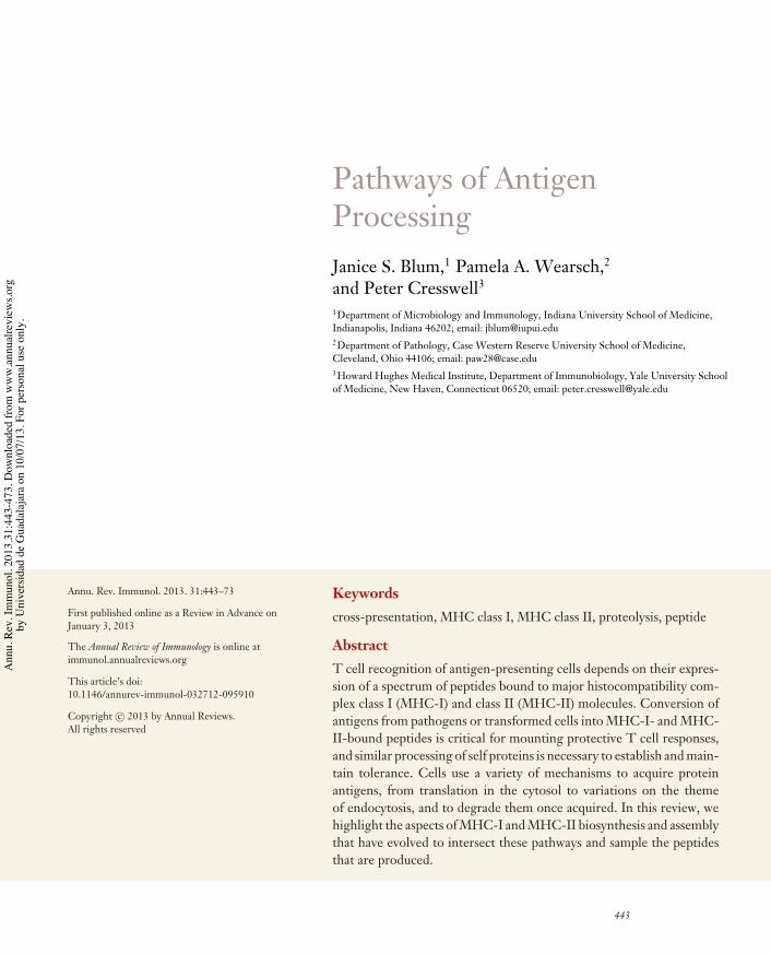
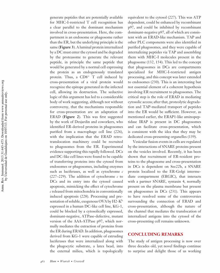
Figure 1Three-dimensional structures of MHC-I and MHC-II molecules with peptideligands. (a,b) Structure of the MHC-I molecule: HLA-A2 complexed withresidues 58–66 of the influenza matrix protein (232) (teal, MHC-I heavy chain;gray, !2-microglobulin; red, peptide). (c,d ) Structure of the MHC-II molecule:HLA-DR1 complexed with residues 306–318 of influenza hemagglutinin (233)( gray, MHC-II " chain; teal, MHC-II ! chain; red, peptide). Ribbon diagramswere generated with the Protein Workshop software available from the RCSBProtein Data Bank (http://www.rcsb.org). Highly polymorphic residues ofHLA-A (b) and HLA-DR (d ) proximal to the peptide binding groove(http://hla.alleles.org) are highlighted in yellow. Note that the polymorphismof the MHC-II " chains is limited; HLA-DR " chains are essentiallynonpolymorphic.
are also T cells that recognize lipid antigensassociated with CD1 molecules (1), but CD1functions and the processing mechanisms thatregulate their interaction with lipids are notconsidered here.
All vertebrates possess an MHC, a largemultigenic region with many conserved genesin addition to MHC-I and MHC-II molecules.Some of these encode products essentialto MHC-I and MHC-II function. In manyspecies, the MHC encodes multiple MHC-Iand MHC-II molecules, which are presumed tohave arisen by gene duplication. For example,in mice, depending on the strain, there aretwo to three genes encoding so-called classicalMHC-I molecules, called H2-D, -K, and -L,within the H2 complex, and most strains havetwo MHC-II molecules, called I-A and I-E.Humans have three genes encoding classicalMHC-I molecules within the HLA complex,called HLA-A, -B, and -C, and there are threeMHC-II molecules, called HLA-DR, -DQ,and -DP. In both mice and humans, there areother class I genes present in the MHC. Theseare known as class Ib genes and are discussedelsewhere in this volume (2).
Multiple structures of MHC-I and MHC-II molecules have been determined, and aschematic structure of each is presented inFigure 1. MHC-I and MHC-II genes exhibitenormous allelic polymorphism, and aminoacid sequence variation is heavily concentratedin the part of each structure that interacts withpeptides, allowing different alleles to bind a dif-ferent range of peptides. The peptide-bindingstructure consists of a membrane-distal grooveformed by two antiparallel !-helices overlayingan eight-strand "-sheet. In the case of MHC-I,the groove corresponds to a contiguous aminoacid sequence formed by the N-terminal regionof the single MHC-encoded subunit, or heavychain, whereas for MHC-II it is formed bythe juxtaposition of the N-terminal regions oftwo MHC-encoded !- and "-chains. For bothmolecules, the membrane-proximal regionconsists of two conserved domains that are ho-mologous to immunoglobulin (Ig) constant re-gion domains. For MHC-I, one is provided by
444 Blum ·Wearsch · Cresswell
Ann
u. R
ev. I
mm
unol
. 201
3.31
:443
-473
. Dow
nloa
ded
from
ww
w.a
nnua
lrevi
ews.o
rgby
Uni
vers
idad
de
Gua
dala
jara
on
10/0
7/13
. For
per
sona
l use
onl
y.

IY31CH16-Cresswell ARI 15 February 2013 4:42
the heavy chain and the other is a separate pro-tein, "2-microglobulin ("2m), a soluble productof a non-MHC-linked gene. For MHC-II, oneconserved domain is part of the !-subunit andthe other is part of the "-subunit. The MHC-Iheavy chain and the MHC-II !- and "-subunitsare transmembrane glycoproteins with shortcytoplasmic domains. The theme that emergesis that MHC-I and MHC-II molecules eachhave a structurally homologous platform capa-ble of binding peptides with very high affinitythat can engage the T cell receptor. A signifi-cant difference is that for MHC-I the peptide isconfined by binding groove interactions at boththe N and C termini, whereas for MHC-II eachend of the peptide can overhang the bindinggroove.
Peptides are the products of proteolysis,and there are two major proteolytic systemsoperating within the cell that contribute toMHC-dependent T cell recognition (Figure2). In the cytosol, most proteolysis is mediatedby the proteasome. The proteasome (reviewedin 3) is not discussed extensively here, but inbrief its core is a barrel-shaped 20S structureconsisting of four stacked rings of seven sub-units each. The outer rings are composed of!-subunits and the middle two of "-subunits,three of which, "1, "2, and "5, constitute theactive proteolytic components. Variants of theactive "-subunits are induced by interferon-#(IFN-#) and replace the constitutive versions.These were historically called LMP1, LMP2,and MECL1, and the genes encoding LMP1and LMP2 are MHC-linked. Commonly, theIFN-#-inducible subunits are now called "1i,"2i, and "5i, and proteasomes that containthem are called immunoproteasomes. Thecleavage specificities of standard proteasomesand immunoproteasomes differ. The 20S coreis capped at each end by an additional 19S mul-tisubunit complex that recognizes ubiquitin-conjugated proteins targeted for degradation.The 19S component has deubiquitinase activityand an unfoldase activity that allows the tar-geted proteins to enter the channel in the centerof the barrel where the "-subunit active sitesreside. The unfolding function, in particular,
necessitates that proteolysis by the capped(26S) proteasome is ATP-dependent. Thereis an alternative capping structure (11S) com-posed of a different set of IFN-#-inducibleproteins that allow a level of ATP-independentproteolysis of peptides but not of foldedproteins. The end products of proteolysisby the 26S proteasome (20S plus 19S) formthe dominant source of peptides for MHC-Ibinding.
Proteins that are internalized by a cell fromexogenous sources are degraded by lysosomalproteolysis (Figure 2). In brief, endocytosedproteins enter a vesicular pathway consisting ofprogressively more acidic and proteolyticallyactive compartments classically referred toas early endosomes, late endosomes, andlysosomes (4). Particles internalized by phago-cytosis follow a similar path, terminating inphagolysosomes that are formed by the fusionof phagosomes and lysosomes. Lysosomesand phagolysosomes have a pH of 4 to 4.5and contain a number of acid pH-optimumproteases generically called cathepsins (5). Inhighly degradative cells such as macrophages,successive cleavages by these enzymes result invery short peptides and free amino acids thatare translocated into the cytosol to replenishtRNAs for new protein synthesis, but inless proteolytically active antigen-presentingcells (APCs), larger intermediates form thedominant source of peptides for MHC-IIbinding.
The trafficking of exogenous and endoge-nous proteins for antigen processing andpresentation are summarized in Figure 2.In general, MHC-I molecules bind peptidesgenerated by proteasomal proteolysis, and theybind them in the endoplasmic reticulum (ER)after the peptides are translocated from the cy-tosol. Peptide binding by MHC-I is integratedinto the assembly pathway of the heavy chain-"2m dimer. MHC-II molecules generally bindpeptides generated by lysosomal proteolysis inthe endocytic and phagocytic pathways. How-ever, both can access peptides from endogenousand exogenous antigens. For example, MHC-II binds peptides derived from endogenous
www.annualreviews.org • Antigen Processing 445
Ann
u. R
ev. I
mm
unol
. 201
3.31
:443
-473
. Dow
nloa
ded
from
ww
w.a
nnua
lrevi
ews.o
rgby
Uni
vers
idad
de
Gua
dala
jara
on
10/0
7/13
. For
per
sona
l use
onl
y.

IY31CH16-Cresswell ARI 15 February 2013 4:42
EndocytosisMacropinocytosisPhagocytosis
Phagosome
MIIC/late endosome Autophagosome
Macroautophagy
CellularRNA
ViralRNA
Viral infection
Proteasome
Golgi
Viral proteinExogenous protein
TAP
Self protein
MHC-II
Lysosome
Cathepsins
Cathepsins
Endoplasmicreticulum
MHC-I
Retro-translocation
ERAAP/ERAP2
Figure 2Trafficking of antigens for processing and presentation with major histocompatibility complex (MHC) molecules: basic pathways andexceptions to the “rules.” Cytosolic proteins are processed primarily by the action of the proteasome. The short peptides are thentransported into the endoplasmic reticulum (ER) by the transporter associated with antigen processing (TAP) for subsequent assemblywith MHC-I molecules. In certain antigen-presenting cells, particularly dendritic cells, exogenous proteins can also be fed into thispathway by retrotranslocation from phagosomes, a phenomenon known as cross-presentation. The retrotranslocation channels may berecruited from the ER, where they are used for ER-associated degradation, or ERAD, of misfolded transmembrane or secretoryproteins. Exogenous proteins are primarily presented by MHC-II molecules. Antigens are internalized by several pathways, includingphagocytosis, macropinocytosis, and endocytosis, and eventually traffic to a mature or late endosomal compartment, often called theMHC-II compartment, or MIIC, where they are processed and loaded onto MHC-II molecules. Cytoplasmic/nuclear antigens can alsobe trafficked into the endosomal network via autophagy for subsequent processing and presentation with MHC-II molecules.
membrane proteins that are degraded inthe lysosome. In addition, MHC-I can bindpeptides derived from exogenous proteinsinternalized by endocytosis or phagocytosis,a phenomenon called cross-presentation.
Specific subsets of dendritic cells (DCs) areparticularly adept at mediating this process,which is critically important for the initiationof a primary response by naive CD8+ T cellswhen it is termed cross-priming.
446 Blum ·Wearsch · Cresswell
Ann
u. R
ev. I
mm
unol
. 201
3.31
:443
-473
. Dow
nloa
ded
from
ww
w.a
nnua
lrevi
ews.o
rgby
Uni
vers
idad
de
Gua
dala
jara
on
10/0
7/13
. For
per
sona
l use
onl
y.

IY31CH16-Cresswell ARI 15 February 2013 4:42
Transporterassociated withantigen processing(TAP): anATP-dependenttransporter composedof two subunits, TAP1and TAP2, thattranslocates peptidesfrom the cytosol intothe endoplasmicreticulum
Peptide-loadingcomplex (PLC):protein complexconsisting of theMHC-I heavy chainand !2-microglobulin,TAP, tapasin,calreticulin, andERp57 that facilitatesMHC-I-peptideloading
ERp57: anendoplasmicreticulum–residenthomolog of proteindisulfide isomerase
PEPTIDE BINDING TO MHC-IMOLECULESPeptides generated in the cytosol are translo-cated into the ER by the transporter associatedwith antigen processing (TAP), which is a mem-ber of the ATP-binding cassette (ABC) fam-ily of transporters (6). TAP is a heterodimericprotein, and the TAP1 and TAP2 subunits areencoded by closely linked genes in the MHC.These are widely distributed in both prokary-otes and eukaryotes and transfer a variety ofmolecules across membranes. Biochemical evi-dence combined with molecular modeling sug-gests that each TAP subunit consists of a centralcore domain of six transmembrane !-helices,which constitute the channel, that is immedi-ately N-terminal to the nucleotide-binding do-main (NBD) (7). The NBD structure is knownfor TAP1 and it is similar to that of other ABCfamily members, with the classical Walker Aand B motifs present in many ATPases (8).Cytosolic loops in the core domains that areproximal to the NBDs constitute the peptiderecognition site, and ATP hydrolysis mediatesthe translocation event (7). Both subunits haveadditional N-terminal domains (N-domains),comprising four transmembrane segments forTAP1 and three for TAP2, which have no coun-terparts in other members of the ABC family oftransporters (7).
The TAP heterodimer associates with anumber of other proteins to form the peptide-loading complex, or PLC (Figure 3). Thetransmembrane glycoprotein tapasin, which isencoded by an MHC-linked gene (9), interactswithin the membrane with the N-domains (10–13). Tapasin has a bridging function, recruitingMHC-I-"2m dimers and the chaperone cal-reticulin (CRT) to the PLC (14). Recentexperiments have confirmed that there are twotapasin molecules in the PLC, one associatedwith each TAP subunit (13, 15). Tapasin in turnis stably linked via a disulfide bond to a secondmolecule, the protein disulfide isomerasehomolog ERp57, and the structure of thelumenal region of human tapasin conjugated toERp57 has been solved (16). The N-domain of
tapasin consists of a " barrel fused to an Ig-likedomain, and, as for the MHC-I and MHC-IIproteins, the membrane proximal domain isIg-like. ERp57 has a slightly twisted U-shapedstructure, and tapasin is inserted into the U ina way that results in extensive protein-proteininteractions with ERp57, particularly with thea and a! domains, each of which contains adouble cysteine “CXXC” motif that constitutesits two redox active sites. As predicted by earlierbiochemical experiments (17), a disulfide bondconnects cysteine 95 of tapasin with cysteine57 of ERp57, which is the N-terminal cysteineresidue of the a domain CXXC motif. Nor-mally, disulfide bonds involving cysteine 57 aretransiently formed during the reduction of adisulfide-containing ERp57 substrate protein,and reduction of this enzyme-substrate bondby the second cysteine in the motif releasesthe substrate. The interactions of tapasinwith the a and a! domains appear to trap thedisulfide-linked species, explaining the stabilityof the tapasin-ERp57 disulfide bond.
ERp57 assists the folding of newly synthe-sized glycoproteins in the ER by mediatingdisulfide bond isomerization. Its specificity forglycoproteins results from its ability to associatevia its b! domain with CRT and a second lectin-like ER chaperone, the transmembrane CRThomolog calnexin (CNX). Both CNX and CRTare important in MHC-I assembly (Figure 3).CNX and CRT normally function in a quality-control cycle that depends on their interactionswith the N-linked glycans of the glycoproteins(18). They then recruit ERp57, which mediatesproper disulfide bond formation in the foldingglycoprotein. Glycan binding to CNX or CRTis dependent on the precise structure of the N-linked glycan, which must bear a single terminalglucose residue and is a biosynthetic interme-diate maintained in this form by the competingactions of two enzymes. One, glucosidase II, re-moves the glucose and the other, UDP-glucoseglycoprotein transferase-1 (UGT1), replacesthe glucose only if the glycoprotein bearingthe glycan is partially unfolded (19–21). Thiscycle plays a role in MHC-I-peptide loading
www.annualreviews.org • Antigen Processing 447
Ann
u. R
ev. I
mm
unol
. 201
3.31
:443
-473
. Dow
nloa
ded
from
ww
w.a
nnua
lrevi
ews.o
rgby
Uni
vers
idad
de
Gua
dala
jara
on
10/0
7/13
. For
per
sona
l use
onl
y.
IY31CH16-Cresswell ARI 15 February 2013 4:42
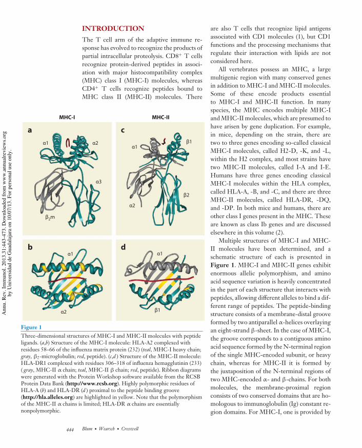
(Figure 3), but the one step that does not ap-pear to be involved is the reduction-oxidationcycle mediated by ERp57 (see below).
Cells that lack TAP1 or TAP2 do notform MHC-I-peptide complexes because nopeptides are imported into the ER. There are afew published exceptions to this rule, some ofwhich lead to CD8+ T cell recognition (22, 23),
but the only major one, in terms of quantitativeeffects on MHC-I assembly, is the unusual andspecific ability of HLA-A2 molecules to bindpeptides derived from signal sequences of cer-tain ER-targeted molecules (24). Because of theinherent instability of so-called empty MHC-Imolecules, and because they do not fold intoa transport-competent structure in the ER,
Native proteins
DRiPsTranslation
Proteasome
Phagosome
Viral RNA
Cellular RNA
Retro-translocation
Exogenousprotein
Golgi
MHC-I withsuboptimal
ligands
UDP-glucose
G
G
G
G
G
G
UGT1
CRT ERp57
Tapa
sin
TAP1 TAP2
ERAAP/ERAP1
ERAP2
Peptideloading
complex
Gls I/Gls II "2m
GG
G
Endoplasmic reticulum
CNX
NascentMHC-I
High-a#nitypeptide
Gls II
MHC-I
448 Blum ·Wearsch · Cresswell
Ann
u. R
ev. I
mm
unol
. 201
3.31
:443
-473
. Dow
nloa
ded
from
ww
w.a
nnua
lrevi
ews.o
rgby
Uni
vers
idad
de
Gua
dala
jara
on
10/0
7/13
. For
per
sona
l use
onl
y.
IY31CH16-Cresswell ARI 15 February 2013 4:42
TAP-negative cells express very little surfaceMHC-I. Cells that lack tapasin also exhibitreduced surface MHC-I, but the defect is muchless drastic than in TAP-negative cells, and themagnitude of the effect depends on the individ-ual MHC-I allele expressed (25–28). Data fromtapasin knockout mice showed an essentialfunction for tapasin in generating CD8+ T cellresponses. Furthermore, data based on T cellrecognition demonstrated that tapasin plays apeptide-editing role, mediating the binding ofhigh-affinity peptides at the expense of peptideswith lower but still significant affinity and that,for this reason, surface MHC-I molecules ontapasin-negative cells are less stable than thoseon tapasin-positive cells (27–30). Subsequently,in vitro data produced using recombinanttapasin-ERp57 conjugates confirmed thattapasin facilitates high-affinity peptide bindingand further showed that its association withERp57 is essential (31). The addition oftapasin-ERp57 conjugates to extracts of humantapasin-negative cells expressing HLA-B8was found to facilitate the binding of addedhigh-affinity peptides to HLA-B8-"2m dimers.Lower-affinity peptides were much less suc-cessful competitors for binding in the presenceof the conjugate than in its absence, indicativeof a peptide-editing effect. The tapasin-ERp57conjugate was also found to mediate peptidebinding to purified, soluble, recombinant
HLA-B8-"2m dimers, provided that the HLA-B8 molecules expressed a monoglucosylatedN-linked glycan (32). Although this reactiondepended on the addition of recombinantCRT, presumably to provide a bridge betweenMHC-I and the tapasin-associated ERp57, noother components were required. In a moresimplified in vitro system, neither CRT nortapasin-associated ERp57 were needed for pep-tide binding when the MHC-I heavy chain andtapasin were artificially coupled by the additionof leucine zippers to their C termini (33).
ERp57-negative cells, as well as CRT-negative cells, also have reduced numbers ofMHC-I molecules on the cell surface (34, 35).The initial identification of ERp57 in the PLCled to considerable speculation that its redoxactivity was important for generating stableMHC-I-peptide complexes. However, thestructural data indicated that tapasin obstructsboth of the ERp57 active sites, rendering thisunlikely. In fact, when the second active sitecysteine in the a domain and both active sitecysteine residues in the a! domain were mutatedto serine residues, the combined substitutionshad no effect on the ability of tapasin to recon-stitute MHC-I cell surface expression when itwas introduced into an ERp57-deficient cellline (36). This triply mutated ERp57 was stilldisulfide-linked to tapasin. However, furtheranalysis in both cell-free systems and intact
"##########################################################################################
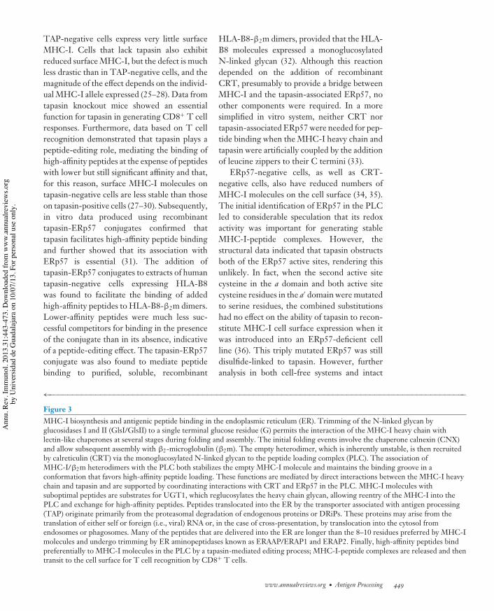
Figure 3MHC-I biosynthesis and antigenic peptide binding in the endoplasmic reticulum (ER). Trimming of the N-linked glycan byglucosidases I and II (GlsI/GlsII) to a single terminal glucose residue (G) permits the interaction of the MHC-I heavy chain withlectin-like chaperones at several stages during folding and assembly. The initial folding events involve the chaperone calnexin (CNX)and allow subsequent assembly with !2-microglobulin (!2m). The empty heterodimer, which is inherently unstable, is then recruitedby calreticulin (CRT) via the monoglucosylated N-linked glycan to the peptide loading complex (PLC). The association ofMHC-I/!2m heterodimers with the PLC both stabilizes the empty MHC-I molecule and maintains the binding groove in aconformation that favors high-affinity peptide loading. These functions are mediated by direct interactions between the MHC-I heavychain and tapasin and are supported by coordinating interactions with CRT and ERp57 in the PLC. MHC-I molecules withsuboptimal peptides are substrates for UGT1, which reglucosylates the heavy chain glycan, allowing reentry of the MHC-I into thePLC and exchange for high-affinity peptides. Peptides translocated into the ER by the transporter associated with antigen processing(TAP) originate primarily from the proteasomal degradation of endogenous proteins or DRiPs. These proteins may arise from thetranslation of either self or foreign (i.e., viral) RNA or, in the case of cross-presentation, by translocation into the cytosol fromendosomes or phagosomes. Many of the peptides that are delivered into the ER are longer than the 8–10 residues preferred by MHC-Imolecules and undergo trimming by ER aminopeptidases known as ERAAP/ERAP1 and ERAP2. Finally, high-affinity peptides bindpreferentially to MHC-I molecules in the PLC by a tapasin-mediated editing process; MHC-I-peptide complexes are released and thentransit to the cell surface for T cell recognition by CD8+ T cells.
www.annualreviews.org • Antigen Processing 449
Ann
u. R
ev. I
mm
unol
. 201
3.31
:443
-473
. Dow
nloa
ded
from
ww
w.a
nnua
lrevi
ews.o
rgby
Uni
vers
idad
de
Gua
dala
jara
on
10/0
7/13
. For
per
sona
l use
onl
y.

IY31CH16-Cresswell ARI 15 February 2013 4:42
Endoplasmicreticulum–associateddegradation (ERAD):pathway that promotesthe translocation ofmisfolded ER proteinsinto the cytoplasm forproteolysis
ERAAP: endoplasmicreticulumaminopeptidaseassociated with antigenprocessing (murine); inhumans, known as ERaminopeptidase-1(ERAP1)
cells using ERp57 mutated in the b! domainshowed that the ability of ERp57 to bind CRTis essential for MHC-I recruitment to the PLCand normal MHC-I-peptide loading (32). Inaddition to the CRT-dependent interactionswith the MHC-I glycan and ERp57 thatmediate MHC-I binding to the PLC, thereis also a direct interaction between MHC-Iand tapasin. Mutagenesis of specific tapasinresidues and expression of the mutants as re-combinant tapasin-ERp57 conjugates revealeda patch on the surface of tapasin that binds tothe MHC-I molecule, and there was a positivecorrelation between the relative abilities ofdifferent mutants to bind MHC-I and theirefficiency in mediating peptide binding toMHC-I in vitro (16). In addition, a tapasinmutant that was nonfunctional in cell-freeassays also failed to function when expressed asa full-length protein in a tapasin-negative cell.
The PLC consists of the TAP heterodimerand two tapasin-ERp57 conjugates, and up totwo CRT molecules and MHC-I-"2m dimerscan be recruited (Figure 3). The MHC-I heavychain glycan must be in the monoglucosylatedform, consistent with the CRT requirement(32). Cellular expression of UGT1 is essentialfor optimal MHC-I-peptide loading, and invitro the enzyme can discriminate betweenMHC-I molecules bound to high-affinity pep-tides and those associated with lower-affinitypeptides (37). This suggests a mechanism thatresembles the normal CRT/CNX quality-control cycle. A plausible model is that there aretwo discriminatory events that regulate peptideediting (Figure 3). First, after peptide-freeMHC-I-"2m dimers bearing a monogluco-sylated N-linked glycan are recruited to thePLC by CRT, there is a direct interaction ofthe MHC-I molecule with tapasin. This in-teraction is sensitive to the peptide occupancyof the MHC-I molecule such that, when apeptide is bound, the affinity of the MHC-Iinteraction with tapasin is reduced, perhapsby a conformational change in the MHC-Iheavy chain similar to that proposed to explainthe ability of HLA-DM/H2-DM moleculesto regulate peptide binding to MHC-II (see
below). Thus, peptide binding induces disso-ciation of the MHC-I molecule from tapasin,and because the affinity of the CRT interactionwith the monoglucosylated MHC-I glycan islow, the glucose residue becomes accessible tothe enzyme glucosidase II, which removes it.If the peptide affinity is sufficiently high, theMHC-I molecule can be transported from theER through the Golgi apparatus and ultimatelyto the cell surface. If the affinity of the peptideis low, there are two possible scenarios for thesecond stage. Either the peptide dissociates andthe transiently empty MHC-I molecule nowbecomes a substrate for UGT1 and glucoseis added back to the N-linked glycan, or theUGT1 can recognize that the conformationof the MHC-I-peptide complex is in some wayimperfect and reglucosylates the glycan. Ineither case, the consequence of the additionof the glucose residue is that the MHC-Imolecule reassociates with CRT, reintegratescompletely into the PLC, and is subjected tofurther rounds of tapasin-mediated peptidebinding and selection. Ultimately, the MHC-Imolecule will escape with a high-affinity pep-tide, or, in common with other glycoproteinsthat are subject to the CRT/CNX/ERp57quality-control cycle, enzymatic removal ofmannose residues from the N-linked glycanwill render it unsusceptible to reglucosylationby UGT1. This acts as a timer, leading to ir-reversible dissociation of the MHC-I from thePLC and its degradation by the ER-associateddegradation (ERAD) pathway (38).
One other ER luminal component that iscritical for the proper generation of MHC-I-peptide complexes is an aminopeptidase; in themouse it is called ER aminopeptidase associatedwith antigen processing (ERAAP) and in hu-mans it is called ER aminopeptidase-1 (ERAP1)(Figure 3) (39, 40). A second aminopeptidase,ERAP2, is present in humans but not in miceand can also play a role (41). Peptides associatedwith MHC-I are generally 8–10 amino acids inlength, but TAP can translocate peptides intothe ER that are significantly longer (42). Thesepeptides can be amino-terminally trimmed inthe ER by ERAAP/ERAP1 to yield peptides
450 Blum ·Wearsch · Cresswell
Ann
u. R
ev. I
mm
unol
. 201
3.31
:443
-473
. Dow
nloa
ded
from
ww
w.a
nnua
lrevi
ews.o
rgby
Uni
vers
idad
de
Gua
dala
jara
on
10/0
7/13
. For
per
sona
l use
onl
y.
IY31CH16-Cresswell ARI 15 February 2013 4:42
of the appropriate length for MHC-I binding.A structural change required for cleavagethat can only be induced by a longer peptideprevents ERAP1 from so-called over-trimmingTAP-translocated peptides to a length thatwould eliminate their ability to bind MHC-I(43). Many of the peptides associated withMHC-I molecules expressed on cells derivedfrom ERAAP knockout mice are elongated,and the MHC-I molecules are relatively unsta-ble (44–46). The absence of ERAAP results insuch a severe alteration in the range of boundpeptides that wild-type and knockout mice onthe same background are actually histoincom-patible, with wild-type mice able to generateCD8+ T cell responses, and even antibodyresponses, against knockout cells (45). Theantibodies generated recognize the MHC-Imolecules complexed with elongated peptidesand can block recognition of ERAAP-negativecells by the ERAAP-positive CD8+ T cells.
PEPTIDE BINDING TO MHC-IIMOLECULESMHC-II molecules assemble within the ER,followed by functional maturation in endoso-mal compartments rich in antigenic peptides.Upon ER translocation, MHC-II ! and "
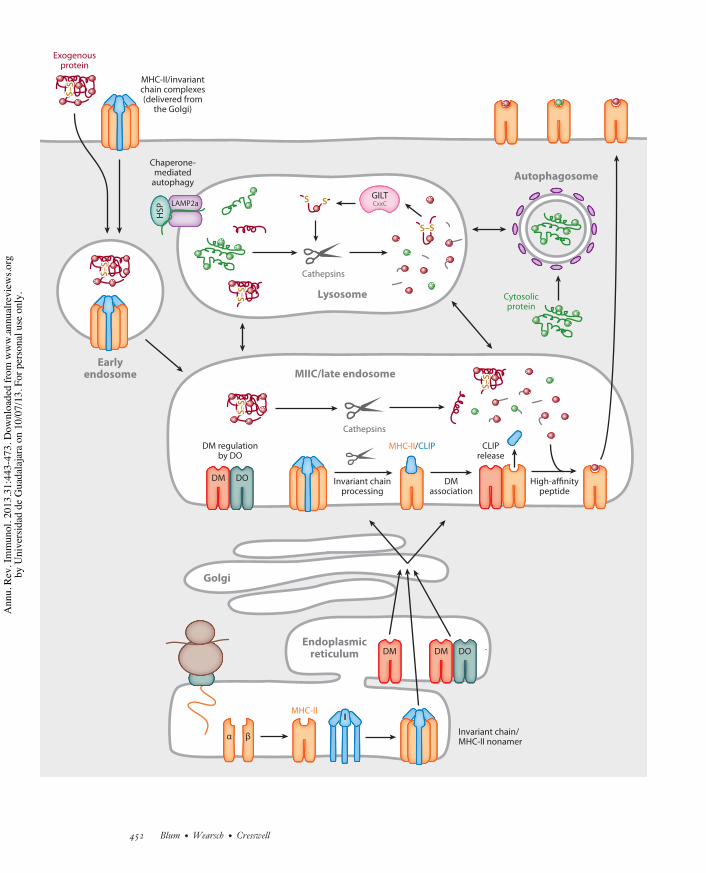
subunits associate in a process facilitated bya specific chaperone, the invariant chain (Ichain), or CD74 (Figure 4). Studies using Ichain–deficient cells and animals have shownthat I chain promotes MHC-II !" folding,protects the MHC-II ligand binding groove,and directs MHC-II molecules to endosomalcompartments for ligand capture. I chain isa nonpolymorphic type II transmembraneglycoprotein not encoded in the MHC. Severalforms of I chain exist due to alternative splicingand the use of alternate start codons (47).Nomenclature for the variants is based on theirmolecular mass, with the shortest form, p33,being most abundantly expressed. A largersplice variant, p41, contains a glycosylateddomain, homologous to domains present inthyroglobulin, which can inhibit the activityof the protease cathepsin L (48). All forms of
I chain contain a conserved di-leucine motif inthe N-terminal cytoplasmic domain requiredfor targeting I chain and associated MHC-IIto late endosomal compartments (49, 50). Inhumans, an alternate upstream translationalstart site gives rise to two additional forms of Ichain, p35 and p43, each with an N-terminal 16amino acid extension. This extended cytoplas-mic domain encodes an ER retention motif,which may facilitate ER accumulation andthe folding of nascent MHC-II !". A limitednumber of I chain molecules are also modifiedvia linkage of a chondroitin sulfate chain; thesemolecules reach the cell surface and facilitatecell-cell adhesion (51, 52). Several othermolecules involved in antigen presentationor transport have been reported to associatewith I chain, including CD1, MHC-I, andthe neonatal Fc# receptor (53–55). AlthoughI chain expression is not required for thefunction of CD1 or MHC-I, it may enhanceantigen presentation by these molecules (56,57). I chain expression negatively regulates DCmotility in vitro, but it is unknown whetherthis facilitates antigen presentation or if it isrelated to the role of I chain as a receptor forthe macrophage and stem cell chemoattractantmigration inhibitory factor (55, 56).
Newly synthesized I chain variants formhomo- or mixed trimers, involving p33, p35,p41, and p43 in humans, which accumulate inthe ER (58). These multimers act as nuclei forMHC-II ! and " assembly, giving rise to non-amers with three !, three ", and three I chains(Figure 4) (59). Distinct MHC-II alleles havedifferent affinities and requirements for I chainbinding that can influence their expression andfunction. In the absence of I chain, some MHC-II !" complexes are unstable, resulting in theiraggregation, retention in the ER, and failure toreach the cell surface (60–62). Association of Ichain with MHC-II !" dimers prevents anti-genic peptide binding, consistent with minimalpeptide acquisition early in MHC-II biosyn-thesis (63, 64). After assembly, the MHC-II-Ichain complexes leave the ER and are routedto the endocytic pathway by the I chain di-leucine motifs (47). This may occur by direct
www.annualreviews.org • Antigen Processing 451
Ann
u. R
ev. I
mm
unol
. 201
3.31
:443
-473
. Dow
nloa
ded
from
ww
w.a
nnua
lrevi
ews.o
rgby
Uni
vers
idad
de
Gua
dala
jara
on
10/0
7/13
. For
per
sona
l use
onl
y.
IY31CH16-Cresswell ARI 15 February 2013 4:42
Endoplasmicreticulum
Invariant chain/MHC-II nonamer
DM DM DO
! "
II
DM regulationby DO
High-a#nitypeptide
Invariant chainprocessing
DMassociation
CLIPrelease
Cathepsins
S–S
S–S
S–S
S–S
Lysosome
Cathepsins
S–SS–S
SS SSGILTCxxC
Chaperone-mediated
autophagy
S–S
S–S
MIIC/late endosome
MHC-II
Golgi
MHC-II/invariantchain complexes(delivered from
the Golgi)
Earlyendosome
Exogenousprotein
S–S
S–S
S–S
S–S
Autophagosome
LAMP2a
HSP
MHC-II/CLIP
Cytosolicprotein
DM DO
452 Blum ·Wearsch · Cresswell
Ann
u. R
ev. I
mm
unol
. 201
3.31
:443
-473
. Dow
nloa
ded
from
ww
w.a
nnua
lrevi
ews.o
rgby
Uni
vers
idad
de
Gua
dala
jara
on
10/0
7/13
. For
per
sona
l use
onl
y.
IY31CH16-Cresswell ARI 15 February 2013 4:42
CLIP: classII–associated invariantchain peptide
BCR: B cell receptorfor antigen
targeting from the trans-Golgi network (TGN)or by endocytosis from the plasma membrane(Figure 4) (65).
I chain release is initiated by progressiveproteolysis in acidic endosomes (66). Thisculminates in a variably extended peptide ofroughly 20 residues that is associated with theMHC-II binding groove (Figure 4). This iscalled CLIP, for class II–associated invariantchain peptide (67, 68). The structure of CLIPbound to HLA-DR3 is virtually identical tothe structure of MHC-II bound to antigenicpeptides indicated in Figure 1. (69). There aresome MHC-II alleles with a low affinity forCLIP, and they are genetically associated withthe development of autoimmunity (70). Thismay reflect a role for MHC-II-CLIP complexesin regulating thymic selection or skewing ofT helper cell subset differentiation (71, 72).Alternatively, premature release of CLIP fromthese disease-associated MHC-II alleles mayfavor the selection of epitopes from autoanti-gens or the capture of self-peptides withindistinct endosomal compartments (73, 74).
CLIP release from MHC-II is facilitated byanother MHC-encoded heterodimeric glyco-protein, DM, which is highly homologous toconventional MHC-II (Figure 4) (75, 76). Inhumans DM is known as HLA-DM and in miceas H2-DM. The DM ! and " subunits displaylimited genetic polymorphism, and the assem-bled dimer lacks an open or accessible ligandbinding groove (77, 78). The cytoplasmic do-main of the DM " chain contains a tyrosine mo-tif that is responsible for sorting assembled DMmolecules to late endosomes; DM may also bindI chain, which may facilitate but is not required
for DM assembly and stability (79–81). DMinteraction with MHC-II-CLIP complexesoccurs in late endosomes, where DM acts topromote a conformational change that inducesCLIP dissociation (Figure 4). This reactioncan be replicated using purified MHC-II-CLIPand DM, and it displays Michaelis-Mentenkinetics and an acidic pH optimum (76, 82,83). CLIP removal facilitates MHC-II loadingwith antigenic peptides, which influences therepertoire of CD4+ T cells selected in the thy-mus (84, 85). DM can remove any low-affinitypeptides from MHC-II, and analogous to therole of tapasin in MHC-I peptide editing dis-cussed above (Figure 3), repetitive interactionswith DM lead to the accumulation of MHC-IIcomplexes with high-affinity peptides (86).Whereas MHC-II binding to peptides derivedfrom endocytosed antigens is inefficient in theabsence of DM, there is a slow release of CLIPfrom MHC-II even in DM-negative APCs.As a consequence, synthetic peptides bindefficiently to surface MHC-II in these cellsand presentation of endogenous antigens canbe detected, whereas in B cells BCR-mediatedtargeting of antigens can overcome the lossof DM, presumably by increasing the amountinternalized over a critical threshold (87–89).
The function of DM is modulated byanother MHC-encoded MHC-II-like !"
heterodimer, DO, and it is generally acceptedthat DO inhibits DM function (90, 91). DO isexpressed in B cells and thymic epithelium andat low levels in select DC subsets, where thereis evidence that it is regulated by Toll-likereceptor (TLR) agonists (92–95). DO !"
dimers associate tightly with DM molecules
"##########################################################################################
Figure 4Major histocompatibility complex (MHC)-II biosynthesis and antigenic peptide binding in the endocytic pathway. MHC-II " and !associate with invariant chain (I chain) trimers to form nonamers. These complexes transit to mature endosomes either via thetrans-Golgi network (TGN) or by recycling from the cell surface. Within endosomes, I chain is sequentially proteolyzed to yield theresidual I chain fragment, class II-associated invariant chain peptide (CLIP). Displacement of CLIP from the ligand groove of MHC-II"! is mediated by the MHC-II-related chaperone HLA-DM (DM) and blunted by HLA-DO (DO). Expression of DO and regulationof DM function involves the assembly of DM-DO complexes in the endoplasmic reticulum and cotransport to endocytic compartments.Antigens delivered to late endosomes by phagocytosis, pinocytosis, endocytosis, and autophagy are processed by cathepsins and thethiol oxidoreductase GILT (#-interferon-inducible lysosomal thiol), and acquisition of high-affinity peptides by MHC-II is facilitatedby DM. The MHC-II-peptide complexes are subsequently transported to the cell surface for T cell recognition by CD4+ T cells.
www.annualreviews.org • Antigen Processing 453
Ann
u. R
ev. I
mm
unol
. 201
3.31
:443
-473
. Dow
nloa
ded
from
ww
w.a
nnua
lrevi
ews.o
rgby
Uni
vers
idad
de
Gua
dala
jara
on
10/0
7/13
. For
per
sona
l use
onl
y.

IY31CH16-Cresswell ARI 15 February 2013 4:42
and are retained in the ER in the absenceof DM, suggesting that in DO-positive cellsDM and DO move in concert to endosomes(Figure 4) (96). Studies using Forster (fluores-cence) resonance energy transfer (FRET) andmutational analysis that defined the DM/DRinterface suggested that DO and DR bindto the same region of DM (97). Recently,the crystal structure of the DO/DM complexconfirmed this and demonstrated an apparentdisplacement of a segment of the DO !-chain!-helix compared with that of the !-chain!-helix in MHC-II-peptide complexes, whichmay reflect the conformational alteration thatDM imparts to induce the dissociation oflow-affinity peptides (98).
A precise biological function for DO hasbeen hard to define. Studies in mice deficientin DO have revealed subtle defects in MHC-IIantigen presentation, although the effects ob-served were influenced by the genetic back-ground of the mice and the MHC-II alleleexamined (91, 99). In vivo, overexpression ofDO in DCs can impair MHC-II presentationof antigenic epitopes and, presumably becauseof this, reduce type I diabetes development inNOD mice (100, 101).
ANTIGEN INTRODUCTION ANDPROTEOLYSIS IN THEENDOCYTIC PATHWAYExploiting conserved pathways establishedfor nutrient and growth factor uptake, APCssample soluble and particulate matter fromextracellular fluids. Many pathogens, includingviruses, bacteria, and fungi, use these samepathways as conduits into cells, favoringimmune recognition and antigen presentation.Pathogen-driven disruption of these pathwaysallows immune evasion (102–104). Amongthese transport pathways, three routes—clathrin-mediated endocytosis, phagocytosis,and macropinocytosis—efficiently promoteantigen internalization and sorting to vesicularorganelles for processing and presentation byMHC molecules (Figure 2). During clathrin-mediated endocytosis, cell surface receptor-
ligand complexes, membrane proteins, andsoluble macromolecules are internalized.Regulated capture of particulate antigens andpathogens is mediated by phagocytosis, a pro-cess that synchronizes engulfment with deliveryinto a microenvironment containing reactiveoxygen species, proteases, and antimicrobialagents to promote pathogen destruction. Thenonselective process of macropinocytosis cap-tures larger quantities of extracellular material,including proteins, bacteria, and viruses, viaplasma membrane ruffling and folding. Allthese pathways exist in DCs, macrophages, andB lymphocytes, although there are variationsin efficiency and regulation. For example,B cells are less efficient at fluid-phase endo-cytosis than are DCs or macrophages (105).However, soluble antigen uptake and MHC-IIpresentation by B cells can be detected invivo using antibodies recognizing specificMHC-II-peptide complexes (50). Surface Ig asa component of the BCR promotes rapid andefficient internalization of antigens, enhancingthe potency of antigen-specific B cells 103-to 104-fold as stimulators of CD4+ T cells(106).
APCs in general display multiple cell sur-face receptors that can capture antigens orintact pathogens to promote internalizationand processing. Enhanced antigen presenta-tion by MHC-II has been observed follow-ing antigen uptake via several receptors thatcluster in clathrin-coated domains, includingthe BCR, Fc receptors, and the C-type lectinfamily receptor DEC205, as well as mannoseand transferrin receptors (107–111). MHC-Icross-presentation was also increased followingthe internalization of ovalbumin (OVA) via themannose receptor on DCs and macrophages(112). DEC205 can promote efficient antigeninternalization and presentation by both MHC-I and MHC-II, and conjugation of antigens toantibodies recognizing DEC205 has been usedto induce tolerance (109). APCs also expressreceptors for self and microbial heat shock pro-teins such as Hsp70, Hsp90, and gp96, whichpromote endocytic uptake of these chaperonesand associated ligands (including peptides and
454 Blum ·Wearsch · Cresswell
Ann
u. R
ev. I
mm
unol
. 201
3.31
:443
-473
. Dow
nloa
ded
from
ww
w.a
nnua
lrevi
ews.o
rgby
Uni
vers
idad
de
Gua
dala
jara
on
10/0
7/13
. For
per
sona
l use
onl
y.

IY31CH16-Cresswell ARI 15 February 2013 4:42
antigens) for MHC-I and MHC-II presenta-tion (113–116).
Receptors on the surface of APCs pro-mote the phagocytosis of bacteria, fungi,select viruses, and apoptotic or necroticcells (117–119). Macrophages and DCs arewell-established phagocytes, but this processcan also be observed in B cells, which canpresent phagocytosed antigens to CD4+ T cells(120–122). MHC-I cross-presentation as wellas MHC-II presentation of opsonized antigensis enhanced by receptor engagement uponphagocytosis, which may reflect intracellularreceptor signaling rather than simply enhanceduptake of these particles. Thus, IgG-coatedbacteria were effectively presented to CD8+
T cells while complement C3 opsonization ofbacteria facilitated phagocytosis but not antigenpresentation (123). Signaling by receptors suchas the C-type lectin family receptor DNGR-1promotes MHC-I and MHC-II presentationof antigens from phagocytosed necrotic cells(124, 125). Internalization and presentationof self-antigens associated with necrotic cellsmay contribute to autoimmunity or allograftrejection. Indeed, while all the above pathwayspromote uptake of extracellular antigens byAPCs, internalization and recycling of theplasma membrane also delivers endogenousproteins for processing; peptides derived frommembrane proteins, such as transferrin recep-tor and MHC-I heavy chain, are abundantlyassociated with MHC-II molecules (126).
Endocytic Compartments in AntigenProcessing and PresentationInternalized antigens enter organelles withmicroenvironments favoring protein denatura-tion and proteolysis. Although these pathwayspermit MHC-II access to exogenous antigens,MHC-I molecules also use these routesto acquire antigens for cross-presentation(Figure 2). Electron microscopy initiallyrevealed an abundance of MHC-II moleculesdistributed in the endocytic pathway, concen-trated in late endosomal vesicles, originallydefined as MHC-II compartments, or MIICs
(Figure 2), in contrast to only limited amountsof MHC-I (127–129). The role for endosomalMHC-I in cross-presentation has been debated.Disrupting expression of HS-1, a modulator ofendocytic invaginations, demonstrated thatendocytosis delivers extracellular antigens forpresentation by MHC-I as well as by MHC-IIin DCs (130). However, in DCs antigenscan transit from within endosomes to thecytoplasm or the ER, raising questions aboutthe role of endocytosed MHC-I in antigencross-presentation (131). A tyrosine motif inthe cytoplasmic tail of MHC-I heavy chainfacilitates recycling of low levels of thesemolecules from the cell surface into endo-somes, but direct delivery of immature MHC-Ifrom the ER may also occur in DCs, possiblyfacilitated by associated I chain (57, 132–135).
Early endosomes mature into late endo-somes and lysosomes driven in part by processessuch as increased luminal acidification and fu-sion with TGN-derived vesicles delivering en-zymes that promote antigen denaturation andproteolysis. Low temperature (18$C) can blockthe maturation step and disrupt the presenta-tion of several exogenous antigens by MHC-II(136). However, MHC-II presentation ofselect antigenic epitopes processed withinearly endosomes can be detected (137, 138).MHC-I-restricted cross-presentation via themannose receptor was favored by its delivery ofantigen into early endosomes (112). Whetherthis is due to limited antigen processing inthese vesicles, favoring epitope recovery by en-docytic MHC-I, or to enhanced translocationof antigens into the cytoplasm for redirectionvia TAP to MHC-I is not clear. Colocalizationof MHC-I in endosomes with the insulin-regulated aminopeptidase (IRAP), potentiallya substitute for ERAP1, also promoted antigencross-presentation (139). MHC-I presentationwas also facilitated by liposome-mediated anti-gen delivery into early but not late endosomes,and neutralization of the acidic pH in the latterenhanced antigen presentation by MHC-I. Bycontrast, antigens delivered via liposomes intoearly or late endosomes were processed forMHC-II presentation (140).
www.annualreviews.org • Antigen Processing 455
Ann
u. R
ev. I
mm
unol
. 201
3.31
:443
-473
. Dow
nloa
ded
from
ww
w.a
nnua
lrevi
ews.o
rgby
Uni
vers
idad
de
Gua
dala
jara
on
10/0
7/13
. For
per
sona
l use
onl
y.

IY31CH16-Cresswell ARI 15 February 2013 4:42
GILT:#-interferon-induciblelysosomalthiolreductase
Mature or late endosomal vesicles areheterogeneous in morphology and content andinclude translucent and electron dense vesicles,multivesicular bodies containing intralumenalvesicles, multilamellar vesicles, and prelyso-somes. Antigen processing in these vesicles isinfluenced by their pH, which regulates theactivity of resident proteases and other rele-vant enzymes, such as #-interferon-induciblelysosomal thiolreductase (GILT) (Figure 4)(141–143). Differences in the ability of distinctAPCs to regulate endocytic processing havealso been documented. For example, thelimited protease content and higher pH of DCendocytic compartments may enhance theircapacity for presenting antigens via MHC-Iand MHC-II compared with macrophages(144). The precise steps in I chain processingvary between APC types, consistent withtheir differential expression of cathepsins.Studies using protease inhibitors and protease-deficient mice revealed that several enzymes,including cathepsins (S, L, F) and asparaginylendopeptidase (AEP), mediate I chain cleavage(5). Although cathepsin S plays a key role in thelate stages of I chain processing in DCs and Bcells, in macrophages cathepsin F is required.Cathepsin L or V is necessary for terminal Ichain proteolysis in cortical thymic epithelialcells. Disruptions in I chain processing can im-pede MHC-II binding to peptides as well as thetransit of the complexes to the cell surface (145).
Although it is well established that I chainguides MHC-II to endosomes, the regulation ofMHC-II transport within and out of endosomalcompartments is not well understood and maydiffer between APC types. Myosin II, an actin-based motor, may modulate this process in Bcells, whereas in DCs MHC-II internalizationis mediated by ubiquitination of the cytoplasmictail of the " chain; DC maturation promotesthe expression of MHC-II-peptide complexeson the cell surface (146, 147). Recently, down-regulation of the MIR (modulator of immunerecognition) family ubiquitin ligase MARCH-1 has been implicated in the reduction ofMHC-II ubiquitination and retention of sur-face expression (148, 149). Subcompartments
within mature endosomes may also regulateMHC-II acquisition of peptides. In multivesic-ular bodies, the interaction of DM and DOfavors their colocalization with HLA-DR inthe outer or limiting membrane of these endo-somes, whereas DM without DO migrates intointernal vesicles that can be shed from cells asexosomes (150, 151). At the cell surface, MHC-II-peptide presentation is greatly enhancedby the clustering in lipid raft microdomains(152).
Phagocytosis, Macropinocytosis,and Antigen PresentationMHC-I and MHC-II are both detectablewithin phagosomes (132, 153, 154). Phago-somal antigen processing and MHC-IIpresentation are well established, and newlyformed MHC II-peptide complexes can bedetected in these organelles (155). In contrastwith endocytosed antigens, MHC-II presen-tation of phagocytosed antigens is impairedin DCs lacking the cytoplasmic adaptor AP-3owing to defective transit of MHC-II-peptidecomplexes to the cell surface (156). Recentstudies have revealed the importance of phago-cytosis in cross-presentation (117), whichtypically leads to antigen translocation into thecytoplasm for processing and subsequent de-livery for presentation by MHC-I (Figure 2).Processing of phagocytosed antigen by cathep-sins has been observed to promote MHC-Icross-presentation, in some cases by a vacuolarpeptide exchange pathway (117, 157). InDCs, antigen cross-presentation by MHC-I isenhanced within newly formed phagosomes,which maintain a neutral pH by regulateddelivery of NADPH oxidase to the phagosomalmembrane (157). In contrast, phagosomematuration and acidification can facilitateMHC-II presentation of pathogen-associatedantigens (158).
Exposure of APCs to TLR ligands andproinflammatory cytokines can influence themicroenvironment within phagosomes byreducing protease content, controlling luminalpH, and modulating the binding of cytoplasmic
456 Blum ·Wearsch · Cresswell
Ann
u. R
ev. I
mm
unol
. 201
3.31
:443
-473
. Dow
nloa
ded
from
ww
w.a
nnua
lrevi
ews.o
rgby
Uni
vers
idad
de
Gua
dala
jara
on
10/0
7/13
. For
per
sona
l use
onl
y.
IY31CH16-Cresswell ARI 15 February 2013 4:42
regulatory proteins such as LC3 and GTPases,which mediate phagosome maturation (159–162). In macrophages, phagosome maturationwas found to be independent of TLR2 orTLR4 signaling (163), whereas in DCs, TLR4activation within a specific phagosome drivesmaturation and MHC-II-restricted antigenpresentation within the organelle (164). ThepH is higher and the protease content lowerwithin DC endosomes and phagosomes than inmacrophages, which preserves epitopes and fa-vors antigen presentation (144). Macrophages,however, are more proficient in killing en-gulfed pathogens, at least partly because oftheir higher phagosomal protease content andmore acidic phagosomal pH (165).
Macropinocytosis does not rely on recep-tors (Figure 2) but nevertheless captures largeantigens and extracellular material into vesi-cles termed pinosomes (166). These vesiclesshare features with early and late endosomesbut are distinct, although pinosomes eventu-ally fuse with lysosomes (167). TLR ligandscan promote a rapid burst of macropinocyto-sis in DCs that then abruptly halts, stimulat-ing preferential MHC-I and MHC-II presen-tation of the bolus of internalized antigen (168).A lack of specific inhibitors has limited analysisof macropinocytosis in APCs, although stud-ies suggest a role for this pathway in MHC-IIpresentation of the autoantigen type II collagenand liposome-coupled antigen presentation viaMHC-I (169, 170).
Although TLR signals clearly influenceantigen-processing functions, studies inmacrophages suggest that intracellular MHC-II molecules help sustain TLR signaling,whereas phosphorylation of intracellularMHC-I molecules promotes SHP-2 inhibitionof TLR activation (171, 172). Conceivably,these effects could influence antigen processingand presentation, although whether they do soremains unknown.
Autophagy and Antigen PresentationBetween 10% and 30% of the peptides boundto MHC-II are derived from cytoplasmic and
nuclear proteins (173). Within APCs, threeroutes of autophagy promote the deliveryof proteins and peptides from the cytoplasmand nucleus into the endosomal network(173, 174). In macroautophagy, nuclear andcytoplasmic material, including mitochondria,peroxisomes, and some intracellular bacteria,are engulfed by isolation membranes to formautophagosomes. These fuse with endosomesand lysosomes, facilitating antigen presentationby MHC-II (Figure 4) as well as the delivery ofnucleic acids to TLRs. MHC-II presentation ofEpstein-Barr virus (EBV) nuclear antigen I aswell as ectopically expressed recombinant viraland bacterial antigens were perturbed in APCsdeficient in macroautophagy (175). Macroau-tophagy is readily detected in thymic epithelialcells, and disruption of Atg5, a regulator ofthis process, perturbed the selection of thymicCD4+ but not CD8+ T cells, implying an effecton MHC-II but not on MHC-I processing(176). The induction of macroautophagy inmacrophages and DCs also enhanced MHC-IIpresentation of mycobacteria, likely owing tomore efficient phagosome maturation (177).In B cells, chaperone-mediated autophagy alsopromoted MHC-II presentation of autoanti-gens to CD4+ T cells (173). In this pathway,cytoplasmic chaperones such as Hsc70 andHsp90, together with the lysosomal transmem-brane protein LAMP-2A, selectively deliverepitopes to MHC-II (Figure 4). Proteinsmay also be captured by microautophagy fordelivery into endosomes via Hsc70 and theESCRT system, although whether this con-tributes to antigen presentation is unclear (174).
APCs readily acquire and present antigensfrom target or dying cells for MHC-I andMHC-II, promoting graft rejection and au-toimmunity as well as immune responses topathogens. In APCs, MHC-II presentationof cytoplasmic antigens derived from targetcells with diminished TAP, ERAAP, andproteasome activity was enhanced, suggestinga role for these molecules in subverting cross-presentation of cytoplasmic antigens (178).In addition, induction of macroautophagyin tumor or target cells can enhance their
www.annualreviews.org • Antigen Processing 457
Ann
u. R
ev. I
mm
unol
. 201
3.31
:443
-473
. Dow
nloa
ded
from
ww
w.a
nnua
lrevi
ews.o
rgby
Uni
vers
idad
de
Gua
dala
jara
on
10/0
7/13
. For
per
sona
l use
onl
y.
IY31CH16-Cresswell ARI 15 February 2013 4:42
phagocytosis and MHC-I cross-presentationto CD8+ T cells (179). By contrast, in DCs,MHC-II direct presentation of membraneantigens from influenza virus required TAPand proteasome activity (180). A requirementfor proteasomal processing of some cytoplas-mic antigens in MHC-II presentation, as wellas a role for ERAAP, has been reported, butthe mechanisms by which these componentsinfluence the MHC-II pathway remain unclear(178, 181–183).
Epitope Selection and GuidedAntigen ProcessingProteins can contain multiple sequences ca-pable of binding MHC molecules, but onlya handful of peptides are selected for presen-tation to T cells. T cell responses are influ-enced by the diversity of the T cell reper-toire, but the steps in antigen processing andpresentation play a major role. The conceptthat a hierarchy of antigenic epitopes is rec-ognized by the immune system is well estab-lished; the strongest are called immunodomi-nant, and there are subdominant and crypticepitopes. Immunodominant epitopes are im-portant for immunity to tumors and pathogens,whereas a shift in the hierarchy of T cell re-sponses to subdominant epitopes is associatedwith autoimmune disorders (184, 185). Multi-ple factors contribute to the process of epitopeselection by MHC-I and MHC-II molecules. Inthe case of MHC-I, the specificity of the protea-some, ERAAP/ERAP1, tapasin, and TAP caninfluence epitope generation and transport toreceptive MHC-I molecules (184). For MHC-II, antigen unfolding and proteolysis influenceprocessing and epitope presentation (186, 187).Multiple endocytic proteases have been impli-cated in processing antigens for MHC-II, in-cluding cathepsins B, D, L, and S and AEP,and several of these enzymes also function inI chain processing (5). Antigen reduction facil-itates protease access for processing, influenc-ing the generation of antigenic epitopes, andGILT is the key enzyme implicated in this pro-cess (142). In melanoma cells, the hierarchy of
epitopes presented by MHC-II is GILT de-pendent (143). GILT expression also influencesautoantigen processing and the developmentof experimental autoimmune encephalomyeli-tis and tolerance development to melanocyteantigens (188, 189). MHC-I and MHC-II epi-topes can also be destroyed by proteases, whichmay result in differential epitope presentationby different APC types as well as tissue-specificdifferences in presentation (5, 190).
The open groove of MHC-II allows largefragments of antigen to bind (Figure 1) (191).This led to the concept of guided antigen pro-cessing, in which MHC-II binding to epitopeswithin antigens shapes proteolytic cleavage(192, 193). In B cells, the specific interactionof antigens with the Ig component of the BCRalso influences processing and presentation byMHC-II (194). An in vitro system reconstitut-ing antigen binding to the BCR followed bydigestion with the enzyme AEP favored epitopecapture by proximal MHC-II (193). Similarly,MHC-II binding to immunodominant epi-topes from an intact protein was reconstitutedin vitro using soluble purified components,including cathepsins to yield peptides and DMto promote editing of the resulting MHC-II-peptide complexes (195). Epitopes may bindMHC-II in an unstable conformation, andediting of these complexes by DM alters thehierarchy of peptides displayed to CD4+ Tcells (196). Notably, DM-independent epitopeconformations can persist, particularly whenthe antigen is available to APCs as a peptiderather than an intact protein, and may induceunusual CD4+ T cells (so-called Type B Tcells) that can lead to autoimmunity (74, 197).Far less is known about the endosomal factorsthat influence epitope selection for MHC-Icross-presentation, although GILT expressionis required for cross-presentation of a disulfide-containing glycoprotein antigen from herpessimplex virus 1 (198). Notably, innate signalingvia TLRs during cross-presentation appears toinfluence antigen presentation, as suggested bya shift in the dominant CD8+ T cell epitopesduring lymphocytic choriomeningitis virusinfection (199).
458 Blum ·Wearsch · Cresswell
Ann
u. R
ev. I
mm
unol
. 201
3.31
:443
-473
. Dow
nloa
ded
from
ww
w.a
nnua
lrevi
ews.o
rgby
Uni
vers
idad
de
Gua
dala
jara
on
10/0
7/13
. For
per
sona
l use
onl
y.
IY31CH16-Cresswell ARI 15 February 2013 4:42
Defective ribosomalproduct (DRiP):misfolded cytosolicprotein or truncatedprotein that arises as aresult of prematuretermination oftranslation in host cells
ANTIGEN INTRODUCTION ANDPROTEOLYSIS IN THE CYTOSOLProtein antigens are conventionally intro-duced into the cytosol by the cellular proteinsynthetic machinery. When a virus infectsa cell the viral genes are transcribed intomRNAs and these are translated on hostribosomes to generate viral proteins. Althoughautophagic mechanisms can give them accessto the MHC-II pathway, cytosolic antigensare the prime source of MHC-I-associatedpeptides. Their proteolysis generates peptidesthat are translocated into the ER by TAPand ultimately bind to MHC-I molecules(Figure 3). If they are too long, they aretrimmed in the ER by ERAAP/ERAP1/2 asdescribed above. This process is not specificto viral proteins; host proteins are similarlydegraded and generate peptides that bind toMHC-I. In fact, in the case of autoimmunityor tumor immunity, MHC-I-associated hostprotein–derived peptides can be recognizedby CD8+ T cells. For example, CD8+ Tcell–mediated killing of melanoma cells,which is exploited for immunotherapy, ofteninvolves the recognition of MHC-I-associatedpeptides derived from melanocyte-specificglycoproteins (200). These proteins are foundin melanosomes, the pigment-containing or-ganelles of melanocytes from which melanomasoriginate. In an infected cell, viral proteinsmust compete with host proteins for repre-sentation in the peptide profile presented toCD8+ T cells.
Protein Sources of MHC-I-AssociatedPeptidesEpitopes from viral glycoproteins, as wellas from melanosomal glycoproteins, can berecognized by CD8+ T cells. These peptidesare generally derived from parts of the antigenthat are luminal, not cytosolic (201, 202).Nevertheless, the generation of these MHC-I-peptide complexes is virtually always TAPand proteasome dependent. This implies that,in spite of the presence of a signal sequenceand the potential for translocation into the
ER, the processing mechanisms at work are nodifferent from those involved in the generationof peptides from exclusively cytosolic antigens.These observations have contributed to the hy-pothesis that intact, folded, cytosolic proteinsare not the major source of peptides that bindto MHC-I. Instead, the sources are proteinsthat are either incomplete, perhaps because ofpremature termination, or misfolded becausecytosolic chaperones are not 100% effective inmediating the folding of newly synthesized pro-teins. In mammalian cells, approximately 30%of total proteins are degraded extremely rapidlyfollowing synthesis (Figure 3) (203). Yewdellhas been a strong advocate of the hypothesisthat this rapidly degraded pool is the primarysource of MHC-I-associated peptides, coin-ing the acronym DRiP (defective ribosomalproduct) to describe them, and has recently re-viewed the evidence supporting the hypothesis(204). Briefly, very early experiments showedthat expression in cells of truncated proteins,which are unstable, generated MHC-I-peptidecomplexes as effectively as full-length proteins.In fact, the experiments that mapped anddefined the first MHC-I-restricted epitope, aninfluenza nucleoprotein–derived peptide thatbinds to H2-Db, relied on the expression oftruncated proteins (205). Work by Neefjes andcoworkers (206) suggested that newly synthe-sized proteins are the primary source of TAP-translocated peptides. They showed by FRAP(fluorescence recovery after photobleaching)analysis that the lateral mobility of TAP in theER membrane decreases when active peptidetranslocation is occurring and that inhibitingprotein synthesis by cycloheximide additionrapidly enhanced TAP mobility. Kinetic anal-ysis of the synthetic rates of cytosolic antigensversus the rates at which complexes of MHC-Iand peptide, which are derived from them, aregenerated confirmed a general principle thatthe accumulation of the protein lags consider-ably behind the acquisition of the complexes(207). Using the SILAC (stable isotope labelingwith amino acids in cell culture) technique,in which cellular proteins, and the peptidesderived from them, are labeled with specific
www.annualreviews.org • Antigen Processing 459
Ann
u. R
ev. I
mm
unol
. 201
3.31
:443
-473
. Dow
nloa
ded
from
ww
w.a
nnua
lrevi
ews.o
rgby
Uni
vers
idad
de
Gua
dala
jara
on
10/0
7/13
. For
per
sona
l use
onl
y.
IY31CH16-Cresswell ARI 15 February 2013 4:42
isotopic variants of amino acids upon synthesisand identified by mass spectrometry, inves-tigators have observed that there is no clearrelationship between the abundance of MHC-I-bound peptides and the abundance of theproteins from which they derive (208). In fact,some MHC-I-associated peptides are derivedfrom proteins that are undetectable in the cell.
Exactly what mechanisms drive DRiPformation are still not entirely clear, althoughone component may involve modificationsto normal translational processes. Work byFahraeus and coworkers (209, 210) adapted thephenomenon of nonsense-mediated decay, inwhich mRNA with a premature stop codon isdegraded after only a single round of transla-tion, to show that an epitope encoded by suchan mRNA is produced with high efficiency forT cell recognition. More recently, Granadoset al. (211) used the SILAC method to an-alyze MHC-I-associated peptides in humanEBV-transformed B cell lines and made theintriguing observations that, first, many of thepeptides were derived from proteins associatedwith B cell differentiation rather than moreabundant housekeeping proteins and, second,the peptides were preferentially derived fromproteins encoded by transcripts that were thetargets of microRNAs, which are known toregulate transcript stability. Analysis of datacovering multiple epitopes and their sourcesdetermined that this is a general phenomenon,not specific to transformed B cell lines. Theprecise mechanistic connection betweenmRNA instability and the generation ofMHC-I-associated peptides remains unknown.
Chaperones and CytosolicPeptide GenerationAlthough DRiPs are a significant and perhapsmajor source, MHC-I-associated peptidescan be derived from intact proteins. Proteinsintroduced directly into the cytosol of a cell—for example, listeriolysin and other proteinssecreted by Listeria monocytogenes after itsinternalization by macrophages (212)—can beprocessed and recognized by CD8+ T cells.What, then, are the intracellular processing
steps that proteins, or DRiPs, follow beforethey degenerate into the peptides that aretranslocated into the ER by TAP? Shastriand coworkers (213) developed exceptionallyclever techniques to identify the cytosolicprecursors of MHC-I-binding peptides andhave shown that they are associated withcytosolic chaperones. The approach drawson the ability of exogenous MHC-I-bindingpeptides to sensitize cells for recognition byCD8+ T cells. In the most refined version ofthe method, the epitope, derived from OVA, isflanked with lysine residues and embedded ina protein that is then expressed in cells. Theprecise epitope (SIINFEHL, a modificationof the classical H2-Kb-associated SIINFEKLepitope with histidine substituted for thenormal internal lysine residue) is released fromany cytosolic precursor of the peptide by diges-tion with trypsin, which produces the correctN-terminal amino acid, and carboxypeptidaseB, which removes the C-terminal lysine. Theexceptional sensitivity of a T cell hybridomarecognizing this epitope allowed the identifica-tion of precursors that coimmunoprecipitatedwith antichaperone antibodies, assaying theproteolytically released epitope by sensitizationof an H2-Kb-positive target cell. Large inter-mediate degradation fragments of the proteinwere found in association with the chaperoneHsp90! (213). shRNA-mediated knockdownof Hsp90! inhibited accumulation of thefragments and processing of the antigen, aswell as its recognition by CD8+ T cells, as didknockdown of a cochaperone, CHIP (carboxylterminus of Hsp70-interacting protein), whichubiquitinates Hsp70 or Hsp90!-associatedproteins and delivers them to proteasomes fordegradation. This suggests that these frag-ments are pre-proteasomal. Consistent withthis, the addition of a proteasome inhibitor tothe cell increased the amounts of the fragments,and they were extended at the C terminusbeyond the actual epitope; the C-terminalresidue of peptides translocated by TAP andassociated with MHC-I is usually generated byproteasomal cleavage (214). Other fragmentswere associated with another chaperone, the
460 Blum ·Wearsch · Cresswell
Ann
u. R
ev. I
mm
unol
. 201
3.31
:443
-473
. Dow
nloa
ded
from
ww
w.a
nnua
lrevi
ews.o
rgby
Uni
vers
idad
de
Gua
dala
jara
on
10/0
7/13
. For
per
sona
l use
onl
y.
IY31CH16-Cresswell ARI 15 February 2013 4:42
tailless complex polypeptide-1 (TCP-1) ringcomplex, or TRiC (215). These fragments wereN-terminally extended but not C-terminallyextended; i.e., all of them ended with theprecise epitope sequence that was originallyembedded in the protein. This indicates thatthey are postproteasomal. Thus, the pathwaythat has emerged is that a cytosolic protein,usually a recently synthesized or somehow de-fective one (a DRiP), associates with Hsp90!,is ubiquitinated by CHIP, and is degraded bythe proteasome to yield truncated fragments,which then associate with TRiC. Cytosolicamino terminal trimming, for example, byleucine aminopeptidase (216), can then reducethem to an appropriate size for TAP-mediatedtransport into the ER. For individual epi-topes, cytosolic peptidases, including leucineaminopeptidase and/or tripeptidyl peptidase II,may facilitate or inhibit their generation (217).
Nonconventional Sources ofMHC-I-Associated PeptidesThe extraordinary sensitivity of T cell recog-nition is well established. Very low numbers ofMHC-I-peptide complexes are required; evena single complex may be sufficient to trigger a Tcell (218). Possibly because of this, some MHC-I-associated peptides have origins that do notdepend on conventional translation. There areexamples of antigenic peptides that are out offrame with regard to their proteins of origin andothers derived from sequences embedded in in-trons (219). There are peptides that derive fromtranslation initiated at codons other than theconventional methionine codon, ATG. Shas-tri and coworkers (220) have identified a noveltranslational mechanism that involves leucine-tRNA-mediated initiation of translation at aCUG codon and suggest that other codonsmay be functional. These experiments consti-tute recent examples of a historically commonphenomenon: Immunological studies often en-hance our understanding of molecular biologi-cal processes.
There are also examples of peptide epitopesderived from noncontiguous sequences inproteins. Many of these derive from studies of
human epitopes recognized by patient-derivedtumor-specific CD8+ T cells. Vigneron et al.(221) described an HLA-A32-associatedepitope derived from the melanosomal glyco-protein gp100 (or pmel17) that was a nonamerbut was derived from a 13 amino acid precursorby removal of four internal residues. Theyshowed that this excision/splicing event wasmediated by the proteasome and involveda mechanism in which the hydrolysis of abond between the peptide and the active sitethreonine residue of the proteolytic protea-some "-subunit, normally the final step ofproteolysis, is replaced by reaction with theN-terminal amino group of a second peptideinstead of water. Several other examples of thishave been described, including one peptide inwhich the N-terminal sequence of the peptideis actually C-terminal to the N-terminalpeptide sequence in the intact protein (222).
Another example of an epitope that doesnot represent the primary sequence of aprotein also involves a melanosomal glyco-protein. In this case, an asparagine residuepresent in the melanosomal enzyme tyrosinasewas replaced by an aspartic acid residue ina tyrosinase-derived HLA-A2.1-associatednonameric peptide (223). This occurs becausethe peptide is generated from the protein afterits signal sequence–mediated entry into theER and subsequent degradation followingretrotranslocation into the cytosol. This is theconventional mechanism for disposal of mis-folded proteins and glycoproteins and is knownas ERAD (224). The proteasome is the normaldestination for such retrotranslocated proteins.A component of the pathway for glycoproteinsinvolves their cytosolic deglycosylation by anN-glycanase that converts the glycan-bearingasparagine residue to an aspartic acid (225); theepitope encompassed a glycosylated sequencein tyrosinase that was deglycosylated in thecytosol.
Implications of ERAD forCross-PresentationA pathway in which proteins that enter theER are retrotranslocated into the cytosol and
www.annualreviews.org • Antigen Processing 461
Ann
u. R
ev. I
mm
unol
. 201
3.31
:443
-473
. Dow
nloa
ded
from
ww
w.a
nnua
lrevi
ews.o
rgby
Uni
vers
idad
de
Gua
dala
jara
on
10/0
7/13
. For
per
sona
l use
onl
y.
IY31CH16-Cresswell ARI 15 February 2013 4:42
generate peptides that are potentially availablefor MHC-I-restricted T cell recognition hasa clear parallel to the dominant mechanisminvolved in cross-presentation. Here, the com-partment is an endosome or phagosome ratherthan the ER, but the underlying principle is thesame (Figure 3). A luminal protein internalizedby a DC must enter the cytosol and be degradedby the proteasome to generate the relevantpeptide, in principle the same peptide thatwould be generated by a normal cell expressingthe protein as an endogenously translatedprotein. Thus, a CD8+ T cell induced bycross-presentation of a viral protein wouldrecognize the epitope generated in the infectedcell, allowing its destruction. The seductivelogic of this argument has led to a considerablebody of work suggesting, although not withoutcontroversy, that the mechanisms responsiblefor cross-presentation are an adaptation ofERAD (Figure 2). This was first suggestedby the work of Desjardin and coworkers, whoidentified ER-derived proteins in phagosomespurified from a macrophage cell line (226),with the implication that the ERAD retro-translocation machinery could be recruitedto phagosomes from the ER. Experimentalevidence supporting this rapidly followed. DCsand DC-like cell lines were found to be capableof transferring proteins into the cytosol fromendosomes or phagosomes, including enzymessuch as luciferases, as well as cytochrome c(227–229). The addition of cytochrome c toDCs and its entry into the cytosol causedapoptosis, mimicking the effect of cytochromec released from mitochondria in conventionallyinduced apoptosis (228). Processing and pre-sentation of soluble, exogenous OVA by H2-Kb
expressed in a human DC-like cell line, KG-1,could be blocked by a cytosolically expressed,dominant-negative, ATPase-defective, mutantversion of the AAA-ATPase p97, which nor-mally mediates the extraction of proteins fromthe ER during ERAD. In addition, phagosomesderived from KG-1 were capable of extrudingluciferases that were internalized along withthe phagocytic substrate, a latex bead, intothe external milieu, which is topologically
equivalent to the cytosol (227). This was ATPdependent, could be enhanced by recombinantp97, and could be inhibited by recombinantdominant-negative p97, all of which are consis-tent with an ERAD-like mechanism. TAP andother PLC components were also identified inpurified phagosomes, and they were capable ofinternalizing peptides via TAP and assemblingthem with MHC-I molecules present in thephagosome (132, 154). This led to the conceptthat phagosomes in DCs are compartmentsspecialized for MHC-I-restricted antigenprocessing, and this concept was later extendedto endosomes (230). This is an interesting butnot essential element of a coherent hypothesisinvolving ER recruitment to phagosomes. Thecritical step is the role of ERAD in mediatingcytosolic access; after that, proteolytic degrada-tion and TAP-mediated transport of peptidesinto the ER would be sufficient. However, asmentioned earlier, the ERAP1-like aminopep-tidase IRAP is present in DC phagosomesand can facilitate cross-presentation, whichis consistent with the idea that they may bededicated cross-presenting organelles (139).
Vesicular fusion events in cells are regulatedby the interactions of SNARE proteins presenton the vesicles involved. Recently, it has beenshown that recruitment of ER-resident pro-teins to the phagosome and cross-presentationin DCs is dependent on Sec22b, a SNAREprotein localized to the ER-Golgi interme-diate compartment (ERGIC), that interactswith a partner SNARE, syntaxin 4, normallypresent on the plasma membrane but presenton phagosomes in DCs (231). This appearsto have resolved some of the controversiessurrounding the connection of ERAD andcross-presentation, although the nature ofthe channel that mediates the translocation ofinternalized antigens into the cytosol of thecross-presenting cell remains unknown.
CONCLUDING REMARKSThe study of antigen processing is now overthree decades old, yet novel findings continueto surprise and delight those of us working
462 Blum ·Wearsch · Cresswell
Ann
u. R
ev. I
mm
unol
. 201
3.31
:443
-473
. Dow
nloa
ded
from
ww
w.a
nnua
lrevi
ews.o
rgby
Uni
vers
idad
de
Gua
dala
jara
on
10/0
7/13
. For
per
sona
l use
onl
y.
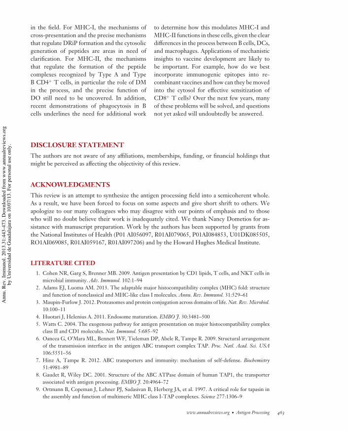
IY31CH16-Cresswell ARI 15 February 2013 4:42
in the field. For MHC-I, the mechanisms ofcross-presentation and the precise mechanismsthat regulate DRiP formation and the cytosolicgeneration of peptides are areas in need ofclarification. For MHC-II, the mechanismsthat regulate the formation of the peptidecomplexes recognized by Type A and TypeB CD4+ T cells, in particular the role of DMin the process, and the precise function ofDO still need to be uncovered. In addition,recent demonstrations of phagocytosis in Bcells underlines the need for additional work
to determine how this modulates MHC-I andMHC-II functions in these cells, given the cleardifferences in the process between B cells, DCs,and macrophages. Applications of mechanisticinsights to vaccine development are likely tobe important. For example, how do we bestincorporate immunogenic epitopes into re-combinant vaccines and how can they be movedinto the cytosol for effective sensitization ofCD8+ T cells? Over the next few years, manyof these problems will be solved, and questionsnot yet asked will undoubtedly be answered.
DISCLOSURE STATEMENTThe authors are not aware of any affiliations, memberships, funding, or financial holdings thatmight be perceived as affecting the objectivity of this review.
ACKNOWLEDGMENTSThis review is an attempt to synthesize the antigen processing field into a semicoherent whole.As a result, we have been forced to focus on some aspects and give short shrift to others. Weapologize to our many colleagues who may disagree with our points of emphasis and to thosewho will no doubt believe their work is inadequately cited. We thank Nancy Dometios for as-sistance with manuscript preparation. Work by the authors has been supported by grants fromthe National Institutes of Health (P01 AI056097, R01AI079065, P01AI084853, U01DK085505,RO1AI069085, R01AI059167, R01AI097206) and by the Howard Hughes Medical Institute.
LITERATURE CITED
1. Cohen NR, Garg S, Brenner MB. 2009. Antigen presentation by CD1 lipids, T cells, and NKT cells inmicrobial immunity. Adv. Immunol. 102:1–94
2. Adams EJ, Luoma AM. 2013. The adaptable major histocompatibility complex (MHC) fold: structureand function of nonclassical and MHC-like class I molecules. Annu. Rev. Immunol. 31:529–61
3. Maupin-Furlow J. 2012. Proteasomes and protein conjugation across domains of life. Nat. Rev. Microbiol.10:100–11
4. Huotari J, Helenius A. 2011. Endosome maturation. EMBO J. 30:3481–5005. Watts C. 2004. The exogenous pathway for antigen presentation on major histocompatibility complex
class II and CD1 molecules. Nat. Immunol. 5:685–926. Oancea G, O’Mara ML, Bennett WF, Tieleman DP, Abele R, Tampe R. 2009. Structural arrangement
of the transmission interface in the antigen ABC transport complex TAP. Proc. Natl. Acad. Sci. USA106:5551–56
7. Hinz A, Tampe R. 2012. ABC transporters and immunity: mechanism of self-defense. Biochemistry51:4981–89
8. Gaudet R, Wiley DC. 2001. Structure of the ABC ATPase domain of human TAP1, the transporterassociated with antigen processing. EMBO J. 20:4964–72
9. Ortmann B, Copeman J, Lehner PJ, Sadasivan B, Herberg JA, et al. 1997. A critical role for tapasin inthe assembly and function of multimeric MHC class I-TAP complexes. Science 277:1306–9
www.annualreviews.org • Antigen Processing 463
Ann
u. R
ev. I
mm
unol
. 201
3.31
:443
-473
. Dow
nloa
ded
from
ww
w.a
nnua
lrevi
ews.o
rgby
Uni
vers
idad
de
Gua
dala
jara
on
10/0
7/13
. For
per
sona
l use
onl
y.
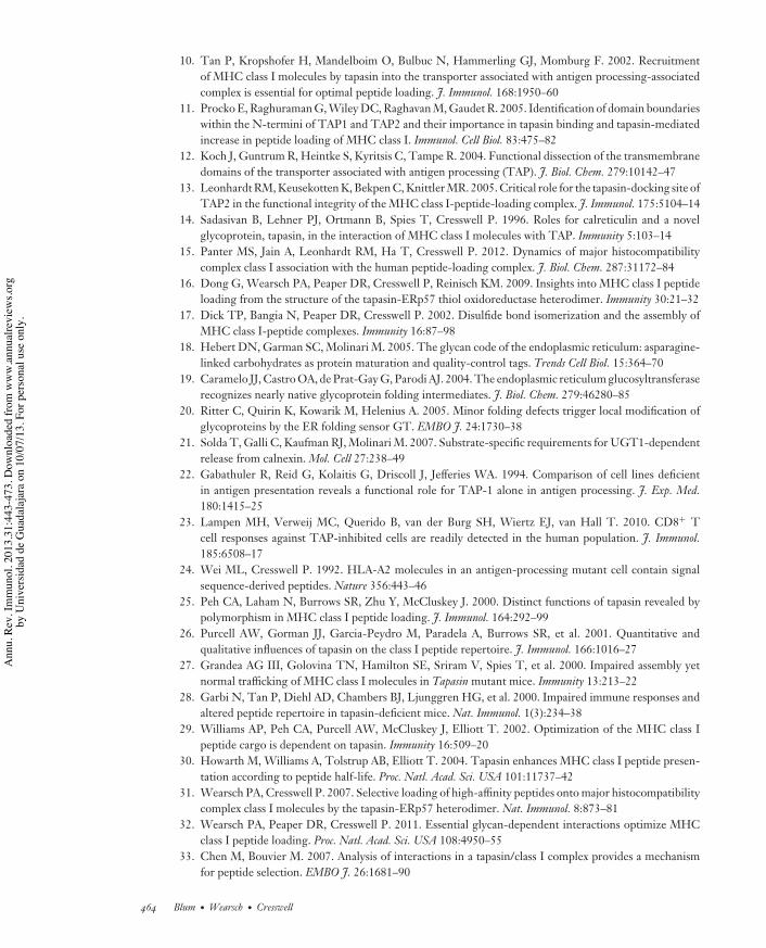
IY31CH16-Cresswell ARI 15 February 2013 4:42
10. Tan P, Kropshofer H, Mandelboim O, Bulbuc N, Hammerling GJ, Momburg F. 2002. Recruitmentof MHC class I molecules by tapasin into the transporter associated with antigen processing-associatedcomplex is essential for optimal peptide loading. J. Immunol. 168:1950–60
11. Procko E, Raghuraman G, Wiley DC, Raghavan M, Gaudet R. 2005. Identification of domain boundarieswithin the N-termini of TAP1 and TAP2 and their importance in tapasin binding and tapasin-mediatedincrease in peptide loading of MHC class I. Immunol. Cell Biol. 83:475–82
12. Koch J, Guntrum R, Heintke S, Kyritsis C, Tampe R. 2004. Functional dissection of the transmembranedomains of the transporter associated with antigen processing (TAP). J. Biol. Chem. 279:10142–47
13. Leonhardt RM, Keusekotten K, Bekpen C, Knittler MR. 2005. Critical role for the tapasin-docking site ofTAP2 in the functional integrity of the MHC class I-peptide-loading complex. J. Immunol. 175:5104–14
14. Sadasivan B, Lehner PJ, Ortmann B, Spies T, Cresswell P. 1996. Roles for calreticulin and a novelglycoprotein, tapasin, in the interaction of MHC class I molecules with TAP. Immunity 5:103–14
15. Panter MS, Jain A, Leonhardt RM, Ha T, Cresswell P. 2012. Dynamics of major histocompatibilitycomplex class I association with the human peptide-loading complex. J. Biol. Chem. 287:31172–84
16. Dong G, Wearsch PA, Peaper DR, Cresswell P, Reinisch KM. 2009. Insights into MHC class I peptideloading from the structure of the tapasin-ERp57 thiol oxidoreductase heterodimer. Immunity 30:21–32
17. Dick TP, Bangia N, Peaper DR, Cresswell P. 2002. Disulfide bond isomerization and the assembly ofMHC class I-peptide complexes. Immunity 16:87–98
18. Hebert DN, Garman SC, Molinari M. 2005. The glycan code of the endoplasmic reticulum: asparagine-linked carbohydrates as protein maturation and quality-control tags. Trends Cell Biol. 15:364–70
19. Caramelo JJ, Castro OA, de Prat-Gay G, Parodi AJ. 2004. The endoplasmic reticulum glucosyltransferaserecognizes nearly native glycoprotein folding intermediates. J. Biol. Chem. 279:46280–85
20. Ritter C, Quirin K, Kowarik M, Helenius A. 2005. Minor folding defects trigger local modification ofglycoproteins by the ER folding sensor GT. EMBO J. 24:1730–38
21. Solda T, Galli C, Kaufman RJ, Molinari M. 2007. Substrate-specific requirements for UGT1-dependentrelease from calnexin. Mol. Cell 27:238–49
22. Gabathuler R, Reid G, Kolaitis G, Driscoll J, Jefferies WA. 1994. Comparison of cell lines deficientin antigen presentation reveals a functional role for TAP-1 alone in antigen processing. J. Exp. Med.180:1415–25
23. Lampen MH, Verweij MC, Querido B, van der Burg SH, Wiertz EJ, van Hall T. 2010. CD8+ Tcell responses against TAP-inhibited cells are readily detected in the human population. J. Immunol.185:6508–17
24. Wei ML, Cresswell P. 1992. HLA-A2 molecules in an antigen-processing mutant cell contain signalsequence-derived peptides. Nature 356:443–46
25. Peh CA, Laham N, Burrows SR, Zhu Y, McCluskey J. 2000. Distinct functions of tapasin revealed bypolymorphism in MHC class I peptide loading. J. Immunol. 164:292–99
26. Purcell AW, Gorman JJ, Garcia-Peydro M, Paradela A, Burrows SR, et al. 2001. Quantitative andqualitative influences of tapasin on the class I peptide repertoire. J. Immunol. 166:1016–27
27. Grandea AG III, Golovina TN, Hamilton SE, Sriram V, Spies T, et al. 2000. Impaired assembly yetnormal trafficking of MHC class I molecules in Tapasin mutant mice. Immunity 13:213–22
28. Garbi N, Tan P, Diehl AD, Chambers BJ, Ljunggren HG, et al. 2000. Impaired immune responses andaltered peptide repertoire in tapasin-deficient mice. Nat. Immunol. 1(3):234–38
29. Williams AP, Peh CA, Purcell AW, McCluskey J, Elliott T. 2002. Optimization of the MHC class Ipeptide cargo is dependent on tapasin. Immunity 16:509–20
30. Howarth M, Williams A, Tolstrup AB, Elliott T. 2004. Tapasin enhances MHC class I peptide presen-tation according to peptide half-life. Proc. Natl. Acad. Sci. USA 101:11737–42
31. Wearsch PA, Cresswell P. 2007. Selective loading of high-affinity peptides onto major histocompatibilitycomplex class I molecules by the tapasin-ERp57 heterodimer. Nat. Immunol. 8:873–81
32. Wearsch PA, Peaper DR, Cresswell P. 2011. Essential glycan-dependent interactions optimize MHCclass I peptide loading. Proc. Natl. Acad. Sci. USA 108:4950–55
33. Chen M, Bouvier M. 2007. Analysis of interactions in a tapasin/class I complex provides a mechanismfor peptide selection. EMBO J. 26:1681–90
464 Blum ·Wearsch · Cresswell
Ann
u. R
ev. I
mm
unol
. 201
3.31
:443
-473
. Dow
nloa
ded
from
ww
w.a
nnua
lrevi
ews.o
rgby
Uni
vers
idad
de
Gua
dala
jara
on
10/0
7/13
. For
per
sona
l use
onl
y.
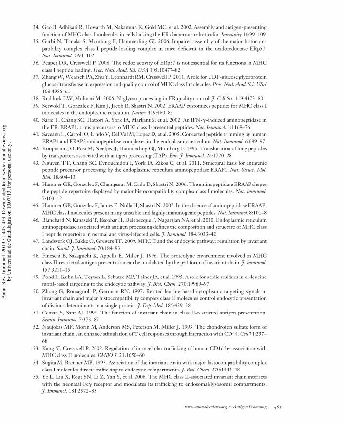
IY31CH16-Cresswell ARI 15 February 2013 4:42
34. Gao B, Adhikari R, Howarth M, Nakamura K, Gold MC, et al. 2002. Assembly and antigen-presentingfunction of MHC class I molecules in cells lacking the ER chaperone calreticulin. Immunity 16:99–109
35. Garbi N, Tanaka S, Momburg F, Hammerling GJ. 2006. Impaired assembly of the major histocom-patibility complex class I peptide-loading complex in mice deficient in the oxidoreductase ERp57.Nat. Immunol. 7:93–102
36. Peaper DR, Cresswell P. 2008. The redox activity of ERp57 is not essential for its functions in MHCclass I peptide loading. Proc. Natl. Acad. Sci. USA 105:10477–82
37. Zhang W, Wearsch PA, Zhu Y, Leonhardt RM, Cresswell P. 2011. A role for UDP-glucose glycoproteinglucosyltransferase in expression and quality control of MHC class I molecules. Proc. Natl. Acad. Sci. USA108:4956–61
38. Ruddock LW, Molinari M. 2006. N-glycan processing in ER quality control. J. Cell Sci. 119:4373–8039. Serwold T, Gonzalez F, Kim J, Jacob R, Shastri N. 2002. ERAAP customizes peptides for MHC class I
molecules in the endoplasmic reticulum. Nature 419:480–8340. Saric T, Chang SC, Hattori A, York IA, Markant S, et al. 2002. An IFN-#-induced aminopeptidase in
the ER, ERAP1, trims precursors to MHC class I-presented peptides. Nat. Immunol. 3:1169–7641. Saveanu L, Carroll O, Lindo V, Del Val M, Lopez D, et al. 2005. Concerted peptide trimming by human
ERAP1 and ERAP2 aminopeptidase complexes in the endoplasmic reticulum. Nat. Immunol. 6:689–9742. Koopmann JO, Post M, Neefjes JJ, Hammerling GJ, Momburg F. 1996. Translocation of long peptides
by transporters associated with antigen processing (TAP). Eur. J. Immunol. 26:1720–2843. Nguyen TT, Chang SC, Evnouchidou I, York IA, Zikos C, et al. 2011. Structural basis for antigenic
peptide precursor processing by the endoplasmic reticulum aminopeptidase ERAP1. Nat. Struct. Mol.Biol. 18:604–13
44. Hammer GE, Gonzalez F, Champsaur M, Cado D, Shastri N. 2006. The aminopeptidase ERAAP shapesthe peptide repertoire displayed by major histocompatibility complex class I molecules. Nat. Immunol.7:103–12
45. Hammer GE, Gonzalez F, James E, Nolla H, Shastri N. 2007. In the absence of aminopeptidase ERAAP,MHC class I molecules present many unstable and highly immunogenic peptides. Nat. Immunol. 8:101–8
46. Blanchard N, Kanaseki T, Escobar H, Delebecque F, Nagarajan NA, et al. 2010. Endoplasmic reticulumaminopeptidase associated with antigen processing defines the composition and structure of MHC classI peptide repertoire in normal and virus-infected cells. J. Immunol. 184:3033–42
47. Landsverk OJ, Bakke O, Gregers TF. 2009. MHC II and the endocytic pathway: regulation by invariantchain. Scand. J. Immunol. 70:184–93
48. Fineschi B, Sakaguchi K, Appella E, Miller J. 1996. The proteolytic environment involved in MHCclass II-restricted antigen presentation can be modulated by the p41 form of invariant chain. J. Immunol.157:3211–15
49. Pond L, Kuhn LA, Teyton L, Schutze MP, Tainer JA, et al. 1995. A role for acidic residues in di-leucinemotif-based targeting to the endocytic pathway. J. Biol. Chem. 270:19989–97
50. Zhong G, Romagnoli P, Germain RN. 1997. Related leucine-based cytoplasmic targeting signals ininvariant chain and major histocompatibility complex class II molecules control endocytic presentationof distinct determinants in a single protein. J. Exp. Med. 185:429–38
51. Ceman S, Sant AJ. 1995. The function of invariant chain in class II-restricted antigen presentation.Semin. Immunol. 7:373–87
52. Naujokas MF, Morin M, Anderson MS, Peterson M, Miller J. 1993. The chondroitin sulfate form ofinvariant chain can enhance stimulation of T cell responses through interaction with CD44. Cell 74:257–68
53. Kang SJ, Cresswell P. 2002. Regulation of intracellular trafficking of human CD1d by association withMHC class II molecules. EMBO J. 21:1650–60
54. Sugita M, Brenner MB. 1995. Association of the invariant chain with major histocompatibility complexclass I molecules directs trafficking to endocytic compartments. J. Biol. Chem. 270:1443–48
55. Ye L, Liu X, Rout SN, Li Z, Yan Y, et al. 2008. The MHC class II-associated invariant chain interactswith the neonatal Fc# receptor and modulates its trafficking to endosomal/lysosomal compartments.J. Immunol. 181:2572–85
www.annualreviews.org • Antigen Processing 465
Ann
u. R
ev. I
mm
unol
. 201
3.31
:443
-473
. Dow
nloa
ded
from
ww
w.a
nnua
lrevi
ews.o
rgby
Uni
vers
idad
de
Gua
dala
jara
on
10/0
7/13
. For
per
sona
l use
onl
y.
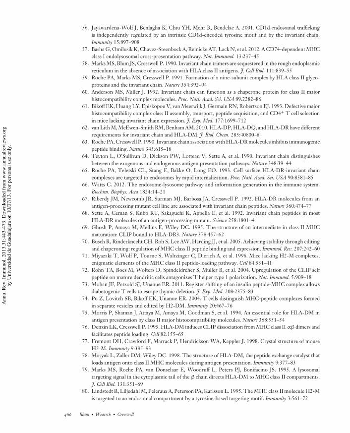
IY31CH16-Cresswell ARI 15 February 2013 4:42
56. Jayawardena-Wolf J, Benlagha K, Chiu YH, Mehr R, Bendelac A. 2001. CD1d endosomal traffickingis independently regulated by an intrinsic CD1d-encoded tyrosine motif and by the invariant chain.Immunity 15:897–908
57. Basha G, Omilusik K, Chavez-Steenbock A, Reinicke AT, Lack N, et al. 2012. A CD74-dependent MHCclass I endolysosomal cross-presentation pathway. Nat. Immunol. 13:237–45
58. Marks MS, Blum JS, Cresswell P. 1990. Invariant chain trimers are sequestered in the rough endoplasmicreticulum in the absence of association with HLA class II antigens. J. Cell Biol. 111:839–55
59. Roche PA, Marks MS, Cresswell P. 1991. Formation of a nine-subunit complex by HLA class II glyco-proteins and the invariant chain. Nature 354:392–94
60. Anderson MS, Miller J. 1992. Invariant chain can function as a chaperone protein for class II majorhistocompatibility complex molecules. Proc. Natl. Acad. Sci. USA 89:2282–86
61. Bikoff EK, Huang LY, Episkopou V, van Meerwijk J, Germain RN, Robertson EJ. 1993. Defective majorhistocompatibility complex class II assembly, transport, peptide acquisition, and CD4+ T cell selectionin mice lacking invariant chain expression. J. Exp. Med. 177:1699–712
62. van Lith M, McEwen-Smith RM, Benham AM. 2010. HLA-DP, HLA-DQ, and HLA-DR have differentrequirements for invariant chain and HLA-DM. J. Biol. Chem. 285:40800–8
63. Roche PA, Cresswell P. 1990. Invariant chain association with HLA-DR molecules inhibits immunogenicpeptide binding. Nature 345:615–18
64. Teyton L, O’Sullivan D, Dickson PW, Lotteau V, Sette A, et al. 1990. Invariant chain distinguishesbetween the exogenous and endogenous antigen presentation pathways. Nature 348:39–44
65. Roche PA, Teletski CL, Stang E, Bakke O, Long EO. 1993. Cell surface HLA-DR-invariant chaincomplexes are targeted to endosomes by rapid internalization. Proc. Natl. Acad. Sci. USA 90:8581–85
66. Watts C. 2012. The endosome-lysosome pathway and information generation in the immune system.Biochim. Biophys. Acta 1824:14–21
67. Riberdy JM, Newcomb JR, Surman MJ, Barbosa JA, Cresswell P. 1992. HLA-DR molecules from anantigen-processing mutant cell line are associated with invariant chain peptides. Nature 360:474–77
68. Sette A, Ceman S, Kubo RT, Sakaguchi K, Appella E, et al. 1992. Invariant chain peptides in mostHLA-DR molecules of an antigen-processing mutant. Science 258:1801–4
69. Ghosh P, Amaya M, Mellins E, Wiley DC. 1995. The structure of an intermediate in class II MHCmaturation: CLIP bound to HLA-DR3. Nature 378:457–62
70. Busch R, Rinderknecht CH, Roh S, Lee AW, Harding JJ, et al. 2005. Achieving stability through editingand chaperoning: regulation of MHC class II peptide binding and expression. Immunol. Rev. 207:242–60
71. Miyazaki T, Wolf P, Tourne S, Waltzinger C, Dierich A, et al. 1996. Mice lacking H2-M complexes,enigmatic elements of the MHC class II peptide-loading pathway. Cell 84:531–41
72. Rohn TA, Boes M, Wolters D, Spindeldreher S, Muller B, et al. 2004. Upregulation of the CLIP selfpeptide on mature dendritic cells antagonizes T helper type 1 polarization. Nat. Immunol. 5:909–18
73. Mohan JF, Petzold SJ, Unanue ER. 2011. Register shifting of an insulin peptide-MHC complex allowsdiabetogenic T cells to escape thymic deletion. J. Exp. Med. 208:2375–83
74. Pu Z, Lovitch SB, Bikoff EK, Unanue ER. 2004. T cells distinguish MHC-peptide complexes formedin separate vesicles and edited by H2-DM. Immunity 20:467–76
75. Morris P, Shaman J, Attaya M, Amaya M, Goodman S, et al. 1994. An essential role for HLA-DM inantigen presentation by class II major histocompatibility molecules. Nature 368:551–54
76. Denzin LK, Cresswell P. 1995. HLA-DM induces CLIP dissociation from MHC class II "!dimers andfacilitates peptide loading. Cell 82:155–65
77. Fremont DH, Crawford F, Marrack P, Hendrickson WA, Kappler J. 1998. Crystal structure of mouseH2-M. Immunity 9:385–93
78. Mosyak L, Zaller DM, Wiley DC. 1998. The structure of HLA-DM, the peptide exchange catalyst thatloads antigen onto class II MHC molecules during antigen presentation. Immunity 9:377–83
79. Marks MS, Roche PA, van Donselaar E, Woodruff L, Peters PJ, Bonifacino JS. 1995. A lysosomaltargeting signal in the cytoplasmic tail of the ! chain directs HLA-DM to MHC class II compartments.J. Cell Biol. 131:351–69
80. Lindstedt R, Liljedahl M, Peleraux A, Peterson PA, Karlsson L. 1995. The MHC class II molecule H2-Mis targeted to an endosomal compartment by a tyrosine-based targeting motif. Immunity 3:561–72
466 Blum ·Wearsch · Cresswell
Ann
u. R
ev. I
mm
unol
. 201
3.31
:443
-473
. Dow
nloa
ded
from
ww
w.a
nnua
lrevi
ews.o
rgby
Uni
vers
idad
de
Gua
dala
jara
on
10/0
7/13
. For
per
sona
l use
onl
y.
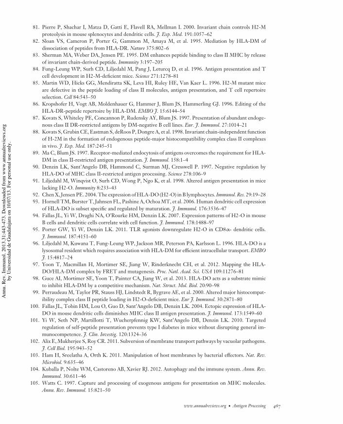
IY31CH16-Cresswell ARI 15 February 2013 4:42
81. Pierre P, Shachar I, Matza D, Gatti E, Flavell RA, Mellman I. 2000. Invariant chain controls H2-Mproteolysis in mouse splenocytes and dendritic cells. J. Exp. Med. 191:1057–62
82. Sloan VS, Cameron P, Porter G, Gammon M, Amaya M, et al. 1995. Mediation by HLA-DM ofdissociation of peptides from HLA-DR. Nature 375:802–6
83. Sherman MA, Weber DA, Jensen PE. 1995. DM enhances peptide binding to class II MHC by releaseof invariant chain-derived peptide. Immunity 3:197–205
84. Fung-Leung WP, Surh CD, Liljedahl M, Pang J, Leturcq D, et al. 1996. Antigen presentation and Tcell development in H2-M-deficient mice. Science 271:1278–81
85. Martin WD, Hicks GG, Mendiratta SK, Leva HI, Ruley HE, Van Kaer L. 1996. H2-M mutant miceare defective in the peptide loading of class II molecules, antigen presentation, and T cell repertoireselection. Cell 84:543–50
86. Kropshofer H, Vogt AB, Moldenhauer G, Hammer J, Blum JS, Hammerling GJ. 1996. Editing of theHLA-DR-peptide repertoire by HLA-DM. EMBO J. 15:6144–54
87. Kovats S, Whiteley PE, Concannon P, Rudensky AY, Blum JS. 1997. Presentation of abundant endoge-nous class II DR-restricted antigens by DM-negative B cell lines. Eur. J. Immunol. 27:1014–21
88. Kovats S, Grubin CE, Eastman S, deRoos P, Dongre A, et al. 1998. Invariant chain-independent functionof H-2M in the formation of endogenous peptide-major histocompatibility complex class II complexesin vivo. J. Exp. Med. 187:245–51
89. Ma C, Blum JS. 1997. Receptor-mediated endocytosis of antigens overcomes the requirement for HLA-DM in class II-restricted antigen presentation. J. Immunol. 158:1–4
90. Denzin LK, Sant’Angelo DB, Hammond C, Surman MJ, Cresswell P. 1997. Negative regulation byHLA-DO of MHC class II-restricted antigen processing. Science 278:106–9
91. Liljedahl M, Winqvist O, Surh CD, Wong P, Ngo K, et al. 1998. Altered antigen presentation in micelacking H2-O. Immunity 8:233–43
92. Chen X, Jensen PE. 2004. The expression of HLA-DO (H2-O) in B lymphocytes. Immunol. Res. 29:19–2893. Hornell TM, Burster T, Jahnsen FL, Pashine A, Ochoa MT, et al. 2006. Human dendritic cell expression
of HLA-DO is subset specific and regulated by maturation. J. Immunol. 176:3536–4794. Fallas JL, Yi W, Draghi NA, O’Rourke HM, Denzin LK. 2007. Expression patterns of H2-O in mouse
B cells and dendritic cells correlate with cell function. J. Immunol. 178:1488–9795. Porter GW, Yi W, Denzin LK. 2011. TLR agonists downregulate H2-O in CD8"- dendritic cells.
J. Immunol. 187:4151–6096. Liljedahl M, Kuwana T, Fung-Leung WP, Jackson MR, Peterson PA, Karlsson L. 1996. HLA-DO is a
lysosomal resident which requires association with HLA-DM for efficient intracellular transport. EMBOJ. 15:4817–24
97. Yoon T, Macmillan H, Mortimer SE, Jiang W, Rinderknecht CH, et al. 2012. Mapping the HLA-DO/HLA-DM complex by FRET and mutagenesis. Proc. Natl. Acad. Sci. USA 109:11276–81
98. Guce AI, Mortimer SE, Yoon T, Painter CA, Jiang W, et al. 2013. HLA-DO acts as a substrate mimicto inhibit HLA-DM by a competitive mechanism. Nat. Struct. Mol. Biol. 20:90–98
99. Perraudeau M, Taylor PR, Stauss HJ, Lindstedt R, Bygrave AE, et al. 2000. Altered major histocompat-ibility complex class II peptide loading in H2-O-deficient mice. Eur J. Immunol. 30:2871–80
100. Fallas JL, Tobin HM, Lou O, Guo D, Sant’Angelo DB, Denzin LK. 2004. Ectopic expression of HLA-DO in mouse dendritic cells diminishes MHC class II antigen presentation. J. Immunol. 173:1549–60
101. Yi W, Seth NP, Martillotti T, Wucherpfennig KW, Sant’Angelo DB, Denzin LK. 2010. Targetedregulation of self-peptide presentation prevents type I diabetes in mice without disrupting general im-munocompetence. J. Clin. Investig. 120:1324–36
102. Alix E, Mukherjee S, Roy CR. 2011. Subversion of membrane transport pathways by vacuolar pathogens.J. Cell Biol. 195:943–52
103. Ham H, Sreelatha A, Orth K. 2011. Manipulation of host membranes by bacterial effectors. Nat. Rev.Microbiol. 9:635–46
104. Kuballa P, Nolte WM, Castoreno AB, Xavier RJ. 2012. Autophagy and the immune system. Annu. Rev.Immunol. 30:611–46
105. Watts C. 1997. Capture and processing of exogenous antigens for presentation on MHC molecules.Annu. Rev. Immunol. 15:821–50
www.annualreviews.org • Antigen Processing 467
Ann
u. R
ev. I
mm
unol
. 201
3.31
:443
-473
. Dow
nloa
ded
from
ww
w.a
nnua
lrevi
ews.o
rgby
Uni
vers
idad
de
Gua
dala
jara
on
10/0
7/13
. For
per
sona
l use
onl
y.

IY31CH16-Cresswell ARI 15 February 2013 4:42
106. Lanzavecchia A. 1985. Antigen-specific interaction between T and B cells. Nature 314:537–9107. Davidson HW, West MA, Watts C. 1990. Endocytosis, intracellular trafficking, and processing of mem-
brane IgG and monovalent antigen/membrane IgG complexes in B lymphocytes. J. Immunol. 144:4101–9108. Gil-Torregrosa BC, Lennon-Dumenil AM, Kessler B, Guermonprez P, Ploegh HL, et al. 2004. Control
of cross-presentation during dendritic cell maturation. Eur J. Immunol. 34:398–407109. Bonifaz LC, Bonnyay DP, Charalambous A, Darguste DI, Fujii S, et al. 2004. In vivo targeting of
antigens to maturing dendritic cells via the DEC-205 receptor improves T cell vaccination. J. Exp. Med.199:815–24
110. Lorenz RG, Blum JS, Allen PM. 1990. Constitutive competition by self proteins for antigen presentationcan be overcome by receptor-enhanced uptake. J. Immunol. 144:1600–6
111. McCoy KL, Page MS, Merkel BJ, Inman JK, Stutzman R. 1993. Differences among various lineagesof antigen-presenting cells in processing exogenous antigen internalized through transferrin receptors.J. Immunol. 151:6757–68
112. Burgdorf S, Kautz A, Bohnert V, Knolle PA, Kurts C. 2007. Distinct pathways of antigen uptake andintracellular routing in CD4 and CD8 T cell activation. Science 316:612–16
113. Castellino F, Boucher PE, Eichelberg K, Mayhew M, Rothman JE, et al. 2000. Receptor-mediateduptake of antigen/heat shock protein complexes results in major histocompatibility complex class Iantigen presentation via two distinct processing pathways. J. Exp. Med. 191:1957–64
114. Tobian AA, Harding CV, Canaday DH. 2005. Mycobacterium tuberculosis heat shock fusion protein en-hances class I MHC cross-processing and -presentation by B lymphocytes. J. Immunol. 174:5209–14
115. Houlihan JL, Metzler JJ, Blum JS. 2009. HSP90" and HSP90! isoforms selectively modulate MHCclass II antigen presentation in B cells. J. Immunol. 182:7451–58
116. Matsutake T, Sawamura T, Srivastava PK. 2010. High efficiency CD91- and LOX-1-mediated re-presentation of gp96-chaperoned peptides by MHC II molecules. Cancer Immun. 10:7
117. Ramachandra L, Simmons D, Harding CV. 2009. MHC molecules and microbial antigen processing inphagosomes. Curr. Opin. Immunol. 21:98–104
118. Schelhaas M. 2010. Come in and take your coat off—how host cells provide endocytosis for virus entry.Cell. Microbiol. 12:1378–88
119. Schulz O, Reis e Sousa C. 2002. Cross-presentation of cell-associated antigens by CD8"+ dendritic cellsis attributable to their ability to internalize dead cells. Immunology 107:183–89
120. Vidard L, Kovacsovics-Bankowski M, Kraeft SK, Chen LB, Benacerraf B, Rock KL. 1996. Analysis ofMHC class II presentation of particulate antigens of B lymphocytes. J. Immunol. 156:2809–18
121. Gao J, Ma X, Gu W, Fu M, An J, et al. 2012. Novel functions of murine B1 cells: active phagocytic andmicrobicidal abilities. Eur. J. Immunol. 42:982–92
122. Parra D, Rieger AM, Li J, Zhang YA, Randall LM, et al. 2012. Pivotal advance: peritoneal cavity B-1B cells have phagocytic and microbicidal capacities and present phagocytosed antigen to CD4+ T cells.J. Leukoc. Biol. 91:525–36
123. Kim SH, Visser A, Cruijsen C, van der Velden AW, Boes M. 2008. Recruitment of Rab27a to phagosomescontrols microbial antigen cross-presentation by dendritic cells. Infect. Immun. 76:5373–80
124. Sancho D, Joffre OP, Keller AM, Rogers NC, Martinez D, et al. 2009. Identification of a dendritic cellreceptor that couples sensing of necrosis to immunity. Nature 458:899–903
125. Joffre OP, Sancho D, Zelenay S, Keller AM, Reis e Sousa C. 2010. Efficient and versatile manipulation ofthe peripheral CD4+ T-cell compartment by antigen targeting to DNGR-1/CLEC9A. Eur. J. Immunol.40:1255–65
126. Chicz RM, Urban RG, Gorga JC, Vignali DA, Lane WS, Strominger JL. 1993. Specificity and promis-cuity among naturally processed peptides bound to HLA-DR alleles. J. Exp. Med. 178:27–47
127. Guagliardi LE, Koppelman B, Blum JS, Marks MS, Cresswell P, Brodsky FM. 1990. Co-localization ofmolecules involved in antigen processing and presentation in an early endocytic compartment. Nature343:133–39
128. Harding CV, Unanue ER, Slot JW, Schwartz AL, Geuze HJ. 1990. Functional and ultrastructuralevidence for intracellular formation of major histocompatibility complex class II-peptide complexesduring antigen processing. Proc. Natl. Acad. Sci. USA 87:5553–57
468 Blum ·Wearsch · Cresswell
Ann
u. R
ev. I
mm
unol
. 201
3.31
:443
-473
. Dow
nloa
ded
from
ww
w.a
nnua
lrevi
ews.o
rgby
Uni
vers
idad
de
Gua
dala
jara
on
10/0
7/13
. For
per
sona
l use
onl
y.
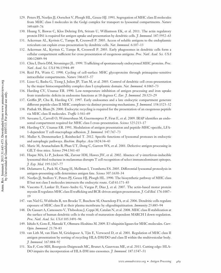
IY31CH16-Cresswell ARI 15 February 2013 4:42
129. Peters PJ, Neefjes JJ, Oorschot V, Ploegh HL, Geuze HJ. 1991. Segregation of MHC class II moleculesfrom MHC class I molecules in the Golgi complex for transport to lysosomal compartments. Nature349:669–76
130. Huang Y, Biswas C, Klos Dehring DA, Sriram U, Williamson EK, et al. 2011. The actin regulatoryprotein HS1 is required for antigen uptake and presentation by dendritic cells. J. Immunol. 187:5952–63
131. Ackerman AL, Kyritsis C, Tampe R, Cresswell P. 2005. Access of soluble antigens to the endoplasmicreticulum can explain cross-presentation by dendritic cells. Nat. Immunol. 6:107–13
132. Ackerman AL, Kyritsis C, Tampe R, Cresswell P. 2003. Early phagosomes in dendritic cells form acellular compartment sufficient for cross presentation of exogenous antigens. Proc. Natl. Acad. Sci. USA100:12889–94
133. Chiu I, Davis DM, Strominger JL. 1999. Trafficking of spontaneously endocytosed MHC proteins. Proc.Natl. Acad. Sci. USA 96:13944–49
134. Reid PA, Watts C. 1990. Cycling of cell-surface MHC glycoproteins through primaquine-sensitiveintracellular compartments. Nature 346:655–57
135. Lizee G, Basha G, Tiong J, Julien JP, Tian M, et al. 2003. Control of dendritic cell cross-presentationby the major histocompatibility complex class I cytoplasmic domain. Nat. Immunol. 4:1065–73
136. Harding CV, Unanue ER. 1990. Low-temperature inhibition of antigen processing and iron uptakefrom transferrin: deficits in endosome functions at 18 degrees C. Eur. J. Immunol. 20:323–29
137. Griffin JP, Chu R, Harding CV. 1997. Early endosomes and a late endocytic compartment generatedifferent peptide-class II MHC complexes via distinct processing mechanisms. J. Immunol. 158:1523–32
138. Pathak SS, Blum JS. 2000. Endocytic recycling is required for the presentation of an exogenous peptidevia MHC class II molecules. Traffic 1:561–69
139. Saveanu L, Carroll O, Weimershaus M, Guermonprez P, Firat E, et al. 2009. IRAP identifies an endo-somal compartment required for MHC class I cross-presentation. Science 325:213–17
140. Harding CV, Unanue ER. 1991. Modulation of antigen presentation and peptide-MHC-specific, LFA-1-dependent T cell-macrophage adhesion. J. Immunol. 147:767–73
141. Muller S, Dennemarker J, Reinheckel T. 2012. Specific functions of lysosomal proteases in endocyticand autophagic pathways. Biochim. Biophys. Acta 1824:34–43
142. Maric M, Arunachalam B, Phan UT, Dong C, Garrett WS, et al. 2001. Defective antigen processing inGILT-free mice. Science 294:1361–65
143. Haque MA, Li P, Jackson SK, Zarour HM, Hawes JW, et al. 2002. Absence of #-interferon-induciblelysosomal thiol reductase in melanomas disrupts T cell recognition of select immunodominant epitopes.J. Exp. Med. 195:1267–77
144. Delamarre L, Pack M, Chang H, Mellman I, Trombetta ES. 2005. Differential lysosomal proteolysis inantigen-presenting cells determines antigen fate. Science 307:1630–34
145. Neefjes JJ, Stollorz V, Peters PJ, Geuze HJ, Ploegh HL. 1990. The biosynthetic pathway of MHC classII but not class I molecules intersects the endocytic route. Cell 61:171–83
146. Vascotto F, Lankar D, Faure-Andre G, Vargas P, Diaz J, et al. 2007. The actin-based motor proteinmyosin II regulates MHC class II trafficking and BCR-driven antigen presentation. J. Cell Biol. 176:1007–19
147. van Niel G, Wubbolts R, ten Broeke T, Buschow SI, Ossendorp FA, et al. 2006. Dendritic cells regulateexposure of MHC class II at their plasma membrane by oligoubiquitination. Immunity 25:885–94
148. De Gassart A, Camosseto V, Thibodeau J, Ceppi M, Catalan N, et al. 2008. MHC class II stabilization atthe surface of human dendritic cells is the result of maturation-dependent MARCH I down-regulation.Proc. Natl. Acad. Sci. USA 105:3491–96
149. Ishido S, Goto E, Matsuki Y, Ohmura-Hoshino M. 2009. E3 ubiquitin ligases for MHC molecules. Curr.Opin. Immunol. 21:78–83
150. van Lith M, van Ham M, Griekspoor A, Tjin E, Verwoerd D, et al. 2001. Regulation of MHC class IIantigen presentation by sorting of recycling HLA-DM/DO and class II within the multivesicular body.J. Immunol. 167:884–92
151. Xiu F, Cote MH, Bourgeois-Daigneault MC, Brunet A, Gauvreau ME, et al. 2011. Cutting edge: HLA-DO impairs the incorporation of HLA-DM into exosomes. J. Immunol. 187:1547–51
www.annualreviews.org • Antigen Processing 469
Ann
u. R
ev. I
mm
unol
. 201
3.31
:443
-473
. Dow
nloa
ded
from
ww
w.a
nnua
lrevi
ews.o
rgby
Uni
vers
idad
de
Gua
dala
jara
on
10/0
7/13
. For
per
sona
l use
onl
y.
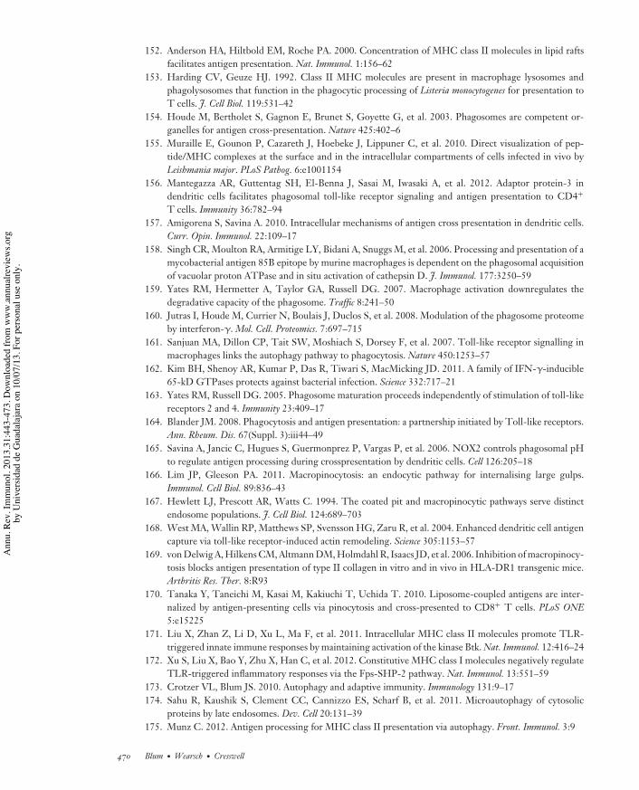
IY31CH16-Cresswell ARI 15 February 2013 4:42
152. Anderson HA, Hiltbold EM, Roche PA. 2000. Concentration of MHC class II molecules in lipid raftsfacilitates antigen presentation. Nat. Immunol. 1:156–62
153. Harding CV, Geuze HJ. 1992. Class II MHC molecules are present in macrophage lysosomes andphagolysosomes that function in the phagocytic processing of Listeria monocytogenes for presentation toT cells. J. Cell Biol. 119:531–42
154. Houde M, Bertholet S, Gagnon E, Brunet S, Goyette G, et al. 2003. Phagosomes are competent or-ganelles for antigen cross-presentation. Nature 425:402–6
155. Muraille E, Gounon P, Cazareth J, Hoebeke J, Lippuner C, et al. 2010. Direct visualization of pep-tide/MHC complexes at the surface and in the intracellular compartments of cells infected in vivo byLeishmania major. PLoS Pathog. 6:e1001154
156. Mantegazza AR, Guttentag SH, El-Benna J, Sasai M, Iwasaki A, et al. 2012. Adaptor protein-3 indendritic cells facilitates phagosomal toll-like receptor signaling and antigen presentation to CD4+
T cells. Immunity 36:782–94157. Amigorena S, Savina A. 2010. Intracellular mechanisms of antigen cross presentation in dendritic cells.
Curr. Opin. Immunol. 22:109–17158. Singh CR, Moulton RA, Armitige LY, Bidani A, Snuggs M, et al. 2006. Processing and presentation of a
mycobacterial antigen 85B epitope by murine macrophages is dependent on the phagosomal acquisitionof vacuolar proton ATPase and in situ activation of cathepsin D. J. Immunol. 177:3250–59
159. Yates RM, Hermetter A, Taylor GA, Russell DG. 2007. Macrophage activation downregulates thedegradative capacity of the phagosome. Traffic 8:241–50
160. Jutras I, Houde M, Currier N, Boulais J, Duclos S, et al. 2008. Modulation of the phagosome proteomeby interferon-#. Mol. Cell. Proteomics. 7:697–715
161. Sanjuan MA, Dillon CP, Tait SW, Moshiach S, Dorsey F, et al. 2007. Toll-like receptor signalling inmacrophages links the autophagy pathway to phagocytosis. Nature 450:1253–57
162. Kim BH, Shenoy AR, Kumar P, Das R, Tiwari S, MacMicking JD. 2011. A family of IFN-#-inducible65-kD GTPases protects against bacterial infection. Science 332:717–21
163. Yates RM, Russell DG. 2005. Phagosome maturation proceeds independently of stimulation of toll-likereceptors 2 and 4. Immunity 23:409–17
164. Blander JM. 2008. Phagocytosis and antigen presentation: a partnership initiated by Toll-like receptors.Ann. Rheum. Dis. 67(Suppl. 3):iii44–49
165. Savina A, Jancic C, Hugues S, Guermonprez P, Vargas P, et al. 2006. NOX2 controls phagosomal pHto regulate antigen processing during crosspresentation by dendritic cells. Cell 126:205–18
166. Lim JP, Gleeson PA. 2011. Macropinocytosis: an endocytic pathway for internalising large gulps.Immunol. Cell Biol. 89:836–43
167. Hewlett LJ, Prescott AR, Watts C. 1994. The coated pit and macropinocytic pathways serve distinctendosome populations. J. Cell Biol. 124:689–703
168. West MA, Wallin RP, Matthews SP, Svensson HG, Zaru R, et al. 2004. Enhanced dendritic cell antigencapture via toll-like receptor-induced actin remodeling. Science 305:1153–57
169. von Delwig A, Hilkens CM, Altmann DM, Holmdahl R, Isaacs JD, et al. 2006. Inhibition of macropinocy-tosis blocks antigen presentation of type II collagen in vitro and in vivo in HLA-DR1 transgenic mice.Arthritis Res. Ther. 8:R93
170. Tanaka Y, Taneichi M, Kasai M, Kakiuchi T, Uchida T. 2010. Liposome-coupled antigens are inter-nalized by antigen-presenting cells via pinocytosis and cross-presented to CD8+ T cells. PLoS ONE5:e15225
171. Liu X, Zhan Z, Li D, Xu L, Ma F, et al. 2011. Intracellular MHC class II molecules promote TLR-triggered innate immune responses by maintaining activation of the kinase Btk. Nat. Immunol. 12:416–24
172. Xu S, Liu X, Bao Y, Zhu X, Han C, et al. 2012. Constitutive MHC class I molecules negatively regulateTLR-triggered inflammatory responses via the Fps-SHP-2 pathway. Nat. Immunol. 13:551–59
173. Crotzer VL, Blum JS. 2010. Autophagy and adaptive immunity. Immunology 131:9–17174. Sahu R, Kaushik S, Clement CC, Cannizzo ES, Scharf B, et al. 2011. Microautophagy of cytosolic
proteins by late endosomes. Dev. Cell 20:131–39175. Munz C. 2012. Antigen processing for MHC class II presentation via autophagy. Front. Immunol. 3:9
470 Blum ·Wearsch · Cresswell
Ann
u. R
ev. I
mm
unol
. 201
3.31
:443
-473
. Dow
nloa
ded
from
ww
w.a
nnua
lrevi
ews.o
rgby
Uni
vers
idad
de
Gua
dala
jara
on
10/0
7/13
. For
per
sona
l use
onl
y.
IY31CH16-Cresswell ARI 15 February 2013 4:42
176. Nedjic J, Aichinger M, Emmerich J, Mizushima N, Klein L. 2008. Autophagy in thymic epitheliumshapes the T-cell repertoire and is essential for tolerance. Nature 455:396–400
177. Jagannath C, Lindsey DR, Dhandayuthapani S, Xu Y, Hunter RL Jr, Eissa NT. 2009. Autophagyenhances the efficacy of BCG vaccine by increasing peptide presentation in mouse dendritic cells. Nat.Med. 15:267–76
178. Dragovic SM, Hill T, Christianson GJ, Kim S, Elliott T, et al. 2011. Proteasomes, TAP, andendoplasmic reticulum-associated aminopeptidase associated with antigen processing control CD4+
Th cell responses by regulating indirect presentation of MHC class II-restricted cytoplasmic antigens.J. Immunol. 186:6683–92
179. Li Y, Wang LX, Pang P, Cui Z, Aung S, et al. 2011. Tumor-derived autophagosome vaccine: mechanismof cross-presentation and therapeutic efficacy. Clin. Cancer Res. 17:7047–57
180. Tewari MK, Sinnathamby G, Rajagopal D, Eisenlohr LC. 2005. A cytosolic pathway for MHC classII-restricted antigen processing that is proteasome and TAP dependent. Nat. Immunol. 6:287–94
181. Lich JD, Elliott JF, Blum JS. 2000. Cytoplasmic processing is a prerequisite for presentation of anendogenous antigen by major histocompatibility complex class II proteins. J. Exp. Med. 191:1513–24
182. Mukherjee P, Dani A, Bhatia S, Singh N, Rudensky AY, et al. 2001. Efficient presentation of bothcytosolic and endogenous transmembrane protein antigens on MHC class II is dependent on cytoplasmicproteolysis. J. Immunol. 167:2632–41
183. Dorfel D, Appel S, Grunebach F, Weck MM, Muller MR, et al. 2005. Processing and presentation ofHLA class I and II epitopes by dendritic cells after transfection with in vitro-transcribed MUC1 RNA.Blood 105:3199–205
184. Akram A, Inman RD. 2012. Immunodominance: a pivotal principle in host response to viral infections.Clin. Immunol. 143:99–115
185. Moudgil KD, Sercarz EE. 2005. Crypticity of self antigenic determinants is the cornerstone of a theoryof autoimmunity. Discov. Med. 5:378–82
186. Dai YD, Jensen KP, Marrero I, Li N, Quinn A, Sercarz EE. 2008. N-terminal flanking residues ofa diabetes-associated GAD65 determinant are necessary for activation of antigen-specific T cells indiabetes-resistant mice. Eur. J. Immunol. 38:968–76
187. Mimura Y, Mimura-Kimura Y, Doores K, Golgher D, Davis BG, et al. 2007. Folding of an MHC classII-restricted tumor antigen controls its antigenicity via MHC-guided processing. Proc. Natl. Acad. Sci.USA 104:5983–88
188. Bergman CM, Marta CB, Maric M, Pfeiffer SE, Cresswell P, Ruddle NH. 2012. A switch in pathogenicmechanism in myelin oligodendrocyte glycoprotein-induced experimental autoimmune encephalomyeli-tis in IFN-#-inducible lysosomal thiol reductase-free mice. J. Immunol. 188:6001–9
189. Rausch MP, Irvine KR, Antony PA, Restifo NP, Cresswell P, Hastings KT. 2010. GILT acceleratesautoimmunity to the melanoma antigen tyrosinase-related protein 1. J. Immunol. 185:2828–35
190. Delamarre L, Couture R, Mellman I, Trombetta ES. 2006. Enhancing immunogenicity by limitingsusceptibility to lysosomal proteolysis. J. Exp. Med. 203:2049–55
191. Davidson HW, Reid PA, Lanzavecchia A, Watts C. 1991. Processed antigen binds to newly synthesizedMHC class II molecules in antigen-specific B lymphocytes. Cell 67:105–16
192. Sercarz EE, Maverakis E. 2003. MHC-guided processing: binding of large antigen fragments. Nat. Rev.Immunol. 3:621–29
193. Moss CX, Tree TI, Watts C. 2007. Reconstruction of a pathway of antigen processing and class II MHCpeptide capture. EMBO J. 26:2137–47
194. Brooks K, Knight AM. 2004. Lowering the affinity between antigen and the B cell receptor can enhanceantigen presentation. Eur. J. Immunol. 34:837–43
195. Hartman IZ, Kim A, Cotter RJ, Walter K, Dalai SK, et al. 2010. A reductionist cell-free major his-tocompatibility complex class II antigen processing system identifies immunodominant epitopes. Nat.Med. 16:1333–40
196. Nanda NK, Sant AJ. 2000. DM determines the cryptic and immunodominant fate of T cell epitopes.J. Exp. Med. 192:781–88
www.annualreviews.org • Antigen Processing 471
Ann
u. R
ev. I
mm
unol
. 201
3.31
:443
-473
. Dow
nloa
ded
from
ww
w.a
nnua
lrevi
ews.o
rgby
Uni
vers
idad
de
Gua
dala
jara
on
10/0
7/13
. For
per
sona
l use
onl
y.
IY31CH16-Cresswell ARI 15 February 2013 4:42
197. Mohan JF, Levisetti MG, Calderon B, Herzog JW, Petzold SJ, Unanue ER. 2010. Unique autoreactiveT cells recognize insulin peptides generated within the islets of Langerhans in autoimmune diabetes.Nat. Immunol. 11:350–54
198. Singh R, Cresswell P. 2010. Defective cross-presentation of viral antigens in GILT-free mice. Science328:1394–98
199. Siddiqui S, Basta S. 2011. CD8+ T cell immunodominance in lymphocytic choriomeningitis virus in-fection is modified in the presence of Toll-like receptor agonists. J. Virol. 85:13224–33
200. Boon T, Coulie PG, Van den Eynde BJ, van der Bruggen P. 2006. Human T cell responses againstmelanoma. Annu. Rev. Immunol. 24:175–208
201. Hahn YS, Braciale VL, Braciale TJ. 1991. Presentation of viral antigen to class I major histocompatibilitycomplex-restricted cytotoxic T lymphocyte. Recognition of an immunodominant influenza hemagglu-tinin site by cytotoxic T lymphocyte is independent of the position of the site in the hemagglutinintranslation product. J. Exp. Med. 174:733–36
202. Hanke T, Graham FL, Rosenthal KL, Johnson DC. 1991. Identification of an immunodominant cy-totoxic T-lymphocyte recognition site in glycoprotein B of herpes simplex virus by using recombinantadenovirus vectors and synthetic peptides. J. Virol. 65:1177–86
203. Schubert U, Anton LC, Gibbs J, Norbury CC, Yewdell JW, Bennink JR. 2000. Rapid degradation of alarge fraction of newly synthesized proteins by proteasomes. Nature 404:770–74
204. Yewdell JW. 2011. DRiPs solidify: progress in understanding endogenous MHC class I antigen process-ing. Trends Immunol. 32:548–58
205. Townsend AR, Gotch FM, Davey J. 1985. Cytotoxic T cells recognize fragments of the influenza nu-cleoprotein. Cell 42:457–67
206. Reits EA, Vos JC, Gromme M, Neefjes J. 2000. The major substrates for TAP in vivo are derived fromnewly synthesized proteins. Nature 404:774–78
207. Princiotta MF, Finzi D, Qian SB, Gibbs J, Schuchmann S, et al. 2003. Quantitating protein synthesis,degradation, and endogenous antigen processing. Immunity 18:343–54
208. Mackay LK, Long HM, Brooks JM, Taylor GS, Leung CS, et al. 2009. T cell detection of a B-celltropic virus infection: newly-synthesised versus mature viral proteins as antigen sources for CD4 andCD8 epitope display. PLoS Pathog. 5:e1000699
209. Apcher S, Daskalogianni C, Lejeune F, Manoury B, Imhoos G, et al. 2011. Major source of antigenicpeptides for the MHC class I pathway is produced during the pioneer round of mRNA translation. Proc.Natl. Acad. Sci. USA 108:11572–77
210. Apcher S, Manoury B, Fahraeus R. 2012. The role of mRNA translation in direct MHC class I antigenpresentation. Curr. Opin. Immunol. 24:71–76
211. Granados DP, Yahyaoui W, Laumont CM, Daouda T, Muratore-Schroeder TL, et al. 2012. MHCI-associated peptides preferentially derive from transcripts bearing miRNA response elements. Blood119:e181–91
212. Villanueva MS, Sijts AJ, Pamer EG. 1995. Listeriolysin is processed efficiently into an MHC class I-associated epitope in Listeria monocytogenes-infected cells. J. Immunol. 155:5227–33
213. Kunisawa J, Shastri N. 2006. Hsp90"chaperones large C-terminally extended proteolytic intermediatesin the MHC class I antigen processing pathway. Immunity 24:523–34
214. Cascio P, Hilton C, Kisselev AF, Rock KL, Goldberg AL. 2001. 26S proteasomes and immunoprotea-somes produce mainly N-extended versions of an antigenic peptide. EMBO J. 20:2357–66
215. Kunisawa J, Shastri N. 2003. The group II chaperonin TRiC protects proteolytic intermediates fromdegradation in the MHC class I antigen processing pathway. Mol. Cell 12:565–76
216. Beninga J, Rock KL, Goldberg AL. 1998. Interferon-# can stimulate post-proteasomal trimming of theN terminus of an antigenic peptide by inducing leucine aminopeptidase. J. Biol. Chem. 273:18734–42
217. Urban S, Textoris-Taube K, Reimann B, Janek K, Dannenberg T, et al. 2012. The efficiency of humancytomegalovirus pp65495#503 CD8+ T cell epitope generation is determined by the balanced activitiesof cytosolic and endoplasmic reticulum-resident peptidases. J. Immunol. 189:529–38
218. Sykulev Y, Joo M, Vturina I, Tsomides TJ, Eisen HN. 1996. Evidence that a single peptide-MHCcomplex on a target cell can elicit a cytolytic T cell response. Immunity 4:565–71
472 Blum ·Wearsch · Cresswell
Ann
u. R
ev. I
mm
unol
. 201
3.31
:443
-473
. Dow
nloa
ded
from
ww
w.a
nnua
lrevi
ews.o
rgby
Uni
vers
idad
de
Gua
dala
jara
on
10/0
7/13
. For
per
sona
l use
onl
y.

IY31CH16-Cresswell ARI 15 February 2013 4:42
219. Vigneron N, Van den Eynde BJ. 2011. Insights into the processing of MHC class I ligands gained fromthe study of human tumor epitopes. Cell. Mol. Life Sci. 68:1503–20
220. Starck SR, Jiang V, Pavon-Eternod M, Prasad S, McCarthy B, et al. 2012. Leucine-tRNA initiates atCUG start codons for protein synthesis and presentation by MHC class I. Science 336:1719–23
221. Vigneron N, Stroobant V, Chapiro J, Ooms A, Degiovanni G, et al. 2004. An antigenic peptide producedby peptide splicing in the proteasome. Science 304:587–90
222. Warren EH, Vigneron NJ, Gavin MA, Coulie PG, Stroobant V, et al. 2006. An antigen produced bysplicing of noncontiguous peptides in the reverse order. Science 313:1444–47
223. Skipper JC, Hendrickson RC, Gulden PH, Brichard V, Van Pel A, et al. 1996. An HLA-A2-restrictedtyrosinase antigen on melanoma cells results from posttranslational modification and suggests a novelpathway for processing of membrane proteins. J. Exp. Med. 183:527–34
224. Smith MH, Ploegh HL, Weissman JS. 2011. Road to ruin: targeting proteins for degradation in theendoplasmic reticulum. Science 334:1086–90
225. Suzuki T, Seko A, Kitajima K, Inoue Y, Inoue S. 1994. Purification and enzymatic properties ofpeptide:N-glycanase from C3H mouse-derived L-929 fibroblast cells. Possible widespread occurrenceof post-translational remodification of proteins by N-deglycosylation. J. Biol. Chem. 269:17611–18
226. Gagnon E, Duclos S, Rondeau C, Chevet E, Cameron PH, et al. 2002. Endoplasmic reticulum-mediatedphagocytosis is a mechanism of entry into macrophages. Cell 110:119–31
227. Ackerman AL, Giodini A, Cresswell P. 2006. A role for the endoplasmic reticulum protein retrotranslo-cation machinery during crosspresentation by dendritic cells. Immunity 25:607–17
228. Lin ML, Zhan Y, Proietto AI, Prato S, Wu L, et al. 2008. Selective suicide of cross-presenting CD8+
dendritic cells by cytochrome c injection shows functional heterogeneity within this subset. Proc. Natl.Acad. Sci. USA 105:3029–34
229. Giodini A, Cresswell P. 2008. Hsp90-mediated cytosolic refolding of exogenous proteins internalizedby dendritic cells. EMBO J. 27:201–11
230. Burgdorf S, Scholz C, Kautz A, Tampe R, Kurts C. 2008. Spatial and mechanistic separation of cross-presentation and endogenous antigen presentation. Nat. Immunol. 9:558–66
231. Cebrian I, Visentin G, Blanchard N, Jouve M, Bobard A, et al. 2011. Sec22b regulates phagosomalmaturation and antigen crosspresentation by dendritic cells. Cell 147:1355–68
232. Madden DR, Garboczi DN, Wiley DC. 1993. The antigenic identity of peptide-MHC complexes: acomparison of the conformations of five viral peptides presented by HLA-A2. Cell 75:693–708
233. Stern LJ, Brown JH, Jardetzky TS, Gorga JC, Urban RG, et al. 1994. Crystal structure of the humanclass II MHC protein HLA-DR1 complexed with an influenza virus peptide. Nature 368:215–21
www.annualreviews.org • Antigen Processing 473
Ann
u. R
ev. I
mm
unol
. 201
3.31
:443
-473
. Dow
nloa
ded
from
ww
w.a
nnua
lrevi
ews.o
rgby
Uni
vers
idad
de
Gua
dala
jara
on
10/0
7/13
. For
per
sona
l use
onl
y.
IY31-Frontmatter ARI 27 February 2013 7:16
Annual Review ofImmunology
Volume 31, 2013
ContentsYears in Cologne
Klaus Rajewsky ! ! ! ! ! ! ! ! ! ! ! ! ! ! ! ! ! ! ! ! ! ! ! ! ! ! ! ! ! ! ! ! ! ! ! ! ! ! ! ! ! ! ! ! ! ! ! ! ! ! ! ! ! ! ! ! ! ! ! ! ! ! ! ! ! ! ! ! ! ! ! ! ! ! ! ! ! ! ! ! ! 1The Biology of Recent Thymic Emigrants
Pamela J. Fink ! ! ! ! ! ! ! ! ! ! ! ! ! ! ! ! ! ! ! ! ! ! ! ! ! ! ! ! ! ! ! ! ! ! ! ! ! ! ! ! ! ! ! ! ! ! ! ! ! ! ! ! ! ! ! ! ! ! ! ! ! ! ! ! ! ! ! ! ! ! ! ! ! ! ! ! ! ! ! !31
Immunogenic Cell Death in Cancer TherapyGuido Kroemer, Lorenzo Galluzzi, Oliver Kepp, and Laurence Zitvogel ! ! ! ! ! ! ! ! ! ! ! ! ! ! ! ! !51
Recognition of Bacteria by InflammasomesJakob von Moltke, Janelle S. Ayres, Eric M. Kofoed, Joseph Chavarrıa-Smith,
and Russell E. Vance ! ! ! ! ! ! ! ! ! ! ! ! ! ! ! ! ! ! ! ! ! ! ! ! ! ! ! ! ! ! ! ! ! ! ! ! ! ! ! ! ! ! ! ! ! ! ! ! ! ! ! ! ! ! ! ! ! ! ! ! ! ! ! ! ! ! ! ! ! ! !73
The Immunology of FibrosisGeorg Wick, Cecilia Grundtman, Christina Mayerl, Thomas-Florian Wimpissinger,
Johann Feichtinger, Bettina Zelger, Roswitha Sgonc, and Dolores Wolfram ! ! ! ! ! ! ! ! ! 107
Memory T Cell Subsets, Migration Patterns, and Tissue ResidenceScott N. Mueller, Thomas Gebhardt, Francis R. Carbone, and William R. Heath ! ! ! ! ! 137
Control of Human Viral Infections by Natural Killer CellsStephanie Jost and Marcus Altfeld ! ! ! ! ! ! ! ! ! ! ! ! ! ! ! ! ! ! ! ! ! ! ! ! ! ! ! ! ! ! ! ! ! ! ! ! ! ! ! ! ! ! ! ! ! ! ! ! ! ! ! ! ! ! ! ! ! 163
Functional T Cell Immunodeficiencies (with T Cells Present)Luigi D. Notarangelo ! ! ! ! ! ! ! ! ! ! ! ! ! ! ! ! ! ! ! ! ! ! ! ! ! ! ! ! ! ! ! ! ! ! ! ! ! ! ! ! ! ! ! ! ! ! ! ! ! ! ! ! ! ! ! ! ! ! ! ! ! ! ! ! ! ! ! ! ! ! ! 195
Controlling Natural Killer Cell Responses: Integration of Signals forActivation and InhibitionEric O. Long, Hun Sik Kim, Dongfang Liu, Mary E. Peterson,
and Sumati Rajagopalan ! ! ! ! ! ! ! ! ! ! ! ! ! ! ! ! ! ! ! ! ! ! ! ! ! ! ! ! ! ! ! ! ! ! ! ! ! ! ! ! ! ! ! ! ! ! ! ! ! ! ! ! ! ! ! ! ! ! ! ! ! ! ! ! ! 227
Metabolic Regulation of T LymphocytesNancie J. MacIver, Ryan D. Michalek, and Jeffrey C. Rathmell ! ! ! ! ! ! ! ! ! ! ! ! ! ! ! ! ! ! ! ! ! ! ! ! 259
Mesenchymal Stem Cell: Keystone of the Hematopoietic Stem Cell Nicheand a Stepping-Stone for Regenerative MedicinePaul S. Frenette, Sandra Pinho, Daniel Lucas, and Christoph Scheiermann ! ! ! ! ! ! ! ! ! ! ! 285
Interleukin-4- and Interleukin-13-Mediated Alternatively ActivatedMacrophages: Roles in Homeostasis and DiseaseSteven J. Van Dyken and Richard M. Locksley ! ! ! ! ! ! ! ! ! ! ! ! ! ! ! ! ! ! ! ! ! ! ! ! ! ! ! ! ! ! ! ! ! ! ! ! ! ! ! ! ! ! ! 317
Brain-Reactive Antibodies and DiseaseB. Diamond, G. Honig, S. Mader, L. Brimberg, and B.T. Volpe ! ! ! ! ! ! ! ! ! ! ! ! ! ! ! ! ! ! ! ! ! ! ! 345
v
Ann
u. R
ev. I
mm
unol
. 201
3.31
:443
-473
. Dow
nloa
ded
from
ww
w.a
nnua
lrevi
ews.o
rgby
Uni
vers
idad
de
Gua
dala
jara
on
10/0
7/13
. For
per
sona
l use
onl
y.

IY31-Frontmatter ARI 27 February 2013 7:16
Immunology of the Maternal-Fetal InterfaceAdrian Erlebacher ! ! ! ! ! ! ! ! ! ! ! ! ! ! ! ! ! ! ! ! ! ! ! ! ! ! ! ! ! ! ! ! ! ! ! ! ! ! ! ! ! ! ! ! ! ! ! ! ! ! ! ! ! ! ! ! ! ! ! ! ! ! ! ! ! ! ! ! ! ! ! ! ! ! 387
Regulation of Ligands for the NKG2D Activating ReceptorDavid H. Raulet, Stephan Gasser, Benjamin G. Gowen, Weiwen Deng,
and Heiyoun Jung ! ! ! ! ! ! ! ! ! ! ! ! ! ! ! ! ! ! ! ! ! ! ! ! ! ! ! ! ! ! ! ! ! ! ! ! ! ! ! ! ! ! ! ! ! ! ! ! ! ! ! ! ! ! ! ! ! ! ! ! ! ! ! ! ! ! ! ! ! ! ! ! 413
Pathways of Antigen ProcessingJanice S. Blum, Pamela A. Wearsch, and Peter Cresswell ! ! ! ! ! ! ! ! ! ! ! ! ! ! ! ! ! ! ! ! ! ! ! ! ! ! ! ! ! ! ! 443
The Immune Response in TuberculosisAnne O’Garra, Paul S. Redford, Finlay W. McNab, and Chloe I. Bloom,
Robert J. Wilkinson, and Matthew P.R. Berry ! ! ! ! ! ! ! ! ! ! ! ! ! ! ! ! ! ! ! ! ! ! ! ! ! ! ! ! ! ! ! ! ! ! ! ! ! ! ! ! ! 475
The Adaptable Major Histocompatibility Complex (MHC) Fold: Structureand Function of Nonclassical and MHC Class I–Like MoleculesErin J. Adams and Adrienne M. Luoma ! ! ! ! ! ! ! ! ! ! ! ! ! ! ! ! ! ! ! ! ! ! ! ! ! ! ! ! ! ! ! ! ! ! ! ! ! ! ! ! ! ! ! ! ! ! ! ! ! ! 529
The Dendritic Cell Lineage: Ontogeny and Function of Dendritic Cells andTheir Subsets in the Steady State and the Inflamed SettingMiriam Merad, Priyanka Sathe, Julie Helft, Jennifer Miller, and Arthur Mortha ! ! ! 563
T Cell–Mediated Host Immune Defenses in the LungKong Chen and Jay K. Kolls ! ! ! ! ! ! ! ! ! ! ! ! ! ! ! ! ! ! ! ! ! ! ! ! ! ! ! ! ! ! ! ! ! ! ! ! ! ! ! ! ! ! ! ! ! ! ! ! ! ! ! ! ! ! ! ! ! ! ! ! ! ! ! ! 605
Human Hemato-Lymphoid System Mice: Current Use and Future Potentialfor MedicineAnthony Rongvaux, Hitoshi Takizawa, Till Strowig, Tim Willinger,
Elizabeth E. Eynon, Richard A. Flavell, and Markus G. Manz ! ! ! ! ! ! ! ! ! ! ! ! ! ! ! ! ! ! ! ! ! ! 635
Signaling by the Phosphoinositide 3-Kinase Family in Immune CellsKlaus Okkenhaug ! ! ! ! ! ! ! ! ! ! ! ! ! ! ! ! ! ! ! ! ! ! ! ! ! ! ! ! ! ! ! ! ! ! ! ! ! ! ! ! ! ! ! ! ! ! ! ! ! ! ! ! ! ! ! ! ! ! ! ! ! ! ! ! ! ! ! ! ! ! ! ! ! ! ! 675
Broadly Neutralizing Antiviral AntibodiesDavide Corti and Antonio Lanzavecchia ! ! ! ! ! ! ! ! ! ! ! ! ! ! ! ! ! ! ! ! ! ! ! ! ! ! ! ! ! ! ! ! ! ! ! ! ! ! ! ! ! ! ! ! ! ! ! ! ! ! 705
Molecular Control of Steady-State Dendritic Cell Maturation and ImmuneHomeostasisGianna Elena Hammer and Averil Ma ! ! ! ! ! ! ! ! ! ! ! ! ! ! ! ! ! ! ! ! ! ! ! ! ! ! ! ! ! ! ! ! ! ! ! ! ! ! ! ! ! ! ! ! ! ! ! ! ! ! ! 743
Indexes
Cumulative Index of Contributing Authors, Volumes 21–31 ! ! ! ! ! ! ! ! ! ! ! ! ! ! ! ! ! ! ! ! ! ! ! ! ! ! ! 793
Cumulative Index of Articles Titles, Volumes 21–31 ! ! ! ! ! ! ! ! ! ! ! ! ! ! ! ! ! ! ! ! ! ! ! ! ! ! ! ! ! ! ! ! ! ! ! ! 800
Errata
An online log of corrections to Annual Review of Immunology articles may be found athttp://immunol.annualreviews.org/errata.shtml
vi Contents
Ann
u. R
ev. I
mm
unol
. 201
3.31
:443
-473
. Dow
nloa
ded
from
ww
w.a
nnua
lrevi
ews.o
rgby
Uni
vers
idad
de
Gua
dala
jara
on
10/0
7/13
. For
per
sona
l use
onl
y.