pathway-specific engagement of ephrina5-epha4/ epha5 … · pathway-specific engagement of...
TRANSCRIPT
Pathway-specific engagement of ephrinA5-EphA4/EphA5 system of the substantia nigra pars reticulatain cocaine-induced responsesKensuke Kimuraa,b, Takatoshi Hikidaa,c, Satoshi Yawataa, Takashi Yamaguchia,d, and Shigetada Nakanishia,1
aDepartment of Systems Biology, Osaka Bioscience Institute, Suita, Osaka 565-0874, Japan; bDepartment of Biological Sciences, Kyoto University Facultyof Medicine, Yoshida, Sakyo-ku, Kyoto 606-8501, Japan; cPrecursory Research for Embryonic Science and Technology, Japan Science and TechnologyAgency, 4-1-8 Honcho Kawaguchi, Saitama 332-0012, Japan; and dDepartment of Aging Science, Graduate School of Medicine, Osaka University, Suita,Osaka 565-0871, Japan
Contributed by Shigetada Nakanishi, May 12, 2011 (sent for review April 28, 2011)
The nucleus accumbens (NAc) serves as a key neural substrate thatcontrols acute and adaptive behavioral responses to cocaineadministration. In this circuit, inputs from the NAc are transmittedthrough two parallel pathways, named the direct and indirectpathways, and converge at the substantia nigra pars reticulata(SNr). Our previous study using reversible neurotransmissionblocking (RNB) of each pathway revealed that the dual stimulationof the SNr by both pathways is necessary for the acute response,but that the direct pathway predominantly controls the adaptiveresponse to repeated cocaine administration. This study aimed atexploring the pathway-specific mechanism of cocaine actions atthe convergent SNr. We examined a genome-wide expressionprofile of the SNr of three types of experimental mice: the directpathway-blocked D-RNB mice, the indirect pathway-blocked I-RNBmice, and wild-type mice. We identified the up-regulation ofephrinA5, EphA4, and EphA5 specific to D-RNB mice during bothacute and adaptive responses to cocaine administration. Theactivation by EphA4 and EphA5 in the SNr of wild-type mice byuse of the immunoadhesin technique suppressed the adaptiveresponse to repeated cocaine administration. Furthermore, cocaineexposure stimulated the phosphorylation of Erk1/2 in ephrinA5-expressing SNr cells in a direct pathway-dependent manner. Theresults have demonstrated that the ephrinA5-EphA4/EphA5 systemplays an important role in the direct pathway-dependent regula-tion of the SNr in both acute and adaptive cocaine responses andwould provide valuable therapeutic targets of cocaine addiction.
basal ganglia | drug addiction | Eph-ephrin signaling | gene regulation |transmission blocking
The basal ganglia are the key neural substrates that controlmotor balance and reward-based and aversive learning (1, 2).
Dysfunction of the basal ganglia leads to devastating neurologi-cal disorders, such as Parkinson disease and drug addiction (3–5). The projection neurons in the striatum and the nucleusaccumbens (NAc), the ventral part of the striatum, are GABA-containing medium-sized spiny neurons, which are divided intotwo subpopulations: striatonigral neurons in the direct pathwayand striatopallidal neurons in the indirect pathway (1, 3, 6). Theinputs of these two pathways converge at the substantia nigrapars reticulata (SNr) and control the dynamic balance of thebasal ganglia-thalamocortical circuitry (1, 7). Cocaine and otherpsychostimulants massively increase dopamine levels in the NAcand the striatum and induce abnormal behavioral responses bothacutely and chronically (8). We previously developed a gene-manipulating technique that allows separate and reversibleneurotransmission blocking (RNB) of the direct pathway (D-RNB mice) and the indirect pathway (I-RNB mice) in vivo (9).The use of this technique revealed the distinct regulatory func-tion of the two pathways in acute and chronic responses to co-caine exposure (9). Blockade of the direct pathway abrogates theacute response and then markedly attenuates the chronic re-
sponse to cocaine administration. In contrast, blockade of theindirect pathway abolishes the acute response as well; but theability to induce normal levels of the chronic response after re-peated cocaine administration is retained. The two pathways arethus necessary for the acute cocaine response but the directpathway plays a predominant role in the adaptive response torepeated cocaine administration (9). However, the molecularand signaling mechanisms that underlie these different adaptivereactions by the two pathways remain to be clarified.The SNr is composed mostly of GABAergic projection neu-
rons and serves as a main target nucleus that receivesGABAergic inputs from the direct pathway and both GABAergicand glutamatergic inputs from the indirect pathway (1, 7). In thisstudy, we investigated what signaling molecules are involved inthe pathway-dependent regulation of the SNr after cocaine ad-ministration. To address this question, we examined a genome-wide expression profile of the SNr of the D-RNB, I-RNB, andWT mice by using microarray and quantitative RT-PCR techni-ques. We identified the specific up-regulation of ephrinA5,EphA4, and EphA5 in the D-RNB mice after cocaine adminis-tration. We also revealed the inhibitory role and downstreamsignaling of the ephrinA5-EphA4/EphA5 system in cocaine-induced behaviors. This study has thus disclosed an importantmechanism of the pathway-specific regulation of cocaine actionsin the basal ganglia circuitry and would provide valuable thera-peutic targets of drug addiction.
ResultsProfiling of Gene Expression of the SNr in the D-RNB Mice AfterCocaine Administration. In this study, we used previously de-veloped RNB transgenic mice, in which the tetanus toxin lightchain (TN) is restrictedly expressed in cells of either the direct orthe indirect pathway (9). TN is a bacterial toxin that cleaves thesynaptic vesicle-associated membrane protein-2 and thus blockstransmitter release from the synaptic vesicles. In RNB mice, theexpression of TN is controlled by the tetracycline-responsiveelement (TRE) and thus driven by its interaction with thetetracycline-repressive transcription factor (tTA) in a tetracy-cline-derivative doxycycline-regulated manner. The restrictedexpression of tTA in either pathway is achieved by using theadeno-associated virus (AAV)-mediated gene-expression system,in which the expression of tTA is directed by the substance Ppromoter or the enkephalin promoter. Recombinant AAVs werebilaterally injected into the NAc, and 2 wk after the viral in-jection, locomotor activity was measured for 10 min immediatelyafter cocaine (10 mg/kg) or saline administration. Both D-RNB
Author contributions: K.K., T.H., and S.N. designed research; K.K., T.H., S.Y., and T.Y.performed research; K.K. and T.H. analyzed data; and K.K., T.H., and S.N. wrote the paper.
The authors declare no conflict of interest.1To whom correspondence should be addressed. E-mail: [email protected].
www.pnas.org/cgi/doi/10.1073/pnas.1107592108 PNAS | June 14, 2011 | vol. 108 | no. 24 | 9981–9986
NEU
ROSC
IENCE
and I-RNB mice failed to show acute hyperlocomotion aftercocaine administration (9).The SNr, which is a main target nucleus of the direct and in-
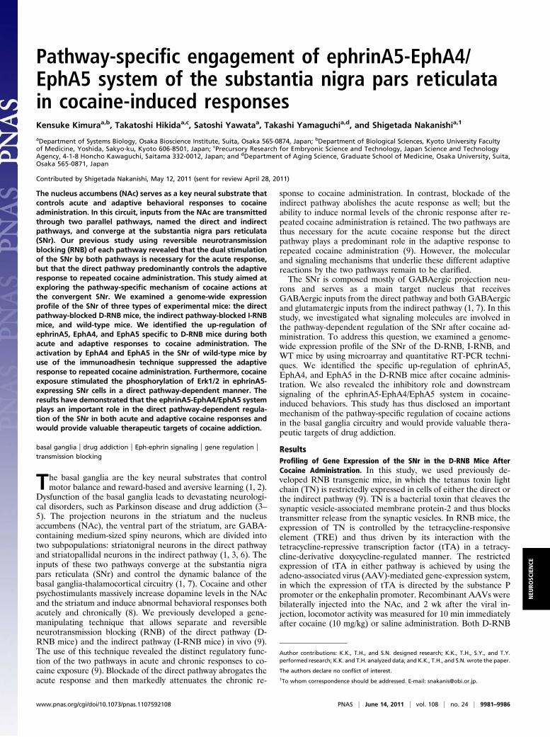
direct pathways, is rich in glutamic acid decarboxylase67 (GAD67)-immunoreactive cells and is located adjacent to the substantianigra pars compacta (SNc), which is characterized by a high densityof tyrosine hydroxylase (TH) immunoreactivity (Fig. 1A) (10, 11).The SNr and the SNc could thus be easily separated and dissectedby the characteristic architecture and cell shapes of these nuclei.We performed quantitative RT-PCR of dissected SNrs and con-firmed that the SNrs used exhibited a high level of the GAD67mRNA and a minimal contamination of the TH mRNA from theSNc (Fig. 1B). Furthermore, there was no difference in expressionlevels of the GAD67 mRNA among the D-RNB, I-RNB, and WTmice, regardless of whether the animals were treated or not withcocaine (Fig. 1B).After confirmation of the lack of cocaine-induced hyper-
locomotion in individual D-RNB and I-RNB mice, SNrs weremechanically isolated fromD-RNB, I-RNB, andWTmice 1haftercocaine or saline administration. Total RNA was extracted frommicrodissected SNrs and subjected to microarray analysis. Ascriteria for the selection of candidate genes, we used hybridizationsignals of>150 at least in oneof the three types of the experimentalanimals and more than 1.4-fold changes between cocaine and sa-line treatments in eitherD-RNBor I-RNBmice, but not in theWTmice. Candidate genes thus selected were further confirmed byquantitative RT-PCR analysis. Among a few candidate genes, wefocused on and analyzed in detail the ephrinA5, EphA4, andEphA5mRNAs, all of which were up-regulated in the SNr of onlythe D-RNB mice (Fig. 1C) (one-way ANOVA analysis for eph-rinA5, P < 0.001–0.01; for EphA4, P < 0.01–0.05; for EphA5, P <0.01–0.05). The up-regulation of these mRNAs was not only spe-cific to D-RNB mice after cocaine administration but in addition,the expression of these mRNAs was not altered in saline-treatedRNBorWTmice (Fig. 1CandD), indicating that theup-regulation
of thesemRNAs depended on both blockade of the direct pathwayand cocaine administration.Because the previous blockade study indicated a key role of
the direct pathway in the adaptive response to repeated cocaineadministration (9), we next addressed whether the ephrinA5,EphA4, and EphA5 mRNAs remained up-regulated in the SNrof D-RNB mice after repeated administration of cocaine. TheSNr was isolated and microdissected after repeated cocaine ad-ministration for 5 d. Quantitative RT-PCR showed that theephrinA5 and EphA5 mRNAs remained up-regulated in the SNrof only the D-RNB mice (Fig. 1D) (one-way ANOVA analysisfor ephrinA5, P < 0.05; for EphA5, P < 0.05). The EphA4mRNA, although not being statistically significant, tended to beup-regulated in the D-RNB mice (Fig. 1D). These results in-dicate that ephrin-Eph receptor signaling molecules are specifi-cally up-regulated in the D-RNB mice not only at the acutephase but also at the adaptive phase of cocaine administration.
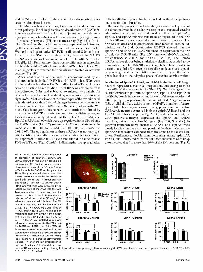
Localization of EphrinA5, EphA4, and EphA5 in the SNr. GABAergicneurons represent a major cell population, amounting to morethan 90% of the neurons in the SNr (12). We investigated thecellular expression patterns of ephrinA5, EphA4, and EphA5 intheSNr by double immunostaining for eachof thesemolecules andeither gephyrin, a postsynaptic marker of GABAergic neurons(13), or glial fibrillary acidic protein (GFAP), a marker of astro-cytes (14). This analysis showed that gephyrin-immunoreactiveGABAergic neurons expressed both the ephrinA5 ligand and theEphA4 and EphA5 receptors (Fig. 2 A, C, and E). In contrast, theGFAP-positive astrocytes expressed the EphA4 and EphA5receptors, but not the ephrinA5 ligand (Fig. 2 B, D, and F). Ingephyrin-immunoreactive neurons, EphA4 and EphA5 weremostly localized in the soma and proximal dendrites, whereas theephrinA5 localization extended from the soma to the distal den-drites. Furthermore, double immunostaining among ephrinA5,EphA4, and EphA5 indicated that all three molecules were ubiq-uitously colocalized in more than 80% of the SNr neurons (Fig. 3).
Fig. 1. Direct pathway-specific regulationof expression of ephrinA5, EphA4, andEphA5 mRNAs in the SNr by cocaine ad-ministration. (A) Double immunostainingof coronal sections of the SNr and SNc ofWT mice with the GAD67 antibody and theTH antibody. A merged view showed thatthe GAD67-immunoreactive SNr (red) is lo-cated adjacent to the TH-immunoreactiveSNc (green). (Scale bar, 100 μm.) (B) D-RNB,I-RNB, and WT mice were prepared by bi-lateral injection of the AAVs into the NAc.Two weeks after the viral injection, theanimals received a single intraperitonealinjection of either cocaine (10 mg/kg) orsaline and were killed 1 h later. The SNrwas then isolated, and the levels of theGAD67 and TH mRNAs were quantified byRT-PCR. mRNA levels were normalized byreferring to that level of the β-actin mRNAas 1 (n = 6 for D-RNB and I-RNB; n = 12 forWT). (C) The SNr was isolated as in B, andmRNA levels were quantified by PCR (n = 6for D-RNB and I-RNB; n = 12 for WT). (D)Experiments were performed as in B, ex-cept that the animals daily received a singleintraperitoneal injection of cocaine (10 mg/kg) or saline for 5 d and the SNr was thenisolated 1 h after the last intraperitonealinjection (n = 6 each). In C and D, levels ofeach mRNA were expressed by referring to those of the corresponding mRNA in saline-injected WT mice. Columns and bars represent the mean ± SEM; *P < 0.05,**P < 0.01, ***P < 0.001.
9982 | www.pnas.org/cgi/doi/10.1073/pnas.1107592108 Kimura et al.
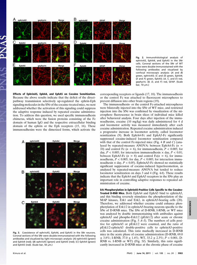
Effects of EphrinA5, EphA4, and EphA5 on Cocaine Sensitization.Because the above results indicate that the deficit of the direct-pathway transmission selectively up-regulated the ephrin-Ephsignalingmolecules in the SNr of the cocaine-treatedmice, wenextaddressed whether the activation of this signaling could suppressthe adaptive response induced by repeated cocaine administra-tion. To address this question, we used specific immunoadhesinchimeras, which were the fusion proteins consisting of the Fcdomain of human IgG and the respective extracellular bindingdomain of the ephrin or the Eph receptors (15, 16). Theseimmunoadhesins were the dimerized forms, which activate the
corresponding receptors or ligands (17, 18). The immunoadhesinor the control Fc was attached to fluorescent microspheres toprevent diffusion into other brain regions (19).The immunoadhesin- or the control Fc-attached microspheres
were bilaterally injected into the SNr of WT mice, and restrictedinjection into the SNr was confirmed by visualization of the mi-crosphere fluorescence in brain slices of individual mice killedafter behavioral analysis. Four days after injection of the immu-noadhesins, cocaine (10 mg/kg) was daily administered for 4 dand locomotor activity was measured immediately after eachcocaine administration. Repeated cocaine administration induceda progressive increase in locomotor activity, called locomotorsensitization (9). Both EphA4-Fc and EphA5-Fc significantlysuppressed cocaine-induced locomotor sensitization comparedwith that of the control Fc-injected mice (Fig. 4 B and C) (ana-lyzed by repeated-measure ANOVA: between EphA4-Fc (n =10) and control Fc (n = 6), for immunoadhesin, P < 0.005; forday, P < 0.005; for interaction immunoadhesin × day, P < 0.01;between EphA5-Fc (n = 8) and control Fc(n = 6), for immu-noadhesin, P < 0.005; for day, P < 0.005; for interaction immu-noadhesin × day, P < 0.05). EphrinA5-Fc showed no statisticallysignificant suppression of cocaine-induced hyperlocomotion, asanalyzed by repeated-measure ANOVA but tended to reducelocomotor sensitization on days 3 and 4 (Fig. 4A). These resultsindicate that the EphA4 and EphA5 receptors in the SNr play animportant role in controlling adaptive responses to repeated ad-ministration of cocaine.
Erk Phosphorylation in EphrinA5-Positive Cells Specific to the Cocaine-Treated D-RNB Mice. Both EphA4 and EphA5 bind to ephrinA5,and this binding reversely stimulates the phosphorylation of theMAP kinases, Erk1 and Erk2, in ephrinA5-bearing cells (20).Therefore, we addressed whether cocaine could enhance phos-phorylation of Erk1/2 in ephrinA5-bearing neurons specific to theSNr of D-RNB mice. The SNr of D-RNB, I-RNB, or WT micewas analyzed by double immunostaining with antibodies againstephrinA5 and phospho-Erk1/2 (pErk1/2) after acute or chroniccocaine administration (Fig. 5 A–I). The numbers of cells posi-tive for ephrinA5 or pErk1/2 were counted, and the ratio ofpErk1/2-ephrinA5 double-positive cells to ephrinA5-positivecells was calculated. This ratio markedly increased in D-RNBmice in the acute phase of cocaine administration (D-RNB, 69.0± 3.8%; I-RNB, 37.0 ± 1.4%; WT, 33.2 ± 2.3%; P < 0.001, D-RNB vs. I-RNB or WT) (Fig. 5J). Similarly, this ratio signifi-cantly increased in D-RNB mice at the chronic phase of cocaine
Fig. 2. Immunohistological analysis ofephrinA5, EphA4, and EphA5 in the SNrcells. Coronal sections of the SNr of WTmice were double-immunostained with thefollowing antibodies and visualized byconfocal microscopic analysis: (A and B)green, ephrinA5; (C and D) green, EphA4;(E and F) green, EphA5; (A, C, and E) red,gephyrin; (B, D, and F) red, GFAP. (Scalebar, 10 μm.)
Fig. 3. Coexistence of ephrinA5, EphA4, and EphA5 in the SNr neurons.Coronal sections of the SNr were double-immunostained with the followingantibodies and visualized by light microscopic analysis: (A) ephrinA5 (green)and EphA4 (red); (B) ephrinA5 (green) and EphA5 (red); (C) EphA4 (green)and EphA5 (red). (Scale bar, 50 μm.)
Kimura et al. PNAS | June 14, 2011 | vol. 108 | no. 24 | 9983
NEU
ROSC
IENCE

administration (D-RNB, 70.4 ± 7.8%; I-RNB, 37.8 ± 2.9%; WT,31.8 ± 7.8%; P < 0.001–0.01, D-RNB vs. I-RNB or WT) (Fig.5K). Importantly, there was no difference in the relative ratio oftwo types of cells in three groups of saline-treated mice (Fig. 5 J
and K). Upon double immunostaining for NeuN, a marker ofmature neurons (21), ephrinA5-immunoreactive cells amountedto 94% to 98% of the NeuN-positive cells in all three groups ofmice, regardless of treatment or not with cocaine. Thus, there
Fig. 4. Suppression of cocaine-induced hyperlocomotion by EphA4 and EphA5 in the SNr. Fluorescent microspheres with attached ephrinA5-Fc (A), EphA4-Fc(B), EphA5-Fc (C), or control Fc (A–C) were bilaterally injected into the SNr of WT mice. One day after immunoadhesin injection, animals received in-traperitoneal saline once a day and were habituated for 3 d. Cocaine (10 mg/kg) was then intraperitoneally injected once a day from day 1 to day 4; andimmediately after each cocaine injection, locomotor activity was counted for a 10-min period. Symbols and bars represent the mean ± SEM (ephrinA5-Fc, n =14; EphA4-Fc, n = 10; EphA5-Fc, n = 8; control Fc, n = 6). Statistical significance was analyzed by repeated-measure ANOVA; **P < 0.01, ***P < 0.001 (EphA4-Fcor EphA5-Fc vs. control Fc).
Fig. 5. Activation of Erk1/2 in ephrinA5-expressing SNr neurons specific to D-RNB mice. For Cocaine Acute (A, D, and G) and Saline (C, F, and I), D-RNB, I-RNB,and WT mice received a single intraperitoneal injection of cocaine (10 mg/kg) and saline, respectively, and the SNr was isolated 6 h after cocaine or salineinjection. For Cocaine Chronic (B, E, and H), three groups of mice daily received a single intraperitoneal injection of cocaine (10 mg/kg) for 5 d and the SNr wasisolated 1 h after cocaine injection. Coronal sections were double-immunostained with antibodies against ephrinA5 (green) and pErk1/2 (red) and visualizedby light microscopic analysis. Arrows and arrowheads indicate the pErk1/2-positive and pErk1/2-negative cells, respectively, that were also immunopositive forephrinA5. (Scale bar, 50 μm.) (J and K) The numbers of Erk1/2-immunopositive and Erk1/2-immunonegative cells among the ephrinA5-immunoreactive cellswere counted, and the ratios of pErk1/2-ephrinA5 double-immunopositive cells to ephrinA5-immunoreactive cells are indicated in J and K. Columns and errorbars represent the mean ± SEM (n = 4 each). The statistical significance was analyzed by one-way ANOVA. **P < 0.01, ***P < 0.001.
9984 | www.pnas.org/cgi/doi/10.1073/pnas.1107592108 Kimura et al.

was a good correlation in the pathway specificity between theregulation of the EphA4/EphA5-ephrinA5 system and phos-phorylation of Erk1/2 in the SNr neurons. This correlation sug-gests that the Erk1/2 signaling downstream of ephrin actions in theSNr is important for the cocaine-induced behavioral responses.
DiscussionThe principal striatal neurons receive inputs from the cerebralcortex and thalamus and send their inputs to the SNr through twoparallel pathways (1, 7). In the basal ganglia circuit, cocaineinhibits the dopamine transporter and massively increases dopa-mine levels in the striatum and the NAc (8). This rapid increase indopamine activates both the low-affinity D1 receptor in the directpathway and the high-affinity D2 receptor in the indirect pathway(22). The chronic cocaine exposure then persistently activates theD1 and D2 receptors and differentially induces long-term poten-tiation at striatonigral neurons of the direct pathway and long-termdepression at striatopallidal neurons of the indirect pathway (23).The long-term potentiation of the direct pathway is thought to becritical for inducing the adaptive response to chronic cocaine ex-posure (9, 23). However, the pathway-specific regulatory mecha-nisms of cocaine actions at the convergent SNr remained to beclarified. This investigation has revealed an importantmechanism,in which the ephrinA5 ligand-EphA4/EphA5 receptors are regu-lated in the SNr via a direct pathway-specific mechanism in boththe acute and chronic phases of cocaine responses. These ephrin-Ephmolecules were up-regulated specifically by blocking inputs ofthe direct pathway after cocaine administration. Conversely, theEphA4 and EphA5 receptors in the SNr suppressed adaptive re-sponse to repeated cocaine administration. Furthermore, cocaineexposure activated the Erk1/2 signaling cascade in ephrinA5-expressing SNr cells in a direct pathway-dependentmanner. Theseresults indicate that the ephrinA5-EphA4/EphA5 signaling mol-ecules are specifically regulated by inputs of the direct pathway andplay an important role in the acute and adaptive responses tococaine exposure.The ephrin-Eph system consists of the large family of both
ephrins and Eph receptors and controls a large variety of cellularresponses, including contact-mediated attraction or repulsion,synapse formation, spinemorphogenesis, and neural plasticity (24,25). One of the characteristic features of the ephrin-Eph system isits bidirectional signaling cascade, in which the interaction ofephrin with the Eph receptor induces a forward signaling in theEph-bearing cells and simultaneously elicits a reverse signaling inthe ephrin-bearing cells (20, 26). Although ephrinA5, EphA4, andEphA5 were all up-regulated by blocking the direct pathway atleast at the acute phase of cocaine administration, activation byEphA4 and EphA5 was more effective than activation by eph-rinA5 in suppressing the cocaine sensitization. This reverse sig-naling could thus play a predominant role in the transmissionregulation of the direct pathway in the SNr. This regulation mayoccur by interaction of the presynaptic EphA4/EphA5 of striatalcells (27, 28) and the postsynaptic ephrinA5 of the SNr neurons.Astrocytes also highly express EphA4 and EphA5, which maystimulate ephrinA5 in neurons (26). Recently, the ephrin-Eph cisinteraction within the same cellular membrane has been shown totransduce a key signaling in the ephrin-Eph system (26, 29, 30).Because both ephrinA5 and EphA4/EphA5 are commonly dis-tributed in most of the SNr neurons, the cis interaction of thissystem could be involved in the direct pathway-specific regulationof cocaine responses. Whatever the mechanisms of the ephrinA5-EphA4/EphA5 system in the SNr, our finding that the Erk1/2 sig-naling is pathway-specifically regulated in ephrinA5-expressing cellsstrongly suggests that the ephrinA5-EphA4/EphA5–expressing SNrneurons play an important role in cocaine-induced input trans-mission of the direct pathway.No alteration of the ephrinA5-EphA4/EphA5 system was
observed in saline-treated D-RNB mice, indicating that the ob-
served changes in this ephrin-Eph system were linked to theaction of cocaine and not a consequence of impaired trans-mission per se of the direct pathway. Our previous study usingthe RNB technique revealed that blockade of either the direct orthe indirect pathway abolished the acute cocaine response (9).The dual stimulation of the two pathways is thus necessary forthe rapid response to cocaine administration (9). In the chronicresponse to repeated cocaine administration, blockade of thedirect pathway—but not that of the indirect pathway—severelyimpaired cocaine-induced adaptive responses, indicating that thedirect pathway plays a predominant role in input transmission forthe adaptive response to cocaine (9). However, despite the de-fectiveness of the acute response by blockade of either of the twopathways (9), up-regulation of the ephrinA5-EphA4/EphA5system as well as activation of Erk1/2 was observed in a directpathway-selective manner at both acute and chronic phases ofcocaine administration. The ephrinA5-EphA4/EphA5 system isthus most likely to contribute to triggering the acute responseand then inducing the adaptive response to cocaine actions. Thecellular response to ephrin-Eph engagement is often repulsivebetween the two cells, although a repulsive or attractive responsedepends on the cellular context (26). This ephrin-Eph system isalso important for cell-cell communication by controlling spinemorphogenesis and neural plasticity (18, 24, 26, 31). Presentfindings of the pathway-selective ephrin-Eph engagement incocaine-induced responses thus shed light on the action of co-caine and would provide valuable therapeutic targets for thetreatment of drug addiction.
Materials and MethodsAnimals and Behavioral Analysis. All animal handling procedures were per-formed according to the guidelines of the Osaka Bioscience Institute. The RNBmice, in which transmission of either the direct or the indirect pathway wasselectively blocked, was generated as described previously (9). Briefly, theexpression of TN was driven in the TN mice by the TRE and induced by in-teraction with the tTA (32). The expression of tTA was restricted to the director indirect pathway by injecting one of two types of the recombinant AAVsinto the NAc, in which tTA was exclusively expressed in either the direct orthe indirect pathway under the control of the substance P promoter or theenkephalin promoter, respectively (9). The recombinant AAV was bilaterallyinjected into four sites of the NAc by stereotaxic techniques (9). The RNBmice and their WT littermates were used for all experiments.
Locomotor activity was measured with an infrared activity monitor (MEDAssociates). For measurement of cocaine-induced hyperlocomotion, animalsreceived intraperitoneal saline once a day and were habituated to a novelchamber for 3 d. Cocaine (10 mg/kg) or saline was then intraperitoneallyinjected once a day from day 1 to day 4, and immediately thereafter thelocomotor activity was counted for a 10-min period.
Microdissection of the SNr. One hour after cocaine or saline administration,mice were killed, and frozen coronal sections (40 μm) were obtained from thebrain embedded in OCT compound. Microdissection was performed by usinga Micro Dissector PPMD (Eppendorf), consisting of a 1-mm diameter stain-less-steel needle (Eppendorf) set at a 45° angle to the surface of the mi-croscope table. A micropipette was mounted on a 3-axis–controlled,motorized micromanipulator (Eppendorf) attached to the microscope. Aftercryosections were covered with a pool of 15 μL of xylene for visualization,the SNr was dissected as an ultrasonically oscillating needle was movedalong a selected tissue area.
Microarray Analysis. Total RNA of dissected SNrs was extracted with thereagents of an RNeasy Mini Kit (Qiagen) after evaporation of xylene in avacuum concentrator. Approximately 5 ng of total RNA was labeled by usingGeneChip Two-Cycle Target Labeling and Control Reagents (Affymetrix).Hybridization signals were calculated by analyzing raw data with MicroarraySuite 5.0 (Affymetrix) and further analyzed with GeneSpring GX 11.0 soft-ware (Agilent Technologies) and Ingenuity Pathway Analysis 6.0 software(Ingenuity Systems). The data were normalized to the 75th percentile for per-chip normalization.
Kimura et al. PNAS | June 14, 2011 | vol. 108 | no. 24 | 9985
NEU
ROSC
IENCE
Quantitative RT-PCR. Reverse transcription was carried out by using the Su-perScript First-Strand Synthesis System (Affymetrix) with the T7-oligo(dT)primer. cDNAs thus synthesized were amplified by a cycle of T7 amplificationby using the MEGAscript High Yield Transcription Kit (Applied Biosystems).Specific primers were designed to generate 60- to 150-bp PCR productscorresponding to the 3′ region of each mRNA. All reactions were performedin duplicate, and β-actin mRNA was used as an internal control for mRNAquantification.
Immunohistological Analysis. Immunohistochemistry of frozen coronal sec-tions (20 μm) of the adult mouse brain was performed as described bySchneider Gasser et al. (33) by using the primary antibodies against ephrinA5(Abcam), EphA4, EphA5 (for both, Abcam or Santa Cruz), pErk1/2 (SantaCruz), GAD67, TH (Millipore), gephyrin (Synaptic Systems), and GFAP (SigmaAldrich). The secondary antibodies used were Alexa488- or Alexa594-con-jugated goat IgG (Molecular Probes), and specific immunoreactivity wasconfirmed by performing immunohistochemical analysis without addition ofthe primary antibody.
Immunoadhesin Analysis. Three different immunoadhesins were used; that is,fusion proteins consisting of the binding domain of either ephrinA5, EphA4,or EphA5 attached to the Fc domain of human IgG (R&D Systems). To preventdiffusion of immunoadhesins into other brain regions, we attached fluo-
rescent microspheres (Lumafluor) to each immunoadhesin, as described byRiddle et al. (19). Immunoadhesin was injected stereotaxically at four sites inthe SNr of WT mice (3.4-mm and 3.6-mm posterior to the bregma, ± 1.5-mmlateral from the midline, 4.0-mm depth from the dura). Four days afterimmunoadhesin injection, locomotor activity was measured immediatelyafter daily administration of cocaine (10 mg/kg). After the behavioralanalysis, injection sites of immunoadhesins were confirmed by visualizationof immunoadhesin-attached fluorescent microspheres in the SNr of brain-slice preparations.
Statistical Analysis. Statistical analysis was conducted by using Graph PadPRISM 5.0 (GraphPad Software). Data were analyzed by one-way ANOVA orrepeated-measure ANOVA and were presented as the mean ± SEM.
ACKNOWLEDGMENTS. This work was supported by Research GrantsKAKENHI 22220005 (to S.N.), 22659069, and 23110522 (to T.H.) from theMinistry of Education, Culture, Sports, Science, and Technology of Japan,Grant on Regulatory Science of Pharmaceuticals and Medical Devicesfrom the Ministry of Health and Labour and Welfare (to T.H.), a grantfrom the Japan Science and Technology Agency Precursory Research forEmbryonic Science and Technology Program (T.H.), and grants from theTakeda Science Foundation (to S.N.) and Daiichi-Sankyo Foundation ofLife Science (to T.H.)
1. Graybiel AM (2000) The basal ganglia. Curr Biol 10:R509–R511.2. Wickens JR, Reynolds JNJ, Hyland BI (2003) Neural mechanisms of reward-related
motor learning. Curr Opin Neurobiol 13:685–690.3. Israel Z, Bergman H (2008) Pathophysiology of the basal ganglia and movement
disorders: From animal models to human clinical applications. Neurosci Biobehav Rev32:367–377.
4. Obeso JA, et al. (2008) The basal ganglia in Parkinson’s disease: Current concepts andunexplained observations. Ann Neurol 64(Suppl 2):S30–S46.
5. Hyman SE, Malenka RC, Nestler EJ (2006) Neural mechanisms of addiction: The role ofreward-related learning and memory. Annu Rev Neurosci 29:565–598.
6. Redgrave P, et al. (2010) Goal-directed and habitual control in the basal ganglia:Implications for Parkinson’s disease. Nat Rev Neurosci 11:760–772.
7. Deniau JM, Mailly P, Maurice N, Charpier S (2007) The pars reticulata of the substantianigra: A window to basal ganglia output. Prog Brain Res 160:151–172.
8. Riddle EL, Fleckenstein AE, Hanson GR (2005) Role of monoamine transporters inmediating psychostimulant effects. AAPS J 7:E847–E851.
9. Hikida T, Kimura K, Wada N, Funabiki K, Nakanishi S (2010) Distinct roles of synaptictransmission in direct and indirect striatal pathways to reward and aversive behavior.Neuron 66:896–907.
10. Esclapez M, Tillakaratne NJ, Kaufman DL, Tobin AJ, Houser CR (1994) Comparativelocalization of two forms of glutamic acid decarboxylase and their mRNAs in rat brainsupports the concept of functional differences between the forms. J Neurosci 14:1834–1855.
11. Oertel WH, Tappaz ML, Berod A, Mugnaini E (1982) Two-color immunohistochemistryfor dopamine and GABA neurons in rat substantia nigra and zona incerta. Brain ResBull 9:463–474.
12. Mugnaini E, Oertel WH (1985) in Handbook of Chemical Nenroanatomy, Volume 4:GABA and Neuropeptides in the CNS, Part I, eds Björklund A, Hökfelt T (Elsevier,Amsterdam), pp 436–595.
13. Sassoè-Pognetto M, Fritschy J-M (2000) Mini-review: Gephyrin, a major postsynapticprotein of GABAergic synapses. Eur J Neurosci 12:2205–2210.
14. Ghandour MS, Langley OK, Vincendon G, Gombos G (1979) Double labelingimmunohistochemical technique provides evidence of the specificity of glial cellmarkers. J Histochem Cytochem 27:1634–1637.
15. Ashkenazi A, Chamow SM (1997) Immunoadhesins as research tools and therapeuticagents. Curr Opin Immunol 9:195–200.
16. Gerlai R, et al. (1998) Protein targeting in the analysis of learning and memory: Apotential alternative to gene targeting. Exp Brain Res 123:24–35.
17. Lim BK, Matsuda N, Poo M-M (2008) Ephrin-B reverse signaling promotes structuraland functional synaptic maturation in vivo. Nat Neurosci 11:160–169.
18. Fu W-Y, et al. (2007) Cdk5 regulates EphA4-mediated dendritic spine retractionthrough an ephexin1-dependent mechanism. Nat Neurosci 10:67–76.
19. Riddle DR, Katz LC, Lo DC (1997) Focal delivery of neurotrophins into the centralnervous system using fluorescent latex microspheres. Biotechniques 23:928–934, 936–937.
20. Davy A, Robbins SM (2000) Ephrin-A5 modulates cell adhesion and morphology in anintegrin-dependent manner. EMBO J 19:5396–5405.
21. Mullen RJ, Buck CR, Smith AM (1992) NeuN, a neuronal specific nuclear protein invertebrates. Development 116:201–211.
22. Hikosaka O (2007) Basal ganglia mechanisms of reward-oriented eye movement. AnnN Y Acad Sci 1104:229–249.
23. Goto Y, Grace AA (2005) Dopamine-dependent interactions between limbic andprefrontal cortical plasticity in the nucleus accumbens: Disruption by cocainesensitization. Neuron 47:255–266.
24. Lai K-O, Ip NY (2009) Synapse development and plasticity: Roles of ephrin/Ephreceptor signaling. Curr Opin Neurobiol 19:275–283.
25. Scicolone G, Ortalli AL, Carri NG (2009) Key roles of Ephs and ephrins in retinotectaltopographic map formation. Brain Res Bull 79:227–247.
26. Egea J, Klein R (2007) Bidirectional Eph-ephrin signaling during axon guidance.Trends Cell Biol 17:230–238.
27. MartoneME, Holash JA, Bayardo A, Pasquale EB, EllismanMH (1997) Immunolocalizationof the receptor tyrosine kinase EphA4 in the adult rat central nervous system. Brain Res771:238–250.
28. Cooper MA, Crockett DP, Nowakowski RS, Gale NW, Zhou R (2009) Distribution ofEphA5 receptor protein in the developing and adult mouse nervous system. J CompNeurol 514:310–328.
29. Carvalho RF, et al. (2006) Silencing of EphA3 through a cis interaction with ephrinA5.Nat Neurosci 9:322–330.
30. Marquardt T, et al. (2005) Coexpressed EphA receptors and ephrin-A ligands mediateopposing actions on growth cone navigation from distinct membrane domains. Cell121:127–139.
31. Fu AKY, et al. (2011) APCCdh1 mediates EphA4-dependent downregulation of AMPAreceptors in homeostatic plasticity. Nat Neurosci 14:181–189.
32. Yamamoto M, et al. (2003) Reversible suppression of glutamatergic neurotransmissionof cerebellar granule cells in vivo by genetically manipulated expression of tetanusneurotoxin light chain. J Neurosci 23:6759–6767.
33. Schneider Gasser EM, et al. (2006) Immunofluorescence in brain sections: Simultaneousdetection of presynaptic and postsynaptic proteins in identified neurons. Nat Protoc 1:1887–1897.
9986 | www.pnas.org/cgi/doi/10.1073/pnas.1107592108 Kimura et al.