pathway for lipid a biosynthesis in arabidopsis ... ·...
TRANSCRIPT

Pathway for lipid A biosynthesis in Arabidopsisthaliana resembling that of Escherichia coliChijun Li, Ziqiang Guan, Dan Liu, and Christian R. H. Raetz1
Department of Biochemistry, Duke University Medical Center, P.O. Box 3711, Durham, NC 27710
Contributed by Christian R. H. Raetz, June 1, 2011 (sent for review March 24, 2011)
The lipid A moiety of Escherichia coli lipopolysaccharide is a hexa-acylated disaccharide of glucosamine that makes up the outermonolayer of the outer membrane. Arabidopsis thaliana containsnuclear genes encoding orthologs of key enzymes of bacterial lipidA biosynthesis, including LpxA, LpxC, LpxD, LpxB, LpxK and KdtA.Although structurally related lipid A molecules are found in mostother Gram-negative bacteria, lipid A and its precursors have notbeen directly detected in plants previously. However, homozygousinsertional knockout mutations or RNAi knock-down constructsofArabidopsis lpx and kdtAmutants revealed accumulation (or dis-appearance) of the expected monosaccharide or disaccharide lipidA precursors by mass spectrometry of total lipids extracted from10-day old seedlings of these mutants. In addition, fluorescencemicroscopy of lpx-gfp fusions in transgenic Arabidopsis plantssuggests that the Lpx and KdtA proteins are expressed and tar-geted to mitochondria. Although the structure of the lipid A endproduct generated by plants is still unknown, our work demon-strates that plants synthesize lipid A precursors using the sameenzymatic pathway present in E. coli.
glucosamine-based lipids ∣ Lpx genes ∣ higher plants ∣electrospray ionization
Lipid A, the hydrophobic portion of LPS, is a glucosamine-based lipid that makes up the outer monolayer of the outer
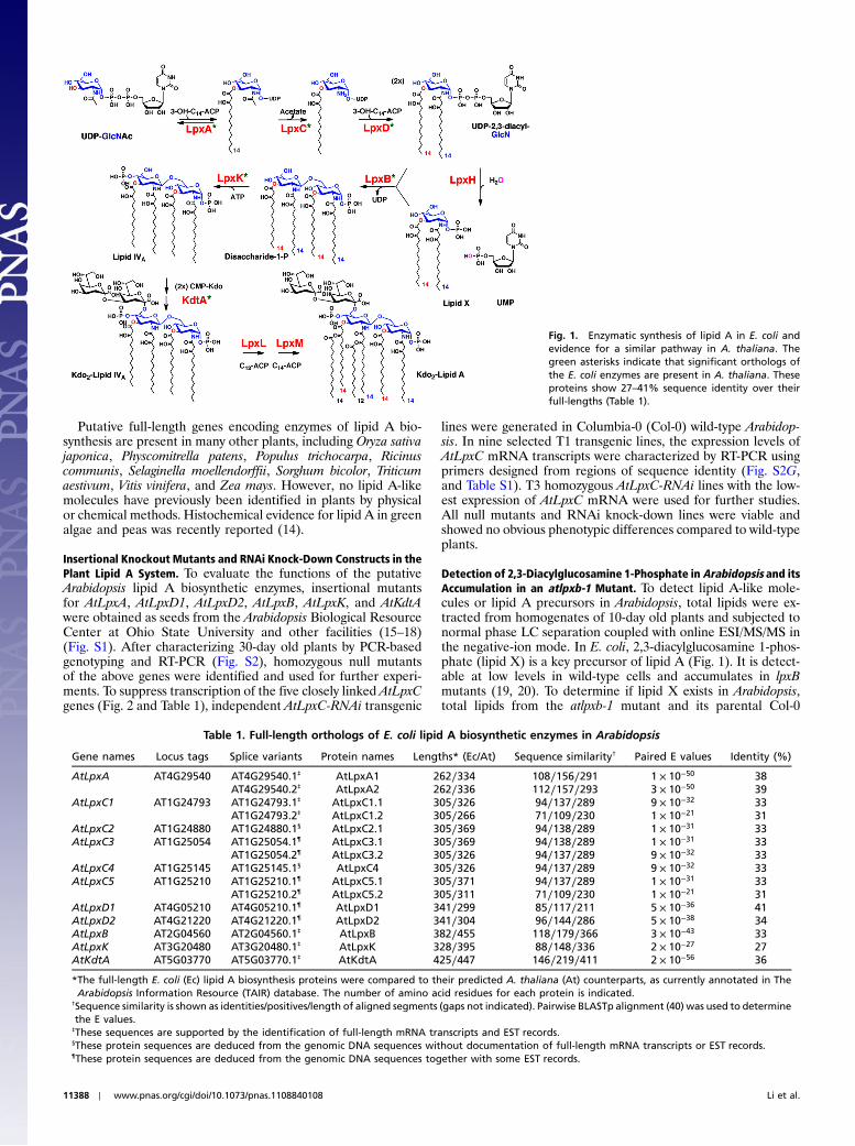
membrane of Gram-negative bacteria (1, 2). Kdo2-lipid A (Fig. 1)usually represents the minimal LPS substructure required forgrowth (1, 2). In a few systems, like Neisseria meningitides, lipidA biosynthesis is not essential (3). However, in most cases lipid Abiosynthesis inhibitors that target LpxC (Fig. 1) are potent Gram-negative-selective antibiotics (4). The structure of lipid A isrelatively conserved in diverse Gram-negative pathogens (1, 2).Picomolar Kdo2-lipid A is recognized by the TLR4/MD2 receptorcomplex of animal cells (5), triggering cytokine production thatcan contribute to the complications of Gram-negative sepsis (6).
Most Gram-negative bacteria encode single copy homologuesof the nine Escherichia coli enzymes that assemble the Kdo2-lipidA moiety of E. coli LPS (Fig. 1) (1, 2). Gram-positive bacteria,Archaea, fungi, insects, worms, and vertebrates do not containthese genes. Although lipid A-like molecules have not beenreported in plants, many plants, including Arabidopsis thaliana,encode full-length nuclear orthologs of six of the nine enzymesof the E. coli system (Fig. 1, green asterisks; and Table 1) (1, 7).A homologue of the UDP-2,3-diacylglucosamine pyrophospha-tase LpxH of E. coli (Fig. 1) is missing in plants, but it is alsomissing in some Gram-negative bacteria in which it may be re-placed with a different pyrophosphatase (8).
We now report the isolation and characterization of homozy-gous insertional knockout mutants in the A. thaliana genes encod-ing LpxA, LpxD, LpxB, LpxK, and KdtA (Table 1 and Fig. 2).Given the presence of multiple genes encoding LpxC-like pro-teins in Arabidopsis (Fig. 2), we chose an RNAi strategy tosuppress AtLpxC expression. Mutants generated from both geneinactivation strategies are viable under laboratory conditions.However, they accumulate (or lose) the expected lipid A precur-sors (Fig. 1) when analyzed by sensitive liquid chromatography–electrospray ionization/mass spectrometry (LC–ESI/MS) proto-
cols. The lipid A biosynthetic genes of higher plants may havebeen acquired from Gram-negative bacteria with the endo-symbiosis of mitochondria. Plant lipid A may therefore have astructural role in mitochondrial or perhaps chloroplast outermembranes. Alternatively, it may serve a regulatory role, or itmay participate in signal transduction, for instance, in the plantresponse to infection by bacterial pathogens (9). This workdefinitively demonstrates the presence of lipid A precursors in aeukaryotic system.
ResultsIdentification of Orthologs of Lipid A Biosynthesis Genes in Arabidop-sis. A position-specific iterated-BLAST search of the nonredun-dant protein sequences database was conducted using the E. coliprotein sequences of LpxA, LpxC, LpxD, LpxB, LpxH, LpxK,KdtA, LpxL, and LpxM as probes (Fig. 1). Eleven nuclear se-quences were identified in A. thaliana, encoding proteins withsignificant homology to E. coli LpxA, LpxC, LpxD, LpxB, LpxK,or KdtA (Table 1). Homologues of LpxH, LpxL, and LpxM werenot found in Arabidopsis, but these are also absent in some Gram-negative bacteria (10). The AtLpxA gene (Table 1) encodes twomRNA splice variants, one of which directs the synthesis of aprotein with two additional amino acid residues inserted in theC-terminal region. These protein variants, designated AtLpxA1and AtLpxA2, are otherwise identical to each other; they share38 and 37% sequence identity with E. coli LpxA, respectively,(Table 1), with conservation of all active site residues (11). Thereare five AtLpxC orthologs located in a 68-kb region on chromo-some one (Fig. 2). They have very similar nucleotide sequences,suggesting possible gene duplications from a common ancestor.Expressed sequence tag (EST) and deduced cDNA databases in-dicate these AtLpxC genes generate at least eight different butrelated mRNA transcripts (Table 1). Previous studies of E. coliLpxC had shown that three amino acid residues (H238, D246,and H265) in the C-terminal region of the protein are criticalfor activity (12). These residues are truncated in two of the eightAtLpxC proteins, specifically in AtLpxC1.2 and AtLpxC5.2(Table 1), suggesting that these two proteins are not catalyticallyactive.
Two AtLpxD orthologs (designated AtLxpD1 and AtLpxD2)were encoded on chromosome 4 (Fig. 2 and Table 1); theseproteins share 41 and 34% identity with E. coli LpxD (Table 1),with conservation of key catalytic residues (13); however, onlyAtLpxD2 contains the putative N-terminal nucleotide bindingdomain of LpxD (13), which is required for E. coli LpxD activity.Single copy genes encoding AtLpxB (AT2G04560), AtLpxK(AT3G20480) and AtKdtA (AT5G03770) (Table 1) were alsofound, with 31, 26, and 34% sequence identity to the correspond-ing E. coli proteins, respectively (Table 1).
Author contributions: C.R.H.R. designed research; C.L. and Z.G. performed research;C.L. and D.L. contributed new reagents/analytic tools; C.L., Z.G., and C.R.H.R. analyzeddata; and C.L., Z.G., and C.R.H.R. wrote the paper.
The authors declare no conflict of interest.1To whom correspondence should be addressed. E-mail: [email protected].
This article contains supporting information online at www.pnas.org/lookup/suppl/doi:10.1073/pnas.1108840108/-/DCSupplemental.
www.pnas.org/cgi/doi/10.1073/pnas.1108840108 PNAS ∣ July 12, 2011 ∣ vol. 108 ∣ no. 28 ∣ 11387–11392
BIOCH
EMISTR
Y
Putative full-length genes encoding enzymes of lipid A bio-synthesis are present in many other plants, including Oryza sativajaponica, Physcomitrella patens, Populus trichocarpa, Ricinuscommunis, Selaginella moellendorffii, Sorghum bicolor, Triticumaestivum, Vitis vinifera, and Zea mays. However, no lipid A-likemolecules have previously been identified in plants by physicalor chemical methods. Histochemical evidence for lipid A in greenalgae and peas was recently reported (14).
Insertional Knockout Mutants and RNAi Knock-Down Constructs in thePlant Lipid A System. To evaluate the functions of the putativeArabidopsis lipid A biosynthetic enzymes, insertional mutantsfor AtLpxA, AtLpxD1, AtLpxD2, AtLpxB, AtLpxK, and AtKdtAwere obtained as seeds from the Arabidopsis Biological ResourceCenter at Ohio State University and other facilities (15–18)(Fig. S1). After characterizing 30-day old plants by PCR-basedgenotyping and RT-PCR (Fig. S2), homozygous null mutantsof the above genes were identified and used for further experi-ments. To suppress transcription of the five closely linked AtLpxCgenes (Fig. 2 and Table 1), independent AtLpxC-RNAi transgenic
lines were generated in Columbia-0 (Col-0) wild-type Arabidop-sis. In nine selected T1 transgenic lines, the expression levels ofAtLpxC mRNA transcripts were characterized by RT-PCR usingprimers designed from regions of sequence identity (Fig. S2G,and Table S1). T3 homozygous AtLpxC-RNAi lines with the low-est expression of AtLpxC mRNA were used for further studies.All null mutants and RNAi knock-down lines were viable andshowed no obvious phenotypic differences compared to wild-typeplants.
Detection of 2,3-Diacylglucosamine 1-Phosphate in Arabidopsis and itsAccumulation in an atlpxb-1 Mutant. To detect lipid A-like mole-cules or lipid A precursors in Arabidopsis, total lipids were ex-tracted from homogenates of 10-day old plants and subjected tonormal phase LC separation coupled with online ESI/MS/MS inthe negative-ion mode. In E. coli, 2,3-diacylglucosamine 1-phos-phate (lipid X) is a key precursor of lipid A (Fig. 1). It is detect-able at low levels in wild-type cells and accumulates in lpxBmutants (19, 20). To determine if lipid X exists in Arabidopsis,total lipids from the atlpxb-1 mutant and its parental Col-0
Fig. 1. Enzymatic synthesis of lipid A in E. coli andevidence for a similar pathway in A. thaliana. Thegreen asterisks indicate that significant orthologs ofthe E. coli enzymes are present in A. thaliana. Theseproteins show 27–41% sequence identity over theirfull-lengths (Table 1).
Table 1. Full-length orthologs of E. coli lipid A biosynthetic enzymes in Arabidopsis
Gene names Locus tags Splice variants Protein names Lengths* (Ec/At) Sequence similarity† Paired E values Identity (%)
AtLpxA AT4G29540 AT4G29540.1‡ AtLpxA1 262∕334 108∕156∕291 1 × 10−50 38AT4G29540.2‡ AtLpxA2 262∕336 112∕157∕293 3 × 10−50 39
AtLpxC1 AT1G24793 AT1G24793.1‡ AtLpxC1.1 305∕326 94∕137∕289 9 × 10⁻³² 33AT1G24793.2‡ AtLpxC1.2 305∕266 71∕109∕230 1 × 10⁻²¹ 31
AtLpxC2 AT1G24880 AT1G24880.1§ AtLpxC2.1 305∕369 94∕138∕289 1 × 10⁻³¹ 33AtLpxC3 AT1G25054 AT1G25054.1¶ AtLpxC3.1 305∕369 94∕138∕289 1 × 10⁻³¹ 33
AT1G25054.2¶ AtLpxC3.2 305∕326 94∕137∕289 9 × 10⁻³² 33AtLpxC4 AT1G25145 AT1G25145.1§ AtLpxC4 305∕326 94∕137∕289 9 × 10⁻³² 33AtLpxC5 AT1G25210 AT1G25210.1¶ AtLpxC5.1 305∕371 94∕137∕289 1 × 10⁻³¹ 33
AT1G25210.2¶ AtLpxC5.2 305∕311 71∕109∕230 1 × 10⁻²¹ 31AtLpxD1 AT4G05210 AT4G05210.1¶ AtLpxD1 341∕299 85∕117∕211 5 × 10−36 41AtLpxD2 AT4G21220 AT4G21220.1¶ AtLpxD2 341∕304 96∕144∕286 5 × 10−38 34AtLpxB AT2G04560 AT2G04560.1‡ AtLpxB 382∕455 118∕179∕366 3 × 10−43 33AtLpxK AT3G20480 AT3G20480.1‡ AtLpxK 328∕395 88∕148∕336 2 × 10⁻²⁷ 27AtKdtA AT5G03770 AT5G03770.1‡ AtKdtA 425∕447 146∕219∕411 2 × 10−56 36
*The full-length E. coli (Ec) lipid A biosynthesis proteins were compared to their predicted A. thaliana (At) counterparts, as currently annotated in TheArabidopsis Information Resource (TAIR) database. The number of amino acid residues for each protein is indicated.
†Sequence similarity is shown as identities/positives/length of aligned segments (gaps not indicated). Pairwise BLASTp alignment (40) was used to determinethe E values.
‡These sequences are supported by the identification of full-length mRNA transcripts and EST records.§These protein sequences are deduced from the genomic DNA sequences without documentation of full-length mRNA transcripts or EST records.¶These protein sequences are deduced from the genomic DNA sequences together with some EST records.
11388 ∣ www.pnas.org/cgi/doi/10.1073/pnas.1108840108 Li et al.

wild-type plant were compared by LC–ESI/MS/MS. As shown inFig. 3A, a small peak at m∕z 710.43, consistent with the ½M-H�−ion of a lipid X molecule bearing two R-3-hydroxymyristoylchains (Fig. 1) (8, 19), was present in the wild type. It accumu-lated more than 10-fold in the atlpxb-1mutant (Fig. 3B). No otherlipid X molecular species were detected. ESI/MS/MS analysis ofthe m∕z 710.43 ion in the atlpxb-1 mutant revealed the fragmen-tation pattern expected for lipid X (Fig. 3C).
To confirm the presence of lipid X and to quantify its level inthe mutants (Fig. 4), a sensitive multiple reaction monitoring(MRM) protocol was developed in conjunction with LC prefrac-tionation. Singly charged precursor/product ion pairs (710.4∕240.0) were used to detect lipid X selectively (Figs. S3 and S4).The MRM peak area of the lipid X ion was normalized to that ofthe major species of Arabidopsis phosphatidylethanolamine (pre-cursor ½M-H�− ion at m∕z 714.5 and product ion at m∕z 279.2),which is acylated with palmitate (C16∶0) and linoleate (C18∶2)(Fig. S3). Lipid X accumulates 42-fold in the atlpxb-1 mutantcompared to the Col-0 wild type by this criterion (Fig. 4A).
Quantification of Lipid X in Other Arabidopsis Lipid A Mutants. Thelevel of lipid X in the atlpxa-1 insertion mutant was approximately21% of its matched wild-type control [Wassilewskija (Ws)],indicating that the loss of function of AtLpxA did not completelyeliminate the production of lipid X (Fig. 4A). This result impliesthat plants may have a second pathway for the acylation of UDP-GlcNAc not present in bacteria.
The lipid X level decreased in AtLpxC-RNAi transgenic plantsto approximately 37% of the amount present in the Col-0 wildtype (Fig. 4A). To confirm the function of the AtLpxC proteinsin production of lipid X and the order of reactions in Fig. 1, theAtLpxC-RNAi transgenes were introduced into the atlpxb-1 mu-tant by pollination to generate AtLpxC-RNAi atlpxb-1 F2 homo-zygous plants. Fig. S5 shows that accumulation of lipid X in theatlpxb-1 mutant was greatly attenuated by suppressing the tran-scription of the AtLpxC genes, consistent with the proposal thatLpxC functions before LpxB (Fig. 1).
The lipid X level in the atlpxd1-1mutant was not reduced fromthat of wild type (Fig. 4A). Introducing the atlpxd1-1 mutationinto the atlpxb-1 mutant background did not lower its elevatedlipid X levels (Fig. S5). However, lipid X was undetectable in theatlpxd2-1 mutant compared with its parental plant [Landsbergerecta (Ler)] (Fig. 4A), indicating that AtLpxD2 is the predomi-nant UDP-3-O-acyl-GlcN N-acyltransferase (Fig. 1). Introducingthe atlpxd2-1 mutation into the atlpxb-1 mutant background sig-nificantly lowered lipid X levels (Fig. S5). The presence of someresidual lipid X in this setting leaves open the possibility that
AtLpxD1 generates a small portion of the lipid X pool. In theatlpxk-1 and atkdta-1mutants, lipid X levels were 2.6- and 2.2-foldhigher than in the Col-0 wild type, respectively (Fig. 4A), consis-tent with the pathway (Fig. 1).
Detection of Additional Lipid A Precursors in Arabidopsis Mutants.UDP-2,3-diacyl-GlcN is the donor substrate for LpxB in E. coli(Fig. 1) (20, 21). By monitoring appropriate pairs of precursor/product ions (1;016.5∕385.0) (Fig. S3), UDP-2,3-diacyl-GlcNcould be detected by LC–MRM in the atlpxb-1 mutant lipids(Fig. 4B and Fig. S6) at levels that are approximately 100 timeslower than those of lipid X. UDP-2,3-diacyl-GlcN was not detect-able in theArabidopsiswild types or in any of themutants (Fig. 4B).
The disaccharide-1-phosphate intermediate generated byLpxB (Fig. 1) accumulated in the atlpxk-1 and atkdta-1 mutants(Fig. 4C and Fig. S7), as judged by MRM analysis, but was notdetectable in wild type or other mutant strains. Lipid IVA (Fig. 1)was detected only in the atkdta-1 mutant (Fig. 4D and Fig. S8),but was not seen in the wild type and other mutants. The inde-pendently isolated atlpxb-2, atlpxk-2, and atlpxk-3mutants showedsimilar lipid accumulation patterns as the atlpxb-1 and atlpxk-1mutants.
Lipid A Pathway Enzymes are Localized in Arabidopsis Mitochondria.The AtKdtA protein was recently localized to mitochondria ofArabidopsis (22), although no characterization of its activity wasreported. To determine the subcellular localization of AtLpxA,
Fig. 2. Chromosomal locations of the putative Arabidopsis lipid A genes.The gene locations were obtained from The Arabidopsis Information Re-source database. Multiple AtLpxC genes may have arisen by duplication.
Fig. 3. Detection of the lipid A precursor 2,3-diacylglucosamine 1-phosphatein A. thaliana. (A) Negative-ion LC–ESI/MS analysis of lipids, eluting from anormal phase LC column between minutes 22.7 and 23.3, from 10-day oldCol-0 wild-type seedlings. (B) Corresponding analysis of the lipids from10-day old seedlings of the atlpxb-1 mutant. The m∕z 710.43 ion peak thataccumulates in this mutant corresponds to the ½M-H�− ion of 2,3-diacylgluco-samine 1-phosphate (lipid X) with the same acyl chain composition as E. colilipid X (Fig. 1). (C) ESI/MS/MS analysis of the lipid X ½M-H�− ion at m∕z 710.43,which accumulates in the atlpxb-1mutant. The fragment ions are the same asthose seen for E. coli lipid X. The mass spectra were acquired using a highresolution QSTAR XL quadrupole time-of-flight tandem mass spectrometer(Applied Biosystems).
Li et al. PNAS ∣ July 12, 2011 ∣ vol. 108 ∣ no. 28 ∣ 11389
BIOCH
EMISTR
Y

AtLpxC1.1, AtLpxD1, AtLpxD2, AtLpxB, and AtLpxK, C-term-inal GFP fusions were generated by cloning either the full-lengthcoding sequences or the 5′ termini of the coding sequences in-frame with GFP under the control of the CaMV 35S promoter(see SI Materials andMethods). These constructs were transformedinto Arabidopsis wild-type Col-0, and T2 generation plants wereanalyzed by confocal microscopy. In mesophyll cells, GFP signalswere shown as 1 μm dots, but were not colocalized with chloro-plasts (data not shown). To avoid interference from chlorophyllauto-fluorescence, only root cells of the transgenic lines were ex-amined. As shown in Fig. 5, the fluorescence of the GFP fusionproteins was localized to small but distinct regions. To confirm thatthis fluorescence reflectedmitochondrial localization,MitoTrackerstain (23) was used in parallel. GFP and MitoTracker fluorescencesignals were recorded simultaneously. The GFP fluorescence fromthe full-length GFP fusions of AtLpxA and AtLpxC1.1 localized inmitochondria (Fig. 5A). Full-length GFP fusions of AtLpxD1,AtLpxD2, AtLpxB, and AtLpxK did not emit sufficient GFP fluor-escence to permit localization. However, 5′-terminal fusions ofAtLpxD1, AtLpxD2, AtLpxB, and AtLpxK to GFP showed thesame mitochondrial pattern as full-length AtLpxA and AtLpxC(Fig. 5B).
The mitochondrial localization of the GFP fusion proteinssuggested that lipid A-like molecules of plants might also belocalized in mitochondria. Subcellular fractions from atlpxb-1plants were subjected to LC–ESI/MS. Lipid X levels in mitochon-
dria were 3- and 48-fold higher than in chloroplasts or wholecell homogenates, respectively. Lipid X was undetectable in theplasma membrane.
DiscussionThe Kdo2-lipid A portion of LPS (Fig. 1) makes up the outermonolayer of the outer membranes of most Gram-negative bac-teria (1, 2). The structure and biosynthesis of Kdo2-lipid A arerelatively conserved (1, 2). In E. coli, nine enzymes are requiredfor Kdo2-lipid A biosynthesis, designated LpxA, LpxC, LpxD,LpxH, LpxB, LpxK, KdtA, LpxL, and LpxM (Fig. 1) (1, 2).Although Kdo2-lipid A and its precursors have not been identi-fied previously as components of plant lipids, the emerging data-bases of plant protein, DNA, and EST sequences have revealedthe presence of nuclear genes encoding full-length orthologs ofLpxA, LpxC, LpxD, LpxB, LpxK, and KdtA in many higher plants(Fig. 1 and Table 1), including A. thaliana. The functions of thesegenes are unknown, but their presence suggests that plantssynthesize lipid A-like molecules. We previously showed thatAtLpxA can complement an E. coli mutant defective in its ownchromosomal lpxA gene (1, 7), and that all active site residuesof E. coli LpxA (11) are conserved in AtLpxA.
By combining the power of Arabidopsis genetics with thesensitivity of ESI mass spectrometry, we have demonstratedthe presence of lipid A precursors in Arabidopsis with the samestructures as their E. coli counterparts. Of the precursors shown
Fig. 4. Quantification of 2,3-diacylglucosamine 1-phosphate and other lipid A precursors in Arabidopsis mutants. MRM analysis and quantification of 2,3-diacylglucosamine 1-phosphate (lipid X) (A), UDP-2,3-diacyl-GlcN (B), disaccharide 1-phosphate (C), and lipid IVA (D) was carried out using lipids extracted fromthe indicated 10-day old seedlings of three parental wild types, six Arabidopsis insertional mutants, and one RNAi transgenic line. These Arabidopsis lipid Aprecursors, which have the same acyl chain compositions and molecular weights as their E. coli counterparts (Fig. 1), were detected using the precursor/production pairs shown in Fig. S3. Peak areas were normalized to the major phosphatidylethanolamine molecular species (C16∶0∕C18∶2) present in the same sample(Fig. S3). Error bars represent standard deviations of three biological replicates. The full LC–MRM tracings for each precursor/product ion pair are shown inFigs. S4, S6, S7, and S8. All LC–MRM experiments were performed using a 4,000 Q-Trap hybrid triple quadrupole linear ion trap mass spectrometer, equippedwith a Turbo V ion source (Applied Biosystems).
11390 ∣ www.pnas.org/cgi/doi/10.1073/pnas.1108840108 Li et al.

in Fig. 1, only lipid X was detected in the wild type (Figs. 3 and 4).The accumulation of lipid X together with UDP-2,3-diacyl-GlcNin three independently grown atlpxb-1 null mutant plants (Figs. 3and 4) demonstrates that both compounds are likely substratesfor AtLpxB in vivo (Fig. 1). Furthermore, the genes encodingAtLpxA, AtLpxC, and AtLpxD2 are required for the efficientproduction of lipid X, as judged by analyzing lipids extracted fromthe single null mutants or from the AtLpxC-RNAi-7 transgenicline (Fig. 4A). Some residual lipid X (∼21% of wild type) is pre-sent in atlpxA mutants (Fig. 4A), suggesting the presence of anadditional UDP-GlcNAc acyltransferase (see below).
Five duplicated AtLpxC genes are found in a short region ofchromosome 1 (Fig. 2 and Table 1). The lipid X level was signifi-cantly reduced (to ∼37% of wild type) by suppressing AtLpxCgene expression with RNAi. However, it remains unclear whichof the closely related AtLpxC enzymes is functional in vivo(Table 1).
Although the AtLpxD1 and AtLpxD2 proteins share morethan 60% sequence identity, lipid X levels are reduced only inatlpxd2 mutants (Fig. 4). These results suggest that AtLpxD2 is
the primary UDP-3-O-acyl-GlcN N-acyltransferase in Arabidop-sis. Our previous study of E. coli LpxD revealed that R293 may berequired for R-3-OHC14-ACP binding (13, 24). R293 is conservedbetween E. coli LpxD and AtLpxD2, but is replaced by lysinein AtLpxD1. AtLpxD1 also lacks two aromatic residues in itsN-terminal domain that are required for acceptor substrate bind-ing (13, 24).
At present, we cannot exclude the intriguing possibility thatAtLpxD1 has LpxA activity, possibly accounting for the residuallipid X seen in atlpxA mutants (Fig. 4). Construction of doublemutants lacking AtLpxA and AtLpxD1 should clarify this issue.
In E. coli, LpxK phosphorylates the disaccharide 1-phosphateproduct made by LpxB to generate lipid IVA (Fig. 1) (1, 2). Thisstep is followed by the transfer of two Kdo residues to lipid IVAby KdtA (Fig. 1) (1, 2). The disaccharide 1-phosphate was notseen in wild-type Arabidopsis, but accumulates in the atlpxk-1and the atkdta-1 mutants (Fig. 4C). Lipid IVA is detected onlyin the atkdta-1 mutant (Fig. 4D). We were unable to detectKdo-lipid IVA, Kdo2-lipid IVA, and putative penta- or hexa-acy-lated derivatives (Fig. 1). The end products of the plant lipid Apathway remain unknown. The numbers and lengths of additionalacyl chains linked to Kdo2-lipid IVA in Arabidopsis might differfrom those in E. coli, and other covalent modifications cannot beexcluded (2).
The physiological role of lipid A-like molecules in Arabidopsisis unclear. Null mutants of AtLpxA, AtLpxD2, AtLpxB, AtLpxK,and AtKdtA, as well as AtLpxC-RNAi knock-down plants, showno obvious phenotypic differences compared to wild type underlaboratory conditions, demonstrating that these genes are notessential for plant growth and development. Our investigationswith GFP fusions show that the proteins required for lipid A pre-cursor biosynthesis are targeted to the mitochondria, and thelipid X that accumulates in the atlpxb-1 mutant is mainly locatedin mitochondria and chloroplasts. These findings suggest thatlipid A precursors are synthesized in mitochondria and may betransported from mitochondria to chloroplasts. Conversely, plantdigalactosyl-diacylglycerol is synthesized in chloroplasts, but it isfound in both chloroplasts and mitochondria (25). Lipid A-likemolecules of plants may serve as structural components of theouter membranes of mitochondria and/or chloroplasts, and prob-ably were introduced by association with endosymbiotic bacteriaduring evolution.
Alternatively, lipid A-like molecules in Arabidopsis may be in-volved in signal transduction or plant defense responses. LPS orlipid A from some Gram-negative bacteria can induce variousplant defense responses, such as an oxidative burst, NO genera-tion, and up-regulation of pathogenesis-related genes (9, 26–31).In some cases, pretreatment with LPS helps plant cells survivesubsequent attack by phytopathogens, mainly by suppressing thehypersensitive response (26, 32). Although the mechanisms bywhich plants detect LPS remain unknown, lipid A-like moleculesin plants might serve as signals to regulate cellular responsesduring plant pathogen invasion.
Materials and MethodsMethods for RNA extraction and cDNA synthesis, isolation, and genotypingof insertional null mutants, plasmid constructions, and plant transformationsare described in SI Materials and Methods. All primers used in this study arelisted in Table S1.
Plant Materials and Growth Conditions. A. thaliana wild-type ecotypes areCol-0, Ws, and Ler. The atlpxa-1 mutation was in Ws background and wasgenerated by theArabidopsis Knockout Facility of theArabidopsis FunctionalGenomics Consortium (17). The atlpxd1-1 (CS855902) mutation was in theCol-0 background and was obtained through the University of Wisconsin-Madison (Wisconsin DsLox lines) (15). The atlpxd2-1 (ET116191) mutationwas in the Ler background and obtained from the Arabidopsis Gene TrapCollection at the Cold Spring Harbor Laboratory (16). The atlpxa-2(SALK_092408), atlpxa-3 (SALK_101521), atlpxb-1 (SALK_087537), atlpxb-2(SALK_087529), atlpxk-1 (SALK_063783C), atlpxk-2 (SALK_100275C), atlpxk-
Fig. 5. Subcellular localization of GFP-fusion proteins of AtLpxA, AtLpxC1.1,AtLpxD1, AtLpxD2, AtLpxB, and AtLpxK. Roots from seven-day old Arabidop-sis plants expressing the indicated GFP fusion proteins (green) were stainedsimultaneously with MitoTracker (red). The GFP and MitoTracker imageswere merged to show the colocalization of the GFP signal and mitochondria.Col WT, the Col-0 WTwithout the GFP transgene; 35S∷GFP, overexpression ofGFP cDNA under the control of 35S CaMV promoter in the Col-0 background.Scale bar: 20 μm.
Li et al. PNAS ∣ July 12, 2011 ∣ vol. 108 ∣ no. 28 ∣ 11391
BIOCH
EMISTR
Y

3 (SALK_145083), and atkdta-1 (SALK_035981) mutations were obtainedfrom the Arabidopsis Biological Resource Center and were in Col-0 back-ground (18).
Plants were grown in long-day conditions (16 h light∕8hdark) at 22 °C ineither soil or half-strength Murashige–Skoog medium (33), solidified with0.8% agar containing 1% sucrose. For sterile growth conditions, seeds weresurface-sterilized by treatment with 50% Clorox and 0.2% Triton X-100 for10 min, and then washed five times with sterile distilled water. All seeds sus-pended in sterile water were kept in the dark at 4 °C for 3 d to break seeddormancy. For lipid analysis, sterilized seeds (typically 2,000) were germi-nated and grown for 10 d in 250 mL full-strength Murashige–Skoog liquidmedium (16 h light∕8hdark), supplemented with 0.1% Plant PreservativeMixture (Plant Cell Technology, Inc.).
Subcellular Localization Experiments. To investigate the subcellular localiza-tion of the AtLpx-GFP fusion proteins, T2 generation plants of the transgeniclines were used to observe GFP fluorescence. Seven-day old seedlings grownon half-strength Murashige–Skoog plates were harvested in half-strengthMurashige–Skoog medium containing 200 nM MitoTracker (MitoTrackerOrange CMTMRos, Molecular Probes). After staining for 15min, the seedlingswere washed in half-strength Murashige–Skoog medium for 10 min. Theseedlings were viewed with a Zeiss LSM 510 upright confocal microscope.To observe GFP and MitoTracker fluorescence simultaneously, GFP wasexcited at 488 nm andMitoTracker Orange at 543 nmwith appropriate lasers.Fluorescence emissions were detected using BP505–530 (GFP) and LP560(MitoTracker Orange) filters. Images were exported as digital files usingthe Zeiss LSM Image Browser. At least five T2 generation lines were viewedin this manner for each construct.
Plasma membranes, chloroplasts and mitochondria were isolated from10-day old seedlings grown under sterile conditions (34–36). Protein concen-tration was determined by the bicinchoninic acid assay (37).
Lipid Extraction from Plant Seedlings. The 10-day old seedlings grown understerile conditions were harvested, washed with cold distilled water, andground with a mortar and pestle at 4 °C in a buffer (4 mL∕g fresh plantweight) consisting of 0.3 M sorbitol, 5 mM MgCl2, 5 mM EGTA, 5 mM EDTA,20 mM Hepes, pH 8.0, 10 mM NaHCO3, and 1% Protease Inhibitor Cocktail(P9599-5ML, Sigma). Homogenates were filtered through two layers of Mira-cloth (Calbiochem Ltd.). Aliquots of plant homogenates (each containing10 mg protein in 2–3 mL) were stored at −80 °C. Lipids were extracted bythe Bligh–Dyer method (38): Homogenates from wild-type or mutant plants,containing equal amounts of protein (10 mg), were diluted into 8 mL phos-phate-buffered saline (39). Each 8-mL sample was extracted for 1 h at roomtemperature by conversion to a single phase Bligh–Dyer system, consisting ofchloroform/methanol/water (1∶2∶0.8 , vol∕vol). The supernatants were col-lected after brief centrifugation and converted to acidic two-phase Bligh–Dyer mixtures, consisting of chloroform∶methanol∶0.1 M HCl (2∶2∶1.8,vol∕vol), by adding chloroform and aqueous HCl. The lower phases werecollected after low-speed centrifugation and the solvent removed by rotaryevaporation. The lipids were stored at −80 °C.
LC–MRM and LC–ESI/MS/MS Detection of Lipid A Precursors in Arabidopsis.Detailed conditions for normal phase LC separation of total Arabidopsislipids, coupled with ESI/MS/MS or MRM detection of the lipid A precursors,are provided in the SI Materials and Methods.
ACKNOWLEDGMENTS. The authors thank Drs. Jinshi Zhao, Hak Suk Chung,Sam Gattis, Jinhua Qian, Louis Metzger, and other lab members for helpfuldiscussions. This research was supported by National Institutes of HealthGrant GM-051310 (to C.R.H. Raetz), the Large Scale Collaborative GrantGM-069338, (to Z.G.), and the LIPID MAPS mass spectrometry facility at DukeUniversity.
1. Raetz CRH, Whitfield C (2002) Lipopolysaccharide endotoxins. Annu Rev Biochem71:635–700.
2. Raetz CRH, Reynolds CM, Trent MS, Bishop RE (2007) Lipid A modification systems inGram-negative bacteria. Annu Rev Biochem 76:295–329.
3. BosMP, Robert V, Tommassen J (2007) Biogenesis of the Gram-negative bacterial outermembrane. Annu Rev Microbiol 61:191–214.
4. McClerren AL, et al. (2005) A slow, tight-binding inhibitor of the zinc-dependentdeacetylase LpxC of lipid A biosynthesis with antibiotic activity comparable to cipro-floxacin. Biochemistry 44:16574–16583.
5. Park BS, et al. (2009) The structural basis of lipopolysaccharide recognition by theTLR4-MD-2 complex. Nature 458:1191–1195.
6. Russell JA (2006) Management of sepsis. N Engl J Med 355:1699–1713.7. Liu D, Sun TP, Raetz CRH (2003) Arabidopsis thaliana genes encoding orthologs of en-
zymes involved in Escherichia coli lipid A biosynthesis. FASEB J 17(Suppl S):A579 (abstr).8. Metzger LE, 4th, Raetz CRH (2010) An alternative route for UDP-diacylglucosamine
hydrolysis in bacterial lipid A biosynthesis. Biochemistry 49:6715–6726.9. Zeidler D, et al. (2004) Innate immunity in Arabidopsis thaliana: Lipopolysaccharides
activate nitric oxide synthase (NOS) and induce defense genes. Proc Natl Acad Sci USA101:15811–15816.
10. Deckert G, et al. (1998) The complete genome of the hyperthermophilic bacteriumAquifex aeolicus. Nature 392:353–358.
11. Williams AH, Raetz CRH (2007) Structural basis for the acyl chain selectivity andmechanism of UDP-N-acetylglucosamine acyltransferase. Proc Natl Acad Sci USA104:13543–13550.
12. Jackman JE, Raetz CRH, Fierke CA (2001) Site directed mutagenesis of the bacterialmetalloamidase UDP-(3-O-acyl)-N-acetylglucosamine deacetylase (LpxC). Identifica-tion of the zinc binding site. Biochemistry 40:514–523.
13. Bartling CM, Raetz CRH (2009) Crystal structure and acyl chain selectivity of Escherichiacoli LpxD, the N-acyltransferase of lipid A biosynthesis. Biochemistry 48:8672–8683.
14. Armstrong MT, et al. (2006) Histochemical evidence for lipid A (endotoxin) in eukar-yote chloroplasts. FASEB J 20:2145–2146.
15. Woody ST, Austin-Phillips S, Amasino RM, Krysan PJ (2007) The WiscDsLox T-DNAcollection: An arabidopsis community resource generated by using an improvedhigh-throughput T-DNA sequencing pipeline. J Plant Res 120:157–165.
16. Martienssen RA (1998) Functional genomics: Probing plant gene function and expres-sion with transposons. Proc Natl Acad Sci USA 95:2021–2026.
17. Krysan PJ, Young JC, Sussman MR (1999) T-DNA as an insertional mutagen in Arabi-dopsis. Plant Cell 11:2283–2290.
18. Alonso JM, et al. (2003) Genome-wide insertional mutagenesis of Arabidopsis thali-ana. Science 301:653–657.
19. Takayama K, et al. (1983) Fatty acyl derivatives of glucosamine 1-phosphate in Escher-ichia coli and their relation to lipid A: Complete structure of a diacyl GlcN-1-P found ina phosphatidylglycerol-deficient mutant. J Biol Chem 258:7379–7385.
20. Bulawa CE, Raetz CRH (1984) The biosynthesis of Gram-negative endotoxin: Identifi-cation and function of UDP-2,3-diacylglucosamine in Escherichia coli. J Biol Chem259:4846–4851.
21. Metzger LE, 4th, Raetz CRH (2009) Purification and characterization of the lipid Adisaccharide synthase (LpxB) from Escherichia coli, a peripheral membrane protein.Biochemistry 48:11559–11571.
22. SevenoM, et al. (2010) Characterization of a putative 3-deoxy-D-manno-2-octulosonicacid (Kdo) transferase gene from Arabidopsis thaliana. Glycobiology 20:617–628.
23. Poot M, et al. (1996) Analysis of mitochondrial morphology and function with novelfixable fluorescent stains. J Histochem Cytochem 44:1363–1372.
24. Bartling CM, Raetz CRH (2008) Steady-state kinetics and mechanism of LpxD, theN-acyltransferase of lipid A biosynthesis. Biochemistry 47:5290–5302.
25. Jouhet J, et al. (2004) Phosphate deprivation induces transfer of DGDG galactolipidfrom chloroplast to mitochondria. J Cell Biol 167:863–874.
26. Newman MA, Daniels MJ, Dow JM (1997) The activity of lipid A and core componentsof bacterial lipopolysaccharides in the prevention of the hypersensitive response inpepper. Mol Plant Microbe Interact 10:926–928.
27. Livaja M, Zeidler D, von Rad U, Durner J (2008) Transcriptional responses of Arabidop-sis thaliana to the bacteria-derived PAMPs harpin and lipopolysaccharide. Immuno-biology 213:161–171.
28. Newman MA, Dow JM, Molinaro A, Parrilli M (2007) Priming, induction and modula-tion of plant defence responses by bacterial lipopolysaccharides. J Endotoxin Res13:69–84.
29. Dow M, Newman MA, von Roepenack E (2000) The induction and modulation ofplant defense responses by bacterial lipopolysaccharides. Annu Rev Phytopathol38:241–261.
30. Gerber IB, Zeidler D, Durner J, Dubery IA (2004) Early perception responses of Nicoti-ana tabacum cells in response to lipopolysaccharides from Burkholderia cepacia.Planta 218:647–657.
31. Meyer A, Puhler A, Niehaus K (2001) The lipopolysaccharides of the phytopathogenXanthomonas campestris pv. campestris induce an oxidative burst reaction in cellcultures of Nicotiana tabacum. Planta 213:214–222.
32. Sequeira L (1983) Mechanisms of induced resistance in plants. Annu Rev Microbiol37:51–79.
33. Murashige T, Skoog F (1962) A revised medium for rapid growth and bioassay withtobacco tissue cultures. Physiol Plant 15:473–497.
34. Kubis SE, Lilley KS, Jarvis P (2008) Isolation and preparation of chloroplasts fromArabidopsis thaliana plants. Methods Mol Biol 425:171–186.
35. Millar AH, Liddell A, Leaver CJ (2001) Isolation and subfractionation of mitochondriafrom plants. Methods Cell Biol 65:53–74.
36. Santoni V (2007) Plant plasma membrane protein extraction and solubilization forproteomic analysis. Meth Mol Biol 355:93–109.
37. Smith PK, et al. (1985) Measurement of protein using bicinchoninic acid. Anal Biochem150:76–85.
38. Bligh EG, Dyer JJ (1959) A rapid method of total lipid extraction and purification. Can JBiochem Physiol 37:911–917.
39. Dulbecco R, Vogt M (1954) Plaque formation and isolation of pure lines with polio-myelitis viruses. J Exp Med 99:167–182.
40. Altschul SF, et al. (1997) Gapped BLAST and PSI-BLAST: A new generation of proteindatabase search programs. Nucleic Acids Res 25:3389–3402.
11392 ∣ www.pnas.org/cgi/doi/10.1073/pnas.1108840108 Li et al.