pathology of the parathyroid gland - wordpress.com · 01/12/2015 · • function: –parathyroid...
TRANSCRIPT

Pathologie Prof. Dr. med. Katharina Glatz
Pathology of the Parathyroid Gland Hyperplasia & Tumors
2015-12-15

• Function: – Parathyroid hormone
– Calcium homeostasis
• Number: – At least 4
– 13% with >4 up to 16
• Size: – 6x4x2mm
– tot. total weight ♂ 120mg, ♀ 142mg
– pathologic: single gland >60mg
Normal Parathyroid Gland


• Histology: – Chief cells – Clear cells – Transitional oxyphilic cells – Oxyphilic cells
– Fat cells:
• Child: none • Puberty until 30 y:
Increase of fat cells (up to 10-30%)
• Pathological in adults: >50% or no fat (fat content varies from gland to gland)
Normal Parathyroid Gland

Immunohistochemistry
• Parathormone – Patchy staining – Expression decreases with increased activity
• Neuroendocrine markers – CGA+ (expression decreases with increased activity) – Synaptophysin +
• Cytokeratins
– CK8,18,19+
• Negative – Thyreoglobulin, CEA, TTF1 – Calcitonin (hyperplastic parathyroid may be +)
• Mib1
– Not useful for DD adenoma/carcinoma – >5% closer and longer follow up

GATA3
GATA3 is sensitive and relatively specific for parathyroid differentiation (paragangliomas 78% +)
Adenoma with acini
Water-clear adenoma
Oxyphilic adenoma

Frozen Section
In the past: Hyperplasia Adenoma Carcinoma
Today: Is it a parathyroid?

Presence of Birefringent Crystals Is Useful in Distinguishing Thyroid From Parathyroid Gland Tissues. Am J Surg Pathol 2002; 26(6):813-814 Isotalo PA et al. Parathyroid
Frozen Section
Thyroid

Hyperparathyroidism
Primary hyperparathyroidism (pHPT) Secondary hyperparathyroidism (sHPT) Tertiary hyperparathyroidism (tHPT)
Hypercalcemia

Primary Hyperparathyroidism
Chief cell adenoma 80-85% Chief cell hyperplasia 15% (20% fam.) Carcinoma <1% Inflammation very rare Familial syndromes MEN 1, MEN 2A, familial hypocalciuric hypercalcemia, familial isolated HPT HPT-Jaw-Tumor-Syndrome, neonatal HPT
Hypercalcemia due to oversecretion of PTH secretion independent of the needs of the organism stones, bones, groans, and psychiatric overtones

Chief Cell Adenoma
Hyperplastic chief cells. Suppressed parathyroid tissue.
Encapsulated solid tumor. No lobulation. No fat cells.

Primary Hyperparathyroidism
a b
c d
a Clear cell adenoma b Adenoma of oxyphilic cells c Lipoadenoma d Chief cell hyperplasia 20% MEN 1 or MEN 2A
Def. >90% oxyphilic cells

Atypical Adenoma
Atypical features but no clearcut criteria of malignancy (no capsular/vascular invasion) Close and prolonged follow up indicated. Mostly benign course of R0 resected lesions.
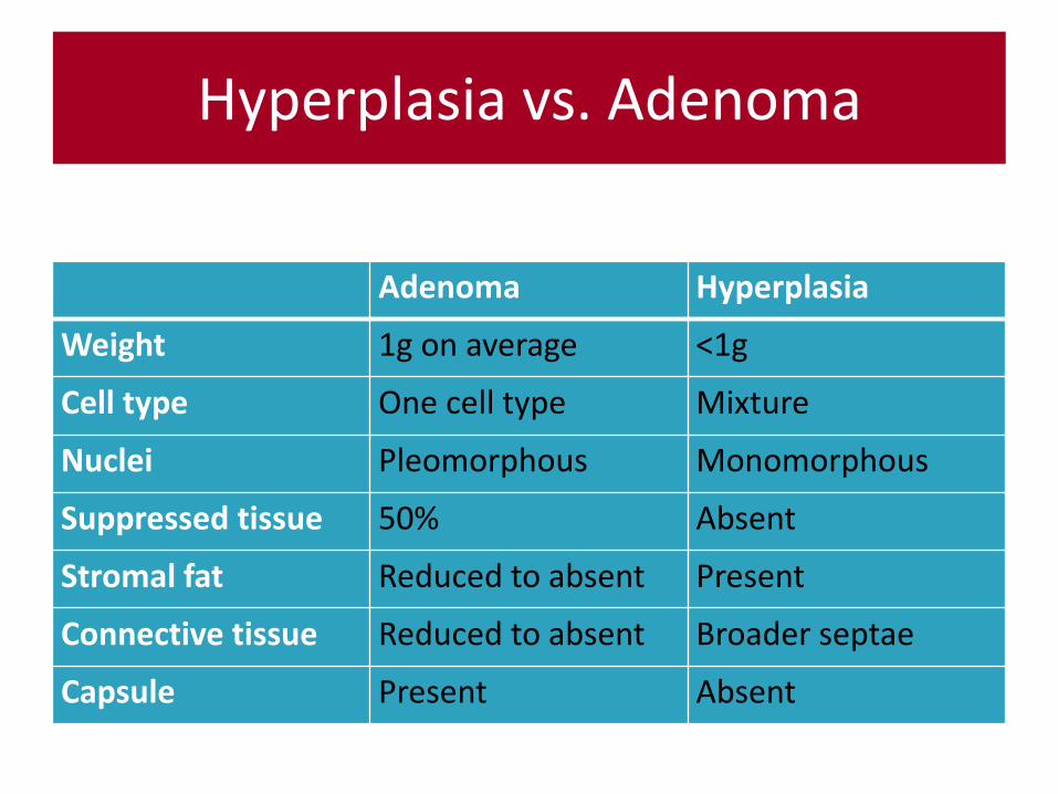
Hyperplasia vs. Adenoma
Adenoma Hyperplasia
Weight 1g on average <1g
Cell type One cell type Mixture
Nuclei Pleomorphous Monomorphous
Suppressed tissue 50% Absent
Stromal fat Reduced to absent Present
Connective tissue Reduced to absent Broader septae
Capsule Present Absent

• Chronic renal failure
• Vitamin D shortage
• Malnutrition/malabsorption/maldigestion
Secondary Hyperparathyroidism
Regulative disturbance of calcium homeostatis: bone symptoms but no hypercalcemia

Secondary Hyperparathyroidism Secondary Hyperparathyroidism
Chief cell hyperplasia Diffuse nodular hyperplasia

• patients with chronic renal failure/malabsorption
• long-term secondary hyperparathyroidism
• asymmetric nodular hyperplasia
Tertiary Hyperparathyroidism
hypercalcemia with loss of response to serum calcium levels subtotal parathyroidectomy or total parathyroidectomy with autotransplantation

Tertiary Hyperparathyroidism

Carcinoma of the Parathyroid
• Criteria of malignancy (min. 1)
– Invasion of adjacent tissue like thyroid, esophagus, nerves, cervical soft tissue
– Lymph node or distant metastases (histologically proved)

Carcinoma of the Parathyroid
• Criteria associated with malignancy (min. 2-3) – Penetration of the capsule (60%)
– Vascular invasion (10-15%)
– >5 mitoses/10 HPF
– Broad intratumoral fibrous septae
– Coagulation necrosis
– High nuclear:cytoplasmic ratio
– Diffuse cellular atypia
– Macronucleoli

Carcinoma of the Parathyroid
No grading No TNM classification
Muscle invasion
Invasion of the capsule
Broad fibrous septae

Adenoma Atypical Adenoma Carcinoma
capsule Thin, complete variable thick fibrous capsule
suppressed tissue present in 50% variable rare
macroscopy red-brown variable grey-white,
lobulated, nodular
stromal fat reduced to lacking reduced to lacking lacking
mitoses 1/10 HPF >1/10 HPF >1/10 HPF
capsule/vascular
invasion
no +/- CI: 2/3
V1: 10-15%
prognosis very good variable Recurrences, distant
metastases in 1/3
Neoplasias

• Persistent disease: – Ectopic position
– Double gland disease
– Unsuspected hyperplasia
– More than four glands
– Inexperienced surgeon
• Recurrent disease (> 6 months) – Subtotal parathyroidectomy for hyperplasia
– Recurrent or metastatic parathyroid cancer
– Parathyromatosis
Failed Parathyroid Surgery
Ectopic locations of parathyroid