pathological variations in mummified feet between two near
TRANSCRIPT

RESEARCH Open Access
Pathological variations in mummified feetbetween two near-distance/long-timepopulations in Ancient EgyptAlbert Isidro1,5*, Beatrice Huber2, Aamer Malik3 and Assumpció Malgosa4*
Abstract
Background: In ancient populations, a significant quantity of foot pathology was related either to the type offootwear they used or the underlying terrain they walked on. Our study was carried out to analyze theseparameters with the foot pathologies the mummies presented.
Methods: Between 2006 and 2012, more than 650 individuals were recovered from the Sharuna and Qararanecropolis (Middle Egypt) dating from the VIth Dynasty of the first Ptolemaic Period to the second Coptic Period.From among them, a total of 73 mummified feet (41 from Sharuna and 32 from Qarara) were studied. We took intoaccount the differences existing between both sites in location (15 km apart) and in time (2500 years apart).
Results: Almost all feet from Sharuna were wrapped and impregnated with a preservative substance (anthropologicalmummification), while the mummification process in Qarara was quite natural. Pathologies were found in 36 of the 73ft (20 from Sharuna and 16 from Qarara). The differences in foot pathologies between the two sites were analysed.
Conclusions: The foot pathologies we found in both necropolises have led us to hypothesise that the majority of thediachronic differences could be related more to progressive changes in the type of the terrain brought out throughdroughts, than the changes in footwear habits.
Keywords: Ancient pathologies, Shoes, Egypt, Mummies
BackgroundIn ancient populations, apart from congenital abnormalitiesand tumours, a significant number of foot alterations andpathologies were related to the type of footwear and the na-ture of the terrain. Despite the importance of this relation-ship few studies have referenced types of foot pathologies inrelation to their lifestyles in ancient times [1].Between 2007 and 2012, archaeological teams from the
Museu Egipci in Barcelona (Catalonia, Spain) and the Äegyp-tolisches Institut of the Eberhart-Karls University in Tübin-gen (Germany) collaborated on site at Sharuna and Qarara.[2] (Fig. 1). These archaeological sites have revealed morethan 650 individuals to date. However, most human remainsof these individuals were discovered dismembered, with poor
anatomical association or even moved from the original bur-ial site where they were interred by specific burial rituals [3].Sharuna (S1) is a large Egyptian necropolis located on the
east bank of the Nile River in Middle Egypt (about 200 kmsouth of Cairo and 60 north of Minia). This site covers a widerange of periods between the 3rd Dynasty and the CopticPeriod with the main anthropological site being the TombU.20. Wilkinson first mentioned this necropolis in 1835 andNestor l’Hôte in 1838 had described theTomb of the PharaohPepi II of the 6thDynasty as an important tomb at the site [4].The Qarara (Q2) necropolis is located 15 Km north of
Sharuna and is a huge burial area in which people from theCoptic Period were interred. Its time period ranges fromthe 5th to the 14th century AD. Although the two necropo-lises are not far each other there is a time difference ofabout 2500 years between them. In Qarara, most of the in-dividuals were discovered in a partially or completely mum-mified state (near 75 %) different to Sharuna in which themajority of them were found as raw-bone (near 85 %).
* Correspondence: [email protected]; [email protected] Universitari del SagratCor, Barcelona, Spain4Department Biologia Animal, Biologia Vegetal i Ecologia, Unitatd’Antropologia Biològica Universitat Autònoma de Barcelona,Bellaterra - Cerdanyola del Valles, SpainFull list of author information is available at the end of the article
JOURNAL OF FOOTAND ANKLE RESEARCH
© 2015 Isidro et al. Open Access This article is distributed under the terms of the Creative Commons Attribution 4.0International License (http://creativecommons.org/licenses/by/4.0/), which permits unrestricted use, distribution, andreproduction in any medium, provided you give appropriate credit to the original author(s) and the source, provide a link tothe Creative Commons license, and indicate if changes were made. The Creative Commons Public Domain Dedication waiver(http://creativecommons.org/publicdomain/zero/1.0/) applies to the data made available in this article, unless otherwise stated.
Isidro et al. Journal of Foot and Ankle Research (2015) 8:58 DOI 10.1186/s13047-015-0115-4

The aim of this study is to compare the pathologies foundin the feet of the mummified individuals between the twonecropolises which had a short distance but large time spanbetween them analysing in the basis of bioclimatic differ-ences, the soil types and differences in footwear.
MethodsThe fact that most of the documented anthropologicalremains were been found outside their original settings(especially in S1), made it difficult for us to ascribe theretrieved individuals to a particular period. However,through some typical characteristics such as the typeand form of bandages in which the mummies werewrapped, the presence or absence of nasal tamponade,and the amount of resins in the abdominal, thoracicand/or cranial cavities, we were able to estimate whenthe mummification procedure took place [5]. In most in-dividuals, we did not find any abdominal wall in an ac-ceptable enough condition to enable us to determine theexistence, or not, of an incision that was used to extractthe internal organs. These different types of incisions
would have provided more information about the periodto which the mummy belonged [6].Nonetheless, the characteristics of the mummification
process that we did find, allowed us to ascribe S1 speci-mens to be from the 6th Dynasty to First IntermediatePeriod (2323–2040 BC) [7]. The specimens coming fromQ2 belonged to the First Coptic period (between theIVth to VIIIth centuries).A total of 73 ft, 41 from S1 and 32 from Q2, belonging
to a minimum of 69 individuals were studied. There was asignificant presence of infant and juvenile feet with 9 beingfrom S1 and 8 from Q2. With reference to the adult indi-viduals, we found that those from Q2 were older thanthose from S1. The age range in S1 was 30–40 years versus45–55 years in the in Q2 individuals. In both the necropo-lises with respect to adult individuals we found a slightlyhigher prevalence of males (30 in S1 and 22 in Q2).The Q2 ft did not present any problems to study due
to their natural mummification process. However, thefeet belonging to the occasional dismembered mummiesfound in the S1 necropolis needed to be carefully
Fig. 1 Map of the region. Enlarged map shows the North-Upper Egypt where the two sites are located
Isidro et al. Journal of Foot and Ankle Research (2015) 8:58 Page 2 of 6
unwrapped. This procedure consisted of progressivelymoistening the outermost bandages and moving in-wards; sometimes this process could take several daysdue to the sheets being glued together with a resin-likemass. The most difficult dressing to remove was those incontact with the skin. In order to study the morphology,as well as the pathologies, some of partial specimenswere taken to the laboratory at the archaeological sitewhere macroscopic, microscopic (x 5) and photographicstudies were performed. Unfortunately, it was impossibleto carry out field X-rays and Ca14 analysis.
ResultsMummificationUpon initial examination, there were clear differencesbetween the mummified feet found in S1 and those fromQ2. At the end of the Old Kingdom and the First Inter-mediate Period in S1, a different mummification processhad been employed, depending on the social status, withthe use of resin-like substances. In the Coptic period inQ2, about 2500 years later, people were sometimes bur-ied with preservative substances like salt and juniperberries within the inner clothing layers. In Q2, the ma-jority of individuals present no signs of body treatment[8, 9]. Probably, between the Vth and the Xth centuryAD, the Coptic people in Egypt, abandoned the ancientmummification methods [6].Almost all of the feet recovered from S1 were wrapped
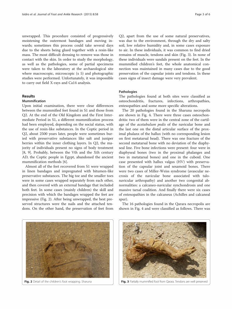
in linen bandages and impregnated with bitumen-likepreservative substances. The big toe and the smaller toeswere in some cases wrapped separately from each other,and then covered with an external bandage that includedboth feet. In some cases (mainly children) the skill andprecision with which the bandages wrapped the feet areimpressive (Fig. 2). After being unwrapped, the best pre-served structures were the nails and the attached ten-dons. On the other hand, the preservation of feet from
Q2, apart from the use of some natural preservatives,was due to the environment, through the dry and saltysoil, low relative humidity and, in some cases exposureto air. In these individuals, it was common to find driedremains of muscle, tendons and skin (Fig. 3). In none ofthese individuals were sandals present on the feet. In themummified children’s feet, the whole anatomical con-nection was maintained in many cases due to the goodpreservation of the capsular joints and tendons. In thesecases signs of insect damage were very prevalent.
PathologiesThe pathologies found at both sites were classified asosteochondritis, fractures, infections, arthropathies,entesopathies and some more specific alterations.The 20 pathologies found in the Sharuna necropolis
are shown in Fig. 4. There were three cases osteochon-dritis: two of them were in the central zone of the cartil-age of the acetabulum pedis of the navicular bone andthe last one on the distal articular surface of the prox-imal phalanx of the hallux (with no corresponding lesionon first metatarsal head). There was one fracture of thesecond metatarsal bone with no deviation of the diaphy-seal line. Five bone infections were present: four were indiaphyseal bones (two in the proximal phalanges andtwo in metatarsal bones) and one in the cuboid. Onecase presented with hallux valgus (HV) with preserva-tion of the capsular joint and sesamoid bones. Therewere two cases of Miller-Weiss syndrome (avascular ne-crosis of the navicular bone associated with talo-navicular arthropathy) and another two congenital ab-normalities: a calcaneo-navicular synchondrosis and onemassive tarsal coalition. And finally there were six casesof entesopathies in the calcaneus (Achilles and calcanealspur).The 16 pathologies found in the Qarara necropolis are
shown in Fig. 4 and were classified as follows. There was
Fig. 2 Detail of the children’s foot wrapping. Sharuna Fig. 3 Partially mummified foot from Qarara. Tendons are well preserved
Isidro et al. Journal of Foot and Ankle Research (2015) 8:58 Page 3 of 6

one case of talar osteochondritis of the medial lip; onepseudocyst in the diaphysis of the 2nd metatarsal bone(of probable infectious origin from outside to inside);one massive tarsal coalition (Fig. 5); one case of talipes(this particular case was not due to a taphonomic orpost-mortem cause when compared to all the other indi-viduals buried in the same method) (Fig. 6); five cases ofcalcaneal spurs and seven cases of subtalar arthropathy(the most significant pathology in this area).Some cases of pseudo-pathology are present in both
necropolises. It was very important not to confuse theseconditions with diseases. From Q2, there is a clear caseof a false hallux extensus in a complete mummified foot(Fig. 7) with a similar case present in a hallux from S1.
DiscussionIn order to approximate a comprehensible picture of thehealth status in these two populations, we compared thepathologies present in the individuals of the two necrop-olises. The comparison allowed us to characterize thepeople and study their possible lifestyles and hence ex-plore the sources of variations between them. In arch-aeological specimens, there is not always a clear cutborder between those which are normal and those that
are pathological. External agents can mimic abnormal-ities, either on the dry bone or in the mummified tissues.Sometimes these can be due to physical or chemicalagents produced in the soil, sunlight, water, etc.; or bythe direct action of living organisms such as plants(mainly roots) or animals (insects through bites andscratches) (Fig. 8). The position after death could havechanged from the original burial one, either through
Fig. 4 Distribution on feet pathologies present in Sharunaand Qarara
Fig. 5 Massive tarsal coalition from Sharuna
Fig. 6 Talipes from Qarara. This case is not a post-mortem condition
Fig. 7 Effects of the wrap and the foot position after deathmimicking a hallux extensus. Qarara
Isidro et al. Journal of Foot and Ankle Research (2015) 8:58 Page 4 of 6

natural or human actions. For instance, the preservedsoft tissues of mummies could be in unusual positionswhich could be confused with deformities; and it is im-portant to draw attention to the fact that these situationscan also occur in our study. This was the case in thefalse hallux extensus above mentioned.Taking into account all these considerations, a total of
36 ft with pathologies or related conditions were foundamong the 73 ft analysed, which represent 49.3 % of thesample. A similar percentage of pathologies were foundin the two necropolises, but their distribution was differ-ent. Infections and osteochondritis were the most fre-quent pathologies found in Sharuna, while they arehardly present in Qarara. Conversely, entesopathies andarthropathies were common in the Qarara site and arescarce in Sharuna.To explain these differences, we must bear in mind
that most of the people buried in both necropolisesbelonged to the working class (workers in S1 and monksin Q2). Although humans used footwear (initially madeof plant fibres or leather) in the Upper Palaeolithicperiod [10], it is probable that most of the exhumed in-dividuals we studied walked barefoot all their lives (nei-ther site had any specimen with footwear in thedressing). We believe that differences between the foot-wear habits of the inhabitants of both these areas are in-sufficient to explain the differences in pathologies foundhere. Sandals are known to have existed from the middleof the 3rd millennium (Pyramids text) and in the middleof the 2nd sandals were frequently used in Egypt, usingwood and leather for soles [11]. At the end of the 6thDynasty and the First Intermediate Period, shoes weremade of hemp and linen and were totally flat, while inthe early Coptic Period, shoes were also flat and made ofleather [12]. We believe that the terrain and ground onwhich these people walked was more important than thefootwear. The landscape in the Old Kingdom and FirstIntermediate Period was very different to what it is
today, as the areas of alluvial soils were greater [13]. Inthe Coptic Period, the terrain had yet to change to onemore or less similar to that of the present day, withsandy and rocky desert soils.This terrain could shed some light on the high preva-
lence of degenerative subtalar arthropathy (7 of 21 morethan 20 % of adult feet) in Q2, an alteration that couldbe associated with walking on irregular ground [14]. Onthe other hand, in S1, there were 5 notable cases of in-fection (12.2 %), a pathology that is absent in Q2, andwhich could be related to walking barefoot in marshes.In S1, there are 3 cases of osteochondritis in bones ofthe medial column of the foot (navicular and F1). It isremarkable that there is an almost complete absence oftraumatic injuries (only 1 fracture in the neck of a sec-ond metatarsal bone - healed with a deviation of 30°) inboth necropolises, a condition that could be related towalking barefoot [15]. It is also uncommon to find, inarchaeological remains, 2 cases (1 in each necropolis) oftarsal coalition [16] although the lack of in-depth ana-lysis, with radiological techniques for instance, meansthat we cannot rule out an ankylosing condition of non-congenital aetiology. The equine foot from Q2 is not theresult of any post-mortem deformity, although it used tobe a frequent condition in many mummified feet due ei-ther to muscular imbalance during the preservationprocess, or to external forces from bandaging and thesarcophagus [17]. Finally, in relation to the case of hal-lux valgus from S1, it is remarkable that in AncientEgypt the bunion was not described in paleopathologicalliterature, although diseases of the big toe must havebeen common. The importance that Egyptians attachedto the big toe can be seen from its relevance in the artof the human figure, from the preservation of this partof the anatomy for the after-life and the presence of exo-prosthesis of the hallux in two mummies. The first casebelonged to a female individual from the 21th Dynastywhich consisted of a two-component hallux prosthesisof the right foot [18], and the second was a superblycrafted wooden prosthesis after hallux amputation in anindividual from the early Third Intermediate Period(21th to 22th Dynasty) [19].
ConclusionsThe analysis of foot pathologies and their incidence inSharuna and Qarara necropolises show the influence ofenvironment and customs on the lives of these people.The majority of diachronic differences that we found ap-pear more to be related to the type of terrain encoun-tered than to their footwear habits.
Competing interestsThe author(s) declare that they have no competing interests.
Fig. 8 Natural mummified feet of a Coptic individual from Qararanecropolis. The holes are due at insect damage
Isidro et al. Journal of Foot and Ankle Research (2015) 8:58 Page 5 of 6

Authors’ contributionsAI and BH carried out the field research and drafted the manuscript; AMsupervised development of work and helped in data interpretation; AMperformed literature research and interpreted data. All authors read andapproved the final manuscript.
AcknowledgmentsThe authors would like to express their gratitude to Mariangela Taulé, LuisGonzálvez and Lourdes Moret for their support during the fieldwork. One ormore of the authors (AM, AI) belongs to GREAB, a research groupacknowledged by the Generalitat of Catalunya (2014-SGR-1420).
Author details1Hospital Universitari del SagratCor, Barcelona, Spain. 2Äegyptolisches Institut,Eberhart-Karls University, Tübingen, Germany. 3Hospital Universitari SagratCor, Barcelona, Spain. 4Department Biologia Animal, Biologia Vegetal iEcologia, Unitat d’Antropologia Biològica Universitat Autònoma de Barcelona,Bellaterra - Cerdanyola del Valles, Spain. 5Serv. C.O.T. Hosp., Sagrat Cor/Unidad Docente U.B., Viladomat 288, 08029 Barcelona, Spain.
Received: 16 April 2015 Accepted: 14 October 2015
References1. Isidro A. Paleopatología del pie. Rev Medicina y Cirugía del Pie.
2000;XIV(2):41–9.2. Gonzálvez LM. Kom El-Ahmar/Sharuna. Primera misión de la Universidad de
Tübingen / MuseuEgipci de Barcelona. Arqueoclub. 2007;8:18–21.3. Isidro A, Gonzalvez L, Taulé MA, Moret L, González E, Galtés I, et al.
Preliminary report of the anthropological remains from the Necrópolis ofSharuna. (MuseuEgipci de Barcelona / Universitat de Tübingen, 2006–2008archaeologicalseasons). Munibe (Antropologia-Arkeologia). 2009;60:243–52.
4. Schenkel W, Gomaà F, Scharuna I. Der Grabungplatz. Die Nekropole. Gräberaus der Alte-Reichs-Nekropole. Tübingen: Philipp von Zabern; 2004.
5. Ikram S, Dodgson A. The Mummy in Ancient Egypt. London: Thames andHudson Ltd; 1998.
6. Aufderheide AC. The Scientific Study of Mummies. Cambridge: CambridgeUniversity Press; 2003.
7. Baines J, Malek J. Atlas of Ancient Egypt. Oxford: Oxford University Press; 1980.8. Lösch S, Hower-Tilmann E, Zink A. Mummies and skeletons from the Coptic
monastery complex Deir el-Bachit in Thebes-West, Egypt. Anthro Anz.2012;70(1):27–41.
9. Gessler-Löhr B, Grabbe E, Raab B-W, Schultz M. Ausklang:EinekoptischeMumieauschristlicherZeit, ÄegypischeMumien.Untersblichkeitim Land der Pharaonen. Mainz: Philipp von Zabern; 2007. p.255–65.
10. Trinkaus E. Anatomical evidence for the antiquity of human footwear use.J Archaeol Sci. 2005;32:1515–26.
11. Stewart SF. Footgear. Its history, uses and abuses. Clin Orth Relat Research.1972;80:119–30.
12. Huber B. Etude de deux paires de sandales provenant des fouilles récentesde Qarara. In: De Moor A, Fluck C, editors. Dress accessories from Roman toEarly Islamic Egypt and neighbouring countries. Proceedings of the 6th
Meeting of the study group “Textiles of the Nile Valley”.Lanoo, Antwerp:2011, p. 139–145.
13. Butzer KW. Physical conditions in Eastern Europe, Western Asia and Egyptbefore the period of agricultural and urban settlement. In: The CambridgeAncient History. Cambridge: Cambridge University Press. 1965;33(1):1–39.
14. Martinez MJ, Baixarias J, Isidro A, Vila S, Campillo D. A case of taluscalcaneous arthrosis secondary to a valgus foot in a late roman individual.JoP. 1999;11:78.
15. Zipfel B, Berger LR. Shod versus unshod: The emergence of forefootpathology in modern humans? Foot. 2007;17:205–13.
16. Isidro A, Castellana C, Malgosa A. Massive tarsal ankylosis in a prehistoricskeleton. Foot & Ankle Surg. 2000;6(4):239–47.
17. Isidro A, Rodriguez C. Club-foot in a mummy from Canary Islands. Foot &Ankle Surg. 2004;10(3):163–5.
18. Wagle WA. Toe prosthesis in an Egyptian human mummy. Am Jour Roengt.1994;162:999–1000.
19. Nierlich AG, Zink A, Szeimies U, Hagedorn HG. Ancient Egyptian prosthesisof the big toe. Lancet. 2000;356:2176–9.
Submit your next manuscript to BioMed Centraland take full advantage of:
• Convenient online submission
• Thorough peer review
• No space constraints or color figure charges
• Immediate publication on acceptance
• Inclusion in PubMed, CAS, Scopus and Google Scholar
• Research which is freely available for redistribution
Submit your manuscript at www.biomedcentral.com/submit
Isidro et al. Journal of Foot and Ankle Research (2015) 8:58 Page 6 of 6