pathological studies on pigeon trichomoniasis with ... · pathogenic strain of trichomonas gallinae...
TRANSCRIPT

Egypt. J. Comp. Path. & Clinic. Path. Vol. 22 No. 2 (March) 2009; 67 - 87
67
Referred byReferred by Prof. Dr. Osman A. Atallah Professor of Pathology, Fac. Vet. Med.,
Zagazig University Prof. Dr. Magda M. Ali Professor of Poultry Diseases, Fac. Vet.
Med., Cairo University
Pathological studies on pigeon trichomoniasis with reference to the associated bacteria
By Mohamed, I.E.*, Gehan, H. El-Sakkar* and **Magda, M.M. Moursi
*Department of Pathology, Animal Health Research Institute, Zagazig Lab. **Department of Microbiology, Animal Health Research Institute, Zagazig Lab.
SUMMARY
O ne hundred and twenty apparently healthy and diseased squabs (up to 2 month old) were collected from Sharkia Governorate our work
revealed that 80 out of 120 examined squabs were positive for T. gallinae. The birds suffered depression, off food and dyspnea. Offensive odor-fluid drilled from their opened beak. Macroscopically, the oropharynx esopha-gus and crop revealed caseated materials, besides necrotic foci in the liver and heart. Experimental infection with trichomonas gallinae was done on twenty examined healthy squabs and treated with flagyle. The result of these experimentally infected squabs revealed typical symptoms and post- mortum lesions of Trichomoniasis, and flagyle was effective against T.gallinae. Specimens were collected from the oropharynx, liver, heart-blood and lungs for bacteriological examination and from the crop, esophagus and brain for histopathological examination. The isolated bacteria were Pasteurella multocida type D1, Strepto-coccus bovis and E.coli at the rates of 65%, 22.5% and 12.5% respec-tively. Microscopically the natural and experimental trichomoniasis showed mild to massive caseous necrosis with bacterial colonies were found on the pharyngeal mucosa. Submucosal hemorrhage and leukocytic infiltration were noticed. The esophagus showed caseous necrosis and congestion. The hepatic parenchyma showed hydropic degeneration and focal replacement with radially arranged pericytes. The portal areas were infiltrated with numerous leukocytes. The myocardium showed hemor-rhage, mononuclears, and necrosis of the myocytes. The lungs showed congestion and hemorrhage with leukocytic aggregations. The brain showed necrotic Purkenje cells and necrosis of the large cerebral neurons. The most effective drugs, against the isolated bacteria were Avitryle, Nor-floxacin, Gentamicin and Ampicillin. Flagyl was effective against tricho-moniasis. Our work revealed that the trichomonas infection and the secondary

Egypt. J. Comp. Path. & Clinic. Path. Vol. 22 No. 2 (March) 2009; 67 - 87
68
INTRODUCTION
P igeons contribute to the poul-try industry, in Egypt. Their
meat is palatable for the majority of the Egyptians and other nation-alities.
Pigeons are affected with dis-eases of diverse etiological nature. Less effort has been attempted, in Egypt, to investigate the prevalent diseases in pigeons except for a few reports which dealt with cer-tain infections (Shihata 1978, Nagwa 1995, Eman 2005 and Hebat-Allah and Abd El-motelib (2007). Little attention has been directed toward diseases of pi-geons. This may be due to the fact that the majority of these birds are unconfined as they usually wander over a long distance searching for food, and the population of pi-geons is few when compared with chickens, that are reared inten-sively (Abd El-Motelib and Galal, 1993).
Trichomoniasis is a common disease of squabs that affects the upper digestive and respiratory tracts, causing high losses (Levine, 1995). The severity of the disease and mortality, in squabs, may vary according to the susceptibility of the squabs and the virulence of the pathogenic strain of Trichomonas gallinae (Abd El-Motelib and Galal 1993, MacDaugald 2003 and Swinnerton et al. 2005).
Trichomoniasis in squabs, is sometimes associated with some bacterial infection (Shihata, 1981 and Pennycott, 1994). Streptococ-cus sp. causes septicemia in pi-gones (Vagel 1983, Devriese 1986,Baele et al. 2001 and Hebat Allah & Abd El-Motelib 2007). The streptococcal infection is usually peracute or acute, and rarely chronic (Devriese et al. 1990) Meanwhile Andreasen and Sandhu (1993), found that Pas-teurella is associated with respira-tory diseases, in pigeons, as pneu-monia and tracheitis. Gross (1991) found that E.coli infection led to great economic losses in poultry.
The objectives of this study were aimed to detect natural (single or mixed bacterial infec-tion) associated with trichomonas with isolation and idenitifecation of bacterial causes besides study-ing the macro and microscopic le-sions, and the antibiogram for iso-lated bacteria. Moreover, traile for treatment of experimentally in-fected squabs with trichomonas gallinae.
MATERIAL AND METHODS
SAMPLES:- A total of 120 (40 apparently healthy and 80 diseased) pigeons up to 2 month old and from differ-ent breeds were collected from dif-ferent markets in Sharkia Gover-norate. The specimens were col-
Egypt. J. Comp. Path. & Clinic. Path. Vol. 22 No. 2 (March) 2009; 67 - 87
69
lected from oropharynx, liver, heart blood and lungs (from each birds) for bacteriological examina-tion. And the crop, esophagus and brain were used for histopathologi-cal examination.
Parasitological examination and preparation.
a- Microscopical examination for detection of the parasite:
Samples were collected from the different lesions and caseated materials of 80 diseased birds and examined microscopically for de-tection of trichomonas gallinae.
b-Preparation of Trichomonas gallinae culture :
Wet swabs were prepared, by gentle rotation, through the le-sions. The swaps were dipped into sterile saline or culture-media (Glucose-serum broth medium) ac-cording to Nagwa (1995). The in-oculated tubes were incubated at 37°C for 7 days. One drop, from the bottom of each tube, was mi-croscopically examined 4 and 7 day post-inoculation (PI) for the presence of trichomonads accord-ing to Abd El-Motelib and Galal (1993). The positive tubes were subcultured into new tubes of me-dia and incubated for 3 days.
Estimation of the population density of the parasite: A measuring dropper (giving
3 drops/0.1 ml) was used to count number of the parasites in one drop according to Nagwa (1995).
Inocula were used to seed the culture-tubes. Those cultures, yielding between 800-1000 para-site /ml, were used according to Nagwa (1995).
c- Experimental infection:
Thirty apparently healthy squabs up to one month age were collected from different markets in Sharkia governorate and were sub-jected to parasitolgical Examina-tion to assure that the examined squabs were free from infection. The examined squabs were reared in two separated rooms under com-plete hyagenic measures, and devided into 2 group the first group (20) squabs were infected orally by dropper with 1ml con-taining (800 – 1000) parasite. The second group 10 squabs were left as control.
Bacteriological examination:
1- Isolation and identification: The collected Specimens
(oropharynx, liver, heart-blood and lung) were inoculated into tubes of nutrient broth and incubated aero-bically at 37°C over night fol-lowed by subculturing into nutrient agar, MacConkey’s agar and blood agar plates and incubated for 24-48

Egypt. J. Comp. Path. & Clinic. Path. Vol. 22 No. 2 (March) 2009; 67 - 87
70
hours at 37°C. The suspected colo-nies were identified morphologi-cally according to Cruickshank et al. (1975) and confirmed bio-chemically according to Krieg and Holt (1984). 2- Serological identification of Pasteurella Sp.:
Pasteurella organism identi-fied serologically according to Carter and Roppy (1963) for cap-sular typing with indirect hemoag-glutination test and somatic typing using gell diffusion precipitation test.
3- Pathogenicity of Pasteurella for mice:
All strains of Pasteurella mul-tocida were tested for pathogenic-ity to mice according to Wilson and Milies (1975).
4- Antibiotic sensitivity test:
In vitro the disc diffusion technique was performed on the isolated bacteria using seven che-motherapeutic discs namely (Avitryl, Erythromycine, Gentamy-cine, Septrin, Norfloxacin and Ampicillin and Amoxycillin) sus-ceptibility of the predominant pathogenic isolates to different chemotherapeutic agents according to Finegold and Martin (1982).
Histopathological preparation:
Tissue specimens, of 0.5 cm thickness were collected from the
pharynx, esophagus, crop, liver, lungs, heart and brain, from freshly dead and slaughtered infected squabs, to detect the different tis-sue reactions against the parasitic and bacterial infections. The speci-mens were fixed in 10% neutral buffered formalin. Five micron-thick paraffin sections were pre-pared and stained with hematoxy-lin and eosin and examined micro-scopically (Bancroft and Gamble 2002).
The experimentally infected squabs were treated with Flagyl at a dose of one gm/liter drinking wa-ter for five successive days (Sampurnanand et al. 2002).
RESULTS AND DISCUSSION
O ur work revealed that, Pas-teurella multocida and Strep-
tococcus bovis are considered fac-ultative pathogens. They may be a part of the normal pharyngeal and intestinal flora in pigeons. Predis-posing factors may cause some streptococci to break through the intestinal mucosa and enter the blood to stream to induce septice-mia (Vanrobaey et al. 2000 & Kimpe, et al. 2003 and Hebat-Allah & Abd El-Motelib 2007).
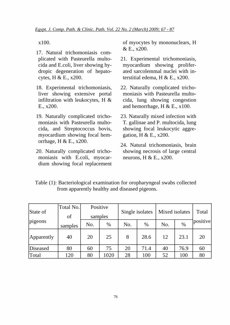
Table (1) shows the percentage of single and mixed bacterial infection in the appar-

Egypt. J. Comp. Path. & Clinic. Path. Vol. 22 No. 2 (March) 2009; 67 - 87
71
ently healthy and diseased pigeons. Table (2) reveals that the isolated Streptococcus bovis was 22.5% which is nearly in agreement with Hebat - Allah and Abd - El -Motelib (2007). Meanwhile more isolates were recorded by De Herdt et al. (1993) who isolated bacteria was Pasteurella multocida type D1 at percentage of 65%, Streptococcus bovis at the rate of 40% from diseased pigeons of all ages. The rate of E. coli isolation in our study was 12.5% which are in accordance with Pedersen et al. (2006) who isolated E. coli at rate of 10%. In the vitro sensitivity test table (3) showed that Ampicillin, Norfloxacin, Erythromycin, Genta-m y c i n . and Avitryle were highly effective against the isolated bacteria. Simi-lar results were reported by De-Herdt et al. (1993) who found that Ampicillin, Doxycycline and Erythromycin were that most ef-fective against the bacterial iso-lates. Hebat-Allah and Abd El-Motelib (2007) showed that am-picillin and Enrofloxacin was the most effective drugs. Shereen (2007) found that E.coli isolated from ostrich was sensitivs to enro-floxacin Gehan (2006) found that the isolated E.coli from quails was sensitive to Enrofloxacin and less sensitive to Erythromycin. Table (4) showed that bacterial isolates from oropharynx of apparently healthy and diseased pigeons but
in liver, lung and heart blood the bacterial isolates were from dis-eased pigeons only by percentage as shown in the table.
All mice were tested for pathogenicity to P. multocida dead within 24-48 h. These results agreement with Okerman et al. (1979).
The natural and experimental affected squabs with trichomoni-asis showed depression, off-food and dyspnea, in addition to offen-sive-odor-fluid, drilling from the opened peak, due to lesions in the oral cavity. Moreover, a large caseated mass was present in the buccal cavity. Sometimes greenish watery diarrhea was noticed. Simi-lar signs were reported by Abd-El-Motelib and Galal (1993), Sohair and Effat (2004), Eman (2005) and Hebat-Allah and Abd-El-Motelib (2007). The treated squabs against trichomoniasis with Flagyl led to improvement in the clinical signs with decreased mor-tality rate within few days after the beginning of the treatment. The complicated trichomoniasis, with bacterial infection, frequently showed early death or chronic lameness. Similar findings were observed by Devriese et al. (1990) in trichomoniasis complicated with streptococcal infection in pigeons.
Macroscopically large whit-ish-yellow caseated masses of

Egypt. J. Comp. Path. & Clinic. Path. Vol. 22 No. 2 (March) 2009; 67 - 87
72
variable sizes (3 – 30 mm diame-ter) were seen in the oropharynx (Fig.1). They reached the esopha-gus and crop. Sometimes the lu-men of the crop was occluded with such caseated material. Similar ob-servations were previously re-corded by Abd-El-Motelib and Galal (1993), Nagwa (1995) and Sohair and Effat (2004). More-over, trichomonas caused yellow-ish necrotic foci in the liver and lungs. Similar lesions were ob-served by Nagwa (1995), El-Metenawy (2000), MacDaugald (2003), Sohair and Effat (2004), Mohamed and Nahla (2005) and Eman (2005). Pneumonia and he-patic congestion were noticed. Si-milar lesions were recorded by He-bat-Allah and Abd-El-Motelib (2007). The ability of the parasites to reach these organs may be at-tributed to a disturbance between the resistance of the host and viru-lence of the parasite (Charlton et al. 1991; Narcisi et al., 1991 and MacDaugald 2003). This study revealed that the lesions varied from mild to moderate. The mild lesions could indicate single infec-tion or a high resistance of the pi-geons.
Microscopically, the exam-ined pharynx of the naturally in-fected squabs with Trichomonas gallinae showed submucosal hem-orrhage (Fig. 2). Another case, of natural trichomoniasis complicated with Pasteurlla multocida showed
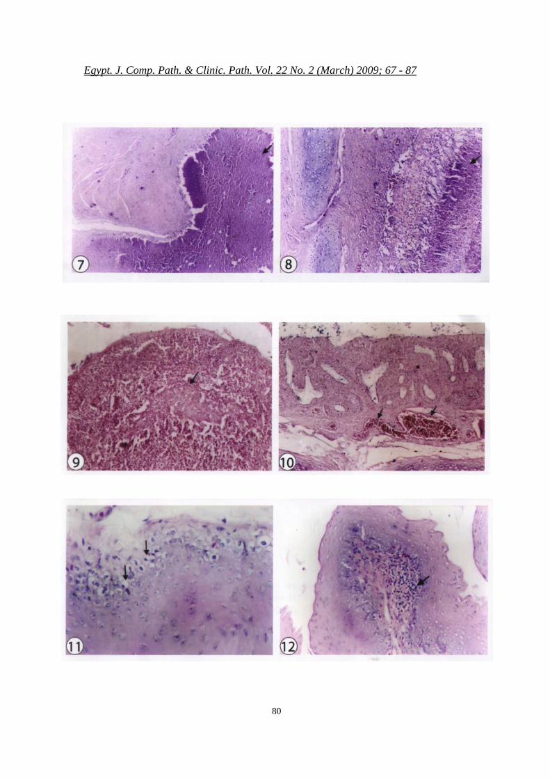
partial replacement of the pharyn-geal wall by caseous necrosis (Fig. 3). Massive infiltration with heter-ophils and macrophages was de-tected in the pharyngeal wall of the naturally infected squab (Fig. 4). Similar lesions were previously detected by Shihata (1978), Nar-cisi et al. (1991), Nagwa (1995) and Eman (2005). The concurrent findings are in a partial agreement wi th Mohamed and Nahla (2005), who described focal ho-mogenous and vacuolated necro-sis. Another case showed acantho-sis, necrosis and mucosal leuko-cytic infiltration, associated with submucosal hemorrhage of the pharynx (Fig. 5). These changes are in a partial agreement with Honigberg (1978) and Eman (2005). Massive caseous necrosis associated with bacterial colonies (Pasteurella multocida and Strep-tococcus bovis) were noticed in natural trichomoniasis (Fig. 6). Massive caseous necrosis was de-tected in the pharyngeal wall of the experimentally infected squab (Fig. 7). These results are in partial concurrence with those reported by Eman (2005) who found caseous necrosis in the pharynx. Large masses of caseous necrosis oc-cluded the pharyngeal lumen of the naturally infected squab (Fig. 8). The previously reported lesions are in complete agreement with Eman (2005). Moreover, the esophagus of the experimentally infected

Egypt. J. Comp. Path. & Clinic. Path. Vol. 22 No. 2 (March) 2009; 67 - 87
73
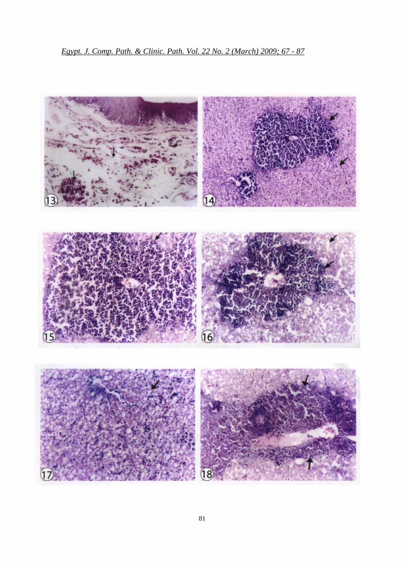
squabs revealed caseous necrosis (Fig. 9). Moderately necrotic and congested mucosa was noticed (Fig. 10), due to natural trichomo-niasis complicated with Strepto-coccus bovis. These changes are in agreement with those reported by Sohair and Effat (2004). Vacuo-lated crop mucosal epithelium was observed in experimental tricho-moniasis (Fig. 11). Focal submu-cosal leukocytic aggregations were seen (Fig. 12). This lesion is in ac-cordance with those recorded by Narcisi et al. (1991) and Nagwa (1995). Edema and submucosal hemorrhage were found in natural trichomoniasis complicated with E.coli (Fig. 13). The naturally compliced trichomoniasis with Pasteurella multocida and E.coli showed hepatic hydropic degen-eration and focal replacement of the hepatic parenchyma by radially arranged pericytes around the cen-tral vein (Figs. 14, 15). These le-sions are in a partial agreement with Abd-El-Rahman and Mousa (2000) who reported that E. coli infection in chicks induced coagu-lative necrosis and congestion. So-hair and Moursi (2003) found vacuolar degeneration and necrosis of hepatic cells due to Pasteurello-sis in ostrich. Sami and El-Oksha (2006) found coagulative necrosis of hepatic tissue due to Pasteurella infection in turkey. Complicated natural trichomoniasis with Pas-teurella multocida revealed focal
replacement of the hepatic paren-chyma by radially arranged peri-cytes around the central vein (Fig. 16). These lesions are partially in agreement with Mostafa (2002) who found coagulative necrosis of hepatic parenchyma due to pas-teurellosis in poultry. Natural trichomoniasis, complicated with Pasteurella multocida and E. coli showed hydropic degeneration of hepatocytes (Fig. 17). Almost nearly similar lesions were re-ported by Sohair and Moursi (2003) who found vacuolar degen-eration and necrosis of the hepatic cells due to pasteurellosis in os-trich. Eman (2005) found hy-dropic degeneration of some hepa-tocytes in pigeons infected with trichomoniasis. Moreover our find-ings are in complete agreement with Mousa and Magda (2006) who found hydropic generation in duck pasteurellosis. Medani et al. (2008) found diffuse vacuolar de-generation in the liver of infected seagulls with E. coli. The experi-mental trichomoniasis induced ex-tensive portal infiltration with leu-kocytes (Fig. 18). This result is in complete agreement with that re-ported by Sohair and Effat (2004) and Eman (2005) due to tricho-moniasis in pigeons..
The myocardium in the natu-ral trichomonas complicated with Pasteurella multocida and Strepto-coccus bovis revealed focal hem-orrhage (Fig. 19). Similar result

Egypt. J. Comp. Path. & Clinic. Path. Vol. 22 No. 2 (March) 2009; 67 - 87
74
were prevously reported by Sohair and Effat (2004). The myocar-dium of the naturally infected squabs complicated with E.coli was focally replaced with mononu-clears (Fig. 20). Similar results were previously recorded by Abd El-Rahaman et al. (2003) due to E. coli infection. The experimental trichomoniasis led to proliferation of sarcolemmal nuclei with inter-stitial edema (Fig. 21). Similar re-sults were previously reported by Sohair and Effat (2004) and Eman (2005). Such lesions could be attributed to the toxic metabo-lites, produced by the infective agents, these metabolites damaged the vascular endothelium, leading to edema and hemorrhage.
The lung of natural trichomo-niasis complicated with Pasteurella multocida showed congestion and hemorrhage (Fig. 22) due to endo-thelial damage by the bacterial and parasitic metabolites which in-creased the permeability of the blood vessels leading to edema and hemorrhage (Jones et al. 1997). These changes are almost in agree-ment with Sohair and Moursi (2003) due to pasteurellosis in os-trich and Sohair and Effat (2004) due to trichomoniasis. Focal leuco-cytic aggregation were seen in naturally trichomoniasis and mixed infection with Pasteurella multocida (Fig. 23).
The brain of the naturally
trichomoniasis revealed necrotic Purkenje cells of the cerebellum. Moreover, nerosis of large cerebral neurons was noticed (Fig. 24). These lesions are nearly similar to those reported by Narcisi et al. (1991) and Sohair and Effat (2004) who found neuronal degen-eration and mild gliosis due to trichomoniasis in pigones.
Our work revealed that the treatment of the trichomonas ex-perimentally infected squabs with flagyl at a dose of 1 gm/liter drink-ing water for five successive days improved the general condition of the birds and decreased the mortal-ity rate. A nearly similar results were reported by Sohair and Effat (2004), within few days after the beginning of the treatment.
I t could be concluded that tricho-moniasis, in most infected
squabs was associated with secon-dary bacterial infection. P. multo-cida E. coli and Streptococcus bo-vis which became pathogenic un-der the stress of trichomoniasis.
Acknowledgement: We approciate the fruitful advices of Dr. Nagwa Anwar helmy for her great helping with the parasitological study.
LEGENDS Fig. 1. Natural trichomoniasis, squab
showing whitish-yellow caseated

Egypt. J. Comp. Path. & Clinic. Path. Vol. 22 No. 2 (March) 2009; 67 - 87
75
masses (3-30 mm in diameter) in the oropharynx.
2. Natural trichomoniasis, pharynx showing submucosal hemoar-rhage, arrow, H & E., x 200.
3. Natural trichomoniasis, pharynx complicated with Pasteurella multocida showing partial re-placement of the pharyngeal wall with caseous necrosis, H & E., x 200.
4. Natural trichomoniasis, pharynx showing massive infiltration with heterophils and macrophages, ar-rows, H & E., x200.
5. Natural trichomoniasis, pharynx acanthosis (short arrow), necro-sis, mucosal leukocytic infiltra-tion (long arrow) associated with submucosal (L) hemorrhage, H & E., x 200.
6. Natural trichomoniasis compli-cated with Pasteurella multocida, pharynx showing massive cas-eous necrosis and bacterial colo-nies, (arrows), H & E., x200.
7. Experimental trichomoniasis, pharynx showing massive cas-eous necrosis of the pharyngeal wall, H & E., x200.
8. Natural trichomoniasis, pharynx showing a large mass of caseous necrosis occluding the pharyn-geal lumen, H & E., x 100.
9. Experimental trichomoniasis,
esophagus showing caseous ne-crosis, H & E., x200.
10. Natural trichomoniasis, esophagus complicated with Streptococcus bovis showing moderately necrotic and con-gested mucosa (arrows), (H & E., x100).
11. Experimental trichomoniasis, crop showing vacuolation of mu-cosal epithelium, H & E., x400.
12. Natural trichomoniasis, crop showing focal submucosal leuko-cytic aggregations, H & E., x200.
13. Natural trichomoniasis, crop complicated with E.coli showing submucosal edema and hemor-rhage, H & E., x200.
14. Naturally complicated tricho-moniasis with E.coli and Pas-teurella sp., liver showing hy-dropic degeneration associated with focal replacement of the he-patic parenchyma by radially ar-ranged pericytes around the cen-tral vein, H & E., x100.
15. A high power for Fig. 14 to show the radially arranged histio-cytes around the central vein, H & E., x200.
16. Naturally complicated tricho-moniasis with Pasteurella multo-cida, liver showing focal replace-ment of the hepatic parenchyma by radially arranged pericytes around a central vein, H & E.,
Egypt. J. Comp. Path. & Clinic. Path. Vol. 22 No. 2 (March) 2009; 67 - 87
76
x100.
17. Natural trichomoniasis com-plicated with Pasteurella multo-cida and E.coli, liver showing hy-dropic degeneration of hepato-cytes, H & E., x200.
18. Experimental trichomoniasis, liver showing extensive portal infiltration with leukocytes, H & E., x200.
19. Naturally complicated tricho-moniasis with Pasteurella multo-cida, and Streptococcus bovis, myocardium showing focal hem-orrhage, H & E., x200.
20. Naturally complicated tricho-moniasis with E.coli, myocar-dium showing focal replacement
of myocytes by mononuclears, H & E., x200.
21. Experimental trichomoniasis, myocardium showing prolifer-ated sarcolemmal nuclei with in-terstitial edema, H & E., x200.
22. Naturally complicated tricho-moniasis with Pasteurella multo-cida, lung showing congestion and hemorrhage, H & E., x100.
23. Naturally mixed infection with T. gallinae and P. multocida, lung showing focal leukocytic aggre-gation, H & E., x200.
24. Natural trichomoniasis, brain showing necrosis of large central neurons, H & E., x200.
Table (1): Bacteriological examination for oropharyngeal swabs collected from apparently healthy and diseased pigeons.
State of
pigeons
Total No.
of
samples
Positive
samples Single isolates Mixed isolates Total
positive No. % No. % No. %
Apparently 40 20 25 8 28.6 12 23.1 20
Diseased 80 60 75 20 71.4 40 76.9 60 Total 120 80 1020 28 100 52 100 80

Egypt. J. Comp. Path. & Clinic. Path. Vol. 22 No. 2 (March) 2009; 67 - 87
77
Table (2): Prevalence of bacterial isolates in the examined pigeons.
Isolated bacteria
Total No. of
samples
Positive samples
Single isolate Mixed isolates No. %
No. % No. % Pasteurella multocida 80 52 65 18 64.3 P. multocida (12)
Strept. bovis (4) 16 30.8
Streptococcus bovis 80 18 22.5 6 21.4
P. multocida (14) E. coli (4) , Strept. bovis
(3) 21 40.4
E. coli 80 10 12.5 4 14.3 P. multocida (8)
E. coli (2) , Strept. bovis (5)
15 28.8
Total 80 100 28 100 Total 52 100
Table (3): Sensitivity test for bacterial isolates from the examined pigeons.
Antibiotic disc P. multocida E. coli Strept. sp.
Avitryl (10 Mg) +++ +++ ++
Erythromycin (15 Mg) +++ ++ +++
Gentamicin (10 Mg) +++ ++ ++
Septrin (100 Mg) + + +
Norfloxacin (10 Mg) +++ +++ ++
Ampicillin (10 Mg) ++ +++ +++
Amoxycillin (25 Mg) +ve +ve +ve
+++ Highly sensitive ++ Moderate sensitive + Less sensitive

Egypt. J. Comp. Path. & Clinic. Path. Vol. 22 No. 2 (March) 2009; 67 - 87
78
Table (4): The total isolates of bacterial groups in examined pigeons.
Oropharyngeal swabs Liver Heart Lungs
Apparent Diseases Apparent Diseases Apparent Diseases Apparent Diseases Bacterial
isolates No. % No. % No. % No. % No. % No. % No. % No. %
Total
P. multocida 14 70 48 80 00 00 35 58 0.0 0.0 42 70 0.0 0.0 44 73 183
St. bovis 5 25 7 17 0.0 0.0 15 25 0.0 0.0 12 20 0.0 0.0 9 15 48
E. coli 1 5 5 3 0.0 0.0 10 17 0.0 0.0 6 10 0.0 0.0 7 12 29
Total 20 100 60 100 0.0 0.0 60 100 0.0 0.0 60 100 0.0 0.0 60 100 260
No. = number

Egypt. J. Comp. Path. & Clinic. Path. Vol. 22 No. 2 (March) 2009; 67 - 87
79
Egypt. J. Comp. Path. & Clinic. Path. Vol. 22 No. 2 (March) 2009; 67 - 87
80
Egypt. J. Comp. Path. & Clinic. Path. Vol. 22 No. 2 (March) 2009; 67 - 87
81

Egypt. J. Comp. Path. & Clinic. Path. Vol. 22 No. 2 (March) 2009; 67 - 87
82

Egypt. J. Comp. Path. & Clinic. Path. Vol. 22 No. 2 (March) 2009; 67 - 87
83
REFERENCES
Abd El-Rahman, A.; Mousa, H. and Abou Zead, A. (2003): "Studies on some aerobic bac-teria causing death of broiler chickens." Egypt. J. Agric. Res. 2003 (81), 2: 727-738.
Abdel El-Motelib, T.Y. and Galal, B.G. (1993): "Some studies on Trichomonas gallinae infection in pigeons." Assiut Vet. Med. J., 30 (59): 277-288.
Abd-El-Rahman, M. and Mousa, H. (2000): "Studies on some aerobic bacterial causing of broilers." Egypt. J. Agric. Res. 78 (1): 25-34.
Andreasen, J.R.Jr. and Sandhu, T. (1993): "Respiratory dis-eases in pigeons." Avian Dis. 1993 Jul – Supt.; 37 (3): 901-911.
Baele, M.; Devriese, L.A. and Haesebrouck, F. (2001): "Lactic acid bacteria in pi-geons crop." J. of Applied. Mi-crobiology V. 91 (3): 488-491.
Bancroft, J.D. and Gamble, M. (2002): "Theory and Practice of Histological Techniques." 5th edition, Churchill Living-stone, London, Edinburgh, New York, Philadelphia, St. Lous, Sydney.
Carter, G.R. and Roppy, D.E. (1963): "A haemagglutination
test specific lipopolysaccharide for detection of Pasteurella an-tibody." Brit. Vet. 112: 37-77.
Charlton, B.R.; Bickford,A.A.; Cooper, G.L. and Chiu, H.W. (1991): "Systemic trichomoniasis in squabs op-eration." Avian Diseases, 35 (2): 426-432.
Cruickshank, R.; Duguid, J.P.; Marmia, B.P. and Swain, R.W.A. (1975): "Medical Mi-crobiology." 12th Ed. Vol. 11, Churchill, Livengston, Edin-burgh, London and New York.
De-Herdt, P.; Devriese, L.A.; Du-cotelle, R. and Haesebrouck, F. (1993): "Antibiotic treat-ment of Streptococcus bovis infection in pigeons." Avian Pathology, 22: 605-615.
Devriese, L.A. (1986): "Ziekten van sicrvogels en Duiven", P. 176 and P. 216. 3rd Ed. Gent: Faculty of Veterinary Medi-cine.
Devriese, L.A.; Uyttebrock, E.; Gevaert, D.; Vandekerck-houe, P. and Ceyssens, K. (1990): "Streptococcus bovis infection in pigeons." Avian Pathology, 19: 429-434.
El-Metenawy, T. (2000): "A sur-vey on some parasitic protozoa infecting birds in Saudi Ara-bia." Egyptian J. of Comp. Path. & Clinic. Path. Vol. (13)

Egypt. J. Comp. Path. & Clinic. Path. Vol. 22 No. 2 (March) 2009; 67 - 87
84
No. (1): 114-118.
Eman, A. (2005): "Some studies on trichomoniasis in pigeons i n S h a r k i a P r o v i n c e . " M.V.Sc. Thesis, Avian and Rabbit Disease Dept., Fac. of Vet. Med., Zagazig Univ.
Finegold.; M. and Martin (1982): "Diagnostic microbi-ology." 6th Ed., C.V. Mosby Company, St. Louis, Toranto, London.
Gehan, E.M. (2006): "Characteri-zation of Escherichia coli iso-lated from quails." M.V.Sc. Thesis, Dept. of Bacteriol., Faculty of Vet. Med., Zaga-zig Univ.
Gross, W.B. (1991): "Colibacillo-sis in: Diseases of Poultry", 9th ed. Editors: B.W. Calnek, et al., Iowa State University Press, Ames., Iowa, USA, 138-144.
Hebat-Allah, M. and Abd El-Motelib, T. Y. (2007): "Studies on gram-positive or-ganisms in sick pigeons." As-siut Vet. Med. J. 53 (114): 280-290.
H o n i g b e r g , B . M . ( 1 9 7 8 ) : "Trichomonads of Veterinary Importance Parasitic Proto-zoa." Vol. II. Krier, J. (eds). Academic Press. New York, 165-273.
Jones, T.C.; Hunt, R.D. and King, N.W. (1997): "Disease due to protozoa." In veteri-nary pathology. 6th edition. C. Cannad S. Hunsberger (eds.). Lippincott Company. Phila-delphia, Penasylvania, P. 418-423.
Kimpe, A.; Decostere, A.; Mar-tel, A.; Haesebrouck, F. and Devriese, L.A. (2003): "Prevalence of antimicrobial resistance among pigeons." Avian Pathology 31: 4, 393-397.
Krieg., N.R. and Holt., J.G. (1984): "Bergey’s Manual of Systematic Bacteriology." Vol. (1): William and Wil-kins Baltimore, London.
Levine, N.D. (1995): "Kaleta out-break of trichomoniasis in wood pigeon (Columba palambus) wintening roost." European J. Wildlife Res., 50 (2): 73-77.
MacDaugald, L.R. (2003): "Diseases of Poultry." 11th Edition. Edited by Saif YM, Arnes HJ, Glisson JR, Fadly AM, McDaugald LR and Swayne DE. Editorial Boad for American Association of Avian Pathologists, 809-811.
Medani, G.; Moustafa, M.; Am-ina, A. and Hammad, M. (2008): "Studies upon some

Egypt. J. Comp. Path. & Clinic. Path. Vol. 22 No. 2 (March) 2009; 67 - 87
85
bacterial affections in Sea-gulls in Fayoum Protector-ate." SCVMJ, XIII (2). Swiz Canal Vet. Medical Journal.
Mohamed, M. and Nahla, R. (2005): "Studies on the pa-thology of pigeon nervous syndrome in Sharkia Gover-noarte with special reference to hematozoa." Egyptian J. of Comp. Path. And Clinic. Path. (V.) 18 No. (1): 392-407.
Mostafa, M.I. (2002): "Pathologic studies on the bacterial dis-eases affecting the liver of poultry." M.V.Sc. Thesis, De-partment of Pathology, Fac. of Vet. Med. Zagazig Univ.
Mousa, H. and Magda, M. (2006): "Pathological and bacteriological studies on natural pasteurellosis infec-tion in ducks at Sharkia gov-ernorate." Egyptian J. Comp. Path. & Clinic Path. (19), 3: 251-264.
Nagwa, H.A. (1995): "Biological studies on Trichomoniasis in bi rds ." M.V.Sc. Thesis (Parasitology Dept.) Fac. of Vet. Med. Zagazig Univ.
Narcisi, N.; Sevoian, M. and Honigberg, B.M. (1991): "Pathologic changes in pi-geons infected with a virulent Trichomonas gallinae strain
(Eiberg)." Avian Dis., 35 (2) : 55-61.
Okerman, L.; Spanoghe and Bruycher, R.M. (1979): "Experimental infection of mice with P.multocida strains isolated from rabbits." J. Comp. Path., 89: 51-55.
Pedersen, K.; Clark, L.; An-delt,W. and Salman, M. (2006): "Prevalence of Shiga toxin-producing Esherichia coli and Salmonella entercica in Rock piegons captured in fort collins, Colorado." J. of Wild Life Disease, 42 (1): 46-55.
Pennycott, T. (1994): "Pigeon diseases-results from a Scot-tish Diagnostic laboratory." Proceedings Annual Confer-ence Association of Avian Veterinarians, pp. 231-239.
Sami, A. and El-Oksha, S. (2006): "Bacteriological and pathological studies on pas-teurollosis in turkey poults." 8th Sci. Vet. Med. Zag.; Con-ference : 587-594.
Sampurnanand, C.; Tyothi, N. and Pandanrinath, G.N. (2002): "Trichomoniasis in love birds (Budgerigars)." In-dian Vet. J.,79: 1195.
Shereen, K.A. (2007): "Bacter-iological studies on enteric bacterial isolates from os-

Egypt. J. Comp. Path. & Clinic. Path. Vol. 22 No. 2 (March) 2009; 67 - 87
86
trich." M.V.Sc. Thesis, Bac-teriology Department. Fac-ulty of Vet. Med. Zagazig Univ.
Shihata, A.B. (1978): "Some studies on protozoal parasites in pigeons and its control in Sharkia Province" M.V.Sc., Thesis, Poultry Diseases, Fac. of Vet. Med., Zagazig Univ.
Shihata, A.B. (1981): "Studies on the causes of squab mortality in pigeon lofts in Sharkia with special consideration to bacterial agents." Ph. D. The-sis, Poultry Disease Dept., Faculty of Vet. Med. Zagazig University.
Sohair, M. and Moursi, K. (2003): "Pasteurellosis in os-trich." Egypt. J. Agric. Res., 81 (2): 709-726.
Sohair, Y. and Effat, A. (2004): "Studies on trichomoniasis in love birds and pigeons." SCVMJ, VII (2) (2004).
Swinnerton, KJ.; Greenwood, A.G.; Chapman, R.E. and Jones, C.G. (2005): "The in-cidence of the parasitic dis-ease Trichomoniasis and its treatment in reintroduced and Wild Pink Pigeons." Ibis 147: 725-737.
Vagel,K. (1983): "Observation on bacteria associated with pi-geon crop. Die Taube-
Taubenkra-nkheiten", 137-138.4th Ed. DDR-Berlin: Deutscher Landwirtschafts verlag.
Vanrobaeys, M.; Haesebrouck, F.; Ducatelle, R. and De Herdt, P. (2000): "Adhesion of Streptococcus gallolyticus strains to extracellular matrix proteins." Vet. Microbiol. 74: 273-280.
Wilson, and Milies, (1975): "Toply Wilsons." Principle of Bacteriology, Virology and Immunity, 2: 3138-3141.
