pathological fractures from benign tumours - sun.ac.za · pathological fractures from benign...
TRANSCRIPT

PathologicalFracturesfrombenigntumours
ICMRobertson–Feb.2012 Page1
Slide 1 PATHOLOGICAL FRACTURES: BENIGN BONE TUMOURSOF CHILDHOOD
ICM Robertson – February 2012
Destructive processes cause defects in the bony architecture. This leads to stress raisers that can cause bone to fracture. Children have more plastic bones (when healthy) and thus can tolerate a greater loss of bone and mineral content than an adult bone before it fractures.
Slide 2 Causes Path. fractures in children Rickets from Vitamin D deficiency
Osteogenesis Imperfecta
Renal Osteodystrophy
Osteomyelitis
Child abuse
Preterm birth resulting in osteopenia – neonates
Fibrous dysplasia
Osteomalacia
Copper deficiency – infants: first 6 months
Bone tumours and cancers
Chronic Vitamin A toxicity
Metabolic diseases – leading to calcium wasting and demineralization
Prolonged administration of prostaglandins, glucocorticoids, or methotrexate
Congenital syphilitic periostitis
Hypophospatasia
Juvenile Osteoporosis
These lesions are will not be presented in detail in this talk. In this presentation the main causes of pathological fractures due to of benign tumours will be dealt with. I will also discuss how to deal with the patient who presents either in a pre‐fracture state, and those whose tumour is discovered by chance.
Slide 3 Benign tumours causing #s
Localised lesion Non Ossifying fibroma
Simple bone cyst
Aneurysmal bone cyst
Listed are the most common lesions. These are usually seen in isolation. Pathological fracture through a malignant lesion is relatively rare.

PathologicalFracturesfrombenigntumours
ICMRobertson–Feb.2012 Page2
Slide 4 Non ossifying fibroma
• Common – seen in 25% children*
• Ignore • Small painless lesion• Incidental finding
* Dormans, AAOS Instructional course lectures, Vol 51, 2002
Small typical fibrous cortical defects can be ignores and do not warrant a biopsy. The common lesions are > 2cm in diameter; occasionally the lesion is larger and may be at risk of fracturing. If a lesion is painful or > 50% of cortex it needs prophylactic fixation. Lesions about the proximal femur are in particular risk of fracturing as the bone here is highly stressed.
Slide 5 NOF
NOFs are seen in 25% of children (Selby, JBJS 43A, 1961). They are usually asymptomatic. Of affected patients 33% have multiple lesions. The NOF usually spontaneously regresses after a few years. This one also regressed, but was helped in doing so by a bone graft. The photo on the right was taken 3 years after the one on the left. Most regress anyway within 29‐53 months, according to Dormans.
Slide 6 NOF
Another example of a pathological fracture through a non ossifying fibroma.
PathologicalFracturesfrombenigntumours
ICMRobertson–Feb.2012 Page3
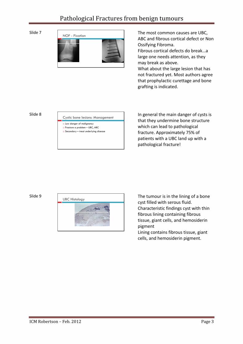
Slide 7 NOF - Fixation
The most common causes are UBC, ABC and fibrous cortical defect or Non Ossifying Fibroma. Fibrous cortical defects do break…a large one needs attention, as they may break as above. What about the large lesion that has not fractured yet. Most authors agree that prophylactic curettage and bone grafting is indicated.
Slide 8 Cystic bone lesions: Management
Low danger of malignancy
Fracture a problem – UBC, ABC
Secondary – treat underlying disease
In general the main danger of cysts is that they undermine bone structure which can lead to pathological fracture. Approximately 75% of patients with a UBC land up with a pathological fracture!
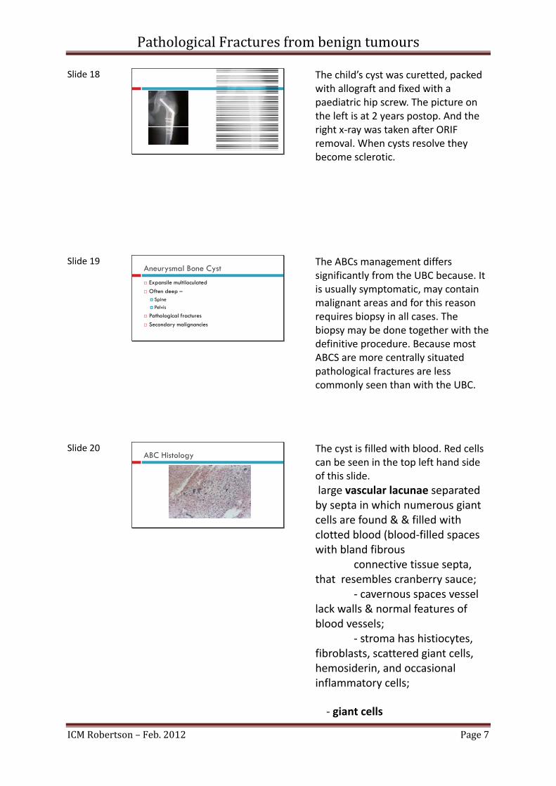
Slide 9 UBC Histology
The tumour is in the lining of a bone cyst filled with serous fluid. Characteristic findings cyst with thin fibrous lining containing fibrous tissue, giant cells, and hemosiderin pigment Lining contains fibrous tissue, giant cells, and hemosiderin pigment.
PathologicalFracturesfrombenigntumours
ICMRobertson–Feb.2012 Page4
Slide 10 Simple Bone Cyst
The usual way a UBC presents. 70% already have pathological fractures at presentation. The rest usually are symptomatic because the cyst is developing a stress fracture. Many other smaller cysts probably go undiagnosed as they never cause symptoms.
Slide 11 UBC – Classical Treatment
Open curettage
Pack with bone graft
Open methods are the gold standard, but are probably an overkill. As you will see there are less invasive methods to deal with this tumour. No sclerosing agents need be used as the danger of recurrence is small and outweighs the risk these agents pose.
Slide 12
* Scaglietti O. JBJS, 1979:61-B 200-4
UBC -Non invasive management
Methylprednisolone * 90% regress
Does not always prevent path fracture
Alcohol (Ethibloc Injection)
Minimally invasive grafting
The preferred method. Scaglietti showed that introducing methylprednisolone (40 to 80 mg) into a cyst via 2 needles (one for vent, the other for introduction) Produced 90% favorable results. Others (Capanna) still got some complications such as path fracture (7 of 90) and growth disturbances. Ethibloc – an alcohol preparation gives similar results. Confirm the cyst is one chambered using a radio opaque dye and there should also be serous fluid and not blood on aspiration. Small window made in the cyst and contents curetted and bone graft using a funnel.

PathologicalFracturesfrombenigntumours
ICMRobertson–Feb.2012 Page5
Slide 13
* Scaglietti O. JBJS, 1979:61-B 200-4
UBC -Non invasive treatment
This picture shows the technique of percutaneous injection of a cyst. Two needles are used and one is attached to a syringe whereby 40 to 80 mg prednisolone is injected.
Slide 14
* Scaglietti 0, JBJS 1979; 61-B:200-4 @ Capanna RDM. Orthop 1982; 161:204-11
UBC – Pathological Fracture
70% incidence
Reduction and plaster cast Only 10% resolve completely *
Intramedullary rods @
70% present with the cyst already fractured. Most can be treated like an uncomplicated fracture and require Plaster casts rather than ORIF. Cysts about the hip are the exception. Capanna has described using flexible intramedullary rods. These both decompress the cyst and provide stability.
Slide 15
Dormans JP; AAOS Instructional Course Lectures 2002; 457 - 67
Proximal Femur
Dormans classification of cysts about the hip – can be applied to any cystic lesion about the hip. All need ORIF. Type 1 is not in the femoral neck. Type 2 are in the femoral neck. Subdivision B means there is not enough bone stock on the lateral side and a sliding screw is needed in Type IB but in type 2 there is no purchase for the device as the cyst abuts the growth plate. In all type 2 cysts smooth K Wires are used crossing the growth plate. A Spica cast is used postoperatively in Type 2 cysts. Type 3 are cysts in mature bone and there is no problem with growth plates and a solid purchase is achieved with the screw (Type III – A) or sliding screw (Type III‐B).

PathologicalFracturesfrombenigntumours
ICMRobertson–Feb.2012 Page6
Slide 16
Type I-B
This cyst involves the lateral buttress, but there is a reasonable buttress between the growth plate and the cyst medially. It is therefore a Dormans I‐B and will qualify for a paediatric sliding hip screw and plate. This should extend to just lateral to the growth plate.
Slide 17
The cyst in the previous slide at operation. The cyst was curetted, packed with bone graft and a paediatric hip screw was used to fix the fracture.
PathologicalFracturesfrombenigntumours
ICMRobertson–Feb.2012 Page7
Slide 18
The child’s cyst was curetted, packed with allograft and fixed with a paediatric hip screw. The picture on the left is at 2 years postop. And the right x‐ray was taken after ORIF removal. When cysts resolve they become sclerotic.
Slide 19 Aneurysmal Bone Cyst
Expansile multiloculated
Often deep – Spine
Pelvis
Pathological fractures
Secondary malignancies
The ABCs management differs significantly from the UBC because. It is usually symptomatic, may contain malignant areas and for this reason requires biopsy in all cases. The biopsy may be done together with the definitive procedure. Because most ABCS are more centrally situated pathological fractures are less commonly seen than with the UBC.
Slide 20 ABC Histology
The cyst is filled with blood. Red cells can be seen in the top left hand side of this slide.
large vascular lacunae separated by septa in which numerous giant cells are found & & filled with clotted blood (blood‐filled spaces with bland fibrous connective tissue septa, that resembles cranberry sauce; ‐ cavernous spaces vessel lack walls & normal features of blood vessels; ‐ stroma has histiocytes, fibroblasts, scattered giant cells, hemosiderin, and occasional inflammatory cells;
‐ giant cells

PathologicalFracturesfrombenigntumours
ICMRobertson–Feb.2012 Page8
‐ ABC may be difficult to distinguish from that of GCT of bone; ‐ large amounts of hemosiderin;
Slide 21 ABC X Ray Features
In the fibula the ABC may have atypical very sclerotic edges. The MRI on the right shows it’s lobulated and expansile nature.
Slide 22
Fluid filled spaces are a characteristic MRI finding.
PathologicalFracturesfrombenigntumours
ICMRobertson–Feb.2012 Page9
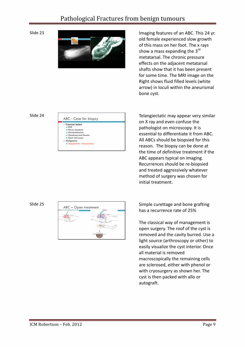
Slide 23
Imaging features of an ABC. This 24 yr. old female experienced slow growth of this mass on her foot. The x rays show a mass expanding the 3rd metatarsal. The chronic pressure effects on the adjacent metatarsal shafts show that it has been present for some time. The MRI image on the Right shows fluid filled levels (white arrow) in loculi within the aneurismal bone cyst.
Slide 24 ABC - Case for biopsy Precursor lesions
NOF Fibrous dysplasia Chondroblastoma Chondromyxiod fibroma Giant Cell tumour
Malignancy Telangiectatic osteosarcoma
Telangiectatic may appear very similar on X ray and even confuse the pathologist on microscopy. It is essential to differentiate it from ABC. All ABCs should be biopsied for this reason. The biopsy can be done at the time of definitive treatment if the ABC appears typical on imaging. Recurrences should be re‐biopsied and treated aggressively whatever method of surgery was chosen for initial treatment.
Slide 25 ABC – Open treatment
Simple curettage and bone grafting has a recurrence rate of 25% The classical way of management is open surgery. The roof of the cyst is removed and the cavity burred. Use a light source (arthroscopy or other) to easily visualize the cyst interior. Once all material is removed macroscopically the remaining cells are sclerosed, either with phenol or with cryosurgery as shown her. The cyst is then packed with allo or autograft.
PathologicalFracturesfrombenigntumours
ICMRobertson–Feb.2012 Page10
Slide 26
* Rastogi S., JBJS Br, 2006. 88-B(9): P. 1212-1216
ABC - Percutaneous methods
Methylprednisolone – fails
Polidocanol injection *
Ethibloc
Calcitonin
Selective Arterial Embolisation Avoid near spine
Methylprednisolone fails here. Make sure that you are dealing not dealing with an ABC when you use this method on what you think is a UBC. Polidocanol injected percutaneously has been shown by Rastogi to be effective (2 recurrences / 72). As in the case of UBC the cyst gradually becomes sclerotic and does not disappear completely. Success is measured by sclerosis and resolution of pain. Multiple injections over several months may be necessary.
Slide 27
Rastogi S. JBJS -Br, 2006. 88-B: p. 1212: Gibbs C., JBJS 1999 81-A, p. 1671
ABC Results
Polidocanol – 2 recurrences / 72 – Rasclogi
Traditional Curette & Graft 90% Gibbs
As can be seen the open and closed methods compare well. The Superficial ABC can be treated openly. In deep situations consider using a per‐cutaneous method.
Slide 28
Slide on the right shows an MRI of ABC of the sacrum in a 14 yr. old boy. The cyst was curetted and packed with bone. The X Ray R is 3 yr. later – the cyst is resolving and becoming sclerotic. Note that there are still some lytic areas present. His pain had resolved completely. The message is – even successfully treated cysts never resolve completely.
PathologicalFracturesfrombenigntumours
ICMRobertson–Feb.2012 Page11
Slide 29
Acetabular ABC – this need bone graft and curettage and successfully resolved. Despite the advice to treat deep ABCs with Percutaneous methods, at this site bony support for a possible future hip replacement is important, and can only be addressed by bone grafting the lesion.
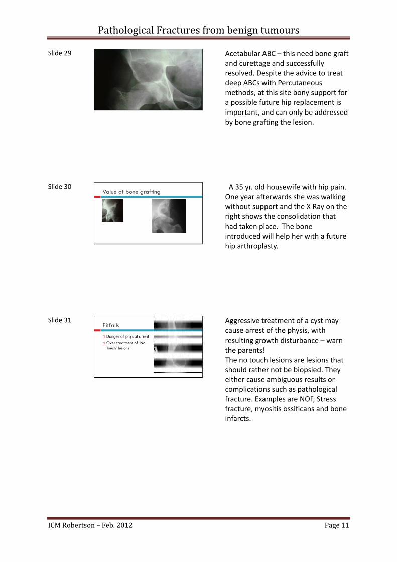
Slide 30 Value of bone grafting
A 35 yr. old housewife with hip pain. One year afterwards she was walking without support and the X Ray on the right shows the consolidation that had taken place. The bone introduced will help her with a future hip arthroplasty.
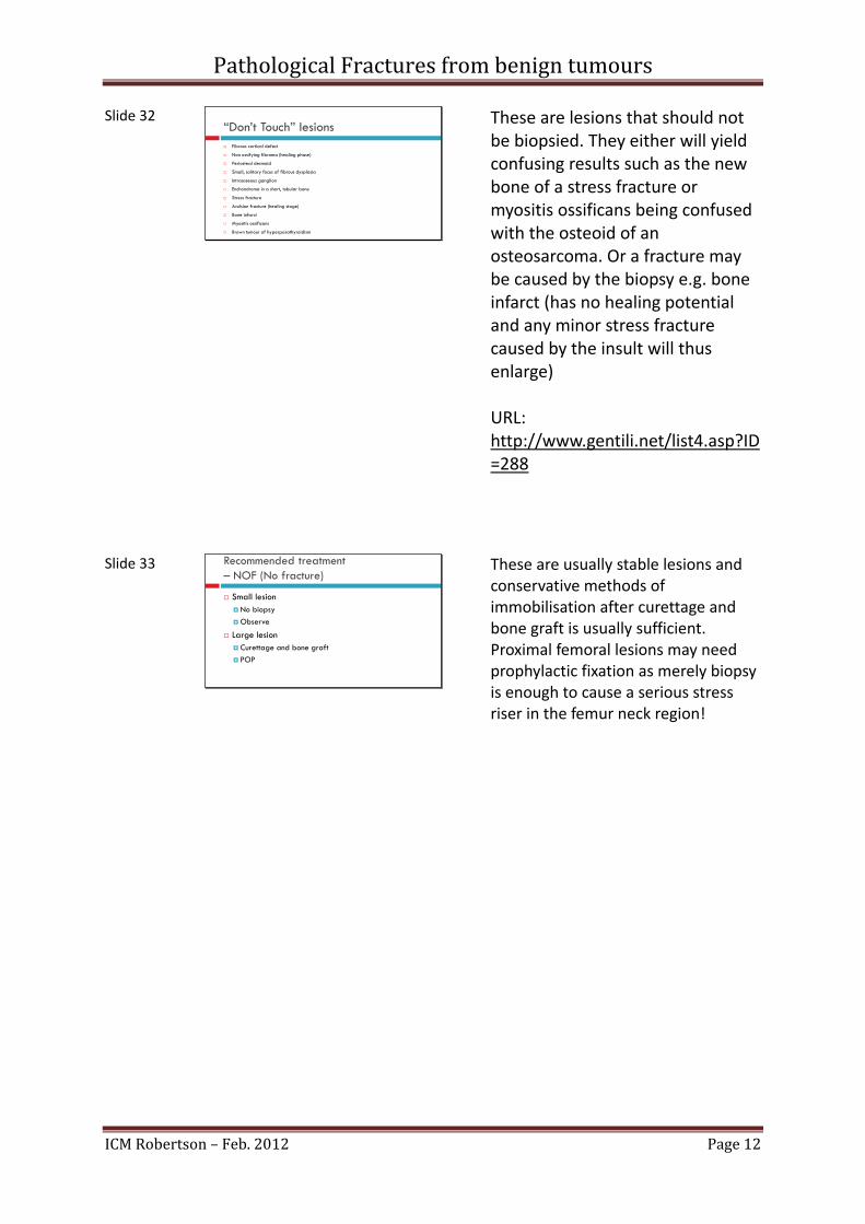
Slide 31 Pitfalls
Danger of physial arrest
Over treatment of ‘No Touch’ lesions
Aggressive treatment of a cyst may cause arrest of the physis, with resulting growth disturbance – warn the parents! The no touch lesions are lesions that should rather not be biopsied. They either cause ambiguous results or complications such as pathological fracture. Examples are NOF, Stress fracture, myositis ossificans and bone infarcts.
PathologicalFracturesfrombenigntumours
ICMRobertson–Feb.2012 Page12
Slide 32 “Don’t Touch” lesions Fibrous cortical defect
Non ossifying fibroma (healing phase)
Periosteal desmoid
Small, solitary focus of fibrous dysplasia
Intraosseous ganglion
Enchondroma in a short, tubular bone
Stress fracture
Avulsion fracture (healing stage)
Bone infarct
Myositis ossificans
Brown tumour of hyperparathyroidism
These are lesions that should notbe biopsied. They either will yield confusing results such as the new bone of a stress fracture or myositis ossificans being confused with the osteoid of an osteosarcoma. Or a fracture may be caused by the biopsy e.g. bone infarct (has no healing potential and any minor stress fracture caused by the insult will thus enlarge) URL: http://www.gentili.net/list4.asp?ID=288
Slide 33 Recommended treatment – NOF (No fracture)
Small lesion No biopsy
Observe
Large lesion Curettage and bone graft
POP
These are usually stable lesions and conservative methods of immobilisation after curettage and bone graft is usually sufficient. Proximal femoral lesions may need prophylactic fixation as merely biopsy is enough to cause a serious stress riser in the femur neck region!

PathologicalFracturesfrombenigntumours
ICMRobertson–Feb.2012 Page13
Slide 34 UBC: Recommended management
Pre-fracture: Rest Allow healing
Treat cyst Percutaneous methods
Open bone graft
Pre‐fracture cysts are large and have become painful due to mico‐fractures. Rest will allow the cyst to begin healing. The definitive treatment is probably best done with a non‐invasive method. In the case of a large cyst that may later fracture open methods that include bone grafting arte preferred.
Slide 35 UBC: Recommended Management
Displaced fracture: Non Hip Conservative
Proximal femur ORIF and bone graft
Pre‐fracture cysts are large and have become painful due to mico‐fractures.
Slide 36 ABC: Preferred Treatment
Confirm diagnosis Open biopsy
Phenol – if aggressive
ORIF if unstable (Prox. femur)
Biopsy is mandatory due to the possibility of malignancy (either misdiagnosis or some portions of the tumour having changed to a higher grade). This may be done with the definitive procedure if MRI findings are typical. Phenol is reserved to aggressive or recurrent ABCs.

PathologicalFracturesfrombenigntumours
ICMRobertson–Feb.2012 Page14
Slide 37 References:
Fractures through bone cysts: Dormans, AAOS Instructional Course Lectures, Vol 51, 2002
Cyst injection: Scaglietti O. JBJS, 1979:61-B 200-4 Polidaconol Injection: Rastogi S., JBJS Br, 2006. 88-
B(9): P. 1212-1216 Traditional curettage and bone graft: Gibbs C., JBJS
1999 81-A, p. 1671
In Summary: In children, pathological fractures caused by tumours are common. These fractures are usually through benign bone lesions. The NOF is common but only large lesions warrant prophylactic management. The simple bone cyst has a high fracture rate. Pre‐fracture cases may be treated by non‐invasive methods such as steroid injection. Once fracture has occurred, treat it as you would manage a non‐pathological fracture except, in the proximal femur, which requires ORIF. A residual or very large cyst may require traditional curettage and open bone grafting. The ABC is usually more central and is a less common cause of pathological fracture. The ABC does not respond well to steroid injection. Biopsy is needed and open curettage with adjuvant (cryosurgery or phenol) + bone graft.