pathologic effects of suramin, hetrazan and …hist.library.paho.org/spanish/bol/v28n11p1107.pdf ·...
TRANSCRIPT

PATHOLOGIC EFFECTS OF SURAMIN, HETRAZAN AND ARSENAMIDE ON ADULT ONCHOCERCA VOLV ULUX*
ByDrs.L.L. ASHBURN,THOMAS A. BTJRCH andF.J. BRADY
National Institutes of Heallh, Bethesda, Md.
Great strides have been made within recent years in the treatment of filariasis. Onchocerciasis is the only filarid infection of man in which adult worms may be consistently obtained by biopsy. In view of this, the experimental therapy of onchocerciasis is of great value because the effects of drugs on adults may be determined. In other human filarid infections the effects of antimonials (1, 2) and hetrazan (3) on adult worms have been discussed on the basis of indirect evidente.
The effect of drugs on adult filarids is of importance in determining whether or not a drug is permanently effective in eliminating micro- filariae. If the compound is effective only against microfilariae, then their recurrence may be expected.
Evidente has accumulated indicating that certain antimony com- pounds not only are microfilaricidal but have a sterilizing effect on adult LXroJiEarz’a and Onchocerca. Hetrazan is microfilaricidal in Wuchereria (3), Onchocerca (4, 5) and Dirojilaria (6) infections. The arsenical prep- aration, arsenamide, is microfilaricidal in Wuchereria infections (7) and is effective against the adult Dirofilaria (8).
Suramin (Bayer 205) has been used successfully both against the microfilariae and adult Onchoeerca. This was reported in a preliminary note by van Hoof, Henrard, Pee1 and Wanson (9). Burch (5) has treated 56 patients with this drug and found it to be very effective in causing disappearance of microfilariae.
The present study was designed to determine histologically the effects of suramin and hetrazan on the adults of Onchocerca volvulus.
EXPERIMENTAL PROCEDVRE
Nodules containing adult Onchocerca were removed from patients during the study previously reported by Burch. In addition, ll nodules removed by Dr. Luis Mazzotti from 3 patients treated with arsenamide and sent to us by Dr. Gilbert Otto are included in this study.
In this analysis, only those nodules are included which were removed from patients considered to have been adequately treated. Adequate treatment** in the case of suramin is considered to be a mínimum total
* Th% work waa done BS a collsborative project of the National Institutes of Health and the Pan Ameritan Sanitary Bureau.
** Adequate treatment as deiined above is not intended as a recommendation as to proper dosage of suramin and hetrazan in the treatment of onchocerciasis. The minimum dose indicated as adequate is only for the purpose of this analysis. In the case of hetrazan, the figure given may be too low.
1107

1108 BULLETIN OF THE PAN AMERICAN SANITARY BUREAU
dose of 0.14 gm./kg. and similarly with hetrazan a minimum total dose of 20 mg./kg. The 3 patients treated with arsenamide received this drug in 3 CC. doses (2Qj, solution) daily for 15 days.
Using these criteria of adequacy of treatment, microscopic examina- tion was made of 34 nodules from 21 patients receiving suramin and of 16 nodules from 9 patients treated with hetrazan. Nineteen nodules from untreated patients were used as controls for the study.
Nodules were fixed in 10% formalin and later blocked in such a man- ner that sections were made through what appeared the greatest concen- tration of adult worms. The tissue was embedded in parafin, sectioned, and stained by azure eosin and by the Van Gieson or Masson tech- niques. Frozen sections of some nodules were stained with oil red 0 for the demonstration of fat.
RESULTS
Histopathology of Control Modules.-The Onchocerca nodules were basically connective tissue masses, largely collagenous, infiltrated by variable numbers of inflammatory cells and showed from few to mod- erate numbers of fixed tissue cells. Throughout the masses sections of adult worms occurred, either widely separated or compactly grouped, depending on the number of worms present and the size of the nodule.
The fibrous tissue was usually quite dense at the periphery where it formed a fairly well delineated layer of variable thickness which is com- monly referred to as a capsule. Some nodules (5 of 19) were densely fibrous throughout and in such cases there was no part that could be correctly considered as a capsule. Fibrous tissue in the central portion of the nodules usually was of only slight to moderate density and was roughly inversely correlated with the degree of inflammatory infiltra- tion.
Fibroblasts generally were present in small numbers. In only one of the nodules was fibroblast proliferation particularly prominent . Large oval to round mononuclear cells with solid or foamy cytoplasm were seen in most nodules. When present in moderate numbers, they tended to be concentrated in patches in the stroma or about the worms. Fat stains done on a few nodules showed much neutral fat both in the large mononuclears and in the spindle shaped stroma cells. In an occasional nodule a sheath of irregularly oriented oval to spindle-shaped histio- cytes occurred about one or very few worm segments. This sheathing was seen only around worms showing some degenerative change. A single clear cut granuloma of foreign body type was found. An occasional giant cell of the foreign body type was seen in a little more than half oî the nodules.
Cellular infiltration of different nodules varied markedly both as to degree and ce11 type. In two, the tissue was densely infiltrated. It was of moderate degree in 5, slight in 8 and very slight in 4. In those cases

November 19431 ONCHOCERCIASIS 1109
showing only slight infiltration, the cells usually occurred in foci or clustered about the adult worms. The inhltrating cells were lympho- cytes, plasma cells, eosinophils and polymorphonuclear leucocytes. Tak- ing the nodules as a group, no one of these cell types could be considered as predominant but in ditIerent nodules and in different areas of the same nodule, any one of them may form the bulk of the inílltrate. Of the lym- phoid cells, lymphocytes were seen in a greater number of nodules, but in some foci plasma cells were abundant. Eosinophils were the most variable ce11 as to frequency of appearance. They were not seen in five nodules but were numberous in two. They usually showed a patchy dis- tribution but there was no tendency for them to be concentrated in the vicinity of adult worms or about microfilaria present in the stroma. Polymorphonuclear leucocytes generally were present in quite small numbers and were most often seen in areas of tissue softening or necrosis around segments of worms. Such areas of necrosis, usually quite small, were seen in al1 nodules. Medium sized areas of necrosis, tissue collapse, fluid and polymorphonuclear leucocytic exudation were seen in 6 of the 19 nodules.
Histology of Adult Onchocerca in Nodules from Treated and Un- treated Patients.-Because of the entwined position of female Oncho- cerca in the nodules, a single section usually passed through various levels of the worms. By study of multiple nodules from untreated pa- tients, it was easily possible to follow the reproductive sequence from ovary to the fully developed microfilariae. It appears that morpholog- ically the sequence of differentiation is similar to that described and illustrated for DiroJiEaria immitis (10). In view of the fact that the ovary is restricted to a relatively short segment of the worm, it was seen infre- quently.
The majority of female Onchocerca in the 19 control nodules from untreated patients showed the normal developmental stages and numer- ous microfilariae in their uteri. Occasionally one or two ova (in uterus) or multicell stages showed karyorrhexis or frank necrosis. There were a few worms which showed defmite abnormalities. These changes varied from necrosis of uterine contents of variable extent to death of the worm.
In an effort to grade the worms of both control and “treated nodules” and to arrange them in order from normal to severe damage, the females of each nodule were classified as follows:
Croup 1. The worms placed in this category showed developmental forms in the proper uterine leve1 and morphologically differentiated microfilariae in the anterior portion of the uteri. Some worms showed slight variation from normal in the tinctorial quality of the cytoplasm of the cells of the developing and dif- ferentiated microfilariae. Since the signifìcance of this minar change was not clear, these worms were considered essentially normal and placed in this group.
Grozcp II. The worms placed in this group usually showed both develop- mental stages and dierentiated microíilariae. However, when present they
1110 BULLETIN OF THE PAN AMERICAN SANITARY BUREAU
showed frank necrosis which in many instances was extensive. Worms which did not show microfilariae but in which a few multicell forms were present, were also included in this group.
Group III. In this group were placed those worm which showed complete suppression of microfilarial production and absence of developing stages. The uteri of the worms of this group, at most, eontained a few undeveloped and often degenerating ova.
Group IV. This group includes dead worms and a few nodules showing clear- cut foreign body granulomas which beyond doubt marked the site of recently disappeared worms.
The classification given above is similar to that used in a previous report (ll) in which the effect of treatment with trivalent antimony was evaluated. The only significant difference is that dead Onchocerca and nodules showing only foreign body granulomas are placed in group 4 whereas they were previously included in group 3. Of all groups, group 2 contains worms which showed most variation in severity of damage. This may possibly mask minor differences of therapeutic effec- tiveness between hetraaan and suramin. However, in view of the num- ber of variables in the study, such as dose of drug and interval between completion of treatment and removal of nodules, Subdivision of this group was not practical.
For reasons given earlier, ovaries were seen in a relatively small num- ber of worms. In these no clear-cut changes were found. Male worms also were recognized infrequently.
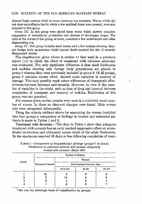
Using the criteria outlined above for separating the worms (nodules) into four groups a comparison of findings in treated and untreated pa- tients is made in Tables 1 and 2.
Treatment with Suramin.-The data in Table 1 show that adequate treatment with suramin has an early marked suppressive effect on micro- filaria1 production and ultimately causes death of the adult Onchocerca. In the specimens removed 60 days or less following completion of treat-
TABLE l.-Comparison of histopathologic Jindings (groupe*) in female Onchocerca in untreated patients and patients adequately
treated with suramin (Bayer 9006)
I Number of Nodules
Treated Group’
Untreated Controls Post-treatment fnterva14ays
60 o* less 90-120 121-323
1 13 0 0 0 2 2 14 0 0 3 3 1 6 1 4 1 2 3 7
* See text for histologic basis of classification by groups.

November 19.@] ONCHOCERCIASIS 1111
TABLE 2.-Comparison of histopathologic jindings (group*) in female Onchocerca in untreated patients and patients “adequately”
treated with hetrazan
Groups* // t
1 2
‘i 3 4
Number of Nodules
Treated
Untreated Controls Post-treatment interval-days
60 or les 61-90
13 8 2 2 1 2 3 0 3 1 0 0
t * Same as table 1.
ment, there were no normal worms. The majority (group 2) shomed either necrosis of variable extent of the uterine contents or uteri con- taining only normal OP degenerating ova. In 90 to 120 days the changes were more strilting. The worms of 3 nodules were dead (group 4) and 6 showed complete suppression of reproduction (group 3). After an
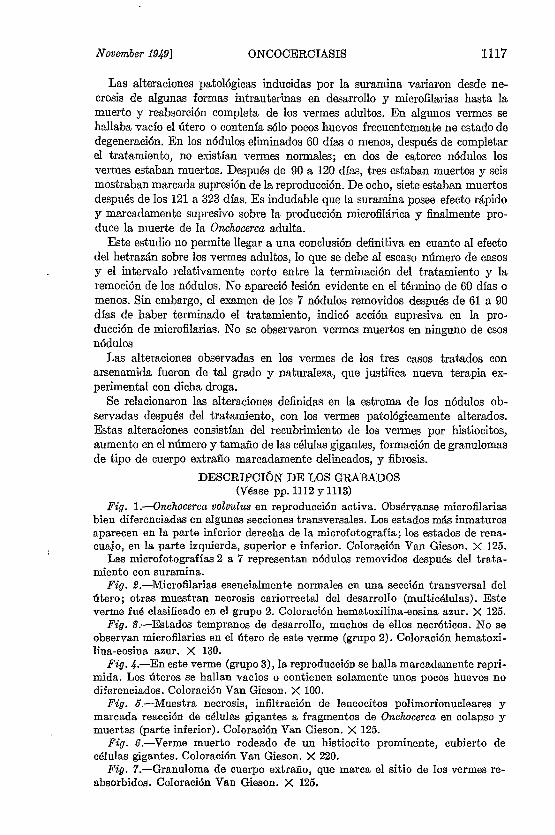
c interval of 121 to 323 days, 7 of 8 nodules showed only dead worms or foreign body granulomas. Photomicrographs illustrating normal Oncho- cerca and changes that occurred following treatment are shown in Figures 1 to 7.
Treatment with Hetrazan-Table 2 shows the grouping of female Onchocerca from nodules after “adequate” treatment with hetrazan. Nodules removed within 60 days of completion of treatment showed no effect either on microfilarial production or on the adult worms. The worms of 8 of 9 nodules mere normal. After 61 to 90 days some effect of treatment became evident. The worms of 2 nodules were normal, 2 showed minimal degenerative changes in uterine contents and 3 showed complete suppression of reproduction.
Treatment with Arsenamide.-The 11 nodules from the 3 patients
i treated with arsenamide showed only one with normal adult worms. In 9, the worms showed complete suppression of reproduction (group 3) and in one, the Onchocerca uteri contained a few ova but no multicell stages or microfilariae. None of the worms appeared dead.
Histological Appearance of Nodules Following Trealment.-The changes that occurred in nodules following treatment appeared to be re- lated to pathologic alteration of the Onchocerca and not to the specific drug. Hence nodules from al1 patients treated with the three drugs employed are considered together in this section. As stated earlier in this report, nodules from untreated patients shom considerable variation as to de- gree and type of cellular infiltration, necrosis, and amount of fibrous tissue. In viem of this fact, only very general statements can be made as to the influente of treatment on these features of the nodules. Aver-

1112 BULLETIN OF THE PAN AMERICAN SANITARY BUREAU
I FIG. l.---Normal Onchocerca volv~~.l~ts showing active reproduction. Weil dif- ferentiated microfilariae are seen in a number of cross sect’ions. The most immature st’ages in this photomicrograph are at lower right. Tadpole stages are seen at upper and lower left. Van Gieson stain. @X 125. Photomicrographs 2 t.o 7 are of nodules removed from patients following treatment with suramin.
FIG. 2.-Essentially normal microfilaria are seen in one cross scction of uterus; others show karyorrhectic necrosis of developing (multicell) was classified as group 2. Hematoxylin azure eosin stain. X 125.
stages. This worm
FIG. 3.-Shows only early developing stages, many of which are necrotic. Microfilariae were not seen in the uteri of this worm (group 2). Hematoxylin azure eosin stain. X 130.

November 19@] ONCHOCERCIASIS 1113
FIG. 4.-In this worm (group 3), reproduction 1s markedlr suppressed. Uteri are empty or contain only a few non-differentiating ova. Van Gieson stain. x 100.
FIG. 5.-Shows necrosis, polymorphonuclear leucocyte infiltration and marked giant ce11 reaction to fragments of collapsed and dead Onchocerca aipearing near bottom. Van Gieson stain. X 125.
FIG. 6.-Dead worm surrounded by a prominent histiocyte and giant cell sheath. Van Gieson stain. X 220.
FIG. 7.-Foreign body granulomas marking site of resorbed worms. Van Gieson stain. X 125.

1114 MJLT,ETIX OF TIIE PAh- AMERICAN SANITARY BUREAU
age total cellulsr infiltration was not apparently altered by treatment but in many nodules of group 4, especially those showing fem or no worms, there were relat’ively fewer inflammatory cells present, and among these, lymphocytes predominated. Eosinophils mere seen less fre- quently. In nodules of group 2, which includes worms showing acute degenerative changes, large mononuclear cells appeared to be increased in number. Areas of necrosis, usually with associated polymorphonuclear leucocytic exudation, showed the same variation in size noted in con- trolsand showed nodefinite increased frequency that could be appreciated by histologic examination. In nodules of group 4, fibrous tissue was definitely increased in the main central portion and blended with the commonly densely fibrous L’capsule.”
Three features of certain nodules from treated patients contrasted sharply with nodules from untreated patients. These mere sheathing of worms by oval or spindle-shaped histiocytes, frequency of giant cells of foreign body type and the presente of few to many foreign body granulomas. The histiocyte sheaths were of variable thickness and stood out in Sharp contrast against the surrounding fibrous tissue in those nodules showing relatively little cellular infiltration. Multinucleate giant cells, often very large and of irregular shape, were numerous in many nodules. They generally were seen adjacent to worms intermingled with and forming part of the sheaths mentioned. They also were seen as part of foreign body granulomas, some of which were formed largely of these cells. These granulomas were seen in a few nodules of group 2, in a greater number of nodules of group 3, and were present in practically al1 nodules of group 4. Giant cells and histiocyte sheathing also increased in frequency and prominente in nodules classified from group 2 to 4.
Microfilariae are found in the stroma of most nodules removed from untreated patients and appear to be related to the reproductive activity of the adult. In this connection the following data from all treated (whether adequate or not) patients is cited. Microfilariae were not found in any nodules classified in groups 3 and 4. They were present in al1 16 classified as group 1 and in approximately half of the 29 classified as group 2.
DISCUSSION Y The goal of treatment of onchocerciasis is the killing of the disscmi-
nated microfilariae as me11 as the adult worms in the nodules. Van Hoof et al. (9) in a preliminary note reported that Bayer 205 (suramin) caused the disappearance of microfilariae from the tissues and that dead and flabby adults were found in the nodules. Burch (5) in a series of 56 cases also found the suramin to be an effective microfilaricide and the present report, which is a part of the same study, gives histopathologic evidente as to the drug’s lethal effect on the adult worms. Definite injury of the adults can be seen in those nodules removed within 60 days following

November 19491 ONCHOCERCIASI~ 1115
completion of treatment. However, incontrovertible evidente of the drug’s lethal effect was obtained only from those nodules removed after a 121 to 323 day interval; most were nearer the latter period. Good evidente of the drug’s potential lethal effect was seen in nodules re- moved 114 days following completion of treatment. The worms of many such nodules showed changes that were considered to be irreversible. Distortion, collapse, and fragmentation were not infrequent and oc- casionally portions of worms were dead. The histocyte sheathing and giant cell reaction is interpreted as indirect evidente of damage to the adult Onchocerca.
This histologic study does not permit a definite conclusion as to the effect of hetrazan on the adult worms. This is true since the number of “adequately” treated cases is small and since all nodules were re- moved 90 days or less following completion of therapy. The 7 nodules studied after a 61 to 90 day interval indicate a suppressive action on microfilarial production but no changes were seen in the adults which would suggest that death was likely to occur. Examination of nodules after a longer post treatment period will be necessary before a definite conclusion can be drawn as to the effect of hetrazan. However, suramin produced a definite suppression of reproduction within 60 days after treatment whereas a similar but less pronounced effect was evident with hetrazan only after a longer period (61-90 days).
The patients treated with arsenamide are too few to permit any con- conlusion. However, the changes seen in the worms from these cases were such as to suggest that the drug is Worth further trial.
%MMARY AND CONCLUSIONS
Histopathologic examination mas made of 34 onchocercotic nodules from 21 patients who had been treated with a minimum of 0.14 gm./kg. (total dose) of suramin, of 16 nodules from 9 cases treated with a mini- mum of 20 mg./kg. (total dose) of hetrazan and of ll nodules from 3 cases treated with 3 CC. of arsenamide daily for 15 days. The nodules were removed after intervals of 29 to 323 days following completion of treatment. From this study it was evident that suramin had a lethal effect on adult Onchoc&ca which was demonstrable in nodules removed after 121 days. In most nodules removed after shorter intervals, there usually were considerable pathologic alterations of the uterine contents.
A definite conclusion as to the effect of hetrazan on adult worms can- not be made from this study. This drug caused some suppression of reproduction but its action in this respect was slower than that of sura- min.
The changes seen in the worms of the 3 patients treated with arsena- mide was of such nature and degree as to suggest that further experi- mental trial of this drug ís indicated.

1116 : BULLETIN OF THE PAN AMERICAN SANITARY BUREAU
BIBLIOGRAPHY (1) Brown, H. W. : The treatment of fllariasis (Wuchereria buncroj~i). J. A. M. A.
1%: 952-958 (1944). (2) Cubertson, James T.; Rose, Harry M.; and Oliver-Gonzalez, J.: Chemo-
therapy of human filariasis by the administration of neostibosan. Am. J. Trop. Med., &6: 271-274 (1945).
(3) Santiago-Stevenson, D.; Oliver-Gonzalez, J.; and Hewitt, R. 1.: Treatment of filariasis bancrofti with 1-diethylcarbamyl+methylpiperazine hydrochloride (“Hetrazan”). J. A. M. A., 136: 708-712 (1947). 4 (4) Mazzotti, Luis, and Hewitt, R.: Tratamiento de la oncocercosis por el cloruro de 1-dietilcarbamil4-metilpiperazina (Hetrazan). Med. Rev. MCx., 28: 548, 3942 (1948).
(5) Burch. T. A.: Experimental therapy of onchocerciasis with suramin and hetrazan. Bol. Of. San. Pan. 28: 233-248 (1949).
(6) Hewitt, R. 1.; Kushner, S.; Stewart, I-1. W.; White, E.; Wallace, W. S.; and Subba Row, Y.: Experimental chemotherapy of filariasis. III. Effect of l- diethylcarbamyl-4-methylpiperazine hydrochloride against naturally acquired filaria1 infections in cotton rats and dogs. Jour. Lab. 85 Clin. Med. 32: 1314-1329 (1947).
(7) Thctford, N. D.; Otto, G. F.; Brown, H. W.; and Maren, T. H. : The use of phenyl arsenoxide in the treatment of Wuchereria buncrofti infection. Am. Jour. Trop. Med., 68: 577-583, (1948).
(8) Otto, G. F.; and Maren, T. H.: Filaricidal activity of substituted phenyl arsenoxides. Science, 106: 105-107, (1947).
\I (9) van Hoof, L. ; Henrard, C. ; Peel, E. ; and Wanson, M. : Sur la chimiotherapie de l’onchoccrcose. Annales’Soc. Belge. de Med. Trop., 27: l, l-5 (1947).
(10) Ashburn, L. L.; Perrin, T. L.; Brady, F. J.; and Lawton, A. H.: Histologic changes in ovary and uterus of live Dirojilaria immitis recovered from dogs treated with trivalent antimony compounds. Arch. Path., 40: 334-339 (1945). 1 (11) Bartter, F. C.; Burch, T. A.; Cowie, D. B.; Ashburn, L. L.; and Brady,
F. J. : Experimental therapy of Onchocerciasis with trivalent antimonials. Annals of the N. Y. Atad. of Sci., W: S9-96 (1948).
EFECTOS PATOLÓGICOS DE LA SURAMINA, EL HETRAZAN Y LA ARSENAMIDA S0BR.E LA OiVCHOCERCA VOLVULUX
ADULTA (Sumario)
En el tratamiento de las infecciones filáricas es importante conocer el efecto de la droga sobre los vermes adultos. Para ser completamente efectiva ésta, debe exterminar los vermes adultos o esterilizar permanentemente a las hem- bras. La oncocerciasis es la Ilnica filariasis del hombre en la cual pueden ob- tenerse consistentemente vermes adultos por medio de la biopsia.
Este estudio se realizó con el fin de determinar histológicamente el efecto de la suramina (Bayer 205) y del hetrazán sobre los vermes adultos de Onchocerca volvulus. En un estudio previamente publicado por Burch (véase Boletin, mzo. 1949) se extirparon núdulos conteniendo vermes adultos, en enfermos vo- luntarios residentes en la zona endémica de Guatemala. Los nódulos fueron extirpados de 29 a 323 días después de completar el tratamiento. En este anná- lisis se tomaron en cuenta solamente aquellos enfermos que habían recibido un mfnimo de 0.14 gm/kg (dosis total) de suramina, o 20 mg/kg (dosis total) de hetrazán. Por cortesía de los Dres. Mazzotti y Otto, se incluyen 3 casos tratados diariamente con 3 CC. de una solución de arsenamida al 2% durante 15 dfas. Sirvieron como testigos diecinueve nódulos de enfermos no tratados.
November 19.&] ONCOCERCLASIS 1117
Las aIteraciones patol6gicas inducidas por la suramina variaron desde ne- crosis de algunas formas intrauterinas en desarrollo y microfilarias hasta Ia muerte y reabsorción completa de los vermes adultos. En algunos vermes se hallaba vacío eI útero o contenfa ~610 pocos huevos frecuentemente ne estado de degeneración. En los nódu!os eliminados 60 días o menos, despu& de completar el tratamiento, no e,xistían vermes normales; en dos de catorce nódulos los vermes estaban muertos. Después de 90 a 120 días, tres estaban muertos y seis mostraban marcada supresión de la reproducción. De ocho, siete estaban muertos después de los 121 a 323 días, Es indudabIe que la suramina posee efecto rápido y marcadamente supresivo sobre la producción microfilárica y finalmente pro- duce la muerte de la Onchocwca adulta.
Este estudio no permite llegar a una conclusi6n definitiva en cuanto al efecto del het.razan sobre los vermes adultos, lo que se debe aI escaso numero de casos y el intervalo relativamente corto entre la terminación del tratamiento y la remoción de los nódulos. No apareció lesión evidente en el término de 60 dfas o menos. Sin embargo, el examen de los 7 nódulos removidos después de 61 a 90 dias de haber terminado el tratamiento, indicó acción supresiva en la pro- ducción de microfilarias. No se observaron vermes muertos en ninguno de esos nódulos
Las alteraciones observadas en los vermes de los tres casos tratados con arsenamida fueron de- tal grado y naturaleza, que justifica nueva terapia ex- perimental con dicha droga.
Se relacionaron las alteraciones definidas en la estroma de los nódulos ob- servadas despu& del tratamiento, con los vermes patológicamente alterados. Estas alteraciones consistían del recubrimiento de los vermes por histiocitos, aumento en el nCunero y tamaño de las células gigantes, formación de granulomas de tipo de cuerpo extraño marcadamente delineados, y fibrosis.
DESCRIPCIÓN DE LOS GRABADOS (Véase pp. 1112 y 1113)
Fig. l.-Onchocerca volvulus en reproducción activa. Obsérvanse microhlarias bien diferenciadas en algunas secciones transversales. Los estados más inmaturos aparecen en la parte inferior derecha de la microfotografía; los estados de rena- cuajo, en la parte izquierda, superior e inferior. Coloración Van Gieson. X 125.
Las microfotografías 2 a 7 representan nódulos removidos despues del trata- miento con suramina.
Fig. B.-Microfilarias esencialmente normales en una sección transversal del dtero; otras muestran necrosis cariorrectal del desarrollo (multicélulas). Este verme fu6 clasiftcado en el grupo 2. Coloración hematoxilina-eosina azur. X 125.
Fig. S.-Estados tempranos de desarrollo, muchos de ellos necrótioos. No se observan microfilarias en el útero de este verme (grupo 2). Coloración hematoxi- lina-eosina azur. X 130.
Fig. .&-En este verme (grupo 3), la reproduccidn se halla marcadamente repri- mida. Los úteros se hallan vacíos o contienen solamente unos pocos huevos no diferenciados. Coloración Van Gieson. X 100.
Fig. B.-Muestra necrosis, infiltración de leucocitos polimorfonucleares y marcada reacción de c6lulae gigantes a fragmentos de Onchocerca en colapso y muertas (parte inferior). Coloración Van Gieson. X 125.
Fig. B.-Verme muerto rodeado de un histiocito prominente, cubierto de células gigantes. Coloración Van Gieson. X 220.
Fig. 7.-Granuloma de cuerpo extraño, que marca el sitio de los vermes re- absorbidos. Coloración Van Gieson. X 125.