passive protective effect of chicken egg against ...iai.asm.org/content/60/3/998.full.pdfsulfate,...
TRANSCRIPT

INFEcrION AND IMMUNITY, Mar. 1992, p. 998-10070019-9567/92/030998-10$02.00/0Copyright © 1992, American Society for Microbiology
Passive Protective Effect of Chicken Egg Yolk Immunoglobulinsagainst Experimental Enterotoxigenic Eschenichia coli
Infection in Neonatal PigletsHIDEAKI YOKOYAMA,* ROBERT C. PERALTA, ROGER DIAZ, SADAKO SENDO,
YUTAKA IKEMORI, AND YOSHIKATSU KODAMA
Immunology Research Institute in Gifu, Minamiyama, Sano, Gifu City 501-11, Japan
Received 11 October 1991/Accepted 12 December 1991
Passive protection of neonatal piglets against fatal enteric colibacillosis was achieved with powderpreparations of specific antibodies against K88, K99, and 987P fimbrial adhesins of enterotoxigenic Escherichiacoil. The antibody powders were obtained by spray drying the water-soluble protein fraction of egg yolks fromimmunized hens after the lipid components were precipitated with an aqueous dispersion of acrylic resins(Eudragit L3OD-55; Rohm pharma). The anti-K88, -K99, and -987P antibody preparations reacted specificallyagainst the corresponding fimbrial antigens in an enzyme-linked immunosorbent assay. The orally adminis-tered antibodies protected in a dose-dependent fashion against infection with each of the three homologousstrains of E. coli in passive immunization trials with a colostrum-deprived piglet model of enterotoxigenic E.coli diarrhea. Scanning electron microscopy revealed adherence of enterotoxigenic E. coli in intestinal epithelialsurfaces of control piglets, whereas in treated piglets treated with high-titer antibodies, a resistance to bacterialadhesion was observed. An enzyme immunoassay with avidin-biotin complex demonstrated specific localantibody activity in target areas of the small intestines. In vitro, E. coli K88+, K99+, and 987P+ strainsadhered equally to porcine duodenal and ileal epithelial cells but failed to do so in the presence of homologousanti-fimbrial antibodies. Absorption of egg yolk antibodies with fimbrial immunosorbent removed theanti-fimbrial antibody fraction and reduced significantly the protective nature of the antibody preparation ina passive immunization experiment, suggesting that anti-fimbrial antibodies were the active components.
Diarrheal disease caused by enterotoxigenic Escherichiacoli (ETEC) is by far the most common enteric colibacillosisencountered in neonatal piglets (22). Previous investigationsindicated that colonization of the small intestine of the pigletby ETEC adhering to the epithelium accounts for mostgastrointestinal disorders (1, 4, 5, 26). The fimbrial K88,K99, and 987P antigens of porcine ETEC that are associatedwith intestinal colonization have been extensively investi-gated with respect to their genetic background, proteinchemistry, and immunological properties (7, 16, 21). Theyhave been widely employed with promising results as vac-cine antigens in controlling porcine colibacillosis. In passiveimmunization experiments, antibodies raised against thesefimbrial antigens have been administered orally to pigletsand have offered potential therapeutic value in controllingthe disease.
Oral administration of antibodies derived from serum andcolostrum and even with monoclonal antibodies has beenvery successful; however, it is prohibitively expensive toobtain the large amounts of antibodies required (18). Ofparticular veterinary interest is the use of chicken egg yolkantibodies for the treatment of porcine colibacillosis. Vacci-nation of laying hens provides a cheaper and good alterna-tive antibody source; the eggs are collected after a high levelof antibodies is reached in the egg yolk. This principle is notnew; some authors have used chicken egg yolk antibodies inthe prevention or control of rotaviral infection in mice andcats (3, 11), and their promising results have led to thesuggestion that egg preparations might serve as a source ofantiviral antibodies for humans (28). There has been no
* Corresponding author.
report so far on the use of chicken egg yolk antibodies in theprevention and control of enteric colibacillosis in swine,although the use of egg yolk antibodies in the treatment andprevention of diseases in other animals and even humans hasbeen described. To our knowledge, the present study de-scribes the first clinical use of spray-dried chicken egg yolkantibodies against ETEC infection in piglets.A number of methods for extraction of antibodies from egg
yolk have been described (2, 6, 10, 14, 23, 27). Traditionally,organic solvents were used to extract antibodies from eggyolk, but because bioactive proteins are denatured withthese methods and because of the health hazards of solventslike chloroform used during production, alternative methodshave been sought. Polson et al. (23) and Jensenius et al. (14)successfully used polyethylene glycol and sodium dextransulfate, respectively, as protein precipitants in the isolationof pure immunoglobulin fractions from egg yolks, but theirmethods, like others, were time-consuming and the agentsused have not been approved as food additives. We triedusing an aqueous dispersion of acrylic resins to isolate thewater-soluble proteins from egg yolk and to extract antibod-ies; the advantages of this method include the absence oftoxic effects and compatibility of the aqueous medium withspray-dry processing.The objective of the present study was to evaluate a crude
chicken egg yolk immunoglobulin fraction as a treatmentagent for experimentally induced ETEC diarrhea in colos-trum-deprived piglets reared in an isolated environment.
MATERUILS AND METHODS
Animals. A total of 76 colostrum-deprived, newborn LargeWhite pigs were utilized in protection trials with antibody
998
Vol. 60, No. 3
on July 6, 2018 by guesthttp://iai.asm
.org/D
ownloaded from
PROTECTIVE EFFECT OF CHICKEN EGG YOLK ANTIBODIES 999
powder preparations and absorbed antibody solutions. Five-month-old White Leghorn chickens (strain Hyline W36)were utilized for immunization, and New Zealand Whiterabbits (3 kg) and Japanese Black cattle (4 years old) were
used for antiserum production.Bacteria and cultivation conditions. ETEC strains 19304
(0157:K88ac:NM, LT'), 431 (O101:K30:K99:NM, ST'),and 987 (09:K103:987P:NM, ST') were obtained fromSalsbury Laboratories Inc. (Charles City, Iowa). K88+ andK99+ ETEC were cultured in 20 liters of minca broth (9),and 987P+ ETEC were cultured in Trypticase soy broth ofequal volume. After incubation for 18 h at 37°C with shaking(200 rpm), the cells were harvested by centrifugation at12,000 x g for 20 min. The cells were used for preparativeextraction of fimbriae. K88+ and 987P+ ETEC were sus-pended in 200 ml of sterile phosphate-buffered saline (PBS;pH 7.2) supplemented with 0.01% Tween 80. The suspen-
sions were then homogenized by using a Polytron homoge-nizer (Kinematica, Luzern, Switzerland) for 30 min on an icebath to detach pilus fractions from the bacterial cells. K99+ETEC was suspended in PBS, and the pilus fraction wasdetached from the bacterial cells by heating in a water bathat 60°C for 30 min with stirring. Then each suspension oftreated bacteria was centrifuged as described above, and thesupernatant was filtered through a 0.45-pum-pore-size mem-
brane filter to remove any remaining whole cells. The pilusconcentration in the crude extract was determined by usingan enzyme-linked immunosorbent assay (ELISA).A fraction of the crude extract was saved for pilus
purification. The K88 and K99 fimbriae were further purifiedby affinity column chromatography by the method of Ku-zuya et al. (19), and 987P pili were purified by the method ofIsaacson and Richter (13) with the following slight modifica-tion of the crystallization procedure. After the crystallizingbuffer was added to solubilizing buffer containing pili, themixture was stirred for 30 min at room temperature and thenallowed to stand for 48 h at 4°C. The purity of each piluspreparation was analyzed by sodium dodecyl sulfate-poly-acrylamide gel electrophoresis (SDS-PAGE) in 15% acryl-amide gels (20) with prestained standards (Bio-Rad Labo-ratories, Richmond, Calif.) and examined by using a
transmission electron microscope (H-300; Hitachi, Tokyo,Japan) and negative staining (19). For protein measurement,a Bio-Rad protein assay system was used with bovineplasma albumin as the reference protein. The purified piliwere used for hyperimmunization of chickens, cattle, andrabbits and for the ELISA.
Immunization with fimbrial vaccine. Each dose of crudefimbrial vaccine, containing 0.5 mg of pilus antigen inemulsion oil mixed with 5% Arlacel 80 (Maine BiologicalLaboratories), was injected intramuscularly in the breastmuscle of a chicken. Six weeks after the initial injection, theanimals were boosted in the same manner, and the eggs were
harvested 2 weeks later.Purified pilus antigen (1 mg/ml) in complete Freund adju-
vant was injected into each chicken as described above on
day 0; two consecutive booster immunizations were given ondays 14 and 28 with same dose of antigen in incompleteFreund adjuvant. The anti-pilus agglutinating titers andELISA optical density (OD) values of several egg yolksamples were monitored regularly for 10 weeks after the firstimmunization.
Serological methods. Six rabbits and six cattle were used to
produce specific anti-fimbrial antisera by injection of 1 mg ofpurified fimbriae per ml in complete Freund adjuvant intoeach animal. The antigens were administered by multiple
intradermal injections into the backs of animals. The re-sponses to this immunization were monitored regularly overa period of 5 weeks by determining anti-K88, -K99, and-987P agglutination titers in microdilution plates.ELISAs. (i) Fimbrial concentration. Microdilution plates
(Immulon 2; Dynatech Laboratories Inc., Alexandria, Va.)were coated with 100 RI of a 7-,ug/ml solution of the firstantibody (bovine anti-pilus immunoglobulin G [IgG]) in 0.05M carbonate buffer (pH 9.6) per well at 4°C for 18 h. Theplates were emptied and blocked with 150 ,ul of PBS con-taining 3% bovine serum albumin (PBS-BSA) per well at37°C for 1 h and then washed with 0.02% Tween 20-saline(T-S) three times. Then 100 ,u of twofold serial dilutions ofpurified K88, K99, or 987P pili in 0.05% Tween 20-PBS(T-PBS) per well, at initial protein values of 0.4, 1.0, and 4.0pgIml, respectively, were applied to appropriate wells asreference antigens. Dilutions of pilus extract specimens(same volume per well) were then added at 37°C for 1 h, andthe plates were washed as described above. A secondantibody (100 Rl of rabbit anti-pilus IgG per well) diluted1:2,000 in 0.05% T-PBS was applied at 37°C for 30 min. Theamount of pilus antigens binding with antibodies was mea-sured colorimetrically by incubating samples at 25°C for 30min with 100 RI of goat anti-rabbit IgG conjugated withhorseradish peroxidase (Cappel, Organon Teknika Co., Pa.)diluted 1:16,000 in 0.05% T-PBS and (after five furtherwashes with T-S) with o-phenylenediamine dihydrochloridesubstrate solution per well. The reaction was stopped with 3N H2SO4, and the ELISA OD at 492 nm was determined ina microdilution plate reader (MR650; Dynatech).
(ii) Titer determination. Microdilution plates were coatedovernight at 4°C with purified pilus antigen solution (at finalconcentrations of 5 ,ug/ml for K88 and K99 and 25 ,ug/ml for987P). The plates were blocked with PBS-BSA as describedabove, and subsequent washings were done in the samemanner. Sample antibody powders were reconstituted inPBS (1:10 dilution) to make working antibody solutions;1:1,000 dilutions of antibody solutions were added, and theplates were incubated at 37°C for 1 h. Rabbit anti-chickenIgG conjugated with horseradish peroxidase (Cappel) diluted1:8,000 in 0.05% T-PBS was applied and incubated at 25°Cfor 30 min; then the o-phenylenediamine dihydrochloridesubstrate was added. The color reaction was inhibited by theaddition of 3 N H2SO4, and the ELISA OD values wereobtained as described above. Titers were determined byplotting the OD values against a standard curve formed bycorrelated ELISA OD values (1:1,000 dilutions) and agglu-tination titers of several chloroform-extracted egg yolk anti-bodies (6) obtained from chickens hyperimmunized withpurified fimbrial antigens (Fig. 1).
(iii) Chicken IgG. The ELISA was performed as describedabove, except the plates were coated with goat anti-chickenIgG (Cappel) at a final concentration of 5 ,ug/ml. Chromato-graphically purified chicken IgG (Cappel) was used as thereference antibody. A 1:1,000 dilution of peroxidase-conju-gated rabbit anti-chicken IgG (Cappel) was utilized to mea-sure colorimetrically the anti-chicken IgG antibody activitywith the o-phenylenediamine dihydrochloride substrate.
Separation of antibodies from chicken egg yolk. The yolkwas carefully separated from the egg white and the yolkmembrane and mixed with 4 volumes of distilled water. Anaqueous dispersion of 30% Eudragit L30D-55 (Rohmpharma, Darmstadt, Germany) was added to the diluted eggyolk to make a 5% (vol/vol) mixture. After centrifugation at12,000 x g for 20 min, the water-soluble fraction wasremoved and passed through a 0.45-,um-pore-size membrane
VOL. 60, 1992
on July 6, 2018 by guesthttp://iai.asm
.org/D
ownloaded from

1000 YOKOYAMA ET AL.
TABLE 2. K88 antibody products with the spray dry method atdifferent temperatures
40 160 640 2560 10240
201 B
0~~~~~~~
op.~~~ ~ ~ ~ ~ ~ ~~
40 160 640 2560 10240
40 161 640 2560 10240agglutiuatiem titers
FIG. 1. Standard curve from ELISA OD on agglutination titersof egg yolk anti-purified pilus antibodies. The titers of the antibodysolutions used corresponded to the following ODs: A, 1.44 for K88(titer, 2,500); B, 1.03 for K99 (titer, 4,100); C, 1.842 for 987P (titer,8,200).
filter to remove solid lipid materials and bacteria. Theremaining lipid content of the fraction was determined byextracting with chloroform-methanol (3:1), evaporating todryness, and weighing the dried lipid residues. For proteinmeasurement, the Bio-Rad protein assay was used withbovine gamma globulin as the reference protein. In detectingegg yolk IgG, an indirect ELISA was performed with chro-matographically purified chicken IgG as the reference pro-tein. Egg yolk IgG was isolated and purified as described byPolson et al. (23) (Table 1).For production of standard egg yolk antibody solutions,
egg yolk antibodies were separated from chickens hyperim-
TABLE 1. Purification of IgG from egg yolk
Total amt (mg) of:
PrepnPuiyo gProtein Lipid IgG Purity of IgG
Egg yolk' 1,240 (100)" 3,470 (100) 65 (100) 5.2Supernatant 323 (26.0) 9 (0.3) 40 (61.5) 12.4Polson methodc 16 (1.3) 0 (0.0) 15 (23.1) 93.8
a A 10-g egg yolk was used.b Numbers within parentheses indicate the mean yield count as a percent-
age of that of the original volume.c Precipitation by polyethylene glycol (22).
Prepn and air Air outlet Wt of powdera Moisture Antibody titer"(°C) temp (OC) (g) content (%) by ELISA
Supernatant 99.1 250
Spray dry140 72 170 4.0 2,500150 76 170 3.7 2,580155 79 165 3.2 2,510160 82 165 3.3 2,540165 85 155 2.9 2,360170 88 150 2.4 1,980
Freeze-dry 19 0.7 2,590
a The amounts of supernatant used were 20 kg for the spray dry method and2 kg for the freeze-dry method.
b Antibody titer of antibody solution (1:10 dilution of antibody powder).
munized with purified fimbrial antigens by chloroform ex-traction (6); the anti-K88, -K99, and -987P agglutinationtiters were determined in microdilution plates. Dilutions(1:1,000) of chloroform-extracted egg yolk antibodies wereassayed by ELISA, and the OD values were plotted againsttheir corresponding agglutination titers.
Production of antibody powders by spray drying. Thewater-soluble protein fraction of egg yolk was used in aprotection experiment after it was assayed for immunoglob-ulin content and converted to powder by using a spray dryer(model L-12; Ohkawara kakohki, Kanagawa, Japan) at an airinlet temperature of 140'C and an air outlet temperature of72'C. The material was introduced into a feeder at the rate of5 liters per h with a pump and then sprayed in rotary fashionat a high speed (22,000 rpm) in the application zone, where itwas mixed with temperature-controlled air. At the bottom ofthe dryer, the dried material was transported by a flow oflower-temperature air to the collection vat. Several temper-ature settings were tried to evaluate effects on antibodypotency, moisture content, and consistency of the antibodyproducts; the results were compared with those with anantibody powder obtained by freeze-drying (Labconco LL-12, Labconco Corp.) (Table 2). The antibody activitiesagainst the three fimbrial antigens of E. coli were analyzedby using the ELISA, and the moisture contents of thepowders were determined by the Karl Fischer volumetrictitration method with moisture analyzer MKS-3p (KyotoElectronics, Tokyo, Japan).Absorption of anti-fimbrial antibodies. A 500-mg sample of
purified pili was linked to 100 g of Formyl-Cellulofine (Sei-kagaku Kogyo Co., Ltd., Tokyo, Japan) in 0.7% sodiumcyanoborohydride as recommended by Seikagaku kogyo Co.The pilus-linked gels were packed into a column (2.5 by 50cm) and washed with 0.2 M PBS (pH 7.2). Then 150 ml of a1:100 dilution of the antibody powder was applied to thecolumn. The column was washed with 10 column volumes ofstarting buffer to remove unbound substances before elutionwith 0.2 M glycine-HCl buffer (pH 2.5). Unbound sub-stances were pooled and concentrated 10-fold and are re-ferred to as the absorbed yolk antibodies (Table 3). Theiranti-O agglutinating titers were determined as described byGross and Rowe (8), with a slight modification. The aggluti-nating titers in microdilution plates were obtained by using0-antigen suspensions prepared from vaccine strains bygrowing overnight broth cultures at 37'C and then heatingthem for 1 h at 100'C.
A
INFECT. IMMUN.
on July 6, 2018 by guesthttp://iai.asm
.org/D
ownloaded from

PROTECTIVE EFFECT OF CHICKEN EGG YOLK ANTIBODIES 1001
TABLE 3. Absorbed antibody preparation
Antibody Pilus titer 0-antigen titerby ELISAb by agglutination
K88 solutiona 2,500 8After absorptionc 30 8
K99 solution 4,100 16After absorption 50 16
987P solution 8,200 8After absorption 100 8
a A 1:10 dilution of antibody powder was used.b A 1:1,000 dilution of antibody solution was used for the ELISA.I Absorption of anti-fimbrial antibodies by immunosorbent.
Infection procedure and antibody treatment. Large Whitepiglets were collected at birth, deprived of colostrum, andinfected 4 h after birth. Trial 1 consisted of 32 pigletschallenged with K88+ ETEC at dose of 1012 CFU of viableorganisms per piglet, trial 2 consisted of 20 piglets infectedwith K99+ ETEC at the same challenge dose, and trial 3consisted of 24 piglets receiving 1010 CFU of viable 987P+ETEC each. The bacterial inoculum was delivered orally toeach piglet with a 5-ml syringe attached to a length ofsilicone tube that was held in place in the piglet's oral cavity.Before infection with ETEC was started, the piglets were
randomly distributed into three antibody powder treatmentgroups and one control group. At the onset of diarrhea,piglets in treatment groups 1, 2, and 3 were treated withantibody solutions specific against the challenge bacteria attiters of 156, 625, and 2,500, respectively, whereas controlgroups received placebo treatment. Antibody solutions wereprepared from K88-, K99-, and 987P-specific antibody pow-ders of known antibody activity. Another group of pigletsdesignated as treatment group 4 were treated with absorbedegg yolk antibody preparations with highly reduced anti-fimbrial immunoglobulin. Treated piglets each received 4 mlof an antibody solution of a given titer and with specificityagainst the infecting strain of ETEC three times a day for 3consecutive days after the occurrence of diarrhea. Theclinical response of each piglet was noted throughout theexperiment in terms of fecal consistency score, weight loss,enumeration of infecting strains from rectal swabs, andmortality rate. Fecal scoring was based on the followingindex used by Sherman et al. (25): 0, normal (i.e., feces firmand well formed); 1, soft consistency (i.e., feces soft andformed); 2, mild diarrhea (i.e., fluid feces, usually yellow-ish); 3, severe diarrhea (i.e., feces watery and projectile).The degree of colonization of the small intestine (duodenum,jejunum, and ileum) with ETEC strains in piglets that diedwith the diarrheal disease and in surviving piglets that weresacrificed on day 6 of the experiment period was evaluatedby culturing intestinal swabs taken at the time of necropsy.Sections of small intestines were also taken at necropsy froma representative number of surviving piglets in the differentantibody treatment groups and from control piglets forhistopathological studies. Experimental animals subsistedon Borden SPF-LAC formula milk given three times dailythroughout the trial.
Isolation and identification of ETEC. The procedure ofRutter and Anderson (24), with slight modifications, wasfollowed. Rectal swabs were taken twice a day for 6 daysafter infection. Enumeration of the ETEC strain in swab wasdone by culturing on desoxycholate-hydrogen sulfide-lactoseagar (DHL agar; Eiken Chemical Co., Ltd., Tokyo, Japan)
plates and Trypticase soy agar with 5% defibrinated sheepblood and counting the number of colonies produced byenterotoxigenic strain as detected by a slide anti-pilus agglu-tination test in a total of 100 colonies. A piglet was consid-ered to be excreting ETEC if the organism predominated inthe culture.
Isolation of piglet small intestine epithelial cells. Isolatedsmall intestine epithelial cells were prepared from newborn,colostrum-deprived piglets by a modification of the methodof Knutton et al. (17). Piglets were sacrificed, and the entiresmall intestines were immediately exposed and removed.Segments (25 cm) of the duodenum and ileum were excisedfrom the small intestines and processed individually. Theintestinal contents were washed away from the intestine byrepeated flushing with PBS. The distal end of the intestinewas tied with a suture, and the intestine was filled withEDTA buffer solution (8 mM KH2PO4, 6.5 mM KCl, 10 mMEDTA [pH 6.8]). The proximal end of the intestine wassecured with a knot, and the fully distended intestine wasimmersed in PBS and incubated at 37°C for 5 min withshaking. After the incubation, the intestine was gently mas-saged by pressing between the fingers to facilitate theexfoliation of epithelial cells, which were then collected bypouring the intestinal contents into a centrifuge tube. Theintestinal segment was longitudinally split open, and themucosal surface was gently scraped with a microscope slideto remove the remaining epithelial cells; the cell mass wasadded to the previous collection. Clumps of cells weredisrupted and dispersed into single-cell suspensions by re-peated passage through a pipette. Epithelial cells were thencollected by centrifugation at 100 x g for 10 min at 4°C,washed once with Hanks balanced salt solution (HBSS), andcentrifuged again. The concentration of the epithelial cellswas determined by evaluation with a microscope, and thecells were immediately used in the binding assay.
In vitro adhesion assay. Piglet small intestine epithelialcells (1 ml; 106 cells per ml) were added to 1 ml of E. coli (108CFU/ml) and incubated at 37°C for 30 min. Nonadherentbacteria were removed by centrifugation at 100 x g for 10min for about four wash repetitions. The final cell pellet wassuspended in 4 ml of HBSS, 1-ml aliquots of epithelial andbacterial cells were laid on a Lab-Tek chamber slide (Nunc,Inc.), and the cells were centrifuged as described above. Thecells were fixed with ice-cold ethanol for 15 min and thenstained with Giemsa for 30 min and examined under a lightmicroscope (magnification, x 1,000). The number of bacteriaattached to 25 epithelial cells was determined, and theaverage number of bacterial cells attached to a single epithe-lial cell was calculated. Only well-defined cells with visiblebrush borders were counted. This assay was repeated threetimes with each of the three piliated E. coli strains and withepithelial cells from either the duodenum or the ileum.Bacterial attachment to epithelial cells was inhibited withanti-fimbrial antibody preparations by preincubating themixture of 1 ml of bacteria and 1 ml of antibody solution(titer, 2,500) for 15 min, adding 1 ml of epithelial cells, andthen incubating at 37°C for 30 min.Immunocytochemical ABC test. The small intestine of a
piglet that survived infection with ETEC was examined. Thetissues were fixed in 10% buffered Formalin and embeddedin paraffin. Five-micrometer serial sections were cut, depar-affinized, and processed in alcohol as in routine processing.Sections were stained by the avidin-biotin complex (ABC)method (12) with rabbit anti-pilus (K88, K99, or 987P) IgG asthe first antibody and horseradish peroxidase biotinylatedgoat anti-rabbit IgG as the second antibody (Vectastain ABC
VOL. 60, 1992
on July 6, 2018 by guesthttp://iai.asm
.org/D
ownloaded from

1002 YOKOYAMA ET AL.
TABLE 4. Clinical response of newborn piglets after challenge with ETEC K88+, K99+, and 987P+ strains and treatment with antibodypowder at various titers
Trial and Antibody treatment No. of piglets with diarrhea/total (FC score)a on day: No. dead/strain (titer) 1 3 5 total
1. K88 0 7/7 (3.0) 4/4 (2.8) 1/1 (2.0) 6/7 (86)156 6/7 (2.6) 3/5 (1.6) 0/5 (0.4) 2/7 (29)625 5/7 (2.1) 0/7 (0.0)b 0/7 (00)b 0/7b (0)
2,500 3/7 (1-3)b 0/7 (0.1)b 0/7 (0.0)" 0/7b (0)Absorbed 4/4 (3.0) 2/2 (3.0) 1/1 (2.0) 3/4 (75)
2. K99 0 4/4 (3.0) 0/0 0/0 4/4 (100)156 4/4 (3.0) 2/2 (3.0) 0/2 (0.0) 2/4 (50)625 4/4 (3.0) 4/4 (2.8) 1/4 (0.5) 0/4C (0)
2,500 3/4 (1.5) 0/4 (0.5) 0/4 (0.0) 0/4C (0)Absorbed 4/4 (3.0) 1/1 (3.0) 0/0 4/4 (100)
3. 987P 0 5/5 (3.0) 1/1 (3.0) 1/1 (3.0) 4/5 (80)156 5/5 (3.0) 2/3 (2.0) 2/3 (1.3) 2/5 (40)625 5/5 (3.0) 0/5 (0.4)c 0/5 (0.0)b 0/5C (0)
2,500 4/5 (2.0) 0/5 (0.0)b 0/5 (0.0)b 0/5C (0)Absorbed 4/4 (3.0) 1/1 (3.0) 1/1 (3.0) 3/4 (75)
a The FC score is the mean fecal consistency score: 0, normal; 1, soft feces; 2, mild diarrhea; 3, severe diarrhea.bP < 0.01.c p < 0.05.
kit; Vector Laboratories, Inc., Burlingame, Calif.) and post-stained with hematoxylin stain.
Scanning electron microscopy. Tissues were fixed in Kar-novsky fixative (15), which is composed of 1.6% paraform-aldehyde and 1.7% glutaraldehyde in 0.08 M cacodylatebuffer (pH 7.4), for 2 h at 4°C and then postfixed with 1%osmium tetroxide in 0.1 M cacodylate buffer at room tem-perature for 1 h. The tissue strips were dehydrated througha graded series of acetone, treated with isoamyl acetate,dried in a critical-point drying apparatus (JCPD-5; JEOL,Tokyo, Japan), coated with a gold layer 20 nm thick in an ioncoater (IB-3; Eikoh Engineering, Tokyo, Japan), and exam-ined with a scanning electron microscope (JSM-T300;JEOL).
Statistical analysis. The statistical significance of differ-ences in mortality rates between the treated and controlgroups was assessed by using the Fischer exact test, andthose of the fecal consistency score, bacterial count, andweight gain were assessed by using the variance t test.
RESULTS
Extraction of antibodies from egg yolk. Isolation of eggyolk immunoglobulin IgG by using a 30% aqueous dispersionof Eudragit was effective for the precipitation of yolk lipo-protein (Table 1). The lipid content in the supernatant afterremoval of precipitate was reduced to 0.3% of that of originalegg yolk suspension. The IgG remaining in the supernatant(yield, 61.5%) was further purified by the method of Polsonet al. (23). The purity of the IgG obtained was 93.8%.Because of lowered antibody yields after purification due toloss of antibodies during the process, the supernatantsobtained after removal of lipoprotein with Eudragit werespray dried and used in the protection trials. The antibodytiters of specific K88, K99, and 987P antibody solutions were2,500, 4,100, and 8,200, respectively, and about 580 mg ofpowder was produced from one egg. No significant alter-ations in antibody potency and quality of powders wereobserved when several spray-drying temperatures were tried(Table 2).
Passive immunization of piglets with antibody powder. Theclinical responses of piglets after challenge and treatmentwith antibodies at various titers are shown in Table 4. Allpiglets in the groups with antibody titers of 625 and 2,500survived the infection, giving a 100% recovery rate fromdiarrhea caused by individual ETEC strains (P < 0.05). Ineach of the groups with the antibody titer of 156, two pigletsdied with the infection within the 3-day treatment period. Sixof seven control piglets died with K88+ ETEC infection,giving 86% mortality, and control piglets challenged withK99+ and 987P+ ETEC had mortality rates of 100 and 80%,respectively, at the end of the experiment.
All piglets had mild to severe diarrhea within 12 h afterinfection with ETEC strains. Treatment with antibodies withtiters of 156 and 625 did not affect the incidence of diarrheaon day 1 after challenge with ETEC strains. There was nodifference in the proportion of treated and control pigletsthat became diarrheic in any of the three trials, as judgedfrom the mean fecal consistency scores of 2 or 3 (Table 4).However, there was significant difference in fecal consis-tency score between the control piglets and piglets treatedwith antibodies with a titer of 2,500 (P < 0.01). At the end oftreatment period (day 3), there were significant differences inthe incidence of diarrhea and fecal consistency scores be-tween the treated and control piglets (P < 0.01). Ourobservation showed that piglets that received higher-titeredantibodies had their fecal consistency returned to normalfaster than did piglets that received lower-titered antibodies.In all piglets in the K88 and 987P antibody treatment groupstreated with antibodies at titers of 625 and 2,500, the diarrheaceased within 2 or 3 days after treatment. However, diarrheapersisted in one piglet in the K99 antibody group despitetreatment with antibody with a titer of 625.
Excretion of ETEC strains in feces. Within 24 h of oraladministration of bacterial suspension, ETEC was isolatedin cultures from rectal swabs of all piglets (Table 5). Al-though there was no difference in the proportion of treatedand control piglets excreting the ETEC strains on day 1 afterchallenge, the mean viable counts of organisms in culturesfrom rectal swabs of piglets treated with antibodies at a titer
INFECT. IMMUN.
on July 6, 2018 by guesthttp://iai.asm
.org/D
ownloaded from

PROTECTIVE EFFECT OF CHICKEN EGG YOLK ANTIBODIES 1003
TABLE 5. Rates of isolation of ETEC K88+, K99+, and 987P+strains from newborn piglets after challenge and treatment
with antibody powder at various titers
Antibody No. of rectal swabs positive/total (%)Trial and treatment on day:strain (titer) 1 3 5
1. K88 0 7/7 (96) 4/4 (50) 1/1 (80)156 7/7 (79) 5/5 (43) 4/5 (34)625 7/7 (58)- 4/7 (14) 4/7 (14)
2,500 4/7 (23)a 2/7 (1)b 0/7 (O)aAbsorbed 4/4 (95) 2/2 (60) 1/1 (60)
2. K99 0 4/4 (80) 0/0 0/0156 4/4 (96) 2/2 (43) 0/2 (0)625 4/4 (81) 4/4 (25) 1/4 (1)
2,500 3/4 (25)a 1/4 (3) 0/4 (0)Absorbed 4/4 (90) 1/1 (80) 0/0
3. 987P 0 5/5 (92) 1/1 (40) 1/1 (40)156 5/5 (95) 3/3 (47) 3/3 (25)625 5/5 (94) 3/5 (18) 0/5 (°)a
2,500 5/5 (21)a 0/5 (O)a 0/5 (O)aAbsorbed 4/4 (95) 1/1 (50) 1/1 (50)
a p < 0.01.b p < 0.05.
of 2,500 were significantly lower (P < 0.01). As calculatedfrom K88 and 987P antibody trials only, fewer piglets in thegroup receiving the antibody at a titer of 2,500, as comparedwith the control and other antibody titer groups, wereexcreting the ETEC strains at the end of treatment period(day 3). ETEC was not recovered from rectal swabs ofpiglets treated with high-titer (titer, 2,500) antibodies on day5 postchallenge in all three antibody treatment groups.ETEC was also not isolated from rectal swabs of pigletschallenged with 987P and treated with antibodies at a titer of625. However, ETEC was persistently recovered from rectalswabs of piglets challenged with K88+ and K99+ ETEC andtreated with antibodies at a titer of 625.Enumeration of ETEC strains from small intestines. The
ETEC K88+, K99+, and 987P+ strains were recovered fromall parts of the small intestine of dead piglets in the control
group and in the groups treated with antibodies at a titer of156 (Table 6); the exception was one dead control piglet inthe 987P treatment group in which bacteria were not isolatedfrom the duodenum. Only K88+ and 987P+ ETEC strainswere recovered from the small intestines of surviving pigletsin both groups. The K99+ ETEC strain was not isolated fromthe small intestines of piglets that survived in the grouptreated with antibodies at a titer of 156. The counts of the987P+ ETEC strain recovered from the jejunums and ileumsof surviving piglets in the control group and the grouptreated with antibodies at a titer of 156 were lower than thosefrom piglets that died in these groups. Piglets treated withantibodies at titers of 625 and 2,500 in the three antibodytrials successfully cleared the pathogenic bacteria from thealimentary tract. In light of these observations, the mortali-ties obtained from the protection experiment were generallyencountered in piglets whose alimentary tract, including theanterior, middle, and posterior small intestine, had beenextensively populated with the enteropathogenic bacteria.
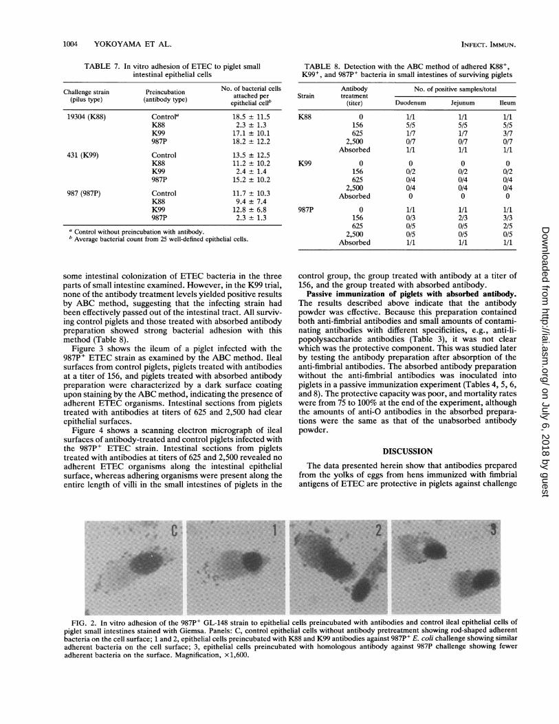
In vitro adhesion to piglet small intestine epithelial cells.K88, K99, and 987P ETEC strains adhered to isolatedporcine small intestinal epithelial cells from either the duo-denum or the ileum. The average numbers of adherentbacterial cells per epithelial cell were 18.5 for strain K88,13.5 for strain K99, and 11.7 for strain 987P. The piliated E.coli strains demonstrated the same degree of attachment toeither duodenal or ileal epithelial cells, suggesting that no
difference in epithelial cell receptor activities exists betweenthe two. However, the attachment of bacterial cells to bothduodenal and ileal epithelial cells was strongly inhibitedwhen homologous anti-fimbrial antibody solutions were usedin the in vitro adhesion assay (Table 7).
Figure 2 shows the in vitro adhesion of the 987P+ ETECstrain to ileum epithelial cells from a piglet. The controlepithelial cells (without pretreatment with antibody) andepithelial cells preincubated with K88 and K99 antibodiesshowed adherent bacteria on their surfaces. Epithelial cellspreincubated with 987P antibody and challenged with 987PE. coli showed minimal bacterial adhesion on their surfaces.
Detection by the ABC method of bacterial adherence insmall intestines of survived piglets. In K88 and 987P trials,piglets that were treated with lower-titer antibodies had
TABLE 6. Rates of isolation of K88+, K99+, and 987P+ strains of ETEC from the small intestines of piglets
No. of positive samples/total (%) from piglets that:Trialand AntibodyTrial and treatment Died Survivedstrain (titer)
Duodenum Jejunum Ileum Duodenum Jejunum Ileum
1. K88 0 6/6 (92) 6/6 (91) 6/6 (85) 1/1 (95) 1/1 (95) 1/1 (90)156 2/2 (100) 2/2 (98) 2/2 (98) 5/5 (81) 5/5 (73) 5/5 (70)625 0 0 0 1/7 (14) 1/7 (14) 3/7 (16)
2,500 0 0 0 0/7 (0) 0/7 (0) 0/7 (0)Absorbed 3/3 (100) 3/3 (90) 3/3 (90) 1/1 (95) 1/1 (90) 1/1 (90)
2. K99 0 4/4 (54) 4/4 (81) 4/4 (83) 0 0 0156 2/2 (88) 2/2 (98) 2/2 (100) 0/2 (0) 0/2 (0) 0/2 (0)625 0 0 0 0/4 (0) 0/4 (0) 0/4 (0)
2,500 0 0 0 0/4 (0) 0/4 (0) 0/4 (0)Absorbed 4/4 (60) 4/4 (100) 4/4 (100) 0 0 0
3. 987P 0 3/4 (30) 4/4 (100) 4/4 (100) 1/1 (20) 1/1 (80) 1/1 (95)156 2/2 (15) 2/2 (70) 2/2 (100) 0/3 (0) 2/3 (42) 3/3 (75)625 0 0 0 0/5 (0) 0/5 (0) 2/5 (16)
2,500 0 0 0 0/5 (0) 0/5 (0) 0/5 (0)Absorbed 3/3 (40) 3/3 (100) 3/3 (100) 1/1 (10) 1/1 (90) 1/1 (95)
VOL. 60, 1992
on July 6, 2018 by guesthttp://iai.asm
.org/D
ownloaded from
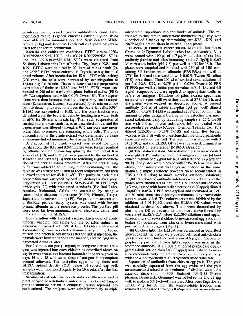
1004 YOKOYAMA ET AL.
TABLE 7. In vitro adhesion of ETEC to piglet smallintestinal epithelial cells
Challenge strain Preincubation No. of bacterial cells(pilus type) (antibody type) attached perepithelial cell'
19304 (K88) Controla 18.5 ± 11.5K88 2.3 ± 1.3K99 17.1 + 10.1987P 18.2 ± 12.2
431 (K99) Control 13.5 + 12.5K88 11.2 ± 10.2K99 2.4 ± 1.4987P 15.2 ± 10.2
987 (987P) Control 11.7 ± 10.3K88 9.4 ± 7.4K99 12.8 ± 6.8987P 2.3 ± 1.3
a Control without preincubation with antibody.b Average bacterial count from 25 well-defined epithelial cells.
some intestinal colonization of ETEC bacteria in the threeparts of small intestine examined. However, in the K99 trial,none of the antibody treatment levels yielded positive resultsby ABC method, suggesting that the infecting strain hadbeen effectively passed out of the intestinal tract. All surviv-ing control piglets and those treated with absorbed antibodypreparation showed strong bacterial adhesion with thismethod (Table 8).
Figure 3 shows the ileum of a piglet infected with the987P+ ETEC strain as examined by the ABC method. Ilealsurfaces from control piglets, piglets treated with antibodiesat a titer of 156, and piglets treated with absorbed antibodypreparation were characterized by a dark surface coatingupon staining by the ABC method, indicating the presence ofadherent ETEC organisms. Intestinal sections from pigletstreated with antibodies at titers of 625 and 2,500 had clearepithelial surfaces.
Figure 4 shows a scanning electron micrograph of ilealsurfaces of antibody-treated and control piglets infected withthe 987P+ ETEC strain. Intestinal sections from pigletstreated with antibodies at titers of 625 and 2,500 revealed noadherent ETEC organisms along the intestinal epithelialsurface, whereas adhering organisms were present along theentire length of villi in the small intestines of piglets in the
TABLE 8. Detection with the ABC method of adhered K88+,K99+, and 987P+ bacteria in small intestines of surviving piglets
Antibody No. of positive samples/totalStrain treatment
(titer) Duodenum Jejunum Ileum
K88 0 1/1 1/1 1/1156 5/5 5/5 5/5625 1/7 1/7 3/7
2,500 0/7 0/7 0/7Absorbed 1/1 1/1 1/1
K99 0 0 0 0156 0/2 0/2 0/2625 0/4 0/4 0/4
2,500 0/4 0/4 0/4Absorbed 0 0 0
987P 0 1/1 1/1 1/1156 0/3 2/3 3/3625 0/5 0/5 2/5
2,500 0/5 0/5 0/5Absorbed 1/1 1/1 1/1
control group, the group treated with antibody at a titer of156, and the group treated with absorbed antibody.
Passive immunization of piglets with absorbed antibody.The results described above indicate that the antibodypowder was effective. Because this preparation containedboth anti-fimbrial antibodies and small amounts of contami-nating antibodies with different specificities, e.g., anti-li-popolysaccharide antibodies (Table 3), it was not clearwhich was the protective component. This was studied laterby testing the antibody preparation after absorption of theanti-fimbrial antibodies. The absorbed antibody preparationwithout the anti-fimbrial antibodies was inoculated intopiglets in a passive immunization experiment (Tables 4, 5, 6,and 8). The protective capacity was poor, and mortality rateswere from 75 to 100% at the end of the experiment, althoughthe amounts of anti-O antibodies in the absorbed prepara-tions were the same as that of the unabsorbed antibodypowder.
DISCUSSION
The data presented herein show that antibodies preparedfrom the yolks of eggs from hens immunized with fimbrialantigens of ETEC are protective in piglets against challengeC* ] .... ... ... : . : ............ . i...... .. ...... ...... ...... . :
..=; . v .......... .... . I I - :..... -. .............. .... S I I} I I IWF' . :*.*; | l l m ....... . :.S .. ...
*: .fiS.. v....
:: :...:. .::: :.:S | | _.. E | i, w , FsF. : ........ : ......... : :.._:-...._S _!e
.. ;.R_
FIG. 2. In vitro adhesion of the 987P+ GL-148 strain to epithelial cells preincubated with antibodies and control ileal epithelial cells ofpiglet small intestines stained with Giemsa. Panels: C, control epithelial cells without antibody pretreatment showing rod-shaped adherentbacteria on the cell surface; 1 and 2, epithelial cells preincubated with K88 and K99 antibodies against 987P' E. coli challenge showing similaradherent bacteria on the cell surface; 3, epithelial cells preincubated with homologous antibody against 987P challenge showing feweradherent bacteria on the surface. Magnification, x1,600.
INFECT. IMMUN.
on July 6, 2018 by guesthttp://iai.asm
.org/D
ownloaded from

PROTECTIVE EFFECT OF CHICKEN EGG YOLK ANTIBODIES 1005
2#A1'
lb~~~~~~A
404
s t ;) 4 .;f~~~~~~~~~~~~~~~~~~~~~~~~~~~~~~~~~~~~~,
sE3g2,.,. .............................'.........:#,.§'.............................5
3 -.
I,. *1
FIG. 3. Localization of 987P+ GL-148 strain in the small intestines of antibody-treated and control piglets by ABC method. Panels: C,villus from the ileum of a control piglet, indicating adhering bacteria on epithelial surface as a dark surface coating; 1, villus from the ileumof a piglet treated with antibody at a titer of 156, showing a moderate dark coating on the surface; 2, clear villus surface of a piglet treatedwith antibody at a titer of 625; 3, villus from the ileum of a piglet treated with antibody at a titer of 2,500, with no bacteria on the epithelialsurface; 4, villus from the ileum of a piglet treated with the absorbed antibody preparation, indicating adherent bacteria on the surface.Magnification, x480.
with homologous ETEC strains. The different therapeuticregimens with various titers of anti-K88, -K99, and -987Pantibodies reduced considerably the severity of diarrhea inpiglets receiving high-titer antibodies. Piglets treated withantibodies at titers of 625 or 2,500 had 100% survival afterchallenge with ETEC strains, whereas piglets in the groupstreated with antibodies at a titer of 156 had mortality rates of29 to 50%. When these differences in acquired resistance aretaken into account, the optimum protection from mortalityof piglets was achieved with oral administration of higher-titer antibodies (titers of 625 and 2,500) against all threechallenge strains of E. coli. The difference in protectionbetween the antibody-treated and control piglets was statis-tically significant (P < 0.01). Although treatments withhomologous antibodies against all three challenge strains ofE. coli at titers of 625 and 2,500 conferred protection fromdeath, the efficacy achieved with antibodies at a titer of 2,500
proved superior in reducing the morbidity and severity of thedisease. Piglets that received antibodies at a titer of 2,500 didnot manifest severe watery diarrhea on day 1 after challenge,as did piglets that received other treatments. Moreover, thediarrhea was of shorter duration, and piglets in this grouphad recovered better body weight gain at the end of exper-iment (data not shown). The enterotoxigenic strains enumer-ated in cultures from rectal swabs of piglets treated withantibodies at a titer of 2,500 represented not more than 25%(mean bacterial count) of the aerobic bacterial flora, which ismuch lower than the percentages observed with piglets thatreceived antibodies at titers of 625 and 156. Also, the degreeof intestinal colonization was significantly reduced in pigletstreated with this antibody level; ETEC strains were notdetected in anterior duodenum, jejunum, and ileum samplestaken at necropsy. On the basis of these findings, weconsider the antibody titer of 2,500 as the most efficacious in
FIG. 4. Demonstration of bacterial adhesion by scanning electron microscopy of the 987P+ GL-148 strain in small intestines ofantibody-treated and control piglets. Panels: C, villus from the ileum of a control piglet showing the rod-shaped morphology of adherentbacteria on the microvillous surface; 1, villus from the ileum of a piglet with antibody at a titer of 156, showing similar adherent bacteria onthe microvillous surface; 2, villus from the ileum of a piglet treated with antibody at a titer of 625; 3, microvillous surface of a villus from theileum of a piglet treated with antibody at a titer of 2,500, showing the absence of adherent rod shaped bacteria; 4, ETEC-laden villus surfaceof an ileum from a piglet treated with absorbed antibody preparation. Scanning electron microscopy was used. Bar, 10 p,m.
VOL. 60, 1992
t
16
on July 6, 2018 by guesthttp://iai.asm
.org/D
ownloaded from

1006 YOKOYAMA ET AL.
protecting piglets from the proliferation of ETEC strains inthe intestinal tract and its harmful consequences.Adherence of K88+, K99+, and 987P+ E. coli to isolated
porcine small intestine epithelial cells in vitro was demon-strated in the absence but not in the presence of homologousanti-fimbrial antibodies prepared with the fimbrial antigen ofeach bacterium. The adhesion of E. coli was associated withspecific attachment to epithelial cell surface receptors by thefimbrial structures. Both duodenal and ileal epithelial cellsurfaces shared similar characteristics with regard to E. colipilus receptors, allowing for attachment of the three piliatedE. coli strains. Inhibition of bacterial attachment to epithelialcells by neutralizing the adhesive property of fimbriae withhomologous anti-fimbrial antibodies demonstrates the spec-ificity of the antibodies directed against ETEC pili in this invitro system. It also provides evidence in support of the roleof fimbriae in promoting bacterial adhesion to epithelial cellsand the direct action of the antibodies against these adhesivestructures. If the antibodies had no specific effect, no inhi-bition of bacterial attachment would occur. More convincingevidence in support of the specificity of these antibodiescomes from inhibition studies with heterologous 4ntibodies.There was no cross-inhibition observed among the threetypes of antibody used. Neither the K88 antibody nor theK99 antibody acted as an inhibitory agent against 987Padherence to epithelial cells or vice versa.The antibody preparation contained small amounts of
antibodies directed at other cellular components of E. coli.Agglutinating anti-O activity was detected to a slight degree.To test whether anti-O antibodies were important for theprotection in our experimental system, we compared theprotective capacity of the antibody preparation before andafter absorption, which reduced the anti-fimbrial titer to1/100 of its preabsorption value and retained the anti-Oantibody activity. Since the protective capacity was reducedsignificantly, we consider it unlikely that the anti-O antibod-ies in our antibody preparation were responsible for theprotection in our study.Comparison of the isolation rates of pathogenic E. coli
from the small intestines of piglets that died with clinicalsigns of diarrhea and surviving piglets revealed a correlationbetween the degree of intestinal colonization and mortality.Swabs taken from the small intestines of piglets treated withantibodies at a titer of 2,500 at a time when shedding ofbacteria had ceased and clinical signs had disappearedalways yielded negative isolation results, whereas intestinalswabs taken from dead piglets almost always yielded highconcentrations of the challenge strains of E. coli. Survivingpiglets treated with antibodies at lower levels also had fewerETEC isolated from the small intestine or none at all. Veryoccasionally, however, large numbers of challenge organ-isms were isolated from surviving piglets, or pure colonieswere isolated. It seems clear from our observations that,given the colonization and establishment of pathogenic E.coli, the final outcome of infection is primarily determinedby the susceptibility of individual piglets to virulence mech-anisms elicited by the thriving bacterial population. In thesame manner, the survival of a piglet is primarily dependentupon its ability to prevent the colonization and subsequentproliferation of pathogenic bacteria in the intestinal tract.Therefore, the therapeutic value of orally administered an-tibodies against experimental ETEC infection in neonatalpiglets lies in their ability to effect blocking of colonization ofenteropathogenic strains. To achieve this antibody effect,apparently, a high antibody activity in the target site afteroral administration of antibodies is needed.
Discontinuation of immunoglobulin therapy after day 3 didnot alter the course of recovery of effectively treated piglets,although excretion of challenge ETEC strains was stillevident from rectal swab cultures of some piglets. Therecovery of infecting strains from either rectal or intestinalswab cultures was observed not only in persistently diar-rheic piglets that did not respond to the 3-day immunoglob-ulin therapy but also in some of the piglets that respondedwell to the therapy. There were instances when pigletsapparently recovered from severe diarrhea and regainedbody weight and appetite but still excreted the bacteria intheir feces; some of these surviving piglets still' harbored theorganisms in their small intestines. However, the rates ofisolation of ETEC from piglets that received lower-titerantibodies and that persistently harbored these bacteria werelower than those from piglets treated with antibodies at titersof 2,500. The bacterial counts obtained from rectal culturesof surviving piglets after day 3 did not show an increasingtrend, but we are still studying whether these bacteria couldrecolonize the intestinal tract and cause a recurrence of thedisease and whether continued immunoglobulin therapy isnecessary.
Since piglets, particularly those deprived of maternalcolostrum, appear to be most susceptible to ETEC infectionduring the first day of life, the challenge and therapeuticregimens were initiated within 4 h after birth to evaluate theefficacy of oral application of antibodies in countering theenteropathogenicity of E. coli. We have demonstrated thatneonatal piglets can be protected from infection with entero-pathogenic E. coli by repeated oral administration of solubleyolk antibody powder. This result raises the possibility ofwide application of chicken egg yolk antibodies in thetreatment of enteric diseases caused by other pathogens inanimals and humans; the antibody powder can be added tofeed or formula or applied as a separate therapeutic agent.
ACKNOWLEDGMENT
We thank Mitsutaka Kuzuya, Gifu Laboratory, Ghen Corp., Gifu,for his valuable technical advice.
REFERENCES
1. Arbuckle, J. B. R. 1970. The location of Escherichia coli in thepig intestine. J. Med. Microbiol. 3:333-340.
2. Bade, H., and H. Stegemann. 1984. Rapid method of extractionof antibodies from hen egg yolk. J. Immunol. Methods 72:421-426.
3. Bartz, C. R., R. H. Conklin, C. B. Tunstall, and J. H. Steele.1980. Prevention of murine rotavirus infection with chicken eggyolk immunoglobulins. J. Infect. Dis. 142:439-441.
4. Bertschinger, H. U., H. W. Moon, and S. C. Whipp. 1972.Association of Escherichia coli with the small intestinal epithe-lium. I. Comparison of enteropathogenic and non-enteropatho-genic porcine strains in pigs. Infect. Immun. 5:595-605.
5. Dress, D. T., and G. L. Waxder. 1970. Enteric colibacillosis ingnotobiotic swine: a fluorescence microscopic study. Am. J.Vet. Res. 31:1147-1157.
6. Faith, R. E., and L. W. Clem. 1973. Passive cutaneous anaphy-laxis in the chicken: biological fractionation of the mediatingantibody population. Immunology 25:151-164.
7. Fusco, P., A. To, S. To, and C. Brinton, Jr. 1978. The purifica-tion and characterization of four types of E. coli pili and thespecificity of E. coli pili for immunity, colonization and adhe-sion, p. 60-70. In C. Miller (ed.), XIIIth United States-JapanConference on Cholera, Atlanta, Ga., September 1977. NationalInstitutes of Health, Bethesda, Md.
8. Gross, R. J., and B. Rowe. 1985. Serotyping of Escherichia coli,
INFECT. IMMUN.
on July 6, 2018 by guesthttp://iai.asm
.org/D
ownloaded from

PROTECTIVE EFFECT OF CHICKEN EGG YOLK ANTIBODIES 1007
p. 345-363. In M. Sussman (ed.), The virulence of Eschenchiacoli. Academic Press, Inc., London.
9. Guinee, P. A. M., J. Veldkamp, and W. H. Jansen. 1977.Improved minca medium for the detection of K99 antigen in calfenterotoxigenic strains of Escherichia coli. Infect. Immun.15:676-678.
10. Hatta, H., J. S. Sint, and S. Nakai. 1988. Separation of phos-pholipids from egg yolk and recovery of water-soluble proteins.J. Food Sci. 53:425-427.
11. Hiraga, C., Y. Kodama, T. Sugiyama, and Y. Ichikawa. 1990.Prevention of human rotavirus infection with chicken egg yolkimmunoglobulins containing rotavirus antibody in cat. J. Jpn.Assoc. Infect. Dis. 64:118-123.
12. Hsu, S-M., L. Raine, and H. Fanger. 1981. Use of avidin-biotin-peroxidase complex (ABC) in immunoperoxidase techniques. J.Histochem. Cytochem. 29:577-580.
13. Isaacson, R. E., and P. Richter. 1981. Escherichia coli 987Ppilus: purification and partial characterization. J. Bacteriol.146-784-789.
14. Jensenius, J. C., I. Andersen, J. Hau, M. Crone, and C. Koch.1981. Eggs: conveniently packaged antibodies. Methods forpurification of yolk IgG. J. Immunol. Methods 46:63-68.
15. Karnovsky, M. J. 1965. A formaldehyde-glutaraldehyde fixativeof high osmolarity for use in electron microscopy. J. Cell. Biol.27:137A-138B.
16. Klemm, P. 1985. Fimbrial adhesins of Escherichia coli. Rev.Infect. Dis. 7:321-336.
17. Knutton, S., R. D. Lloyd, A. D. C. Candy, and A. McNeish.1985. Adhesion of enterotoxigenic Eschenchia coli to humansmall intestine enterocytes. Infect. Immun. 48:824-831.
18. Kuhlman, R., V. Wiedemann, P. Schmidt, R. Wanke, E. Linckh,and U. Losch. 1988. Chicken egg antibodies for prophylaxis andtherapy of infectious intestinal diseases. I. Immunization andantibody determination. J. Vet. Med. B 35:610-616.
19. Kuzuya, M., H. Yokoyama, and Y. Kodama. 1988. Purifica-tion of K88 and K99 pili from porcine enterotoxigenic Esche-
richia coli by affinity chromatography. Jpn. J. Vet. Sci. 50:951-953.
20. Laemmli, U. K. 1970. Cleavage of structural proteins during theassembly of the head of bacteriophage T4. Nature (London)227:680-685.
21. Mooi, F. R., and F. K. deGraaf. 1985. Molecular biology offimbriae of enterotoxigenic Escherichia coli. Curr. Top. Micro-biol. Immunol. 118:119-138.
22. Morris, J. A., and W. J. Sojka. 1985. Escherichia coli as apathogen in animals, p. 47-77. In M. Sussman (ed.), Thevirulence of Escherichia coli. Academic Press, Inc., London.
23. Polson, A., T. Coetzer, J. Kruger, E. von Maltzahn, and K. J.van der Merwe. 1985. Improvements in the isolation of IgY fromthe yolks of eggs laid by immunized hens. Immunol. Invest.14:323-327.
24. Rutter, J. M., and J. C. Anderson. 1972. Experimental neonataldiarrhoea caused by an enteropathogenic strain of Escherichiacoli in piglets: a study of the disease and the effect of vaccinatingthe dam. J. Med. Microbiol. 5:197-210.
25. Sherman, D. M., S. D. Acres, P. L. Sadowski, J. A. Springer, B.Bray, T. J. G. Raybould, and C. C. Muscoplat. 1983. Protectionof calves against fatal enteric colibacillosis by orally adminis-tered Escherichia coli K99-specific monoclonal antibody. In-fect. Immun. 42:656-658.
26. Wilson, M. R., and A. W. Hohmann. 1974. Immunity to Esch-enchia coli in pigs: adhesion of enteropathogenic Escherichiacoli to isolated intestinal epithelial cells. Infect. Immun. 10:776-782.
27. Yamamoto, H., H. Watanabe, G. Sato, and T. Mikami. 1975.Identification of immunoglobulins in chicken eggs and theirantibody activity. Jpn. J. Vet. Res. 23:131-140.
28. Yolken, R. H., F. Leister, S.-B. Wee, R. Miskuff, and S.Vonderfecht. 1988. Antibodies to rotaviruses in chickens' eggs:a potential source of antiviral immunoglobulins suitable forhuman consumption. Pediatrics 81:291-295.
VOL. 60, 1992
on July 6, 2018 by guesthttp://iai.asm
.org/D
ownloaded from