part 1: general retinal anatomy · part 1: general retinal anatomy. general anatomy. ... less than...
TRANSCRIPT


PART 1: GENERAL RETINAL ANATOMY
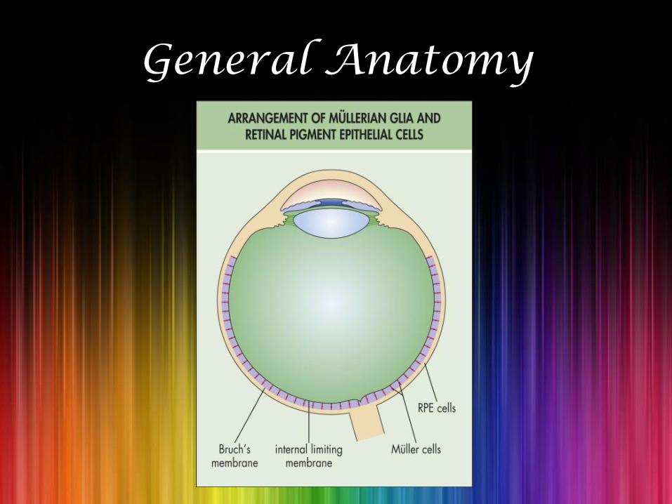
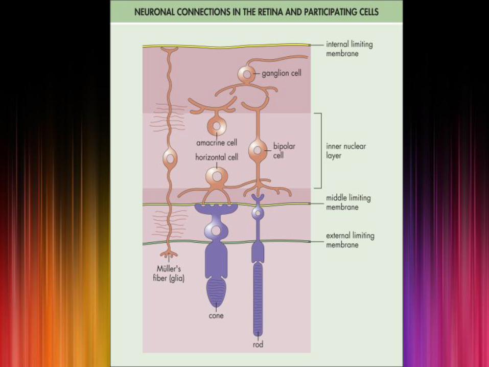
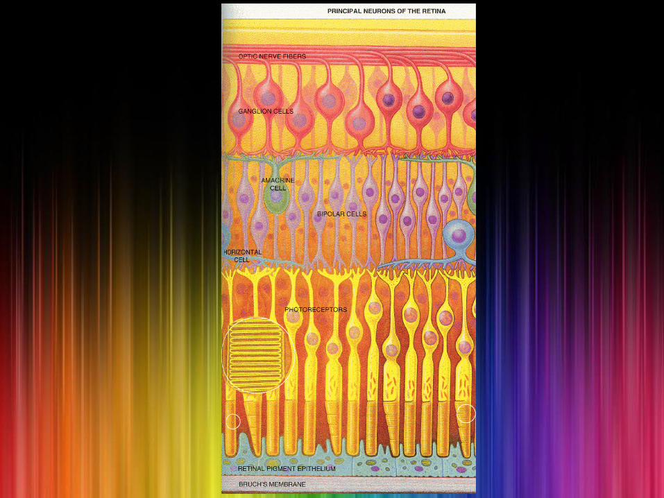
General Anatomy

At Ora Serrata

At Optic Nerve Head
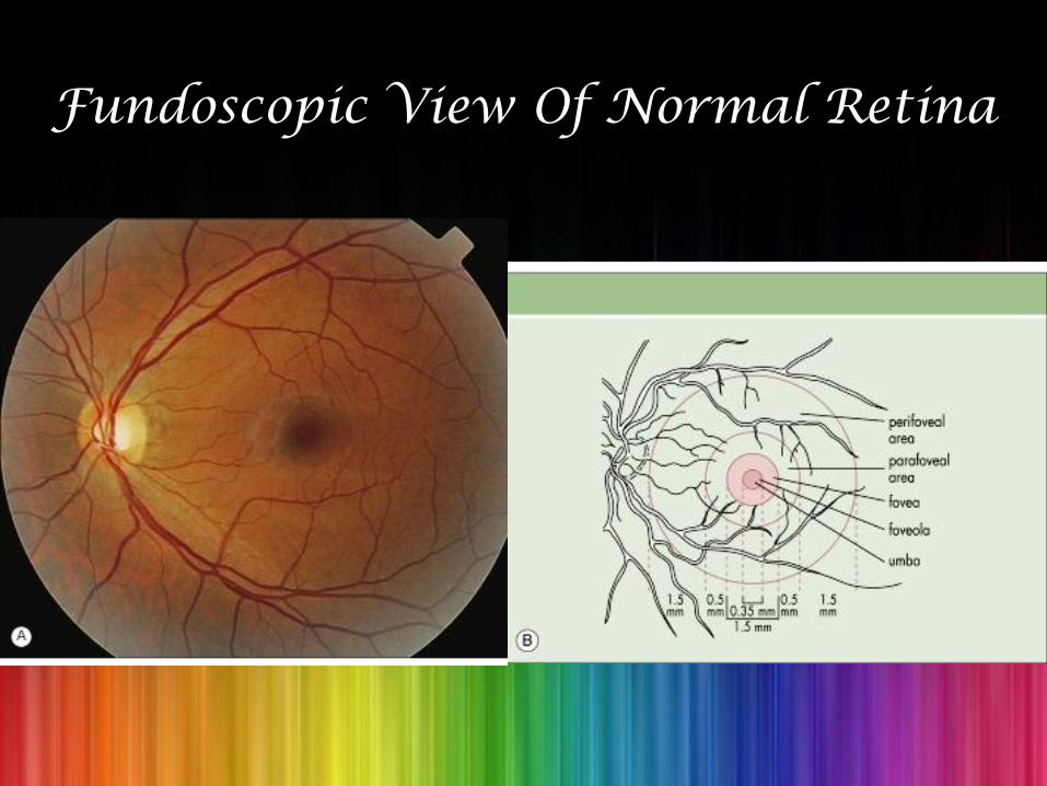
Fundoscopic View Of Normal Retina
What Is So Special About Diabetic Retinopathy?
• The WHO definition of blindness is a vision less than 3/60 in the better eye with best available spectacle correction.
• Diabetic Retinopathy is the most common cause of blindness amongst individuals of working age (20-65 years).

Part 2: Diabetic Retinopathy
Pathogenesis
• ALDOSE REDUCTASE PATHWAY
• CENTRAL ROLE OF VEGF
• MORPHOLOGICAL CHANGES IN PLATELETS
• BLOOD VISCOCITY

Aldose Reductase Pathway
• Aldose reductase converts glucose to sorbitol and galactose to galactitol.
• These sorbitol and galactitol are harmful for the eye in excess amount.
• Aldose reductase is present in high levels in:
1. Lens epithelial cells: Responsible for cataract formation.
2. Retinal cells: Responsible for Diabetic retinopathy.
• An effective aldose reductase inhibitor has not been developed yet.
Central Role Of VEGF
• VEGF NORMALLY INHIBITS THE GROWTH OF RETINAL EPITHELIAL CELLS.
• VEGF has a direct role in the proliferative retinal vascular abnormalities that are found in diabetes.
• The concentration of VEGF in aqueous and vitreous directly correlates with the severity of retinopathy.
Morphological Changes In Platelets & High Blood Viscosity
• They cause focal capillary occlusion and focal areas of ischemia in the retina which, in turn, contribute to the development of diabetic retinopathy.
Stages Of Diabetic Retinopathy
1. Nonproliferative Diabetic Retinopathy [NPDR]: Early and advanced.
2. Proliferative Diabetic Retinopathy [PDR].
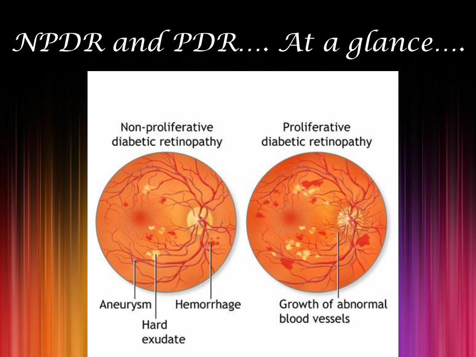
NPDR and PDR…. At a glance….
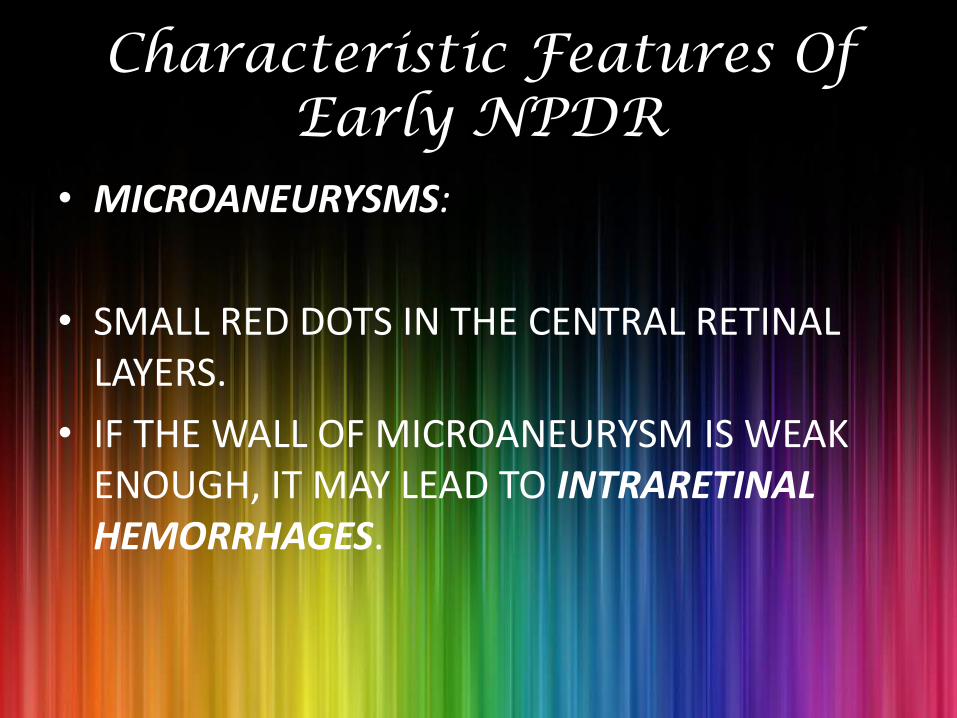
Characteristic Features Of Early NPDR
• MICROANEURYSMS:
• SMALL RED DOTS IN THE CENTRAL RETINAL LAYERS.
• IF THE WALL OF MICROANEURYSM IS WEAK ENOUGH, IT MAY LEAD TO INTRARETINAL HEMORRHAGES.
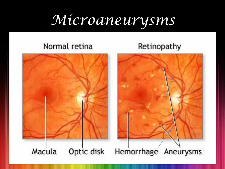
Microaneurysms
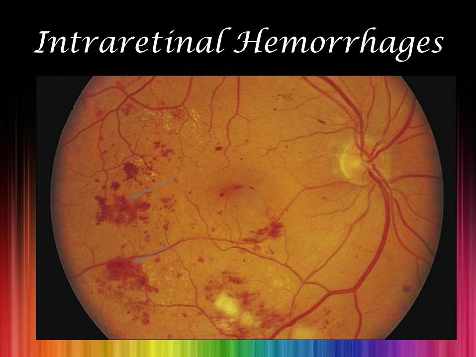
Intraretinal Hemorrhages
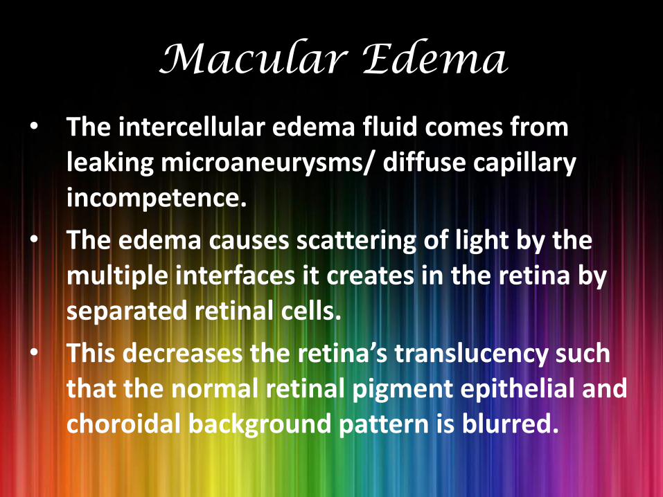
Macular Edema
• The intercellular edema fluid comes from leaking microaneurysms/ diffuse capillary incompetence.
• The edema causes scattering of light by the multiple interfaces it creates in the retina by separated retinal cells.
• This decreases the retina’s translucency such that the normal retinal pigment epithelial and choroidal background pattern is blurred.
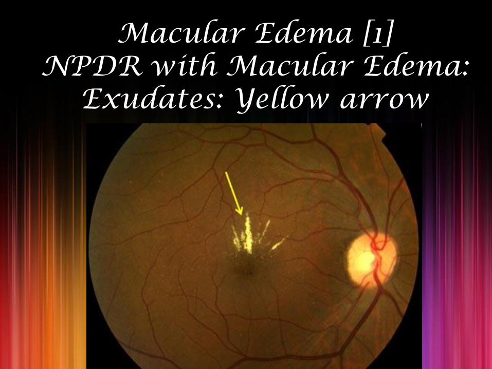
Macular Edema [1]NPDR with Macular Edema:
Exudates: Yellow arrow

Macular Edema [2]
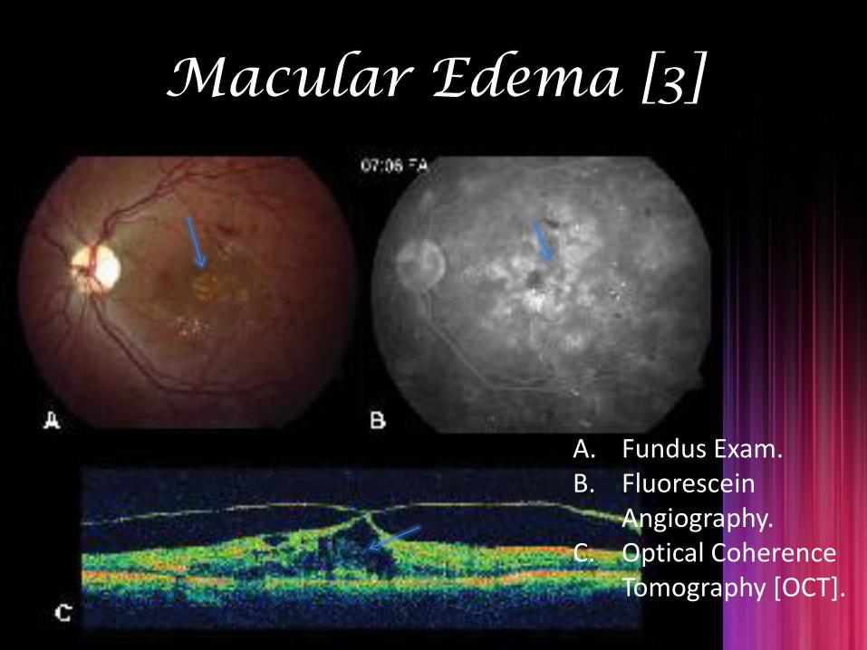
Macular Edema [3]
A. Fundus Exam.B. Fluorescein
Angiography.C. Optical Coherence
Tomography [OCT].
Hard Exudates And Circinate Retinopathy
• If the leakage of fluid is severe enough, lipid accumulates and precipitates in the retina.
• In some cases, lipid is scattered through the macula. Then it is called “ ”.
• In others, it accumulates in a ring around a group of leaking microaneurysms/ around microaneurysms surrounding an area of capillary nonperfusion. This pattern is called “ ”.
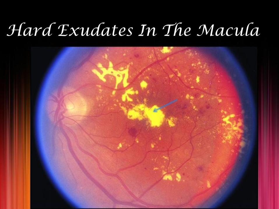
Hard Exudates In The Macula

Circinate Retinopathy
Characteristic Features Of Advanced NPDR
• Due to increased retinal hypoxia, following changes are seen in the retina:
1. Intraretinal microvascular abnormalities (IRMA).
2. Cotton-wool spots.
3. Venous beading.
Intraretinal Microvascular Abnormalities (IRMA)
• They are dilated capillaries, which seem to function as collateral channels.
• Capillary hypoperfusion often surrounds IRMA.

Severe IRMA
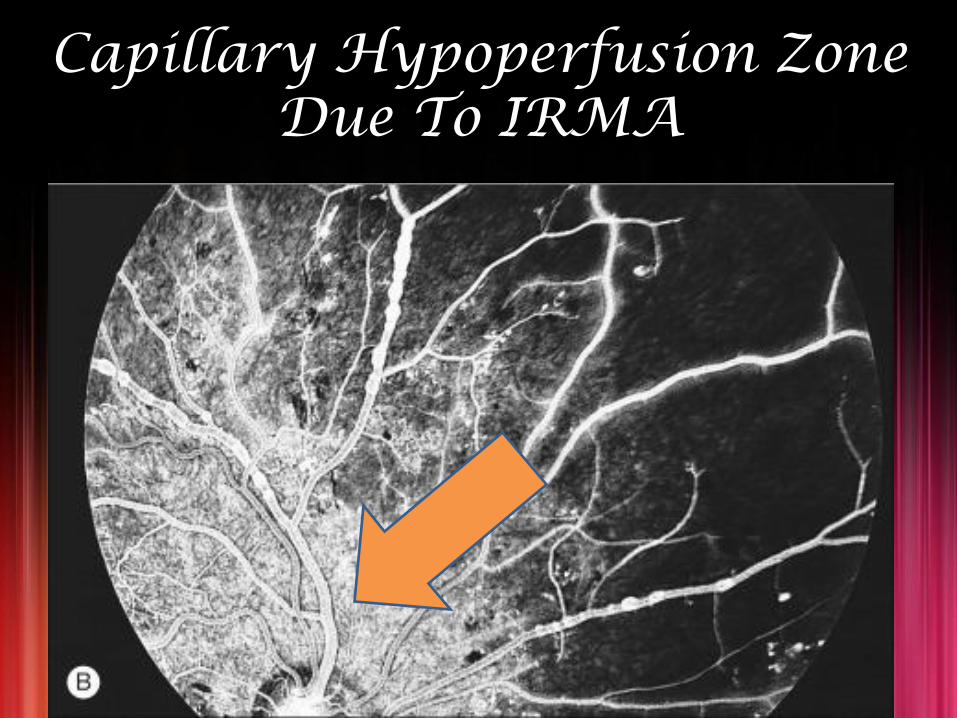
Capillary Hypoperfusion Zone Due To IRMA

Cotton Wool Spots(Soft Exudates)
• The main cause of this feature is ischemic changes.
• Local ischemia causes effective obstruction of axoplasmic flow in the normally transparent nerve fiber layer AND,
• Subsequent swelling of the nerve fibers gives a characteristic white fluffy appearance to the cotton-wool spots.
• Fluorescein angiography shows no capillary perfusion in the area corresponding to a cotton-wool spot.
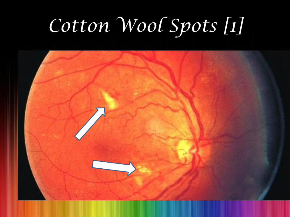
Cotton Wool Spots [1]
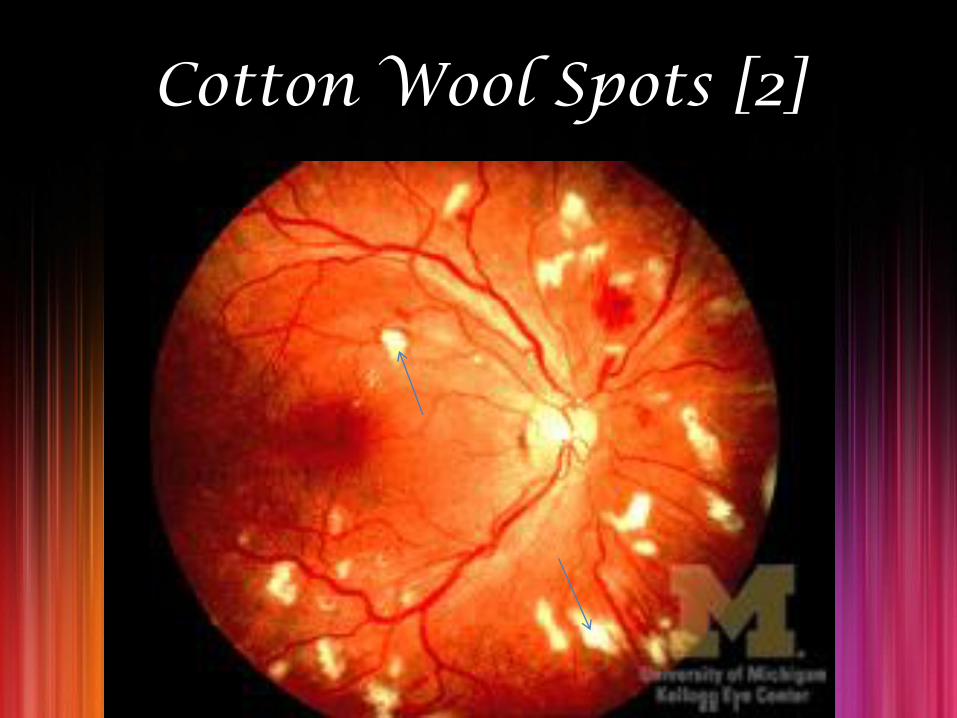
Cotton Wool Spots [2]
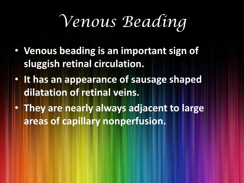
Venous Beading
• Venous beading is an important sign of sluggish retinal circulation.
• It has an appearance of sausage shaped dilatation of retinal veins.
• They are nearly always adjacent to large areas of capillary nonperfusion.
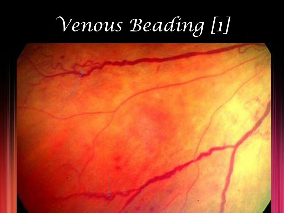
Venous Beading [1]
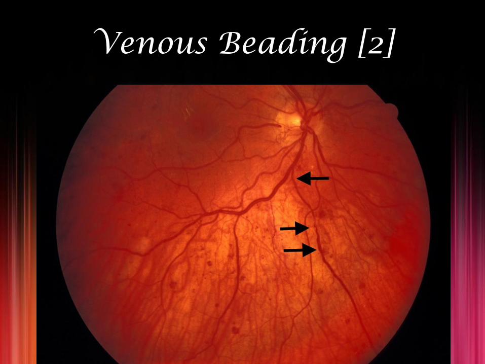
Venous Beading [2]
PDR
• It is characterized by neovascularization [new blood vessel formation], which is of 2 types:
1. Neovascularization of the disc [NVD],
2. Neovascularization elsewhere [NVE].
• NVD: New vessels arise within ≤1 disc diameter of optic nerve.
• NVE: New vessels arise from >1 disc diameter of optic nerve.
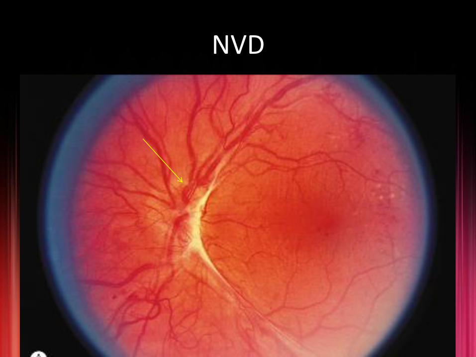
NVD
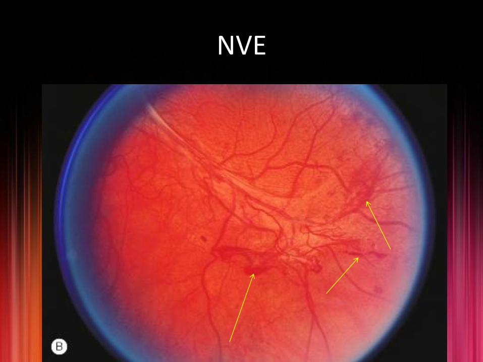
NVE

Vitreous Traction And Retinal Detachment
• The new vessels usually progress through a stage of further proliferation, with associated connective tissue formation.
• As PDR progresses, the fibrous component becomes more prominent.
• Vitreous traction is transmitted to the retina along these proliferations and may lead to traction retinal detachment.
*. Davis et al. have stressed the role of the contracting vitreous in the production of vitreous hemorrhage, retinal breaks, and retinal detachment.
Types Of Diabetic Retinal Detachments
• Two types of diabetic retinal detachments occur:
1. Those that are caused by traction alone (nonrhegmatogenous) and,
2. Those caused by retinal break formation (rhegmatogenous).
• Optical coherence tomography (OCT) is used to describe/ determine those detachments.
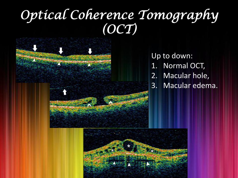
Optical Coherence Tomography (OCT)
Up to down:1. Normal OCT,2. Macular hole,3. Macular edema.
Diagnosis Is Done By…..
1. Direct ophthalmoscopy.
2. Detection of systemic hyperglycemia:
A. Fasting blood sugar testing,
B. Glucose tolerance test, and
C. Hemoglobin A1c determinations.
3. Optical coherence tomography (OCT), where available.

Treatment
• Antiplatelet therapy.
• Antihypertensive drugs.
• Anti-VEGF agents.
• PRP.
• Vitrectomy.

Antiplatelet Therapy
Aspirin 650 mg daily
• It does not influence the progression of retinopathy/ affect visual acuity/ influence the incidence of vitreous hemorrhages.
• But it reduces the incidence of stroke in diabetic patient.
ANTIHYPERTENSIVE DRUGS
• The Hypertension in Diabetes Study group has demonstrated that with better blood pressure control, a 37% risk reduction in microvascular changes can be achieved.
Anti VEGF Agents
• Anti-VEGF drugs are available for the treatment of macular degeneration.
• Recently, a protein kinase C inhibitor [PKCI] has been shown to reduce diabetes-induced hemodynamic abnormalities in patients with diabetic retinopathy and reduce the risk of vision loss in patients with macular edema.
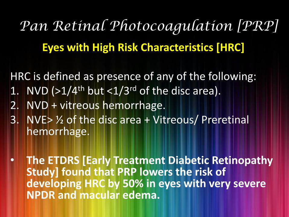
Pan Retinal Photocoagulation [PRP]
Eyes with High Risk Characteristics [HRC]
HRC is defined as presence of any of the following:1. NVD (>1/4th but <1/3rd of the disc area). 2. NVD + vitreous hemorrhage.3. NVE> ½ of the disc area + Vitreous/ Preretinal
hemorrhage.
• The ETDRS [Early Treatment Diabetic Retinopathy Study] found that PRP lowers the risk of developing HRC by 50% in eyes with very severe NPDR and macular edema.

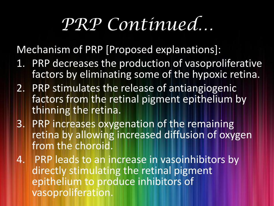
PRP Continued…Mechanism of PRP [Proposed explanations]:1. PRP decreases the production of vasoproliferative
factors by eliminating some of the hypoxic retina.2. PRP stimulates the release of antiangiogenic
factors from the retinal pigment epithelium by thinning the retina.
3. PRP increases oxygenation of the remaining retina by allowing increased diffusion of oxygen from the choroid.
4. PRP leads to an increase in vasoinhibitors by directly stimulating the retinal pigment epithelium to produce inhibitors of vasoproliferation.
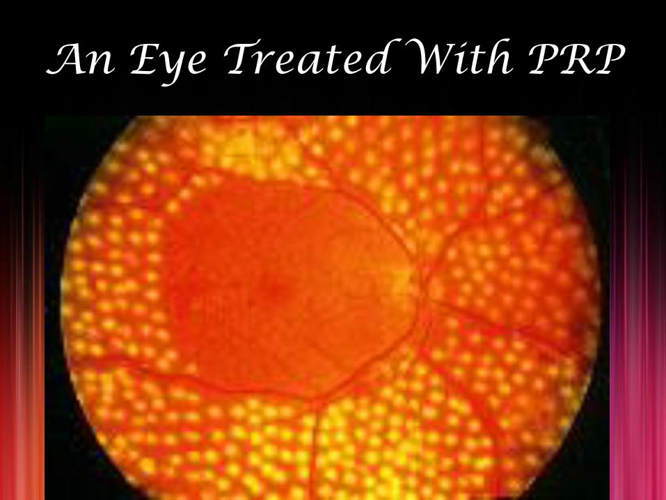
An Eye Treated With PRP

Treatment Of Macular Edema By PRP

Vitrectomy
• The major indications are of vitrectomy in diabetics are:
1. Macular-involving/ macular-threatening traction retinal detachment
2. Nonclearing vitreous hemorrhage and,
3. Combined traction-rhegmatogenous retinal detachment.

Vitrectomy….Continued….• The surgical objectives are:
1. To clear the media,
2. To release all anterior-posterior traction,
3. To release tangential traction via delamination or segmentation (cutting the fibrotic bridges between areas of tractional detachment), and
4. To perform endophotocoagulation.
• A possible cause of failure following an otherwise successful vitrectomy is
Thank You….
Courtesy:1. Parson’s Disease Of The Eye.2. Yanoff’s Ophthalmology.3. Michigan State University.4. Atlas Of Clinical Endocrinology.5. Emedicine Medscape.6. Wikipedia.7. Early Treatment Diabetic
Retinopathy Study [ETDRS].8. Medline Plus Medical Encyclopedia.