paris urine cytology presentation - legeforeningen.no doughty.pdf · outline ¨ urine – the...
TRANSCRIPT

URINE CYTOLOGY
Richard Doughty Avd for patologi Ahus

The main purpose of urine cytology is
To detect High Grade Urothelial Carcinoma


Outline
¨ Urine – the basics ¨ A little on the history of claasification systems. ¨ What is the goal of urine cytology? ¨ Why to standardize, why Paris? ¨ What is the guiding principle? ¨ What are diagnostic categories? ¨ What are the criteria? ¨ What adjuvant studies? ¨ What are future clinical and research needs?

Bladder cancer � current status ¨ ~ 76,900 new cases in 2016 in the USA ¨ ~ 16,390 deaths due to bladder cancer ¨ 4th most common ca in men and 9th in women (1 in 44 people) ¨ 9th most common cause of cancer death (F>M) ¨ ~ 75% non�muscle invasive bladder cancers (superficial bladder ¨ cancers), Ta, Tis, T1 ¨ ~ 30% � 70% � recurrence ¨ ~ 5% � 15% � progression (<1% LG Ta) ¨ > 535,000 people in the US are survivors of this cancer
¨ Highest per patient cost from dx to death of all cancers ¨ $4.1 billion/year spent to tx bladder cancer Nielsen ME et al. Trends in stage�specific incidence rates for urothelial carcinoma of the bladder in the United States: 1998 to 2006. Cancer 2014:120:86

In Norway....
¨ 2015: 1731 new cases of cancer in the urinary bladder, ureters or urethra ¤ 1262 men ¤ 469 women


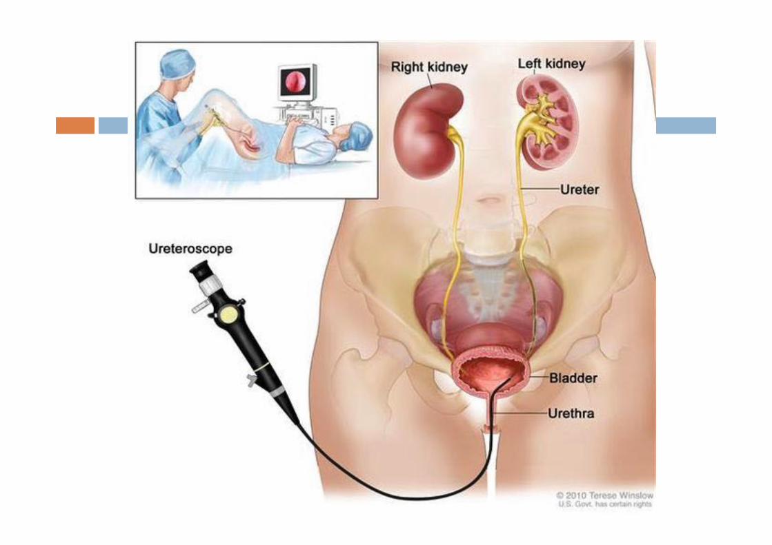
The urinary tract – the basics
Upper urinary tract
Lower urinary tract


What is urine?

Urine – a definition
¨ Urine is a liquid by-product of the metabolism in humans and in many animals. Urine flows from the kidneys through the ureters to the urinary bladder.

Urine composition
Not the perfect cell presevation medium!!

Clinical Indications of Urine Cytology
q Hematuria q Follow-up of patients treated for Urothelial
Carcinoma (UC) q High-risk of bladder cancer

What can you expect to find in a normal urine?

Normal Urinary Elements
q Urothelial cells q Intermediate and superficial (umbrella) cells (voided urine) q Intermediate, superficial and basal cells (catheterized urine,
washing) q Squamous cells q Miscellaneous findings
q Prostate and seminal vesicle epithelial cells q Renal tubular cells and casts q Corpora amylacea q Crystals q Inflammatory cells
q Degenerated intestinal epithelial cells (ileal conduit)

Umbrella cells
• Low N/C ratio • Pale finely granular chromatin • Smooth nuclear shapes • Multinucleation common • Cytoplasm transparent

Intermediate and basal cells
• High N/C ratio • Chromatin darker than
superficial cells • Nuclei smaller than
superficial cells • Nuclear shape round • Even nuclear spacing

Normal Urinary Elements
Melamed-Wolinska Bodies

Casts
q Renal Diseases: q RBC casts: Glomerular diseases q WBC casts: Tubulointerstitial diseases and transplant
rejection q Renal tubular casts: Renal parenchymal diseases q Fatty casts: Nephrotic syndrome
q Physiologic: q Hyaline and granular casts: Secondary to dehydration,
fever, exercise etc

Normal Urinary Elements
Rbc cast
Rbc cast
Rbc cast
RBC Cast Renal Tubular Cast
Corpora Amylacea

Non-Urinary Elements
Seminal Vesicle Cells Endometrial Cells

Infections -Fungal

Crystals
q Common finding, no clinical significance in most cases q Crystals analysis part of routine urinalysis rather than urine
cytology q Uric acid: most common, variable shape q Triple-phosphate: prism shaped and resemble coffin lids q Ammonium biurate: “Thorn apples” q Calcium Oxalate: Oval, dumbbell shaped q Pathologic crystals: much less common, bilirubin (brown
granules and needles), cholesterol , cysteine (hexagonal plates), leucine (spheres with radiating striations) and tyrosine (slender needles)

Types of Urinary Specimens
q Voided Urine q Catheterized Urine q Bladder Washings q Upper Tract Washings and Brushings q Ileal Conduit Samples
Urine samples is a relatively easy sample to obtain….maybe

Physicians have it easy....
= + +
+ = + =

Voided Urine
q Collected 3-4hrs after the last void (100-300ml) q Sparse cellularity, superficial and intermediate cells q Degenerative changes q Squamous cells common
q Trigone or genital tract contamination in women q Inflammation or irritation
q Non-cellular constituents such as crystals, casts, corpora amylacea
q Non-invasive technique and no instrumentation effect

Catheterized Urine
q Moderate to highly cellular q Superficial, intermediate and basal cells q Poor preservation with pronounced degenerative
changes in pooled specimens q Urethra not sampled q Instrumentation artifacts: Urothelial clusters can mimic
low-grade urothelial carcinoma q Risk of infection

Catheterized Urine
Basal Urothelial Cells in Catheterized Urine Specimen

Bladder Washings
q Obtained through a catheter by irrigating the bladder with 5-10 pulses of 50 ml sterile saline
q Better cellularity and preservation q Less contamination by background debris q Increase sensitivity (66%-77%) q Only bladder epithelium represented – upper tract
not sampled q Quality of sample dependent on the skill of urologist

Bladder Washings

Upper Tract Washings / Brushings
q Comparable sensitivity to other type of urinary specimens
q Technically and morphologically challenging q Prone to false positive results – marked cellularity q Comparison of bilateral specimens (normal vs
lesional) helpful in making diagnosis q Cytological diagnosis with conservative approach
q Ureterectomy or nephrectomy

Urethral Brushings

Urethral Brushings

Ileal Conduit
q Surveillance of ureters and renal pelves post cystectomy
q Cellular specimen with large amount of degenerated intestinal epithelial cells and background debris
q Malignant cells may be obscured

Take home from sampling:
• There is a balance between the invasiveness of the sampling method and the cellularity obtained

The history of systems for reporting urine samples

Urine comtemplation
¨ Avicenna, physician and philosopher (980 – 1037), advocated systematic analysis of urine: ¤ Colour ¤ Density ¤ Sendiment – calculus, abscess or tumor ¤ Odor – tumor

The age of uroscopy...
¨ Historic medical practice of visually examining a patient's urine for pus, blood, or other symptoms of disease

From urine analysis to urine cytology
¨ Rise of modern light microscopes – 1600s ¨ No mention of cellular elements in urine until 1800s
Alfred Donne 1801 - 1878 Hermann Lebert 1813 - 1878

Modern times.....
1928 Georgios Papanikolaou Pap smear
1940s - onwards Leopold Koss `Father of urine cytology’
Dorothy Rosenthal The Johns Hopkins Hospital template for urologic cytology samples

Onwards to Paris!
18th International Congress of Cytology, Paris, May, 2013 • “Paris Group” – all participants of two Urine
Cytology Symposia • Outline of the Paris System for Reporting
Urinary Cytopathology • Ultimate goal – detection of HGUC • Sponsorship by the ASC and IAC • Contract with Springer • Numerous face�to�face meetings

The move to standardise...
2001 2007

The Paris System for Reporting Urinary Cytology

Why to standardize reporting of urinary cytology?
¨ Reproducibility ¨ Improvement of communication ¨ Atypical cells
¤ Wide intraobserver variability
¨ Nationally rates of atypical vary among institution ¤ Range from 2% to 30% (51% atypical + suspicious)

For example
Irregulære, degenererte urotelceller av usikker betydning.....

The Paris System
1. Pathogenesis of Urothelial Carcinoma 2. Adequacy 3. Negative for High Grade Urothelial Carcinoma 4. Atypical Urothelial Cells 5. Suspicious for High Grade Urothelial Carcinoma 6. High Grade Urothelial Carcinoma 7. Low Grade Urothelial Neoplasm 8. Other malignancies, both primary and secondary 9. Ancillary Studies 10. Clinical management 11. Preparatory techniques relative to Urinary Tract samples

System has to be build based on:
¨ Consensus ¨ Evidence ¨ Inclusion ¨ Acceptance ¨ Understanding

A little on grading and staging
¨ Grading ¤ Histological appearance
n Low grade n High grade
¨ Staging ¤ Non muscle invasive bladder cancer (NMIBC)
n Tis, Ta, T1
¤ Muscle invasive bladder cancer (MIBC) n >T1

TNM classification for bladder cancer
Tis

Normal Urothelium
Hyperplasia Dysplasia
Low Grade Carcinoma High Grade Carcinoma Carcinoma in situ
Invasive Carcinoma
9p-, 9q- p16
Genetically Stable FGFR3 (~85%)
Genetically Unstable p53 (~60%) <10%
Recurrence Recurrence
RAS (?)
Pathogenesis of Urothelial Carcinoma
Eva M. Wojcik and Stefan E. Pambuccian
Normal Urothelium
Hyperplasia Dysplasia
High Grade Carcinoma Carcinoma in situ
Invasive Carcinoma
Papillary Pathway
80%
Non-Papillary Pathway
20% 9p-, 9q-
p16
Genetically Unstable p53 (~60%) <10%

Bladder cancer – more than one disease?
• ~ 75 % Non�Muscle�Invasive (Ta/T1) • Good prognosis • Recurrence • 10%�15% progression (LG Ta <1%)*
• ~ 25 % Muscle�Invasive (> T2) • >60% overall survival

“Approximately 80% (of Ta bladder tumors) appear to follow a benign course without developing invasive tumors or dying of bladder cancer”

Question…. “Carcinoma”?
GU GI

CARCINOMA ADENOMA
Question…. “Carcinoma”?

Mr. Smith - You have a bladder cancer Hansen

What really matters?
High Grade Urothelial Carcinoma

Diagnostic Categories
HGUC Everything else
Hope Positive Negative Atypical/Suspicious
Reality

Classifications
WHO 1973
WHO/ISUP 2004
Papilloma
Papilloma
Grade I Grade III Grade II
Low Grade High Grade PUNLMP
~ 80-90% ~ 10-20% ~ 50-60%
URINE CYTOLOGY SENSITIVITY
Very high probability that we are going to be wrong

Evolution of the Classification
Owens et al. Cancer Cytopathology 2013
?LG
?HG

NEW paradigm
• It is all about High Grade Urothelial Carcinoma • Negative for High Grade Urothelial Carcinoma
• LGUN – Low Grade Urothelial Neoplasm
AUC SHGUC HGUC Quality and Quantity Quantity
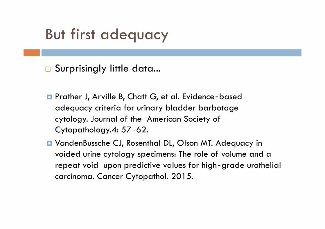
But first adequacy
¨ Surprisingly little data...
¤ Prather J, Arville B, Chatt G, et al. Evidence�based adequacy criteria for urinary bladder barbotage cytology. Journal of the American Society of Cytopathology.4: 57�62.
¤ VandenBussche CJ, Rosenthal DL, Olson MT. Adequacy in voided urine cytology specimens: The role of volume and a repeat void upon predictive values for high�grade urothelial carcinoma. Cancer Cytopathol. 2015.

Prather J, Arville B, Chatt G, et al.
Table1.Prospectivestudy.
Cellularity Sensitivities
AUC+ HGUC
<10per10hpfs 60.5 37.2
≥10per10hpfs 95.2 76.2
Pvalue 0.0001 0.0004
<20per10hpfs 68.3 43.3
≥20per10hpfs 100.0 88.0
Pvalue 0.001 0.0001

Volume is important…

Adequacy of Urine Specimens (Adequacy)

Lets take a break!

Diagnostic categories
1. Negative for High Grade Urothelial Carcinoma
2. Atypical Urothelial Cells 3. Suspicious for High Grade
Urothelial Carcinoma 4. High Grade Urothelial Carcinoma 5. Low Grade Urothelial Neoplasm 6. Other malignancies, both primary
and secondary


Negative for High�Grade Urothelial Carcinoma (Negative)
Definition of Negative for High�Grade Urothelial Carcinoma • A sample of urine, either voided or instrumented, may
be considered benign, i.e., NHGUC, if any of the following components are present in the specimen: – Benign urothelial, glandular, and squamous cells – Benign urothelial tissue fragments (BUTF) and
urothelial sheets or clusters – Changes associated with lithiasis – Viral cytopathic effect; polyoma virus (BK virus—
decoy cells) – Post�therapy effect, including epithelial cells from
urinary diversions

Benign Superficial (Umbrella) Urothelial Cells

“Atypical” Umbrella Cells

Glandular Cells
• Sources: endometrium, prostate, kidneys, urachal remnants, metaplasia

Cystitis cystica/glandularis

Renal Tubular Epithelial Cells

Benign Urothelial Tissue Fragments � BUTF
Onur, I., Rosenthal, D. L., & VandenBussche, C. J. (2015). Benign�appearing urothelial tissue fragments in noninstrumented voided urine specimens are associated with low rates of urothelial neoplasia. Cancer cytopathology, 123(3), 180-185.

Nephrolithiasis – 3D fragments

Stone Atypia

Systemic Chemotherapy Changes
q Degenerative changes with frayed cell borders
q Enlarged hyperchromatic but smudgy nuclei
q Vacuolated cytoplasm
q Irregular dark nucleoli q Multinucleation

Chemotherapy Changes
High-grade UC Mitomycin
Thiotepa

Intravesical Chemotherapy Changes
q Predominantly effect the superficial cells
q Marked cytomegaly with abundant vacuolated cytoplasm and one or more nuclei
q Nuclear chromatin chunky, clumped, deeply staining or structureless and smudgy with smooth borders
q Prominent nucleoli q Frayed borders q No significant effect on neoplastic
cells

Immunotherapy
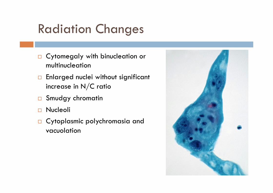
Radiation Changes
¨ Cytomegaly with binucleation or multinucleation
¨ Enlarged nuclei without significant increase in N/C ratio
¨ Smudgy chromatin
¨ Nucleoli ¨ Cytoplasmic polychromasia and
vacuolation

Malakoplakia
Malakoplakia with Michealis Gutmann Bodies
Von Kossa Stain
Malakoplakia: Histiocytes with abundant granular cytoplasm filled with bacteria and bacterial fragments

Seminal Vesicle Cells

Bladder Diversion Urine
Melamed – Wolinska body

Infections -Viral
q Polyomavirus q Infects both healthy and immunocompromised individuals q 4% of urine specimens q No clinical significance in immunocompetent
q Herpes: Uncommon, immunocompromised patients q CMV: Most commonly effects renal tubular cells q HPV: Vaginal contamination

Infections - Viral
CMV HSV HPV

Infections - Viral
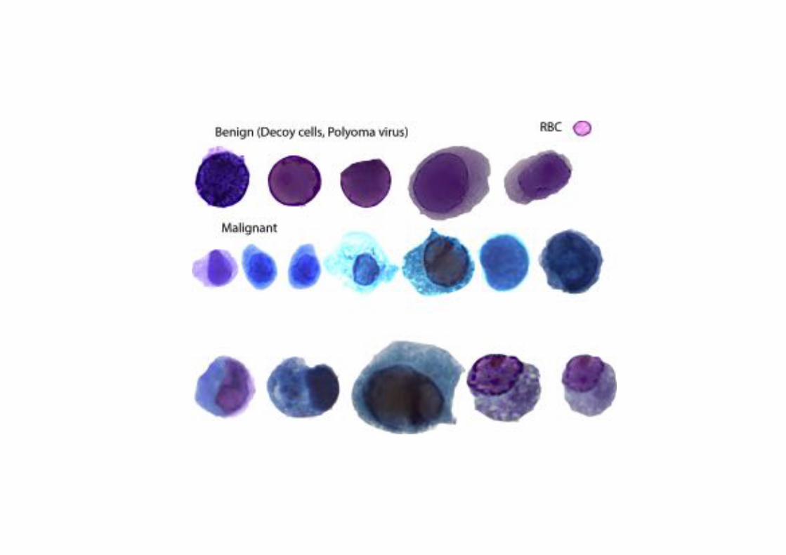
Polyomavirus Cytopathic Changes

Things are never that easy......

88-year-old man with a history of T1 HGUC previously treated by local excision. F/U bx negative. Cystoscopy – negative.

• Polyoma → Negative for High Grade Urothelial Carcinoma
How about these?
Positive Suspicious Atypical Negative
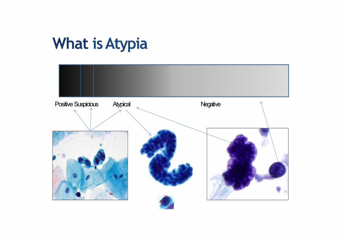
What is Atypia


What do YOU call atypia in urine specimens?
1. There are rare cells, reminiscent to that of high grade UC
2. Lots of cs, worrisome for low grade UC
3. Other (degenerated cells, cells/groups that don’t fit in either group above)
Negative for High Grade Urothelial Carcinoma

Diagnostic categories

What is atypia? Findings in literature
1. Highnuclearcytoplasmicra5o(>0.7)2. Nuclearhyperchromasia3. Coarse,clumpedchroma5n4. Irregularnuclearmembranes
Atypia Suspicious Positive

Criteria for AUC • Non�superficial and non�degenerated urothelial cells
with an high N/C ratio > 0.5 (required)
and one of the following: • Hyperchromasia (compared to the umbrella cells
or the intermediate squamous cell nucleus) • Irregular clumpy chromatin • Irregular nuclear contours
Atypical Urothelial Cells (AUC)


Degeneration

N:C ratio of 0.5???


Suspicious for High�Grade Urothelial Carcinoma (Suspicious)
Criteria for SHGUC
• Non�superficial and non�degenerated urothelial cells with an high N/C ratio > 0.7 (required)
• Hyperchromasia (compared to the umbrella cells or the intermediate squamous cell nucleus) (required)
and one of the following: • Irregular clumpy chromatin • Irregular nuclear membranes
<10 cells


Suspicious for HGUC vs. Positive HGUC Quantity matters..
“The number of atypical urothelial cells is an important criterion to classify urine cytology specimens into the ‘positive’ or the ‘suspicious’ categories. A cut�off number of >10 cells to render a definitive diagnosis of HGUCA seems valid from the clinical standpoint .”
5 – 10 cells – gray zone, based on experience, history, individual threshold, etc
JASC 2015;4(4)232–238

Not only quantity and quality matter...


High�Grade Urothelial Carcinoma (HGUC) • Cellularity: At least 5–10 abnormal cells • N/C ratio: 0.7 or greater • Nucleus: Moderate to severe hyperchromasia • Nuclear membrane: Markedly irregular • Chromatin: Coarse/clumped

High-grade UC
Bladder Washing Squamous differentiation

Other Notable Cytomorphologic Features
• Cellular pleomorphism • Marked variation in cellular size and shapes, i.e.,
oval, rounded, elongated, or plasmacytoid (Comet cells)
• Scant, pale, or dense cytoplasm • Prominent nucleoli • Mitoses • Necrotic debris • Inflammation

High-grade UC - Differential Diagnosis
q Polyomavirus q Stone atypia q Normal upper tract washing or brushings q Treatment effect q Non specific reactive changes

Squamous differentiation Glandular differentiation

What happened to Low grade urothelial neoplasia (LGUN)??
• Almost impossible to diagnose without a mini�biopsy with fibrovascular core
• Cytologically normal nuclei • Is it truly a carcinoma? • More common than HGUC • BUT, not life threatening

Low�Grade Urothelial Neoplasia (LGUN)
• LGUN � combined cytologic term for low grade papillary urothelial neoplasms (LGPUN) (which include urothelial papilloma, PUNLMP and LGPUC) and flat, low grade intraurothelial neoplasia
LGUC
LGUN

Cytologic Criteria of Low Grade Urothelial Neoplasia (LGUN) (regardless of the specimen type: voided or instrumented):
• Three�dimensional cellular papillary clusters (defined as clusters of cells with nuclear overlapping, forming "papillae") with fibrovascular cores with capillaries

Cytologic Criteria of Low Grade Urothelial Neoplasia (LGUN) (regardless of the specimen type: voided or instrumented)
Cell Block

How about these????
Negative for HGUC
Suggestive of LGUN

G. Barkan, MD

Nuclear:Cytoplasm Ratios
Zhang ML, Guo AX, VandenBussche CJ. Morphologists overestimate the nuclear-to-cytoplasmic ratio. Cancer Cytopathol2016;124:669–677.
SHGUC + HGUC
AUC

What does the urologist do the cytology report?????

Clinical Management
• From the standpoint of the urologist, the workup for AUC should be individualized based on the risk assessment of the patient
• From a practical standpoint, the clinical management of “suspicious for HGUC” is similar to a “positive for HGUC” diagnosis
• Transurethral resection establishes the histologic diagnosis and is therapeutic for most solitary low grade tumors

Clinical Management
Category Risk of
Malignancy Management
Unsatisfactory/Nondiagnostic ? (<5%) Repeat cytology, cystoscopy in 3 months if increased clinical suspicion
Negative for HGUC 0-2% Clinical follow up as needed
Atypical Urothelial Cells (AUC) 8-35% Clinical follow up as needed. Use of ancillary testing.
Suspicious for HGUC 50-90% More aggressive follow up, cystoscopy, biopsy
LGUN ~10% Need biopsy to further evaluate grade and stage
High Grade UC >90% More aggressive follow up, cystoscopy, biopsy, staging
Other malignancy >90% More aggressive follow up, cystoscopy, biopsy, staging

Rate of Atypia at Loyola per pathologist
0,00 %
5,00 %
10,00 %
15,00 %
20,00 %
25,00 %
30,00 %
35,00 %
2008 2009 2010 2011 2012 2013 2014 2015 2016

Diagnostic categories
1. Negative for High Grade Urothelial Carcinoma
2. Atypical Urothelial Cells 3. Suspicious for High Grade
Urothelial Carcinoma 4. High Grade Urothelial Carcinoma 5. Low Grade Urothelial Neoplasm 6. Other malignancies, both primary
and secondary

Other Malignancies Primary and Metastatic and Miscellaneous Lesions

Melanoma
ADC
Lymphoma
Clear cell adc bladder

Squamous Cell Carcinoma
q 5% of bladder cancers q Pure squamous cell carcinoma rare
– associated with caliculi, diverticuli, schistosomiasis
q Squamous differentiation in UC
q Cytoplasmic keratinization q Hyperchromatic angulated nuclei

Primary Adenocarcinoma
q Rare, <2% of bladder cancer
q Colonic type, most common q Signet ring type q Clear cell adenocarcinoma
Clear cell adenocarcinoma

Secondary Tumors
q Prostatic Adenocarcinoma q Seen in high-grade prostatic
carcinoma q Large cohesive three
dimensional clusters with ill-defined cell borders
q Prominent nucleoli with relatively abundant cytoplasm
q Dark nuclei resembling UC q History helpful !

Secondary Tumors
Colonic Adenocarcinoma Endometrial Adenocarcinoma

ANCILLARY TECHNIQUES

Nuc
lear
/ cy
tolo
gic
atyp
ia
low moderate/high certain Probability of high grade UC
AUC/SHGUC 8%-30%
HGUC NFHG

Ancillary Urine Based Techniques
q DNA ploidy q Bladder Tumor Antigen (Bard BTA stat®) q Nuclear Matrix Proteins (NMP22™) q UroVysion™ q ImmunoCyt/uCyt™ q Telomerase q Hyaluronic Acid Hyaluronidase q Fibrin-Fibrinogen Degradation Product

UroVysion
q Chromosomal abnormalities in UC first described in 1990s
q Initial studies tested single chromosome probes q Suklova et al published first study with multiple probes
(10 probes tested) q Highest sensitivity achieved with combination of 4 probes
q Chromosome 3 (CEP) q Chromosome 7 (CEP) q Chromosome 17 (CEP) q Chromosome 9p21 (LSI probe)
q Sensitivity: 84% Specificity: 92% q Cutoff: 5 abnormal cells

Ancillary Studies in Urinary Cytology

UroVysion
q Multicolor multitarget FISH UroVysion test approved by FDA in 2001
q Approved Indications: q Surveillance of patients with bladder cancer q Detection of bladder cancer in persons with hematuria
suspected of having bladder cancer
q Meta-analysis of several studies by Hajdinijak q Sensitivity (72%) ; Specificity (83%)
q Targeted-UroVysion (CK7 immunophenotyping followed by UroVysion) improves diagnostic efficiency

Summary
q Most urine specimens are negative q Diagnosis of low-grade UC remains challenging due to
overlapping features with reactive atypia
q Urine cytology has high accuracy for high-grade lesions
q FISH (UroVysion) more sensitive than cytology in detection of UC but produces more false positive results. Data suggest its use as a reflex test following equivocal cytologic diagnosis
q Upper tract urinary samples including FISH should be interpreted with reserve due to higher false positive rate

FISH vs. Cytology
¨ FISH more sensitive but less specific than urine cytology
¨ PPV of urine cytology in HGUC > 90% ¤ PPV of FISH: as low as 50% ¤ Cytology= 7-10 times cheaper (Murphy 2009) ¤ Combined FISH & Cytology
n 98% sensitivity and > 95% specificity
¨ FISH-neg patients (low risk) may be allowed extended time intervals between cystoscopies

Final take home message
¨ HGUC – this is the one that matters – Negative for HGUC
¨ The diagnosis “atypia” should not be used as a waste basket and dx should be based on criteria
¨ LGUN – new diagnostic category, based on presence of fibrovascular cores
¨ Not all malignant cells in urines are urothelial carcinoma
¨ Future studies are needed for validation of TPS
TPS
Atypia

Thank you for listening!
Any questions?


References
1. Reynolds JP, Voss JS, Kipp BR, Karnes RJ, Nassar A, Clayton AC, Henry MR, Sebo TJ, Zhang J, Halling KC. Comparison of urine cytology and fluorescence in situ hybridization in upper urothelial tract samples. Cancer Cytopathol. 2014 Jun;122(6):459-67
2. Bostowick DG, Cheng L. (2014), Urologic Surgical Pathology. Philadelphia, PA: Elsevier Saunders
3. Dimashkieh H, Wolff DJ, Smith TM, Houser PM, Nietert PJ, Yang J. Evaluation of urovysion and cytology for bladder cancer detection: a study of 1835 paired urine samples with clinical and histologic correlation. Cancer Cytopathol. 2013 Oct;121(10):591-7.
4. Rosenthal DL, Vandenbussche CJ, Burroughs FH, Sathiyamoorthy S, Guan H, Owens C. The Johns Hopkins Hospital template for urologic cytology samples: part I-creating the template. Cancer Cytopathol. 2013 Jan;121(1):15-20.
5. VandenBussche CJ, Sathiyamoorthy S, Owens CL, Burroughs FH, Rosenthal DL, Guan H. The Johns Hopkins Hospital template for urologic cytology samples: parts II and III: improving the predictability of indeterminate results in urinary cytologic samples: an outcomes and cytomorphologic study. Cancer Cytopathol. 2013 Jan;121(1):21-8.
6. Flezar MS. Urine and bladder washing cytology for detection of urothelial carcinoma: standard test with new possibilities. Radiol Oncol. 2010 Dec;44(4):207-14.
7. Owens CL, Vandenbussche CJ, Burroughs FH, Rosenthal DL. A review of reporting systems and terminology for urine cytology. Cancer Cytopathol. 2013 Jan;121(1):9-14.
8. Thiryayi SA, Rana DN. Urine cytopathology: challenges, pitfalls, and mimics. Diagn Cytopathol. 2012 Nov;40(11):1019-34.
9. Kini RS,. (2011), Color atlas of differential diagnosis in exfoliative and aspiration cytopathology. Philadelphia, PA: Lippincott Williams & Wilkins.