parathyroid cyst: case report and literature review
TRANSCRIPT
Remedy Publications LLC.
American Journal of Otolaryngology and Head and Neck Surgery
2019 | Volume 2 | Issue 10 | Article 10751
Parathyroid Cyst: Case Report and Literature Review
OPEN ACCESS
*Correspondence:Rodrigo Arrangoiz, Department
of General Surgery and Surgical Oncology, Sociedad Quirurgica (S.C.),
American British Cowdray Medical Center, Av Carlos Graef Fernandez
#154-515, Colonia Tlaxala, Cuajimalpa Delegation, Mexico City, Mexico, Tel:
+52-5516647200; Fax: +52-1664-7164;E-mail: [email protected]
Received Date: 21 Sep 2019Accepted Date: 25 Oct 2019
Published Date: 04 Nov 2019
Citation: Arrangoiz R, Margain D, Cordera F, Caba D, Luque-de-Leon E, Munoz Juarez M, et al. Parathyroid Cyst:
Case Report and Literature Review. Am J Otolaryngol Head Neck Surg.
2019; 2(10): 1075.
Copyright © 2019 Rodrigo Arrangoiz. This is an open access
article distributed under the Creative Commons Attribution License, which permits unrestricted use, distribution,
and reproduction in any medium, provided the original work is properly
cited.
Case ReportPublished: 04 Nov, 2019
AbstractBackground: Parathyroid Cyst (PC) is a rare and unsuspected disease. There are more than 300 cases reported in the international literature. We present a case with an extensive literature review.
Methods: We are reporting a patient with a PC that is a 65-years-old female with a neck ultrasound showing well-defined cystic lesion measuring 25 mm × 10 mm × 12 mm in size. She had a past medical history of non-toxic multinodular goiter. She was asymptomatic, with normal laboratory results. The patient had no evidence of parathyroid dysfunction or compressive symptoms. A Fine Needle Aspiration Biopsy (FNAB) was performed, which confirmed the diagnosis of a PC. A left inferior parathyroidectomy was programmed successfully performed through a minimal invasive focused incision and uneventful recovery. Histopathology was confirmed with unilocular cyst with an intact capsule.
Conclusion: PC is extremely rare and difficult to diagnose due to their nonspecific presentation and imaging features.
Keywords: Hyperparathyroidism; Parathyroid cyst; Non-functional parathyroid cyst; Functional parathyroid cyst
Rodrigo Arrangoiz*, Daniel Margain, Fernando Cordera, David Caba, Luque-de-Leon E, Munoz Juarez M and Eduardo Moreno
Department of General Surgery and Surgical Oncology, Sociedad Quirurgica S.C., American British Cowdray Medical Center, Mexico
IntroductionParathyroid Cysts (PC) are rare lesions representing 0.5% of parathyroid gland pathologies and
account 1% to 5% of neck masses [1]. PC usually originates from the lower parathyroid glands [2]. The underlying mechanism of cyst formation is either developmental or due to a degeneration of an adenoma [1,3]. Some authors have suggested that these cysts originate due to fusion of the smaller microcysts often seen in normal parathyroid glands, while others believe that these cysts may represent embryologic remnants of the pharyngeal pouches, which undergo cystic degeneration with entrapped parathyroid tissue [4]. PC is usually large ranging in size from 1 cm to 6 cm in size [1]. Grossly, they are unilocular, smooth walled, containing watery clear fluid, with a high in Parathyroid Hormone (PTH) concentration [4].
Parathyroid cysts can be divided into two main categories: Functioning and non-functioning cysts [1,2,5]. Most of them are non-functional, however 10% to 15% may secrete Parathyroid Hormone (PTH), and they are more common in older patients [6].
Parathyroid cysts can be difficult to distinguish them from a cystic thyroid nodule at Fine-Needle Aspiration (FNA) [7]. The aspiration sample almost always consists of watery clear fluid, which can be assayed for PTH to confirm the diagnosis [4].
Case PresentationA 65-years-old woman underwent a screening thyroid Ultra Sound (US) and was identified
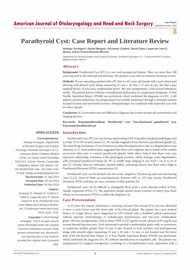
to have a cystic lesion near the lower pole of the thyroid gland. The patient has a past medical history of a right breast cancer diagnosed in 1997 treated with a modified radical mastectomy without adjuvant chemotherapy or radiotherapy; hypertension, and non-toxic multinodular goiter. She was asymptomatic, with an intact PTH of 44.5 pg/ml, serum calcium of 9.0 mg/dl, and normal thyroid function tests. Neck ultrasound reported a multinodular goiter with no prominent or suspicious nodules greater than 10 mm; it also showed an oval, anechoic and heterogeneous image with smooth edges, measuring 25 mm × 10 mm × 12 mm in size located near the lower pole of the left thyroid lobe (Figure 1). A Fine Needle Aspiration Biopsy (FNAB) was performed, which confirmed the diagnosis of a PC without identification of neoplastic cells. The patient was programmed for surgical management consisting of a focused/limited neck exploration with a
Rodrigo Arrangoiz, et al., American Journal of Otolaryngology and Head and Neck Surgery
Remedy Publications LLC. 2019 | Volume 2 | Issue 10 | Article 10752
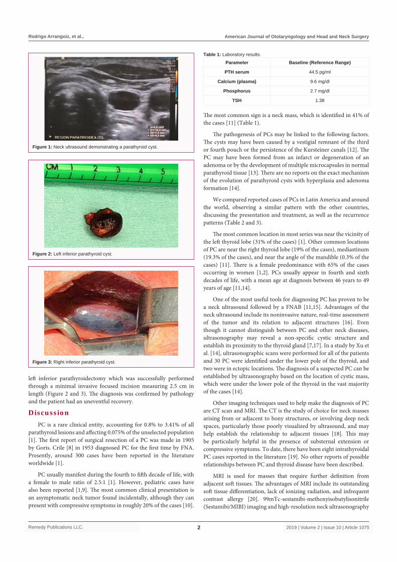
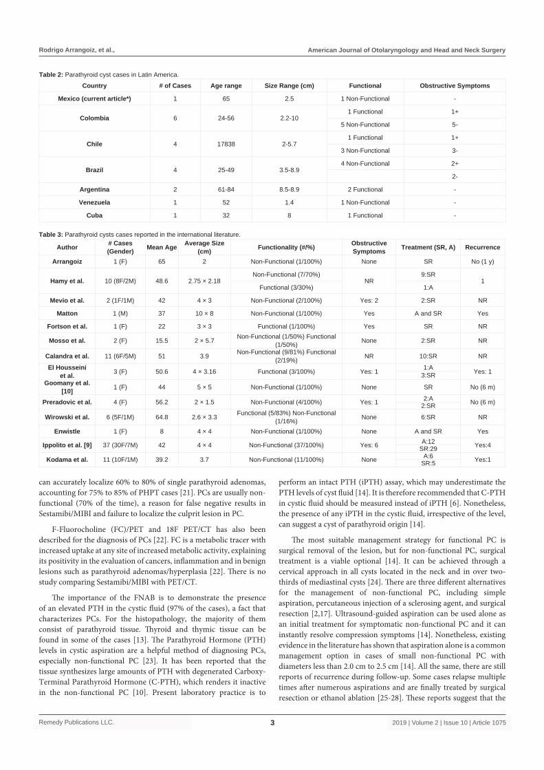
left inferior parathyroidectomy which was successfully performed through a minimal invasive focused incision measuring 2.5 cm in length (Figure 2 and 3). The diagnosis was confirmed by pathology and the patient had an uneventful recovery.
DiscussionPC is a rare clinical entity, accounting for 0.8% to 3.41% of all
parathyroid lesions and affecting 0.075% of the unselected population [1]. The first report of surgical resection of a PC was made in 1905 by Goris. Crile [8] in 1953 diagnosed PC for the first time by FNA. Presently, around 300 cases have been reported in the literature worldwide [1].
PC usually manifest during the fourth to fifth decade of life, with a female to male ratio of 2.5:1 [1]. However, pediatric cases have also been reported [1,9]. The most common clinical presentation is an asymptomatic neck tumor found incidentally, although they can present with compressive symptoms in roughly 20% of the cases [10].
The most common sign is a neck mass, which is identified in 41% of the cases [11] (Table 1).
The pathogenesis of PCs may be linked to the following factors. The cysts may have been caused by a vestigial remnant of the third or fourth pouch or the persistence of the Kursteiner canals [12]. The PC may have been formed from an infarct or degeneration of an adenoma or by the development of multiple microcapsules in normal parathyroid tissue [13]. There are no reports on the exact mechanism of the evolution of parathyroid cysts with hyperplasia and adenoma formation [14].
We compared reported cases of PCs in Latin America and around the world, observing a similar pattern with the other countries, discussing the presentation and treatment, as well as the recurrence patterns (Table 2 and 3).
The most common location in most series was near the vicinity of the left thyroid lobe (31% of the cases) [1]. Other common locations of PC are near the right thyroid lobe (19% of the cases), mediastinum (19.3% of the cases), and near the angle of the mandible (0.3% of the cases) [11]. There is a female predominance with 65% of the cases occurring in women [1,2]. PCs usually appear in fourth and sixth decades of life, with a mean age at diagnosis between 46 years to 49 years of age [11,14].
One of the most useful tools for diagnosing PC has proven to be a neck ultrasound followed by a FNAB [11,15]. Advantages of the neck ultrasound include its noninvasive nature, real-time assessment of the tumor and its relation to adjacent structures [16]. Even though it cannot distinguish between PC and other neck diseases, ultrasonography may reveal a non-specific cystic structure and establish its proximity to the thyroid gland [7,17]. In a study by Xu et al. [14], ultrasonographic scans were performed for all of the patients and 30 PC were identified under the lower pole of the thyroid, and two were in ectopic locations. The diagnosis of a suspected PC can be established by ultrasonography based on the location of cystic mass, which were under the lower pole of the thyroid in the vast majority of the cases [14].
Other imaging techniques used to help make the diagnosis of PC are CT scan and MRI. The CT is the study of choice for neck masses arising from or adjacent to bony structures, or involving deep neck spaces, particularly those poorly visualized by ultrasound, and may help establish the relationship to adjacent tissues [18]. This may be particularly helpful in the presence of substernal extension or compressive symptoms. To date, there have been eight intrathyroidal PC cases reported in the literature [19]. No other reports of possible relationships between PC and thyroid disease have been described.
MRI is used for masses that require further definition from adjacent soft tissues. The advantages of MRI include its outstanding soft tissue differentiation, lack of ionizing radiation, and infrequent contrast allergy [20]. 99mTc-sestamibi-methoxyisobutylisonitrile (Sestamibi/MIBI) imaging and high-resolution neck ultrasonography
Figure 1: Neck ultrasound demonstrating a parathyroid cyst.
Figure 2: Left inferior parathyroid cyst.
Figure 3: Right inferior parathyroid cyst.
Parameter Baseline (Reference Range)
PTH serum 44.5 pg/ml
Calcium (plasma) 9.6 mg/dl
Phosphorus 2.7 mg/dl
TSH 1.38
Table 1: Laboratory results.
Rodrigo Arrangoiz, et al., American Journal of Otolaryngology and Head and Neck Surgery
Remedy Publications LLC. 2019 | Volume 2 | Issue 10 | Article 10753
can accurately localize 60% to 80% of single parathyroid adenomas, accounting for 75% to 85% of PHPT cases [21]. PCs are usually non-functional (70% of the time), a reason for false negative results in Sestamibi/MIBI and failure to localize the culprit lesion in PC.
F-Fluorocholine (FC)/PET and 18F PET/CT has also been described for the diagnosis of PCs [22]. FC is a metabolic tracer with increased uptake at any site of increased metabolic activity, explaining its positivity in the evaluation of cancers, inflammation and in benign lesions such as parathyroid adenomas/hyperplasia [22]. There is no study comparing Sestamibi/MIBI with PET/CT.
The importance of the FNAB is to demonstrate the presence of an elevated PTH in the cystic fluid (97% of the cases), a fact that characterizes PCs. For the histopathology, the majority of them consist of parathyroid tissue. Thyroid and thymic tissue can be found in some of the cases [13]. The Parathyroid Hormone (PTH) levels in cystic aspiration are a helpful method of diagnosing PCs, especially non-functional PC [23]. It has been reported that the tissue synthesizes large amounts of PTH with degenerated Carboxy-Terminal Parathyroid Hormone (C-PTH), which renders it inactive in the non-functional PC [10]. Present laboratory practice is to
perform an intact PTH (iPTH) assay, which may underestimate the PTH levels of cyst fluid [14]. It is therefore recommended that C-PTH in cystic fluid should be measured instead of iPTH [6]. Nonetheless, the presence of any iPTH in the cystic fluid, irrespective of the level, can suggest a cyst of parathyroid origin [14].
The most suitable management strategy for functional PC is surgical removal of the lesion, but for non-functional PC, surgical treatment is a viable optional [14]. It can be achieved through a cervical approach in all cysts located in the neck and in over two-thirds of mediastinal cysts [24]. There are three different alternatives for the management of non-functional PC, including simple aspiration, percutaneous injection of a sclerosing agent, and surgical resection [2,17]. Ultrasound-guided aspiration can be used alone as an initial treatment for symptomatic non-functional PC and it can instantly resolve compression symptoms [14]. Nonetheless, existing evidence in the literature has shown that aspiration alone is a common management option in cases of small non-functional PC with diameters less than 2.0 cm to 2.5 cm [14]. All the same, there are still reports of recurrence during follow-up. Some cases relapse multiple times after numerous aspirations and are finally treated by surgical resection or ethanol ablation [25-28]. These reports suggest that the
Country # of Cases Age range Size Range (cm) Functional Obstructive Symptoms
Mexico (current article*) 1 65 2.5 1 Non-Functional -
Colombia 6 24-56 2.2-101 Functional 1+
5 Non-Functional 5-
Chile 4 17838 2-5.71 Functional 1+
3 Non-Functional 3-
Brazil 4 25-49 3.5-8.94 Non-Functional 2+
2-
Argentina 2 61-84 8.5-8.9 2 Functional -
Venezuela 1 52 1.4 1 Non-Functional -
Cuba 1 32 8 1 Functional -
Table 2: Parathyroid cyst cases in Latin America.
Author # Cases (Gender) Mean Age Average Size
(cm) Functionality (#/%) Obstructive Symptoms Treatment (SR, A) Recurrence
Arrangoiz 1 (F) 65 2 Non-Functional (1/100%) None SR No (1 y)
Hamy et al. 10 (8F/2M) 48.6 2.75 × 2.18Non-Functional (7/70%)
NR9:SR
1Functional (3/30%) 1:A
Mevio et al. 2 (1F/1M) 42 4 × 3 Non-Functional (2/100%) Yes: 2 2:SR NR
Matton 1 (M) 37 10 × 8 Non-Functional (1/100%) Yes A and SR Yes
Fortson et al. 1 (F) 22 3 × 3 Functional (1/100%) Yes SR NR
Mosso et al. 2 (F) 15.5 2 × 5.7 Non-Functional (1/50%) Functional (1/50%) None 2:SR NR
Calandra et al. 11 (6F/5M) 51 3.9 Non-Functional (9/81%) Functional (2/19%) NR 10:SR NR
El Housseini et al. 3 (F) 50.6 4 × 3.16 Functional (3/100%) Yes: 1 1:A Yes: 13:SR
Goomany et al. [10] 1 (F) 44 5 × 5 Non-Functional (1/100%) None SR No (6 m)
Preradovic et al. 4 (F) 56.2 2 × 1.5 Non-Functional (4/100%) Yes: 1 2:A No (6 m)2:SR
Wirowski et al. 6 (5F/1M) 64.8 2.6 × 3.3 Functional (5/83%) Non-Functional (1/16%) None 6:SR NR
Enwistle 1 (F) 8 4 × 4 Non-Functional (1/100%) None A and SR Yes
Ippolito et al. [9] 37 (30F/7M) 42 4 × 4 Non-Functional (37/100%) Yes: 6 A:12 Yes:4SR:29
Kodama et al. 11 (10F/1M) 39.2 3.7 Non-Functional (11/100%) None A:6 Yes:1SR:5
Table 3: Parathyroid cysts cases reported in the international literature.
Rodrigo Arrangoiz, et al., American Journal of Otolaryngology and Head and Neck Surgery
Remedy Publications LLC. 2019 | Volume 2 | Issue 10 | Article 10754
larger the cyst is, the more likely it is to relapse after the procedure [25-28]. Injections of sclerosing agents are not recommended because they can flow out from the capsule and cause serious complications such as fiber degeneration or damage to the recurrent laryngeal nerve, resulting in vocal cord paralysis [28,29]. Some authors recommended that the optimal treatment for symptomatic non-functional PCs is surgical resection [5,30]. There is evidence in the literature that has shown that surgical resection is an effective and safe treatment for non-functional PCs with diameters equal or greater than 2.5 cm [14].
ConclusionCystic lesions located under the lower pole of the thyroid
gland should be considered to have originated at the parathyroid gland. Ultrasound guided cystic aspiration with PTH detection or postoperative immunopathology can help with the differential diagnosis. Resection of PC is still a common practice and effective treatment with minimal morbidity.
References1. Cappelli C, Rotondi M, Pirola I, De Martino E, Leporati P, Magri F, et
al. Prevalence of parathyroid cysts by neck ultrasound scan in unselected patients. J Endocrinol Invest. 2009;32(4):357-9.
2. Arduc A, Tutuncu YA, Dogan BA, Arikan Ileri AB, Tuna MM, Ozcan HN, et al. Parathyroid cysts. Am Surg. 2015;81(4):E163-5.
3. Fortson JK, Patel VG, Henderson VJ. Parathyroid cysts: a case report and review of the literature. Laryngoscope. 2001;111(10):1726-8.
4. Bilezikian JP, Marcus R, Levine M, Marcocci C, Silverberg SJ, Potts J. Parathyroids. 3rd ed. Basic and Clinical Concepts: Elsevier Inc; 2015;946.
5. Fustar Preradovic L, Danic D, Dzodic R. Small nonfunctional parathyroid cysts: Single institution experience. Endocr J. 2017;64(2):151-6.
6. Lee SL. Parathyroid cyst fluid: Discrepancy between C-terminal and intact parathyroid hormone assays. Thyroid. 2000;10(12):1125-6.
7. Ujiki M, Sturgeon C, Nayar R, Angelos P. Parathyroid cyst: often mistaken for a thyroid cyst. World J Surg. 2008;32(6):1234.
8. Crile G Jr, Perryman RG. Parathyroid cysts: Report of five cases. Surgery. 1953;34(1):151-4.
9. Ippolito G, Palazzo FF, Sebag F, Sierra M, De Micco C, Henry JF. A single-institution 25-year review of true parathyroid cysts. Langenbecks Arch Surg. 2006;391(1):13-8.
10. Goomany A, Rafferty A, Smith I. An unusual neck mass: A case of a parathyroid cyst and review of the literature. Case Rep Surg. 2015;2015:243527.
11. Papavramidis TS, Chorti A, Pliakos I, Panidis S, Michalopoulos A. Parathyroid cysts: A review of 359 patients reported in the international literature. Medicine (Baltimore). 2018;97(28):e11399.
12. Rossi ED, Revelli L, Giustozzi E, Straccia P, Stigliano E, Lombardi CP, et al. Large non-functioning parathyroid cysts: Our institutional experience of a rare entity and a possible pitfall in thyroid cytology. Cytopathology, 2015;26(2):114-21.
13. Pontikides N, Karras S, Kaprara A, Cheva A, Doumas A, Botsios D, et al. Diagnostic and therapeutic review of cystic parathyroid lesions. Hormones (Athens), 2012;11(4):410-8.
14. Xu P, Xia X, Li M, Guo M, Yang Z. Parathyroid cysts: experience of a rare phenomenon at a single institution. BMC Surg. 2018;18(1):9.
15. Collins B, Stoner JA, Digoy GP. Benefits of ultrasound vs. computed tomography in the diagnosis of pediatric lateral neck abscesses. Int J Pediatr Otorhinolaryngol, 2014;78(3):423-6.
16. Gritzmann N, Hollerweger A, Macheiner P, Rettenbacher T. Sonography of soft tissue masses of the neck. J Clin Ultrasound. 2002;30(6):356-73.
17. Sung JY. Parathyroid ultrasonography: The evolving role of the radiologist. Ultrasonography. 2015;34(4):268-74.
18. McCoy KL, Yim JH, Zuckerbraun BS, Ogilvie JB, Peel RL, Carty SE. Cystic parathyroid lesions: Functional and nonfunctional parathyroid cysts. Arch Surg. 2009;144(1):52-6.
19. Ahmad MM, Almohaya M, Almalki MH, Aljohani N. Intrathyroidal parathyroid Cyst: An unusual neck mass. Clin Med Insights Endocrinol Diabetes, 2017;10:1179551417698135.
20. Ishikawa M, Anzai Y. MR imaging of lymph nodes in the head and neck. Neuroimaging Clin N Am. 2004;14(4):679-94.
21. Dutta D, Selvan C, Kumar M, Datta S, Narayan Das R, Ghoshet S, et al. Needle aspirates PTH in diagnosis of primary hyperparathyroidism due to intrathyroidal parathyroid cyst. Endocrinol Diabetes Metab Case Rep. 2013;2013:130019.
22. Michaud L, Burgess A, Huchet V, Lefèvre M, Tassart M, Ohnona J, et al. Is 18F-fluorocholine-positron emission tomography/computerized tomography a new imaging tool for detecting hyperfunctioning parathyroid glands in primary or secondary hyperparathyroidism? J Clin Endocrinol Metab. 2014;99(12):4531-6.
23. Caleo A, Vitale M, Valvano L, Siano M, Angrisani B, Forlenza M, et al. Fine needle cytology pre-surgical differentiation of parathyroid neoplasms: Is it reliable? Cytopathology. 2017;28(4):273-9.
24. Shields TW, Immerman SC. Mediastinal parathyroid cysts revisited. Ann Thorac Surg. 1999;67(2):581-90.
25. Katz AD, Dunkleman D. Needle aspiration of nonfunctioning parathyroid cysts. Arch Surg. 1984;119(3):307-8.
26. Okamura K, Ikenoue H, Sato K, Yoshinari M, Nakagawa M, Kuroda T, et al. Sclerotherapy for benign parathyroid cysts. Am J Surg. 1992;163(3):344-5.
27. Shi B, Guo H, Tang N. Treatment of parathyroid cysts with fine-needle aspiration. Ann Intern Med. 1999;131(10):797-8.
28. Sung JY, Baek JH, Kim KS, Lee D, Ha EJ, Lee JH. Symptomatic nonfunctioning parathyroid cysts: Role of simple aspiration and ethanol ablation. Eur J Radiol. 2013;82(2):316-20.
29. Lorenzo J, Fernández G, Iglesias B, Boente R, Sas M. Recurrent parathyroid cyst: A clinical case. An Med Interna. 2008;25(5):231-3.
30. Roman-Gonzalez A, Aristizábal N, Aguilar C, Palacios K, Camilo Pérez, Vélez-Hoyos A, et al. Parathyroid cysts: the Latin-American experience. Gland Surg. 2016;5(6):559-64.