parasitology international - github pages
TRANSCRIPT

Contents lists available at ScienceDirect
Parasitology International
journal homepage: www.elsevier.com/locate/parint
Emendation and new species of Hapalorhynchus Stunkard, 1922 (Digenea:Schistosomatoidea) from musk turtles (Kinosternidae: Sternotherus) inAlabama and Florida rivers
Jackson R. Robertsa, Kenneth M. Halanychb, Cova R. Ariasc, Brian Foltd, Jeffrey M. Goesslingd,Stephen A. Bullarda,⁎
a Aquatic Parasitology Laboratory, School of Fisheries, Aquaculture, and Aquatic Sciences, College of Agriculture, Auburn University, 203 Swingle Hall, Auburn, AL36849, USAb Department of Biological Sciences and Molette Biology Laboratory for Environmental & Climate Change Studies, Auburn University, 101 Rouse Life Sciences Building,Auburn, AL 36830, USAc Aquatic Microbiology Laboratory, School of Fisheries, Aquaculture, and Aquatic Sciences, Auburn University, 559 Duvall Drive, Auburn, AL 36832, USAd Department of Biological Sciences, Auburn University, 331 Funchess Hall, Auburn, AL 36849, USA
A R T I C L E I N F O
Keywords:Turtle blood flukeMorphology28SMud turtleNorth America
A B S T R A C T
Hapalorhynchus Stunkard, 1922 is emended based on morphological study of existing museum specimens (typeand voucher specimens) and newly-collected specimens infecting musk turtles (Testudines: Kinosternidae:Sternotherus spp.) from rivers in Alabama and Florida (USA). Hapalorhynchus conecuhensis n. sp. is described froman innominate musk turtle, Sternotherus cf. minor, (type host) from Blue Spring (31°5′27.64″N, 86°30′53.21″W;Pensacola Bay Basin, Alabama) and the loggerhead musk turtle, Sternotherus minor (Agassiz, 1857) from theWacissa River (30°20′24.73″N, 83°59′27.56″W; Apalachee Bay Basin, Florida). It differs from congeners bylacking a body constriction at level of the ventral sucker, paired anterior caeca, and a transverse ovary as well asby having a small ventral sucker, proportionally short posterior caeca, nearly equally-sized anterior and pos-terior testes, a small cirrus sac, and a uterus extending dorsal to the ovary and the anterior testis. Specimens ofHapalorhynchus reelfooti Byrd, 1939 infected loggerhead musk turtles, stripe-necked musk turtles (Sternotheruspeltifer Smith and Glass, 1947), Eastern musk turtles (Sternotherus odoratus [Latreille in Sonnini and Latreille,1801]), and S. cf. minor. Those of Hapalorhynchus cf. stunkardi infected S. minor and S. odoratus. Sternothorusminor, S. peltifer, and S. cf. minor plus S. minor and S. odoratus are new host records for H. reelfooti and H. cf.stunkardi, respectively. This is the first report of an infected musk turtle from the Coosa and Tallapoosa Rivers(Mobile-Tensaw River Basin), Pensacola Bay Basin, or Apalachee Bay Basin. Sequence analysis of the largesubunit rDNA (28S) showed a strongly-supported clade for Hapalorhynchus.
1. Introduction
Hapalorhynchus Stunkard, 1922 includes 19 accepted species thatinfect both snapping turtles (Testudines: Chelydridae) as well as muskand mud turtles (Kinosternidae) from the Nearctic realm, side-neckedturtles (Pelomedusidae) from the Ethiopian realm, and both pond tur-tles (Geoemydidae) and softshell turtles (Trionychidae) from theIndomalayan realm (Smith [1,2]; Platt and Snyder [3]; Platt andSharma [4]; Roberts et al. [5]). Three of the six species of Hapa-lorhynchus reported from North America infect kinosternids: Hapa-lorhynchus reelfooti Byrd, 1939 from the Eastern musk turtle (Ster-notherus odoratus [Latreille in Sonnini and Latreille, 1801]),
Hapalorhynchus stunkardi Byrd, 1939 from the razor-backed musk turtle(Sternotherus carinatus [Gray, 1856]), and Hapalorhynchus albertoi La-mothe-Argumedo, 1978 from the white- lipped mud turtle (Kinosternonleucostomum Duméril and Bibron in Duméril and Duméril, 1851). Thepresent work focuses on musk turtles (Sternotherus spp.) in Alabama andFlorida because only two of the six North American musk turtles hadpreviously been reported as blood fluke hosts and no infected muskturtle had been reported from Alabama or Florida. Herein, loggerheadmusk turtles (Sternotherus minor [Agassiz, 1857]), stripe-necked muskturtles (Sternotherus peltifer Smith and Glass, 1947), Eastern musk tur-tles, and specimens of an innominate loggerhead musk turtle (Ster-notherus cf. minor) were examined for blood fluke infections.
http://dx.doi.org/10.1016/j.parint.2017.09.001Received 8 May 2017; Received in revised form 25 August 2017; Accepted 3 September 2017
⁎ Corresponding author.E-mail address: [email protected] (S.A. Bullard).
Parasitology International 66 (2017) 748–760
Available online 08 September 20171383-5769/ © 2017 Elsevier B.V. All rights reserved.
MARK

Table1
Hap
alorhy
nchu
sspp.
infectingmud
andmuskturtles(K
inosternidae).
Turtle
host
Bloo
dfluk
eSite
inho
stRiverinelocality
Accession
nos.
Referen
ce
Kinosternon
leucostomum
Dum
érilan
dBibron
inDum
érilan
dDum
éril,
1851
H.a
lbertoiL
amothe
-Argum
edo,
1978
bloo
d(adu
lt)
GrijalvaRiver,V
illah
ermosa,
Taba
sco,
Mexico
Colección
Helmintológica
delInstitutode
Biolog
íade
laUNAM
229-19
,229
-20
Lamothe
-Argum
edo
[7]
Sterno
therus
carina
tus(G
ray,
1856
)(proba
bly
S.minor;s
eeSe
ction3.4.3Rem
arks)
H.stunk
ardi
Byrd,1
939
pulm
onarybloo
dve
ssels(adu
lt)
North
Oco
neeRiver,A
then
s,Geo
rgia,U
SAUSN
M13
2196
7By
rd[8]
Sterno
therus
minor
(Aga
ssiz,18
57)
H.reelfootiByrd,
1939
mesen
tericbloo
dve
ssels,
viscerawash(adu
lt)
Wac
issa
River
(tribu
tary
ofAuc
illa
River)
(30°19
′39.80
″N,83
°59′6.13
″W),
Flor
ida,
USA
Retaine
dby
Bullard
labo
ratory
Presen
tstud
y
Wac
issa
River
(30°20
′24.73
″N,83
°59′27
.56″
W)
USN
M14
3760
6Pr
esen
tstud
yH.cf.s
tunk
ardi
heart(adu
lt)
Wac
issa
River
(30°20
′24.73
″N,83
°59′27
.56″
W)
USN
M14
3761
0Pr
esen
tstud
yH.co
necu
hensis
n.sp
.he
art(adu
lt)
Wac
issa
River
(30°20
′24.73
″N,83
°59′27
.56″
W)
Gen
Ban
kMF5
6803
7Pr
esen
tstud
ySterno
therus
cf.minor
H.reelfooti
mesen
tericbloo
dve
ssels,
viscerawash,
heart(adu
lt)
BlueSp
ring
(Yellow
River)(31°5′27
.64″
N,
86°30′53
.21″
W),
Alaba
ma,
USA
USN
M14
3760
7Pr
esen
tstud
y
H.co
necu
hensis
heart,
viscerawash(adu
lt)
BlueSp
ring
(31°5′27
.64″
N,86
°30′53
.21″
W)
USN
M14
3761
2−
1437
614
Presen
tstud
ySterno
therus
odoratus
(Latreillein
Sonn
ini
andLa
treille
,180
1)H.reelfo
oti
bloo
d(adu
lt)
Reelfoo
tLa
ke(M
ississippi
River)(36°21
′12.23
″N,
89°25′21
.50″W),Te
nnessee,
USA
USN
M13
2196
8By
rd[8]
viscerawash(adu
lt)
North
Cha
inLa
ke(K
anka
keeRiver)(41°41
′57.03
″N,
86°22′16
.51″W),Indian
a,USA
USN
M13
9385
5Platta
ndSn
yder
[3]
viscerawash(adu
lt)
Westham
pton
Lake
(Jam
esRiver)(37°34
′38.01
″N,
77°32′26
.90″W),Richm
ond,
Virginia,
USA
USN
M13
9385
7Platta
ndSn
yder
[3]
viscerawash(adu
lt)
Can
oeLa
ke(C
oosa
River)(33°47
′56.16
″N,
86°29′25
.02″
W),
Alaba
ma
USN
M14
3760
8Pr
esen
tstud
y
mesen
tericbloo
dve
ssels
(adu
lt)
Pond
offOdo
mCreek
(Tallapo
osaRiver)
(32°30
′9.58″
N,85
°26′6.07
″W),
Alaba
ma
Gen
Ban
kMF5
6803
0Pr
esen
tstud
y
H.cf.stun
kardi
heart(adu
lt)
Pond
offPa
rkerso
nMillC
reek
(Tallapo
osaRiver)
(32°35
′20.04
″N,85
°29′41
.51″
W),
Alaba
ma
USN
M14
3761
1Pr
esen
tstud
y
Sterno
therus
peltifer
(Smithan
dGlass,
1947
)H.reelfooti
mesen
tericbloo
dve
ssels,
lung
(adu
lt)
Che
wac
laCreek
(Tallapo
osaRiver)
(32°31
′58.64
″N,85
°29′53
.09″
W),
Alaba
ma
USN
M14
3760
9Pr
esen
tstud
y
Bold
indicatesreco
rdsco
mprisingthepresen
tstud
y.
J.R. Roberts et al. Parasitology International 66 (2017) 748–760
749
Hapalorhynchus reelfooti Byrd, 1939, Hapalorhynchus cf. stunkardi, and anew species of Hapalorhynchus were identified. Based on these newcollections and borrowed type materials of related blood flukes, weemend Hapalorhynchus, describe the new congener (the first new spe-cies of the genus described from North America in 29 years; Platt [6]),report the new host and geographic locality records for H. reelfooti, andprovide a large subunit ribosomal DNA (28S) phylogeny for these taxa.
2. Materials and methods
2.1. Specimen collection and preparation
Turtles were collected from the Coosa River (Canoe Lake),Tallapoosa River (Odom Creek, Parkerson Mill Creek, Chewacla Creek),and Yellow River (Blue Spring) in Alabama as well as the Aucilla River(Wacissa River) in Florida (Table 1). Two loggerhead musk turtles werecaptured by hand from two sites (30°20′24.73″N, 83°59′27.56″W;30°19′39.80″N, 83°59′6.13″W, respectively) in the Wacissa River on 23April 2016. One Eastern musk turtle was captured with a monitored,baited hoop-net from Canoe Lake (33°47′56.16″N, 86°29′25.02″W), onewas captured by hand from a pond off Parkerson Mill Creek(32°35′20.04″N, 85°29′41.51″W), and two were captured by hand froma pond off Odom Creek (32°30′9.58″N, 85°26′6.07″W) on 24 June 2015,7 September 2015, and 22−24 September 2015, respectively. Twospecimens of the innominate loggerhead musk turtle were captured byhand from Blue Spring (31°5′27.64″N, 86°30′53.21″W) on 19 May2016. One stripe-necked musk turtle was captured from ChewaclaCreek (32°31′58.64″N, 85°29′53.09″W) on 15 July 2015. All turtleswere decapitated immediately upon returning to the laboratory. Ne-cropsies were performed with aid of 7.0 g/L sodium citrate saline so-lution to prevent blood coagulation. Live turtle blood flukes (TBFs)intended as whole mounts for morphology were heat-killed on a glassslide with coverslip (no coverslip pressure) using a butane hand lighter.Resulting specimens were transferred and maintained in 5% neutralbuffered formalin for morphology or placed in 95% non-denaturedethanol (EtOH) for molecular biology (see below). Specimens formorphology were rinsed in distilled water, stained in Van Cleave's he-matoxylin with several drops of Ehrlich's hematoxylin, dehydratedthrough an ethanol series, made basic at 70% EtOH with lithium car-bonate and butly-amine, cleared in clove oil, and permanently mountedin Canada balsam. The resulting whole mounts were examined and il-lustrated with Leica DM 2500 and Leica DMR (Leica, Wetzler, Ger-many) microscopes each equipped with differential interference con-trast (DIC) optical components, an ocular micrometer, and a drawingtube. Measurements of the TBF specimens are reported in micrometers(μm) followed by the mean and number of specimens measured inparentheses. Measurements for the new species are reported for theholotype and paratype, respectively. Those for Hapalorhynchus cf.stunkardi are reported separately for each voucher specimen (NationalMuseum of National History, Smithsonian Department of InvertebrateZoology [USNM] 1437610 and 1437611, respectively), and those forthe holotype of H. stunkardi (USNM 1321967) immediately follow inbrackets (“n/a” indicates not available due to poor specimen quality).All of the newly-collected specimens were deposited in the USNM.Turtle scientific and common names follow Spinks et al. [9], van Dijket al. [10], and Guyer et al. [11]. Classification and anatomical termsfor TBFs follow Roberts et al. [5,12,13] and Platt [6] except that‘anterolateral diverticula’ are herein referred to as ‘anterior caeca’.
2.2. DNA extraction, amplification, and sequencing
Specimens for molecular analyses were handled with camel hairbrushes, fine-tipped forceps, or syringe tips and immediately preservedin a vial of 95% EtOH and stored at −20 °C. Total genomic DNA(gDNA) was extracted using DNeasyTM Blood and Tissue Kit (Qiagen,Valencia, California, USA) according to the manufacturer's protocol
except that the incubation period with proteinase-K was extended toovernight and that the final elution step was performed using only100 μL of elution buffer to increase the final DNA concentration. Thepartial 28S rDNA (domains D1–D3; ~1400 bp) was amplified using theforward primer “U178” (5′-GCA CCC GCT GAA YTT AAG-3′) and thereverse primer “L1642” (5′-CCA GCG CCATCCATT TTC A-3′) (Lockyeret al. [14]). PCR amplifications were performed using a total volume of50 μl with 2 μl of DNA template, 0.4 μM of each primer along with 1×buffer, 2.5 mM MgCl2, 1 mM dNTP mixture, and 0.3 μl Taq polymerase(5 U/μl) (Promega, Madison, Wisconsin, USA). The thermocyclingprofile comprised an initial 5 min at 95 °C for denaturation, followed by40 repeating cycles of 94 °C for 30 s for denaturation, 50 °C for 30 s forannealing, and 72 °C for 2 min for extension, followed by a final fivemin at 72 °C for extension. All PCR reactions were carried out in a MJResearch PTC-200 (BioRad, Hercules, California, USA). PCR products(10 μl) were verified on a 1% agarose gel and stained with ethidiumbromide. PCR products were purified by microcentrifuge with theQIAquick PCR Purification Kit (Qiagen, Valencia, California, USA) ac-cording to manufacturer's protocol, except that the last elution step wasperformed with autoclaved nanopure H2O rather than the providedbuffer. DNA sequencing was performed by ACGT, Incorporated(Wheeling, Illinois, USA). Reactions were sequenced using BigDye ter-minator version 3.1, cleaned-up with magnetic beads (CleanSeq dyeterminator removal kit), and analyzed using ABI 3730 XL or 3730 Ge-netic Analyzer. Primers used in sequencing of 28S rDNA included thePCR primers and the reverse primer 1200R (5′-GCATAGTTCACCATC-TTTCGG-3′) (Lockyer et al. [14]). Sequence assembly and analysis ofchromatograms were performed with BioNumerics version 7.0 (AppliedMaths, Saint-Martens- Latem, Belgium).
2.3. Sequence alignments and phylogenetic analyses
Assembled sequences (Table 2) were aligned with MAFFT 7.310(Katoh and Standley [17]) and subsequently corrected by eye in Mes-quite 3.2 (Maddison and Maddison [18]). Regions that could not beunambiguously aligned were excluded from further analyses. BayesianInference was performed with MrBayes 3.5.3 (Ronquist et al. [19]).Using a GTR + Gamma model, 4 runs of 4 chains each were conductedfor 1,000,000 generations. Priors were set to default values and burninwas set to 25% of generations (or 250,000). Chains were run until theaverage standard deviation of split frequencies was below 0.01. Theresulting phylogenetic tree was viewed using FigTree v1.4.3 (Rambaut[20]) and then edited in Adobe Illustrator CC 2015.3 (Adobe Systems).
3. Results
3.1. Hapalorhynchus Stunkard, 1922, emended (Figs. 1-7)
3.1.1. DiagnosisBody dorsoventrally flat (not cylindrical), 3.7−17.0 × longer than
wide; body constriction at level of ventral sucker present or absent;hindbody 1.4−3.3 × longer than forebody, aspinous; small ventralbody papillae present or absent; massive, ventrolateral tegumentalpapillae absent. Oral sucker robust, demarcated from body by con-striction; oral sucker spines present or absent; papillae present or ab-sent. Ventral sucker present, aspinous. Pharynx present, envelopinganterior extremity of oesophagus. Oesophagus extending posteriad 1/10−1/4 of body length, ventral to anterior nerve commissure; lateraloesophageal diverticula present or absent; median oesophageal di-verticulum absent; oesophageal gland surrounding oesophagus frompharynx to caecal bifurcation. Intestine comprising non-fused posteriorcaeca bifurcating anterior to ventral sucker; paired anterior caecapresent (in Hapalorhynchus brooksi Platt, 1988) or absent; posteriorcaeca inverse U-shaped, extending 1/2−3/4 of body length directlyposteriad, not extensively convoluted, terminating in posterior bodyextremity. Testes comprising one anterior and one posterior testis, in
J.R. Roberts et al. Parasitology International 66 (2017) 748–760
750

Table2
Largesubu
nitribo
somal
DNA
(28S
)sequ
encesused
herein.
Turtle
bloo
dfluk
eTu
rtle
host
Locality
Gen
Bank
Accession
No.
Referen
ce
Coeuritrem
aplattiRob
erts
andBu
llard,2
016
Pelodiscus
sinensis(W
iegm
ann,
1835
)DaRan
gRiver
Basin,
PhuYen
Prov
ince,Vietnam
KX71
2243
Rob
erts
etal.[
5]Hap
alorhy
nchu
sgracilisStun
kard,1
922
Chelydraserpentin
aLinn
aeus,1
758
Reelfoo
tLa
ke(M
ississippi
River)(36°21
′12.23
″N,8
9°25
′21.50
″W),
Tenn
essee,
USA
AY60
4710
Snyd
er[15]
Hap
alorhy
nchu
sfolio
rchisBroo
ksan
dMay
es,
1975
C.serpentina
Pond
offSa
ugah
atch
eeCreek
(Tallapo
osaRiver)(32°39
′1.36″N,
85°29′4.70
″W),Alaba
ma,
USA
KX71
2242
Rob
erts
etal.[
5]
Hap
alorhy
nchu
sreelfootiByrd,
1939
Sterno
therus
minor
(Aga
ssiz,18
57)
Wac
issa
River
(tribu
tary
ofAuc
illa
River)(30°20
′24.73
″N,
83°59′27
.56″
W),
Flor
ida,
USA
MF5
6803
2Pr
esen
tstud
y
MF5
6803
3Pr
esen
tstud
ySterno
therus
cf.minor
BlueSp
ring
(Yellow
River)(31°5′27
.64″
N,86
°30′53
.21″
W),
Alaba
ma,
USA
MF5
6803
4Pr
esen
tstud
y
MF5
6803
5Pr
esen
tstud
ySterno
therus
odoratus
(Latreille
inSo
nninian
dLa
treille,
1801
)Can
oeLa
ke(C
oosa
River)(33°47
′56.16
″N,86
°29′25
.02″
W),
Alaba
ma,
USA
MF5
6803
1Pr
esen
tstud
y
Pond
offOdo
mCreek
(Tallapo
osaRiver),
(32°30
′9.58″
N,
85°26′6.07
″W),
Alaba
ma,
USA
MF5
6803
0Pr
esen
tstud
y
Hap
alorhy
nchu
sco
necu
hensis
n.sp
.Sterno
therus
cf.minor
BlueSp
ring
(31°5′27
.64″
N,86
°30′53
.21″
W)
MF5
6803
6holog
enop
hore
Presen
tstud
yMF5
6803
8Pr
esen
tstud
yS.
minor
Wac
issa
River
(30°20
′24.73
″N,83
°59′27
.56″
W)
MF5
6803
7Pr
esen
tstud
ySchistosom
ajaponicum
(Katsurada
,190
4)Mus
musculusLinn
aeus,17
58expe
rimen
talinfection;
original
isolatefrom
Phillipines
Z465
04AY15
7607
Lockye
ret
al.[14
];Olson
etal.
[16]
Schistosom
aman
soni
Sambo
n,19
07M.m
usculus
expe
rimen
talinfection
AY15
7173
Lockye
ret
al.[
14]
Schistosom
asinensium
Pao,
1959
M.m
usculus
expe
rimen
talinfection;
original
isolatefrom
Mianz
hu,S
ichu
an,China
AY15
7251
Lockye
ret
al.[
14]
Bold
indicatesreco
rdsco
mprisingthepresen
tstud
y.
J.R. Roberts et al. Parasitology International 66 (2017) 748–760
751

posterior 2/3 of body, intercaecal, smooth or lobed. Vas deferens ex-tending anteriad and ventral to gonads before laterally expanding toform external seminal vesicle; external seminal vesicle posterior toventral sucker, intercaecal, anterior or dextral to genital pore; internalseminal vesicle present (when cirrus sac present); pars prostatica dif-ficult to discern, enveloping distal extremity of internal seminal vesiclewhen present. Cirrus sac absent or pre-testicular and directed posteriadif present. Ovary ovoid or triangular in outline, intercaecal, inter-testicular, transverse or sinistral. Oviduct emerging from dextral marginof ovary, extending directly posteriad or sinistrad; oviducal seminalreceptacle comprising middle portion of oviduct between ovary andposterior testis. Laurer's canal intercaecal, intertesticular; Laurer's canal
ph
esaog
esv
ov
ct
vr
ev
ep
at
scdc
250
µm
vln
cs
vs
os
cb
nc
vr
cgp
lvd
tvdosr
pt
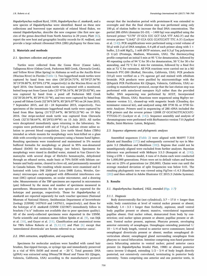
Fig. 1. Adult of Hapalorhynchus conecuhensis n. sp. from Sternotherus cf. minor, from BlueSpring (31°5′27.64″N, 86°30′53.21″W; Yellow River, Alabama, USA). Scale value asidebar. Holotype (National Museum of National History, Smithsonian Department ofInvertebrate Zoology [USNM] 1437612) of H. conecuhensis showing oral sucker (os),pharynx (ph), oesophagus (es), anterior oesophageal gland (aog), nerve commissure (nc),ventrolateral nerve chords (vln), posterior oesophageal gland (pog), vitellarium (vr),caecal bifurcation (cb), sinistral posterior caecum (sc), dextral posterior caecum (dc),ventral sucker (vs), external seminal vesicle (esv), cirrus sac (cs), vas deferens (vd),common genital pore (cgp), anterior testis (at), ovary (ov), lateral vitelline collecting duct(lvd), transverse vitelline collecting duct (tvd), oviducal seminal receptacle (osr), pos-terior testis (pt), caecal terminus (ct), excretory vesicle (ev), and excretory pore (ep).Ventral view.
ovov
osrosr
pve
tvdtvd
mtmt
esvesv
isvisvpppp
atat
vd
125
µm
tvdtvd
ptpt
eggegg
cscs
lvdlvd
lvdlvdlvdlvd
lvdlvd
cscs
pppp
ec
vtvt
ave
Lc
ci
cgpcgp
Fig. 2. Genitalia of adult Hapalorhynchus conecuhensis n. sp. from Sternotherus cf. minor,from Blue Spring (31°5′27.64″N, 86°30′53.21″W; Yellow River, Alabama, USA). Scalevalue aside bar. Genital complex of holotype (USNM 1437612) showing vas deferens (vd),external seminal vesicle (esv), cirrus (ci), cirrus sac (cs), pars prostatica (pp), internalseminal vesicle (isv), common genital pore (cgp), metraterm (mt), egg chamber (ec),uterine egg (egg), anterior trunk of vasa efferentia (ave), anterior testis (at), lateral vi-telline collecting duct (lvd), ovary (ov), transverse vitelline collecting duct (tvd), oviducalseminal receptacle (osr), primary vitelline collecting duct (vt), posterior trunk of vasaefferentia (pve), Laurer's canal (Lc), and posterior testis (pt). Ventral view.
J.R. Roberts et al. Parasitology International 66 (2017) 748–760
752

pore dorsal. Vitellarium follicular, ventrolateral to caeca and gonads,distributing from oesophagus to excretory vesicle or from caecal bi-furcation to ends of posterior caeca; transverse vitelline duct inter-testicular, ventral to ovary, comprising lateral collecting ducts ventralto caeca. Oötype with longitudinal axis parallel to that of body, inter-cecal. Mehlis' gland not observed. Uterus intercaecal, intertesticular,straight (not coiled), difficult to discern from oötype in gravid speci-mens, extending anteriad, dorsal or ventral to ovary; metratermnarrow, anterior to ovary, sinistral to anterior testis. Uterine pouchabsent.
Uterine egg single, tricornate or ovoid (with or without filaments),occupying oötype and uterus proximal to metraterm. Common genitalpore sinistral, dorsal. Excretory vesicle distinctly Y- shaped or sinuous(lacking multiple laterally-directed lobes). Manter's organ absent.Excretory pore terminal or dorsal.
Type species: Hapalorhynchus gracilis Stunkard, 1922
3.1.2. Differential diagnosisBody dorsoventrally flat (not cylindrical), aspinous; ventrolateral
tegumental papillae sensu Roberts et al. [5] absent. Oral sucker robust,spinous or aspinous. Pharynx present. Median oesophageal diverti-culum absent. Testes comprising one anterior and one posterior testis,intercaecal. Ovary ovoid or triangular in outline, intertesticular,transverse or sinistral. Uterus extending anteriad dorsal or ventral toovary; metraterm narrow. Common genital pore sinistral, dorsal. Ex-cretory vesicle distinctly Y-shaped or sinuous (lacking multiple
laterally-directed lobes). Manter's organ absent.
3.1.3. RemarksOur emended diagnosis of Hapalorhynchus adds or further details
numerous features that help differentiate it from that of other TBFgenera: details of the body surface and oral sucker, pharynx, anterior
ms30
µm
mo
op
op
op
vbp
Fig. 3. Oral sucker of Hapalorhynchus conecuhensis n. sp. from Sternotherus cf. minor col-lected from Blue Spring (31°5′27.64″N, 86°30′53.21″W; Yellow River, Alabama, USA).Scale value aside bar. Holotype (National Museum of National History, SmithsonianDepartment of Invertebrate Zoology [USNM] 1437612) showing oral sucker papillae (op),oral sucker spines (ss), mouth (mo), and ventral body papillae (vbp). Ventral view.
30 µ
m
vbp
opop
Fig. 4. Oral sucker of Hapalorhynchus conecuhensis n. sp. from Sternotherus cf. minor col-lected from Blue Spring (31°5′27.64″N, 86°30′53.21″W; Yellow River, Alabama, USA).Scale value aside bar. Paratype (USNM 1437613) showing oral sucker papillae (op) andventral body papillae (vbp). Dorsal view.
ph
og
es
vs
ov
ct
vr
ev
at
sc
dc
125
µm
vr
cs
esv
os
pve
nc
cb
cgp
vd
osrtvd
pt
ep
ut
isv
Fig. 5. Adult fluke from loggerhead musk turtles, Sternotherus minor, collected from theWacissa River (30°20′24.73″N, 83°59′27.56″W; tributary of Aucilla River, Florida, USA).Scale value aside bar. Hapalorhynchus reelfooti Stunkard, 1922 (National Museum ofNational History, Smithsonian Department of Invertebrate Zoology [USNM] 1437606)showing oral sucker (os), pharynx (ph), oesophageal gland (og), oesophagus (es), nervecommissure (nc), vitellarium (vr), caecal bifurcation (cb), dextral posterior caecum (dc),sinistral posterior caecum (sc), ventral sucker (vs), external seminal vesicle (esv), vasdeferens (vd), cirrus sac (cs), internal seminal vesicle (isv), common genital pore (cgp),anterior testis (at), uterus (ut), ovary (ov), transverse vitelline collecting duct (tvd),oviducal seminal receptacle (osr), posterior trunk of vasa efferentia (pve), posterior testis(pt), caecal terminus (ct), excretory vesicle (ev), and excretory pore (ep). Ventral view.
J.R. Roberts et al. Parasitology International 66 (2017) 748–760
753

caeca, oesophageal gland, vas deferens, internal seminal vesicle, parsprostatica, oviduct, transverse vitelline duct, oötype, metraterm, andexcretory pore. Yet, the genus likely needs additional revisionary work:its 19 accepted species are unusual among TBF genera in that theircollective geographic distributions include North America, Asia, andAfrica (Platt and Sharma [4]). The North American species of Hapa-lorhynchus infect snapping turtles (Chelydridae, infecting one of fourspecies belonging to one of two genera) as well as the mud and muskturtles (Kinosternidae, infecting three of 26 species belonging to bothgenera; Table 1): Hapalorhynchus gracilis Stunkard, 1922 (type species),H. reelfooti, H. stunkardi, Hapalorhynchus foliorchis Brooks and Mayes,1975, H. albertoi, and H. brooksi.
The Asian species of Hapalorhynchus infect pond turtles(Geoemydidae, infecting five of 69 species belonging to four of 19genera) and soft-shelled turtles (Trionychidae, infecting four of 31species belonging to four of 13 genera; all previously Coeuritrema unlessotherwise noted): Hapalorhynchus odhnerensis (Mehra, 1933) Byrd,1939, Hapalorhynchus indicus (Thapar, 1933) Price, 1934 (asTremarhynchus Thapar, 1933), Hapalorhynchus yoshidai Ozaki, 1939,Hapalorhynchus ocadiae (Takeuti, 1942) Platt, 2002, Hapalorhynchusoschmarini (Belous, 1963) Platt, 2002, Hapalorhynchus macrotesticularis(Rohde, Lee and Lim, 1968), Brooks and Sullivan, 1981, Hapalorhynchusmica (Oshmarin, 1971) Platt, 2002, Hapalorhynchus sheilae (Mehrotra,1973) Platt, 2002, Hapalorhynchus sutlejensis (Mehrotra, 1973) Platt,2002, Hapalorhynchus snyderi Platt and Sharma, 2012, andHapalorhynchus tkachi Platt and Sharma, 2012. All of these species ex-cept H. snyderi and H. tkachi lack complete taxonomic descriptions andtheir type specimens either do not exist or reside in museums or re-search collections that cannot or choose not to loan materials outside ofthe Indian subcontinent (Brooks and Sullivan [21]; Platt [22]; T. Platt,pers. comm.). The uniqueness of these taxa needs to be reassessed.
Hapalorhynchus snyderi and H. tkachi (for which we examined typematerial) differ from North American species of Hapalorhynchus byhaving a papillate ventral sucker (similar to Vasotrema Stunkard, 1926;
os
ph
aog
es
nc
vrvln
pog
cb
vs
dcsc
esv
vdcsisvcgp
at
ov
ut
tvdosr
pve
pt
ctvr
ev
ep
200
µm
Fig. 6. Adult fluke from loggerhead musk turtles, Sternotherus minor, collected from theWacissa River (30°20′24.73″N, 83°59′27.56″W; tributary of Aucilla River, Florida, USA).Scale value aside bar. Hapalorhynchus cf. stunkardi (USNM 1437610) showing oral sucker(os), pharynx (ph), anterior oesophageal gland (aog), oesophagus (es), nerve commissure(nc), vitellarium (vr), ventrolateral nerve chords (vln), posterior oesophageal gland (pog),caecal bifurcation (cb), dextral posterior caecum (dc), sinistral caecum (sc), ventralsucker (vs), external seminal vesicle (esv), vas deferens (vd), cirrus sac (cs), internalseminal vesicle (isv), common genital pore (cgp), uterus (ut), anterior testis (at), ovary(ov), transverse vitelline collecting duct (tvd), oviducal seminal receptacle (osr), posteriortrunk of vasa efferentia (pve), posterior testis (pt), caecal terminus (ct), excretory vesicle(ev), and excretory pore (ep). Ventral view.
H. foliorchis KX712242
H. gracilis AY604710
H. reelfooti (S. odoratus, AL) MF568030
H. reelfooti (S. odoratus, AL) MF568031
H. reelfooti (S. minor, FL) MF568032
H. reelfooti (S. minor, FL) MF568033
H. reelfooti (S. cf. minor, AL) MF568034
H. reelfooti (S. cf. minor, AL) MF568035
Coeuritrema platti KX712243
H. conecuhensis (S. cf. minor, AL) MF568036
H. conecuhensis (S. minor, FL) MF568037
H. conecuhensis (S. cf. minor, AL) MF568038
Schistosoma japonicum Z46504
Schistosoma mansoni AY157173
Schistosoma sinensium AY157251
0.03
1
1
0.51
1
1
1
1
1
Fig. 7. Phylogenetic relationships of blood flukes reconstructed by Bayesian inferenceand based on partial D1–D3 domains of the large subunit ribosomal DNA (28S) sequencesfrom selected blood flukes (15 TBFs, three schistosomes). Numbers aside tree nodes in-dicate posterior probability.
J.R. Roberts et al. Parasitology International 66 (2017) 748–760
754

see Roberts and Bullard [23]), an external seminal vesicle abutting theanterior margin of the cirrus sac, and a cirrus sac posterior to the genitalpore and directed anteriad).
The African species of Hapalorhynchus infect African side-neckedturtles (Pelomedusidae, infecting two of 18 species belonging to one oftwo genera) and comprise Hapalorhynchus beadlei Goodman, 1987 andHapalorhynchus tchalimi Bourgat and Kulo, 1987. These infections werereported only once for both H. beadlei and H. tchalimi (Goodman [24];Bourgat and Kulo [25]). Goodman [24] described H. beadlei based onspecimens collected from the Albert Nile mud turtle, Pelusios williamsilutescens Laurent, 1965, but lost all of the original type materials beforethe work was accepted for publication. Goodman returned to the typelocality and sampled the type host; however, according to Platt [26,27],these newer specimens (represented by voucher USNM 79375) were notconspecific with those Goodman originally illustrated, measured, andnamed but rather were conspecific with those of H. tchalimi (RoyalMuseum for Central Africa, Tervuren; see Bourgat and Kulo [25]). Thismatter was resolved in a petition (case no. 2653) by Platt to the In-ternational Commission on Zoological Nomenclature (Platt [26]). Sev-eral reported features distinguish H. beadlei and H. tchalimi from con-geners: lateral oesophageal diverticula present, cirrus sac absent in H.beadlei (see Goodman [24]), and Laurer's canal originating near theproximal margin of the seminal receptacle (Fig. 2, p. 438; Bourgat andKulo [25]). Further, given the uniqueness of these taxa regarding theirgeographic origin and their hosts' ancestry, additional specimens ofthese taxa should be collected and studied so that their membership inHapalorhynchus can be confirmed.
Hapalorhynchus resembles Coeuritrema Mehra, 1933,Enterohaematotrema Mehra, 1940, and Cardiotrema Dwivedi, 1967 byhaving a ventral sucker, inverse U-shaped posterior caeca, two testes,and pre-gonadal male terminal genitalia. Hapalorhynchus differs fromCoeuritrema by lacking ventrolateral tegumental papillae and by havinga diminutive metraterm that is difficult to discern from the uterus.Coeuritrema has massive, mound-like, ventrolateral tegumental papillae(sensu Roberts et al. [5]) and a robust metraterm (1/10–1/7 of bodylength) that is easily differentiated from the uterus, even in gravidspecimens. Hapalorhynchus can be differentiated from En-terohaematotrema by having a moderately-sized cirrus sac (1/10-1/7 ofbody length when present), a diminutive metraterm, and a dorsosinis-tral genital pore. Enterohaematotrema has a massive cirrus sac (1/3 ofbody length), a massive metraterm (1/3 of body length), and a ven-tromedial genital pore posterior to the ventral sucker. Lastly, Hapa-lorhynchus differs from Cardiotrema by having a longer oesophagus (1/10–1/4 of body length), a larger ventral sucker (1/4−1/2 of bodywidth), and an intertesticular ovary. Cardiotrema has a short oeso-phagus (< 1/10 of body length), a small ventral sucker (< 1/5 of bodywidth), and an ovary that is either lateral to the anterior testis or in-tertesticular.
No species of Hapalorhynchus had previously been described ashaving papillae on the body surface (Figs. 3, 4). Vouchers (USNM1393855, 1393857) and newly collected specimens of H. reelfooti(USNM 1437606−1437609), newly collected specimens of H. cf.stunkardi (USNM 1437610, 1437611), and specimens of the new species(USNM 1437612−1437614) have papillae on the body surface. Pa-pillae were indistinct in the holotype of H. gracilis, H. stunkardi, and H.brooksi (AMNH 125; USNM 1321967, 1375720, respectively) as well asvouchers of H. foliorchis (USNM 1422462−1422464). We suspect thesepapillae are present in other species of Hapalorhynchus; however, theyare quite difficult to see without exceptionally well-stained material.Regarding the pharynx, like Spirorchis MacCallum, 1918, UnicaecumStunkard, 1925, Vasotrema, Coeuritrema, and Baracktrema Roberts,Platt, and Bullard, 2016, species of Hapalorhynchus have a pharynxlocated in the extreme anterior end of the oesophagus such that it ap-pears in fixed, whole-mounted specimens as immediately dorsal to themouth. This position makes it easily missed, shrouded by the muscularoral sucker, or mistaken for a component of the oral sucker itself
(Figs. 1, 5, 6; Stunkard [28]; Platt [22]; Roberts et al. [5,12,13]; Robertsand Bullard [23]). Regarding the oral sucker spines, no species ofHapalorhynchus has been described as having spines associated with theoral sucker (Stunkard [28]; Platt [22]) but the holotype (USNM1437615) of the new species has spines on the dorsal rim of the mouth
(Fig. 3). Similar spines have been described on the surface of theventral concavity of the oral sucker of Vasotrema robustum Stunkard,1928 (see Roberts and Bullard [23]). No spines were observed in spe-cimens of any other species examined herein. Oral sucker papillae wereherein observed in the new species and Hapalorhynchus cf. stunkardi(Figs. 3, 4). Such papillae have not been reported previously for anyspecies of Hapalorhynchus; however, they have been observed in speciesof Vasotrema (see Roberts and Bullard [23]) and some fish blood flukes(Aporocotylidae) (Bullard and Overstreet [29]; Truong and Bullard[30]; Yong et al. [31]).
Because these papillae are present in species assigned to evidentlyunrelated genera (Orélis- Ribeiro et al. [32; Roberts et al. [12]), theylikely evolved independently; perhaps as a result of strong ‘selectionpressure’ for the fluke being able to detect its position in the host or tolocate conspecifics.
Regarding the cirrus sac, we accept it as being present or absentamong members of the genus (Platt [6,22]): H. gracilis, H. foliorchis, andH. beadlei lack a cirrus sac. The holotype of H. gracilis (American Mu-seum of Natural History [AMNH] Coll. No. 125) and vouchers of H.foliorchis (USNM 1422462−1422464) lack a cirrus sac. As statedabove, type materials of H. beadlei were lost, with Goodman's [24]description designated as the lectotype (Platt, 26; 27).
Examination of newly-collected specimens from the type host(Pelusios sp.) and type locality (Kampala, Uganda) are needed to assessthis feature in H. beadlei (Platt [26,27]).
Hapalorhynchus stunkardi was originally described as having a “thin-walled and non-muscular (sic)” cirrus sac. Platt [6] reported that theprostatic cells of H. stunkardi were irregularly scattered about the cirrus;however, our observations of that region in the holotype of H. stunkardi(USNM 1321967) agree with Byrd's [8] description, i.e., the cirrus sac ispresent, thin- walled, and non-muscular.
3.2. Hapalorhynchus conecuhensis Roberts and Bullard, n. sp. (Figs. 1-4, 7)
3.2.1. Diagnosis of adult (based on light microscopy of two intact specimens[holotype, USNM 1437612; paratype, USNM 1437613] and two partialspecimens [hologenophore, USNM 1437614; paratype, SAB's collection]).
Body 1390 and 1340 long or 4.3× (2) longer than wide, 250 and230 wide or 18% and 17% of body length at level of caecal bifurcation,223–275 (253; 3) wide or 19% and 21% of body length at level ofventral sucker, 245–320 (288; 4) wide or 23% and 24% of body lengthat level of external seminal vesicle, 260–335 (295; 4) wide or 22% and25% of body length at level of ovary, 200–250 (224; 4) wide or 17%and 15% of body length at level of caecal terminus; forebody (middle ofventral sucker to anterior body end) 470 (2) long or 34% and 35% ofbody length; hindbody (middle of ventral sucker to posterior body end)920 and 870 long or 66% and 65% of body length or 2.0× and 1.9×longer than forebody; small ventral body papillae present (Figs. 3, 4).Oral sucker 58 and 73 long or 4% and 5% of body length, 65 and 60wide or 26% (2) of body width at level of caecal bifurcation; oral suckerspines present; papillae present (Figs. 3, 4). Ventral sucker 75 and 95long or 5% and 7% of body length or 1.3×(2) longer than oral sucker,88 and 100 wide or 34% and 26% of body width at level of ventralsucker or 1.4× and 1.7× wider than oral sucker. Nerve commissure190 and 165 long or 14% and 12% of body length from anterior bodyend. Pharynx 40 and 50 long or 13% and 16% of oesophagus length, 63and 58 wide or 1.9× and 1.8× wider than maximum oesophaguswidth. Oesophagus 310 and 308 long or 22% and 23% of body length,20 and 13 wide posterior to pharynx with wall 10 and 8 thick, 33 (2)wide or 13% and 14% of body width at mid-oesophagus, with wall 15and 20 thick, 65 (2) wide or 26% and 28% of body width at caecal
J.R. Roberts et al. Parasitology International 66 (2017) 748–760
755

bifurcation, with wall 55 and 47 thick; anterior oesophageal gland 130and 114 long or 42% and 37% of oesophagus length, 75 and 57 wide or30% and 25% of body width at level of caecal bifurcation; posterioroesophageal gland 125 and 107 long or 40% and 35% of oesophaguslength, 77 and 80 wide or 31% and 35% of body width at level of caecalbifurcation. Intestine bifurcating 335 and 325 or 24% (2) of bodylength from anterior body end; sinistral posterior caecum 830−920(870; 3) long or 66% and 64% of body length, 50−63 (56; 3) wide or22% and 27% of body width at level of caecal bifurcation, 25−43 (33;4) wide or 10−13% (11%; 4) of body width at level of ovary, 38−63(51; 4) wide or 17−30% (23%; 4) of body width at terminus; dextralposterior caecum 775−890 (852; 3) long or 64% and 66% of bodylength, 38−63 (55; 3) wide or 25% and 27% of body width at level ofcaecal bifurcation, 25−45 (39; 4) wide or 10−15% (13%;4) of bodywidth at level of ovary, 38−58 (48; 4) wide or 18−25% (21%; 4) ofbody width at terminus; caecal terminus 140 (2) or 10% (2) of bodylength from posterior body end.
Anterior testis lobed, follicular, 100−178 (150; 4) long or 12% and13% of body length or 88−95% (92%; 4) of posterior testis length,83−150 (127; 4) wide or 32−52% (43%; 4) of body width at level ofovary or 98−106% (101%; 4) of posterior testis width; intertesticularspace 28−73 (45; 4) or 3% (2) of body length. Posterior testis lobed,follicular, 113−188 (161; 4) long or 12% and 14% of body length,78−153 (128; 4) wide or 30−52% (43%; 4) of body width at level ofgonads, 243−313 (282; 3) or 23% and 22% of body length fromposterior body end. Anterior trunk of vasa efferentia emanating fromventral surface of anterior testis, extending 13−50 (26; 3) or 1% (2) ofbody length anteriad, 5−8 (6; 3) wide; posterior trunk of vasa effer-entia emanating from ventral surface of posterior testis, ventral to go-nads, extending 150−255 (206; 3) or 11% and 16% of body lengthanteriad, 5 (4) wide, meeting anterior trunk of vasa efferentia ventraland posterior to anterior testis to form vas deferens. Vas deferensventral, extending 133−195 (156; 3) or 14% and 10% (2) of bodylength anteriad, 5−10 (8; 4) wide, expanding to form external seminalvesicle. External seminal vesicle 63−113 (94; 4) long or 8% and 7% ofbody length, 103−258 (189; 4) wide or 42−81% (64%; 4) of bodywidth or 1.6−2.3× (2.0; 4) wider than long, dorsal to caeca (Figs. 1,2); internal seminal vesicle 102−150 (118; 4) long or 70−80% (74%;4) of cirrus sac length, 25−43 (35; 4) wide or 2.4−4.1× (3.4; 4)longer than wide. Pars prostatica surrounding distal portion of internalseminal vesicle, 60−100 (72; 4) long or 52−67% (61%; 4) of internalseminal vesicle length, 45−63 (50; 4) wide or 1.3−1.6× (1.4; 4)longer than wide (Fig. 2). Cirrus sac obvious, enveloping entirety ofinternal seminal vesicle and cirrus, 128−205 (159; 4) long or 11% and15% of body length, 75−113 (101; 4) wide or 31−39% (35%; 4) bodywidth at level of external seminal vesicle; cirrus 18−28 (24; 4) long,18−50 (34; 4) wide (Fig. 2).
Ovary triangular in outline, sinistral, 305−335 (316; 3) or 24% and23% of body length posterior to middle of ventral sucker, 25−55 (45;4) or 4% and 3% of body length posterior to genital pore, 100−138(118; 4) long or 10% and 8% of body length, 80−110 (92; 4) wide or26−34% (31%; 4) of body width; post-ovarian space 460 and 475 or33% and 35% of body length. Oviduct turning dorsad and extendingposteriad 30−50 (44; 4) or 4% and 3% (2) of body length, 8−15 (12;4) in maximum width, laterally expanding to form seminal receptacle;oviducal seminal receptacle extending sinistrad for 50−73 (63; 4) or57−85% (69%; 4) of ovary width, 20−35 (30; 4) wide or 7−12%(10%; 4) of body width, constricting and turning dorsad, extendinganteriad for 68−100 (87; 3) or 7% (2) of body length before joiningoötype, 13−18 (15; 3) wide or 4−7% (7%; 3) of body width (Figs. 1,2). Laurer's canal originating immediately distal to seminal receptacleterminus, extending posteriad 25−35 (32; 3) or 1% (2) of body length,8 (3) wide, opening dorsally over posterior testis (Fig. 2). Vitellariumcomprising a series of interconnected spheroid masses of follicles(Fig. 1), ventro-lateral to caeca (illustrated as lateral to emphasizecourse of caeca), distributing from level of oesophagus median or 285
and 205 or 21% and 15% of body length from anterior body end toexcretory vesicle or 55 and 88 or 4% and 7% of body length fromposterior body end; lateral collecting ducts 8−20 (15; 16 [4 ducts perspecimen]) wide, ventral to caeca, coalescing at level of posteriormargin of ovary to form transverse vitelline duct; transverse vitellineduct ventral to ovary, 370−390 (378; 3) or 28% (2) of body lengthfrom middle of ventral sucker; primary vitelline collecting duct ex-tending anterodextrad and dorsal 26−53 (35; 4) or 3% and 2% of bodylength before connecting with oviduct (Fig. 2), 9−14 (11; 4) wide or3−5% (4%; 4) of body width. Oötype difficult to discern in gravidindividuals, forming egg chamber with proximal portion of uterus, 33and 28 long, 18 and 20 wide, dorsal to ovary and anterior testis. Uterusoriginating 300 or 22% (1) of body length posterior from middle ofventral sucker. In gravid individuals (2 of 4), egg chamber originating305 and 335 or 22% (1) of body length posterior from middle of ventralsucker, 98 and 100 long or 7% (1) of body length, 55 and 50 wide or18% (2) of body width; metraterm extending anterosinistrad, 35−52(41; 4) long or 3% and 4% of body length, 11−30 (18; 4) wide or3−10% (6%; 4) of body width, 1.1× and 0.9× longer than uterus.Uterine egg ovoid, 73 and 90 long or 5% (1) of body length, 45 and 43wide or 15% and 16% of body width. Common genital pore 225−250(238; 3) or 18% and 17% of body length posterior to middle of ventralsucker, 30−40 (35; 4) in diameter.
Excretory vesicle 25−65 (48; 3) wide or 23% and 13% of bodywidth at level of caecal terminus; wall 10−17 (14; 3) thick (Fig. 2);excretory pore dorsal, subterminal, 13 and 18 or 1% (2) of body lengthfrom posterior body margin.
3.2.2. Taxonomic summaryType host: Sternotherus cf. minor (Testudines: Kinosternidae), in-
nominate musk turtle; other host: Sternotherus minor, loggerhead muskturtle.
Type locality: Blue Spring (31°5′27.64″N, 86°30′53.21″W; YellowRiver, Alabama, USA; S. cf. minor); other locality: Wacissa River(30°20′24.73″N, 83°59′27.56″W; tributary of Aucilla River, Florida,USA; S. minor).
Site in host: Heart, viscera wash.Prevalence and intensity of infection by adults: Two innominate
musk turtles from Blue Spring had two (whole-mounted specimens) andfour (MF568036 [hologenophore], MF568038, three whole-mountedspecimens) specimens of H. conecuhensis. One of two specimens ofloggerhead musk turtle from the Wacissa River had one specimen of H.conecuhensis (MF568037).
Specimens deposited: Holotype (USNM 1437612); Paratype (USNM1437613); Hologenophore (USNM 1437614); GenBank (Nos.MF568036−MF568038).
Specimens examined: Holotype of H. gracilis (AMNH 125); Vouchersof H. foliorchis (USNM 1422462−1422464); Holotype of H. stunkardi(USNM 1321967); Holotype of H. reelfooti (USNM 1321968); Vouchersof H. reelfooti (USNM 1393855, 1393857); Holotype of H. brooksi(USNM 1375720).
Etymology: The specific epithet conecuhensis is for the type locality,Conecuh National Forest, Andalusia, Alabama.
3.2.3. RemarksThe new species resembles H. brooksi and H. stunkardi by having (in
addition to all diagnostic characters of the genus) proportionally longposterior caeca (post-caecal space< 14% of body length), a transverseexternal seminal vesicle (abutting posterior caeca), and lobed testesthat are> 40% of body width. It most closely resembles H. stunkardi bylacking a marked body constriction at level of the ventral sucker, pairedanterior caeca, and a transverse ovary as well as by having a vitellariumdistributing from the oesophagus to the excretory vesicle and an ovoiduterine egg that lacks polar filaments. Hapalorhynchus brooksi has amarked body constriction at level of the ventral sucker, paired anteriorcaeca, a transverse ovary, a vitellarium distributing from the caecal
J.R. Roberts et al. Parasitology International 66 (2017) 748–760
756

bifurcation to the caecal terminus, and an egg with polar filaments. Thenew species differs from H. stunkardi by the combination of having arelatively small ventral sucker (34−36% of body width), short pos-terior caeca (64−66% of body length), nearly equal-sized anterior andposterior testes (anterior testis is 88−95% and 98−106% of posteriortestis length and width, respectively), a small cirrus sac (31−39% ofbody width), and a uterus that extends dorsal to the ovary and theanterior testis (Fig. 2). Hapalorhynchus stunkardi has a large ventralsucker (43% of body width), long posterior caeca (72% and 74% ofbody length), a markedly smaller anterior testis relative to the posteriortestis (< 80% of posterior testis length and width), a large cirrus sac(47% of body width), and a uterus that extends ventrolateral to theanterior testis and anterior margin of ovary.
The type host for H. conecuhensis, S. cf. minor (see Guyer et al. [11];pp. 21, 22), is an innominate species of musk turtle, and its taxonomicdescription is forthcoming (P. Scott [University of California, Los An-geles [UCLA]; Los Angeles, California, USA], personal communicationto JRR).
3.3. Hapalorhynchus reelfooti Byrd, 1939 (Figs. 5, 7)
3.3.1. Diagnosis of adult (based on light microscopy of 13 intact specimens[vouchers USNM 1437606−1437609])
With features of H. reelfooti (see Byrd, [8] and Platt and Snyder, [3])except for the following. Body 40–82 (62; 13) wide or 5–11% (8%; 13)of body width at level of caecal bifurcation, 33–75 (57; 13) wide or5–10% (7%; 13) of body width at level of ventral sucker, 43–111 (78;13) wide or 7–14% (9%; 13) of body length at level of ovary, 35–98 (66;13) wide or 6–11% (8%; 13) at level of caecal terminus; small ventralbody papillae present. Oral sucker 21–36 (26; 13) long or 2–5% (3%;13) of body length, 23–34 (29; 13) wide or 33–63% (48%; 13) of bodywidth at level of caecal bifurcation. Pharynx 13–36 (24; 13) long or7–18% (13%; 13) of body length, 20–33 (26; 13) wide or 0.7–1.4×(1.2; 13) wider than maximum oesophagus width. Oesophagus 5–14 (8;13) wide posterior to pharynx with wall 3–8 (5; 13) thick, 11–25 (18;13) wide or 16–49% (30%; 13) of body width at mid-oesophagus, withwall 7–16 (11; 13) thick, 16–32 (21; 13) wide or 23–45% (35%; 13) ofbody width at caecal bifurcation, with wall 8–24 (15; 13) thick; oeso-phageal gland 96−175 (133; 10) long or 12−18% (15%; 10) of bodylength, 26−44 (36; 10) wide or 39−71% (54%; 10) of body width atlevel of caecal bifurcation. Intestine bifurcating 153−245 (195; 13) or19−27% (24%; 13) of body length from anterior body end; sinistralposterior caecum 297−786 (497; 13) long or 44−89% (60%; 13) ofbody length, 8−23 (16; 13) wide or 18−35% (26%; 13) of body widthat level of caecal bifurcation, 5−14 (8; 13) wide or 5−21% (11%; 13)of body width at level of ovary, 8−14 (11; 13) wide or 11−28% (17%;13) of body width at terminus; dextral posterior caecum 292−761(495; 13) long or 44−89% (60%; 13) of body length, 8−23 (16; 13)wide or 14−35% (26%; 13) of body width at level of caecal bifurca-tion, 3−11 (8; 13) wide or 5−21% (11%; 13) of body width at level ofovary, 3−16 (11; 13) wide or 9−28% (17%; 13) of body width atterminus.
Intertesticular space 43−84 (60; 13) or 5−9% (7%; 12) of bodylength; posterior testis 190−311 (240; 12) or 21−32% (29%; 12) ofbody length from posterior body end. Anterior trunk of vasa efferentiaemanating from ventral surface of anterior testis, extending 5 and 9 (2)or 1% (2) of body length anteriad, 2 and 5 (2) wide; posterior trunk ofvasa efferentia emanating from ventral surface of posterior testis,ventral to gonads, extending 118 and 141 (2) or 15% and 16% (2) ofbody length anteriad, 2 and 5 (2) wide, meeting anterior trunk of vasaefferentia ventral and posterior to anterior testis to form vas deferens.Vas deferens ventral, extending 80 and 95 (2) or 10% and 11% (2) ofbody length anteriad, 2 and 5 (2) wide, expanding to form externalseminal vesicle. Internal seminal vesicle 42−54 (50; 10) long or61−84% (71%; 10) of cirrus sac length, 16−28 (21; 10) wide or1.8−3.3× (2.4; 10) longer than wide. Pars prostatica surrounding
distal portion of internal seminal vesicle, 18−26 (21; 7) long or33−49% (42%; 7) of internal seminal vesicle length, 16−25 (19; 7)wide or 0.8−1.4× (1.1; 7) longer than wide. Cirrus sac transverse,abutting both caeca, enveloping entirety of internal seminal vesicle andcirrus, 40−81 (66; 12) long or 6−10% (8%; 12) of body length,20−53 (40; 12) wide or 45−63% (54%; 12) body width at level ofexternal seminal vesicle; cirrus 11−16 (13; 10) long, 4−11 (7; 10)wide.
Ovary ovoid in outline, transverse, 137−223 (171; 10) or16−24% (20%; 10) of body length posterior to middle of ventralsucker, 28−55 (42; 12) or 4−7% (5%; 12) of body length posterior togenital pore; post-ovarian space 238−400 (326; 12) or 30−43%(39%; 12) of body length.
Oviduct extending posteriad 15−44 (33; 11) or 2−6% (4%; 11) ofbody length, 5−14 (8; 11) in maximum width, laterally expanding toform seminal receptacle; oviducal seminal receptacle extending sinis-trad for 25−41 (33; 11) or 57−85% (69%; 4) of 32−46% (39%; 11)of body width, 11−23 (19; 11) wide or 17−36% (23%; 11) of bodywidth, constricting and turning dorsad, extending anteriad for 26−65(43; 10) or 3−7% (5%; 10) of body length before joining oötype,7−12 (10; 10) wide or 7−18% (11%; 9) of body width (Fig. 5).Laurer's canal originating immediately distal to seminal receptacleterminus, extending posteriad 9−30 (19; 10) or 1−3% (2; 10) of bodylength, 5− 11 (8; 10) wide, opening dorsally over posterior testis.Vitellarium comprising a series of interconnected spheroid masses offollicles (Fig. 5), ventro-lateral to caeca (illustrated as lateral to em-phasize course of caeca), distributing from level of oesophagus medianor 109−189 (141; 11) or 14−19% (16%; 11) of body length fromanterior body end to excretory vesicle or 48−107 (68; 11) or 5−12%(8%; 11) of body length from posterior body end; lateral collectingducts 5−23 (11; 40 [4 ducts per specimen]) wide, ventral to caeca,coalescing at level of posterior margin of ovary to form transverse vi-telline duct; transverse vitelline duct ventral to ovary, 158−234 (195;11) or 19−25% (23%; 11) of body length from middle of ventralsucker; primary vitelline collecting duct extending anteriad and dorsal12 and 16 (2) or 1% and 2% (2) of body length before connecting withoviduct, 4 and 5 (2) wide or 5% and 6% (2) of body width. Oötypedifficult to discern, 9−18 (12; 10) long, 9−16 (12; 10) wide, dorsal toovary. Uterus originating 149−195 (176; 9) posteriad from middle ofventral sucker, 19−56 (32; 10) long or 2−7% (4%; 10) of bodylength, 11−28 (18; 10) wide or 10−35% (21%; 10) of body width;metraterm extending anterosinistrad, 14−35 (25; 10) long or 2−4%(3%; 10) of body length, 5−11 (8; 10) wide or 6−13% (9%; 10) ofbody width, 0.3−1.6× (0.8; 10) of uterus length. Common genitalpore 66−136 (111; 13) or 11−16% (13%; 13) of body length pos-terior to middle of ventral sucker, 4−10 (6; 13) in diameter.
Excretory vesicle 14−40 (25; 13) wide or 19−71% (39%; 13) ofbody width at level of caecal terminus; wall 4−12 (8; 13) thick (Fig. 5);excretory pore dorsal, subterminal, 8−16 (11; 13) or 1−2% (1%; 13)of body length from posterior body margin.
3.3.2. Taxonomic summaryHost(s): Sternotherus minor (Agassiz, 1857) (Testudines:
Kinosternidae), loggerhead musk turtle; Sternotherus odoratus (Latreillein Sonnini and Latreille, 1801) (Kinosternidae), Eastern musk turtle;Sternotherus peltifer Smith and Glass, 1947 (Kinosternidae), stripe-necked musk turtle; Sternotherus cf. minor (Kinosternidae), innominatemusk turtle.
Locality(ies): Wacissa River (30°19′39.80″N, 83°59′6.13″W; tribu-tary of Aucilla River, Florida, USA); Wacissa River (30°20′24.73″N,83°59′27.56″W); Canoe Lake (33°47′56.16″N,
86°29′25.02″W; Coosa River, Alabama, USA); Pond off Odom Creek(32°30′9.58″N, 85°26′6.07″W; Tallapoosa River, Alabama); ChewaclaCreek (32°31′58.64″N, 85°29′53.09″W; Tallapoosa River, Alabama);Blue Spring (31°5′27.64″N, 86°30′53.21″W; Yellow River, Alabama).
Site in host: Mesenteric blood vessels, heart, lung, viscera wash.
J.R. Roberts et al. Parasitology International 66 (2017) 748–760
757

Prevalence and intensity of infection by adults: Two of two (100%)S. minor were infected with four and 42 individuals; two of three (67%)S. odoratus individuals were infected both with two specimens; one S.peltifer individual was infected with five specimens; two of two (100%)S. cf. minor individuals were infected with 10 and 26 specimens.
Specimens deposited: Vouchers (USNM 1437606−1437609);GenBank (Nos. MF568030−MF568035).
Specimens examined: Holotype of H. reelfooti (USNM 1321968);Vouchers of H. reelfooti (USNM 1393855, 1393857).
3.3.3. RemarksThis report comprises the first record of H. reelfooti from a river in
Alabama or Florida, and its previous geographic distribution was lim-ited to Reelfoot Lake (Mississippi River, Tennessee) as well as rivers inVirginia and Indiana (Table 1). It is the third species of Hapalorhynchusreported from Alabama, along with H. conecuhensis and H. foliorchis (seeRoberts et al. [5]). It is the first species of Hapalorhynchus reported fromFlorida. Additionally, all but S. odoratus comprise new host records forH. reelfooti. This is the first record of both S. minor and S. peltifer as TBFhosts.
Because the holotype of H. reelfooti (USNM 1321968) is in poorcondition, apparently destained and strongly contracted (Platt andSnyder [3]), we compared our newly collected specimens to Platt andSnyder's [3] vouchers (USNM 1393855, 1393857) rather than relyingprimarily on the holotype. All of these specimens have minute, spine-like projections in the forebody and hindbody. These projections aredistributed over the nearly the entire body surface and appear to be thedistal tips of minute sensory papillae. Hapalorhynchus conecuhensis, H.reelfooti, and H. cf. stunkardi (below) are the only species of Hapa-lorhynchus confirmed to have them.
3.4. Hapalorhynchus cf. stunkardi (Figs. 6, 7)
3.4.1. Diagnosis of adult (based on light microscopy of 2 specimens[vouchers USNM 1437610, 1437611])
With features of H. stunkardi (see Byrd [8]; Platt [6]) except for thefollowing. Forebody (middle of ventral sucker to anterior body end)415 and 368 [460] or 42% and 49% [32%] of body length; hindbody(middle of ventral sucker to posterior body end) 575 and 382 [980] or58% and 51% [68%] of body length or 1.4× and 1.0× [2.1×] longerthan forebody. Oral sucker 60 and 48 [70] long or 6% (2) [5%] of bodylength, 60 and 58 [93] wide or 38% and 37% [29%] of body width atlevel of caecal bifurcation. Ventral sucker 113 and 95 [136] long or11% and 13% [9%] of body length or 1.9× and 2.0× [1.9×] longerthan oral sucker, 88 and 93 [141] wide or 49% and 52% [43%] of bodywidth at level of ventral sucker or 1.5× and 1.6× [1.5×] wider thanoral sucker. Nerve commissure 183 and 160 [168] long or 18% and21% [12%] of body length from anterior body end. Pharynx 38 (2) [39]long or 14% and 15% [15%] of oesophagus length, 58 and 60 [91] wideor 2.1× and 3.3× [1.6×] wider than maximum oesophagus width.Oesophagus 13 (2) [20] wide posterior to pharynx with wall 8 and 10[15] thick, 28 and 18 [57] wide or 18% and 11% [18%] of body widthat mid-esophagus with wall 18 and 15 [34] thick, 38 and 43 [64] or24% and 27% [20%] of body width at caecal bifurcation, with wall 23and 35 [50] thick; anterior oesophageal gland 115 and 75 [73] long or80% and 29% [27%] of oesophagus length, 58 and 45 [141] wide or36% and 28% [43%] of body width at level of caecal bifurcation;posterior oesophageal gland 125 and 100 [123] long or 47% and 39%[46%] of oesophagus length, 75 and 50 [143] wide or 47% and 32%[44%] of body width at level of caecal bifurcation. Intestine bifurcating320 and 238 [297] or 32% (2) [21%] of body length from anterior bodyend; sinistral posterior caecum 535 and 450 [1042] long or 54% and60% [72%] of body length, 48 and 28 [68] wide or 30% and 18%[21%] of body width at level of caecal bifurcation, 20 and 15 [41] wideor 10% (2) [14%] of body width at level of ovary, 28 and 18 [48] wideor 19% and 13% [18%] of body width at terminus; dextral posterior
caecum 570 and 435 [1069] long or 58% (2) [74%] of body length, 43and 30 [80] wide or 27% and 19% [25%] of body width at level ofcaecal bifurcation, 30 and 20 [18] wide or 15% and 13% [6%] of bodywidth at level of ovary, 40 and 20 [57] wide or 27% and 14% [21%] ofbody width at terminus.
Anterior testis 45 and 38 [127] wide or 23% and 25% [43%] ofbody width at level of ovary or 100% and 76% [76%] of posterior testiswidth; intertesticular space 58 and 40 [86] or 6% and 5% [6%] of bodylength. Posterior testis 45 and 50 [168] wide or 23% and 33% [56%] ofbody width at level of ovary. Anterior trunk of vasa efferentia ema-nating from ventral surface of anterior testis, extending 18 and 21 [n/a]or 2% and 1% [n/a] of body length anteriad, 7 and 4 [n/a] wide;posterior trunk of vasa efferentia emanating from ventral surface ofposterior testis, ventral to gonads, extending 126 and 123 [n/a] or 13%and 16% [n/a] of body length anteriad, 7 and 4 [n/a] wide, meetinganterior trunk of vasa efferentia ventral and posterior to anterior testisto form vas deferens. Vas deferens ventral, extending 72 and 39 [n/a]long or 7% and 5% [n/a] of body length anteriad, 5 and 7 [n/a] wide,expanding to form external seminal vesicle. External seminal vesicle 53and 28 [57] long or 5% and 4% [4%] of body length, 33 and 13 [202]wide or 17% and 7% [64%] of body width or 1.6× and 2.2× [0.3×]longer than wide, intercaecal (Fig. 6); internal seminal vesicle 60 and47 [136] long or 68% and 75% [59%] of anterior testis length, 28 and12 [43] wide or 2.1× and 3.9× [3.2×] longer than wide. Pars pros-tatica surrounding distal portion of internal seminal vesicle, 28 and 20[91] long or 47% and 43% [67%] of internal seminal vesicle length, 25and 13 [59] wide or 1.1× and 1.5× [1.5×] longer than wide. Cirrussac obvious, abutting both caeca or medial, enveloping entirety of in-ternal seminal vesicle and cirrus, 88 and 63 [232] long or 9% and 8%[16%] of body length, 45 and 35 [150] wide or 23% and 20% [47%] ofbody width at level of external seminal vesicle; cirrus 20 and 9 [27]long, 25 and 8 [30] wide.
Ovary triangular in outline, sinistral, 193 and 165 [382] or 19% and22% [27%] of body length posterior to middle of ventral sucker, 38 (2)[70] or 4% and 5% [5%] of body length posterior to genital pore; post-ovarian space 325 and 235 [473] or 33% and 31% [33%] of bodylength. Oviduct extending posteriad 38 and 48 [111] or 4% and 6%[8%] of body length, 8 and 13 [16] in maximum width, laterally ex-panding to form seminal receptacle; oviducal seminal receptacle ex-tending sinistrad for 35 and 38 [68] or 18% and 25% [23%] of ovarywidth, 20 and 18 [39] wide or 10% and 12% [13%] of body width,constricting and turning dorsad, extending anteriad for 63 and 50 [125]or 6% and 7% [9%] of body length before joining oötype, 8 and 10 [25]wide or 4% and 7% [8%] of body width (Fig. 6). Laurer's canal origi-nating immediately distal to seminal receptacle terminus, extendingposteriad 30 and 47 [45] or 3% and 6% [3%] of body length, 5 and 11[25], opening dorsally over posterior testis. Vitellarium weakly devel-oped in specimens (Fig. 6), distributing from level of oesophagusmedian or 214 and 125 [184] or 22% and 17% [13%] of body lengthfrom anterior body end to excretory vesicle or 73 and 43 [45] or 7%and 6% [3%] of body length from posterior body end; lateral collectingducts difficult to discern [n/a]; transverse vitelline duct ventral toovary, 227 and 184 [491] or 23% and 25% [34%] of body length frommiddle of ventral sucker; primary vitelline collecting duct not observed[n/a]. Oötype 23 (2) [34] long, 18 and 13 wide [27], ventrolateral toovary and anterior testis.
Uterus originating 180 and 150 [386] or 18% and 20% [27%] ofbody length posteriad from middle of ventral sucker; metraterm ex-tending anterosinistrad, 20 (2) [45] long or 2% and 3% [3%] of bodylength, 13 and 10 [23] wide or 7% (2) [8%] of body width, 80% and71% [49%] of uterus length. Uterine egg not observed [ovoid], [86]long by [50] wide. Common genital pore 150 and 125 [309] or 15%and 17% [21%] of body length posterior to middle of ventral sucker, 10and 8 [11] in diameter.
Excretory vesicle 43 and 40 [109] wide or 29% (2) [40%] of bodywidth at level of caecal terminus; excretory pore dorsal, subterminal, 15
J.R. Roberts et al. Parasitology International 66 (2017) 748–760
758

and 8 [7] or 2% and 1% [< 1%] of body length from posterior bodymargin (Fig. 6).
3.4.2. Taxonomic summaryHost(s): Sternotherus minor (Agassiz, 1857) (Testudines:
Kinosternidae), loggerhead musk turtle; Sternotherus odoratus (Latreillein Sonnini and Latreille, 1801) (Kinosternidae), Eastern musk turtle.
Locality(ies): Wacissa River (30°20′24.73″N, 83°59′27.56″W;Florida, USA); Pond off Parkerson Mill Creek (32°35′20.04″N,85°29′41.51″W; Tallapoosa River, Alabama, USA).
Site in host: Heart, viscera wash.Prevalence and intensity of infection by adults: One loggerhead
musk turtle had one specimen and one of three (33%) Eastern muskturtles had one specimen.
Specimens deposited: Vouchers (USNM 1437610, 1437611).Specimens examined: Holotype of H. gracilis (AMNH 125); Vouchers
of H. foliorchis (USNM 1422462−1422464); Holotype of H. stunkardi(USNM 1321967); Holotype of H. reelfooti (USNM 1321968); Vouchersof H. reelfooti (USNM 1393855, 1393857); Holotype of H. brooksi(USNM 1375720).
3.4.3. RemarksThis is the first record of H. cf. stunkardi from a river in Alabama or
Florida, and this species was previously known only from an aquaticlocality probably within the North Oconee River (near Athens, Georgia,USA). It is the fourth species of Hapalorhynchus reported from anAlabama river. Both of the reported hosts comprise new host records forH. cf. stunkardi.
Our specimens of H. cf. stunkardi resembled the holotype (USNM1321967) and published descriptions (Byrd [8]; Platt [6]) of H. stun-kardi by having a similar ventral sucker/anterior sucker ratio (1.9×and 2.0× anterior sucker length vs. 2.4× for H. stunkardi), an obviouscirrus sac, a vitellarium extending from the oesophagus to the excretoryvesicle, and a uterus that is lateral and slightly ventral to the anteriortestis and ovary. Specimens of H. cf. stunkardi differed from those of H.stunkardi in numerous subtle ways: having a longer forebody (42% and49% vs. 32% of body length), shorter caeca (52−60% of body lengthvs. 72−74%), smaller testes (ant. testis: 23% and 25% of body widthvs. 43%; post. testis: 23% and 33% of body width vs. 56%), a narrowerexternal seminal vesicle (1.6× and 2.2× longer than wide vs. 0.3×), asmaller cirrus sac (9% and 8% of body length vs. 16%; 23% and 20% ofbody width vs. 47%), and a longer metraterm (80% and 71% of uteruslength vs. 49%). Despite these morphological differences, and becauseByrd's [8] original description is incomplete and we lack molecularsequence data from specimens of H. stunkardi collected from the typehost and type locality, we do not think that the specimens of H. cf.stunkardi warrant a new species description at this time. Moreover, thetwo specimens comprising the description above appear to be youngadults with developing gonads and genitalia (Fig. 6). Larger adults fromthese hosts and localities are needed to address intra- or interspecificvariation among these closely related species of Hapalorhynchus. Futureworkers should rely upon Platt [6] and the description herein to iden-tify newly-collected TBFs that resemble H. stunkardi.
Recent taxonomic revisions and geographic range studies of speciesof Sternotherus suggest that the type host for H. stunkardi must be re-considered. Byrd's [8] description and the holotype slide label indicatedthat the host was a razor-backed musk turtle (S. carinatus) from Athens(see above; Table 1). This turtle ranges throughout the Gulf CoastalPlain from southeastern Oklahoma, central Arkansas, and into Mis-sissippi from the Chickasawhay River south to the Gulf of Mexico (Ernstand Lovich [33]). Blankenship et al. [34] extended the range eastwardto include the Escatawpa River, Mobile County, Alabama. If Byrd [8]necropsied a wild-caught turtle near Athens (North Oconee River), thathost was likely the loggerhead musk turtle; not the razor-backed muskturtle as he reported. The loggerhead musk turtle was once consideredto be a subspecies of the razor-backed musk turtle and ranges from east
central Georgia and east central and southeastern Alabama south tocentral Florida (Iverson [35]; Lindeman [36]; Ernst and Lovich [33];van Dijk et al. [10]). Unpublished records of loggerhead musk turtlesfrom Athens exist (G. Brown, personal communication JRR).
3.5. Phylogenetic analysis
The reconstructed 28S rDNA phylogeny (Fig. 7) did not refute ourmorphology-based conclusions regarding respective conspecificity ofour specimens of H. conecuhensis and H. reelfooti. The clade includingthe blood flukes (H. foliorchis and H. gracilis) that infect the commonsnapping turtle, Chelydra serpentina (Linnaeus, 1758), and those (H.conecuhensis and H. reelfooti) infecting musk turtles was strongly sup-ported (posterior probably, pp = 1.00). Isolates of H. reelfooti wereidentical, and an isolate of H. conecuhensis (MF568038) differed by 1base pair (an inserted T at alignment position 1592) from the otherwiseidentical 28S sequences for the other two conspecific specimens of H.conecuhensis (MF568036, MF568037). An updated, broad-scale mole-cular phylogeny that treats generic interrelationships of TBFs is inpreparation, but that Coeuritrema and Hapalorhynchus share a recentcommon ancestor has been proposed previously (Orélis-Ribeiro et al.[32]).
4. Discussion
4.1. Future directions for kinosternid TBF discovery
This is the first report of an infected musk turtle from the Coosa andTallapoosa rivers (Mobile-Tensaw River Basin), Yellow River(Pensacola Bay Basin), or Wacissa River (Apalachee Bay Basin). Despitethe broad sampling of species of Sternotherus for the present study, ki-nosternids as a whole remain largely unsampled for TBF infections,with only five of the 26 species (19%) reported as hosts. We report fourof the six species of Sternotherus as TBF hosts, with the razor-backedmusk turtle (S. carinatus) and the flattened musk turtle (S. depressus)still unsampled for blood flukes. The flattened musk turtle is a threa-tened species that can only be found in permanent streams of the BlackWarrior River system above the Fall Line in Alabama (Guyer et al.[11]). Future sampling of razor-backed musk turtles is likely, butsampling of flattened musk turtles for TBFs is highly unlikely due totheir federal and state listing (Guyer et al. [11]). The TBFs infectingmud turtles (Kinosternon Spix, 1824) are largely still unknown, withonly one of 19 species reported as a TBF host (H. albertoi ex. white-lipped mud turtle [K. leucostomum]). Hapalorhynchus albertoi is a uniquecongener not only because it is the only TBF known from a mud turtle,but also because it is the only species of Hapalorhynchus reported from akinosternid with large radially extending lateral oesophageal diverti-cula, resembling those of some species of Vasotrema (see Lamothe-Ar-gumedo [7]; Roberts and Bullard [23]). Of the 19 species of Kinos-ternon, only two are reported as ranging in the Southeastern UnitedStates: Kinosternon baurii Garman, 1891 and Kinosternon subrubrum(Bonnaterre, 1789), with the rest (17 species) ranging in the South-western United States down through to South America (van Dijk et al.[10]). Continued parasitological surveying of previously unsampledmud turtle species will help us better understand the true diversity ofkinosternid blood flukes and their phylogenetic position within Hapa-lorhynchus.
4.2. Interrelationships of Hapalorhynchus spp.
Gene sequence data analysis suggested that the species ofHapalorhynchus infecting musk turtles are closely related to H. gracilis(type species) and H. foliorchis (Fig. 7), i.e., the only other congeners forwhich 28S sequence data are published. As the node grouping H. gracilisand H. foliorchis with H. conecuhensis is weakly supported (pp = 0.51),testing monophyly of the musk turtle TBFs is not possible without
J.R. Roberts et al. Parasitology International 66 (2017) 748–760
759

additional data. Despite some morphological similarities between thisgenus and Coeuritrema, these molecular data do not reject our mor-phology-based acceptance of H. reelfooti and H. conecuhensis as con-generic with H. gracilis and H. foliorchis. Our molecular analysis in-cluded four of the seven North American species of Hapalorhynchus(Fig. 7). Hapalorhynchus conecuhensis and H. reelfooti (blood flukes ofSternotherus spp. [Kinosternidae]) differ from H. gracilis and H. foliorchis(blood flukes of common snapping turtles [Stunkard [28]; Brooks andMayes [37]) by having an obvious (well-developed) cirrus sac. Se-quence data from H. brooksi, which infects common snapping turtles notmusk turtles, would be a valuable addition to future analyses becauseH. brooksi is the only congener that reportedly has paired anterior caecain addition to an obvious cirrus sac. Whereas we only have gene se-quence data coverage for just over half of the known North Americanspecies of Hapalorhynchus, we lack a gene sequence for an Asian orAfrican species of Hapalorhynchus. Future discovery and new collectionsof species of Hapalorhynchus from Asian and African turtles is im-perative to better understand the taxonomy and systematics of thisgeographically wide-ranging TBF genus.
Acknowledgements
We thank Peter Scott (UCLA) and Grover Brown (University ofSouthern Mississippi; Hattiesburg, Mississippi, USA) for telling us aboutthe innominate musk turtle species (S. cf. minor) and that the type hostfor H. stunkardi was likely S. minor not S. carinatus; Colin Cai and StaceyLaFrentz (Auburn University) for laboratory assistance in generatingmolecular data; Matthew Womble (National Oceanic and AtmosphericAdministration, Washington DC) for helping sample infected turtles;Dean Black (Springville, Alabama) for providing access to samplingsites; and Estefania Rodriguez (AMNH), Anna Philips, Chad Walter, andWilliam Moser (all USNM) for loaning museum specimens. The presentstudy is a contribution of the Southeastern Cooperative Fish Parasiteand Disease Project and was initially supported by a National ScienceFoundation Division of Environmental Biology Revisionary Systematics(REVSYS) award to SAB and KMH.
References
[1] J.W. Smith, The blood flukes (Digenea: Sanguinicolidae and Spirorchidae) of cold-blooded vertebrates: Part I. A review of the literature published since 1971, andbibliography, Helminth Ab. 66 (1997) 255–−294.
[2] J.W. Smith, The blood flukes (Digenea: Sanguinicolidae and Spirorchidae) of cold-blooded vertebrates: Part II. Appendix I: comprehensive parasite-host list; AppendixII: comprehensive host-parasite list, Helminth Ab. 66 (1997) 329–−344.
[3] T.R. Platt, S.D. Snyder, Redescription of Hapalorhynchus reelfooti Byrd, 1939(Digenea: Spirorchiidae) from Sternotherus odoratus (Latreille, 1801), Comp.Parasitol. 74 (2007) 31−34.
[4] T.R. Platt, R.S.K. Sharma, Two new species of Hapalorhynchus (Digenea:Spirorchiidae) from freshwater turtles (Testudines: Geoemydidae) in Malaysia,Comp. Parasitol. 79 (2012) 202–207.
[5] J.R. Roberts, R. Orélis-Ribeiro, B.T. Dang, K.M. Halanych, S.A. Bullard, Blood flukesof Asiatic softshell turtles: revision of Coeuritrema Mehra, 1933 (Digenea:Schistosomatoidea) and a new species infecting Chinese softshell turtles, Pelodiscussinensis, (Trionychidae) from the Da Rang River, Vietnam, Folia Parasitol. 63 (2016)031, http://dx.doi.org/10.14411/fp.2016.031.
[6] T.R. Platt, Hapalorhychus brooksi sp. n. (Trematoda: Spirorchiidae) from the snap-ping turtle (Chelydra serpentina), with notes on H. gracilis and H. stunkardi, Proc.Helminthol. Soc. Wash. 55 (1988) 317–323.
[7] R. Lamothe-Argumedo, Tremátodos de reptiles 1. Descripción de una especie nuevade la familia Spirorchidae, parásita de Kinosternon leucostomum de Villahermosa,Tabasco, Mexico, An. Inst. Biol. Zool. 49 (1978) 19–−24.
[8] E.E. Byrd, Studies on the blood flukes of the family Spirorchidae. Part II. Revision ofthe family and description of new species, J. Tenn. Acad. Sci. 14 (1939) 116–−161.
[9] P.Q. Spinks, R.C. Thomson, M. Gidiş, H.B. Shaffer, Multilocus phylogeny of theNew-World mud turtles (Kinosternidae) supports the traditional classification of thegroup, Mol. Phylogenet. Evol. 76 (2014) 254–260.
[10] P.P. van Dijk, J.B. Iverson, A.G.J. Rhodin, H.B. Shaffer, R. Bour, Turtles of theworld, 7th edition: annotated checklist of taxonomy, synonymy, distribution withmaps, and conservation status, Chelon. Res. Monogr. 5 (2014) 329–479.
[11] C. Guyer, M.A. Bailey, R.H. Mount, Turtles of Alabama, 1st ed., The University ofAlabama Press, Tuscaloosa, AL, 2015, p. 266.
[12] J.R. Roberts, T.R. Platt, R. Orélis-Ribeiro, S.A. Bullard, New genus of blood fluke(Digenea: Schistosomatoidea) from Malaysian freshwater turtles (Geoemydidae)and its phylogenetic position within Schistosomatoidea, J. Parasitol. 102 (2016)451–462.
[13] J.R. Roberts, R. Orélis-Ribeiro, K.M. Halanych, C.R. Arias, S.A. Bullard, A newspecies of Spirorchis MacCallum, 1918 (Digenea: Schistosomatoidea) and Spirorchiscf. scripta from chicken turtle, Deirochelys reticularia, (Emydidae), with an emen-dation and molecular phylogeny of Spirorchis, Folia Parasitol. 63 (2016) 041,http://dx.doi.org/10.14411/fp.2016.041.
[14] A.E. Lockyer, P.D. Olson, P. Ostergaard, D. Rollinson, D.A. Johnston, S.W. Attwood,et al., The phylogeny of the Schistosomatidae based on three genes with emphasison the interrelationships of Schistosoma Weinland, 1858, Parasitology 126 (2003)203–224.
[15] S.D. Snyder, Phylogeny and paraphyly among tetrapod blood flukes (Digenea:Schistosomatidae and Spirorchiidae), Int. J. Parasitol. 34 (2004) 1385–1392.
[16] P.D. Olson, T.H. Cribb, V.V. Tkach, R.A. Bray, D.T.J. Littlewood, Phylogeny andclassification of the Digenea (Platyhelminthes: Trematoda), Int. J. Parasitol. 33(2003) 733–755.
[17] K. Katoh, D.M. Standley, MAFFT multiple sequence alignment software version 7:improvements in performance and usability, Mol. Biol. Evol. 30 (2013) 772–780.
[18] W.P. Maddison, D.R. Maddison, Mesquite: A Modular System for EvolutionaryAnalysis, Version 3.2, 2017. http://mesquiteproject.org, .
[19] F. Ronquist, M. Teslenko, P. van der Mark, D. Ayres, A. Darling, S. Höhna, et al.,MrBayes 3.2: efficient Bayesian phylogenetic inference and model choice across alarge model space, Syst. Biol. 61 (2011) 539–542.
[20] A. Rambaut, FigTree v1.2.3, Institute of Evolutionary Biology, Univ. of Edinburgh,2009 available at: http://tree.bio.ed.ac.uk/software/figtree.
[21] D.R. Brooks, J.J. Sullivan, Hapalorhynchus rugatus sp. nov. (Digenea: Spirorchidae)from a Malaysian freshwater turtle, Can. J. Zool. 59 (1981) 1335–1338.
[22] T.R. Platt, Family Spirorchiidae Stunkard, 1921, in: D.I. Gibson, A.J. Jones,R.A. Bray (Eds.), Keys to the Trematoda, Vol. 1 CABI Publishing, Wallingford,Oxford, 2002, pp. 453–468.
[23] J.R. Roberts, S.A. Bullard, Revision and new species of Vasotrema Stunkard, 1926(Digenea: Schistosomatoidea): turtle blood flukes of North American softshell tur-tles (Testudines: Trionychidae: Apalone spp.), J. Parasitol. 103 (2017) (in press).
[24] J.D. Goodman, A new blood fluke, Hapalorhynchus beadlei n. sp. (Spirorchiidae),and a note on Allossostomoides [sic] (Paramphistomidae), in Pelusios williamsi lu-tescens from Uganda, T. Am. Microsc. Soc. 106 (1987) 80–84.
[25] R. Bougat, S.-D. Kulo, Hapalorhynchus tchalimi n. sp. (Digenea) premierSpirorchiidae de tortue d'eau douce en Afrique, Rev. Zool. Afr. 100 (1987) 435–441.
[26] T.R. Platt, Hapalorhynchus beadlei Goodman, 1987 (Trematoda, Digenea): proposedreplacement of the holotype by a lectotype, Bull. Zool. Nomen. 45 (1988) 258–259.
[27] T.R. Platt, Notes on the genus Hapalorhynchus (Digenea: Spirorchidae) from Africanturtles, T. Am. Microsc. Soc. 110 (1991) 182–184.
[28] H. Stunkard, Two new genera of North American blood flukes, Am. Mus. Novit. 39(1922) 1–8.
[29] S.A. Bullard, R.M. Overstreet, Two new species of Cardicola (Digenea:Sanguinicolidae) in drums (Sciaenidae) from Mississippi and Louisiana, J. Parasitol.90 (2004) 128–136.
[30] T.N. Truong, S.A. Bullard, Blood flukes (Digenea: Aporocotylidae) of walking cat-fishes (Siluriformes: Clariidae): new genus and species from the Mekong River(Vietnam) and a note on catfish aporocotylids, Folia Parasitol. 60 (2013) 237–247.
[31] R. Q-Y Yong, S.C. Cutmore, R.A. Bray, T.L. Miller, I.W.Y. Semarariana, H.W. Palm,T.H. Cribb, Three new species of blood flukes (Digenea: Aporocotylidae) infectingpufferfishes (Teleostei: Tetraodontidae) from off Bali, Indonesia, Parasitol. Int. 65(2016) 432–443.
[32] R. Orélis-Ribeiro, C.R. Arias, K.M. Halanych, T.H. Cribb, S.A. Bullard, Diversity andancestry of flatworms infecting blood of nontetrapod craniates “fishes”, Adv.Parasitol. 85 (2014) 1–64.
[33] C.H. Ernst, J.E. Lovich, Turtles of the United States and Canada, 2nd ed., The JohnsHopkins University Press, Baltimore, MD, 2009, p. 827.
[34] E.L. Blankenship, M.A. Bailey, K. Schnuelle, B. Hauge, Sternotherus carinatus, Herp.Rev. 26 (1995) 106–107.
[35] J.B. Iverson, Sternotherus minor (Agassiz), loggerhead musk turtle, Cat. Am. Amph.Rep. 195 (1977) 1–2.
[36] P.V. Lindeman, Sternotherus carinatus (Gray, 1856) − razorbacked musk turtle,razor- backed musk turtle, A.G.J. Rhodin, P.C.H. Pritchard, P.P. van Dijk, R.A.Saumure, K.A. Buhlmann, J.B. Iverson. Conservation Biology of Freshwater Turtlesand Tortoises: A compilation Project of the IUCN/SSC Tortoise and FreshwaterTurtle Specialist Group. Chelonian Research Monographs No. 5 (2008) 012.1−012.6.
[37] D.R. Brooks, M.A. Mayes, Platyhelminths of Nebraska turtles with descriptions oftwo new species of spirorchiids (Trematoda: Spirorchiidae), J. Parasitol. 61 (1975)403–406.
J.R. Roberts et al. Parasitology International 66 (2017) 748–760
760