para-cavernous sinus venous structures: anatomic ... research para-cavernous sinus venous...
TRANSCRIPT
ORIGINALRESEARCH
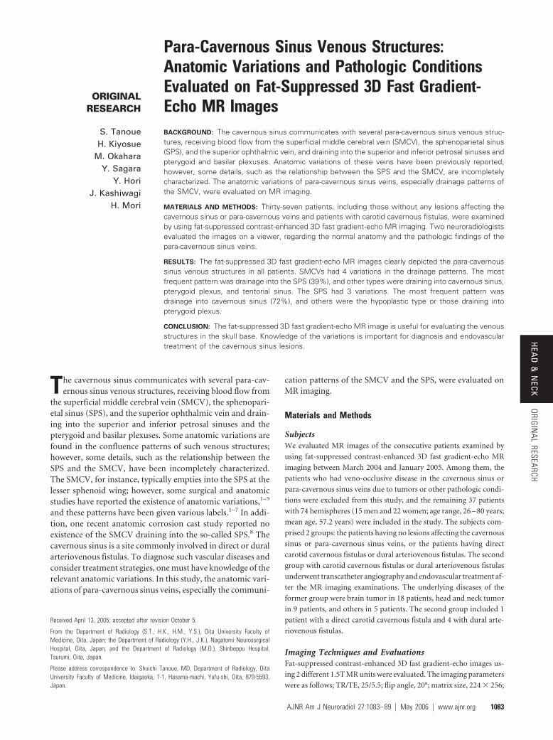
Para-Cavernous Sinus Venous Structures:Anatomic Variations and Pathologic ConditionsEvaluated on Fat-Suppressed 3D Fast Gradient-Echo MR Images
S. TanoueH. Kiyosue
M. OkaharaY. Sagara
Y. HoriJ. Kashiwagi
H. Mori
BACKGROUND: The cavernous sinus communicates with several para-cavernous sinus venous struc-tures, receiving blood flow from the superficial middle cerebral vein (SMCV), the sphenoparietal sinus(SPS), and the superior ophthalmic vein, and draining into the superior and inferior petrosal sinuses andpterygoid and basilar plexuses. Anatomic variations of these veins have been previously reported;however, some details, such as the relationship between the SPS and the SMCV, are incompletelycharacterized. The anatomic variations of para-cavernous sinus veins, especially drainage patterns ofthe SMCV, were evaluated on MR imaging.
MATERIALS AND METHODS: Thirty-seven patients, including those without any lesions affecting thecavernous sinus or para-cavernous veins and patients with carotid cavernous fistulas, were examinedby using fat-suppressed contrast-enhanced 3D fast gradient-echo MR imaging. Two neuroradiologistsevaluated the images on a viewer, regarding the normal anatomy and the pathologic findings of thepara-cavernous sinus veins.
RESULTS: The fat-suppressed 3D fast gradient-echo MR images clearly depicted the para-cavernoussinus venous structures in all patients. SMCVs had 4 variations in the drainage patterns. The mostfrequent pattern was drainage into the SPS (39%), and other types were draining into cavernous sinus,pterygoid plexus, and tentorial sinus. The SPS had 3 variations. The most frequent pattern wasdrainage into cavernous sinus (72%), and others were the hypoplastic type or those draining intopterygoid plexus.
CONCLUSION: The fat-suppressed 3D fast gradient-echo MR image is useful for evaluating the venousstructures in the skull base. Knowledge of the variations is important for diagnosis and endovasculartreatment of the cavernous sinus lesions.
The cavernous sinus communicates with several para-cav-ernous sinus venous structures, receiving blood flow from
the superficial middle cerebral vein (SMCV), the sphenopari-etal sinus (SPS), and the superior ophthalmic vein and drain-ing into the superior and inferior petrosal sinuses and thepterygoid and basilar plexuses. Some anatomic variations arefound in the confluence patterns of such venous structures;however, some details, such as the relationship between theSPS and the SMCV, have been incompletely characterized.The SMCV, for instance, typically empties into the SPS at thelesser sphenoid wing; however, some surgical and anatomicstudies have reported the existence of anatomic variations,1–5
and these patterns have been given various labels.1–7 In addi-tion, one recent anatomic corrosion cast study reported noexistence of the SMCV draining into the so-called SPS.8 Thecavernous sinus is a site commonly involved in direct or duralarteriovenous fistulas. To diagnose such vascular diseases andconsider treatment strategies, one must have knowledge of therelevant anatomic variations. In this study, the anatomic vari-ations of para-cavernous sinus veins, especially the communi-
cation patterns of the SMCV and the SPS, were evaluated onMR imaging.
Materials and Methods
SubjectsWe evaluated MR images of the consecutive patients examined by
using fat-suppressed contrast-enhanced 3D fast gradient-echo MR
imaging between March 2004 and January 2005. Among them, the
patients who had veno-occlusive disease in the cavernous sinus or
para-cavernous sinus veins due to tumors or other pathologic condi-
tions were excluded from this study, and the remaining 37 patients
with 74 hemispheres (15 men and 22 women; age range, 26 – 80 years;
mean age, 57.2 years) were included in the study. The subjects com-
prised 2 groups: the patients having no lesions affecting the cavernous
sinus or para-cavernous sinus veins, or the patients having direct
carotid cavernous fistulas or dural arteriovenous fistulas. The second
group with carotid cavernous fistulas or dural arteriovenous fistulas
underwent transcatheter angiography and endovascular treatment af-
ter the MR imaging examinations. The underlying diseases of the
former group were brain tumor in 18 patients, head and neck tumor
in 9 patients, and others in 5 patients. The second group included 1
patient with a direct carotid cavernous fistula and 4 with dural arte-
riovenous fistulas.
Imaging Techniques and EvaluationsFat-suppressed contrast-enhanced 3D fast gradient-echo images us-
ing 2 different 1.5T MR units were evaluated. The imaging parameters
were as follows; TR/TE, 25/5.5; flip angle, 20°; matrix size, 224 � 256;
Received April 13, 2005; accepted after revision October 5.
From the Department of Radiology (S.T., H.K., H.M., Y.S.), Oita University Faculty ofMedicine, Oita, Japan; the Department of Radiology (Y.H., J.K.), Nagatomi NeurosurgicalHospital, Oita, Japan; and the Department of Radiology (M.O.), Shinbeppu Hospital,Tsurumi, Oita, Japan.
Please address correspondence to: Shuichi Tanoue, MD, Department of Radiology, OitaUniversity Faculty of Medicine, Idaigaoka, 1-1, Hasama-machi, Yufu-shi, Oita, 879-5593,Japan.
HEA
D&
NECK
ORIGINAL
RESEARCH
AJNR Am J Neuroradiol 27:1083– 89 � May 2006 � www.ajnr.org 1083
section, 1 mm in 1 scanner (Excelart Vantage, Toshiba Medical Sys-
tems, Tokyo, Japan); and TR/TE, 21/2.1; flip angle, 20°; matrix size,
222 � 256; section thickness, 1.4 mm in the other scanner (SIGNA
Horizon, GE Healthcare, Milwaukee, Wis). As contrast media, 0.1
mmol/kg of gadolinium chelate (Omniscan) was administered intra-
venously in each patient. The scanning area covered between the level
of the orbital roof and the craniocervical junction. Each image was
obtained in a transverse section combined with a fat-suppression
technique.
The images were transferred to a workstation and independently
evaluated by 2 neuroradiologists with respect to the normal anatomy
and the pathologic findings of the para-cavernous sinus veins. The
vertical continuity of such veins was evaluated by paging the trans-
verse images on an identical window. Each subject with no lesion
affecting the cavernous sinus or para-cavernous sinus veins or with a
carotid cavernous fistula was evaluated in regard to anatomic varia-
tions, focusing on communication patterns between the cavernous
sinus and the para-cavernous sinus veins connecting with the cavern-
ous sinus, and images with arteriovenous fistulas were correlated with
the findings of digital subtraction angiography (DSA).
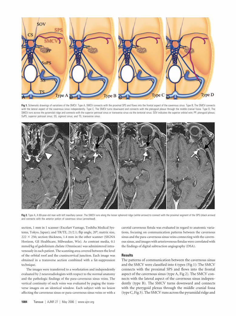
ResultsThe patterns of communication between the cavernous sinusand the SMCV were classified into 4 types (Fig 1): The SMCVconnects with the proximal SPS and flows into the frontalaspect of the cavernous sinus (type A, Fig 2). The SMCV con-nects with the lateral aspect of the cavernous sinus indepen-dently (type B). The SMCV turns downward and connectswith the pterygoid plexus through the middle cranial fossa(type C, Fig 3). The SMCV runs across the pyramidal ridge and
Fig 1. Schematic drawings of variations of the SMCV. Type A, SMCV connects with the proximal SPS and flows into the frontal aspect of the cavernous sinus. Type B, The SMCV connectswith the lateral aspect of the cavernous sinus independently. Type C, The SMCV turns downward and connects with the pterygoid plexus through the middle cranial fossa. Type D, TheSMCV runs across the pyramidal ridge and connects with the superior petrosal sinus or transverse sinus via the tentorial sinus. SOV indicates the superior orbital vein; PP, pterygoid plexus;SuPS, superior petrosal sinus; SS, sigmoid sinus; and TS, transverse sinus.
Fig 2. Type A, A 69-year-old man with left maxillary cancer. The SMCV runs along the lesser sphenoid ridge (white arrows) to connect with the proximal segment of the SPS (black arrows)and connects with the anterior potion of cavernous sinus (arrowhead).
1084 Tanoue � AJNR 27 � May 2006 � www.ajnr.org
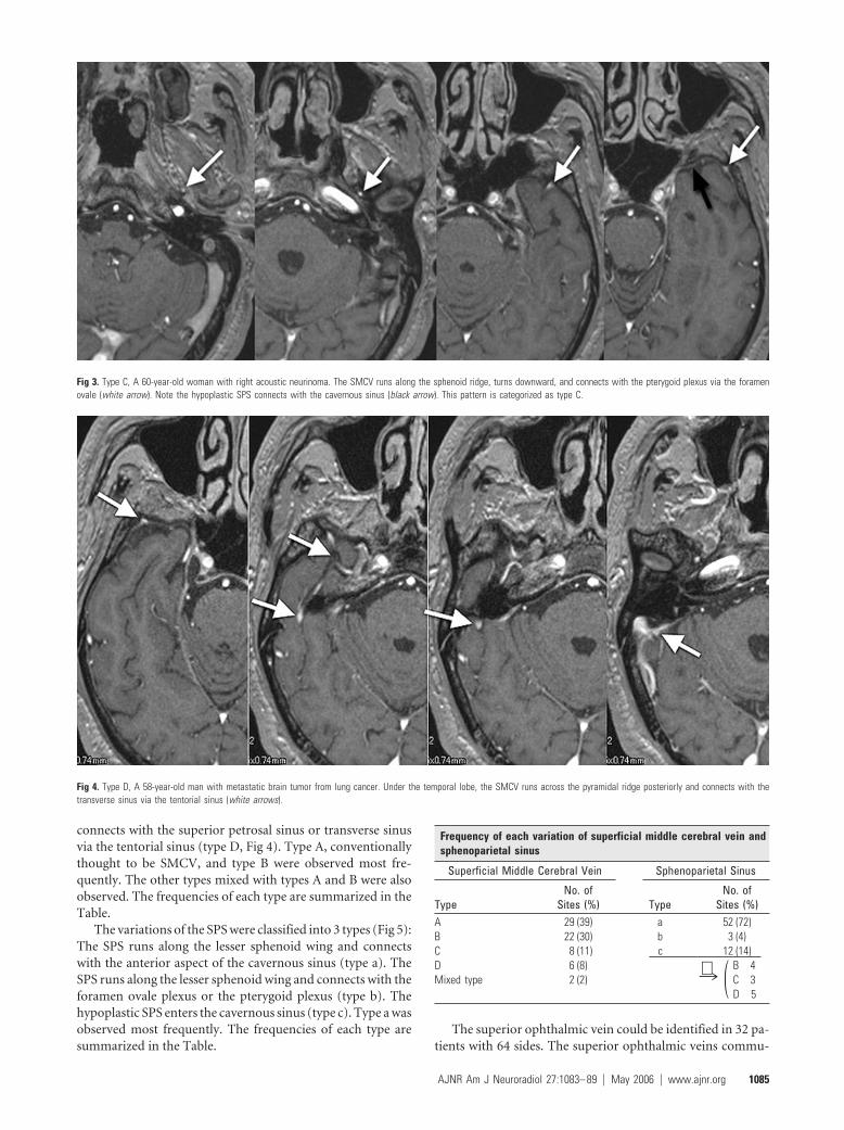
connects with the superior petrosal sinus or transverse sinusvia the tentorial sinus (type D, Fig 4). Type A, conventionallythought to be SMCV, and type B were observed most fre-quently. The other types mixed with types A and B were alsoobserved. The frequencies of each type are summarized in theTable.
The variations of the SPS were classified into 3 types (Fig 5):The SPS runs along the lesser sphenoid wing and connectswith the anterior aspect of the cavernous sinus (type a). TheSPS runs along the lesser sphenoid wing and connects with theforamen ovale plexus or the pterygoid plexus (type b). Thehypoplastic SPS enters the cavernous sinus (type c). Type a wasobserved most frequently. The frequencies of each type aresummarized in the Table.
The superior ophthalmic vein could be identified in 32 pa-tients with 64 sides. The superior ophthalmic veins commu-
Fig 3. Type C, A 60-year-old woman with right acoustic neurinoma. The SMCV runs along the sphenoid ridge, turns downward, and connects with the pterygoid plexus via the foramenovale (white arrow). Note the hypoplastic SPS connects with the cavernous sinus (black arrow). This pattern is categorized as type C.
Fig 4. Type D, A 58-year-old man with metastatic brain tumor from lung cancer. Under the temporal lobe, the SMCV runs across the pyramidal ridge posteriorly and connects with thetransverse sinus via the tentorial sinus (white arrows).
Frequency of each variation of superficial middle cerebral vein andsphenoparietal sinus
Superficial Middle Cerebral Vein Sphenoparietal Sinus
TypeNo. of
Sites (%) TypeNo. of
Sites (%)A 29 (39) a 52 (72)B 22 (30) b 3 (4)C 8 (11) c 12 (14)D 6 (8) B 4Mixed type 2 (2) C 3
D 5
¡ (
AJNR Am J Neuroradiol 27:1083– 89 � May 2006 � www.ajnr.org 1085

Fig 5. Schematic drawings of variations of the SPS.Type a, The SPS runs along the lesser sphenoidwing and connects to the anterior aspect of thecavernous sinus. Type b, The SPS runs along thelesser sphenoid wing and connects with the fora-men ovale plexus or the pterygoid plexus. Type c,the hypoplastic SPS connects with the cavernoussinus. CS indicates the cavernous sinus
Fig 6. Mixed type with type A and B. A 57-year-old woman with left cavernous dural arteriovenous fistula.
A, Lateral view; B, frontal view. Right carotid arteriogram demonstrates a left cavernous dural arteriovenous fistula draining into the cerebellar cortical veins via the superior petrosal sinus(arrows). Retrograde opacification of the SMCV is not shown.
C, On the contrast-enhanced gradient-echo MR image, a small venous structure connecting to lateral wall of the cavernous sinus, thought to be the SMCV, is observed (white arrow). The findingsindicate the potential risk of cortical venous reflux to the medial temporal lobe. Another prominent SMCV flowed into the pterygoid plexus with no connection to the cavernous sinus (black arrow).Note the dilated inferior hypophyseal arteries joining the posteroinferior aspect of the cavernous sinus (arrowheads) and cortical venous reflux to the cerebellar veins (open arrows).
D, This patient was treated by transvenous embolization, and the shunt disappeared completely.
1086 Tanoue � AJNR 27 � May 2006 � www.ajnr.org

nicated with the cavernous sinuses in the anterolateral aspectsvia the superior orbital fissures in all patients. No anatomicvariation could be found in our subjects. The basal vein ofRosenthal could be also observed in some patients; however,there was no communication between the cavernous sinus andthe basal vein of Rosenthal in our study. Thus evaluation of thebasal vein of Rosenthal was abbreviated in this study.
In subjects with arteriovenous fistula, feeding arteries anddraining veins demonstrated on DSA could also be identifiedon MR images (Fig 6). In patients with dural arteriovenousfistulas, SMCVs entering into the cavernous sinuses wereclearly depicted on MR images, even if the SMCV had not beendemonstrated with cortical venous reflux on DSA. The orificeof the direct carotid-cavernous fistulas could not be identified
on MR images; however, drainage veins and para-cavernoussinus veins were clearly depicted (Fig 7).
Discussion
Embryology of Cavernous Sinus and Para-CavernousVeinPadget9 described the detailed developmental anatomy of cra-nial veins in past reports. The cavernous sinus and other ve-nous structures develop from primitive dural plexuses sur-rounding primitive cerebral vesicles.10,11 With developmentof the cerebral hemisphere, these plexuses fuse to form venoussinuses and cortical veins. Variations of para-cavernous sinusveins can occur in venous development.
Fig 7. Type C, A 43-year-old woman with a right direct carotid cavernous fistula.
A, Frontal view of a right carotid arteriogram demonstrates a right carotid cavernous fistula. Retrograde opacification of the SMCV is not shown.
B, On the contrast-enhanced gradient-echo MR image, 2 SMCVs are conjoined beside the cavernous sinus and flow through the middle carnial fossa (arrows). This patient was treatedby transarterial and transvenous coil-packing of the right cavernous sinus.
C, The carotid angiogram immediately after embolization demonstrates no visualization of the shunt. Note the right SMCV flowing beside the coil mass (arrows).
AJNR Am J Neuroradiol 27:1083– 89 � May 2006 � www.ajnr.org 1087

Two major primitive sinuses, the primitive tentorial sinusand prootic sinus, contribute to the development of cavernoussinus and para-cavernous sinus veins.6,12 During the early em-bryonic stage, the prootic sinus receives blood flow from theprimitive maxillary vein, developing to the ophthalmic veinsin the late stage. The cortical and pial veins developing into anSMCV are connected with the transverse sinus through aprimitive tentorial sinus in an 8-week-old embryo (Fig 8A,-B). After a week, the primitive SMCV connecting with theprimitive tentorial sinus is elongated anteromedially and isaccompanied by the development of the cerebral hemisphere.In the 12-week-old embryo, the prootic sinus participates informing the cavernous sinus and foramen ovale venous plexus(draining into pterygoid plexus) and receives blood flow fromsuperior ophthalmic vein (Fig. 8C).12 After developing to the pre-natal stage, the tentorial sinus connecting with the SMCV extendsmedially and anastomoses with the cavernous sinus and para-cavernous venous plexuses or sinuses10,11 (Fig 8D, -E).
Anatomic Variations Regarding EmbryologyAnatomic variations between the cavernous sinus and para-cavernous sinus venous structures can be formed in the courseof developmental anastomosis. Among the variations, theprimitive tentorial sinus performs the most dynamic conver-sion of blood flow to produce the SMCV. The SMCV and SPS
are important venous structures serving as drainage routesfrom the cerebral hemisphere and meninges to the cervicalvenous system via the cavernous sinus; and previous re-ports1–5 have shown these venous structures to have some an-atomic variations. Variations of the SMCV have been investi-gated by anatomic and surgical methods. The SMCV hasgenerally been thought to run along the lesser sphenoid wingand enter into the proximal SPS; however, San Millan Ruiz etal8 reported no existence of an SMCV draining into the SPS inan anatomic study using an autopsy cadaver.
Suzuki and Matsumoto6 investigated variations of theSMCV by using 3D CT angiography and classified some typesfrom a developmental viewpoint. In that report, the variationsof SMCV are subdivided into 7 detailed types, including thesphenoparietal, cavernous, emissary, superior petrosal, basal,squamosal, and undeveloped types; and the classifications inother reports could be included in the classification of Suzukiand Matsumoto. We classified the variations into 4 simplesubtypes. The blood flow from the SMCV drains into the ex-tracranial veins via the cavernous sinus in the former 2 types(types A and B) and drains without direct connection withcavernous sinus in the latter 2 types (types C and D). Depictionof the existence of direct communication between the SMCVand the cavernous sinus is important in diagnosis and endo-vascular treatment of arteriovenous fistula.
Fig 8. Schematic drawings of the developmental anatomyof cavernous and para-cavernous venous structures in theembryonic stage. SSS indicates superior sagittal sinus;PTS, primitive tentorial sinus; PS, prootic sinus; PMS,primitive maxillary sinus; and IPS, inferior petrosal sinus.
A, Lateral view; B, axial view. In an 8-week embryo,cerebral venous structures develop from primitive duralplexuses surrounding primitive cerebral vesicles. The plex-uses fuse to form venous sinuses and cortical veins. Twomajor primitive sinuses, the primitive tentorial sinus andthe prootic sinus, contribute to the development of cav-ernous sinus and para-cavernous sinus veins. The primitiveSMCV is connected with the transverse sinus via theprimitive tentorial sinus.
C, In a 12-week embryo, after several weeks, the primitiveSMCV is elongated and develops anteromedially to formthe SMCV as a development of the cerebral hemisphere.The prootic sinus contributes to form the superior ophthal-mic vein, the cavernous sinus, and the foramen ovalevenous plexus.
D, Axial view; E, lateral view. In a developed embryo, the SMCV makes further anteromedial development in the prenatal stage; however, in many patients, there is no direct connectionbetween the SMCV and the cavernous sinus. Secondary anastomosis after birth may form a connection between the SMCV and the cavernous sinus, and the connection to the primitivetentorial sinus subsequently degenerates.
1088 Tanoue � AJNR 27 � May 2006 � www.ajnr.org

In previous reports, a larger number of SMCVs directlyenter into the SPS or cavernous sinus.6,7 Next evaluated is a typeof draining into the pterygoid plexus via the emissary vein. Sim-ilar results were observed in our series (types A–C). These SMCVvariations occur during the process of joining into the cavernoussinus or surrounding veins, and the difference in connectingpoints is caused by simultaneous development of the cavernoussinus and para-cavernous sinus venous system and the primitivetentorial sinus. The previously reported sphenopetrosal sinus orsphenobasilar type and our type D are the extent of communica-tion to the transverse sinus via the primitive tentorial sinus. Incases with hypoplastic SPSs, the SMCV tends to have communi-cation with the tentorial sinus (transverse sinus).
In contrast to the SMCV, the superior ophthalmic veindevelops from the primitive maxillary sinus, connecting withthe prootic sinus.12 Because the primitive vein of the superiorophthalmic vein communicates with the prootic sinus form-ing the cavernous sinus, the superior ophthalmic vein rarelyforms an anatomic variation. The superior ophthalmic veinsin all our patients drained into the anterolateral aspect of thecavernous sinus via the superior orbital fissure. To our knowl-edge, there is no previous report about an anatomic variationof communication pattern between the superior ophthalmicvein and the cavernous sinus.
The basal vein of Rosenthal develops by anastomoses ofdeep pial veins, and its tributaries may connect with the cav-ernous sinus via the SMCV or superior petrosal sinus.13 Theseconnections have great clinical significance, especially in pa-tients with deep venous thrombosis or other deep venous oc-clusive diseases. However, in our study, there were no imagesthat depicted a communication between the basal vein ofRosenthal and the cavernous sinus. Further examination withpatients having deep venous occlusive diseases is necessary fordiscussing the communication pattern of the basal vein ofRosenthal with the cavernous sinus.
Clinical SignificanceSome recent investigators have evaluated these variations byusing 3D CT angiography.6,13,14 Recent improvements of theperformance of CT scanners have allowed more detailed ob-servation of the venous anatomy. However, there are somedifficulties in observing vessels adjacent to or penetratingbony structures. In contrast, fat-suppressed contrast-en-hanced MR imaging is feasible to evaluate skull base vessels.15
In our series, veins surrounding or penetrating bony struc-tures could be depicted with the high tissue resolution of fastgradient-echo MR imaging.
Knowledge of the variations noted here is important, notonly for diagnosing several diseases involving the cavernoussinus or para-cavernous sinus veins but also for surgery ofpara-cavernous sinus lesions and endovascular treatment ofarteriovenous fistulas. The SMCV and SPS can be a venousrefluxing route in patients with arteriovenous fistulas. Even ifdiagnostic DSA shows no cortical venous reflux, hemody-
namic changes may cause venous reflux into the SMCV or SPSin patients having potential connections among the cavernoussinus, SMCV, and SPS. For endovascular treatment for a ca-rotid cavernous fistula, the contrast-enhanced fat-suppressed3D fast gradient-echo MR images provide important informa-tion for decisions on therapeutic strategies and for the appro-priate positioning of embolic material. There were small num-bers of patients without veno-occlusive disease or witharteriovenous fistulas in this study, but the contrast-enhancedMR images provide more information for diagnosing andtreatment planning of other veno-occlusive diseases, such astumors and venous thrombosis.
ConclusionContrast-enhanced fat-suppressed 3D fast gradient-echo MRimages clearly depicted para-cavernous sinus venous struc-tures in healthy subjects and in patients with arteriovenousfistulas. Several variations in the pattern of drainage of theSMCVs (eg, draining into the SPS, the cavernous sinus, andother para-cavernous veins) were noted. Knowledge of thesevariations is important for diagnosis and treatment strategiesusing endovascular or surgical techniques for cavernous sinuslesions.
References1. Oka K, Rhoton AL Jr, Barry M, et al. Microsurgical anatomy of the superficial
veins of the cerebrum. Neurosurgery 1985;17:711– 482. Yamakami I, Hirai S, Yamaura A, et al. Venous system playing a key role in
transpetrosal approach [in Japanese]. No Shinkei Gaku 1998;26:69 –703. Aydin IH, Tuzun Y, Takci E, et al. The anatomical variations of sylvian veins
and cisterns. Minim Invasive Neurosurg 1997;40:68 –734. Ciszek B, Dabrowska M, Andrzejczak A, et al. Middle superfical cerebral vein.
Folia Morphol (Warsz) 1998;57:149 –555. Aydin IH, Kadioglu HH, Tuzun Y, et al. The variations of sylvian veins and
cisterns in anterior circulation aneurysms: an operative study. Acta Neurochir(Wien) 1996;138:1380 – 85
6. Suzuki Y, Matsumoto K. Variations of the superficial middle cerebral vein:classification using three-dimensional CT angiography. AJNR Am J Neurora-diol 2000;21:932–38
7. Gailloud P, San Millan Ruiz D, Muster M, et al. Angiographic anatomy of thelaterocavernos sinus. AJNR Am J Neuroradiol 2000;21:1923–29
8. San Millan Ruiz D, Fasel JH, Rufenacht DA, et al. The sphenoparietal sinus ofBreschet: does it exist? An anatomic study. AJNR Am J Neuroradiol 2004;25:112–20
9. Padget DH. The cranial venous system in man in reference to development,adult configuration, and relation to the arteries. Am J Anat 1956;98:307–55
10. Truwit CL. Embryology of the cerebral vasculature. Neuroimaging Clin N Am1994;4:663– 89
11. The cerebral veins. In: Osborn AG, Jacobs JM. Diagnostic Cerebral Angiography,2nd ed. Philadelphia, Pa: Lippincott Williams & Wilkins; 1999:217–37
12. Knosp E, Muller G, Perneczky A, et al. Anatomical remarks on the fetal cavern-ous sinus and on the veins of the middle cranial fossa. In: Dolenc VV, ed. TheCavernous Sinus: A Multidisciplinary Approach to Vascular and Tumorous Le-sions. New York: Springer-Verlag; 1987:104 –16
13. Suzuki Y, Ikeda H, Shimadu M, et al. Variations of the basal vein: identificationusing three-dimensional CT angiography. AJNR Am J Neuroradiol 2001;22:670 –76
14. Suzuki Y, Matsumoto K. Detection of the venous system of the skull base usingthree-dimensional CT angiography (3D-CTA): utility of the pterional and an-terior temporal approaches [in Japanese]. No Shinkei Geka 1999;27:1091–96
15. Caruso RD, Rosenbaum AE, Chang JK, et al. Craniocervical junction venousanatomy on enhanced MR images: the suboccipital cavernous sinus. AJNRAm J Neuroradiol 1999;20:1127–31
AJNR Am J Neuroradiol 27:1083– 89 � May 2006 � www.ajnr.org 1089