par-clip (photoactivatable ribonucleoside-enhanced
TRANSCRIPT

CHAPTER EIGHT
PAR-CLIP (PhotoactivatableRibonucleoside-EnhancedCrosslinking andImmunoprecipitation):a Step-By-Step Protocol to theTranscriptome-Wide Identificationof Binding Sites of RNA-BindingProteinsJessica Spitzer*,2, Markus Hafner*,2, Markus Landthaler3,Manuel Ascano*, Thalia Farazi*, Greg Wardle*, Jeff Nusbaum*,Mohsen Khorshid†, Lukas Burger†, Mihaela Zavolan†,Thomas Tuschl*,1*Howard Hughes Medical Institute, Laboratory of RNA Molecular Biology, The Rockefeller University,New York, NY, USA†Biozentrum der Universitat Basel and Swiss Institute of Bioinformatics (SIB), Basel, Switzerland1Corresponding author: e-mail address: [email protected] authors contributed equally to this work3Present address: Berlin Institute for Medical Systems Biology, Max-Delbruck-Center for Molecular Medicine,Berlin, Germany
Contents
1. Theory 1152. Equipment 1173. Materials 119
3.1 Solutions & buffers 1224. Protocol 129
4.1 Preparation 1294.2 Tip 1294.3 Caution 1314.4 Duration 131
5. Step 1 UV Crosslinking of 4-Thiouridine-Labeled Cells (Day 1) 1315.1 Overview 131
Methods in Enzymology, Volume 539 # 2014 Elsevier Inc.ISSN 0076-6879 All rights reserved.http://dx.doi.org/10.1016/B978-0-12-420120-0.00008-6
113

5.2 Duration 1335.3 Tip 133
6. Step 2 Preparation of Cell Lysate for Immunoprecipitation (Day 2) 1346.1 Overview 1346.2 Duration 1346.3 Tip 1356.4 Tip 1356.5 Tip 135
7. Step 3 Preparation of the Magnetic Beads (Day 2) 1367.1 Overview 1367.2 Duration 1367.3 Tip 1367.4 Tip 136
8. Step 4 Immunoprecipitation and Second RNase T1 Treatment (Day 2) 1368.1 Overview 1368.2 Duration 1378.3 Tip 138
9. Step 5 Dephosphorylation and Radiolabeling RNA Segments Crosslinked toImmunoprecipitated Proteins (Day 2) 1389.1 Overview 1389.2 Duration 1389.3 Tip 139
10. Step 6 SDS-PAGE and Electroelution of Cross-Linked RNA-Protein Complexesfrom Gel Slices (Days 2 and 3) 13910.1 Overview 13910.2 Duration 14010.3 Tip 14210.4 Tip 14210.5 Tip 14310.6 Tip 14310.7 Tip 143
11. Step 7 Proteinase K Digestion (Day 3) 14311.1 Overview 14311.2 Duration 14311.3 Tip 14411.4 Tip 145
12. Step 8 30-Adapter Ligation for cDNA Library Preparation (Day 3 overnight, day 4,beginning of day 5) 14512.1 Overview 14512.2 Duration 14512.3 Tip 147
13. Step 9 50-Adapter Ligation for cDNA Library Preparation (Day 5, beginning of day 6) 14813.1 Overview 14813.2 Duration 14813.3 Tip 15013.4 Tip 150
114 Jessica Spitzer et al.

14. Step 10 cDNA Library Preparation/Reverse Transcription (Day 6) 15014.1 Overview 15014.2 Duration 150
15. Step 11 PCR Amplification of cDNA Library & Sample Preparation for Sequencing(Day 6) 15215.1 Overview 15215.2 Duration 15215.3 Tip 15415.4 Tip 15415.5 Tip 15615.6 Tip 156
16. Step 12 Determination of Incorporation Levels of 4SU into Total RNA 15616.1 Overview 15616.2 Duration 156
References 160Source References 160
Abstract
We recently developed a protocol for the transcriptome-wide isolation of RNA recog-nition elements readily applicable to any protein or ribonucleoprotein complex directlycontacting RNA (including RNA helicases, polymerases, or nucleases) expressed in cellculture models either naturally or ectopically (Hafner et al., 2010).
Briefly, immunoprecipitation of the RNA-binding protein of interest is followed byisolation of the crosslinked and coimmunoprecipitated RNA. In the course of lysatepreparation and immunoprecipitation, the mRNAs are partially degraded using Ribonu-clease T1. The isolated crosslinked RNA fragments are converted into a cDNA libraryand deep-sequenced using Solexa technology (see Explanatory Chapter: NextGeneration Sequencing). By introducing photoreactive nucleosides that generate char-acteristic sequence changes upon crosslinking (see below), our protocol allows one toseparate RNA segments bound by the protein of interest from the background un-crosslinked RNAs.
1. THEORY
Posttranscriptional regulation (PTR) of messenger RNAs (mRNAs)
plays important roles in diverse cellular processes (Ambros, 2004;
Halbeisen et al., 2008). The fates of mRNAs are determined predominantly
by their interactions with RNA-binding proteins (RBPs) and noncoding,
guide-RNA-containing ribonucleoprotein complexes (RNPs). Taken
together, they form mRNA-containing ribonucleoprotein complexes
(mRNPs). The RBPs influence the structure and interactions of the RNAs
115PAR-CLIP (Photoactivatable Ribonucleoside-Enhanced Crosslinking and Immunoprecipitation)

and play critical roles in their biogenesis, stability, function, transport, and
cellular localization (Moore, 2005; Keene, 2007; Glisovic et al., 2008).
Given that hundreds of RBPs andRNPs and their networks remain to be
studied and evaluated in a cell-type-dependent manner, the development of
powerful tools to determine their binding sites or RNA recognition ele-
ments (RREs) is critical to enhance our understanding of PTR. It offers
new opportunities for understanding both gene regulation and conse-
quences of genetic variation in transcript regions aside from the open
reading frame.
Typically, a combination of genetic, biochemical, and computational
approaches has been applied to identify mRNA-RBP or mRNA-RNP
interactions. However, each of these methods has limitations. Microarray
profiling of mRNA associated with immunopurified RBPs (RIP-ChIP)
(Tenenbaum et al., 2000) is limited by incomplete enrichment of bound
mRNAs and the difficulty of locating theRRE in the hundreds to thousands
of nucleotide (nt) long target mRNA (Gerber et al., 2006; Landthaler
et al., 2008).
Some of these problems were addressed by an in vivo UV 254-nm
crosslinking and immunoprecipitation (CLIP) protocol (Ule et al., 2003.
See also UV crosslinking of interacting RNA and protein in cultured cells)
that better defines the interaction site by isolating and sequencing small
RNA segments crosslinked to RBPs. However, UV 254-nm crosslinking
is not efficient, and the site of crosslinking is not revealed after sequencing
of the isolated RNA fragment. To separate crosslinked sites from back-
ground noise, additional control crosslinking experiments are needed,
including the use of knockout cells of the protein of interest.
To overcome these limitations, we developed a new protocol referred to
as PAR-CLIP (Photoactivatable-Ribonucleoside-Enhanced Crosslinking
and Immunoprecipitation) (Hafner et al., 2010).
4-Thiouridine (4SU) and 6-thioguanosine (6SG) are readily incorpo-
rated into nascent RNAs by simply supplementing the media of cultured
cells with the modified nucleoside (Favre et al., 1986; Bezerra and Favre,
1990). At the concentrations used in the presented protocol, neither of
the tested photoreactive nucleosides showed any detectable toxic effects
based on mRNA profiling or cell count. Irradiation of the cells by UV light
of 365 nm leads to crosslinking of photoreactive nucleoside-labeled cellular
RNAs to interacting RBPs. Using similar irradiation protocols, 4SU incor-
poration substantially enhances RNA recovery compared to UV 254-nm
crosslinking, 6SG performs in between these two methods.
116 Jessica Spitzer et al.
Most importantly, the sites of crosslinking can be easily identified by
mapping characteristic T to C mutations (G to A in the case of 6SG, though
less pronounced) in the sequenced cDNA libraries obtained from the recov-
ered RNA initiated by the photocrosslinking itself. We presume that the
structural change upon crosslinking of the modified nucleosides to aromatic
amino acid side chains directs the incorporation of a noncognate
deoxynucleoside during reverse transcription of crosslinked RNAs. The
presence of the mutations in sequence reads, together with the observation
that multiple positions within a cluster of sequence reads can be altered, facil-
itates the separation from clusters of unaltered background sequences typi-
cally derived from abundant cellular RNAs.
For details on the bioinformatic analyses, please refer to our recent pub-
lication (Hafner et al., 2010).
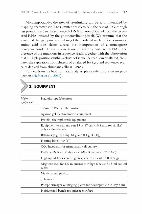
2. EQUIPMENT
Major
equipment
Radioisotope laboratory
365-nm UV-transilluminator
Agarose gel electrophoresis equipment
Protein electrophoresis equipment
Equipment to cast and run 15 � 17 cm � 0.8 mm (or similar)
polyacrylamide gels
Balances (e.g., 0.1 mg–64 g and 0.1 g–4.2 kg)
Heating block (90 �C)
CO2 incubator for mammalian cell culture
D-Tube Dialyzer Midi rack (EMD Biosciences, 71511-3)
High-speed floor centrifuge (capable of at least 13 000 � g)
Magnetic rack for 1.5-ml microcentrifuge tubes and 15-ml conical
tubes
Multichannel pipettor
pH-meter
Phosphorimager & imaging plates (or developer and X-ray film)
Refrigerated bench top microcentrifuge
117PAR-CLIP (Photoactivatable Ribonucleoside-Enhanced Crosslinking and Immunoprecipitation)

Rotating wheel
PCR thermocycler
Thermometer
Thermomixer
UV Stratalinker 2400 equipped with 365-nm light bulbs for
crosslinking (Stratagene)
Vortex mixer
Water bath
Water filter; MilliQBiocel water purification system
X-ray exposure cassette
HPLC with a Supelco Discovery C18 (bonded phase silica 5 mMparticle, 250 � 4.6 mm) reverse phase column (Bellefonte, PA,
USA)
Consumables 1.5-ml polypropylene tubes
1.5-ml siliconized tubes (BIO PLAS Inc., 4165SL)
15- and 50-ml conical tubes (e.g., Falcon) as well as tubes
withstanding high-speed centrifugation (e.g., Sarsted, 13-ml
centrifuge tube, 55.518)
15-cm culture dishes
10-cm tissue culture dishes
5-mm Supor membrane syringe filter (Pall Acrodisc)
Cell scraper (Corning)
D-Tube Dialyzer Midi, MWCO 3.5 kDa (EMD Biosciences,
71506-3)
NuPAGE Novex 4–12% BT Midi 1.0 gel (Invitrogen)
pH paper (covering the range between pH 6.5 and 10)
Plastic wrap
Scalpels or razor blades
Strips of 0.2-ml tubes (Thermo Scientific, AB-0264)
Syringes (10 ml)
118 Jessica Spitzer et al.

3. MATERIALS
Reagents &
Chemicals
Appropriate cell culture medium and selection antibiotics
2-Mercaptoethanol (14.3 M; Sigma, M6250)
Acetonitrile
Agarose, electrophoresis grade (SeaKem LE Agarose, Lonza,
50004)
Agarose, low melting (NuSieve GTG Agarose, Lonza, 50080)
Ammonium persulfate (APS)
Adenosine triphosphate (ATP)
Bacterial Alkaline Phosphatase (Worthington Biochemical,
LS006344)
Bromophenol blue
Bovine serum albumin, acetylated (BSA, acetylated; Ambion,
AM2614)
Calcium chloride (CaCl2�2H2O)
Calf intestinal alkaline phosphatase (CIP)
Chloroform
Citric acid monohydrate
Complete EDTA-free protease inhibitor cocktail (Roche)
Dimethyl sulfoxide (DMSO)
DNA ladder (25 bp)
dNTPs: dATP, dCTP, dGTP, dTTP (0.1 M each; Fermentas,
R0182)
Dithiothreitol (DTT)
Dynabeads Protein G (Invitrogen, 100-03D)
EDTA disodium salt dihydrate (Sigma, E5134)
EGTA (C14H20N2O10Na4; Sigma, E8145-10G)
Ethanol (100%)
119PAR-CLIP (Photoactivatable Ribonucleoside-Enhanced Crosslinking and Immunoprecipitation)

Ethidium bromide
Ficoll type 400
Formamide
[g-32P]-ATP (10 mCi ml�1, 6000 Ci (222 TBq) mmol�1;
Perkin Elmer, NEG002Z500UC)
Glycerol
Glycoblue or glycogen
Hydrochloric acid (HCl) (Fisher Scientific, A144S)
HEPES
Isoamyl alcohol
Isopropyl alcohol
Potassium chloride (KCl)
Potassium hydroxide (KOH)
Magnesium chloride (MgCl2�6H2O)
MOPS SDS running buffer (20�; Invitrogen)
Sodium acetate (NaOAc, Fisher, S210)
Sodium phosphate dibasic (Na2HPO4�7H2O; Sigma, S9390-
100G)
Sodium chloride (NaCl)
Sodium fluoride (NaF)
Sodium hydroxide (NaOH)
NP40 substitute (100%; Sigma [74385])
Phosphate buffered saline (PBS; 10�, commercially available)
Phenol (saturated with 0.1 M citrate buffer, pH 4.3 � 0.2,
Sigma, P4682)
Photoreactive nucleoside (Sigma; 4-thiouridine [T4509]/6-
thioguanosine [858412])
Protein ladder (e.g., Biorad, 161-0374; 10–250 kDa)
Proteinase K (lyophilizate; Roche, 03115801001)
rA, rG, rC, rU, and 4SU (Sigma, T4509)
RNase T1 (Fermentas, EN0541); concentration 1000 U ml�1
Sodium dodecyl sulfate (SDS; Fisher Scientific, BP166-500)
120 Jessica Spitzer et al.

Snake Venom Phosphodiesterase (Worthington Biochemical,
LS003926)
SuperScript III Reverse Transcriptase (Invitrogen, 18080-044);
includes 5� First-strand buffer
T4 Polynucleotide Kinase (T4 PNK; NEB, M0201)
T4 RNA Ligase 1 (NEB, M0204L)
T4 RNA Ligase 2, truncated (e.g., NEB, M0242L); or: Rnl2(1-
249)K227Q (our plasmid for expression of the his-tagged
mutant is available at www.addgene.com, plasmid 14072;
however, the purified enzyme will shortly also be available from
NEB)
Taq DNA polymerase (5 U ml�1)
Tris-Borate–EDTA buffer solution (TBE)
Acetic acid – triethylamine solution 1:1 (TEAA; Sigma, 09748)
Tetramethylethylenediamine (TEMED)
Tris base (Fisher Scientific, BP152-1)
Tris–HCl (Promega, H5121)
Triton X-100
TRIzol reagent (Invitrogen, 15596-026)
UreaGel – SequaGel – System, National Diagnostics, EC-833
Antibody (e.g., for FLAG-tagged RBPs: mouse monoclonal
anti-FLAG M2 (Sigma, F1804))
QIAquick gel purification kit (Qiagen)
RNA & DNA
oligonucleotides
30 adapter (DNA, except for the 50 riboadenylate (rApp)residue): 50 rAppTCGTATGCCGTCTTCTGCTTGT
50 adapter (RNA):
50 GUUCAGAGUUCUACAGUCCGACGAUC
30 PCR primer (DNA):
50 CAAGCAGAAGACGGCATACGA
50 PCR primer (DNA): 50
AATGATACGGCGACCACCGACAGGTTCAGAGTTCT
ACAGTCCGA
19-nt size marker (RNA):
50 CGUACGCGGGUUUAAACGA
121PAR-CLIP (Photoactivatable Ribonucleoside-Enhanced Crosslinking and Immunoprecipitation)

24-nt size marker (RNA):
50 CGUACGCGGAAUAGUUUAAACUGU
33-nt size marker (RNA):
50
CAUCUUGGUCGUACGCGGAAUAGUUUAAACUGU
35-nt size marker (RNA):
50CUCAUCUUGGUCGUACGCGGAAUAGUUUAA
ACUGU
Reference
oligoribonucleotides
CGUACGCGGAAUACUUCGA(4SU)U (e.g., from Thermo
Scientific)
CGUACGCGGAAUACUUCGAUU
3.1. Solutions & buffersStep 1 1 M 4-Thiouridine stock solution
Dissolve 250-mg 4-thiouridine in 960.5-ml DMSO
(For a 1 M 6-thioguanosine solution, first dehydrate the powder supplied by Sigma
in a vacuum oven at room temperature overnight. Then, dissolve 299.3 mg in 1-ml
DMSO)
4-Thiouridine-containing growth medium
Component Final concentration Stock Amount
4-thiouridine (in DMSO) 100 mM 1 M 100 ml
Cell culture medium 1 l
1� PBS
Component Final concentration Stock Amount
PBS 1� 10� 100 ml
H2O 900 ml
Step 2 NP40 lysis buffer
Component Final concentration Stock Amount
HEPES-KOH, pH 7.5 50 mM 1 M 50 ml
KCl 150 mM 1 M 150 ml
122 Jessica Spitzer et al.

EDTA–NaOH, pH 8.0 2 mM 0.5 M 4 ml
NaF 1 mM 0.5 M 2 ml
NP40 substitute 0.5% (v/v) 100% 5 ml
H2O 788.5 ml
DTT (add fresh) 0.5 mM 1 M 0.5 ml
Complete EDTA-free protease
inhibitor cocktail (add fresh)
1 tablet/50 ml
Step 3 Citrate-phosphate buffer, pH 5.0
Component Amount
Citric acid monohydrate 4.7 g
Na2HPO4�7H2O 9.2 g
H2O to 1 l
Step 4 IP-wash buffer
ComponentFinalconcentration Stock Amount
HEPES-KOH, pH 7.5 50 mM 1 M 50 ml
KCl 300 mM 1 M 300 ml
NP40 substitute 0.05% (v/v) 100% 0.5 ml
H2O 649 ml
DTT (add fresh) 0.5 mM 1 M 0.5 ml
Complete EDTA-free protease inhibitor
cocktail (add fresh)
1 tablet/
50 ml
High-salt wash buffer
ComponentFinalconcentration Stock Amount
HEPES-KOH, pH 7.5 50 mM 1 M 50 ml
KCl 500 mM 1 M 500 ml
NP40 substitute 0.05% (v/v) 100% 0.5 ml
H2O 449 ml
123PAR-CLIP (Photoactivatable Ribonucleoside-Enhanced Crosslinking and Immunoprecipitation)

DTT (add fresh) 0.5 mM 1 M 0.5 ml
Complete EDTA-free protease inhibitor
cocktail (add fresh)
1 tablet/
50 ml
10� Dephosphorylation buffer
Component Final concentration Stock Amount
Tris–HCl, pH 7.9 50 mM 1 M 50 ml
NaCl 100 mM 3 M 33.3 ml
MgCl2�6H2O 10 mM 1 M 10 ml
H2O 906.2 ml
DTT (add fresh) 1 mM 1 M 0.5 ml
Step 5 Phosphatase wash buffer
Component Final concentration Stock Amount
Tris–HCl, pH 7.5 50 mM 1 M 50 ml
EGTA–NaOH, pH 7.5 20 mM 0.5 M 40 ml
NP40 substitute 0.5% (v/v) 100% 5 ml
H2O 905 ml
Polynucleotide kinase (PNK) buffer without DTT
Component Final concentration Stock Amount
Tris–HCl, pH 7.5 50 mM 1 M 50 ml
NaCl 50 mM 3 M 16.7 ml
MgCl2�6H2O 10 mM 1 M 10 ml
H2O 923.3 ml
PNK buffer with DTT
Component Final concentration Stock Amount
Tris–HCl, pH 7.5 50 mM 1 M 50 ml
NaCl 50 mM 3 M 16.7 ml
MgCl2�6H2O 10 mM 1 M 10 ml
124 Jessica Spitzer et al.
H2O 918.3 ml
DTT (add fresh) 5 mM 1 M 5 ml
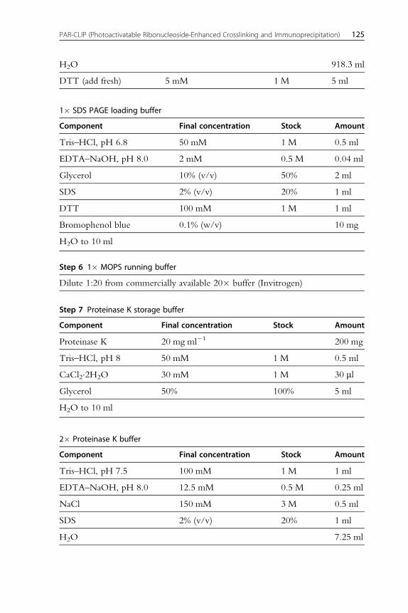
1� SDS PAGE loading buffer
Component Final concentration Stock Amount
Tris–HCl, pH 6.8 50 mM 1 M 0.5 ml
EDTA–NaOH, pH 8.0 2 mM 0.5 M 0.04 ml
Glycerol 10% (v/v) 50% 2 ml
SDS 2% (v/v) 20% 1 ml
DTT 100 mM 1 M 1 ml
Bromophenol blue 0.1% (w/v) 10 mg
H2O to 10 ml
Step 6 1� MOPS running buffer
Dilute 1:20 from commercially available 20� buffer (Invitrogen)
Step 7 Proteinase K storage buffer
Component Final concentration Stock Amount
Proteinase K 20 mg ml�1 200 mg
Tris–HCl, pH 8 50 mM 1 M 0.5 ml
CaCl2�2H2O 30 mM 1 M 30 ml
Glycerol 50% 100% 5 ml
H2O to 10 ml
2� Proteinase K buffer
Component Final concentration Stock Amount
Tris–HCl, pH 7.5 100 mM 1 M 1 ml
EDTA–NaOH, pH 8.0 12.5 mM 0.5 M 0.25 ml
NaCl 150 mM 3 M 0.5 ml
SDS 2% (v/v) 20% 1 ml
H2O 7.25 ml
125PAR-CLIP (Photoactivatable Ribonucleoside-Enhanced Crosslinking and Immunoprecipitation)
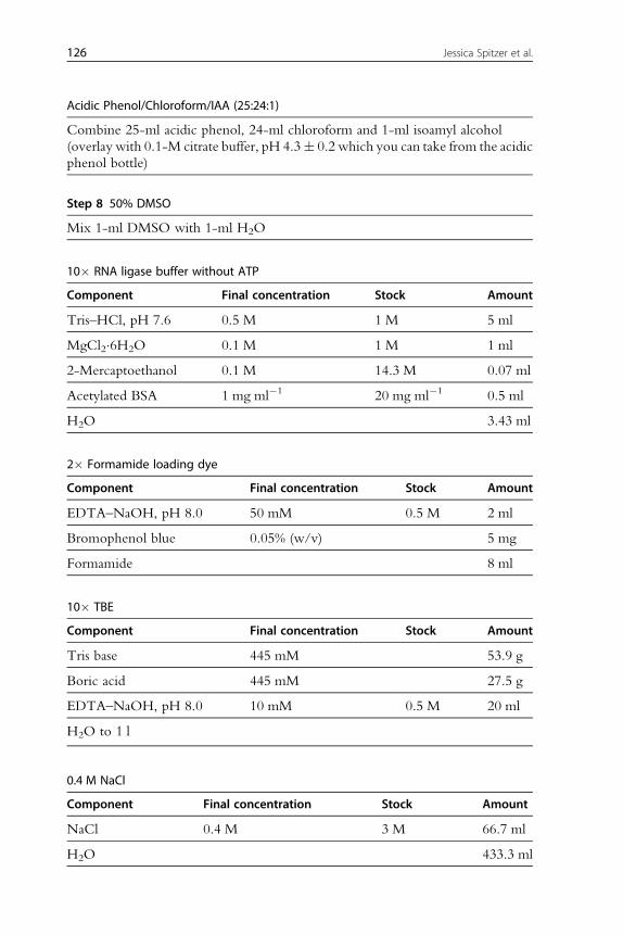
Acidic Phenol/Chloroform/IAA (25:24:1)
Combine 25-ml acidic phenol, 24-ml chloroform and 1-ml isoamyl alcohol
(overlay with 0.1-M citrate buffer, pH 4.3� 0.2 which you can take from the acidic
phenol bottle)
Step 8 50% DMSO
Mix 1-ml DMSO with 1-ml H2O
10� RNA ligase buffer without ATP
Component Final concentration Stock Amount
Tris–HCl, pH 7.6 0.5 M 1 M 5 ml
MgCl2�6H2O 0.1 M 1 M 1 ml
2-Mercaptoethanol 0.1 M 14.3 M 0.07 ml
Acetylated BSA 1 mg ml�1 20 mg ml�1 0.5 ml
H2O 3.43 ml
2� Formamide loading dye
Component Final concentration Stock Amount
EDTA–NaOH, pH 8.0 50 mM 0.5 M 2 ml
Bromophenol blue 0.05% (w/v) 5 mg
Formamide 8 ml
10� TBE
Component Final concentration Stock Amount
Tris base 445 mM 53.9 g
Boric acid 445 mM 27.5 g
EDTA–NaOH, pH 8.0 10 mM 0.5 M 20 ml
H2O to 1 l
0.4 M NaCl
Component Final concentration Stock Amount
NaCl 0.4 M 3 M 66.7 ml
H2O 433.3 ml
126 Jessica Spitzer et al.

Step 9 10� RNA ligase buffer with ATP
Component Final concentration Stock Amount
Tris–HCl, pH 7.6 0.5 M 1 M 5 ml
MgCl2�6H2O 0.1 M 1 M 1 ml
2-Mercaptoethanol 0.1 M 14.3 M 0.07 ml
Acetylated BSA 1 mg ml�1 20 mg ml�1 0.5 ml
ATP 2 mM 100 mM 0.2 ml
H2O 3.23 ml
Step 10 10� dNTP solution
Component Final concentration Stock Amount
dATP 2 mM 0.1 M 0.2 ml
dCTP 2 mM 0.1 M 0.2 ml
dGTP 2 mM 0.1 M 0.2 ml
dTTP 2 mM 0.1 M 0.2 ml
H2O 9.2 ml
150-mM KOH/20-mM Tris base
Component Final concentration Stock Amount
KOH 150 mM 5 M 30 ml
Tris base 20 mM 1 M 20 ml
H2O 950 ml
150-mM HCl
Component Final concentration Stock Amount
HCl, concentrated 150 mM 12.1 M 12.4 ml
H2O 987.6 ml
Step 11 10� PCR buffer
Component Final concentration Stock Amount
Tris–HCl, pH 8.0 100 mM 1 M 1 ml
KCl 500 mM 1 M 5 ml
127PAR-CLIP (Photoactivatable Ribonucleoside-Enhanced Crosslinking and Immunoprecipitation)

2-Mercaptoethanol 10 mM 14.3 M 7 ml
Triton X-100 1% (v/v) 100% 0.1 ml
MgCl2�6H2O 20 mM 1 M 2 ml
H2O 1.9 ml
5� DNA loading dye
Component Final concentration Stock Amount
EDTA–NaOH, pH 8.0 50 mM 0.5 M 1 ml
Bromophenol blue 0.2% (w/v) 20 mg
Ficoll type 400 20% (w/v) 2 g
H2O to 10 ml
Step 12 1 M DTT
Dissolve 1.54-g DTT in 10-ml water
3 M NaOAc (pH 5.2)
Component Stock Final concentration Amount
NaOAc n/a n/a 246.09 g
H2O to 1 l
HPLC buffer A
Component Final concentration Stock Amount
Acetonitrile 3% 100% 30 ml
TEAA 0.1 M 2 M 50 ml
H2O to 1 l
HPLC buffer B
Mix 900-ml acetonitrile with 100-ml water
128 Jessica Spitzer et al.

4. PROTOCOL
4.1. Preparation4.1.1 CellsExpand cells in an appropriate growth medium containing selection antibi-
otics as appropriate to maintain your stable cell line. We usually prepare
lysates from 3 to 5 ml of wet cell pellet from crosslinked cells per experiment.
This corresponds to 20–50 15-cm cell culture plates (for HEK293). How-
ever, if material is limiting, we have performed successful PAR-CLIPs
experiments from <0.5 ml of wet cell pellet (200 � 106 HEK293 cells
(10 15-cm plates) will yield �1 ml of wet cell pellet).
Grow cells to �80% confluence. Fourteen hours before crosslinking,
add 4-thiouridine (4SU) to a final concentration of 100 mM directly to
the cell culture medium. 6-Thioguanosine (6SG, 100 mM) can also be used
as the photoactivatable ribonucleoside. Induce expression of protein, if
necessary.
4.2. TipIf you want to add 4SU to 50 15-cm cell culture plates containing 20 ml of
growth medium each, place 265 ml of growth medium into a sterile, empty bottle
(e.g., an empty media bottle). Add 132.5-ml 1 M 4SU and mix. Additional
reagents such as doxycycline (e.g., 1 mg ml�1) to induce protein expression may
be added. Dispense 5 ml of the prepared growth medium containing 4SU per
15-cm plate.
4.2.1 BuffersBuffer recipes and required reagents are listed above. Allow�1 day for gen-
eral preparations, including buffer preparation. All pH measurements and
adjustments are performed at room temperature. Buffers and all perishable
reagents should be refrigerated for storage. We use water purified by a Mil-
lipore water purification system.
On the day before you start the PAR-CLIP procedure, fill the required
amounts of the individual buffers into 50-ml conical tubes and refrigerate
them. The table below gives a rough guide to the required amounts of
the individual buffers (but only of those that will be used in quantities above
1 ml on the first 2 days). Add DTT and protease inhibitors on the day of the
experiment.
129PAR-CLIP (Photoactivatable Ribonucleoside-Enhanced Crosslinking and Immunoprecipitation)
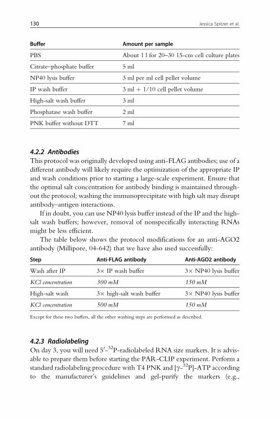
Buffer Amount per sample
PBS About 1 l for 20–30 15-cm cell culture plates
Citrate–phosphate buffer 5 ml
NP40 lysis buffer 3 ml per ml cell pellet volume
IP wash buffer 3 ml þ 1/10 cell pellet volume
High-salt wash buffer 3 ml
Phosphatase wash buffer 2 ml
PNK buffer without DTT 7 ml
4.2.2 AntibodiesThis protocol was originally developed using anti-FLAG antibodies; use of a
different antibody will likely require the optimization of the appropriate IP
and wash conditions prior to starting a large-scale experiment. Ensure that
the optimal salt concentration for antibody binding is maintained through-
out the protocol; washing the immunoprecipitate with high salt may disrupt
antibody–antigen interactions.
If in doubt, you can use NP40 lysis buffer instead of the IP and the high-
salt wash buffers; however, removal of nonspecifically interacting RNAs
might be less efficient.
The table below shows the protocol modifications for an anti-AGO2
antibody (Millipore, 04-642) that we have also used successfully:
Step Anti-FLAG antibody Anti-AGO2 antibody
Wash after IP 3� IP wash buffer 3� NP40 lysis buffer
KCl concentration 300 mM 150 mM
High-salt wash 3� high-salt wash buffer 3� NP40 lysis buffer
KCl concentration 500 mM 150 mM
Except for these two buffers, all the other washing steps are performed as described.
4.2.3 RadiolabelingOn day 3, you will need 50-32P-radiolabeled RNA size markers. It is advis-
able to prepare them before starting the PAR-CLIP experiment. Perform a
standard radiolabeling procedure with T4 PNK and [g-32P]-ATP according
to the manufacturer’s guidelines and gel-purify the markers (e.g.,
130 Jessica Spitzer et al.

phosphorylate 1-mM RNA size marker in a 10-ml reaction volume using
1 ml of [g-32P]-ATP (see RNA Radiolabeling). Keep radioactive gel pieces
from the running front of this gel as markers to implant into gels for align-
ment of phosphorimager printouts to exposed gels later on.
4.3. CautionConsult your institute’s Radiation Safety Officer for proper ordering, handling, and
disposal of radioactive materials.
4.4. Duration
Preparation Expanding cell line(s) Approximately 2 weeks depending on the
desired number of cells
Antibody testing Variable
Buffers etc. 1 day
Radiolabeling of
RNA size markers
1.5 days
Protocol Total 7 days
Day 1 3–4 h
Day 2 10–12 h
Day 3 5–6 h
Day 4 3–4 h
Day 5 5–6 h
Day 6 6–7 h
Day 7 4–5 h
See Figure 8.1 for the flowchart of the complete protocol.
5. STEP 1 UV CROSSLINKING OF4-THIOURIDINE-LABELED CELLS (DAY 1)
5.1. OverviewIn this first step, the RNA-binding protein of interest is cross-linked to its
bound mRNAs targets that incorporated the photoactivatable ribonucleo-
side into nascent transcripts during the labeling step (see also UV crosslinking
131PAR-CLIP (Photoactivatable Ribonucleoside-Enhanced Crosslinking and Immunoprecipitation)

Figure 8.1 Flowchart of the complete protocol, including preparation.
132 Jessica Spitzer et al.
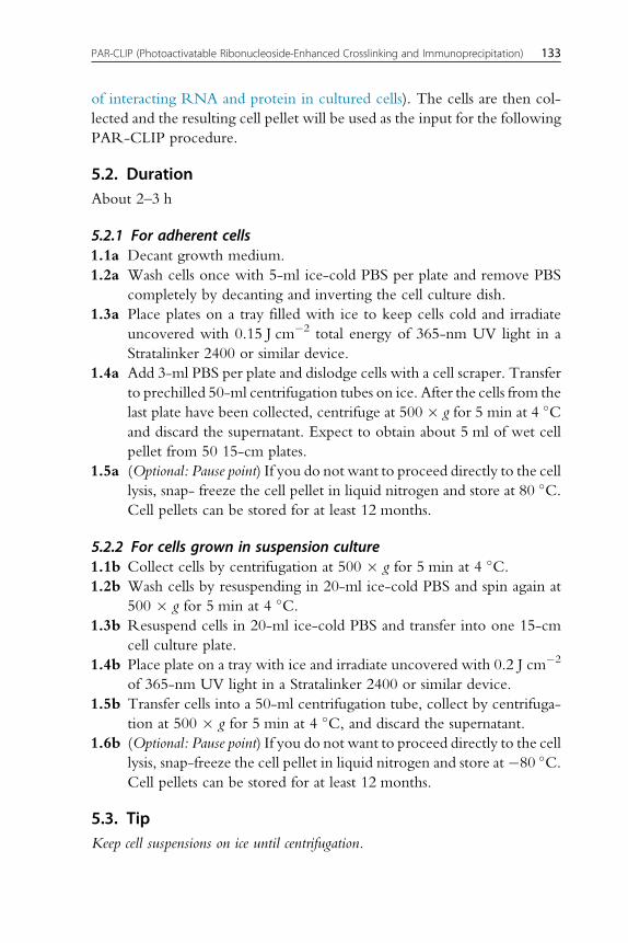
of interacting RNA and protein in cultured cells). The cells are then col-
lected and the resulting cell pellet will be used as the input for the following
PAR-CLIP procedure.
5.2. DurationAbout 2–3 h
5.2.1 For adherent cells1.1a Decant growth medium.
1.2a Wash cells once with 5-ml ice-cold PBS per plate and remove PBS
completely by decanting and inverting the cell culture dish.
1.3a Place plates on a tray filled with ice to keep cells cold and irradiate
uncovered with 0.15 J cm�2 total energy of 365-nm UV light in a
Stratalinker 2400 or similar device.
1.4a Add 3-ml PBS per plate and dislodge cells with a cell scraper. Transfer
to prechilled 50-ml centrifugation tubes on ice. After the cells from the
last plate have been collected, centrifuge at 500 � g for 5 min at 4 �Cand discard the supernatant. Expect to obtain about 5 ml of wet cell
pellet from 50 15-cm plates.
1.5a (Optional: Pause point) If you do not want to proceed directly to the cell
lysis, snap- freeze the cell pellet in liquid nitrogen and store at 80 �C.Cell pellets can be stored for at least 12 months.
5.2.2 For cells grown in suspension culture1.1b Collect cells by centrifugation at 500 � g for 5 min at 4 �C.1.2b Wash cells by resuspending in 20-ml ice-cold PBS and spin again at
500 � g for 5 min at 4 �C.1.3b Resuspend cells in 20-ml ice-cold PBS and transfer into one 15-cm
cell culture plate.
1.4b Place plate on a tray with ice and irradiate uncovered with 0.2 J cm�2
of 365-nm UV light in a Stratalinker 2400 or similar device.
1.5b Transfer cells into a 50-ml centrifugation tube, collect by centrifuga-
tion at 500 � g for 5 min at 4 �C, and discard the supernatant.
1.6b (Optional: Pause point) If you do not want to proceed directly to the cell
lysis, snap-freeze the cell pellet in liquid nitrogen and store at�80 �C.Cell pellets can be stored for at least 12 months.
5.3. TipKeep cell suspensions on ice until centrifugation.
133PAR-CLIP (Photoactivatable Ribonucleoside-Enhanced Crosslinking and Immunoprecipitation)
See Fig. 8.2 for the flowchart of Step 1.
6. STEP 2 PREPARATION OF CELL LYSATE FORIMMUNOPRECIPITATION (DAY 2)
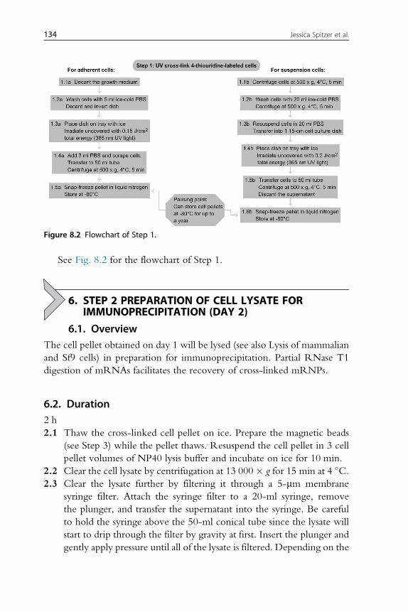
6.1. OverviewThe cell pellet obtained on day 1 will be lysed (see also Lysis of mammalian
and Sf9 cells) in preparation for immunoprecipitation. Partial RNase T1
digestion of mRNAs facilitates the recovery of cross-linked mRNPs.
6.2. Duration2 h
2.1 Thaw the cross-linked cell pellet on ice. Prepare the magnetic beads
(see Step 3) while the pellet thaws. Resuspend the cell pellet in 3 cell
pellet volumes of NP40 lysis buffer and incubate on ice for 10 min.
2.2 Clear the cell lysate by centrifugation at 13 000� g for 15 min at 4 �C.2.3 Clear the lysate further by filtering it through a 5-mm membrane
syringe filter. Attach the syringe filter to a 20-ml syringe, remove
the plunger, and transfer the supernatant into the syringe. Be careful
to hold the syringe above the 50-ml conical tube since the lysate will
start to drip through the filter by gravity at first. Insert the plunger and
gently apply pressure until all of the lysate is filtered. Depending on the
Figure 8.2 Flowchart of Step 1.
134 Jessica Spitzer et al.

initial viscosity of the lysate, it might be necessary to exchange a clogged
filter for a fresh one.
2.4 Add RNase T1 to a final concentration of 1 U ml�1 and incubate in a
water bath for 15 min at 22 �C. Invert to mix from time to time. Cool
reaction for 5 min on ice before proceeding.
2.5 Remove a 10-ml aliquot for immunoblotting as a control for the protein
levels used as input and freeze at �20 �C.
6.3. TipTake the cell pellet out of the�80 �C freezer and put it on ice first thing in the morn-
ing since the thawing process takes a long time.
6.4. TipThe temperature and duration of the incubation with RNase T1 are both critical for
obtaining a controlled partial digestion.
6.5. TipDesignate a set of micropipettors for working with RNases to avoid contamination at
later RNA isolation and cDNA library preparation steps.
See Fig. 8.3 for the flowchart of Step 2.
Figure 8.3 Flowchart of Step 2.
135PAR-CLIP (Photoactivatable Ribonucleoside-Enhanced Crosslinking and Immunoprecipitation)

7. STEP 3 PREPARATION OF THE MAGNETIC BEADS(DAY 2)
7.1. OverviewThe antibody is conjugated to protein G magnetic beads to be used in the
subsequent immunoprecipitation. Protein G is the optimal Ig-binding protein
for anti-FLAG antibodies based on species and isotype. The choice of protein
A versus protein G should be considered depending on the antibody used.
7.2. Duration1.5 h
3.1 Transfer 10 ml of Protein G magnetic particles per ml cell lysate (typ-
ically �100–150 ml of beads) to a 1.5-ml microtube. Put the magnetic
rack on ice. Wash the beads twice with 1 ml of citrate–phosphate
buffer.
3.2 Resuspend the beads in twice the volume of citrate–phosphate buffer
relative to the original volume of bead suspension (i.e., 200–300 ml).3.3 Add antibody to a final concentration of 0.25 mg ml�1 and incubate on
a rotating wheel for 40 min at room temperature.
3.4 Collect the beads and wash twice in 1 ml of citrate–phosphate buffer to
remove unbound antibody.
3.5 Resuspend beads in twice the volume of citrate–phosphate buffer rel-
ative to the original volume of bead suspension.
7.3. TipThis step is performed while the cell pellet is thawing.
7.4. TipBe careful to not let the magnetic beads dry out.
See Fig. 8.4 for the flowchart of Step 3.
8. STEP 4 IMMUNOPRECIPITATION AND SECOND RNaseT1 Treatment (Day 2)
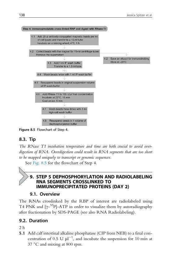
8.1. OverviewThe mRNA-RBP complex of choice is isolated from the lysate by immu-
noprecipitation. A second RNase T1 digestion ensures that only the RNA
136 Jessica Spitzer et al.

segment that was bound, crosslinked, and protected by the RBP is
recovered and sequenced. This enables the precise definition of the
binding sites.
8.2. Duration2 h
4.1 Add 20 ml of freshly prepared antibody-conjugated magnetic beads
per ml of the partially RNase T1-treated cell lysate from Step 2
and incubate in a 15-ml centrifuge tube on a rotating wheel for 1 h
at 4 �C.4.2 Collect magnetic beads on a magnetic particle collector for 15-ml cen-
trifuge tubes (Invitrogen) and remove the supernatant. Save an aliquot
for immunoblotting.
4.3 Add 1 ml of IP wash buffer and transfer to 1.5-ml polypropylene tubes.
4.4 Wash beads 2 times in 1 ml of IP wash buffer.
4.5 Resuspend beads in the original bead volume of IP wash buffer.
4.6 Add RNase T1 (Fermentas, 10 000 U ml�1) to a final concentration of
100 U ml�1 and incubate the bead suspension in a 22 �C water bath for
15 min. Cool on ice for 5 min.
4.7 Wash beads 3 times with 1-ml high-salt wash buffer.
4.8 Resuspend beads in 1 volume of dephosphorylation buffer.
Figure 8.4 Flowchart of Step 3.
137PAR-CLIP (Photoactivatable Ribonucleoside-Enhanced Crosslinking and Immunoprecipitation)
8.3. TipThe RNase T1 incubation temperature and time are both crucial to avoid over-
digestion of RNA. Overdigestion could result in RNA segments that are too short
to be mapped uniquely to transcript or genomic sequences.
See Fig. 8.5 for the flowchart of Step 4.
9. STEP 5 DEPHOSPHORYLATION AND RADIOLABELINGRNA SEGMENTS CROSSLINKED TOIMMUNOPRECIPITATED PROTEINS (DAY 2)
9.1. OverviewThe RNAs crosslinked by the RBP of interest are radiolabeled using
T4 PNK and [g-32P]-ATP in order to visualize them by autoradiography
after fractionation by SDS-PAGE (see also RNA Radiolabeling).
9.2. Duration2 h
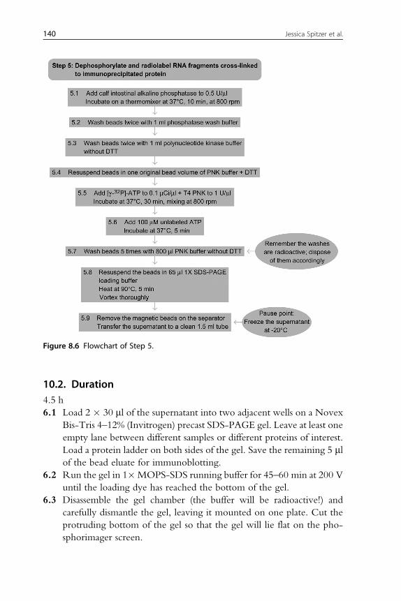
5.1 Add calf intestinal alkaline phosphatase (CIP from NEB) to a final con-
centration of 0.5 U ml�1, and incubate the suspension for 10 min at
37 �C and mixing at 800 rpm.
Figure 8.5 Flowchart of Step 4.
138 Jessica Spitzer et al.

5.2 Wash beads twice with 1 ml of phosphatase wash buffer.
5.3 Wash beads twice with polynucleotide kinase (PNK) buffer
without DTT.
5.4 Resuspend beads in one original bead volume of PNK buffer
containing DTT.
5.5 Add [g-32P]-ATP to a final concentration of 0.1 mCi ml�1 and T4 PNK
(NEB) to 1 U ml�1. Incubate the suspension for 30 min at 37 �C and
800 rpm, mixing manually every 5–10 min.
5.6 Add 100-mM nonradioactive ATP and incubate for another 5 min at
37 �C. This ensures that all RNAs are fully 50 phosphorylated, whichis required for the ligation of the 50 adapter (Step 9).
5.7 Wash the magnetic beads 5 times with 800 ml of PNK buffer without
DTT; dispose of the radioactive buffer according to local guidelines.
5.8 Resuspend the beads in 65 ml of 1� SDS-PAGE loading buffer, incu-
bate for 5 min in a heat block at 90 �C to denature, and release the
immunoprecipitated RBP with the cross-linked, radiolabeled RNAs
from the beads. Vortex.
5.9 Remove the magnetic beads on the separator and transfer the superna-
tant to a clean 1.5-ml microcentrifuge tube. (Pause point: you can freeze
the supernatant and continue with the protocol at another time.)
9.3. TipRemove the [g-32P]-ATP from the freezer and place it at room temperature during the
dephosphorylation incubation so that it is thawed by the time you need it.
See Fig. 8.6 for the flowchart of Step 5.
10. STEP 6 SDS-PAGE AND ELECTROELUTION OF CROSS-LINKED RNA-PROTEIN COMPLEXES FROM GELSLICES (DAYS 2 AND 3)
10.1. OverviewSize fractionation of the radiolabeled and cross-linked RNA protein
complexes is achieved by SDS-PAGE (see One-dimensional SDS-
Polyacrylamide Gel Electrophoresis (1D SDS-PAGE)). The band
corresponding to the expected mass of the protein will be excised and
the cross-linked RNA protein complexes electroeluted. This step ensures
that only the band corresponding to the correct RBP is isolated and addi-
tionally prevents any unbound but labeled RNA from further processing
(see Fig. 8.7(a)).
139PAR-CLIP (Photoactivatable Ribonucleoside-Enhanced Crosslinking and Immunoprecipitation)
10.2. Duration4.5 h
6.1 Load 2 � 30 ml of the supernatant into two adjacent wells on a Novex
Bis-Tris 4–12% (Invitrogen) precast SDS-PAGE gel. Leave at least one
empty lane between different samples or different proteins of interest.
Load a protein ladder on both sides of the gel. Save the remaining 5 mlof the bead eluate for immunoblotting.
6.2 Run the gel in 1�MOPS-SDS running buffer for 45–60 min at 200 V
until the loading dye has reached the bottom of the gel.
6.3 Disassemble the gel chamber (the buffer will be radioactive!) and
carefully dismantle the gel, leaving it mounted on one plate. Cut the
protruding bottom of the gel so that the gel will lie flat on the pho-
sphorimager screen.
Figure 8.6 Flowchart of Step 5.
140 Jessica Spitzer et al.

6.4 To facilitate the alignment of the gel to the phosphorimager paper
printout later on, place three tiny radioactive gel pieces asymmetrically
into three of the four corners of the gel. Radioactive gel pieces could be
collected earlier from the bottom of the gel from radiolabeling the size
markers (see above).
Figure 8.7 Selected PAR-CLIP experimental steps. (a) SDS-PAGE gel of cross-linked and50-radiolabeled RNA-protein complex immunoprecipitates. The red arrow points to theradioactive bands corresponding to the expected size of the RNA-binding protein (FUS,running at 75 kDa), and the yellow to the radioactive running front. (b) 8 M urea poly-acrylamide gel after 30-adapter ligation. The black arrow indicates one of the insertedlittle radioactive gel pieces to facilitate alignment of the gel to printout, the red tothe 30-ligated sizemarkers, and the area that was cut from the gel and further processed;the yellow arrows show the unligated 30-size markers. (c) 8-M urea polyacrylamide gelafter 50-adapter ligation. The black arrow indicates one of the inserted little radioactivegel pieces to facilitate alignment of the gel to printout, the red to the 50-ligated sizemarkers, and the area which was cut from the gel and further processed; the yellowarrows show the unligated 50-size markers. (d) Agarose gel after small-scale trial PCR.The black arrow points to the position of migration of the xylene cyanol loading dye,and the yellow to the bromophenol blue loading dye running close to the gel front.Bands of about 75 and 100 bp are detectable, representing insert-less 50-adapter-30-adapter PCR side product and expected insert-containing PCR product, respectively.The red arrows indicate the number of cycles chosen for the large-scale experiment.A 25-bp ladder is loaded to the left of each set of experiments; the fourth bandfrom the bottom corresponds to 100 bp. The negative control was performed but isnot shown.
141PAR-CLIP (Photoactivatable Ribonucleoside-Enhanced Crosslinking and Immunoprecipitation)

6.5 After placing the gel pieces, wrap the gel in plastic wrap and expose the
gel to a phosphorimager screen for 15 min. Visualize it on a pho-
sphorimager. Have a second screen ready and expose it during the scan-
ning process should the first exposure indicate that a longer exposure is
necessary.
6.6 Print the scanned image file at its original size (100%). Align the trans-
parently wrapped gel on top of the printout guided by the implanted gel
pieces for precise positioning. Cut out the bands that correspond to the
expected size of the RBP (see Fig. 8.7(a)).
6.7 Add 800 ml of water to a D-Tube Dialyzer Midi Tube used for elec-
troelution and let stand at room temperature for 5 min. Remove the
water. Take care not to pierce the membrane.
6.8 Transfer the excised bands to the dialyzer tube and add 800-ml 1�MOPS-SDS running buffer.
6.9 Place the electroelution rack with the tubes in an agarose gel chamber
so that the membrane is exposed to the flow of the current (for details,
see manufacturer’s instructions). Add 1� MOPS-SDS running buffer
to the chamber until it covers the tubes.
6.10 Electroelute the cross-linked RNA-RBP complex at 100 V for 1.5 h.
Reverse the current for 2 min to release any protein attached on the
dialysis membrane.
6.11 Transfer the solution to two siliconized tubes so that each contains
around 350 ml (you will not be able to fully recover the original
800 ml). (Pause point: you can freeze the solution at �20 �C and con-
tinue the next day.)
10.3. TipTo confirm that the correct band was excised from the gel, run a second small-scale
SDS-PAGE gel with 1 or 2 ml of the remaining 5 ml of your sample (see above).After transferring the gel to a nitrocellulose membrane, take an autoradiography expo-
sure (exposing for 1–2 h) and then use protein-specific antibodies to perform a stan-
dard immunoblot (see Western Blotting using Chemiluminescent Substrates). After
overlaying the resulting images, you should be able to establish which band corresponds
to your protein of interest and proceed with the protocol.
10.4. TipIn case you observe more than one band, you can also cut all of them since they should
correspond to other co-purifying RNA cross-linked proteins.
142 Jessica Spitzer et al.

10.5. TipMake sure that the membrane of the dialyzer tube is aligned correctly to allow flow of
current.
10.6. TipUse aerosol barrier tips and take general precautions to avoid any RNase contamina-
tion since you will be working with RNA from now on until the reverse transcription
step on day 6. Clean your pipettors before starting to work with RNA.
10.7. TipUse siliconized tubes until you have obtained your cDNA library; at low concentra-
tions, nucleic acids have a tendency to stick to the tube walls.
See Fig. 8.8 for the flowchart of Step 6.
11. STEP 7 PROTEINASE K DIGESTION (DAY 3)
11.1. OverviewIn this step, the recovered RBP is proteolyzed and the cross-linked RNA is
recovered so that it can serve as the input material for subsequent ligation to
adapters and Solexa sequencing.
11.2. Duration3.5 h
7.1 Add 1 volume of 2� Proteinase K Buffer, followed by the addition of
Proteinase K (Roche) to a final concentration of 1.2 mg ml�1. Incubate
at 55 �C for 30 min. If the volume per tube exceeds 800 ml at this stage,split the sample into two tubes.
7.2 Add 1 volume of acidic phenol/chloroform/isoamyl alcohol, vortex
and spin at 20 000 � g for 10 min at 4 �C. Recover the upper aqueous
phase without disturbing the interphase and pipette into two
siliconized tubes.
7.3 Add an equal volume of chloroform, and vortex and spin at 20 000� g
for 10 min at 4 �C. Recover the aqueous phase without disturbing the
interphase.
7.4 Add 1/10 volume 3 MNaCl, 1 ml of glycogen (10 mg ml�1 stock), and
3 volumes of 100% ethanol.
143PAR-CLIP (Photoactivatable Ribonucleoside-Enhanced Crosslinking and Immunoprecipitation)

7.5 Precipitate the RNA for 1 h on ice and spin at 20 000� g for 15 min at
4 �C. (Pause point: precipitate the RNA overnight at 20 �C. See also
RNA purification – precipitation methods.)
7.6 Remove the supernatant, air-dry the pellets, and dissolve in a total of
10 ml of H2O.
11.3. TipMonitor the radioactivity of the supernatant and the pellet to assess the efficiency of the
ethanol precipitation.
Figure 8.8 Flowchart of Step 6.
144 Jessica Spitzer et al.
11.4. TipRepeat the phenol/chloroform/IAA extraction until there is no precipitate visible in
the interphase (usually, once is sufficient, but two or more times might be needed).
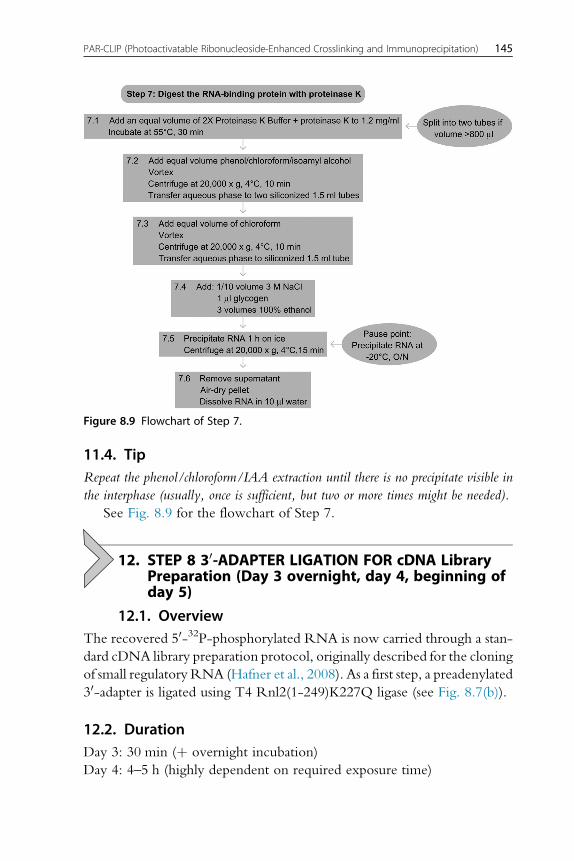
See Fig. 8.9 for the flowchart of Step 7.
12. STEP 8 30-ADAPTER LIGATION FOR cDNA LibraryPreparation (Day 3 overnight, day 4, beginning ofday 5)
12.1. OverviewThe recovered 50-32P-phosphorylated RNA is now carried through a stan-
dard cDNA library preparation protocol, originally described for the cloning
of small regulatory RNA (Hafner et al., 2008). As a first step, a preadenylated
30-adapter is ligated using T4 Rnl2(1-249)K227Q ligase (see Fig. 8.7(b)).
12.2. DurationDay 3: 30 min (þ overnight incubation)
Day 4: 4–5 h (highly dependent on required exposure time)
Figure 8.9 Flowchart of Step 7.
145PAR-CLIP (Photoactivatable Ribonucleoside-Enhanced Crosslinking and Immunoprecipitation)

Day 5: 2 h
8.1 Prepare the following reaction mixture for ligating the 30-adapter, mul-
tiplying the volumes by the number of ligation reactions plus two (one
for the size markers plus another to account for pipetting loss):
2 ml of 10� RNA ligase buffer (without ATP)
6 ml 50% DMSO
1 ml of 100 mM preadenylated 30-adapterAdd 9 ml of the reaction mixture to each sample (from Step 7.6, you
have 19 ml per tube).8.2 Prepare �40 fmol of a 1:100 dilution of 50-32P-labeled RNA size
markers (19-nt and 24-nt size marker at equimolar concentrations,
see preparation step). This controls for successful ligation and indicates
the length of the bands that will be cut out from the gel later on.
8.3 Heat-denature the RNA to disrupt secondary structures by incubating
at 90 �C for 30 s. Immediately place the tubes on ice for 30 s.
8.4 Add 1 ml of Rnl2(1-249)K227Q ligase (1 mg ml�1) to the ligation reac-
tions, mix gently, and incubate overnight on ice in the cold room or in
an insulated ice bucket covered with a lid.
8.5 The next morning, cast a 15% denaturing 8-M urea polyacrylamide gel
(we use the UreaGel system from National Diagnostics. See also RNA
purification by preparative polyacrylamide gel electrophoresis) and wait
until the polymerization process is complete. Our gels measure 15 �17 cm � 0.8 mm and contain about 25-ml gel volume with a
20 well comb.
8.6 Prerun the gel in 1� TBE buffer at 30 W for 30 min. After the prerun,
flush the wells with 1� TBE.
8.7 Add 20 ml of formamide gel loading solution to the samples to stop the
ligation reactions.
8.8 Denature the RNA at 90 �C for 30 s.
8.9 Load each sample into one well (or two) of the gel. Load the size
markers symmetrically on both sides of the gel to allow for approximat-
ing the length of the ligated samples between them.Use the center lanes
of the gel to guarantee even running of the gel. Make sure to space dif-
ferent samples appropriately, typically at a two-well distance, to avoid
cross contamination. Ensure that the overall loading of the gel is
asymmetrical.
8.10 Run the gel at 30 W for 45 min until the bromophenol blue dye is
close to the bottom of the gel.
146 Jessica Spitzer et al.

8.11 Dismantle the gel, leaving it attached to one glass plate. To facilitate
the alignment of the gel to the phosphorimager paper printout, again
implant three tiny radioactive gel pieces asymmetrically at three of the
four corners of the gel. Cover the gel with plastic wrap.
8.12 Expose the gel to a phosphorimager screen for at least 1 h. If the radio-
activity of the recovered RNA is weak, you can expose the gel over-
night, placing the exposure cassette in a �20 �C freezer. Allow the
cassette to return to room temperature before opening it.
8.13 Align the gel on top of a full-scale printout according to the position of
the three radioactive gel pieces. Cut out the bands in between the
ligated products of the 19-nt and above the 24-nt marker (Note:
We do not recommend cutting of RNA that is running below the
19-nt marker line. For our bioinformatic analyses, all sequences
shorter than 20 nucleotides are discarded due to the increased prob-
ability of mapping to multiple locations and the uncertainty defining
its genetic location. Our bioinformatic analysis pipeline discards reads
under 20 nucleotides for that reason. In case you would like to cut a
larger size range, we also have successfully used two larger-sized
markers (33-nt and 35-nt). Cut out the ligated 19- and 24-nt size
markers; they will serve once more as a ligation control in the next
step (see Figure 8.7(b)).
8.14 Place the cut gel pieces in siliconized tubes and add 350-ml 0.4-MNaCl (ensure that the gel pieces are covered by NaCl). Elute the liga-
tion products overnight at 4 �C, shaking at 800 rpm.
8.15 Transfer the supernatant into a new siliconized tube and add 1-ml
100% ethanol. Precipitate the RNA for 1 h on ice and spin at
20 000 � g for 15 min at 4 �C.8.16 Remove the supernatant, air-dry the pellets, and dissolve in a total of
9-ml H2O. Dissolve the ligated markers in 12-ml H2O.
12.3. TipKeep the supernatant from the ethanol precipitation. In case no pellet should form after
the precipitation, you can add 1 ml of glycogen to it and precipitate again. We do not
routinely add glycogen at this stage, since the relatively high amount of glycogen might
interfere with the subsequent reaction, which is performed in a small volume. The lin-
ear acrylamide eluted from the gel is usually a sufficient carrier.
See Fig. 8.10 for the flowchart of Step 8.
147PAR-CLIP (Photoactivatable Ribonucleoside-Enhanced Crosslinking and Immunoprecipitation)

13. STEP 9 50-ADAPTER LIGATION FOR cDNA LibraryPreparation (Day 5, beginning of day 6)
13.1. OverviewIn this step, the 50-adapter is joined to the 30-ligated RNA to enable the
cDNA synthesis in the next step (see Fig. 8.7(c)).
13.2. DurationDay 5: about 5 h (highly dependent on required exposure time)
Day 6: 2 h
Figure 8.10 Flowchart of Step 8.
148 Jessica Spitzer et al.

9.1 Prepare the following reaction mixture for the ligation of the 50-adapter, multiplying the volumes by the number of ligation reactions
to be performed plus two (for the positive control plus one extra to
account for pipetting loss):
1 ml of 100 mM 50-adapter2 ml of 10� RNA ligase buffer with ATP
6 ml 50% DMSO
Combine 9 ml of this mixture with 9 ml of sample.
Remember to also process the ligated markers from the last step.
Ligate 9 ml out of the 12 ml and keep 3 ml as an unligated control for
the next gel.
9.2 Denature the RNA by incubation at 90 �C for 30 s. Immediately place
the tube on ice for 30 s.
9.3 Add 2 ml of T4 RNA ligase 1 (10 U ml�1), mix gently, and incubate for
1 h at 37 �C.9.4 In the meantime, cast a 12% denaturing 8-M urea polyacrylamide gel
and wait until the polymerization process is complete. We again use
0.8-mm spacers and a 20 well comb.
9.5 Prerun the gel 1� TBE buffer at 30 W for 30 min. After the prerun,
gently flush the wells with 1� TBE.
9.6 Add 20 ml of formamide gel loading solution to the samples, incubate
them at 90 �C for 30 s, and load the gel. Make sure to space different
samples appropriately, typically at a two-well distance, to avoid cross
contamination.
Load 50% of the ligated markers on the left side and 50% on the
right side. Load the remaining 3-ml unligated marker on one side
(see Fig. 8.7(c)).
9.7 Run the gel at 30 W for 45 min until the bromophenol blue dye is close
to the bottom of the gel. Disassemble and image the gel as described
above for the 30-ligation (start with an exposure roughly twice as long
as for the 30-ligation) and excise the new ligation product (also excise
the ligated markers).
9.8 Elute the ligation products from the gel slices in 350-ml 0.4-M
NaCl. Shake at 800 rpm overnight at 4 �C. Add 1 ml of 100 mM30 PCR primer as a carrier to facilitate the recovery of the ligation
products.
9.9 Pipet the supernatant into a new siliconized tube and add 1-ml 100%
ethanol. Precipitate the RNA for 1 h on ice and spin at 20 000 � g at
4 �C for 15 min.
149PAR-CLIP (Photoactivatable Ribonucleoside-Enhanced Crosslinking and Immunoprecipitation)
9.10 Remove the supernatant, air-dry the pellets, and dissolve in 5.6-mlH2O.
13.3. TipMake sure that the loading of the gel is asymmetrical.
13.4. TipYou can recover unligated material by excising the gel region below the ligated
19-nt marker line, since this represents 30-ligated, 50-unligated RNA fragments.
Freeze these gel pieces. You will have them stored as a backup in case you later
wish to perform another 50-adapter ligation from the RNAs eluted from these gel
pieces.
See Fig. 8.11 for the flowchart of Step 9.
14. STEP 10 cDNA Library Preparation/ReverseTranscription (Day 6)
14.1. OverviewTheRNA ligated to both sequencing adapters is reverse-transcribed andwill
be used for PCR in the subsequent step.
14.2. Duration1.5 h
10.1 Prepare the following reaction mix (multiplied by the number of sam-
ples plus one for pipetting loss):
1.5-ml 0.1-M DTT
3-ml 5� First-strand buffer (Superscript)
4.2-ml 10� dNTPs
10.2 Denature the RNA by incubating the tube at 90 �C for 30 s and trans-
fer the tube to a 50 �C thermomixer.
10.3 Add 8.7 ml of the reaction mix to each sample and incubate at 50 �Cfor 3 min. Add 0.75 ml of Superscript III Reverse Transcriptase and
incubate at 42 �C for 1 h.
10.4 Prepare 150-mM KOH/20-mM Tris base and 150-mMHCl and use
pH paper to verify that a 1:1 mixture results in a pH between 7.0 and
9.5. If not, change the ratios until the pH is within that range.
10.5 To hydrolyze the RNA, add 40 ml of 150-mM KOH/20-mM Tris
base and incubate at 90 �C for 10 min.
150 Jessica Spitzer et al.

10.6 Neutralize the solution by adding 40 ml of 150-mM HCl (the exact
volume depends on the ratio determined in Step 10.4) and check
the pH of the mixture by spotting 1 ml on pH paper. It should be
between 7.0 and 9.5 so that the subsequent PCR is not inhibited.
If necessary, readjust the pH by adding more base or acid.
Figure 8.11 Flowchart of Step 9.
151PAR-CLIP (Photoactivatable Ribonucleoside-Enhanced Crosslinking and Immunoprecipitation)

See Fig. 8.12 for the flowchart of Step 10.
15. STEP 11 PCR AMPLIFICATION OF cDNA Library &Sample Preparation for Sequencing (Day 6)
15.1. OverviewThis step concludes the PAR-CLIP protocol. To minimize the distortion of
the cDNA library composition by excessive PCR and to recognize possible
failure during reverse transcription leading to false positive PCR results, we
monitor the accumulation of the PCR product during a pilot PCR. To
determine the minimal cycle number, a small-scale trial PCR is performed
before the final large-scale PCR. The PCR product is gel fractionated (see
Agarose Gel Electrophoresis); the appropriately sized fraction is recovered
from the gel and submitted to Solexa sequencing (see Fig. 8.7(d) and Explan-
atory Chapter: Next Generation Sequencing).
15.2. Duration8–9 h
11.1 Prepare the following mix multiplied by the number of samples (plus
one for the negative control):
40-ml 10� PCR buffer
40-ml 10� dNTPs
Figure 8.12 Flowchart of Step 10.
152 Jessica Spitzer et al.
2 ml of 100-mM 50 PCR primer
2 ml of 100-mM 30 PCR primer
272-ml H2O
89 ml of the reaction mix will be used in the pilot PCR reaction to
determine the minimal cycle number; the remainder will be needed
for the large-scale PCR (freeze the reaction mix if you want to run the
large-scale PCR on the following day).
Add to a 0.2-ml thin-walled PCR tube:
89 ml of the reaction mix
10-ml cDNA
1-ml Taq polymerase (5 U ml�1)
Include a negative control using water instead of cDNA.
Use the following cycling conditions:
94 �C 45 s
50 �C 85 s
72 �C 60 s
11.2 To determine the necessary number of cycles for amplifying the
cDNA library, remove 12-ml aliquots every other cycle starting withcycle number 12 and ending with cycle number 26. To remove ali-
quots from the PCR tube, temporarily pause the PCR cycler at the
end of the 72 �C step. You can use a multichannel pipettor to remove
the aliquots.
11.3 Analyze 6 ml of each sample on a 2.5% agarose gel containing
0.4 mg ml�1 of ethidium bromide to check for consistency. Load a
25-bp ladder on each side and load all cycles from one sample next
to one another in an ascending order.
The PCR products might appear as a double band, with the
higher band running at the expected length of about 95–110 nucle-
otides and a lower band corresponding to the 30-adapter-to-50-adapter ligation/template switch products running at about 65 nucle-
otides. Figure 8.7(d) illustrates a typical small-scale PCR. The red
arrows indicate the chosen number of cycles for the large-scale
experiment.
Define the minimal cycle number for the cDNA amplification. It
should be within the exponential amplification phase of the PCR,
about five cycles away from reaching the saturation level of PCR
amplification. For a typical PAR-CLIP experiment, the minimal
number of cycles is between 16 and 20 (Pause point: you can pause
at any time before or after the large-scale PCR).
153PAR-CLIP (Photoactivatable Ribonucleoside-Enhanced Crosslinking and Immunoprecipitation)

11.4 For the large-scale PCR, set up three 100-ml reactions (as in Step
11.1). Perform the PCR using the determined minimal number of
cycles. After the reactions are finished, combine all three PCR reac-
tions. Include a negative control as before.
11.5 Analyze 6 ml of the products next to the corresponding products fromthe pilot PCR on a 2.5% agarose gel containing 0.4 mg ml�1 of
ethidium bromide to check for consistency.
11.6 To the remaining 264 ml, add 26.4-ml 3 M NaCl and 1-ml 100% eth-
anol. Precipitate for 1 h on ice and spin at 20 000 � g at 4 �C for
30 min. Remove the supernatant, air-dry the pellet, and dissolve in
40 ml of 1� DNA loading dye (dilute 5� DNA loading dye in
1� TBE).
11.7 Divide the sample into two wells of a 2.5% low melt agarose gel con-
taining 0.4-mg ml�1 ethidium bromide. Run the gel at 120 V for
2–3 h.
Do not overload the gel since that will compromise its ability to
separate fragments.
11.8 Visualize the DNA on a 365-nm transilluminator and use a clean scal-
pel to excise the region corresponding to 95–110 nucleotides.
11.9 Purify the DNA using the Qiaquick gel extraction kit (Qiagen)
according to the manufacturer’s instructions. Include the iso-
propranol step as described for short fragments. Elute in 30-mlelution buffer.
11.10 Analyze 5 ml of the eluate on a 2.5% agarose containing 0.4 mg ml�1
of ethidium bromide gel to ensure the removal of any unwanted
amplified 50-adapter-30-adapter PCR products.
11.11 Submit 10 ml of the purified cDNA to Solexa sequencing.
15.3. TipIf you have more than one PAR-CLIP sample, prepare a 96-well plate with 3-ml 5�DNA loading dye in the required number of wells so that you only have to pipet once
per cycle using a multichannel pipettor.
15.4. TipThe main goal of the preparative gel is to reduce noninformative sequence reads of
unwanted 50-adapter-30-adapter PCR products. Do not overload the gel and run
the gel as long as possible to achieve the best separation possible. Check intermittently
so that you do not run your samples into the buffer.
154 Jessica Spitzer et al.

Figure 8.13 Flowchart of Step 11.
155PAR-CLIP (Photoactivatable Ribonucleoside-Enhanced Crosslinking and Immunoprecipitation)

15.5. TipBromophenol blue runs roughly at the same position as the samples. Use a DNA gel
loading buffer containing xylene cyanol or do not add any dye to it. Run an aliquot
containing bromophenol blue next to your samples.
15.6. TipPerform a second gel extraction if any 50-adapter-30-adapter products are still seen afterthe first gel extraction.
See Fig. 8.13 for the flowchart of Step 11.
16. STEP 12 DETERMINATION OF INCORPORATIONLEVELS OF 4SU INTO TOTAL RNA
16.1. OverviewTo optimize crosslinking of protein to RNA, it is useful to determine the
fraction of substitution of uridine by 4SU. This is especially necessary when
changing cell growth conditions or cell type. Total RNA is isolated and
enzymatically degraded to monomeric ribonucleosides, which are separated
and quantified by HPLC analysis (Andrus and Kuimelis, 2001).
16.2. DurationCell labeling and harvesting: 16 h þ 20 min
RNA isolation: about 2 h
Dephosphorylation and hydrolysis: 30 min þ overnight incubation
Chromatography: as needed
12.1 Grow two 10-cm plates of HEK293 cells in regular medium. Add
100-mM 4SU to one plate 16 h prior to harvesting cells.
12.2 Decant the growth medium; wash cells once with 1� PBS.
12.3 Add 1 ml of TRIzol reagent directly onto the plate and isolate the total
RNA according to the manufacturer’s instructions.
12.4 Include 0.1-mM DTT in the isopropanol and ethanol-wash steps as
well as in the subsequent reaction to prevent oxidation of the
thiocarbonyl group, which yields disulfides or uridine.
12.5 Dissolve the RNA pellet in 60-ml H2O containing 1-mM DTT.
12.6 Determine the concentration of the obtained RNA. Expect to obtain
about 50–100mg total RNA per 10-cm plate.
12.7 0.2 OD260 (8.0 mg RNA) of total RNA is digested and
dephosphorylated to single nucleosides for HPLC analysis. Set up
the following reaction:
156 Jessica Spitzer et al.

Reagent or solution Final concentration Volume
RNA 0.2 OD260 x ml
10-mM DTT 0.1 mM 1.3 ml
1 M MgCl2 13.8 mM 1.8 ml
0.5-M Tris–HCl (pH 7.5) 34.6 mM 9.0 ml
Bacterial Alkaline Phosphatase 1.6 U x ml
Snake Venom Phosphodiesterase 0.2 U x ml
H2O to 130 ml
12.8 Digest for 16 h at 37 �C. As an additional control, also digest and
analyze synthetic RNAs with and without 4SU.
12.9 Add 2.3-ml 100-mMDTT, 4 ml of 3 MNaOAc (pH 5.2), and 100 mlof ice-cold 100% ethanol, incubate on dry ice for 10 min, and cen-
trifuge the sample at 12 500 � g for 5 min at 25 �C.12.10 Collect the supernatant, add 3 ml of 100-mMDTT and 300 ml of ice-
cold 100% ethanol, incubate on dry ice for 10 min, centrifuge the
sample at 12 500� g for 5 min at 25 �C, and collect the supernatant.12.11 Evaporate the supernatant in a Speed-vac to complete dryness. If the
sample is not completely dried, the retention times during HPLC
analysis are affected.
12.12 Dissolve the sample in 50 ml of H2O, which is the volume of one
HPLC injection.
12.13 Separate ribonucleosides on a Supelco Discovery C18 reverse phase
column (bonded phase silica 5 mM particles, 250 � 4.6 mm).
12.14 Use an isocratic gradient of 0% B for 15 min, 0–10% B for 20 min,
and 10–100% B for 30 min with a 5-min 100% Bwash between runs
(see Fig. 8.14).
12.15 Calculate the absorption ratios from the known sequence of the ref-
erence oligonucleotides. This is used to estimate the incorporation
rate for 4SU (in our experiments, between 1.4 and 2.4% of U is
substituted by 4SU).
12.16 Confirm U and 4SU retention times by co-injection with standards.
12.17 Calculate the substitution ratio of 4SU by dividing the area under the
curve by the extinction coefficients of rU versus 4SU at 260 nm ver-
sus 330 nm.
%Usubstituted by 4SU¼ Area 4SU, 330 nm=e4SU, 330 nmð Þ� eU, 260 nm=Area U, 260 nmð Þ�100
157PAR-CLIP (Photoactivatable Ribonucleoside-Enhanced Crosslinking and Immunoprecipitation)

NucleosideExtinction coefficient at 260 nm(pH 7.0)
Extinction coefficient at330 nm
rA 12 340 0
rC 7020 0
rG 10 240 0
rU 9720 0
4SU 4250 17 000
See Fig. 8.15 for a flowchart of Step 12.
Figure 8.14 HPLC trace of extracted total RNA to estimate 4SU incorporation intoHEK293 cells. Please refer to the main text for a detailed description.
158 Jessica Spitzer et al.
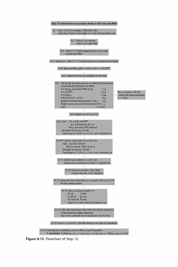
Figure 8.15 Flowchart of Step 12.

VIDEO
Please refer to this link (http://www.jove.com/index/Details.stp?
ID¼2034) for a video illustrating the first day of experiments.
REFERENCESReferenced LiteratureAmbros, V. (2004). The functions of animal microRNAs. Nature, 431(7006), 350–355.Andrus, A., & Kuimelis, R. G. (2001). Base composition analysis of nucleosides using HPLC.
In A. Andrus & R. G. Kuimelis (Eds.), Current Protoccols Nucleic Acid Chemistry. NewYork: Wiley Unit 10 16.
Bezerra, R., & Favre, A. (1990). In vivo incorporation of the intrinsic photolabel4-thiouridine into Escherichia coli RNAs. Biochemical and Biophysical Research Communica-tions, 166(1), 29–37.
Favre, A., Moreno, G., Blondel, M. O., Kliber, J., Vinzens, F., & Salet, C. (1986).4-Thiouridine photosensitized RNA-protein crosslinking in mammalian cells.Biochemical and Biophysical Research Communications, 141(2), 847–854.
Gerber, A. P., Luschnig, S., Krasnow, M. A., Brown, P. O., & Herschlag, D. (2006).Genome-wide identification of mRNAs associated with the translational regulatorPUMILIO in Drosophila melanogaster. Proceedings of the National Academy of Sciences ofthe United States of America, 103(12), 4487–4492.
Glisovic, T., Bachorik, J. L., Yong, J., & Dreyfuss, G. (2008). RNA-binding proteins andpost-transcriptional gene regulation. FEBS Letters, 582(14), 1977–1986.
Halbeisen, R. E., Galgano, A., Scherrer, T., & Gerber, A. P. (2008). Post-transcriptionalgene regulation: From genome-wide studies to principles. Cellular and Molecular LifeSciences, 65(5), 798–813.
Hafner, M., Landgraf, P., Ludwig, J., et al. (2008). Identification of microRNAs and othersmall regulatory RNAs using cDNA library sequencing. Methods, 44(1), 3–12.
Hafner, M., Landthaler, M., Burger, L., et al. (2010). PAR-CLIP – Transcriptome-wideidentification of RNA targets and binding sites of RNA-binding proteins. Cell,141(1), 129–141.
Keene, J. D. (2007). RNA regulons: Coordination of post-transcriptional events. NatureReviews Genetics, 8(7), 533–543.
Landthaler, M., Gaidatzis, D., Rothballer, A., et al. (2008). Molecular characterization ofhuman Argonaute-containing ribonucleoprotein complexes and their bound targetmRNAs. RNA, 14(12), 2580–2596.
Moore, M. J. (2005). From birth to death: The complex lives of eukaryotic mRNAs. Science,309(5740), 1514–1518.
Tenenbaum, S. A., Carson, C. C., Lager, P. J., & Keene, J. D. (2000). Identifying mRNAsubsets in messenger ribonucleoprotein complexes by using cDNA arrays. Proceedings ofthe National Academy of Sciences of the United States of America, 97(26), 14085–14090.
Ule, J., Jensen, K. B., Ruggiu, M., Mele, A., Ule, A., & Darnell, R. B. (2003). CLIP iden-tifies Nova-regulated RNA networks in the brain. Science, 302(5648), 1212–1215.
SOURCE REFERENCESHafner, M., Landgraf, P., Ludwig, J., et al. (2008). Identification of microRNAs and other
small regulatory RNAs using cDNA library sequencing. Methods, 44(1), 3–12.Hafner, M., Landthaler, M., Burger, L., et al. (2010). PAR-CLIP – Transcriptome-wide
identification of RNA targets and binding sites of RNA-binding proteins. Cell,141(1), 129–141.
160 Jessica Spitzer et al.

Referenced Protocols in Methods NavigatorExplanatory Chapter: Next Generation Sequencing.UV crosslinking of interacting RNA and protein in cultured cells.RNA Radiolabeling.Lysis of mammalian and Sf9 cells.One-dimensional SDS-Polyacrylamide Gel Electrophoresis (1D SDS-PAGE).Western Blotting using Chemiluminescent Substrates.RNA purification – precipitation methods.RNA purification by preparative polyacrylamide gel electrophoresis.Agarose Gel Electrophoresis.
161PAR-CLIP (Photoactivatable Ribonucleoside-Enhanced Crosslinking and Immunoprecipitation)