paper submission deadline - microscopy … · paper submission deadline: february 8, 2016 look...
TRANSCRIPT
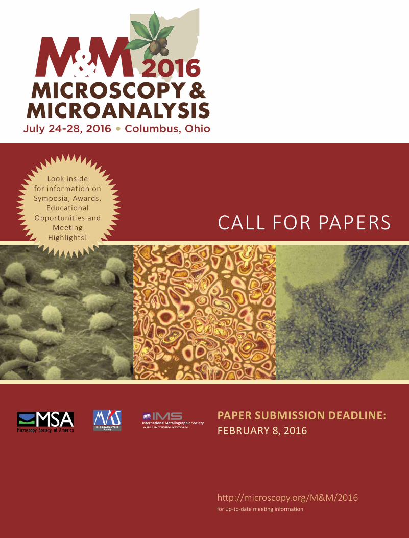
CALL FOR PAPERS
PAPER SUBMISSION DEADLINE: FEBRUARY 8, 2016
Look inside for information on Symposia, Awards,
Educational Opportunities and
Meeting Highlights!
http://microscopy.org/M&M/2016for up-to-date meeting information
Modi�ed A

2 M&M 2016 | July 24-28 | Columbus, OH
QUESTIONS?Questions regarding the technical content of the meeting or regarding specific sessions may be directed to:
2016 PROGRAM CHAIR Joe [email protected]
Registration opens March 1, 2016. Please direct questions regarding registration to: [email protected] Questions regarding exhibits, exhibitors or sponsors may be directed to: [email protected] Please direct all other meeting-related questions to: [email protected]
ARE YOU A MEMBER?Join Today and Save on M&M 2016 Registration Fees
Visit http://microscopy.org to join the Microscopy Society of America online, or call 1-800-538-3672 for more information about the benefits of MSA membership.
Visit http://microanalysissociety.org to join the Microanalysis Society and find out information about MAS membership benefits.
Visit http://metallography.net for membership information on the International Metallographic Society.
Dear Fellow Microscopists, Colleagues, and Friends:
On behalf of the sponsoring societies, we would like to thank everyone who attended M&M 2015 in Portland, Oregon. We hope your time in Portland was enjoyable and informative. We now turn to 2016, and invite you to join us July 24-28 in Columbus, Ohio for Microscopy and Microanalysis 2016. Columbus is the capital of Ohio, home to the Ohio State University, and a vibrant, bustling city with great restaurants, fun nightspots, an up-and-coming culinary scene, and is a great family-friendly place to visit!
We look forward to another exciting and informative M&M meeting in 2016. The Program Committee has created a wonderfully diverse program that illustrates our members’ diverse fields of work. We encourage all, whether brand-new to or veterans of M&M, to submit one or more scientific papers for presentation in Columbus, as M&M 2016 promises to have something for all.
The meeting will officially start on Sunday evening with a welcome reception. The technical program will commence on Monday morning with two plenary lectures; one featuring Prof. Mark Miodownik, University College London Professor of Materials and Society. He is the author of Stuff Matters, the winner of the 2014 Royal Society Winton Prize, and is a well-known BBC presenter of programs on materials science and engineering. The other plenary speaker, Drew Berry, is a biomedical animator with a background in cell biology and microscopy, whose work has been exhibited at storied art venues including the Guggenheim Museum, MoMA, the Royal Institute of Great Britain, and the University of Geneva. He will give a talk entitled: Beyond the Limits of Microscopy: Revealing the Unseeable through Hollywood Visual Effects.
Between the two talks, the winners of our major society and meeting awards will be honored. The exhibit floor will showcase the latest state-of-the-art microscopy-related equipment. The ever-popular free vendor tutorials will again be available onsite. The meeting will also feature the traditional Sunday Short Courses, Physical Sciences and Biological Sciences Tutorials, as well as two Pre-Meeting Congresses: Exploiting the Diffractive Properties of Electrons for Solving Materials Problems, organized by the Electron Crystallography and Automated Mapping Methods FIG, and Essentials of Atom Probe Tomography, organized by the Atom Probe FIG.
Participating at M&M 2016 will give you the opportunity to stay abreast of new technologies, learn new techniques, see the latest instrumentation, and most importantly, network with colleagues and make new connections. The paper submission site will open on December 1, 2015. We hope that you will be able to join us in Columbus for what is certain to be a very exciting and educational meeting.
Mike Marko Thomas F. Kelly Jaret FrafjordPRESIDENT PRESIDENT PRESIDENT
Microscopy Society Microanalysis Society Internationalof America Metallographic Society
ON THE FRONT COVER, FROM LEFT TO RIGHT:
IMAGE 1: Monocytes (White Blood Cells) Adhering to the Inside Surface of an Artery as Part of an Inflammatory Reaction. W. Gray (Jay) Jerome, Vanderbilt University
IMAGE 2: Cast A347 Alloy Made by Semi-solid Melting (Mert Fleming’s Development) Weck’s Reagent in Bright Field. George Vander Voort, Consultant (Struers Inc.)
IMAGE 3: High Density Lipoprotein (HDL; the good cholesterol carrier) Stacking Together in Solution. W. Gray (Jay) Jerome, Vanderbilt University

CALL FOR PAPERSBIOLOGICAL SCIENCES SYMPOSIA
http://microscopy.org/M&M/2016 for up-to-date meeting information 3
B01 Nanostructured Scaffolds for Regenerative Medicine Marco C. Bottino, Caroline A. Miller
• Microscopy-related instrumentation for synthesis and characterization of nanostructured scaffolds (e.g. nanofibers, nanotubes, etc.)
• Role of nanostructured scaffolds in tissue regeneration
• Novel techniques to develop and characterize nanostructured scaffolds
B02 New Technologies for Digital PathologyRohit Bhargava, David Mayerich
• New microscopy technology • Computational methods for better
decision-making• Applications in Digital Pathology
B03 Super-Resolution Visualization of Cellular and Inter-Cellular Processes in Health and Disease Rob Gourdie
• Models of normal and diseased tissues using super-resolution imaging
• Super-resolution imaging of cell culture models
• Methodological application of super-resolution modalities in cell biology
• Super-resolution imaging of intercellular junction formation and organization
• Novel super-resolution visualization modalities
B04 Microscopy and MorphogenesisRich Goodwin, Jay Potts
• Dynamic imaging of embryological development
• Three- and four-dimensional imaging, reconstruction, and analysis
• Cellular processes of organogenesis• Plant development• Microanalysis of the development of
biological shapes• The use of microscopy to elucidate
molecular mechanisms of development and dysgenesis
B05 Pathology: When Normal Goes WrongJay Jerome, Bill Gunning
• Molecular and cellular causes of disease• Novel underlying principles behind
disease progression• Papers on both human and animal
diseases are encouraged• Contributions on the understudied
areas of mitochondropathies and lysomal storage diseases are especially encouraged
B06 Pharmaceuticals and Medical ScienceJohn Bruce Green, Bridget Carragher
• Novel method development (from MicroCT to cryo-TEM)
• Pharmacology challenges (polymorphs, contaminants, particles, etc.)
• Device challenges (failure mode analysis, biocompatibility, sterility, etc.)
• Case studies
B07 3D Structures of Macromolecular Assemblies, Cellular Organelles, and Whole CellsElizabeth R. Wright, Teresa Ruiz, Kristin N. Parent
• Structural and ultrastructural studies of biological systems using advanced EM techniques and hybrid methodologies
• Eukaryotic and prokaryotic architecture
• Cellular metabolism, cell division and protein translation
• Cellular and bacterial adhesion, motility and secretion
• Cell-cell interactions and cell signaling • Virus-host interactions and virus
structure
B08 Utilizing Microscopy for Research and Diagnosis of Diseases in Humans, Plants, and AnimalsJon Charlesworth, Greg Ning, Betty Thompkins, Caroline Miller
• Research findings in renal and ciliopathic diseases
• Technical applications for basic and clinical research
• Use of microscopy for improving the quality and durability of crops and livestock
• Investigation of organisms and their related pathogens in clinical and research laboratories
• Techniques that improve rapid detection and treatment of diseases
Cast Alloy A356 Made in a Permanent Mold, Etched with Weck’s Reagent, 200X, Crossed Polarized Light. George Vander Voort, Consultant (Struers Inc.)

4 M&M 2016 | July 24-28 | Columbus, OH
P01 Dr. Gareth Thomas Symposium: Materials Solutions through MicroscopyDavid B. Williams, Ron Gronsky
• Structure-property relationships in metals and ceramics in the TEM
• High-resolution TEM• High-voltage TEM• TEM of magnetic materials
P02 Electron Microscopy of Materials for Electrochemical Power SystemsMark Aindow, Karren L. More
• Materials for fuel cells, electrolyzers, batteries, and supercapacitors
• Effects of processing on microstructure in components and assemblies
• In-service degradation: microstructure and materials stability
• Strategies for sample preparation from electrochemical devices
• Recent developments of in-situ and in operando studies
P03 Combining Simulation, Experiment, and Data Science for Materials Characterization and DesignPaul Voyles, Jinwoo Hwang, Mark Oxley
• Methods and software for simulating microscopy data
• Combining experiments and simulations to gain new insight
• Data science methods applied to microscopy images and spectra
• Design of new materials using microscopy data and simulations
P04 Nuclear and Irradiated MaterialsChad M. Parish, Khalid Hattar, Peter Hosemann
• Fission, fusion, accelerator, and space materials: metals, ceramics, polymers, semiconductors, fuels, etc.
• Damage phenomena in nuclear environments: dislocation loops, segregation and precipitation, stacking faults, tetrahedra corrosion, etc.
• Microscopy using SEM and TEM with aberration correction, in-situ imaging, tomography, etc.
• Microanalysis using microprobe, atom probe, mass spectroscopy, synchrotron and neutron beams, etc.
• Modelling and theoretical approaches that aid in data interpretation
P05 Microscopy for Thin Films of Metals, Semiconductors, and InsulatorsLaxmikant V. Saraf, C. Barry Carter
• Microscopy of thin films used for future potential alternative-energy applications
• Films for fuel cells, batteries, solar cells, and other alternative-energy storage or transport technologies
• Microscopic investigations of defects in these films
• Correlation of microscopy and other characterization techniques for understanding growth of films and their physical properties
• Understanding sensors or detectors based on thin films
• Microscopic analysis of films for applications in optical, magnetic, photovoltaic, and nuclear technologies
P06 Magnetic Materials, Phenomena, and Imaging at the NanoscaleMarc De Graef, Amanda Petford-Long
• Microscopy of bulk and nanoscale magnetic materials
• Applications of microscopy techniques to explore magnetic structure and behavior
• Development of new microscopy techniques for quantitative analysis of magnetic fields and materials
P07 Failure Analysis Applications of Microanalysis, Microscopy, Metallography, and FractographyDaniel P. Dennies, Noah Budiansky
• Failure investigations where microscopy and/or fractography are of great diagnostic importance
• Unique uses of light and electron microscopy to characterize materials that have failed
• Unusual, interesting, and/or difficult-to-interpret fractographic features
• Challenging, interesting, and/or innovative sample preparation and evaluation
• New or innovative uses of microanalysis or quantitative metallography in failure analysis
P08 Microscopy of Additive Manufacturing and 3D Printing in Materials and Biology Manuel Garcia-Leiner, Daniel P. Dennies, Michael Yost
• Failure investigations in additively manufactured parts where microscopy is of great diagnostic importance
• Unique uses of microscopy for the characterization and design of additively manufactured components
Vitreously Frozen Bacteriophage T4 Imaged at 300 kV with a Hole-free Phase Plate. Mike Marko, Wadsworth Center

CALL FOR PAPERS
http://microscopy.org/M&M/2016 for up-to-date meeting information 5
• Novel materials and their characterization for additive manufacturing processes
• Development and uses of microscopy for novel processes and applications for additively manufactured products
• Process development, application standards for additive manufacturing processes and products
• Regulatory, legal and intellectual property considerations for additive manufacturing processes and products
P09 From Nanometers to AU: Studies of Planet-Forming MaterialsEve L. Berger, Francis M. McCubbin, Adrian J. Brearley
• Mineral analysis with EPMA, EELS, EDS, SIMS, FTIR, APT, EBSD, XCT
• Emergence and evolution of micro-scale analyses
• Coordinated analyses: maximizing scientific return from small samples
• Terrestrial analogs for the analysis of astromaterials
P10 Microscopy and Characterization of Ceramics, Polymers, and CompositesRichard E. Chinn, Ronald J. Parrington
• Materialography/preparation of ceramics, polymers and composites
• The role of light, acoustic, X-ray, cathodoluminescent, laser and electron microscopy for examination of ceramics, polymers and composites
• Unusual, interesting, and/ or difficult-to-interpret microstructural and fractographic features
• Challenging, interesting, and/or innovative sample preparation and evaluation
P11 Metallography and Microstructural Characterization of MetalsCoralee McNee, George Vander Voort
• Characterization of metals and alloys with emphasis on solving problems in metal processing, service, or performance
• Metallic specimen preparation techniques, e.g., manual or automated, ion-beam polishing techniques, chemical and electrolytic etching
• The influence of specimen preparation upon microstructural interpretation, characterization and structure-property correlations
• Characterization methods including light optical, quantitative image analysis, SEM, EBSD, EMPA, TEM, XRD, etc., and the effects of specimen preparation upon results using those techniques
• Novel techniques for the evaluation of mechanical, chemical and other physical properties of metals and
correlation to the structure and/or processing
• Quantitative metallography and the use of standardized test methods
P12 Microscopy and Analysis in Forensic ScienceS. Frank Platek , Stefanie L. Heckman
• Strategies for processing, preparing and analyzing trace evidence, both biological and non-biological
• Identification of unknown particles and materials using multiple microscopy and analysis technologies
• Non-conventional microscopy analysis (i.e. 3D surface metrology, alternative light source fluorescence, etc.) applied to forensic samples
• Case-related applications of microscopy and microanalysis
• Continuing analytical issues related to gunshot residue (GSR) analysis by SEM/EDS
X30 Technologists’ Forum: Analysis of Real Data and Recognition of ArtifactsCathy Johnson, Caroline Miller, Frank Macaluso
• Ability to identify and interpret data correctly
• Recognizing both preparation and imaging artifacts
• Guidance on improving data analysis
X31 Technologists’ Forum Special Topic: Image Analysis and a Practical Approach to Current Software Solutions and Their ApplicationsFrank Macaluso, Cathy Johnson, Caroline Miller
• Image analysis as an integral component of every experiment for extracting meaningful data
• A practical approach to current software solutions and their applications
• Exploring the use of freeware and commercial software packages
X32 Technologists’ Forum: Roundtable Discussion on ArtifactsE. Ann Ellis, Lee Cohen-Gould, Vicky Bryg
• Discussion about artifacts from materials to biological samples
• How to recognize artifacts and determine their causes
• How to correct procedures to avoid artifacts
TECHNOLOGISTS’ FORUM
PHYSICAL SCIENCES SYMPOSIA

6 M&M 2016 | July 24-28 | Columbus, OH
A01 Vendor SymposiumPaul Kotula, Teresa Ruiz
• New methods, techniques and technologies
• Breakthrough and new instrumentation • Improvements to existing
instrumentation
A02 TEM Phase Plate Imaging in Biological and Materials ScienceRadostin Danev, Mike Marko
• Theoretical considerations for phase-contrast TEM
• Design and optimization of phase-shifting devices for TEM
• Best practices for the use of phase plates in biological cryo-EM
• Phase-plate imaging with a Cs-corrected TEM
• Application results in biological and materials science
A03 X-ray Imaging and AnalysisJeff Davis, Ric Wuhrer, Eric Telfeyan
• µCT Imaging and 3D X-ray Analysis• Correlative X-ray imaging (including
light microscopy, XPS and SIMS techniques)
• Post-processing of X-ray images (phase analysis, multivariate statistics and quantitative imaging)
• µXRF and µXRD imaging and analysis• SEM-EDS imaging (including µXRF in the
SEM and traditional EDS imaging)• New detectors, instrumentation
and software• Practical applications of X-ray imaging
for materials analysis
A04 Advances in FIB Instrumentation and Applications in Materials and Biological SciencesLucille A. Giannuzzi, Nabil Bassim, Srinivas Subramaniam
• New sources, columns, ions, detectors, multi-column analyses, etc. for FIB.
• 2D and 3D applications in biology, including cryo-FIB
• 2D and 3D applications in materials/geology
• Techniques for TEM and other specimen preparation
• Micro- and Nano-prototyping
A05 Applications of Correlative Microscopy to Physical and Biological SciencesSi Chen, Renu Sharma, Niels de Jonge, Nestor J. Zaluzec
• Instrument development in correlative microscopy and spectroscopy
• Applications to physical and biological sciences
• Sample preparation, preservation and transfer
• Methods and challenges of image registration
A06 Analytical Electron Microscopy for Advanced Characterization, from Multi-dimensional Data Acquisition to Integrated AnalysisChaoying Ni, Peter A. van Aken, Masashi Watanabe
• AEM: EELS, XEDS, EFTEM, CL, and concurrent signal acquisition and analysis towards quantification
• AEM-based multi-dimensional data acquisition, including tomography
• Improved data analysis strategies for hyperspectral/diffraction imaging in AEM
• Advances in AEM instrumentation and detectors (high-speed signal detectors and pixelated electron detectors for STEM imaging)
• Theoretical approaches related to AEM, including damage effects induced by electron beam irradiation
A07 Surface and Subsurface Microscopy and AnalysisVincent S. Smentkowski, John A Chaney, Chanmin Su
• State-of-the-art surface analysis and instrumentation
• Advances in scanning probe microscopy for quantitative analysis including nano-scale chemical, mechanical and electrical analyses
• How surface methods expand and compliment SEM/TEM
• Correlational imaging by combining multiple analytical technologies
• Hyper-spectral data, 3D imaging, and multivariate statistical analysis
A08 Quantitative and Qualitative Microanalysis by EPMA and SEMPaul Carpenter, Julien Allaz
• Microanalysis by FE-EPMA and FE-SEM using WDS and EDS detectors
• Advances in microanalysis instrumentation, analytical methods, and software tools
• Trace- and light-element analysis by EPMA: instrumentation and analysis methods
• Complementary microanalysis techniques: compositional mapping, cathodoluminescence, LA-ICP-MS, micro-XRF
• Standard reference materials: characterization and use for microanalysis and quality control
• Education and quantitative microanalysis
A09 Advanced Scanning Diffraction: Mapping Functionality in Reciprocal Space at Nanometer ResolutionJim Ciston, Doug Medlin, Alex Eggemann
• Ptychography/ coherent diffractive imaging
CALL FOR PAPERS
http://microscopy.org/M&M/2016 for up-to-date meeting information 7
• Differential phase contrast/ nanobeam diffraction mapping
• Scanning quantitative PACBED/ octahedral rotation mapping
• Orientation imaging• Fluctuation microscopy• STEM/CBED/ diffraction simulations
on a grand scale• Data reduction to produce property/
functionality maps
A10 Advances in Image Processing, Display and AnalysisWilliam A Heeschen, Clifford S Todd
• Innovative or interesting image analysis techniques
• Applications in image-related machine learning
• Advances in pattern recognition• Big data image analysis and handling
A11 Advances in Scanning Electron/Ion Instrumentation and DetectorsBrad Thiel, Matthew Phillips, Milos Toth
• Instrumentation advances in SEM and scanning ion microscopy
• Developments in and applications for miniature and microcolumn optics
• Developments in and applications for multibeam columns/beam splitters
• Aberration-corrected and energy- filtered imaging
• Direct detection of lower energy (100 - 1000 eV) electrons and ions with high efficiency
• Polarization-resolved cathodoluminescence
A12 Research and Applications in Atom Probe TomographyFrederick Meisenkothen, Eric B. Steel
• Materials applications, specimen preparation techniques, and optimization of acquisition conditions
• Correlative techniques, 3-D reconstruction, and data analysis
• Detector performance• Modeling and measurements
to understand specimen and instrument parameters
• Accuracy and precision in APT measurements and the development of APT standards
A13 In-situ Electron Microscopy and Big Data Analytics in 2D and 3DHuolin Xin, Peter Ercius, Kai He
• TEM in liquids and gases• High-speed S/TEM imaging for
capturing material dynamics• Big data storage, mining, and analysis• High-throughput time-lapse EELS and
EDX imaging for chemical/physical transformation of materials
• Development of sample environ-ments and external stimuli (heat, light, x-ray, plasma, microwave)
A14 Single-Atom Electron Microscopy and SpectroscopyJingyue (Jimmy) Liu, Larry Allard
• Novel methods for imaging isolated single atoms and/or determining their 3D distribution
• Spectroscopy of supported or embedded isolated single atoms
• Dynamic behavior of isolated individual atoms
• Anchoring of supported or embedded single atoms
• Novel properties of isolated single-atom systems
A15 Quantitative Measurement of Intensities and Distances in Electron Microscopy James LeBeau, Jinwoo Hwang
• Methods and applications of quantifying image intensities in HRTEM and STEM
• Direct comparisons between scattering theory and experiment
• Improving precision and accuracy to determine atom positions in both lateral and depth directions
• Quantification of intensities by comparing with theory or statistical models
• Alternative imaging methods to improve quantification
A16 New Frontiers in Monochromated EELSIan MacLaren, Peter Crozier
• New developments in instrumentation, experimental techniques and data analysis for monochromated EELS
• Experimental EELS studies at very high-energy resolution and developments to extract physically meaningful information from the raw data
• Theoretical studies of the interactions of low-energy excitations in materials and nanostructures with high-energy electrons
• Correlation to other high-energy resolution spectroscopic techniques (e.g Raman spectroscopy, inelastic neutron scattering, HREELS)
An Array of CBED Simulations. Colin Ophus, LBNL
ANALYTICAL SCIENCES SYMPOSIA

8 M&M 2016 | July 24-28 | Columbus, OH
PLENARY SESSIONSMONDAY, JULY 25, 2016Columbus Convention Center
PLENARY SPEAKER
Professor Mark MiodownikUniversity College London, UK
Materials for the 21st Century
Professor Mark Miodownik is the UCL Professor of Materials and Society. He received his Ph.D in alloys for jet turbine engines from Oxford University, and has worked as a materials engineer in the USA, Ireland and the UK. For more than ten years he has championed materials research that links the arts and humanities to medicine, engineering and materials science. This culminated in the establishment of the UCL Institute of Making where he is Director and runs the research program (www.instituteofmaking.org.uk). Prof. Miodownik is a well-known author and broadcaster. He regularly presents BBC TV programs on materials science and engineering which have reached millions of viewers in more than 200 countries. In 2013 he was awarded the Royal Academy of Engineering Rooke Medal, and he was elected a fellow of the Royal Academy of Engineering in 2014. He is author of Stuff Matters, which won the Royal Society Winton Prize in 2014.
PLENARY SPEAKER
Drew BerryWalter and Eliza Hall Institute of Medical Research, Melbourne, Australia
Beyond the Limits of Microscopy: Revealing the Unseeable through Hollywood Visual Effects
Drew Berry is a biomedical animator who creates scientifically accurate and aesthetically rich visualisations that reveal the cellular and molecular processes for a wide range of audiences. Beginning his career as a cell biologist and microscopist, Drew brings a rigorous scientific approach to each project, immersing himself in relevant research to ensure current data are represented. Since 1995, he has been a biomedical animator at the Walter and Eliza Hall Institute of Medical Research. His animations have exhibited at venues such as the Guggenheim Museum, MoMA, the Royal Institute of Great Britain and the University of Geneva. In 2010 he received a MacArthur Fellowship “Genius Grant.”
EDUCATIONAL OPPORTUNITY
Instrumentation Grant Writing RoundtableMONDAY, JULY 25, 2016Lunchtime session: 12:00 – 1:30 PM
Organized by the Facility Operation and Management (FOM) FIG
Bring your own lunch! Join us for a discussion about Grant Writing for Instrumentation. This Roundtable will center on your experiences with grant writing for the funding of your microscopes and other instrumentation. Many FOM FIG members have been through this process with different granting agencies. They will share with us their successes and failures so that you might gain some insight into the process.
PLENARY SESSIONS
NOTE: Important Information for Submitting Authors
Beginning this year, only three (3) unspecified/general categories will be offered to submitting authors:
1. Contributed Papers in Biological Sciences
2. Contributed Papers in Physical Sciences
3. Contributed Papers in Analytical Sciences
Papers submitted to one of these categories will be assigned to symposia by members of the Program Committee to optimize their fit with the overall scientific program.
“Life is a Puzzle”N. benthamiama epidermal cell infected with barley stripe mosaic virus expressing GFP, co-infiltrated with talin expressing DsRed. Imaged with a Zeiss 710 laser scanning confocal microscope. The 4 window panes utilize various color-coded projections.
2015 MICROGRAPH CONTEST WINNER Jim Kilcrease, USDA-ARS / ORISE
CALL FOR PAPERS
ONSITE AWARDS The M&M meeting’s co-sponsoring societies confer competitively judged awards at the meeting.
MSA Student Poster Awards: We believe poster presentations are an excellent format for all participants to engage in intensive discussion with other researchers in the field. To especially encourage students to take advantage of this opportunity and submit papers for poster presentation, MSA provides cash awards to the most outstanding student posters (first author) each day (up to one in each of three categories).
Diatome Poster Awards: All posters illustrating the use of diamond-knife ultramicrotomy are eligible. Prizes include cash and Swiss watches.
MAS Best Paper Awards: MAS annually confers awards for papers presented at the M&M meeting deemed to be best in four categories. Each comes with a cash award generously provided by MAS Sustaining Members.
MICROGRAPH COMPETITIONS
IMS Metallographic Contest: This annual contest solicits micrographs that illustrate problem-solving using a variety of imaging techniques, with cash prizes awarded in each of several classes.
MSA Micrograph Competition: This competition rewards the innovative blending of art and science. Winning micrographs will be selected on the basis of artistic merit and general audience appeal. See M&M 2016 website for complete information.
The winner of the 2015 Micrograph Competition is featured on Page 8 of this brochure.
2016 AWARDS
http://microscopy.org/M&M/2016 for up-to-date meeting information 9
GENERAL CONSIDERATIONS: Award applicants will automatically be considered for memorial scholarships, conferred by MSA based on the generous support of society sponsors.
Applicants who have previously received an M&M Award will not be considered for a second award in the same category.
STUDENTS:All full-time students enrolled at accredited academic institutions are eligible. High school, undergraduate, and graduate students are encouraged to apply. Applicants are not required to be members of the sponsoring society.
POSTDOCTORAL RESEARCHERS:All full-time postdoctoral researchers are eligible. Applicants are not required to be members of the sponsoring society.
PROFESSIONAL TECHNICAL STAFF MEMBERS:Full-time technologists are eligible. In addition, the applicant must be a member of the sponsoring society, current in his or her dues for the year of the meeting.
AMOUNT OF AWARD:M&M Meeting Awards and memorial awards consist of full meeting registration and up to $1,000 for travel-related expenses. Original receipts must be provided to receive travel reimbursement.
All award winners also receive an invitation to the Presidents’ Reception, held on the Tuesday evening of the meeting.
NOTIFICATION OF AWARD:All award applicants will be notified of their award status approximately eight weeks after the Call for Papers deadline.
Unsuccessful applicants will be permitted to withdraw their papers, should their ability to attend the meeting be contingent on the award, within one week following notification.
REQUIREMENTS OF AWARD:All award winners must present their paper in person at the M&M meeting in order to receive their award.
Awardees are expected to attend and participate in the entire meeting, which runs from Sunday evening’s opening reception through late Thursday afternoon.
Awardees are required to attend the Monday morning plenary session, at which their award will be conferred.
Modi�ed A
Modi�ed A
MEETING AWARDS The Microscopy Society of America (MSA) and the Microanalysis Society (MAS) annually sponsor awards for outstanding papers contributed to the Microscopy and Microanalysis (M&M) meeting, which are competitively judged based upon the quality of the submitted paper. These awards are provided to students, postdoctoral researchers, and professional technical staff members to help defray travel, lodging and other costs of attending the meeting. All awardees must fit the award criteria, as described below, at the time of the M&M meeting.
HOW TO APPLY FOR AN M&M MEETING AWARD: 1. As part of the on-line paper submission process, an applicant must
flag his or her paper for award consideration. Only one paper may be designated per applicant.
2. The applicant must appear as first author and presenter of the paper submitted for award.
3. The applicant must provide the name, title, institution, and e-mail address of his or her supervisor, who will be contacted to provide a supporting letter and confirmation of applicability for the indicated award category (e.g. student, post-doc, or technical staff).
10 M&M 2016 | July 24-28 | Columbus, OH
X10 Cryo-preparation for Biological EM Kent McDonald, Danielle Jorgens, Rick Webb, Helmut Gnaegi
• Introduction to low-temperature sample preparation, especially high pressure freezing and freeze substitution
• Rapid and inexpensive methods for processing frozen specimens into resins
• Cryotechniques for preserving fluorescence within polymerized resin for correlative light and electron microscope analysis
• Cryosectioning for immunolabeling or cryo-TEM imaging
X11 Electron Cryotomography Image Processing Using RELIONSjors Scheres, Tanmay Bharat
• Explanation of the RELION algorithm and explanation of how the algorithm works for single-particle analysis
• Introduction to tomographic data and the missing wedge; explanation of the combined 3D CTF and missing wedge model in RELION
• Setup of files and directories for RELION sub-tomogram averaging, including generation of 3D CTF models
• 2D and 3D classification of tomogram data• Sub-tomogram averaging auto-refinement in RELION
SUNDAY SHORT COURSESOrganizer: Elizabeth Wright, Emory University
• These full-day courses run from 8:30 AM to 5:00 PM on Sunday.
• A certificate of participation will be issued to each participant.
• Two (2) Continuing Microscopy Education Units are available (registration fee $10 for members).
• Morning and afternoon coffee breaks are included. (Breakfast and lunch are on your own.)
• Separate registration/fees required (see registration form for more information).
X12 Imaging and Analysis with Variable Pressure or Environmental SEM Brendan J. Griffin
• Imaging with SE, BSE, CL, and EDX detectors• Monitoring and optimizing instrument performance• Use of charge-related contrast mechanisms• Use of hot, cool, and cold stages• Imaging uncoated specimens with ultra low kV and other beams (He, Ga)
X13 Practical Considerations for Image Analysis and use of ImageJ/FijiJames Grande
• Image analysis considerations• Application of image analysis tools in real-world problems• Using ImageJ/Fiji• ImageJ/Fiji programming considerations• Imaging and image-analysis connections
X14 Advanced Focused Ion Beam MethodsLucille Giannuzzi, Joe Michael
• Use of FIB for TEM and SEM sample preparation• Basics of ion/solid interactions• 3-D imaging and analysis• Nanofabrication
X15 Nanomaterial Microscopy and Microanalysis: Tools and PreparationLou Germinario, Phillip Russell, John Thornton
• Choosing the proper preparation technique and minimizing the introduction of artifacts
• Ensuring that representative samples are identified for subsequent analysis—sample preparation suitable for nanoscale characterization, with case studies to demonstrate the benefits of combining several techniques
• Use of SEM, ESEM, FESEM, and X-ray Microanalysis• AFM sample preparation suitable for nanoscale characterization • Nanoanalysis by TEM and HRTEM and STEM/EELS • Sample preparation and nanofabrication by FIB
X40 Career Tracks in Government and Industry: A Panel DiscussionSpeakers: Benjamin Bammes, Gabriella Kiss, TBA
X41 Effective Tactics for Getting an Equipment GrantSpeaker: Ken Taylor
X42 Building and Validating Atomic Models for EM Density MapsSpeaker: TBA
Please be sure to check the M&M 2016 website often for updates to the Tutorials offerings.
BIOLOGICAL AND PHYSICAL SCIENCES TUTORIALSOrganizers: Scott Stagg, Patrick Phillips

CALL FOR PAPERS
http://microscopy.org/M&M/2016 for up-to-date meeting information 11
MICROSCOPY OUTREACH X90 Microscopy in the Classroom: Strategies
for Education and OutreachCraig Queenan, Alyssa Waldron, Dave Becker
Local educators and registered conference attendees are invited to participate in presentations, round table discussions, and demonstrations of effective strategies for microscopy outreach and education from K-12 and beyond. This session will show how microscopy in education serves as an important learning tool for inspiring our future STEM professionals. Those involved in microscopy education or educational outreach are encouraged to submit a paper about their successful program or lesson for platform or poster presentation.
• Best Practices for incorporating microscopy into K-12 classrooms and curricula
• Corporate and academic institutions and programs involved in microscopy outreach, both locally and nationally.
• Methods to expose students to microscopy in an engaging and successful manner
X91 Family AffairElaine Humphrey, Stuart McKernan
The exciting world of microscopy opens for for attendees, family, and friends. This session includes:
• A mystery to solve using microscopy• Materials science and biological science
X92 A Project MICRO WorkshopElaine Humphrey, Caroline Schooley
• The Project MICRO workshop will change its venue this year to the MegaBooth all week after the Exhibit Hall opens.
• Visit the Outreach booth every day to see how to set up different stations in a classroom
• How to have fun with microscopy outreach and kids (adults love this too)
• See different microscopes for classroom use in action
2016 PRE-MEETING CONGRESSES• Separate registration fee required.• Breakfast, Lunch, and coffee breaks are included.
Exploiting the Diffractive Properties of Electrons for Solving Materials ProblemsSunday, July 24, 2016 • 8:00 AM – 5:00 PM
Organized by the Electron Crystallography and Automated Mapping Methods Focused Interest Group
ORGANIZERS: Jörg Wiezorek, University of PittsburghYoosuf Picard, Carnegie Mellon UniversitySergei Rouvimov, University of Notre DameRobert Stroud, nanoMegas
This pre-meeting congress reviews basic methodologies in the analysis of crystalline materials using electron diffraction. Both scanning electron microscopy and transmission electron microscopy methods are featured. This congress features nine invited speakers, each an internationally renowned expert in the utilization of electron diffraction methods for analyzing one or more of the following structural properties of crystalline materials: phase/symmetry, orientation, defects, and strain. Each speaker will describe the fundamental physics, explain the basic methodologies and approaches, and highlight key recent research findings and/or important technique developments. This session provides an excellent opportunity for electron microscopists to review SEM and TEM methodologies based on electron diffraction, and gain new insights on the latest advances and applications of state-of-the-art diffraction methods.
Essentials of Atom Probe TomographySunday, July 24, 2016 • 8:30 AM – 5:00 PM Organized by the Atom Probe Tomography Focused Interest Group
ORGANIZERS:Richard L. Martens, University of AlabamaArun Devaraj, Pacific Northwest National LaboratoryPrakash Kolli, University of MarylandBaishakhi Mazumder, Oak Ridge National Laboratory
Atom probe tomography (APT) has been growing very rapidly over the past decade. Commercial instrumentation and FIB-based specimen preparation have driven much of this growth. Accordingly, the number of people utilizing the technique has also been rising rapidly. This one-day pre-meeting congress, organized by the MSA Atom Probe Tomography Focused Interest Group (APT-FIG), will present the basics of atom probe tomography in an introductory overview. APT instrumentation and experimental design, theory, specimen preparation, data analysis, and applications will be discussed.
OTHER EDUCATIONAL OPPORTUNITIES
“New York Skyline”The bright-field transmission electron microscopy (TEM) image shows vertically-grown ZnO nanorods on a ZnO/Si(100) substrate by a metal vapor deposition. The crystallographic orientation and diameter of the ZnO nanorods are confined by those of ZnO buffer layer on the substrate. The cross-sectional TEM specimen was prepared by a conventional polishing/ion milling technique. The image was acquired using JEOL JEM3010. The inset shows the original TEM image.
MSA MICROGRAPH CONTEST, 3RD PLACEJong Seok Jeong, University of Minnesota
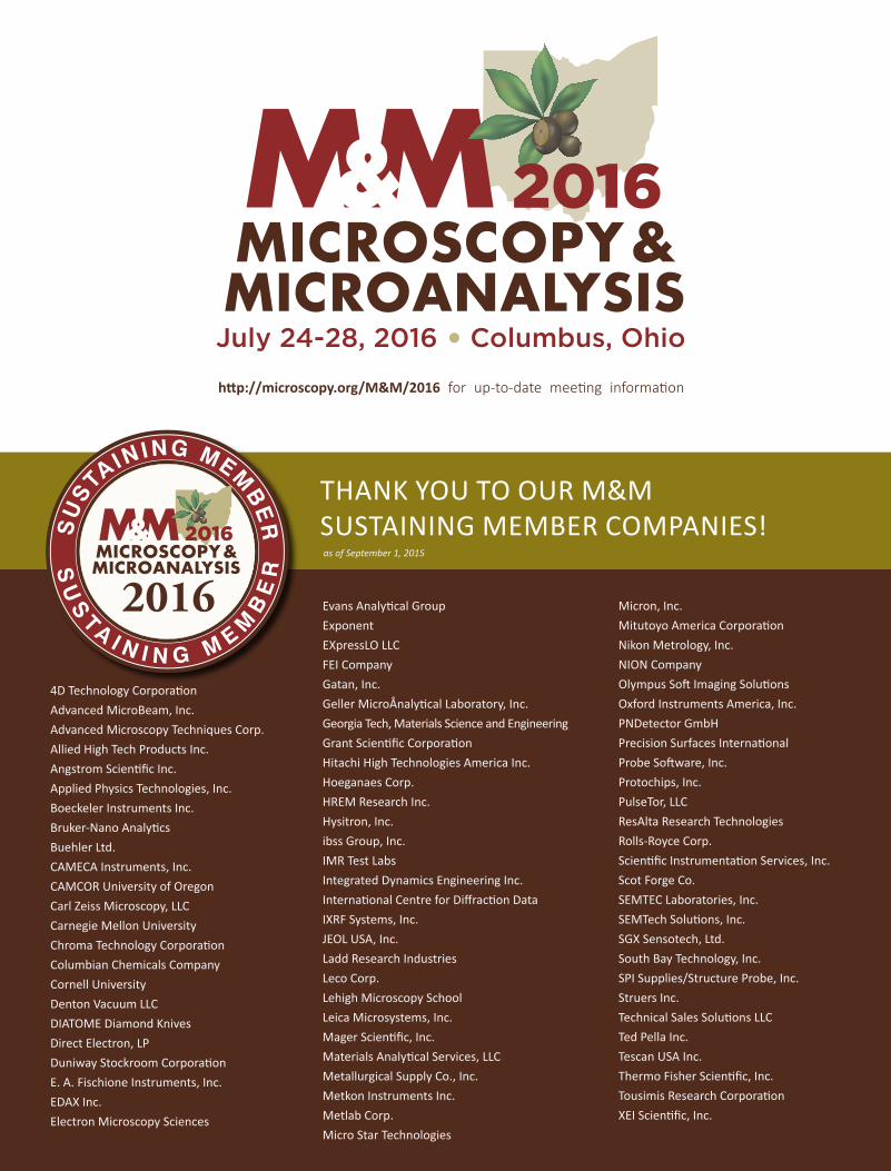
THANK YOU TO OUR M&M SUSTAINING MEMBER COMPANIES!
4D Technology Corporation Advanced MicroBeam, Inc. Advanced Microscopy Techniques Corp. Allied High Tech Products Inc. Angstrom Scientific Inc. Applied Physics Technologies, Inc. Boeckeler Instruments Inc. Bruker-Nano Analytics Buehler Ltd. CAMECA Instruments, Inc. CAMCOR University of Oregon Carl Zeiss Microscopy, LLC Carnegie Mellon University Chroma Technology Corporation Columbian Chemicals Company Cornell University Denton Vacuum LLC DIATOME Diamond Knives Direct Electron, LP Duniway Stockroom Corporation E. A. Fischione Instruments, Inc. EDAX Inc. Electron Microscopy Sciences
Evans Analytical Group Exponent EXpressLO LLC FEI Company Gatan, Inc. Geller MicroÅnalytical Laboratory, Inc. Georgia Tech, Materials Science and Engineering Grant Scientific Corporation Hitachi High Technologies America Inc. Hoeganaes Corp. HREM Research Inc. Hysitron, Inc. ibss Group, Inc. IMR Test Labs Integrated Dynamics Engineering Inc. International Centre for Diffraction Data IXRF Systems, Inc. JEOL USA, Inc. Ladd Research Industries Leco Corp. Lehigh Microscopy School Leica Microsystems, Inc. Mager Scientific, Inc. Materials Analytical Services, LLC Metallurgical Supply Co., Inc. Metkon Instruments Inc. Metlab Corp. Micro Star Technologies
Micron, Inc. Mitutoyo America Corporation Nikon Metrology, Inc. NION Company Olympus Soft Imaging Solutions Oxford Instruments America, Inc. PNDetector GmbH Precision Surfaces International Probe Software, Inc. Protochips, Inc. PulseTor, LLC ResAlta Research Technologies Rolls-Royce Corp. Scientific Instrumentation Services, Inc. Scot Forge Co. SEMTEC Laboratories, Inc. SEMTech Solutions, Inc. SGX Sensotech, Ltd. South Bay Technology, Inc. SPI Supplies/Structure Probe, Inc. Struers Inc. Technical Sales Solutions LLC Ted Pella Inc. Tescan USA Inc. Thermo Fisher Scientific, Inc. Tousimis Research Corporation XEI Scientific, Inc.
http://microscopy.org/M&M/2016 for up-to-date meeting information
as of September 1, 2015