pain associated with invisalign versus clear …
TRANSCRIPT

PAIN ASSOCIATED WITH INVISALIGN VERSUS CLEAR TRADITIONAL
BRACKETS: A RANDOMIZED, PROSPECTIVE TRIAL
A Thesis
by
DAVID W. WHITE
Submitted to the Office of Graduate and Professional Studies of
Texas A&M University
in partial fulfillment of the requirements for the degree of
MASTER OF SCIENCE
Chair of Committee, Peter H. Buschang
Committee Members, Phillip M. Campbell
Katie C. Julien
Head of Department, Phillip M. Campbell
May 2015
Major Subject: Oral Biology
Copyright 2015 David W. White

ii
ABSTRACT
The purpose of the present study is to compare the pain levels, analgesic
consumption, and sleep disturbances associated with Invisalign aligners (Align
Technology, Santa Clara, Calif) and traditional fixed appliances at multiple time points.
A prospective, randomized cohort study was conducted that included 41 adult
patients who were treated with either traditional fixed appliances (6 males and 12
females) or aligners (11 males and 12 females). Patients completed a daily discomfort
initially following bonding or delivery of the aligners, after one month, and after two
months. At each occasion, patients were asked to record their analgesic consumption,
sleep disturbances, along with their pain at rest, while chewing, while biting on their
front teeth, and when biting on their back teeth.
Both treatment modalities demonstrated similar pain values at baseline. There
were no significant sex differences. Patients in the traditional fixed appliances group
consistently reported higher pain scores than the patients in the Invisalign aligners group.
Depending on the question, the treatment group differences were statistically significant
(p<0.05) most days during the first week. Aligner patients also reported significantly
lower pain than the traditional treated patients after the first and second months. Pain
scores after the subsequent adjustments were consistently lower than after the initial
bonding or aligner delivery. A higher percentage of patients in the fixed appliances
group reported taking analgesics during the first week for dental pain, but only the
difference on day 2 was statistically significant.

iii
Aligner treatment is significantly less painful than traditional fixed appliances.
Patients treated with aligners consume fewer analgesics than patients treated with
traditional appliances.

iv
DEDICATION
I would like to dedicate this thesis to my wonderful wife, Bonnie White, and to
our two daughters, Audrey and Eliza White. They have supported me from the beginning
and have made a lot of sacrifices to help me get to this point. I would also like to
dedicate this thesis to my parents, Ronald White and Barbara Snider. Both of them have
done everything possible to help me succeed in life.
v
ACKNOWLEDGEMENTS
I would like to thank my thesis committee members, Dr. Peter Buschang, Dr.
Phillip Campbell, and Dr. Katie Julien, for their mentorship and support during this
project. I would also like to thank Dr. Helder Jacob for his help seeing patients and being
there when I couldn’t.
I would like to thank American Orthodontics and Align Technologies for their
donated supplies and materials, without which we wouldn’t have been able to perform
the project.
A final thanks to my classmates Britney Bare, John Feusier, Jason Morris, Kelly
Owen, and Brittany Wright for all of the great memories. I couldn’t have asked for a
better class to have joined me on this incredible journey. I have added two new brothers
and three new sisters to my family.

vi
NOMENCLATURE
CNS Central Nervous System
CuNiTi Copper Nickel Titanium
GCF Gingival Crevicular Fluid
IASP International Association for Pain
IL-1β Interleukin 1 beta
IL-6 Interleukin 6
LLLT Low Level Laser Therapy
NiTi Nickel Titanium
NRS Numeric Rating Scale
NSAID Non-steroidal Anti-inflammatory Drugs
PDL Periodontal Ligament
PGE2 Prostaglandin E2
PGI2 Prostaglandin I2
TENS Transcutaneous Electrical Nerve Stimulation
TNF-α Tumor Necrosis Factor alpha
VAS Visual Analog Scale
VRS Verbal Rating Scale

vii
TABLE OF CONTENTS
Page
ABSTRACT ...................................................................................................................... ii
DEDICATION ..................................................................................................................iv
ACKNOWLEDGEMENTS ...............................................................................................v
NOMENCLATURE .........................................................................................................vi
TABLE OF CONTENTS ................................................................................................ vii
LIST OF FIGURES ........................................................................................................ viii
LIST OF TABLES ...........................................................................................................ix
CHAPTER I INTRODUCTION AND REVIEW OF THE LITERATURE .................... 1
Introduction.......... .......................................................................................................... 1Review of the Literature................ ................................................................................ 4
CHAPTER II PAIN ASSOCIATED WITH INVISALIGN VERSUS CLEAR
TRADITIONAL BRACKETS: A RANDOMIZED, PROSPECTIVE TRIAL ............... 24
Introduction.................................................................................................................. 24
Materials and Methods................ ................................................................................. 25
Results.......................................................................................................................... 29
Discussion.....................................................................................................................32
CHAPTER III CONCLUSIONS AND CLINICAL APPLICATIONS ........................... 38
Conclusions................................................................................................................... 38
Clinical Applications..................................................................................................... 38
REFERENCES ................................................................................................................. 39
APPENDIX A .................................................................................................................. 46
APPENDIX B .................................................................................................................. 58

viii
LIST OF FIGURES
Page
Figure 1. Diagram of patient flow through the study ....................................................... 46
Figure 2. Patient treatment and daily diary timeline ........................................................ 47
Figure 3. Median pain levels for Invisalign and traditional treatment groups in
response to the question: “Rate the amount of discomfort that you are
currently experiencing with your braces or Invisalign.” .................................. 50
Figure 4. Median pain levels for Invisalign and traditional treatment groups in
response to the question: “Rate how much discomfort you experienced the
last time that you chewed.” ............................................................................... 50
Figure 5. Median pain levels for Invisalign and traditional treatment groups in
response to the question: “Rate how much discomfort you are experiencing
when you bite down on your back teeth?” ........................................................ 51
Figure 6. Median pain levels for Invisalign and traditional treatment groups in
response to the question: “Rate how much discomfort you are experiencing
when you bite down on your front teeth?” ....................................................... 51
Figure 7. Median pain levels for traditional treatment patients at rest and while
chewing. ............................................................................................................ 52
Figure 8. Median pain levels for traditional treatment patients while biting down on
the front and back teeth. .................................................................................... 52
Figure 9. Median pain levels for Invisalign patients at initial bonding, 1 month
adjustment and at the 2 month adjustment in response to the question: “Rate
how much discomfort you are experiencing when you bite down on your
front teeth?” ...................................................................................................... 55
Figure 10. Median pain levels for traditional patients at initial bonding, 1 month
adjustment and at the 2 month adjustment in response to the question: “Rate
how much discomfort you are experiencing when you bite down on your
front teeth?” ...................................................................................................... 55
Figure 11. Percentages of patients who took medications for tooth pain during the
first week after the initial appliance placement or Invisalign delivery. ............ 55
Figure 12. Percentages of patients who had sleep disturbance from tooth pain during
the first week after the initial appliance placement or Invisalign delivery. ...... 57

ix
LIST OF TABLES
Page
Table 1. Pain [medians (Med) and interquartile ranges] associated with Invisalign and
traditional orthodontic treatment at initial delivery or appliance placement. ... 48
Table 2. Pain [medians (Med) and interquartile ranges] associated with Invisalign and
traditional orthodontic treatment for the first month adjustment. ..................... 53
Table 3. Pain [medians (Med) and interquartile ranges] associated with Invisalign and
Traditional orthodontic treatment for the second month adjustment. ............... 54
Table 4. Percentages of patients who took medications for tooth pain after the first,
second, and sixth month adjustments. .............................................................. 56
Table 5. Percentages of patients who had sleep disturbance from tooth pain after the
first, second, and sixth month adjustments. ...................................................... 57

1
CHAPTER I
INTRODUCTION AND REVIEW OF THE LITERATURE
INTRODUCTION
Pain and discomfort are common side effects of orthodontic treatment.
According to a study by Scheurer et al. conducted on 170 patients, 95% of the patients
reported feeling pain within 24 hours of insertion of orthodontic appliances.[1] They
reported that the average pain level was 42% of maximal pain. Kvam et al. confirmed
the high prevalence of pain in orthodontics when they evaluated the experience of
patients treated in both private practice and a graduate program in Oslo, Norway.[2] Of
the 161 patients, 95% had experienced pain during treatment and 11% maintained that
they were constantly in pain. Approximately 85% of the patients felt as though the pain
only lasted two to three days. Another study conducted in Singapore found that 91% of
368 Chinese patients experienced transient pain and 39% experienced pain with each
new archwire or elastic force application.[3]
The main factor causing pain has been shown to be the initial application of
orthodontic forces.[2, 4-6] There are also other situations and appliances that have been
shown to stimulate painful responses in orthodontic patients. While Kvam et al.
demonstrated that archwire changes were the most painful part of their orthodontic
treatment, they also showed that approximately 76% of patients had some form of oral
ulceration during treatment.[2] 2.5% of their patients described the ulcers as being “very
painful.” Similarly, Otasevic et al. found oral ulcers in 42% of their sample of

2
orthodontic patients.[7] Rapid maxillary expanders have also been shown to cause pain
in 85% of patients over the entire course of treatment.[8] Most patients reported pain
between the 4th
to the 7th
day of expansion. The Herbst appliance, which is commonly
used in class II dentoskeletal corrections, has been reported to be a potential cause of
pain in the masticatory system.[9] Lastly, 16% of patients with a class III skeletal
pattern who were treated with chin cup therapy developed symptoms of
temporomandibular joint disorder.[10] Deguchi et al. theorized that posterior
positioning of the condyle caused anterior displacement of the disk, which led to both
pain and clicking upon opening.
As members of a health-care profession, orthodontists traditionally are concerned
about their patient’s well-being. They desire to provide necessary treatment in a time
appropriate manner, while attempting to minimize painful side effects. In addition to
this motivation, orthodontists should be very concerned about causing pain in their
patients, because it alters patients’ behaviors in ways that often lead to negative
consequences. Even before treatment begins, fear of pain is one of the primary reasons
that patients fail to seek orthodontic care.[11] O’Connor conducted a study that found
that injury from wires sticking out and dental pain are the 2nd
and 4th
most commonly
selected fears of patients prior to starting orthodontic treatment.[12] He also found that
pain was the greatest dislike during treatment, above even appearance, headgear, or
waiting-room delays.
During treatment, orthodontic pain has numerous negative effects on both the
patient and the treatment outcomes. Jones and Chan found that 22% of patients reported

3
that pain disturbed their sleep.[13] This was supported by Scheurer et al., who reported
that 18% of patients claimed to have been awoken by pain during the first night after
fixed appliance placement.[1] Patients often have trouble eating the foods they are
accustomed to while undergoing orthodontic treatment. Otasevic et al. found that 54% of
the patients in their study reported difficulties eating and chewing that led to a change in
diet.[7] As treatment continues and patient’s begin to resent being in pain, their
compliance to treatment starts to decrease. Sergl et al. found that compliance is
negatively associated with increasing complaints of pain.[14] Krukemeyer et al. stated
that pain from orthodontic treatment had a negative effect on both oral hygiene efforts
and was a major factor in patient’s missing appointments.[15] Ultimately, some patients
tire from being in pain and seek to end treatment early. Patel reported that almost 1 out
of every 10 patients will interrupt their treatment early due to painful experiences.
Haynes suggested that pain from orthodontic appliances and its effect on a patient’s
daily life were major reasons for discontinuance of treatment.[16]
Invisalign aligners have been used as a treatment modality since 1998 for those
patients who desire an esthetic orthodontic appliance. Since they were brought to the
market, there have been very few studies which have compared the effects of Invisalign
to traditional fixed appliances.[17-19] The purpose of this study was to evaluate
specifically the differences in pain levels and analgesic consumption between the two
treatment modalities at multiple time points. The hypothesis is that Invisalign aligners
will produce less pain than traditional fixed appliances, due to its nature as a removable

4
appliance that provides intermittent forces. The study will help orthodontists to be able
to correctly inform patients on the positives and negatives of both treatment options.
The review of the literature will start with an overview of the pain pathway from
sensation at the receptor to perception in the brain. Second, the orthodontic pain cascade
will be outlined, focusing on the role of inflammatory mediators in the process. Next,
both the treatment and measurement of orthodontic pain will be examined. This will be
followed by an analysis of how variable forces affect pain perception by patients. Lastly,
the literature evaluating Invisalign aligners and the studies comparing Invisalign aligners
to traditional fixed appliances will be evaluated.
REVIEW OF THE LITERATURE
Pain Sensation to Perception
Pain plays an important role in the survival of individuals. Melzack described it
as a “warning signal” that enables an organism to sense impending tissue damage and
thus avoid harm.[20] The International Association for the Study of Pain (IASP) has
defined pain as: “An unpleasant sensory and emotional experience associated with actual
or potential tissue damage, or described in terms of such damage.”[21] The painful
response of individuals has been described as consisting of four stages: signal
transduction, transmission, modulation, and perception.[22]
The first responder to a painful stimulus is the primary afferent neuron located at
the site of injury.[22] Primary afferent neurons are composed of a cell body, dendrites,
and axons. The cell body contains the nucleus and much of the cellular organelles. The

5
dendrites role is to detect and transduce the stimulus, while the axon transmits the
electrical stimulus to the next neuron. The dendritic end of a primary afferent neuron
contains specialized receptors that are able to identify various external stimuli. For
example, Ruffini corpuscles are able to detect changes in light touch or pressure and
Krause corpuscles respond to cold temperatures. Polymodal nociceptors are specialized
receptors that recognize a variety of noxious stimuli, such as physical, thermal or
chemical trauma to a tissue.[23] They are able to convert, or transduce, the initial
stimulus to an electrical current that rapidly passes down the primary afferent cell’s axon
to the second-order neuron, which transmits it eventually to the central nervous system
(CNS). The speed at which the nerve impulse is conducted by the neuron depends upon
the size of the nerve axon and whether or not the axon is myelinated.
The two most common types of neuron cell fibers in the orofacial complex that
transmit pain are the large Aδ (1-20 µm) and smaller C (0.5-1.0 µm) fibers.[22] The
faster Aδ fibers usually transmit pain that is localized and described as being sharp or
bright. While the slower C fibers transmit pain that is more diffuse and is thought to be
dull or aching. While there are exceptions, for the most part in the body, pain is
transmitted through a set of three neurons on its way to the somatosensory cortex of the
brain. The cell bodies for the primary afferent sensory neurons in the orofacial complex
are mostly located in the trigeminal ganglion near the apex of the petrous part of the
temporal bone. When a painful stimulus in the mouth activates these neurons, they
transfer the signal to the secondary neurons, which are located in the trigeminal spinal
tract nucleus of the brainstem. These neurons then relay the message to the tertiary cell

6
bodies located in the thalamus, which ultimately communicate with neurons located in
the somatosensory cortex. It is in the somatosensory cortex that the pain signal is
interpreted and an appropriate response is initiated.
In order to better explain many of the phenomena related to pain sensation and
why it is so varied between individuals, Melzack and Wall suggested what has become
known as the gate control theory.[24] Sensory impulses being relayed from peripheral
receptors to the CNS first must pass through a gating mechanism located in the dorsal
horn of the spinal cord for most of the body. The gating mechanism either inhibits or
allows the impulse to pass up to higher relay stations located in the thalamus.[25] In the
orofacial region the anatomy is slightly different. The impulses are transported by the
trigeminal nerve to the brain stem pons where the trigeminal spinal nucleus is located.
The trigeminal spinal nucleus acts much like the spinal cord dorsal horn. This system
allows the mind to shut down or minimize certain signals from receptors.
Another way that pain signals are modulated is through sensitization of pain
receptors and the neurons that compose the pain signal pathway. When cells are
subjected to brief physical or chemical trauma, and either Aδ or C fibers are activated,
there is a transient pain signal that serves as a physiological warning to the individual.
[26] Part of the body’s reaction to the damaging stimulus is to activate the inflammatory
response, which serves to remove the stimuli and begin the healing process. The
damaged cells release a variety of inflammatory mediators, such as: ions (e.g. K+, H+),
bradykinin, histamine, ATP and nitric oxide. [23] Immune cells are recruited by the
initial release of these chemical messengers. They subsequently release a host of

7
cytokines and growth factors, for example: interleukin 1 β (IL-1β), interleukin 6 (IL-6),
tumor necrosis factor α (TNFα), and nerve growth factor .[23] When released, these
cytokines can induce powerful hyperalgesia.[26] Hyperalgesia has been defined by the
IASP as: “an increased response to a stimulus which is normally painful.”[27] In
addition to the release of all of these inflammatory cytokines, the arachidonic acid
pathway is activated, leading to the synthesis of prostaglandins from the cyclooxegenase
enzymes COX-1 and COX-2.[23] Prostaglandin E2 (PGE2) and prostaglandin I2 (PGI2)
are the lipid metabolites that have the greatest impact on pain signal modulation.[28]
The many inflammatory mediators previously mentioned have a varied and
profound effect upon the primary afferent neuron’s activity. Some of them serve to
directly activate the nociceptors, which causes spontaneous pain. Others indirectly
increase the rate and intensity of the signal by sensitizing the receptor to respond to
either a lower stimulus or a normally non-painful stimuli.[26] When the painful
response is increased by sensitization, it is called hyperalgesia. Whereas, when a
normally harmless stimulus causes pain, it is known as allodynia. The inflammatory
mediators initiate these effects on the neurons by either changing the sensitivity of the
actual receptor molecules or by modulating the activity of voltage-gated ion channels
that are responsible for transmitting the signal. [23]
Pro-inflammatory cytokines, such as TNF-α and IL-1β, play a crucial role in
initiating and maintaining the inflammatory process by helping communication between
cells. In the short term, they increase sensitization by activating receptor-associated
kinases and ion channels. In the long term or chronic situations, they induce the

8
transcriptional up-regulation of receptors.[29] All of these effects serve to increase
hyperalgesia in an individual. Neuropeptides such as substance P and calcitonin-gene
related protein (CGRP) work synergistically to initiate pro-inflammatory actions:
vasodilation, extravasation of plasma, and degranulation of mast cells. [26]
Neuropeptides are released at the peripheral junction of neurons and trigger sensitization
of other neurons.[22] Prostaglandins (e.g. PGE2 and PGI2) are some of the most active
molecules causing inflammatory hyperalgesia. They play a role in reducing the
activation threshold, sensitizing the sensory neurons, and increasing their response to
additional stimuli.[26] They are thought to reduce the activation threshold of Na+
channels, which help propagate the signal from the receptor to the CNS. [30] Local
anesthetics used in dentistry, such as lidocaine, work by blocking these very same Na+
channels.
In order for a noxious stimulus to be perceived as pain, it must be greater than the
pain threshold, or the least experience of pain which a subject can recognize.[27] There
is a significant amount of variation between individuals in the stimulus and the pain
experienced. This is partly due to the variable pain thresholds that occur in each
individual based on a multitude of factors: patients psychological well-being, sex, age,
race/culture. The combination of all of those factors makes it very difficult to predict the
response of any individual to a specific stimulus as well as the patient’s pain tolerance.
The IASP defined pain tolerance level as “the maximum intensity of a pain-producing
stimulus that a subject is willing to accept in a given situation.”[27]

9
According to Klinberg et al., 3-21% of children and adolescents visiting a dentist
reported being fearful or anxious.[31] This is important, because there have been
multiple reports that increased levels of anxiety leads to greater reactions to painful
stimuli.[32-34] Commonly, this is because of past experiences and the expectation of
that painful experience to reoccur.[35] A review of pain and anxiety in the dental setting
concluded that anxiety can lower the pain threshold in patients and cause them to react to
non-painful stimuli as being painful.[36] This has been supported in the orthodontics
literature by Sergl et al., who proposed that psychological well-being of an individual
affected pain perception more than even force levels. Both personalized phone calls and
text messages from the orthodontists have been shown to decrease pain reports and
anxiety in patients. [37, 38]
There are reports that the pain threshold, in general, increases along with age. In
a study looking at 520 individuals, aged 5 to 105, cutaneous pain thresholds were
measured using a constant-current electrical stimulator.[39] The researchers found that
pain thresholds rapidly increase until the age of 25 years, and then following a plateau,
increased only slightly until 75 years. The orthodontic literature is somewhat divided on
whether adolescents or adults report higher levels of pain. Jones and Chan found a
significant association between the age of an orthodontic patient and the subsequent arch
wire pain, with greater ages being associated with greater pain.[13] However, all of their
patients were less than 17 years of age. This was supported by Fernandes et al., who also
found a significant association between age and level of pain or discomfort while
comparing superelastic nickel-titanium wires with nitinol wires in patients less than 17
10
years of age.[5] Lower pain scores were attributed to younger patients. Jones found that
those older than 16 were significantly more likely to report pain than those under 16.[6]
Conversely, Brown and Moerenhout found that adolescents (aged 14-17) reported higher
levels of pain and lower levels of psychological well-being than pre-adolescents and
adults.[4] Other studies have found no significant difference in age groups. Ngan et al.
stated that there was no significant difference in pain reports when comparing patients
less than 16 years of age and those that were greater than 16.[40] The conflicting reports
of age being a factor in pain levels, can be explained by the fact that adults and
adolescents often have very different treatment plans involving different appliances.
In general, the orthodontic literature does not demonstrate a correlation between
gender and perception of orthodontic pain. Multiple studies have failed to show a
significant difference between the two sexes. Jones and Chan found no significant
differences in pain reports between males and females.[13] Erdinc and Dincer stated
that gender differences were not shown to be statistically significant in the perception of
orthodontic pain.[41] However, some studies have shown that females report more
pain/discomfort than males. Kvam et al. showed that girls were more likely to report oral
ulcerations than boys.[2] Scheurer et al. administered a series of eight questionnaires to
170 patients (93 females, 77 males) and found that girls report greater levels of general
pain intensity, analgesic consumption, and pain while chewing than boys.[1] They
believed that some of the variation between their findings and much of the published
literature could be explained by differences in culture. Their sample was drawn from a
predominantly German-speaking region of Switzerland.

11
There is evidence that while some aspects of pain perception are universal, other
aspects are culture-specific.[42] People of Italian or Jewish descent are more likely to
report pain than people of Northern European descent, and to tolerate less intense pain
levels.[43] Also, studies over the last 50 years have generally shown that non-Hispanic
Caucasians have a higher pain tolerance and threshold than African Americans and
Hispanics.[44-46] This demonstrates that certain aspects of pain perception are learned
behaviors. Stoicism is encouraged by some cultures during painful experiences, while
other cultures reinforce pain expression by providing sympathy and attention.
The Orthodontic Pain Cascade
Before discussing how orthodontic forces initiate pain in a patient, it is necessary
to have an understanding of the basic anatomy of a tooth and its surrounding structures.
Each tooth is separated from the surrounding alveolar bone by a densely collagenous
tissue called the periodontal ligament (PDL). [47]. The PDL is approximately 0.5 mm
thick and inserts into the cementum covering the tooth root and into the dense bone
immediately surrounding the root. It contains both cellular elements and tissue fluids,
each of which plays an important role in orthodontic tooth movement. The cells found in
the PDL are predominantly undifferentiated mesenchymal cells and their offspring:
fibroblasts/fibroclasts and osteoblasts/osteoclasts. These cells play an integral role in the
remodeling process by removing both collagen and bone in the direction of force
application, and then laying down collagen and bone behind the tooth. Blood vessels and
nerve endings are also found in the PDL space. The nerve endings are either of the

12
unmyelinated variety that sense pain, or they are of the more complicated type that
differentiates pressure and positional information.
The pressure tension theory is the classical theory in orthodontics used to explain
how tooth movement occurs.[47] When an orthodontic force is applied to a tooth in a
certain direction, the tooth shifts positions. This results in compression of the PDL in
some areas and tension of the PDL in others. On the compression side, pressure builds in
the decreased PDL space, which occludes blood vessels in this area. However, on the
tension side, the reverse occurs; blood flow is either maintained or increased. Changes in
blood flow rapidly lead to adjustments in the chemical composition of the PDL
environment. For example, decreased oxygen levels and increased levels of
inflammatory mediators are found on the compression side. All of these effects serve to
activate cells involved in bone and tissue remodeling, but they also form the basis for
orthodontic pain.
As has been previously discussed, inflammatory mediators increase pain by
either direct activation of nociceptors or indirectly by lowering the activation threshold.
Orthodontic forces have been shown to produce mechanical damage and inflammation in
the periodontium.[48] Numerous studies have looked at the inflammatory metabolites
and chemical mediators that are present in the PDL and gingival crevicular fluid. Most
of them concur that orthodontic pain is a consequence of an inflammatory reaction
initiated by changes in blood flow following orthodontic force application.
Beginning in 1975, Davidovitch and Shanfield looked at biochemical changes in
mechanically stressed bone in cats, who were orthodontically treated anywhere from 1

13
hour to 28 days. They found elevated levels of cyclic adenosine monophosphate
(cAMP, a second messenger important in many biochemical processes, and suggested
that this rise was due to an increase in bone remodeling activity in the compression sites.
Then in 1985, Oliver and Knapman discovered increased levels of PgE and IL-1β
immediately after an increase in pressure.[11] This was supported by the work of Grieve
et al., who used a split-mouth design to evaluate PgE and IL-1β levels drawn from
gingival crevicular fluid (GCF) during human orthodontic tooth movement.[49] The
GCF levels of PgE and IL-1β were significantly elevated, while the levels on the control
side remained the same as the baseline. Uemastsu et al. assessed a host of cytokines
found in the GCF of orthodontic patients using an enzyme-linked immunosorbent assay.
They found elevated levels of IL-1β, IL-6, TNF-α, epidermal growth factor, and β2-
microglobulin in the experimental side when compared to the control side.[50]
Nicolay et al. used a cat model to evaluate neurotransmitters in the PDL
following orthodontic forces.[51] While substance P levels were relatively low before
application of the forces, the density of the positive staining for substance P increased
markedly from 24 hours to 14 days. They hypothesized that substance P may be an
initial trigger for a biochemical cascade that comprised the activation of various cell
types in the PDL. In a review of the literature on the cellular and tissue level reactions to
orthodontic force, Krishnan and Davidovitch concluded that force-induced tissue strain
produces local alterations in vascularity, which leads to the synthesis of various
neurotransmitters, cytokines, growth factors, and metabolites of 13ignificant acid.[52]

14
As has been discussed previously, these molecules are all involved in initiating and
propagating a painful response to a patient.
Treatment of Orthodontic Pain
As Scheurer et al. demonstrated, around 90-95% of orthodontics patients
experience pain while in treatment.[1] Based on how many negative consequences
result from this experience, it is imperative that orthodontists find ways of decreasing
their patients’ pain. Non-steroidal anti-inflammatory drugs (NSAIDS) are the most
commonly used method of treating patients for pain. Ibuprofen was found to be more
effective at controlling pain than aspirin in patients who were treated with both
separators and orthodontic wire changes.[53] Young et al. evaluated the best time to
administer NSAIDS.[54] He split up three groups of adult patients, and asked them to
score the amount of pain that they felt after the initial arch wire was placed. The first
group received 40 mg of Valdecoxib, an NSAID, 30 minutes prior to arch wire
placement; the second group received it 2 hours after arch wire placement, and the third
group received a placebo control. The study showed that 1 day after initial arch wire
placement, the group that received the 40 mg of Valdecoxib 30 minutes before arch wire
placement had the lowest pain scores. Even though NSAIDs have been shown to be
effective in pain management, they are only being used by 13-16% of patients during the
peak pain periods of 1-2 days.[1]
There are three other modalities that have been studied for their ability to manage
pain: low level laser therapy (LLLT), mastication, and transcutaneous electrical nerve
stimulation (TENS). LLLT has been shown to have both anti-inflammatory properties
15
as well as regenerative effects. Turhani et al. found a significant difference in pain levels
between a group treated with a low level laser and a control group.[55] However, the
operator was not blinded and could differentiate between the laser and the control. It has
been theorized that chewing on gum or a plastic wafer during the first few hours after
orthodontic forces would decrease patient’s pain levels by moving the teeth enough to
allow blood flow through compressed areas. This would permit the removal of metabolic
byproducts that have previously been discussed to increase sensitization to pain. White
had 93 patients with full appliances chew on two pieces of Aspergum and report whether
they felt more, the same, or less pain than their normal adjustments.[56] Fifty-eight
patients (63%) reported that they had a decrease in discomfort, while 24 (25%) said that
they experienced no difference, and only 11 (12%) said that they felt an increase in their
discomfort. Hwang et al reported that chewing on something hard, such as a therabite
wafer for 10-12 minutes after an adjustment may reduce pain.[57] While 56% of their
sample reported a decrease in pain, the other 44% found it to be more painful. Lastly,
Roth and Thrash evaluated whether TENS was able to reduce periodontal pain following
separator placement mesial and distal to first molars.[58] Patients were asked to report
their pain every 12 hours for the four days. They found a significant decrease in pain at
the 24, 36, and 48 hour time frames in the treatment group.
Measurement of Orthodontic Pain
Pain is a subjective experience, and therefore difficult to quantify accurately.
Pain intensity is influenced by the meaning of the pain to the patients and its expected
duration, along with a host of other factors.[20] There are many pain rating scales that

16
have been devised and are used in the literature. However, in orthodontics the three most
commonly used scales are the numerical rating scale (NRS), verbal rating scale (VRS),
and the visual analog scale (VAS).
A NRS has a patient rate their pain on a scale of 0-10 or 0-100, where 0
represents no pain and 10 or 100 represents the worst pain imaginable.[24] While it is
not commonly used in orthodontics, it is an interval level scale and can provide data to
which parametric tests can be applied.[59] The VRS provides a list of adjectives that
describe various levels of pain intensity and asks the patient to select the best response.
The list typically consists of words that describe the extremes and then some of the
gradations in between (e.g. no pain, mild, moderate, and severe pain).[59] These are
assigned numbers, but it would be incorrect to assume that the intervals in between each
response are equal, which is not the case according to Jensen et al.[60] VRS data is
ordinal and therefore can only be analyzed by non-parametric tests.[59]
The VAS is the most commonly used scale in orthodontic literature today,
because it has been shown to be the most accurate and reliable. It consists of a 10 cm
line that is anchored on both ends by verbal descriptors (e.g. no pain, worst pain
imaginable). The patient marks a line through the area that represents the pain level that
they are currently experiencing. This allows for 1001 gradations if measurements are
taken to the nearest 1/10th
of a mm. Even young children have been shown to understand
it and respond to the questions.[41] According to Krishnan, VAS has two main
advantages. First, it provides freedom to select the exact intensity of pain, and second, it
allows for maximum expression of an individual response style.[61] VAS has ratio

17
properties, but it is not always normally distributed.[62] If the data is normally
distributed, Williamson et al. suggest the use of parametric statistical analysis.[59]
Similarly, if it is not normally distributed, then non-parametric tests should be used.
Therefore, normality should be determined prior to running any statistical tests.
Variable Orthodontic Forces and their Effects on Pain
Pain during orthodontic treatment is the result of compression of the periodontal
ligament and the changes in blood flow that result. There is still some debate, however,
as to how force levels are related to pain. Burstone devised a classification system that
differentiated painful responses based upon either the relationship of the force
application or the timing of onset.[63] In terms of force application, there are three
degrees. With first degree the patient doesn’t perceive pain unless the doctor is pushing
on the teeth. Second degree is when the patient feels pain when he or she is clenching.
Third degree is when patients have difficulty eating foods of normal consistency. In
terms of the timing of onset, the pain can either be immediate or delayed. Immediate is
characterized by abrupt heavy forces placed on the teeth, while delayed is produced by
various force levels which create hyperalgesia of the PDL space.
The current goal of orthodontists is to provide the optimum force level to
produce fast tooth movement, while limiting tissue damage and maximizing patient
comfort. Traditionally, orthodontic forces have been classified as either being “heavy”
or “light”. Most experts advocate using light forces, because they are thought to be more
physiologic than heavy forces and therefore less painful.[47] It is believed that heavy,
sustained forces cause necrosis and hyalinzation of cells in the PDL. This causes less

18
efficient bone remodeling called undermining resorption. Lighter forces are believed to
lead to healthy cellular activity in the PDL along with efficient and relatively painless
remodeling of the alveolar bone, known as frontal resorption. Luppanapornlarp et al.
tested this theory using a split mouth design while retracting canines with either a 50 g
or 150 g nickel-titanium coil spring. They found that the mean VAS score was
significantly higher in the 150 g control side than the 50 g experimental side.[64]
However, Andreasen et al. also used a split mouth design to evaluate heavy (400-450g)
and light (100-150g) forces between canines and first molars and found no significant
differences in pain levels reported.[65] Jones and Richmond also failed to find a
correlation between pain and applied forces, using severity of crowding as an indicator
of applied forces.[66]
In an effort to minimize orthodontic pain, other researchers have investigated
different wire types and removable appliances. Wires made from a nickel-titanium alloy
have been shown to provide lighter and more constant force levels than stainless steel
wires.[67] Miura et al. found that Japanese NiTi wires exhibited super-elastic properties,
indicating definite stress levels with increasing amounts of strain. A three-point bending
test showed that initial super-elastic NiTi wires (e.g. 0.016 in.) generate forces in the
range of 140-170 grams. When comparing NiTi wires to a multistranded stainless steel
wire, Jones and Chan found similar levels of pain intensity and pain duration.[13]
Superelastic NiTi wires have also been reported to cause lower pain values when
compared with conventional nitinol wires.[5]

19
Traditional fixed appliances generate constant forces while removable appliances
inherently provide intermittent forces. Studies have looked at the effects of the different
types of appliances on pain levels. On one hand, Oliver and Knapman found no
significant differences in patient discomfort between fixed and removable
appliances.[11] However, Sergl et al. found that the severity of pain and discomfort was
significantly higher in patients treated with functional or fixed appliances than in those
treated with removable plates.[14] Stewart et al. also concluded that fixed appliances
were more painful than removable.[68]
Invisalign Clear Aligners
The first mention of using small sequential tooth positioners to make major
changes to the occlusion came almost 70 years ago by Kesling. He recognized the
potential for tooth alignment, but understood that it was not feasible in 1945.[69] At the
time, impressions of the dentition were poured up in plaster and the teeth were separated
and then individually moved to their desired locations. A one-piece pliable rubber
appliance was made to adapt to each surface of the teeth and patients were instructed to
wear it full time. However, the tooth positioning appliances were limited to correcting
slight rotations and making minor modifications to the arch form and axial position of
teeth. Over the following decades, numerous authors wrote about using plastic overlay
appliances to act as invisible retainers and to make minor tooth movements.[70-74]
Raintree Essix (New Orleans, LA) developed a technique to use aligners with small
“divots” to put pressure on teeth and move them into spaces, called “windows.”[75]

20
However, these aligners were limited to tooth movements that were no greater than two
to three mm.
Around the year 2000, Align technologies introduced the Invisalign system,
which utilized computer-aided-design-computer-aided-manufacturing technology to
manufacture clear aligners from stereolithography models.[75] After creating a digital
representation of a patient’s dentition from either a polyvinyl siloxane impression or
from a 3D digital scan, Align technology fabricates a set of clear plastic aligners to
slowly move the teeth to the desired location. Each plastic aligner is 0.030” thick and
allows for minor tooth movements, around 0.25-0.33 mm per tray.[76] The number of
trays produced depends on the number of stages required to move the teeth to their final
position. They are traditionally worn for two weeks before switching to the new set of
aligners.[77] In a recent study by Simon et al., they found that the forces generated by
the Invisalign system are “within the range of orthodontic forces.”[78] Specifically,
intrusion and distalization movements by Invisalign aligners produced forces between
0.5 to 1.0 newtons (N) or around 50-100 grams.
Invisalign Compared to Traditional Braces
Since Invisalign has only been in existence for a little over a decade, there are
limited studies investigating the patient experiences and clinical outcomes. Nedwed and
Miethke were the first to evaluate patient acceptance of Invisalign and the quality of life
of patients undergoing treatment. They administered a questionnaire with twelve
questions to fifty-four consecutively treated Invisalign patients after three to six months
of treatment. The patients were limited to only three verbal answer choices for each
21
question. The question concerning pain found that 35% had no pain, 54% had minimal
pain, and 11% had severe pain. There were several flaws with their study design. Not
only are verbal rating scales limited, but Nedwed and Miethke did not even provide a
fourth answer choice for moderate pain. This could have skewed the responses towards
minimal pain. The surveys were only administered after 3-6 months of treatment. This
only provides a brief look at the patient’s experience and fails to reliably express the
pain levels at different stages of treatment, especially at the beginning.
The first study to actually evaluate differences in pain levels between an
Invisalign sample and one treated with traditional braces was by Miller et al. in
2007.[17] They administered a daily diary to 60 patients from offices in Florida,
Arizona, Kentucky, Texas, and at the University of Florida. The diary was to be
completed every day for the first week and was to assess treatment effects including
functional, psychosocial, and pain-related outcomes. Four of the questions addressed the
pain experienced by the patients. Three of them were Likert style questions, while the
other was a visual analog scale based question. The results show that at baseline there
was no significant difference. Both groups show a dramatic increase during the first day
and then a gradually return to baseline over the course of the week. The results indicated
both a significant treatment group and treatment day effect, with Invisalign patients
demonstrating lower pain levels at each of the seven days. At 24 hours, the fixed group
had a mean VAS score of approximately 50/100, while the Invisalign group had a mean
VAS score closer to 40.

22
Shalish et al. performed a similar study, however, they included a sample of
patients wearing lingual brackets.[18] Sixty-eight patients filled out a health related
quality of life questionnaire composed of numeric rating scale questions for the first
week after initial appliance placement and then again on day 14. While the pain levels
were consistently higher in the Invisalign group than in the buccal fixed group, the
differences were not statistically significant. These are opposite results from those found
by Miller et al. They stated that the difference could have been due to the greater
mechanical force that the Invisalign technique applies early in treatment. They also
hypothesized that the wire’s flexibility may result in a more gradual and lighter force in
the buccal fixed group.
More recently, a group from Japan performed a study comparing the pain levels
of patients treated with Invisalign aligners and fixed edgewise appliances.[19] Fujiyama
et al. administered a VAS style survey to a sample consisting of 55 adult patients in the
fixed appliances group and 38 adult patients in the Invisalign group. Patients were asked
to report their pain at multiple time points during the first week and then again at 3
weeks and 5 weeks after appliance placement or delivery of aligners. They found that
while patients reported more pain with fixed appliances, the difference was only
significant at a couple of time points during each stage. Similar to prior studies, they
failed to randomly assign patients to specific treatment categories.
All of the studies evaluating differences between Invisalign and traditional braces
had flaws in their research design. None of them randomly assigned the patients to their
prospective treatment modality. Shalish et al. claimed that adult patients, who are often

23
concerned with esthetics, would be unwilling to comply with random assignment.[18]
They recognized that lack of randomization was a weakness, because allowing the
patients to choose treatments may provide results that reflect personality traits rather
than true appliance differences. In addition, Miller et al. and Shalish et al. only looked at
the first week or two of treatment, and thereby failed to evaluate treatment effects at any
other point in treatment. There is a possibility that pain levels early in treatment could be
very different at other time points. Lastly, Miller et al. and Shalish et al. used questions
from inferior rating scales. Miller et al. used some Likert style questions, while Shalish
et al. used a NRS for their pain questions.

24
CHAPTER II
PAIN ASSOCIATED WITH INVISALIGN VERSUS CLEAR TRADITIONAL
BRACKETS: A RANDOMIZED, PROSPECTIVE TRIAL
INTRODUCTION
Pain and discomfort are common side effects of orthodontic treatment.[1-3]
Because orthodontists are concerned about their patients’ well-being, they want to
minimize any painful side effects. Orthodontists should also be concerned about causing
pain, because it alters their patients’ behaviors in ways that often lead to negative
consequences. Fear of pain is one of the primary reasons that patients fail to seek
orthodontic care.[11] Sergl et al. found that compliance during treatment is negatively
associated with increasing complaints of pain.[14] Therefore, it would be beneficial to
be able to provide treatment modalities that produce the least amount of pain possible.
There are numerous reports describing the pattern of pain associated with
traditional fixed appliances.[1, 13, 40] For example, Scheurer et al. found that pain
peaked at 42% of maximum pain around 24 hours after initiation of orthodontic
treatment, and decreases thereafter.[1] Subsequent reports confirmed that pain decreases
during the first week after initial bonding, returning to baseline values. While the first
week of treatment has been extensively studied, few studies have evaluated the pain and
discomfort experienced by patients further into treatment. This is important, because
pain reported at future time points could be different than during the first week.

25
Since their introduction to the market in 1997 by Align technologies, clear
aligners have quickly become one of the preferred orthodontic appliances for patients
who are concerned with esthetics. Given how popular aligners have become, there is
surprisingly little research comparing the treatment effects and outcomes of aligners to
traditional braces. The first study to evaluating the differences in pain found that
traditional braces were significantly, approximately 25%, more painful than
Invisalign.[17] However, this study was conducted at multiple locations and was only
conducted during the first week after treatment started. This was followed by a study
from Shalish et al. that reported the exact opposite effect.[18] They utilized the inferior
numeric rating scale and also failed to evaluate pain months after initiation of treatment.
Recently, Fujiyama et al. confirmed the results of Miller et al. and found that traditional
fixed appliances were significantly more painful than Invisalign aligners.[19]
Importantly, none of the previous articles randomly assigned treatment modality, which
increases the possibility of biased comparisons.
The purpose of this study was to compare the pain levels produced by traditional
fixed appliances and Invisalign aligners at multiple time points. Secondarily, the study
sought to compare their effects on pain medication usage and sleep disturbances.
MATERIALS AND METHODS
A power analysis determined that ideally the sample needed to consist of 80 adult
(males > 19 years old and females > 17 years old) patients recruited from advertisements
at the Graduate Orthodontic Clinic of Baylor College of Dentistry. Of the 240 people

26
who were screened, 199 of them did not qualify for the study or declined to participate
(Figure 2.1). Currently there are 41 patients admitted to the study (17 males, 24
females). This study was approved by the Texas A&M University Baylor College of
Dentistry IRB (IRB approval # 2012-21-BCD). Informed consent was obtained from all
of the subjects.
The patients were randomly divided into one of the two treatment groups using a
randomization table created using Microsoft Excel. The experimental group consisted of
23 patients treated with Align Technology’s Invisalign appliance, while the control
group consisted of 18 patients treated using conventional fixed appliances (clear in
maxillary arch, stainless steel in the mandibular arch) as described in detail below. In
order to qualify for the study, patients had to fulfill the following selection criteria:
Inclusion Criteria
1. Class I molar and canine relationships
2. Non-extraction treatments
3. Mandibular crowding of 4 mm or less
4. No missing teeth (from second to second molar)
Exclusion Criteria
1. Anterior or posterior crossbites
2. Anterior or lateral open bites
3. Maxillary overjet exceeding 3 mm
4. Impacted teeth

27
All patients were seen at Texas A&M University Baylor College of Dentistry and
treated by one of two clinicians, Dr. Katie Julien DDS, MS or Dr. Helder Jacob DDS,
PhD. Following randomization and the obtainment of informed consent, initial
diagnostic records were taken (intra and extraoral photographs, lateral cepholagrams,
panoramic radiographs, Blu-Mousse® bite records, and plaster models) at the first
appointment. Subsequent appointments were dependent on whether the patient was in
the experimental group or the control group. The sequence of appointments adhered to
the timeline found in Figure 2
Patients in the Invisalign group had either a polyvinylsiloxane impression made
or the dentition was scanned using an iTero scanner®. The records were then sent to
Align Technology. A series of removable polyurethane aligners were fabricated based
on the treatment plan determined using Align Technology’s proprietary ClinCheck®
software. All composite attachments were placed at the initial aligner delivery
appointment. Patients were given two sets of aligners and instructed to wear them
twenty-two hours per day for two weeks and to change to the new set of trays on the 15th
day. Patients were scheduled every 4 weeks for evaluation appointments and were given
two new sets of trays at each appointment.
Traditional fixed appliances consisted of American Orthodontics’ Radiance clear
brackets in the maxillary arch, and stainless steel brackets in the mandibular arch. The
brackets used were a 0.018” X 0.028” Alexander prescription (American Orthodontics,
Sheboygan, WI). Brackets and tubes were bonded to the maxillary and mandibular arch
from second molar to second molar. A sequence of NiTi (.016” and .017”x.025”) and

28
stainless steel (.016”x.022” and .017”x.025”) wires were used according to the
malocclusion and the progress of correction. To monitor the progress of treatment,
patients were scheduled for appointments every four weeks.
This was a randomized, prospective, clinical trial that assessed patients’ pain and
analgesic consumption using a daily diary. The patients completed a daily discomfort
diary at four different occasions during treatment. The first was completed immediately
following initial bonding or delivery of Invisalign to create a baseline, and each day
thereafter during the first week (Appendix) They also completed diaries on the day of
and for 4 days following each of the next two adjustment appointments (4 and 8 weeks).
Four days was considered to be adequate because patients have been shown to adapt to
the pain and discomfort of orthodontics within the first 3-5 days.[2, 14, 24] The patients
were seen every 4 weeks for adjustment appointments or delivery of new aligners.
The daily discomfort diary consisted of eight questions (Appendix). The first five
questions asked about their discomfort under certain circumstances. The responses were
recorded on a 10 cm VAS, using No Discomfort and Worst Discomfort as the anchors.
The final three questions determined whether the patient’s sleep was affected (Did
discomfort caused by your orthodontic appliance interfere with your sleep?) and the
frequency of analgesic consumption (Did you take medication to relieve tooth pain?).
The daily diaries were collected at the adjustment appointments following a recorded
occasion. The examiner of the surveys was blinded as to which treatment the patients
had received. If a patient failed to bring the daily diary form in to the appointment, they
were asked to either bring it to the next appointment or mail it in a self-addressed and

29
stamped envelope. Four out of one hundred thirty two surveys were either lost or not
returned. After all of the diaries had been collected, they were measured by the principal
investigator, who was blinded as to the treatment modality, to the nearest 0.01 mm using
a digital caliper. The data were transcribed into a Microsoft Excel spreadsheet, using a
numeric label to identify the patients.
Statistical Analysis
All statistics were calculated using SPSS version 18 software (SPSS Inc.,
Chicago, Illinois). Due to skewness and kurtosis of the results, medians and interquartile
ranges were used to describe the results and group differences were compared using the
non-parametric Mann-Whitney test. Significance for all tests was set at P < 0.05.
RESULTS
Based on the first question from the daily diary, both treatment groups
demonstrated strong rating reliability with an intraclass correlation coefficient of 0.917-
0.986, depending on the assessment period.
Initial Adjustment
Traditional braces produced a similar pattern of pain for each of the four
questions. At baseline, or immediately following appliance placement, the patients
reported a low level of pain (Table 1). This was followed by a dramatic and statistically
significant (p < 0.05) increase in pain response (300 – 500%) that peaked between the
first and third day (Figures 3-6). The highest peak pain scores were reported when
30
chewing (VAS score = 46.91) and biting down on the front teeth (VAS score = 50.43).
Following the peak, there was a gradual reduction in pain over the next 4-5 days, ending
with a pain level similar to or slightly above those reported at baseline. Patients in the
traditional treatment group reported significantly higher pain while chewing than at rest
during most of the first week (Figure 7). They also reported higher pain at the front teeth
than the back teeth for the first few days, but the differences were not statistically
significant (Figure 8).
Invisalign also produced a similar pattern of pain for each of the VAS questions.
Initially, patients reported low levels of pain that were followed by slight increases (50 –
100 %), peaking after the first or second day (at VAS pain scores around 15-20) (Figures
3-6). Pain levels then decreased slowly over the rest of the first week. By day 7, patients
in the Invisalign treatment group experienced minimal pain levels, lower than those
reported on day 0.
Both treatment groups reported similar levels of pain on day 0, with no
statistically significant group differences (Table 1). Between day 1 and day 7, the
traditional group consistently demonstrated higher pain levels than the Invisalign group
(Figures 3-6). Depending on the question, the pain values in the traditional group were
found to be significantly higher during the majority of the first week. Due to high
variability between subjects, there were no statistically significant group differences
initially. After day 2 or day 3, group differences were mostly statistically significant at a
p level < 0.05 (Table 1).

31
Subsequent Adjustments
Pain after the first and second month adjustments was also consistently less for
Invisalign than traditional treatments. Many of the group differences after the first month
adjustment were statistically significant (p < 0.05) and all of the others closely
approached significant levels (Table 2). Significant group differences were found on
days 0, 1, 2, and 3. For both groups, the pain levels reported at subsequent adjustments
peaked at much lower levels than during the first week after the initial bonding or
Invisalign delivery.
The first month and second month Invisalign adjustments showed a similar pain
response pattern for each of the VAS questions. At day 0, the pain scores were generally
lower than those reported at the initial delivery and then slowly decreased over the next
four days (e.g. Figure 9). With traditional treatment group, the pain scores at day 0 were
generally higher than at the initial bonding (e.g. Figure 10). Like the Invisalign group,
the pain values decrease gradually over the next four days. However, unlike during the
first week of treatment, there was not a drastic increase in pain values following the
adjustment at months 1 and 2. The pain scores after the initial bonding were significantly
higher than the first month adjustments for both the Invisalign and traditional groups
(Figures 9-10)
Analgesic Consumption
The percentage of patients in the traditional group taking medication to relieve
tooth pain increased by approximately 45% during the first two days, and then decreased
steadily during the remaining five days (Figure 11). In the Invisalign group, the

32
percentage taking medication increased by 11% during the first day, and then decreased
steadily thereafter. While the percentages of patients taking medication was consistently
greater in the traditional than Invisalign group, only the 50% difference that occurred on
day two was statistically significant (p < 0.05). Medications taken after the first and
second adjustments showed no clear pattern and no statistically significant differences
between the traditional and the Invisalign treatments (Table 3.5).
Sleep Disturbance
There was no consistent pattern of group difference and no statistically
significant group difference in the percentage of patients who had sleep disturbances
during the first week (Figure 3.10). The frequency of patients reporting sleep
disturbances decreased from approximately 30% on the first day to 15% on the seventh
day. There also were no significant group differences in the proportion of patients
reporting sleep disturbances during the first and second adjustments (Table 3.6).
DISCUSSION
Patients treated with traditional fixed appliances show a dramatic increase in pain
over the first 24 hours after appliance placement; pain peaks after 24-48 hours, and then
decreases steadily to baseline values over the remainder of the first week. Previous
studies evaluating orthodontic pain during the first week of treatment have also shown
sharp pain increases during the first 24 hours, followed by gradual decreases over the
subsequent 6-7 days to values similar to those observed at appliance placement.[1, 13,
17, 19, 40] Regardless of the amount of crowding or degree of malocclusion, this

33
pattern of pain appears to be consistent. The large increase in pain over the first 24 hours
correlates with an acute inflammatory response [52] Initial orthodontic forces cause
pain through compression of the PDL, which leads to ischemia, edema, and the release
of pro-inflammatory mediators during the first 24-48 hours. The inflammatory
mediators, such as prostaglandins (e.g. PgE) and interleukins (e.g. IL-1β), sensitize
nociceptors in the PDL and lower the pain threshold.[26, 29] The levels of PgE and IL-
1β found in gingival crevicular fluid (GCF) peak 24 hours after initiation of orthodontic
force and fall to baseline after 7 days.[49] As such, the gradual reduction in pain
observed over the course of the first week can be attributed to the decrease of
inflammatory mediators in the PDL.
The amount of peak pain during the first week that occurs with traditional fixed
appliances appears to depend on the initial archwire used. Patients in the traditional fixed
appliances group reported that pain peaked at approximately 33% of the worst pain
imaginable. This peak pain value closely approximates the value found by a study that
used superelastic NiTi (29%) and is less than a study that used nitinol (42%).[1, 13] The
differences could be due to the material used. Initial nitinol archwires have been shown
to produce higher peak pain than superelastic NiTi archwires.[5] This could be due to the
fact that nitinol produces a greater amount of force.[79] The current study used 0.016 in.
copper NiTi (CuNiTi) archwires, which is a type of superelastic archwire. When loaded
2 mm, CuNiTi generates a force of 47 grams while unloading. Classic nitinol generates a
force of 180 grams when loaded under the same conditions.[79]

34
Patients treated with traditional fixed appliances experience significantly more
pain during the first week of treatment than patients treated with Invisalign aligners.
While both treatment groups reported similar baseline values, pain from days 2 through
7 was consistently and significantly higher in the traditional group, regardless of the
question being asked. Significantly less pain among patients treated with Invisalign
aligners compared to those treated with fixed appliances has been previously
reported.[17, 19] The lower pain reported by Invisalign patients could be due the fact
that it is a removable appliance. Removable appliances are generally less painful than
fixed appliances.[14, 68] The difference could be due to the fact that removable
appliances provide intermittent forces, while fixed appliances provide continuous forces.
Thilander proposed that interrupted or intermittent forces are advantageous, because they
allow the tissues enough time to reorganize before the compression force is
reapplied.[80]
The difference in pain between Invisalign and traditional fixed appliances does
not seem to be attributed to the extent of force applied. Invisalign aligners produce
forces between 0.5-1.0 N, or around 50-100 g, for intrusion and distalization
movements.[78] Conversely, initial leveling and aligning archwires, such as the 0.016 in
CuNiTi used in this study generate forces between 47-77 g.[79] Therefore, Invisalign
aligners and NiTi archwires produce similar forces that cannot explain the difference in
pain observed between Invisalign and fixed appliances.
After the first and second month of treatment, patients also reported that
Invisalign was less painful than traditional fixed appliances. One other study that

35
evaluated pain 3 and 5 weeks after appliance delivery showed similar differences.[19]
The reduced pain reported by Invisalign patients can be explained by the role of pro-
inflammatory mediators, such as IL-1β. Over the short term, they increase sensitization
by activating receptor-associated kinases and ion channels. Over the long term, or in
chronic situations, they induce the transcriptional up-regulation of receptors, leading to
hyperalgesia in an individual.[29] If fixed causes greater initial pain due to an increased
inflammatory response, then it is possible that patients with fixed appliances have more
sensitized nociceptors, which affects their pain perception during following adjustments.
Invisalign’s new SmartTrack® (Align Technology, Santa Clara, Calif) material
appears to be less painful for patients than previous clear aligner materials manufactured
by Invisalign. Patients in the Invisalign treatment group reported small increases in pain
that peaked at 13.6% of maximum during the first 24 hours and then decreased gradually
to pain values below baseline by day seven. Miller et al., who found a similar pattern of
slight pain increase followed by a steady pain decrease to values below baseline by the
end of day seven, reported peak pain values at 40% of maximum pain for the original
material.[17] Although more studies are needed, this supports Invisalign’s claims that
SmartTrack® provides greater patient comfort than the original material. [81]
In comparison to pain values during the first week of treatment, both treatment
modalities demonstrated significantly less pain at the subsequent adjustment. Fujiyama
et also found lower levels of pain for Invisalign and fixed appliances at subsequent
adjustments.[19] Soltis et al. showed that there is a decrease in the pain threshold
immediately after orthodontic forces are placed, but that after a period of time, the pain

36
threshold returns to its original level.[82] Therefore, the same stimulus will generate a
less painful signal months into treatment than immediately after appliances are placed.
For patients in the traditional group, pain while chewing was significantly higher
than pain at rest for most of the first week. Inflammatory mediators, such as substance P,
have been shown to be present in the PDL following the initiation of orthodontic
forces.[51] These mediators are believed to sensitize the nociceptors in the PDL.[24] It is
possible that chewing compresses previously sensitized nociceptors in the PDL and
stimulates a more painful signal than at rest. Importantly, this phenomenon was only
evident in the traditional group, which exhibited a more painful response to orthodontic
forces. The nociceptors in the PDL of patients in the traditional group may have been
more sensitized than those in the Invisalign group, due to the continuous nature of the
forces used, making them more susceptible to variations in compression of the PDL.
Patients treated with traditional fixed appliances report that their front teeth are
more sensitive than their back teeth while chewing, but the differences were not found to
be statistically significant. This is supported by the published literature.[1, 40, 41]
Scheurer et al. hypothesized that this difference depends on how involved anterior teeth
are during leveling and aligning, as well as to their smaller root surface.[1] According to
the principles of static equilibrium, the forces exerted on the anterior teeth during initial
leveling stage must be equal to the forces exerted on the posterior teeth.[83] Anterior
teeth could be more painful during chewing, because forces are distributed over smaller
root surface areas than posterior teeth.

37
Patients treated with fixed appliances are more likely to take pain medication
than Invisalign patients. The pattern of analgesic consumption closely mirrored the
pattern of pain at rest during the first week of treatment. There was an increase in
analgesic consumption during the first 24-48 hours, and then a gradual return to baseline
levels by day 7. A correlation between pain medication intake and pain levels throughout
the first week has been previously demonstrated.[17] Patients with fixed appliances have
been shown to have been in more pain than their Invisalign counterparts, and
accordingly, they reported taking more pain medication during the first week.
Interestingly, this phenomenon was not present at subsequent adjustments. This could be
due to the fact that patients, in general, were in less pain at subsequent adjustments and
didn’t require pain control.

38
CHAPTER III
CONCLUSIONS AND CLINICAL APPLICATIONS
CONCLUSIONS
1) Invisalign treatment is significantly less painful than traditional fixed appliances.
2) Patients treated with both Invisalign and traditional fixed appliances report
significantly less pain at subsequent adjustments than at the initial bonding.
3) Chewing is significantly more painful than when a patient is at rest for patients
treated with traditional fixed appliances.
4) Consumption of analgesics closely mirrored the levels of pain reported by patients
and was predominantly during the first week of treatment.
CLINICAL APPLICATIONS
While the first week of treatment may be mildly to moderately painful for
patients with Invisalign or traditional fixed appliances, following appointments will
produce less pain. Invisalign may be an alternative treatment option for patients that are
hesitant to start treatment due to fear of pain. Patients should be warned that biting with
the front teeth is more painful than chewing on the back teeth. Patients should also be
aware that analgesics may be needed during the first week of treatment, but are
traditionally not as necessary after subsequent adjustments.

39
REFERENCES
1. Scheurer, P.A., A.R. Firestone, and W.B. Burgin, Perception of pain as a result
of orthodontic treatment with fixed appliances. Eur J Orthod, 1996. 18(4): p.
349-57.
2. Kvam, E., N.R. Gjerdet, and O. Bondevik, Traumatic ulcers and pain during
orthodontic treatment. Community Dent Oral Epidemiol, 1987. 15(2): p. 104-7.
3. Lew, K.K., Attitudes and perceptions of adults towards orthodontic treatment in
an Asian community. Community Dent Oral Epidemiol, 1993. 21(1): p. 31-5.
4. Brown, D.F. and R.G. Moerenhout, The pain experience and psychological
adjustment to orthodontic treatment of preadolescents, adolescents, and adults.
Am J Orthod Dentofacial Orthop, 1991. 100(4): p. 349-56.
5. Fernandes, L.M., B. Ogaard, and L. Skoglund, Pain and discomfort experienced
after placement of a conventional or a superelastic NiTi aligning archwire. A
randomized clinical trial. J Orofac Orthop, 1998. 59(6): p. 331-9.
6. Jones, M.L., An investigation into the initial discomfort caused by placement of
an archwire. Eur J Orthod, 1984. 6(1): p. 48-54.
7. Otasevic, M., et al., Prospective randomized clinical trial comparing the effects
of a masticatory bite wafer and avoidance of hard food on pain associated with
initial orthodontic tooth movement. Am J Orthod Dentofacial Orthop, 2006.
130(1): p. 6 e9-15.
8. Gecgelen, M., et al., Evaluation of stress and pain during rapid maxillary
expansion treatments. J Oral Rehabil, 2012. 39(10): p. 767-75.
9. Pancherz, H. and M. Anehus-Pancherz, The effect of continuous bite jumping
with the Herbst appliance on the masticatory system: a functional analysis of
treated class II malocclusions. Eur J Orthod, 1982. 4(1): p. 37-44.
10. Deguchi, T., et al., Clinical evaluation of temporomandibular joint disorders
(TMD) in patients treated with chin cup. Angle Orthod, 1998. 68(1): p. 91-4.
11. Oliver, R.G. and Y.M. Knapman, Attitudes to orthodontic treatment. Br J Orthod,
1985. 12(4): p. 179-88.
40
12. O'Connor, P.J., Patients' perceptions before, during, and after orthodontic
treatment. J Clin Orthod, 2000. 34(10): p. 591-2.
13. Jones, M. and C. Chan, The pain and discomfort experienced during orthodontic
treatment: a randomized controlled clinical trial of two initial aligning arch
wires. Am J Orthod Dentofacial Orthop, 1992. 102(4): p. 373-81.
14. Sergl, H.G., U. Klages, and A. Zentner, Pain and discomfort during orthodontic
treatment: Causative factors and effects on compliance. American Journal of
Orthodontics and Dentofacial Orthopedics, 1998. 114(6): p. 684-691.
15. Krukemeyer, A.M., A.O. Arruda, and M.R. Inglehart, Pain and orthodontic
treatment. Angle Orthod, 2009. 79(6): p. 1175-81.
16. Haynes, S., Discontinuation of orthodontic treatment relative to patient age. J
Dent, 1974. 2(4): p. 138-42.
17. Miller, K.B., et al., A comparison of treatment impacts between Invisalign
aligner and fixed appliance therapy during the first week of treatment. Am J
Orthod Dentofacial Orthop, 2007. 131(3): p. 302 e1-9.
18. Shalish, M., et al., Adult patients' adjustability to orthodontic appliances. Part I:
a comparison between Labial, Lingual, and Invisalign. Eur J Orthod, 2012.
34(6): p. 724-30.
19. Fujiyama, K., et al., Analysis of pain level in cases treated with Invisalign
aligner: comparison with fixed edgewise appliance therapy. Prog Orthod, 2014.
15: p. 64.
20. Melzack, R.W., PA, Textbook of Pain, ed. Livingstone. 1989, London.
21. Merksey, H.B., N, Classification of Chronic Pain. Second ed. 1994, Seattle:
IASP Press.
22. Silverman, S., L.R. Eversole, and E.L. Truelove, Essentials of oral medicine.
2001, Hamilton, ON: BC Decker Inc. x, 381 p.
23. Kidd, B.L. and L.A. Urban, Mechanisms of inflammatory pain. Br J Anaesth,
2001. 87(1): p. 3-11.
24. Bergius, M., S. Kiliaridis, and U. Berggren, Pain in orthodontics. A review and
discussion of the literature. J Orofac Orthop, 2000. 61(2): p. 125-37.

41
25. Melzack, R. and P.D. Wall, Pain mechanisms: a new theory. Science, 1965.
150(3699): p. 971-9.
26. Dray, A., Inflammatory mediators of pain. Br J Anaesth, 1995. 75(2): p. 125-31.
27. Loeser, J.D. and R.D. Treede, The Kyoto protocol of IASP Basic Pain
Terminology. Pain, 2008. 137(3): p. 473-7.
28. Kawabata, A., Prostaglandin E2 and pain--an update. Biol Pharm Bull, 2011.
34(8): p. 1170-3.
29. Opree, A. and M. Kress, Involvement of the proinflammatory cytokines tumor
necrosis factor-alpha, IL-1 beta, and IL-6 but not IL-8 in the development of heat
hyperalgesia: effects on heat-evoked calcitonin gene-related peptide release from
rat skin. J Neurosci, 2000. 20(16): p. 6289-93.
30. England, S., S. Bevan, and R.J. Docherty, PGE2 modulates the tetrodotoxin-
resistant sodium current in neonatal rat dorsal root ganglion neurones via the
cyclic AMP-protein kinase A cascade. J Physiol, 1996. 495 ( Pt 2): p. 429-40.
31. Klingberg, G., et al., Child dental fear: cause-related factors and clinical effects.
Eur J Oral Sci, 1995. 103(6): p. 405-12.
32. Weisenberg, M.I., Pain and pain control. Psychol Bull, 1977. 84(5): p. 1008-44.
33. Sternbach, R.A., Pain; a psychophysiological analysis. 1968, New York,:
Academic Press. xv, 185 p.
34. Chapman, C.R. and J.A. Turner, Psychological control of acute pain in medical
settings. J Pain Symptom Manage, 1986. 1(1): p. 9-20.
35. Leventhal, H.E., D, Emotions in personality and psychopathology. Emotion, pain
and physical illness. 1979, New York: Plenum Press.
36. Litt, M.D., A model of pain and anxiety associated with acute stressors: distress
in dental procedures. Behav Res Ther, 1996. 34(5-6): p. 459-76.
37. Bartlett, B.W., et al., The influence of a structured telephone call on orthodontic
pain and anxiety. Am J Orthod Dentofacial Orthop, 2005. 128(4): p. 435-41.
38. Keith, D.J., et al., Effect of text message follow-up on patient's self-reported level
of pain and anxiety. Angle Orthod, 2013. 83(4): p. 605-10.

42
39. Tucker, M.A., et al., Age-associated change in pain threshold measured by
transcutaneous neuronal electrical stimulation. Age Ageing, 1989. 18(4): p. 241-
6.
40. Ngan, P., B. Kess, and S. Wilson, Perception of discomfort by patients
undergoing orthodontic treatment. Am J Orthod Dentofacial Orthop, 1989.
96(1): p. 47-53.
41. Erdinc, A.M. and B. Dincer, Perception of pain during orthodontic treatment
with fixed appliances. Eur J Orthod, 2004. 26(1): p. 79-85.
42. Moore, R.A. and S.F. Dworkin, Ethnographic methodologic assessment of pain
perceptions by verbal description. Pain, 1988. 34(2): p. 195-204.
43. Hardy, J.D., Pain sensations and reactions. 1952, Baltimore,: Williams &
Wilkins. xv, 435 p.
44. Chapman, W.P. and C.M. Jones, Variations in Cutaneous and Visceral Pain
Sensitivity in Normal Subjects. J Clin Invest, 1944. 23(1): p. 81-91.
45. Woodrow, K.M., et al., Pain tolerance: differences according to age, sex and
race. Psychosom Med, 1972. 34(6): p. 548-56.
46. Walsh, N.E., et al., Normative model for cold pressor test. Am J Phys Med
Rehabil, 1989. 68(1): p. 6-11.
47. Proffit, W.R., H.W. Fields, and D.M. Sarver, Contemporary Orthodontics.
Fourth ed. 2007, Saint Louis: Mosby Elsevier.
48. Yamaguchi, M. and K. Kasai, Inflammation in periodontal tissues in response to
mechanical forces. Arch Immunol Ther Exp (Warsz), 2005. 53(5): p. 388-98.
49. Grieve, W.G., 3rd, et al., Prostaglandin E (PGE) and interleukin-1 beta (IL-1
beta) levels in gingival crevicular fluid during human orthodontic tooth
movement. Am J Orthod Dentofacial Orthop, 1994. 105(4): p. 369-74.
50. Uematsu, S., M. Mogi, and T. Deguchi, Interleukin (IL)-1 beta, IL-6, tumor
necrosis factor-alpha, epidermal growth factor, and beta 2-microglobulin levels
are elevated in gingival crevicular fluid during human orthodontic tooth
movement. J Dent Res, 1996. 75(1): p. 562-7.
51. Nicolay, O.F., et al., Substance P immunoreactivity in periodontal tissues during
orthodontic tooth movement. Bone Miner, 1990. 11(1): p. 19-29.

43
52. Krishnan, V. and Z. Davidovitch, Cellular, molecular, and tissue-level reactions
to orthodontic force. Am J Orthod Dentofacial Orthop, 2006. 129(4): p. 469 e1-
32.
53. Ngan, P., et al., The effect of ibuprofen on the level of discomfort in patients
undergoing orthodontic treatment. Am J Orthod Dentofacial Orthop, 1994.
106(1): p. 88-95.
54. Young, A.N., et al., Evaluation of preemptive valdecoxib therapy on initial
archwire placement discomfort in adults. Angle Orthod, 2006. 76(2): p. 251-9.
55. Turhani, D., et al., Pain relief by single low-level laser irradiation in orthodontic
patients undergoing fixed appliance therapy. Am J Orthod Dentofacial Orthop,
2006. 130(3): p. 371-7.
56. White, L.W., Pain and cooperation in orthodontic treatment. J Clin Orthod,
1984. 18(8): p. 572-5.
57. Hwang, J.Y., et al., Effectiveness of thera-bite wafers in reducing pain. J Clin
Orthod, 1994. 28(5): p. 291-2.
58. Roth, P.M. and W.J. Thrash, Effect of transcutaneous electrical nerve stimulation
for controlling pain associated with orthodontic tooth movement. Am J Orthod
Dentofacial Orthop, 1986. 90(2): p. 132-8.
59. Williamson, A. and B. Hoggart, Pain: a review of three commonly used pain
rating scales. J Clin Nurs, 2005. 14(7): p. 798-804.
60. Jensen, M.P., J.A. Turner, and J.M. Romano, What is the maximum number of
levels needed in pain intensity measurement? Pain, 1994. 58(3): p. 387-92.
61. Krishnan, V., Orthodontic pain: from causes to management--a review. Eur J
Orthod, 2007. 29(2): p. 170-9.
62. Price, D.D., et al., The validation of visual analogue scales as ratio scale
measures for chronic and experimental pain. Pain, 1983. 17(1): p. 45-56.
63. Burstone, C., B.S. Kraus, and R.A. Riedel, Vistas in orthodontics: presented to
Alton W. Moore. 1962: Lea & Febiger.
64. Luppanapornlarp, S., et al., Interleukin-1beta levels, pain intensity, and tooth
movement using two different magnitudes of continuous orthodontic force. Eur J
Orthod, 2010. 32(5): p. 596-601.

44
65. Andreasen, G.F. and D. Zwanziger, A clinical evaluation of the differential force
concept as applied to the edgewise bracket. Am J Orthod, 1980. 78(1): p. 25-40.
66. Jones, M.L. and S. Richmond, Initial Tooth Movement - Force Application and
Pain - a Relationship. American Journal of Orthodontics and Dentofacial
Orthopedics, 1985. 88(2): p. 111-116.
67. Miura, F., et al., The super-elastic property of the Japanese NiTi alloy wire for
use in orthodontics. Am J Orthod Dentofacial Orthop, 1986. 90(1): p. 1-10.
68. Stewart, F.N., W.J. Kerr, and P.J. Taylor, Appliance wear: the patient's point of
view. Eur J Orthod, 1997. 19(4): p. 377-82.
69. Kesling, H., The philosophy of the tooth positioning appliance. Am J Orthod
Dentofacial Orthop, 1945(31): p. 297-304.
70. Nahoum, H., The vaccum formed dental contour appliance. NY State Dent J,
1964. 9: p. 385-390.
71. Ponitz, R.J., Invisible retainers. Am J Orthod, 1971. 59(3): p. 266-72.
72. McNamara, J.A., K.L. Kramer, and J.P. Juenker, Invisible retainers. J Clin
Orthod, 1985. 19(8): p. 570-8.
73. Sheridan, J.J., W. LeDoux, and R. McMinn, Essix retainers: fabrication and
supervision for permanent retention. J Clin Orthod, 1993. 27(1): p. 37-45.
74. Rinchuse, D.J. and D.J. Rinchuse, Active tooth movement with Essix-based
appliances. J Clin Orthod, 1997. 31(2): p. 109-12.
75. Wong, B.H., Invisalign A to Z. Am J Orthod Dentofacial Orthop, 2002. 121(5): p.
540-1.
76. Boyd R, M.R., Vlaskalic V., The Invisalign system in adult orthodontics: mild
crowding and space closure cases. J Clin Orthod, 2000: p. 203-12.
77. Kwon, J.S., et al., Force delivery properties of thermoplastic orthodontic
materials. American Journal of Orthodontics and Dentofacial Orthopedics, 2008.
133(2): p. 228-234.
78. Simon, M., et al., Treatment outcome and efficacy of an aligner technique--
regarding incisor torque, premolar derotation and molar distalization. BMC
Oral Health, 2014. 14: p. 68.

45
79. Nakano, H., et al., Mechanical properties of several nickel-titanium alloy wires
in three-point bending tests. Am J Orthod Dentofacial Orthop, 1999. 115(4): p.
390-5.
80. Thilander, B., Tissue Reactions in Orthodontics, in Orthodontics: Current
Principles and Techniques. 2011, Mosby: Philadelphia, PA. p. 253-286.
81. SmartTrack Aligner Material. Available from:
http://provider.invisalign.com/smarttrack.
82. Soltis, J.E., P.R. Nakfoor, and D.C. Bowman, Changes in ability of patients to
differentiate intensity of forces applied to maxillary central incisors during
orthodontic treatment. J Dent Res, 1971. 50(3): p. 590-6.
83. Sifakakis, I., et al., Forces and moments on posterior teeth generated by incisor
intrusion biomechanics. Orthod Craniofac Res, 2009. 12(4): p. 305-11.

46
APPENDIX A
FIGURES AND TABLES
Figure 1. Diagram of patient flow through the study
Assessed for eligibility
Excluded (n=199) Did not meet inclusion
criteria (n=184) Declined to participate
Analysed (n=22)
Lost to follow-up (n=1)
Refused to continue treatment
due to TMJ discomfort.
Allocated to Invisalign group (n=23)
Received allocated intervention
Lost to follow-up (n=0)
Allocated to fixed appliances (n=18)
Received allocated
Analysed (n=18)
Allocation
Analysis
Follow-Up
Randomized
Enrollment

47
Figure 2. Patient treatment and daily diary timeline
Patient Recruitment
Initial Records
Random Assignment
Bonding / Initiate Invisalign
Deliver 7-day Daily Diary
1 Month Adj Appt. / Deliver 4-day Daily Diary
2 Month Adj Appt. / Deliver 4-day Daily Diary

48
Table 1. Pain [medians (Med) and interquartile ranges] associated with Invisalign and traditional orthodontic treatment at
initial delivery or appliance placement.
Day 0 1 2 3
Question 25% Med 75% 25% Med 75% 25% Med 75% 25% Med 75%
Current
Discomfort
Invisalign 3.3 11.87 17.1 7.1 13.59 36.9 8.3 13.08 36.7 4.6 10.68 39.2
Traditional 4.6 10.62 22.1 21.0 26.79 46.4 26.8 28.92 46.7 17.0 32.85 53.0
Prob. .564 .138 .073 .039
Last Time
Chewed
Invisalign 1.5 7.57 19.1 8.5 13.41 27.1 8.0 15.58 42.2 2.0 9.01 44.2
Traditional 3.1 8.59 24.8 30.6 43.00 60.6 31.6 39.20 70.4 21.7 46.91 65.1
Prob. .780 .051 .043 .023
Back Teeth
Invisalign 2.1 9.84 15.7 6.6 10.78 30.3 8.7 14.34 35.3 2.1 10.12 23.4
Traditional 4.5 14.85 22.2 25.8 37.11 57.8 27.3 30.78 42.6 22.1 30.75 56.7
Prob. .138 .026 .051 .012
Front
Teeth
Invisalign 1.6 12.70 23.2 10.0 21.19 61.8 11.9 20.33 57.6 4.3 13.12 58.5
Traditional 4.4 20.18 29.5 38.6 50.43 60.3 26.0 48.26 66.0 23.4 46.53 58.8
Prob. .423 .188 .078 .043

49
Table 1 cont. Pain [medians (Med) and interquartile ranges] associated with Invisalign and traditional orthodontic treatment at
initial delivery or appliance placement.
4 5 6 7
25% Med 75% 25% Med 75% 25% Med 75% 25% Med 75%
6.1 11.31 20.4 0.0 5.70 11.2 0.2 5.54 16.7 0.0 3.19 13.1
12.8 26.85 54.2 13.7 17.83 36.8 10.3 15.03 26.3 77.0 13.80 26.5
.029 .004 .011 .008
0.9 10.28 36.4 0.0 4.32 17.7 0.2 4.00 15.7 0.0 3.20 10.4
12.5 34.76 66.5 14.9 24.05 54.9 14.5 21.67 34.5 11.4 18.80 29.2
.015 .002 .010 .001
0.7 9.13 19.7 0.0 4.94 12.3 0.2 5.62 12.8 0.0 3.23 10.8
10.5 33.82 44.5 11.4 24.18 38.6 9.7 20.52 30.5 6.2 18.75 26.8
.021 .002 .012 .010
0.5 11.04 35.0 0.0 8.36 18.0 0.3 6.63 19.9 0.0 2.92 13.3
13.0 23.84 64.0 15.7 22.64 49.1 5.8 18.16 44.1 7.0 18.08 32.4
.056 .043 .043 .011

50
Figure 3. Median pain levels for Invisalign and traditional treatment groups in response
to the question: “Rate the amount of discomfort that you are currently experiencing with
your braces or Invisalign.”
Figure 4. Median pain levels for Invisalign and traditional treatment groups in response
to the question: “Rate how much discomfort you experienced the last time that you
chewed.”
0
10
20
30
40
50
Day 0 Day 1 Day 2 Day 3 Day 4 Day 5 Day 6 Day 7
VA
S P
ain
Lev
el
Invisalign Traditional
0
10
20
30
40
50
Day 0 Day 1 Day 2 Day 3 Day 4 Day 5 Day 6 Day 7
VA
S P
ain
Lev
el
Invisalign Traditional
* p<.05
* p<.05
* *
* *
*
* * *
* * *

51
Figure 5. Median pain levels for Invisalign and traditional treatment groups in response
to the question: “Rate how much discomfort you are experiencing when you bite down
on your back teeth?”
Figure 6. Median pain levels for Invisalign and traditional treatment groups in response
to the question: “Rate how much discomfort you are experiencing when you bite down
on your front teeth?”
0
10
20
30
40
50
Day 0 Day 1 Day 2 Day 3 Day 4 Day 5 Day 6 Day 7
VA
S P
ain
Lev
el
Invisalign Traditional
* p<.05
* * *
* * *
0
10
20
30
40
50
Day 0 Day 1 Day 2 Day 3 Day 4 Day 5 Day 6 Day 7
VA
S P
ain
Lev
el
Invisalign Traditional
* p<.05
*
* * *

52
Figure 7. Median pain levels for traditional treatment patients at rest and while chewing.
Figure 8. Median pain levels for traditional treatment patients while biting down on the
front and back teeth.
0
10
20
30
40
50
Day 0 Day 1 Day 2 Day 3 Day 4 Day 5 Day 6 Day 7
VA
S P
ain
Lev
el
Chewing At Rest
0
10
20
30
40
50
Day 0 Day 1 Day 2 Day 3 Day 4 Day 5 Day 6 Day 7
VA
S P
ain
Lev
el
Front Back
* p<.05
* p<.05
* *
*
* *

53
Table 2. Pain [medians (Med) and interquartile ranges] associated with Invisalign and traditional orthodontic treatment for the
first month adjustment.
Day 0 1 2 3 4
Question 25% Med 75% 25% Med 75% 25% Med 75% 25% Med 75% 25% Med 75%
Current
Discomfort
Invisalign 1.07 6.37 36.53 0.33 5.05 29.27 0.17 4.19 23.46 0.00 3.56 16.58 0.00 2.40 17.85
Traditional 7.62 16.17 37.32 10.83 16.87 33.13 12.81 19.64 29.27 6.67 10.58 19.73 8.38 16.33 21.94
Prob. .081 .045 .041 .087 .119
Last Time
Chewed
Invisalign 1.19 5.58 34.07 1.16 5.16 42.76 0.17 4.19 23.46 0.00 3.58 16.58 0.00 2.40 17.85
Traditional 9.91 23.04 40.88 12.15 20.72 37.42 11.33 20.84 33.25 7.59 12.93 19.62 9.37 12.70 22.93
Prob. .037 .049 .049 .063 .056
Back Teeth
Invisalign 0.36 5.64 16.08 0.29 3.72 22.84 0.28 3.48 13.01 0.00 2.94 11.59 0.00 2.96 12.55
Traditional 6.73 17.49 34.46 11.33 20.62 32.49 10.48 21.27 27.49 7.90 11.08 17.01 8.84 12.91 20.65
Prob. .049 .014 .034 .041 .051
Front Teeth
Invisalign 1.68 6.35 38.21 1.77 3.86 41.01 1.70 4.01 21.38 0.17 4.67 11.37 0.30 4.91 15.17
Traditional 7.30 22.33 44.31 8.63 16.14 43.39 11.07 20.13 30.48 8.29 10.64 23.50 8.53 14.85 23.37
Prob. .127 .146 0.127 .102 .110

54
Table 3. Pain [medians (Med) and interquartile ranges] associated with Invisalign and Traditional orthodontic treatment for the
second month adjustment.
Day 0 1 2 3 4
Question 25% Med 75% 25% Med 75% 25% Med 75% 25% Med 75% 25% Med 75%
Current
Discomfort
Invisalign 5.55 7.50 24.66 4.38 9.21 24.58 3.16 11.05 23.99 1.35 6.60 16.02 1.17 4.32 18.87
Traditional 11.02 23.26 30.73 14.77 22.39 32.43 11.26 19.06 38.30 7.04 15.15 25.70 6.19 12.16 26.74
Prob. .169 .116 .128 .067 .105
Last Time
Chewed
Invisalign 4.81 8.47 32.87 5.33 8.14 28.85 4.35 6.32 25.50 2.46 6.85 12.79 1.23 7.12 20.36
Traditional 10.22 24.15 33.91 7.43 23.39 35.59 7.88 20.77 32.15 9.46 17.93 24.66 6.98 13.61 25.98
Prob. .259 .141 .141 .095 .202
Back Teeth
Invisalign 2.88 7.63 29.17 3.02 4.70 29.41 0.33 5.98 22.04 0.26 3.41 11.64 0.00 1.88 16.78
Traditional 6.90 21.27 34.35 9.51 21.43 36.74 11.20 22.17 32.15 6.39 15.55 22.99 5.17 12.33 23.29
Prob. .220 .094 .076 .060 .068
Front
Teeth
Invisalign 3.80 6.47 32.66 5.17 7.90 29.06 4.05 9.64 28.49 2.22 6.82 14.32 1.01 6.24 19.92
Traditional 13.8 25.96 38.90 12.64 28.99 33.81 9.49 21.76 28.98 8.52 15.57 26.08 8.16 13.83 21.37
Prob. .185 .141 .141 .193 .220

55
Figure 9. Median pain levels for Invisalign patients at initial bonding, 1 month
adjustment and at the 2 month adjustment in response to the question: “Rate how much
discomfort you are experiencing when you bite down on your front teeth?”
Figure 10. Median pain levels for traditional patients at initial bonding, 1 month
adjustment and at the 2 month adjustment in response to the question: “Rate how much
discomfort you are experiencing when you bite down on your front teeth?”
0.00
10.00
20.00
30.00
40.00
50.00
Day 0 Day 1 Day 2 Day 3 Day 4
VA
S P
ain
Lev
el
Initial Bonding 1 month Adj. 2 Month Adj.
* p<.05
* * * *
0.00
10.00
20.00
30.00
40.00
50.00
Day 0 Day 1 Day 2 Day 3 Day 4
VA
S P
ain
Lev
el
Initial Bonding 1 month Adj. 2 Month Adj.
* *
*
*
* p<.05

56
Figure 11. Percentages of patients who took medications for tooth pain during the first
week after the initial appliance placement or Invisalign delivery.
Table 4. Percentages of patients who took medications for tooth pain after the first,
second, and sixth month adjustments.
0
10
20
30
40
50
60
70
80
Day 0 Day 1 Day 2 Day 3 Day 4 Day 5 Day 6 Day 7Pe
rce
nta
ge o
f P
atie
nts
Wh
o T
oo
k P
ain
Me
dic
atio
n
Invisalign Traditional
Day 0 1 2 3 4
Adjustment
First
Adjustment
Invisalign 6.3 12.5 6.3 6.3 12.5
Traditional 23.1 16.7 0 7.7 9.1
Prob. 0.191 0.755 0.359 0.879 0.782
Second
Adjustment
Invisalign 15.4 15.4 8.3 7.7 8.3
Traditional 9.1 0.0 0.0 0.0 0.0
Prob. 0.642 0.174 0.328 0.347 0.328
Sixth
Adjustment
Invisalign 30.0 40.0 30.0 20.0 20.0
Traditional 22.2 22.2 0.0 0.0 0.0
Prob. 0.701 0.405 0.090 0.180 0.357
* p<.05 *
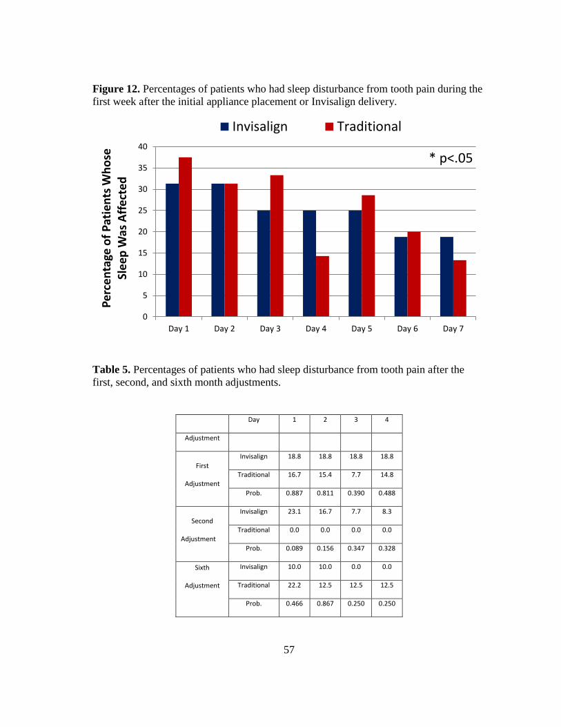
57
Figure 12. Percentages of patients who had sleep disturbance from tooth pain during the
first week after the initial appliance placement or Invisalign delivery.
Table 5. Percentages of patients who had sleep disturbance from tooth pain after the
first, second, and sixth month adjustments.
0
5
10
15
20
25
30
35
40
Day 1 Day 2 Day 3 Day 4 Day 5 Day 6 Day 7
Pe
rce
nta
ge o
f P
atie
nts
Wh
ose
Sl
ee
p W
as A
ffe
cte
d
Invisalign Traditional
Day 1 2 3 4
Adjustment
First
Adjustment
Invisalign 18.8 18.8 18.8 18.8
Traditional 16.7 15.4 7.7 14.8
Prob. 0.887 0.811 0.390 0.488
Second
Adjustment
Invisalign 23.1 16.7 7.7 8.3
Traditional 0.0 0.0 0.0 0.0
Prob. 0.089 0.156 0.347 0.328
Sixth
Adjustment
Invisalign 10.0 10.0 0.0 0.0
Traditional 22.2 12.5 12.5 12.5
Prob. 0.466 0.867 0.250 0.250
* p<.05

58
APPENDIX B
DAILY DISCOMFORT DIARY
Initials_______________
Date________________
Time________________
1. Each of the sheets that follow has 8 questions apiece, except for the first sheet.
a. The first sheet will be filled in before you have received your Invisalign/Braces. b. The other sheets will be filled in by you after the braces or Invisalign are on
your teeth. c. Try to fill out the forms at the same time each day. E.g. If you fill out the first
form at 7 P.M., try and fill out the remaining forms at 7 P.M. at each consecutive day.
2. The 8 questions will ask you about discomfort you have previously experienced and are currently experiencing as well as about pain medications that you have taken.
a. The first question will ask you to remember the worst discomfort of your life and record the discomfort on the line between the phrases, “No Discomfort” and “Worst Discomfort Ever”
Example 1: Please rate the worst physical discomfort that you have ever experienced in your life on the following line. Let’s say that the worst discomfort that I have ever experienced was a broken arm and nothing else that I have ever done has hurt that bad. I would mark this experience near the right end of the line near “Worst Discomfort Ever” No Discomfort Worst Discomfort Ever
Example 2:
In the past 24 hours how often did you take medication to relieve tooth pain? Please answer these questions with a 1 for never, 2 for seldom, 3 for often, and 4 for always by CIRCLING the appropriate response. E.G. In the past 24 hours how often did you take medication to relieve tooth pain? 1 = NEVER 2 = SELDOM 3 = OFTEN 4 = ALWAYS
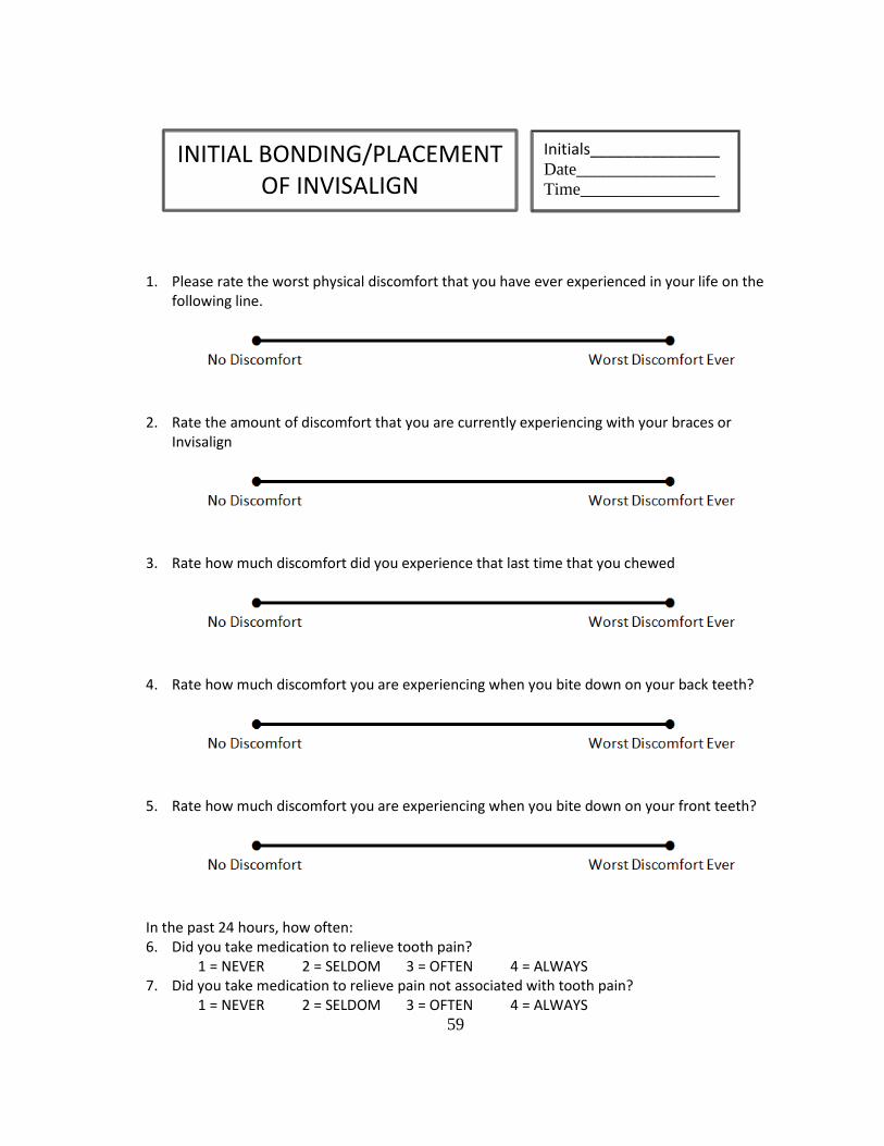
59
1. Please rate the worst physical discomfort that you have ever experienced in your life on the following line.
2. Rate the amount of discomfort that you are currently experiencing with your braces or Invisalign
3. Rate how much discomfort did you experience that last time that you chewed
4. Rate how much discomfort you are experiencing when you bite down on your back teeth?
5. Rate how much discomfort you are experiencing when you bite down on your front teeth?
In the past 24 hours, how often: 6. Did you take medication to relieve tooth pain?
1 = NEVER 2 = SELDOM 3 = OFTEN 4 = ALWAYS 7. Did you take medication to relieve pain not associated with tooth pain?
1 = NEVER 2 = SELDOM 3 = OFTEN 4 = ALWAYS
INITIAL BONDING/PLACEMENT OF INVISALIGN
Initials_______________ Date________________
Time________________

60
1. Please rate the worst physical discomfort that you have ever experienced in your life on the
following line.
2. Rate the amount of discomfort that you are currently experiencing with your braces or
Invisalign
3. Rate how much discomfort did you experience that last time that you chewed
4. Rate how much discomfort you are experiencing when you bite down on your back teeth?
5. Rate how much discomfort you are experiencing when you bite down on your front teeth?
In the past 24 hours, how often: 6. Did discomfort caused by your orthodontic appliance interfere with your sleep?
1 = NEVER 2 = SELDOM 3 = OFTEN 4 = ALWAYS 7. Did you take medication to relieve tooth pain?
1 = NEVER 2 = SELDOM 3 = OFTEN 4 = ALWAYS 8. Did you take medication to relieve pain not associated with tooth pain?
1 = NEVER 2 = SELDOM 3 = OFTEN 4 = ALWAYS
1 DAY AFTER PLACEMENT OF BRACES OR INVISALIGN
Initials_______________ Date________________
Time________________

61
1. Please rate the worst physical discomfort that you have ever experienced in your life on the
following line.
2. Rate the amount of discomfort that you are currently experiencing with your braces or
Invisalign
3. Rate how much discomfort did you experience that last time that you chewed
4. Rate how much discomfort you are experiencing when you bite down on your back teeth?
5. Rate how much discomfort you are experiencing when you bite down on your front teeth?
In the past 24 hours, how often: 6. Did discomfort caused by your orthodontic appliance interfere with your sleep?
1 = NEVER 2 = SELDOM 3 = OFTEN 4 = ALWAYS 7. Did you take medication to relieve tooth pain?
1 = NEVER 2 = SELDOM 3 = OFTEN 4 = ALWAYS 8. Did you take medication to relieve pain not associated with tooth pain?
1 = NEVER 2 = SELDOM 3 = OFTEN 4 = ALWAYS
Initials_______________ Date________________
Time________________
2 DAY AFTER PLACEMENT OF BRACES OR INVISALIGN

62
1. Please rate the worst physical discomfort that you have ever experienced in your life on the
following line.
2. Rate the amount of discomfort that you are currently experiencing with your braces or
Invisalign
3. Rate how much discomfort did you experience that last time that you chewed
4. Rate how much discomfort you are experiencing when you bite down on your back teeth?
5. Rate how much discomfort you are experiencing when you bite down on your front teeth?
In the past 24 hours, how often: 6. Did discomfort caused by your orthodontic appliance interfere with your sleep?
1 = NEVER 2 = SELDOM 3 = OFTEN 4 = ALWAYS 7. Did you take medication to relieve tooth pain?
1 = NEVER 2 = SELDOM 3 = OFTEN 4 = ALWAYS 8. Did you take medication to relieve pain not associated with tooth pain?
1 = NEVER 2 = SELDOM 3 = OFTEN 4 = ALWAYS
Initials_______________ Date________________
Time________________
3 DAY AFTER PLACEMENT OF BRACES OR INVISALIGN
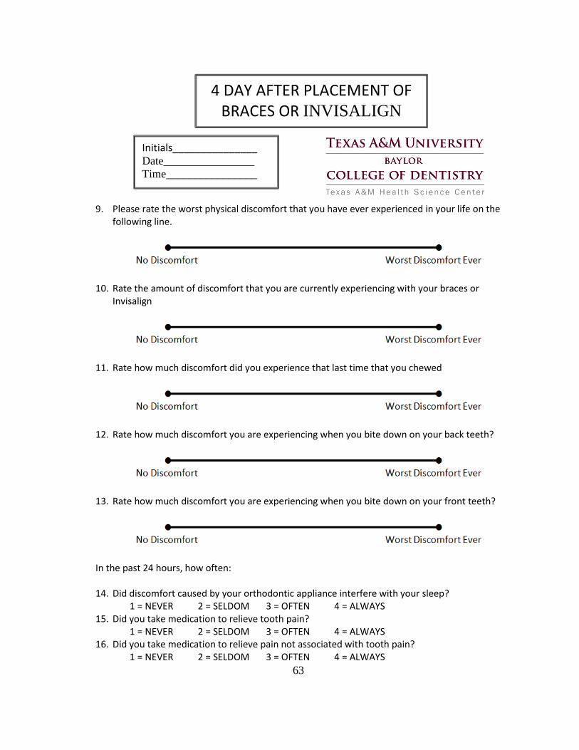
63
9. Please rate the worst physical discomfort that you have ever experienced in your life on the
following line.
10. Rate the amount of discomfort that you are currently experiencing with your braces or
Invisalign
11. Rate how much discomfort did you experience that last time that you chewed
12. Rate how much discomfort you are experiencing when you bite down on your back teeth?
13. Rate how much discomfort you are experiencing when you bite down on your front teeth?
In the past 24 hours, how often: 14. Did discomfort caused by your orthodontic appliance interfere with your sleep?
1 = NEVER 2 = SELDOM 3 = OFTEN 4 = ALWAYS 15. Did you take medication to relieve tooth pain?
1 = NEVER 2 = SELDOM 3 = OFTEN 4 = ALWAYS 16. Did you take medication to relieve pain not associated with tooth pain?
1 = NEVER 2 = SELDOM 3 = OFTEN 4 = ALWAYS
Initials_______________ Date________________
Time________________
4 DAY AFTER PLACEMENT OF BRACES OR INVISALIGN

64
17. Please rate the worst physical discomfort that you have ever experienced in your life on the
following line.
18. Rate the amount of discomfort that you are currently experiencing with your braces or
Invisalign
19. Rate how much discomfort did you experience that last time that you chewed
20. Rate how much discomfort you are experiencing when you bite down on your back teeth?
21. Rate how much discomfort you are experiencing when you bite down on your front teeth?
In the past 24 hours, how often: 22. Did discomfort caused by your orthodontic appliance interfere with your sleep?
1 = NEVER 2 = SELDOM 3 = OFTEN 4 = ALWAYS 23. Did you take medication to relieve tooth pain?
1 = NEVER 2 = SELDOM 3 = OFTEN 4 = ALWAYS 24. Did you take medication to relieve pain not associated with tooth pain?
1 = NEVER 2 = SELDOM 3 = OFTEN 4 = ALWAYS
Initials_______________ Date________________
Time________________
5 DAY AFTER PLACEMENT OF BRACES OR INVISALIGN

65
25. Please rate the worst physical discomfort that you have ever experienced in your life on the
following line.
26. Rate the amount of discomfort that you are currently experiencing with your braces or
Invisalign
27. Rate how much discomfort did you experience that last time that you chewed
28. Rate how much discomfort you are experiencing when you bite down on your back teeth?
29. Rate how much discomfort you are experiencing when you bite down on your front teeth?
In the past 24 hours, how often: 30. Did discomfort caused by your orthodontic appliance interfere with your sleep?
1 = NEVER 2 = SELDOM 3 = OFTEN 4 = ALWAYS 31. Did you take medication to relieve tooth pain?
1 = NEVER 2 = SELDOM 3 = OFTEN 4 = ALWAYS 32. Did you take medication to relieve pain not associated with tooth pain?
1 = NEVER 2 = SELDOM 3 = OFTEN 4 = ALWAYS
Initials_______________ Date________________
Time________________
6 DAY AFTER PLACEMENT OF BRACES OR INVISALIGN

66
33. Please rate the worst physical discomfort that you have ever experienced in your life on the
following line.
34. Rate the amount of discomfort that you are currently experiencing with your braces or
Invisalign
35. Rate how much discomfort did you experience that last time that you chewed
36. Rate how much discomfort you are experiencing when you bite down on your back teeth?
37. Rate how much discomfort you are experiencing when you bite down on your front teeth?
In the past 24 hours, how often: 38. Did discomfort caused by your orthodontic appliance interfere with your sleep?
1 = NEVER 2 = SELDOM 3 = OFTEN 4 = ALWAYS 39. Did you take medication to relieve tooth pain?
1 = NEVER 2 = SELDOM 3 = OFTEN 4 = ALWAYS 40. Did you take medication to relieve pain not associated with tooth pain?
1 = NEVER 2 = SELDOM 3 = OFTEN 4 = ALWAYS
Initials_______________ Date________________
Time________________
7 DAY AFTER PLACEMENT OF BRACES OR INVISALIGN