paediatric drls made easy - eurosafe imaging · paediatric drls made easy ... cristina almeida...
TRANSCRIPT

Visit the EuroSafe Imaging lounge at ECR 2018
© European Society of Radiology
Ask EuroSafe ImagingTips & Tricks
Paediatric Imaging Working Group
Paediatric DRLs made easy
Raija Seuri (HUS Medical Imaging Center, FI)
Cristina Almeida (Centro Hospitalar de Lisboa Central, PT)
Theocharis Berris (University of Crete, GR)

Visit the EuroSafe Imaging lounge at ECR 2018
© European Society of Radiology
Introduction
Establishing quantitative indicators on the value of administered doses allows the comparison between different examination techniques to promote strategies for optimizing patient protection;
The pediatric DRL’s are expected not to be exceeded in the routine procedures when good diagnostic quality practices are applied

Visit the EuroSafe Imaging lounge at ECR 2018
© European Society of Radiology
Diagnostic Reference Levels/ICRP
Advisory dose levels to help in optimisation of imaging practices
Aim to identify unusually high dose levels
Defined for common examinationse.g. chest radiography, pelvis
radiography
Visit the EuroSafe Imaging lounge at ECR 2018
© European Society of Radiology
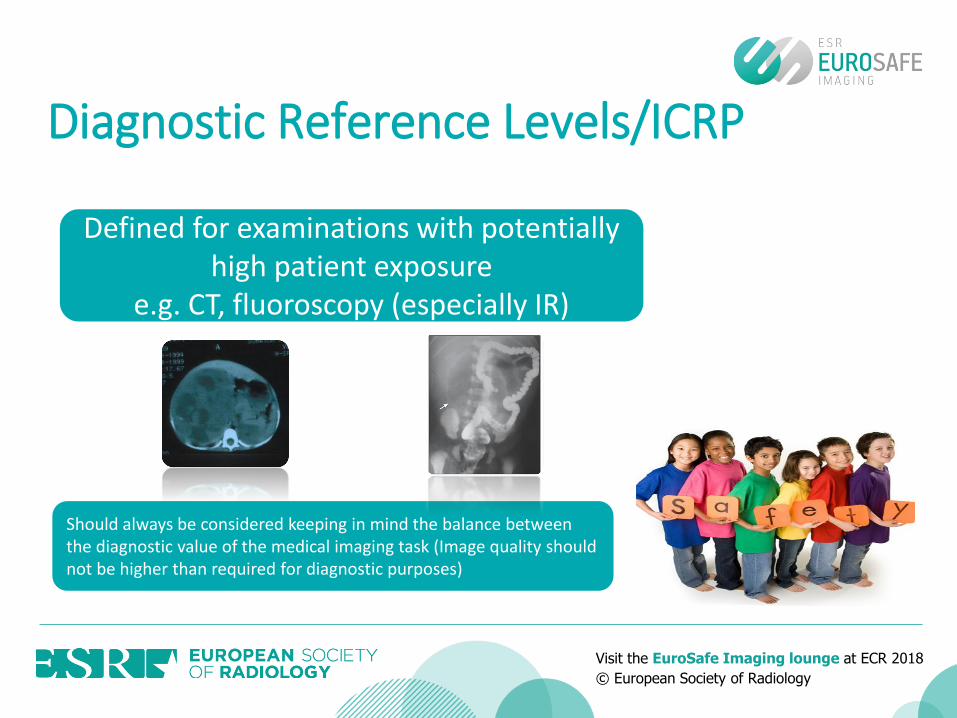
Diagnostic Reference Levels/ICRP
Defined for examinations with potentially high patient exposure
e.g. CT, fluoroscopy (especially IR)
Should always be considered keeping in mind the balance between the diagnostic value of the medical imaging task (Image quality should not be higher than required for diagnostic purposes)
Visit the EuroSafe Imaging lounge at ECR 2018
© European Society of Radiology
Dose restrictions or limits
DRL’s are guiding levels for optimised practices, established for groups of patients, not individual patients.
To separate good and bad practices
DRLs are NOT
Visit the EuroSafe Imaging lounge at ECR 2018
© European Society of Radiology
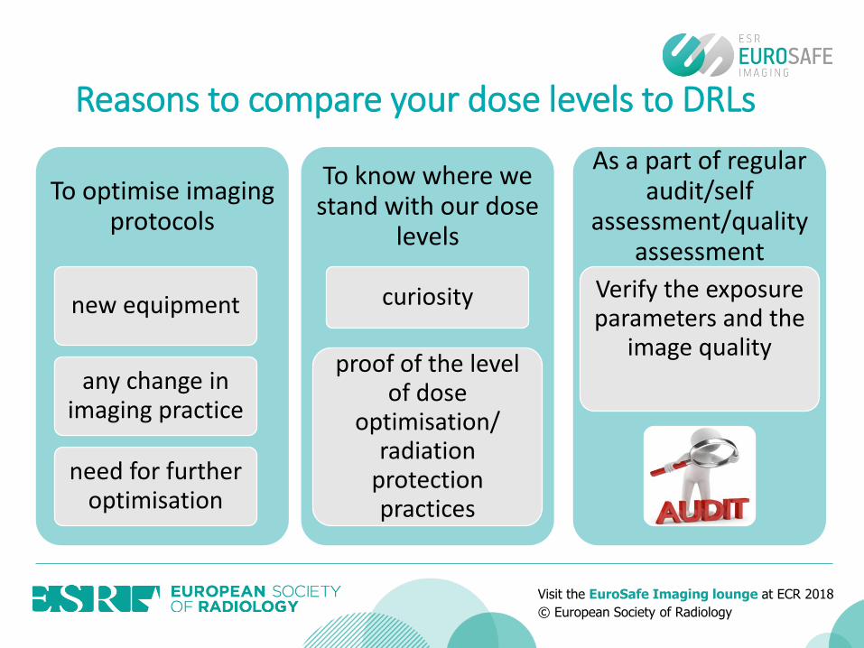
Reasons to compare your dose levels to DRLs
To optimise imaging protocols
new equipment
any change in imaging practice
need for further optimisation
To know where we stand with our dose
levels
curiosity
proof of the level of dose
optimisation/ radiation
protection practices
As a part of regular audit/self
assessment/quality assessment
Verify the exposure parameters and the
image quality

Visit the EuroSafe Imaging lounge at ECR 2018
© European Society of Radiology
DRLs to use• set by national authoritative bodies
• based on national dose collectionsNational DRLs
• if national DRLs do not exist
• based on existing national DRLs in Europe
European DRLs (PiDRL)
• based on local/areal dose collections without official involvement of national authoritative body
• should be considered especially in big units with modern equipment or if the national DRLs are outdated
Local DRLs

Visit the EuroSafe Imaging lounge at ECR 2018
© European Society of Radiology
Child specifics in the useof DRLs
Patient size has to be accounted for
weight is recommended by EC and ICRP
if age is used, the number of patients should be considerably higher
PiDRL report includes conversion tables for age groups -weight groups
DRLs are usually given for specific weight groups
(or as a continuous curve: Finland)
There is variation of the use of 32 cm or 16 cm phantom for the calculation of dose quantities in paediatric body protocols

Visit the EuroSafe Imaging lounge at ECR 2018
© European Society of Radiology
Paediatric categorisation
Rare publications quoting patient age + weight;
Only a few with patient weight;
The majority used patient age;
Most common appears to be:
Jarvinen et al., 2011, 2014Roch & Aubert, 2013
<1 1-55-1010-15 y.o.

Visit the EuroSafe Imaging lounge at ECR 2018
© European Society of Radiology
Head CT examinations
0 - < 4 weeks4 weeks -< 1y
1 -< 6 y≥ 6 y
Body examinations
< 5 kg5 -< 15kg
15-< 30 kg30-<50 kg50-<80 kg
Categorisation recommendations

Visit the EuroSafe Imaging lounge at ECR 2018
© European Society of Radiology
Age equivalent group
Description Weight group
Neonate < 5 kg
Infant, toddler and early childhood
5 -< 15kg
Middle Childhood 15-< 30 kg
Early adolescence 30-<50 kg
Late adolescence 50-<80 kg
Age group based oncharts
Most commonage on NDRLs
< 1 m 0 y
1 m - < 4 y 1 y
4 y - < 10 y 5 y
10 y - < 14 y 10 y
14 y - < 18 y 15 y
Visit the EuroSafe Imaging lounge at ECR 2018
© European Society of Radiology
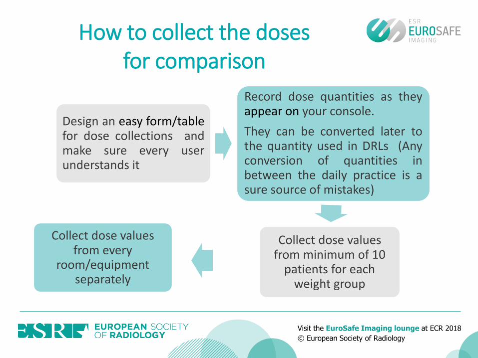
How to collect the doses for comparison
Design an easy form/tablefor dose collections andmake sure every userunderstands it
Record dose quantities as theyappear on your console.
They can be converted later tothe quantity used in DRLs (Anyconversion of quantities inbetween the daily practice is asure source of mistakes)
Collect dose values from minimum of 10
patients for each weight group
Collect dose values from every
room/equipment separately
Visit the EuroSafe Imaging lounge at ECR 2018
© European Society of Radiology
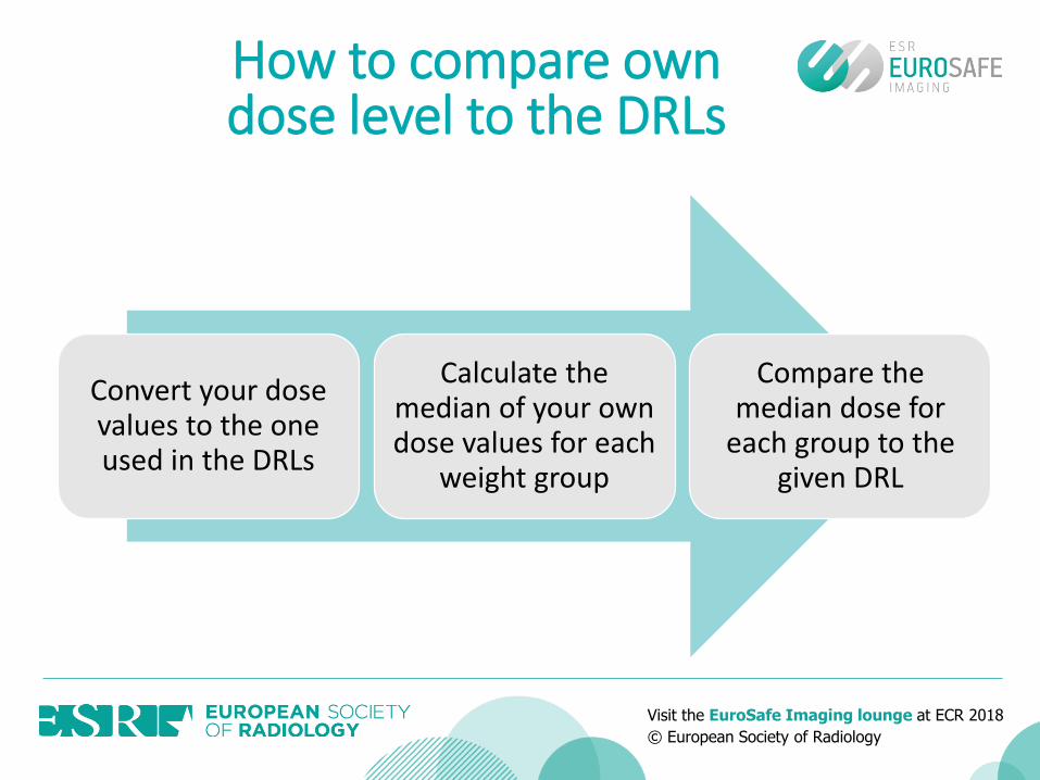
How to compare own dose level to the DRLs
Convert your dose values to the one used in the DRLs
Calculate the median of your own dose values for each
weight group
Compare the median dose for
each group to the given DRL
Visit the EuroSafe Imaging lounge at ECR 2018
© European Society of Radiology
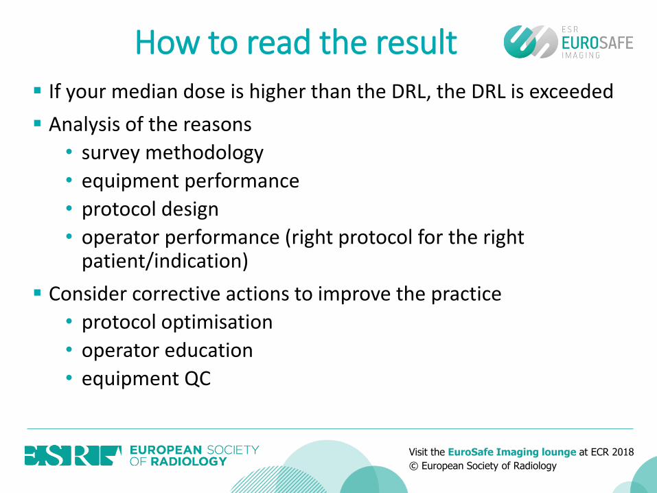
How to read the result If your median dose is higher than the DRL, the DRL is exceeded
Analysis of the reasons
• survey methodology
• equipment performance
• protocol design
• operator performance (right protocol for the right patient/indication)
Consider corrective actions to improve the practice
• protocol optimisation
• operator education
• equipment QC
Visit the EuroSafe Imaging lounge at ECR 2018
© European Society of Radiology
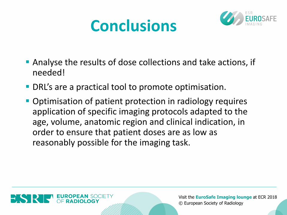
Analyse the results of dose collections and take actions, if needed!
DRL’s are a practical tool to promote optimisation.
Optimisation of patient protection in radiology requires application of specific imaging protocols adapted to the age, volume, anatomic region and clinical indication, in order to ensure that patient doses are as low as reasonably possible for the imaging task.
Conclusions

Visit the EuroSafe Imaging lounge at ECR 2018
© European Society of Radiology
• E.C., European Commission. (2001). Radiation Protection 109: Guidance on Diagnostic Reference Levels (DRL’s) for medical exposures. Luxemburg.
• Medina LS, et all Evidence-Based Imaging in Pediatrics: Optimizing Imaging in Pediatrics. Springer. 2010
• G. Paulo et al. 2016 Analysis of overexposed areas in paediatric plainradiography, ECR 2016
• www.icrp.org, www.unscear.org, www.iaea.org, www.who.int, www.imagegently.org
References