p-glycoprotein (p-gp) mediated efflux in caco-2 cell monolayers: the influence of culturing...
TRANSCRIPT

P-Glycoprotein (P-gp) Mediated Efflux in Caco-2 Cell Monolayers: TheInfluence of Culturing Conditions and Drug Exposure on P-gpExpression Levels
PASCALE ANDERLE,† EVA NIEDERER,‡ WERNER RUBAS,§ CONSTANZE HILGENDORF,| HILDE SPAHN-LANGGUTH,|HEIDI WUNDERLI-ALLENSPACH,† HANS P. MERKLE,*,† AND PETER LANGGUTH†,⊥
Contribution from Department of Pharmacy, ETH, 8057 Zurich, Switzerland, Institute of Biomedical Engineering, ETH,8092 Zurich, Switzerland, Genentech Inc., South San Francisco, California 94080 and Department of Pharmacy,Martin Luther University, 06120 Halle, Germany.
Received September 22, 1997. Accepted for publication February 24, 1998.
Abstract 0 The influence of cell culture conditions and previous drugexposure on P-glycoprotein (P-gp) expression levels in Caco-2 cellswas determined. In this study, the expression of P-gp is demonstrated(i) visually by confocal laser scanning microscopy (CLSM), (ii)functionally by transport studies with substrates of the efflux pump,and (iii) quantitatively by flow cytometry (FCM) analysis using specificmonoclonal antibodies (anti P-gp MRK 16 as an external antibodyand P-GlycoCheck C219 as an internal antibody). Trypsinization ofthe cells after reaching confluence led to a decrease of P-gp expressionlevels, while trypsinization before reaching confluence led to anincrease after long-term cultivation. Culturing the cells on polycar-bonate filters did not elicit a significant change of P-gp expressionover time in culture, whereas in plastic flasks (polystyrene) a decreasewas detected. Using CLSM a strong fluorescence on the apical sideof Caco-2 cell monolayers was observed, as a result of incubationwith MRK 16 as primary and IgG Cy5 as secondary antibody.Previous drug exposure of the cells showed that verapamil, celiprolol,and vinblastine induced the P-gp expression, while metkephamid(MKA) decreased the P-gp expression level as compared to the control.Permeation studies consolidated the theory that P-gp is expressed inthe Caco-2 cells examined. For talinolol and MKA, a higher transportfrom basolateral to apical side than from apical to basolateral couldbe measured. Incubation of the cell monolayer with MRK 16 reducedthe secretion process to the apical side, but did not influence [3H]-mannitol flux. Caco-2 cells seem to be a suitable cell line model forP-gp-mediated secretion studies. However, the variability of the P-gpexpression requires careful control when this model is to be used inquantitative structure/secretion studies.
IntroductionRecent observations on intestinal absorption of drugs
with peptide and nonpeptide structure suggest that thebioavailability of some drugs after peroral administrationmay be limited by an intestinal secretion process that ismediated by P-glycoprotein (P-gp).1 P-gp is a 170 kDaprotein originally found to cause multidrug resistance incancer chemotherapy upon treatments with anticancerdrugs such as vinblastine, actinomycin D, and dauno-mycin.2-5 But also noncytostatic drugs are reported to be
affected by the P-glycoprotein transporter, such as â-recep-tor antagonists6,7 and peptides.8-10 In the gastrointestinaltract the P-gp transporter may secrete drugs at lowconcentrations out of the epithelium back into the intes-tinal lumen. At high intestinal concentrations the secre-tion may be saturable, leading to an apparent increase inbioavailability. In addition to the gastrointestinal tract,P-gp is expressed in the liver, the pancreas, the kidneys,and reproductive organs as well as in the endothelia of thebrain, testes, and adrenal glands.11
Previous studies showed that P-glycoprotein is alsoexpressed in Caco-2 cells, a well-established cell line fordrug transport studies in the gastrointestinal tract.3 SinceCaco-2 cells are frequently used to estimate the fraction ofdrug absorbed based on a compound’s permeability in thisculture system, P-gp expression has to be taken intoaccount. It may be foreseen that in the case of a muchhigher P-gp expression in Caco-2 cells as compared to thehuman intestine, the risk of underestimation of humanintestinal permeability by this in-vitro test system mayexist. In addition, since the P-gp expression level in Caco-2cells is depending on the time in culture12 as well as onthe culturing conditions,13 variable expression levels mustbe taken into account. As interlaboratory differences, suchas passage number, composition of the medium includingthe use of antibiotics, passaging procedure etc. may affectP-gp expression, there is a need for a rigorous standardiza-tion of the Caco-2 model with respect to P-gp expressionlevels.The aim of this investigation was to localize and quantify
the expression of P-gp in Caco-2 cells and to identify factorsthat affect P-gp expression in Caco-2 cells, namely byconfocal laser scanning microscopy (CLSM), flow cytometry(FCM), and drug permeation studies.
Experimental Section
MaterialssCell Culture. Dulbecco’s Modified Eagle’s medium(DMEM) containing 0.5% l-glutamine and 4.5 g/L glucose, nones-sential amino acids (NEAA) solution, fetal calf serum (FCS), 0.05%trypsin/0.025% ethylendiaminetetraacetic acid (EDTA) solution(trypsinization solution, trypsin activity standardized by manu-facturer by cell lift activity test), 0.025% EDTA solution, penicillin/streptomycin solution (penstrep) (10000 units/mL penicillin and10000 µg/mL streptomycin), Hank’s balanced salt solution (HBSS),phosphate-buffered saline (PBS; 0.132 g/L calcium chloride dihy-drate, 0.2 g/L potassium chloride, 0.2 g/L potassium dihydrogenphosphate, 0.1 g/L magnesium chloride hexahydrate, 8.0 g/Lsodium chloride, 1.15 g/L disodium hydrogenphophosphate) wereobtained from Life Technologies (Basel, CH). Transwell Snapwellcell culture inserts, diameter of 1.13 cm2, mean pore diameter of0.4 µm, and Transwell cell culture inserts, diameter of 4.7 cm2,
* Corresponding author: Phone: +41-1-635 60 10, Fax: +41-1-635 68 81, email: [email protected].
† Department of Pharmacy, ETH.‡ Institute of Biomedical Engineering, ETH.§ Genentech.| Martin Luther University.⊥ Present address: Astra Hassle AB, 43183 Molndal, Sweden.
© 1998, American Chemical Society and S0022-3549(97)00372-9 CCC: $15.00 Journal of Pharmaceutical Sciences / 757American Pharmaceutical Association Vol. 87, No. 6, June 1998Published on Web 04/30/1998

were supplied by Costar (Basel, CH). Triton X-100, glycine,n-propylgallate, glycerol, vinblastine sulfate, and 2-morpholino-ethanesulfonic acid (MES) were from Fluka Chemie AG (Buchs,CH), Hoechst 33342 from Hoechst (Frankfurt D), Rhodamin-phalloidin from Molecular Probes (Leiden, NL), and goat anti-mouse IgG Cy5 antibody (affinity purified F(Ab′)2 secondaryantibody) from Chemicon (Leiden, NL). The anti P-gp MRK 16, amonoclonal IgG2R mouse (150 µg/mL in PBS, 0.1% NaN3 1% BSA(reacting specifically with a a surface epitope of human mdr1 P-gp)was supplied by Kamiya Biomedical Company. As a negativecontrol to MRK 16 (MRK 16 NC) a purified myeloma proteinmouse IgG2R (1 mg/mL 0.02 M Tris-buffered saline, pH 8.1,Organon Teknica) was used. Goat anti-mouse Ig FITC (specificto purified mouse IgG1, IgG2R, IgG2â, IgG3) was obtained fromBecton Dickinson. P-GlycoCheck C219, a FITC labeled murinemonoclonal antibody IgG2 (reacting with an internal epitope ofP-gp) and P-GlycoCheck C219 negative control, a FITC labeledmurine monoclonal antibody IgG2, were purchased from CIS biointernational (GIF-sur-Yvette Cedex, France). Fluorescamine andsodium dodecyl sulfate (SDS) were obtained from Sigma Chemicaland Co. (St. Louis, MO). [3H]mannitol was from Du Pont deNemours International S. A. (Regensdorf, CH) and Ultima Goldfrom Packard (Croningen, NL). Trospium was a gift fromMadausAG (Koln, Germany). Talinolol, losartan and its metabolite EXP3174, and celiprolol were a gift from the Martin Luther University(Halle, Germany), metkephamid was donated by Eli Lilly and Co.(Indianapolis, IN), and latex particles, Immuno-Brite Level II,were obtained from Instrumentation Laboratory AG (Schlieren,Switzerland). Caco-2 cells, passage 22 were obtained from ATCC(Rockville, MD), passage 46 from ECACC (London, GB), andpassage 68 from the Department of Physiology (University ofZurich, Switzerland).Cell CulturesCaco-2 cells passage 22-38, 46-59, and 68-83
were cultured in Dulbecco’s modified Eagle’s medium (DMEM,with 4.5 g/L of D-glucose) which was supplemented with 16.5%fetal bovine serum, nonessential amino acids (1% v/v), andL-glutamine (0.5%). Confluent monolayers were subcultured every7 days by treatment with 0.5% trypsin and 0.2% EDTA andseeding at a density of 2 × 10⁵ cells/25 cm2 into plastic flasks.Cultures were incubated at 37 °C in a humidified atmosphere of5%/95% CO2/O2. Special treatments of the cells will be coveredin the subsequent paragraphs.Flow cytometry (FCM)-Caco-2 cells were cultured in 80 cm2
flasks at 37 °C in a 5% CO2 atmosphere using DMEM containing16.5% FCS (v/v) and 1% NEAA (v/v) (medium for cell culture).Caco-2 cells were grown as monolayers by seeding 4 × 10⁵ cells/80 cm2 bottle. For passaging and/or immunolabeling, confluentlyor approximately 70% confluently grown cell monolayers weretrypsinized (trypsin/EDTA) and suspended. Alternatively, con-fluently grown cell monolayers were trypsinized, suspended, andseeded onto polycarbonate filters (Transwell cell culture inserts,diameter of 4.7 cm2, mean pore diameter of 0.4 µm) at a densityof 100000 cells/cm2. For labeling 4 × 10⁶ cells/100 µL of BSA/PBS (10%, v/v) were incubated for 30 min with 5 µL of anti P-gpMRK 16 per 1 mL of PBS or with 2.5 µL of MRK 16 negativecontrol per 1 mL of PBS, respectively. Thereafter the cells wereincubated with goat anti-mouse Ig FITC. For labeling withP-GlycoCheck C219 or P-GlycoCheck C219 negative control, thecells were fixed with 3.6% (v/v) formaldehyde solution (in PBS),permeabilized with aceton at 0 °C, and incubated with the anti-body for 60 min at 4 °C under light exclusion. After washing withPBS (3 times), the cells were suspended in 1 mL 0.36% (v/v) offormaldehyde solution (in PBS) and measured. 10000 cells werecounted on the average by an EPICS Profile Analyzer of Coulter(Miami, FL). Excitation was at 488 nm and emission at 520 nm.As an internal standard, latex particles, Immuno-Brite Level II,were used.Confocal Laser ScanningMicroscopy (CLSM)sCaco-2 cells
(passage 70) were cultured in 80 cm2 flasks at 37 °C in a 5% CO2atmosphere using DMEM containing 16.5% FCS and 1% NEAA.Caco-2 cells were grown as epithelial layers by seeding 4 × 10⁵cells/80 cm2 bottle. Confluently grown cell monolayers weretrypsinized, suspended, and seeded onto polycarbonate filters(Transwell Snapwell cell culture inserts, diameter of 1.13 cm2,mean pore diameter of 0.4 µm) at a density of 100000 cells/cm2
for 28 days using DMEM containing 16.5% FCS, 1% NEAA, and0.011 µM vinblastine sulfate. The cell layers were washed withPBS, pH 7.4, which was also used for all subsequent washing
steps. Fixation and labeling were performed at room temperature.The following standard protocol was applied: Cells were fixed inparaformaldehyde (3.6% in PBS) for 15 min and treated with 0.1M glycine for 5 min. After washing three times they werepermeabilized with Triton X-100 (0.1%, w/v) for 15 min andwashed again three times. Cell nuclei were labeled with Hoechst33342 stain (1:100), and actin was labeled with rhodamine-labeledphalloidin (1:10). For labeling with antibodies, the sandwichtechnique was used. Preparations were incubated for 1 h withthe primary antibody (1:4), i.e., the specific anti P-gp MRK 16, orthe nonspecific MRK 16 negative control. As a secondary antibody,Cy5-labeled anti IgG (1:50) was applied for 1 h. After washingagain (three times), the cells were mounted in a mixture of 7 mLof glycerol 100%, 3 mL of 0.1 Tris-HCl, pH 9.5, and 0.5 g of n-propylgallate.Transport StudiessCaco-2 cells were grown in 80 cm2 flasks
at 37 °C in a 5% CO2 atmosphere using DMEM containing 16.5%FCS and 1% NEAA. Caco-2 cells were grown as epithelial layersby seeding 4 × 10⁵ cells/80 cm2 bottle. Confluently grown cellmonolayers were trypsinized, suspended, and seeded onto poly-carbonate filters (Transwell Snapwell cell culture inserts, diameterof 1.13 cm2, meanpore diameter of 0.4 µm at a density of 100000cells/cm2 ) for 15 to 16 days.The integrity of the cells was checked before and after the
experiment by measuring the TEERs. The permeation studieswere performed by 5% CO2/O2 airlift (15 mL/min) in Ussing typechambers (Precision instruments, Costar, NL). The diffusion cellswere maintained at 37 °C by a water-heated jacket.As a marker for the passive transport [3H]mannitol with an
activity of 1 µCi/mL was used. The activity of the stock solutionwas 15-30 Ci/mmol.After washing twice with HBSS, the inserts were placed into
the diffusion cells; 5.0 mL of transport buffer containing 1 mMmetkephamid or talinolol, respectively, was filled either into theapical (apical to basolateral permeation direction; a f b) or thebasolateral compartment (b f a); and 5.5 mL of transport buffer(10 mM MES in HBBS, pH 6.5) was filled either into thebasolateral (a f b) or apical (b f a) compartment. For the studieswith anti P-gp MRK 16, the inserts were first incubated for 15min at 37 °C with 40 µL of 30 µL MRK 16/300 µL HBSS andtreated as described above. At time zero, samples of 100 µL forthe determination of the [3H]mannitol concentration and 400 µLfor the determination of the drug concentration were taken fromthe donor and receiver compartment and 0.5 mL immediatelyreplaced by transport buffer in the receiver compartment. Samplesof 500 µL were taken after 15, 30, 45, and 60 min from the receivercompartment and immediately replaced, and one sample of 500µL was taken after 60 min from the donor compartment. For [3H]-mannitol analysis, samples (3.0 mL or 3.5 mL) were also takenafter 60 min. All samples for the [3H]mannitol transport studieswere continuously mixed with 7.5 mL or 4 mL of scintillation fluid(Ultima Gold). The samples from the drug transport studies wereanalyzed by HPLC analysis according to Bohner et al.16 (MKA) orto Wetterich et al. (talinolol).7 The [3H]mannitol samples werecounted in a multipurpose scintillation counter Beckman type LS6500 (Beckman Instruments Inc., Fullerton, CA).Calculation of PermeabilitysEffective permeability coefficients,
Peff (cm s-1) were calculated according to:
where (dC/dt)ss is the steady-state change of concentration overtime (µg mL-1 s-1), A is the diffusion area (cm2), V is the volumeof the receiver compartment (mL), and C0 is the initial concentra-tion in the apical (a f b) or basolateral (b f a) compartment (µgmL-1). Data are presented as means ( SD of n ) 3-5 Caco-2cell monolayers.StatisticssStandard F-tests and Student’s unpaired t test (two-
tailed) were used to compare means of flow cytometry andpermeability data. P < 0.05 was considered statistically signifi-cant.
ResultsFlow Cytometry (FCM)sWith both antibodies, MRK
16 as an external antibody and P-GlycoCheck C219 as
Peff ) ( dCdt )ss VAC0
758 / Journal of Pharmaceutical SciencesVol. 87, No. 6, June 1998

internal antibody, P-gp expression is recognized. Basedon FCM of Caco-2 cells incubated with P-GlycoCheck C219,only a small shift of the fluorescence intensity was mea-sured (data not shown). When incubated with MRK 16antibody clearly two populations are apparent, one popula-tion showing P-gp expression, whereas the other does not,having the same mean fluorescence as the negative control(Figure 1). For all subsequent studies MRK 16 labelingwas therefore preferred.Influence of the Cell Culture ConditionssIn our prelimi-
nary studies we have found that no systematic effect oftrypsinization (trypsin/EDTA) versus EDTA alone and ofincubation time (20 versus 40 min), respectively, on P-gpexpression was detectable. Therefore, trypsinization over10-20 min was not expected to have any influence on theP-gp expression level. To evaluate the influence of thetime in culture on P-gp expression, the starting passages29, 49, and 72, after reaching confluence, were trypsi-nized, suspended, and seeded for 9 or 10 times, respec-tively, resulting in passages 38, 59, and 82, respectively.Starting with passage 72, passage 73 was also evaluated.In all cases a significant drop of the P-gp expression levelswas observed (Figure 2). However, trypsinizing the cellsbefore reaching confluence (ca. 70% confluence) resultedin a significant increase of the P-gp expression levels(Figure 3).The time in culture was a major determinant for the P-gp
expression level as demonstrated with passages 73 and 71(following trypsinization). The results are shown in Figure4. Additionally, the P-gp expression as a function of timein culture starting with passage 71 was studied, treatingthe cells with EDTA solution. The amount of measurablefluorescence declined significantly over time in culture,
both upon trypsinization of the cells and upon EDTAtreatment alone (data not shown).On the other hand, cultivation of the cells on polycar-
bonate filters did not result in any statistically significanteffect on P-gp expression levels over time (Figure 5).Influence of Previous Drug Exposure on P-gp
Expression LevelssThe influence of previous drug ex-posure on P-gp expression levels was tested. Cells werealways trypsinized. The results are shown in Table 1.The P-gp expression levels were found to increase,
decrease, or remain constant, depending on the drug addedand its concentration in the medium. Vinblastine sulfate(0.011 µM) added to the culture medium increased the P-gpexpression by a factor of 7, i.e., from a fluorescence of 0.84V ( 0.40 (mean ( SD) without vinblastine sulfate and 6.25V ( 0.41 when vinblastine sulfate was added. On the otherhand, when trospium (1 mM) was added to the culturemedium, no increase in P-gp expression (0.65 V ( 0.08)was observed. Effects of previous drug exposure were alsotested for several other drugs including talinolol, losartanand EXP 3174 (a metabolite of losartan), celiprolol, vera-pamil, and metkephamid in concentrations of 10 µM and100 µM. Only verapamil and metkephamid showed asignificant effect on the P-gp expression level. Verapamilincreased the P-gp expression level compared to control.The same was observed for celiprolol, though at the
Figure 1sP-gp expression in Caco-2 cells. FCM of passage 73, incubatedwith the external antibody, anti P-gp MRK 16. Shown is the formation of twopopulations and the overall shift of the positive sample to higher fluoresenceintensity as compared to the negative control. MRK 16 NC ) negative control.
Figure 2sP-gp expression in Caco-2 cells, 7 days after seeding in bottles of80 cm2 area; incubated with anti P-gp MRK 16; 9 starting passages, 0 laterpassages. Throughout, the cells were trypsinized after reaching confluence.
Figure 3sP-gp expression in Caco-2 cells, 4 days after seeding in bottles of80 cm2 area; incubated with anti P-gp MRK 16; 9 starting passages, 0 laterpassages. Throughout, the cells were trypsinized before reaching confluence(approximately 70% confluence).
Figure 4sInfluence of time in culture on P-gp expression in passages 71(9) and 73 (0). Cells were trypsinized at 10, 13, 16, and 21 days or 3, 7, 10,and 14 days, respectively, after seeding in bottles of 80 cm2 area; incubatedwith anti P-gp MRK 16.
Figure 5sInfluence of the time in culture on P-gp expression in passage 73.Cells were trypsinized 6, 13 and 21 days after seeding on polycarbonatefilters; incubated with anti P-gp MRK 16.
Journal of Pharmaceutical Sciences / 759Vol. 87, No. 6, June 1998
borderline of statistical significance. In contrast, met-kephamid decreased P-gp expression in a concentration-dependent manner. Cells cultured in medium containing100 µM verapamil did not proliferate.Confocal Laser Scanning MicroscopysCell mono-
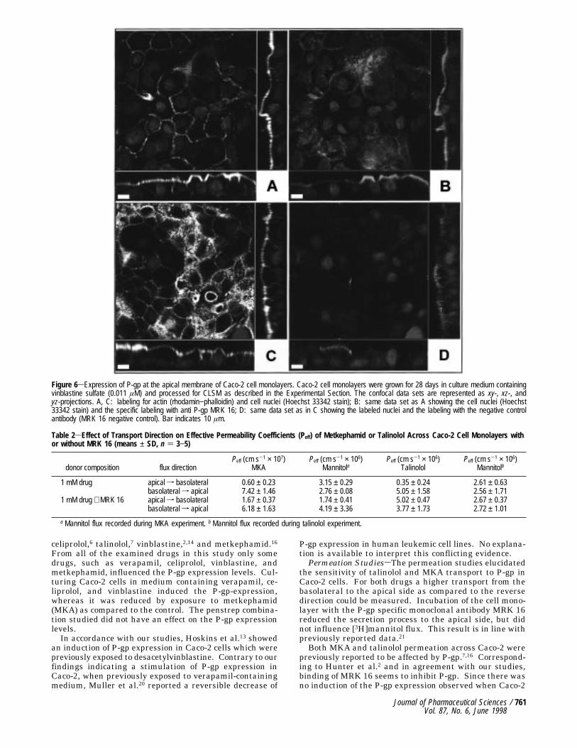
layers of passage 71 were grown for 28 days on a polycar-bonate filter in pure medium containing vinblastine sulfate(0.011 µM). Cultures were fixed and processed for CLSMas described in the Experimental Section. A triple labelingwas performed: for actin, for cell nuclei, and for P-gp, thelatter with specific MRK 16 antibody. As a control thesame protocol was applied except that the negative controlantibody, MRK 16 negative control, was used instead ofMRK 16. As illustrated in Figure 6, Caco-2 cells formmonolayers as judged from the arrangement of the cellnuclei in the x,z sections and the y,z sections. The outlinesof the cells are strongly marked with actin (Figure 6A and6C). P-gp could clearly be localized in the apical membraneof the cells (Figure 6B). The specificity of this labeling wasdemonstrated with the negative control antibody whichonly resulted in a very weak and diffuse fluorescence(Figure 6D).Permeation StudiessThe transport of metkephamid
and talinolol across Caco-2 cell monolayers in the apicalto basolateral direction and in the basolateral to apicaldirection with and without MRK 16 antibody was inves-tigated. As a control [3H]mannitol was used.The transport of MKA and talinolol from the apical to
the basolateral side was significantly slower than in thereverse direction. Binding of MRK 16 antibody signifi-cantly increased the effective permeability of both drugsfrom apical to basolateral. In the case of MKA, thepresence of MRK 16 antibody reduced slightly the differ-ence of Peff apical-basolateral and Peff basolateral-apical,whereas in the case of talinolol, a stronger reduction of thedifference of the two Peff values was observed (Table 2).
DiscussionOn the basis of the significant expression of P-glycopro-
tein in Caco-2 cells, this cell line appears to be suitable for
the investigation of intestinal, P-gp dependent secretionprocesses which may be relevant for the intestinal absorp-tion and bioavailability of drugs. For the elucidation ofquantitative structure/secretion and structure/transportrelationships, however, the variability of the P-gp expres-sion level as a function of Caco-2 cell passage number,culturing conditions, and previous drug exposure needs tobe carefully monitored and possibly standardized. Onlywhen standardized for reproducible P-gp expression canthe Caco-2 model be applied for quantitative studies. Inour study, the expression of P-gp in Caco-2 cells isdemonstrated (i) visually by CLSM, (ii) functionally bytransport studies with substrates of the efflux pump, and(iii) quantitatively by FCM using specific monoclonalantibodies. In addition, the variability of the expressionlevel was investigated as a function of culturing conditionsand exposure to drugs which are potential substrates ofP-gp.Flow CytometrysAfter immunolabeling of Caco-2 with
P-GlycoCheck C219, the shift of fluorescence intensity ascompared to the negative control was much smaller thanafter immunolabeling with MRK 16. Since MRK 16binding to humanmdr1 (multidrug resistance protein 1)17,18is very specific, but P-GlycoCheck C219 reacts additionallywith humanmdr3 and its murine analogues,19 and the shiftof the fluorescence intensity using MRK 16 immunolabelingwas more significant than with P-GlycoCheck C219, weapplied MRK 16 for all subsequent studies.Influence of CulturingsNo practically relevant differ-
ences were observed between treatments with trypsin/EDTA (trypsinization) and with EDTA alone, from whichcan be concluded that trypsin does not seem to interactwith the P-gp epitope reacting with the external (MRK16) or internal (P-GlycoCheck C219) P-gp specific anti-bodies.The time in culture, however, has a major influence on
the P-gp expression in Caco-2 cells. P-gp expressiondecreased in higher passages compared to the startingpassages when the cells were trypsinized after reachingconfluence. On the contrary, the P-gp expression levelincreased significantly in higher passages compared to thestarting passages when the cells were trypsinized beforereaching confluence. It was also apparent that there maybe a difference depending on whether the cells have beencultured in plastic flasks or on polycarbonate filters. Whenthe cells were cultured in plastic flasks a significantdecrease of the P-gp expression level was apparent whereascultivation on polycarbonate filters lead to an increase ofthe P-gp expression level.Using Western blots, Hosoya et al.12 demonstrated that
the order of P-gp expression over a period of 4 weeks was4w > 1w > 2w > 3w, while in permeation studies thefunction of P-gp as a transporter protein increased signifi-cantly from day 17 to day 27 when the cells were seededon polycarbonate filters. In contrast to Hosoya et al.,17however, Wils et al.14,15 using immunoblotting reported thatno increase of P-gp expression from day 4 to 22 occurredwhen Caco-2 cells were cultured in plastic flasks. Ad-ditionally, the authors observed that the capacity to expressP-gp may be lost during long-term cultivation.Obviously P-gp expression in Caco-2 cells strongly
depends on the individual culturing conditions. The timein culture and the material used for the culture primarilyhave an important influence of the P-gp expression levels.Certain conditions, e.g., trypsinization after reaching con-fluence may, even provoke a loss of P-gp expression aftercertain times of cultivation (cf. Figure 2).Influence of Previous Drug Exposure on P-gp Expression
LevelssIt is well-known that the transport of certain drugsacross Caco-2 cells is affected by P-gp expression, e.g. with
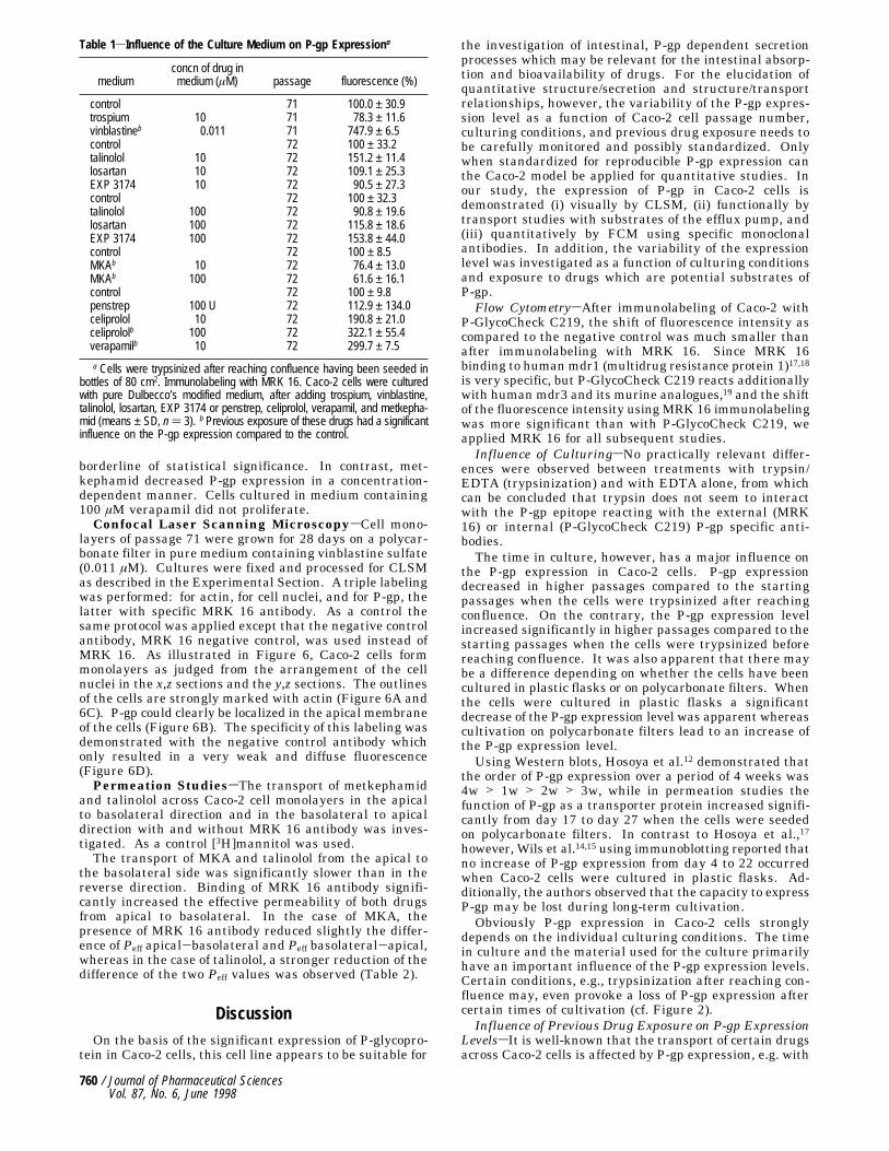
Table 1sInfluence of the Culture Medium on P-gp Expressiona
mediumconcn of drug inmedium (µM) passage fluorescence (%)
control 71 100.0 ± 30.9trospium 10 71 78.3 ± 11.6vinblastineb 0.011 71 747.9 ± 6.5control 72 100 ± 33.2talinolol 10 72 151.2 ± 11.4losartan 10 72 109.1 ± 25.3EXP 3174 10 72 90.5 ± 27.3control 72 100 ± 32.3talinolol 100 72 90.8 ± 19.6losartan 100 72 115.8 ± 18.6EXP 3174 100 72 153.8 ± 44.0control 72 100 ± 8.5MKAb 10 72 76.4 ± 13.0MKAb 100 72 61.6 ± 16.1control 72 100 ± 9.8penstrep 100 U 72 112.9 ± 134.0celiprolol 10 72 190.8 ± 21.0celiprololb 100 72 322.1 ± 55.4verapamilb 10 72 299.7 ± 7.5
a Cells were trypsinized after reaching confluence having been seeded inbottles of 80 cm2. Immunolabeling with MRK 16. Caco-2 cells were culturedwith pure Dulbecco’s modified medium, after adding trospium, vinblastine,talinolol, losartan, EXP 3174 or penstrep, celiprolol, verapamil, and metkepha-mid (means ± SD, n ) 3). b Previous exposure of these drugs had a significantinfluence on the P-gp expression compared to the control.
760 / Journal of Pharmaceutical SciencesVol. 87, No. 6, June 1998
celiprolol,6 talinolol,7 vinblastine,2,14 and metkephamid.16From all of the examined drugs in this study only somedrugs, such as verapamil, celiprolol, vinblastine, andmetkephamid, influenced the P-gp expression levels. Cul-turing Caco-2 cells in medium containing verapamil, ce-liprolol, and vinblastine induced the P-gp-expression,whereas it was reduced by exposure to metkephamid(MKA) as compared to the control. The penstrep combina-tion studied did not have an effect on the P-gp expressionlevels.In accordance with our studies, Hoskins et al.13 showed
an induction of P-gp expression in Caco-2 cells which werepreviously exposed to desacetylvinblastine. Contrary to ourfindings indicating a stimulation of P-gp expression inCaco-2, when previously exposed to verapamil-containingmedium, Muller et al.20 reported a reversible decrease of
P-gp expression in human leukemic cell lines. No explana-tion is available to interpret this conflicting evidence.Permeation StudiessThe permeation studies elucidated
the sensitivity of talinolol and MKA transport to P-gp inCaco-2 cells. For both drugs a higher transport from thebasolateral to the apical side as compared to the reversedirection could be measured. Incubation of the cell mono-layer with the P-gp specific monoclonal antibody MRK 16reduced the secretion process to the apical side, but didnot influence [3H]mannitol flux. This result is in line withpreviously reported data.21Both MKA and talinolol permeation across Caco-2 were
previously reported to be affected by P-gp.7,16 Correspond-ing to Hunter et al.2 and in agreement with our studies,binding of MRK 16 seems to inhibit P-gp. Since there wasno induction of the P-gp expression observed when Caco-2
Figure 6sExpression of P-gp at the apical membrane of Caco-2 cell monolayers. Caco-2 cell monolayers were grown for 28 days in culture medium containingvinblastine sulfate (0.011 µM) and processed for CLSM as described in the Experimental Section. The confocal data sets are represented as xy-, xz-, andyz-projections. A, C: labeling for actin (rhodamin−phalloidin) and cell nuclei (Hoechst 33342 stain); B: same data set as A showing the cell nuclei (Hoechst33342 stain) and the specific labeling with anti P-gp MRK 16; D: same data set as in C showing the labeled nuclei and the labeling with the negative controlantibody (MRK 16 negative control). Bar indicates 10 µm.
Table 2sEffect of Transport Direction on Effective Permeability Coefficients (Peff) of Metkephamid or Talinolol Across Caco-2 Cell Monolayers withor without MRK 16 (means ± SD, n ) 3−5)
donor composition flux directionPeff (cm s-1 × 10⁷)
MKAPeff (cm s-1 × 10⁶)
MannitolaPeff (cm s-1 × 10⁶)
TalinololPeff (cm s-1 × 10⁶)
Mannitolb
1 mM drug apical f basolateral 0.60 ± 0.23 3.15 ± 0.29 0.35 ± 0.24 2.61 ± 0.63basolateral f apical 7.42 ± 1.46 2.76 ± 0.08 5.05 ± 1.58 2.56 ± 1.71
1 mM drug + MRK 16 apical f basolateral 1.67 ± 0.37 1.74 ± 0.41 5.02 ± 0.47 2.67 ± 0.37basolateral f apical 6.18 ± 1.63 4.19 ± 3.36 3.77 ± 1.73 2.72 ± 1.01
a Mannitol flux recorded during MKA experiment. b Mannitol flux recorded during talinolol experiment.
Journal of Pharmaceutical Sciences / 761Vol. 87, No. 6, June 1998

cells were cultured in medium containing talinolol orMKAsfor MKA even a decrease of P-gp expression couldbe detectedshigh concentrations might help to saturate theP-gp mediated secretion and increase bioavailability.Further studies using immunoblotting should help to
elucidate the relationship of various factors studied in thiswork on the level of P-gp expression monitored by FCManalysis. This question may also be studied on the basisof mRNA translation from the multidrug resistance gene.Other potential influences on secretion processes such as,e.g., expression of multidrug resistance associated protein,22must not be neglected.The observed variability of P-gp expression as a function
of cell culture conditions and previous drug exposure raisesseveral questions and issues. First, one may consider todevelop a standard P-gp substrate that can serve as areference in a transport study, but it has to be taken intoaccount that by measuring transport activity with astandard P-gp substrate one determines only the overallfunctional expression. Second, P-gp expression may benormalized with respect to an internal standard, e.g. bymonitoring mdr-1 mRNA taking into account that vari-ability can result from altered transcription, processing,stability, and translation of mRNA, and functional inser-tion of the protein into the plasma membrane. Third, astable transfected cell line may show less variation, buteventually leads to a change of expression levels due toselection pressures. And finally, changes of cell regulationmay also be profitable. At this point, however, this aspectremains speculative.
References and Notes1. Saitoh, H.; Aungst, B. J. Possible involvement of multiple
P-glycoprotein-mediated efflux systems in the transport ofverapamil and other organic cations across rat intestine.Pharm. Res. 1995, 12, 1304-10.
2. Hunter, J.; Jepson, M. A.; Tsuruo, T.; Simmons, N. L.; HirstB. H. Functional expression of P-glycoprotein in apicalmembranes of human intestinal Caco-2 cells. Kinetics ofvinblastine secretion and interaction with modulators. J.Biol. Chem. 1993a, 268, 14991-14997.
3. Hunter, J.; Hirst B. H.; Simmons, N. L. Drug absorptionlimited by P-glycoprotein-mediated secretory drug transportin human intestinal epithelial Caco-2 cell layers. Pharm. Res.1993b, 10: 743-749.
4. Horio, M.; Chin K. V.; Currier S. J.; Goldenberg S.; WilliamsC.; Pastan I.; Gottesman M. M.; Handler J. Transepithelialtransport of drugs by the multidrug transporter in culturedMadin-Darby canine kidney cell epithelia. J. Biol. Chem.1989, 264, 14880-4.
5. Bartels, H.; Korasiak, E.; Daniel, H. Transport of Dauno-mycin in Caco-2 cells: What is the contribution of the mdr-1-gene product to the transepithelial fluxes. H. Presentedat the 13th European Intestinal Transport Group (EITG)Congress, Sept 22-26, 1996, Mikolayki, Poland.
6. Karlsson, J.; Kuo S. M.; Ziemniak J.; Artursson, P. Transportof celiprolol across human intestinal epithelial (Caco-2)cells: Mediation of secretion by multiple transporters includ-ing P-glycoprotein. Brit. J. Pharmacol. 1993, 110, 1009-1016.
7. Wetterich, U.; Spahn-Langguth, H. Mutschler, E.; Terhaag,B.; Rosch, W.; Langguth, P. Evidence for intestinal secretionas an additional clearance pathway of talinolol enanti-omers: concentration- and dose-dependent absorption invitro and in vivo. Pharm. Res. 1996, 13, 514-22.
8. Burton, P. S.; Conradi, R. A.; Hilgers A. R.; Ho, N. F.Evidence for a polarized efflux system for peptides in theapical membrane of Caco-2 cells. Biochem. Biophys. Res.Commun. 1993, 190: 760-6.
9. Toppmeyer, D. L.; Slapak, C. A.; Croop, J.; Kufe, D. W. Roleof P-glycoprotein in dolastatin 10 resistance. Biochem. Phar-macol. 1994, 48, 609-12.
10. Sarkadi, B.; Muller, M.;. Homolya, L.; Hollo, Z.; Seprodi, J.;Germann, U. A.; Gottesman, M. M.; Price, E. M.; Boucher,R. Interaction of bioactive hydrophobic peptides with thehuman multidrug transporter. C. FASEB J. 1994, 8, 766-70.
11. Smit, J. J.; Schinkel, A. H.; Mol, C. A.; Majoor, D.; Mooi, W.J.; Jongsma, A. P.; Lincke, C. R.; Borst, P. Tissue distributionof the human MDR3 P-glycoprotein [see comments] [pub-lished erratum appears in Lab. Invest. 1995, Mar, 72(3),following table of contents]. Lab Invest. 1994, 71, 638-49.
12. Hosoya, K.; Kim K. J.; Lee, V. H. L. Expression of P-glycoprotein, a drug efflux pump, in Caco-2 cell monolayersas a function of age. Pharm. Res. 1996, 13, 885-890.
13. Hoskins, J.; DeHerdt S. V.; Moore R. E.; Bumol T. F. Thedevelopment and characterization of Vinca alkaloid-resistantCaco-2 human colorectal cell lines expressing mdr-1. Int. J.Cancer 1993, 53, 680-8.
14. Wils, P.; Phung-Ba, V.; Warnery, A.; Lechardeur, D.; Raeissi,S.; Hidalgo, I. J.; Scherman, D. Polarized transport ofdocetaxel and vinblastine mediated by P-glycoprotein inhuman intestinal epithelial cell monolayers. Biochem. Phar-macol. 1994a, 48, 1528-30.
15. Wils, P.; Warnery, A.; Phung-Ba, V.; Legrain, S.; SchermanD. High lipophilicity decreases drug transport across intes-tinal epithelial cells. J. Pharmacol Exp. Ther. 1994b, 269,654-658.
16. Bohner Lang, V.; Langguth, P.; Ottiger, C.; Wunderli-Allenspach, H.; Rognan, D.; Rothen-Rutishauser, B.; Perri-ard, J. C.; Lang, S.; Biber, J.; Merkle, H. P. Structure-permeation relations of met-enkephalin peptide analogueson absorption and secretion mechanisms in Caco-2 cellmonolayers. J. Pharm. Sci. 1996, 86, 846-853.
17. Lehel, C.; Los, D.; Wada, H.; Gyorgyei, J.; Horvath, I.; Kovacs,E.; Murata, N.; Vigh, L. A second groEL-like gene, organizedin a groESL operon is present in the genome of Synechocystissp. PCC 6803. J. Biol. Chem. 1993, 268, 1799-804.
18. Thiebaut, F.; Tsuruo, T.; Hamada, H.; Gottesman, M. M.;Pastan, I.; Willingham, M. C. Immunohistochemical localiza-tion in normal tissues of different epitopes in the multidrugtransport protein P170: evidence for localization in braincapillaries and crossreactivity of one antibody with a muscleprotein. J. Histochem. Cytochem. 1989, 37, 159-64.
19. Georges, E.; Bradley, G.; Gariepy, J.; Ling, V. Detection ofP-glycoprotein isoforms by gene-specific monoclonal antibod-ies. Proc. Natl. Acad. Sci. U.S.A. 1990, 87, 152-6.
20. Muller, C.; Bailly, J. D.; Goubin, F.; Laredo, J.;. Jaffrezou,J. P.; Bordier, C.; Laurent, G. Verapamil decreases P-glycoprotein expression in multidrug-resistant human leu-kemic cell lines. Int. J. Cancer 1994, 56, 749-54.
21. Rubas, W.; Jezyk, N.; Grass, G. M. Flux MeasurementsAcross Caco-2 Monolayers May Predict Transport In HumanLarge Intestinal Tissue. Pharm. Res. 1993, 10, 113-8.
22. Endo, K.; Maehara, Y.; Ichiyoshi, Y.; Kusumoto, T.; Sakagu-chi, Y.; Ohno, S.; Sugimachi, K. Multidrug resistance-associated protein expression in clinical gastric carcinoma.Cancer 1996, 77, 1681-7.
AcknowledgmentsThis work was supported by the Swiss National Foundation Nr.
32-39737.93. Technical support for the CLSM studies was kindlygiven by Maja Gunthert. Valuable information on metkephamidanalysis was provided by Dr. Viviane Bohner Lang.
JS970372E
762 / Journal of Pharmaceutical SciencesVol. 87, No. 6, June 1998