oxygen therapy in acute exacerbations of chronic obstructive pulmonary disease
TRANSCRIPT

7/24/2019 Oxygen Therapy in Acute Exacerbations of Chronic Obstructive Pulmonary Disease
http://slidepdf.com/reader/full/oxygen-therapy-in-acute-exacerbations-of-chronic-obstructive-pulmonary-disease 1/12
© 2014 Brill and Wedzicha. This work is published by Dove Medical Press Limited, and licensed under Creative Commons Attribution – Non Commercial (unported, v3.0)License. The full terms of the License are available at http://creativecommons.org/licenses/by-nc/3.0/. Non-commercial uses of the work are permitted without any further
permission from Dove Medical Press Limited, provided the work is properly attributed. Permissions beyond the scope of the License are administered by Dove Medical Press Limited. Information onhow to request permission may be found at: http://www.dovepress.com/permissions.php
International Journal of COPD 2014:9 1241–1252
International Journal of COPD Dovepress
submit your manuscript | www.dovepress.com
Dovepress
1241
R E V I E W
open access to scientific and medical research
Open Access Full Text Article
http://dx.doi.org/10.2147/COPD.S41476
Oxygen therapy in acute exacerbationsof chronic obstructive pulmonary disease
Simon E Brill
Jadwiga A Wedzicha
Airway Disease Section, NationalHeart and Lung Institute, Imperial
College, London, UK
Correspondence: Simon E BrillAirway Disease Section, National Heartand Lung Institute, Imperial College,Emmanuel Kaye Building, Room G39,1b Manresa Road, London, SW3 6LR, UKEmail [email protected]
Abstract: Acute exacerbations of chronic obstructive pulmonary disease (COPD) are important
events in the history of this debilitating lung condition. Associated health care utilization and
morbidity are high, and many patients require supplemental oxygen or ventilatory support. The
last 2 decades have seen a substantial increase in our understanding of the best way to manage
the respiratory failure suffered by many patients during this high-risk period. This review articleexamines the evidence underlying supplemental oxygen therapy during exacerbations of COPD.
We first discuss the epidemiology and pathophysiology of respiratory failure in COPD during
exacerbations. The rationale and evidence underlying oxygen therapy, including the risks when
administered inappropriately, are then discussed, along with further strategies for ventilatory
support. We also review current recommendations for best practice, including methods for
improving oxygen provision in the future.
Keywords: chronic obstructive pulmonary disease (COPD), exacerbation, oxygen therapy,
respiratory failure, hypercapnia
Introduction
Chronic obstructive pulmonary disease (COPD) is a global health problemand is expected to be the third leading cause of mortality worldwide by 2020.1
It is characterized by persistent airflow limitation and acute episodes of symptom
worsening, or exacerbations, that are beyond normal daily variation and that lead to
a change in treatment.1 Acute exacerbations of COPD are key events and are associ-
ated with faster lung function deterioration,2 worsened health status,3 and increased
mortality.4,5 Impaired gas exchange leading to hypoxemia is an important feature of
COPD6 and is likely to underlie many of its pathophysiological consequences.
While the use of supplemental oxygen in stable disease carries a poor prognosis,7
it remains one of the few evidence-based interventions to improve mortality. However,
although hypoxemia worsens at exacerbation,8 there is increasing evidence that
injudicious use of oxygen is also dangerous.This review article will examine the pathophysiology and clinical features of
hypoxemia in exacerbations of COPD, as well as the evidence and recommendations
for supplemental oxygen therapy.
DenitionsOxygen therapy is defined as oxygen given at concentrations greater than that found in
the surrounding air. It is used as a treatment for respiratory failure, itself defined as an
inability of the lungs and respiratory apparatus to ensure adequate systemic oxygenation
Number of times this article has been viewed
This article was published in the following Dove Press journal:
International Journal of COPD
7 November 2014

7/24/2019 Oxygen Therapy in Acute Exacerbations of Chronic Obstructive Pulmonary Disease
http://slidepdf.com/reader/full/oxygen-therapy-in-acute-exacerbations-of-chronic-obstructive-pulmonary-disease 2/12
International Journal of COPD 2014:9submit your manuscript | www.dovepress.com
Dovepress
Dovepress
1242
Brill and Wedzicha
and/or carbon dioxide excretion. This is further classified by
whether there is a failure of oxygenation (a low partial pressure
of oxygen [PaO2]) with a normal partial pressure of carbon
dioxide (PaCO2, “type 1” respiratory failure), or whether the
PaCO2 is high (“type 2,” or hypercapnic, respiratory failure).
Another important variable is whether acidosis is present as a
consequence of hypercapnia, suggesting an acute worsening
without time for compensation via the renal axis, which usually
occurs over hours to days. The general principles and detailed
pathophysiology of respiratory failure are beyond the scope
of this article, but are well reviewed elsewhere.9
The usual arterial oxygen saturation (SaO2) for a young
adult breathing air at sea level is 94%–98%, corresponding
to 11.98–14.82 kPa (89.3–110.5 mmHg), although this
decreases with increasing age such that the range for
those .64 years is 9.02–14.76 kPa (67.3–110.1 mmHg).10
While hypoxemia may be defined as any arterial PaO2
below the normal lower limit, most authors suggest a
value of ,8 kPa (60 mmHg), or SaO2 ,90%, as clinically
hypoxemic because the risk of hypoxic tissue damage increases
below this level.9 The normal range for PaCO2 is 4.6–6.1 kPa
(34–46 mmHg) and type 2 respiratory failure is diagnosed where
hypercapnia is present, even in the absence of hypoxia.9
Prevalence and burden of chronicrespiratory failure in COPDThe prevalence of supplemental oxygen used for respiratory
failure in stable COPD is difficult to quantify and varies widely
according to the population studied. In clinical trials recruiting
patients with moderate disease (forced expiratory volume in
1 second of ,70% predicted) the prevalence of oxygen use
was 2%–4%;11,12 by contrast, in the National Emphysema
Treatment trial it was used by .80% of patients with severe
disease.13 However, there are few studies that assess unselected
patients in primary care, and the true prevalence is likely to
be much lower than this. One audit of COPD patients in the
United Kingdom reported respiratory failure or cor pulmonale
in only five of 397 patients, suggesting a prevalence closer to
1% in the wider COPD population.14 Despite this, long-term
oxygen therapy still accounts for an estimated specific health
care expenditure of $2.3 billion/year in the United States
alone, while the estimated $1.8 billion cost of hospitalization
and emergency visits will also be due in part to the need for
acute oxygen therapy at exacerbation.15
Respiratory failure during acuteexacerbations of COPDAlthough most patients with stable disease are able to
maintain acceptable parameters, decompensation and
respiratory failure are much more common in patients
hospitalized with exacerbations. In a well-characterized
Spanish cohort of 2,487 patients attending the emergency
department with an acute exacerbation of COPD, 50% were
hypoxemic at presentation (SaO2 ,90%) and 57% had a
PaCO2 of.45 mmHg.7 In a UK audit of 9,716 patients from
232 hospitals, 20% had respiratory acidosis at presentation.16
Oxygen therapy is not always administered appropriately,
however, with 24% of patients in one series being hyperox-
emic on arrival to hospital;17 this will be discussed further.
Data from milder community-treated exacerbations are
scarce, although an observational study of outpatients treated
for exacerbations from the United Kingdom found a mean
drop of only 1% in oxygen saturations,18 which reflects the
fact that exacerbations with hypoxemia are more severe and
usually treated in hospital.
Pathophysiology of respiratory
failure at COPD exacerbationGas exchange in COPD is complex and influenced by a
number of processes, although the principal common out-
come is a disruption of the normal ventilation–perfusion
(V/Q) ratio in the lungs, such that the blood returning to the
left atrium remains poorly oxygenated. This results in sys-
temic hypoxemia, while alveolar hypoventilation may also
result in poor elimination of carbon dioxide and consequent
hypercapnia.
The optimum V/Q ratio for gas exchange is 0.8, although
even in health there is a gravitational gradient such that the
ratio is higher at the top than the bottom of the lung. Different
pathological processes in COPD have opposing effects on
the V/Q ratio.19 For example, emphysematous areas of lung
continue to receive adequate ventilation even though the
alveolar capillary networks are destroyed; the physiological
dead space prevents any significant benefit from the high V/Q
ratio. Conversely, small airway inflammation and obstruc-
tion prevent adequate alveolar ventilation, reducing the V/Q
ratio and potentially causing veno–venous shunting. COPD
is a heterogeneous lung condition and different pathological
processes predominate in different lung areas, leading to
admixture of differentially oxygenated blood on return to
the systemic circulation.19
A complex series of events occurs at COPD exac-
erbation, worsening V/Q mismatch and oxygenation.
Consequent to an exacerbation trigger, usually viral
or bacterial,20 airway inflammation increases rapidly,21
causing increased mucus secretion, mucosal edema, and
bronchospasm.22 This causes acute worsening of expiratory
airflow limitation, which, combined with tachypnea, leads to
7/24/2019 Oxygen Therapy in Acute Exacerbations of Chronic Obstructive Pulmonary Disease
http://slidepdf.com/reader/full/oxygen-therapy-in-acute-exacerbations-of-chronic-obstructive-pulmonary-disease 3/12
International Journal of COPD 2014:9 submit your manuscript | www.dovepress.com
Dovepress
Dovepress
1243
Oxygen therapy in acute exacerbations of COPD
a damaging cycle of dynamic hyperinflation and abnormal
breathing.23 These, along with other factors discussed below,
markedly reduce alveolar ventilation. Changes within the
pulmonary vasculature compound the situation. Pulmonary
vascular resistance and pulmonary arterial pressure rise
acutely at exacerbation24 and this, combined with cardiac
dysfunction,25 reduces blood flow to the alveolar capillary
bed. Despite this, perfusion appears to be disproportionately
increased through poorly ventilated lung units,8 and this
worsens systemic hypoxemia still further. These processes
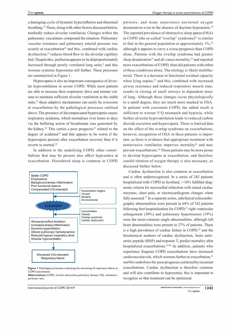
are summarized in Figure 1.
Hypercapnia is also an important consequence of alveo-
lar hypoventilation in severe COPD. While most patients
are able to increase their respiratory drive and minute vol-
ume to maintain sufficient alveolar ventilation in the stable
state,22 these adaptive mechanisms can easily be overcome
at exacerbation by the pathological processes outlined
above. The presence of decompensated hypercapnia causes
respiratory acidemia, which normalizes over hours to days
via the buffering action of bicarbonate ions generated by
the kidney.26 This carries a poor prognosis27 related to the
degree of acidemia28 and that appears to be worse if the
hypercapnia persists after exacerbation recovery than if it
reverts to normal.29
In addition to the underlying COPD, other comor-
bidities that may be present also affect hypoxemia at
exacerbation. Disordered sleep is common in COPD
patients, and many ex perience noc turnal ox ygen
desaturation even in the absence of daytime hypoxemia.30
The reported prevalence of obstructive sleep apnea (OSA)
in COPD (the so-called “overlap” syndrome)31 is similar
to that in the general population at approximately 1%,32
although it appears to car ry a worse prognosis than COPD
alone. Patients with the overlap syndrome had greater
sleep desaturation33 and all-cause mortality,34 and reported
more exacerbations of COPD, than did patients with either
of these conditions alone. The etiology is likely multifac-
torial. There is a decrease in functional residual capacity
when lying supine,35 and this, combined with increased
airway resistance and reduced respiratory muscle tone,
results in closing of small airways in dependent areas
of lung. Although these changes occur physiologically
to a small degree, they are much more marked in OSA.
In patients with coexistent COPD, the added insult is
sufficient to worsen V/Q mismatch and hypoxia, while
further alveolar hypoventilation leads to reduced carbon
dioxide excretion and hypercapnia. There is limited data
on the effect of the overlap syndrome on exacerbations;
however, recognition of OSA in these patients is impor-
tant, as there is evidence that appropriate treatment with
noninvasive ventilation improves mortality36 and may
prevent exacerbations.34 These patients may be more prone
to develop hypercapnia at exacerbation, and therefore
careful titration of oxygen therapy is also necessary, as
discussed further below.
Cardiac dysfunction is also common at exacerbation
and is often underrecognized. In a series of 242 patients
hospitalized with COPD in Scotland, .10% fulfilled diag-
nostic criteria for myocardial infarction with raised cardiac
enzymes, chest pain, or electrocardiogram changes when
fully assessed.37 In a separate series, subclinical echocardio-
graphic abnormalities were present in 64% of 342 patients
following first hospitalization for COPD;25 right ventricular
enlargement (30%) and pulmonary hypertension (19%)
were the most-common single abnormalities, although left
heart abnormalities were present in 27% of patients. There
is a high prevalence of cardiac failure in COPD,38 and the
biochemical markers of cardiac dysfunction, brain natri-
uretic peptide (BNP) and troponin T, predict mortality after
hospitalized exacerbations.39,40 In addition, patients who
experience frequent COPD exacerbations have increased
cardiovascular risk, which worsens further at exacerbation,41
and this underlines the poor prognosis conferred by recurrent
exacerbations. Cardiac dysfunction is therefore common
and will also contribute to hypoxemia; this is important to
recognize so that treatment can be optimized.
Worsened airflow limitation
Increased airway inflammation
Dynamic hyperinflation
Altered pulmonary hemodynamicsReduced hypoxic inspiratory drive
Alveolar hypoventilation
Exacerbation
Stable COPDEmphysema
Background airway inflammation
Poor functional reserve
Compensated V/Q mismatchExacerbation triggers
Viruses
Bacteria
Environmental
Comorbidities
Obesity
Overlap syndrome
Cardiac dysfunction
Worsened V/Q mismatch
Respiratory failure
Figure 1 Pathological processes underlying the worsening of respiratory failure at
COPD exacerbation.
Abbreviations: COPD, chronic obstructive pulmonary disease; V/Q, ventilation–
perfusion ratio.

7/24/2019 Oxygen Therapy in Acute Exacerbations of Chronic Obstructive Pulmonary Disease
http://slidepdf.com/reader/full/oxygen-therapy-in-acute-exacerbations-of-chronic-obstructive-pulmonary-disease 4/12
International Journal of COPD 2014:9submit your manuscript | www.dovepress.com
Dovepress
Dovepress
1244
Brill and Wedzicha
Acute exacerbation of COPD is a clinical diagnosis, and it
is important to exclude other conditions causing hypoxia and
respiratory failure. For example, pulmonary embolism may
be present in up to a fifth of patients presenting to hospital
with COPD exacerbation,42 and this may worsen hypoxemia
by causing further V/Q mismatching. Other conditions caus-
ing acidosis, reduced tissue perfusion, or increased tissue
oxygen requirements, notably systemic sepsis or cardiogenic
shock, may also worsen respiratory failure in COPD patients
and should be treated appropriately.
Benecial effects of oxygentherapy in stable COPDAside from relieving the hypoxia associated with COPD,
supplemental oxygen therapy also reduces symptoms of
dyspnea both in normal subjects and in those with severe
COPD.43,44 A recent Cochrane meta-analysis including
431 patients across 18 studies also showed a highly sig-
nificant improvement in dyspnea in nonhypoxemic COPD
patients who would not otherwise meet physiological cri-
teria for oxygen therapy.45 Breathing supplemental oxygen
versus compressed air improved dyspnea and endurance
during exercise in COPD patients46 and, in those patients
who showed an improvement in exercise tolerance fol-
lowing long-term oxygen therapy (LTOT), cardiac output
was also increased.47 These effects are likely related to
decreased minute ventilation48,49 and decreased dynamic
hyperinflation.50 Hypoxic pulmonary vasoconstriction and
pulmonary hemodynamics may also be improved,46 leading
to increased systemic oxygen delivery and improved respira-
tory muscle function.49 In addition, the flow of oxygen may
stimulate upper airway and facial receptors, and this appears
to reduce the intensity of dyspnea.51
Importantly, LTOT was the first therapy to demonstrate
a reduction in mortality for COPD. Published in 1980, the
Nocturnal Oxygen Therapy Trial52 randomized 203 patients
with hypoxemic chronic lung disease to receive supple-
mental oxygen therapy, either continuously or overnight
only, with follow-up for a minimum of 12 months. Oxygen
therapy was given via nasal prongs with flow titrated to the
minimum amount between 1–4 L/minute that produced a
rise in PaO2. All-cause mortality in the continuous-treatment
group was 52% of that in the nocturnal-only treatment group,
with the difference even more marked in the subgroup
with baseline hypercapnia. Shortly afterwards, the British
Medical Research Council trial in the United Kingdom 53
published results from 87 patients with severe COPD and
hypoxemia, hypercapnia, and congestive cardiac failure
who were randomized to receive oxygen (via nasal prongs
at 2 L/minute, for a minimum of 15 hours per day) or no
oxygen therapy. After 5 years of follow-up, 30/45 (67%) of
the untreated patients had died, compared to 19/42 (45%) of
those who were given oxygen; arterial carbon dioxide tension
and red-cell mass appeared to be useful predictors of poor
prognosis. Together, these trials provide the evidence under-
lying the indications for the use of LTOT in severe COPD,
as well as the recommendation to use LTOT for a mini-
mum of 15 hours/day to ensure maximal benefit. However,
a further trial of LTOT in patients with milder hypoxemia
(PaO2 ,8 kPa [60 mmHg]) showed no mortality benefit.54
Therapy with LTOT should therefore be restricted to hypox-
emic patients with PaO2 ,7.3 kPa (55 mmHg), or ,8 kPa
(60 mmHg) in the presence of complications including pul-
monary hypertension, polycythemia, or peripheral edema.55
“Ambulatory” oxygen therapy may also be given to patients
for use during exercise or activities of daily living, and while
short-term studies show improved exercise performance in
COPD,56 no survival benefit has been demonstrated.
Uncertainty remains regarding the use of LTOT in patients
with isolated exercise desaturation, or only moderate hypox-
emia at rest, and given the scale of LTOT use in COPD there
is therefore a pressing need for further evidence. It is hoped
that the Long-term Oxygen Treatment Trial,57 which aims
to recruit over 1,000 participants with COPD and moderate
resting hypoxemia or exercise desaturation, will answer some
of these questions.
Oxygen therapy at acuteexacerbation of COPDSupplemental oxygen delivered at moderate concentrations
is usually adequate to overcome the hypoxia associated
with COPD exacerbations, with the main risk being the
long-recognized induction of hypercapnia.58 Despite this,
high-flow oxygen has long been the standard treatment for
acutely distressed patients, and until relatively recently this
was also the case in COPD, especially in prehospital care.
However, the risks of injudicious oxygen administration in
these patients are now well recognized. We outline here the
rationale and guidance for the appropriate use of oxygen
during COPD exacerbations, as well as strategies for oxygen
delivery and ventilatory support.
Oxygen-induced hypercapniaand hyperoxia at COPD exacerbationOne important consequence of oxygen therapy is a worsen-
ing of hypercapnia in susceptible patients, and this has long
7/24/2019 Oxygen Therapy in Acute Exacerbations of Chronic Obstructive Pulmonary Disease
http://slidepdf.com/reader/full/oxygen-therapy-in-acute-exacerbations-of-chronic-obstructive-pulmonary-disease 5/12
International Journal of COPD 2014:9 submit your manuscript | www.dovepress.com
Dovepress
Dovepress
1245
Oxygen therapy in acute exacerbations of COPD
been recognized. In 1949, case reports were first published
of patients with chronic cor pulmonale in whom administra-
tion of high concentrations of oxygen induced neurological
changes, including fatal coma and transient increases in
intracranial pressure;59 hypercapnia was hypothesized to
result from reduced minute ventilation (VE) resulting from
reduced hypoxic stimulus to breathe.60 This widely held view
was challenged by a study of patients with severe COPD
and hypercapnia before and after breathing in 100% oxygen
for 20 minutes.61 Although VE decreased transiently, after
15 minutes it was again similar to control values on room air;
despite this, there was a significant rise in PaCO2 values that
was not correlated to changes in VE, and this was attributed to
increased V/Q mismatching within the lungs, possibly due to
the reversal of hypoxic vasoconstriction increasing perfusion
of poorly ventilated lung units. However, this was investi-
gated more recently using the multiple inert-gas elimination
technique in 22 patients during an exacerbation of COPD.62
Intrapulmonary V/Q defects were similar in the 12 patients
who developed hypercapnia with supplemental oxygen as in
those who did not, although alveolar dead space increased
in the hypercapnic patients. Importantly, VE also fell by
approximately 20% in the hypercapnic patients. Respiratory
muscle fatigue, also thought to be a factor, appears now to
only occur at a late stage, and reductions in ventilation may
also reflect a shallower breathing pattern.58
A further potential mechanism underlying oxygen-induced
hypercapnia results from changes in the CO2 –hemoglobin
dissociation curve as the PaO2 increases. Oxygenated hemo-
globin has a lower carbon dioxide binding capacity than does
deoxygenated hemoglobin, and as the proportion of oxygen-
ated hemoglobin increases there is a consequent rightward
shift of the CO2 –hemoglobin dissociation curve.63 This is
known as the Haldane effect, and it results in an increase in
PaCO2. While this is usually overcome by increasing VE,
patients with severe COPD are often unable to do this and
hypercapnia may worsen. This may explain up to 25% of the
total PaCO2 increase due to oxygen administration.61
Oxygen-induced hypercapnia at COPD exacerbation
is therefore due to a complex combination of factors and
remains incompletely understood. The associated risk, how-
ever, is now well recognized. Many studies have focused
on the oxygen therapy given at the point of prehospital
care, usually by a treating paramedic crew during assess-
ment and transport to hospital, and this appears to be par-
ticularly important. In a retrospective case series of nearly
1,000 patients in the United Kingdom, 20% had respiratory
acidosis on presentation to hospital and this was associated
with an increased risk for subsequent tracheal intubation.64
The pH was inversely correlated with the PaO2, and more
than half of hyperoxemic patients (PaO2 .13.3 kPa) were
acidotic, reflecting the role of overoxygenation in the
prehospital setting. Where respiratory acidosis is present,
the degree of acidosis correlates to mortality.65 Similarly,
one retrospective audit of prehospital oxygen therapy given
to 211 patients with acute exacerbations of COPD found
that those treated with oxygen at a concentration of $28%
were significantly more acidotic, with significantly higher
partial pressures of carbon dioxide, than those in whom
controlled oxygen therapy was administered.66 In one series
of 254 patients presenting via ambulance with acute COPD
exacerbation,17 hyperoxemia at presentation carried an odds
ratio of 9.17 (95% confidence interval [CI] =4.08–20.6) over
normoxemia for a composite of serious adverse outcomes
(including hypercapnic respiratory failure), compared to only
2.16 (95% CI =1.11–4.20) for hypoxemia. Further indirect
evidence is provided by a large anonymized audit of arterial
blood gas samples in hospital, which found oxygen satura-
tions above the recommended 92% in 72% of hypercapnic
samples, suggesting that oxygen control is poor.67 Although
hypoxia may be more acutely dangerous than hypercapnia,
hyperoxia therefore also carries significant excess risk.
A further consideration in hypercapnic patients treated
with high oxygen concentrations is the phenomenon of
rebound hypoxia upon withdrawal of supplemental oxygen.
This occurs because oxygen and carbon dioxide displace
each other from the limited space within the alveoli accord-
ing to their relative partial pressures, as described by the
alveolar gas equation.9 In the presence of a very high alveolar
PaO2 (as seen when breathing a high concentration of
supplemental oxygen), systemic oxygenation is maintained
even as the PaCO2 rises to a level higher than it was when
the patient was breathing room air. The human body stores
more carbon dioxide than oxygen, and if the supplemental
oxygen is abruptly withdrawn there may be a sharp fall in
the alveolar PaO2 as the remaining carbon dioxide displaces
the (now lower-pressure) oxygen. The result is a fall in arte-
rial PaO2
, which can be precipitous and may cause death by
acute arterial hypoxemia even though the PaCO2 is stable or
improving.68 For this reason, oxygen therapy should always
be stepped down gradually.9
Despite compelling observational data, until relatively
recently there has been a lack of high-quality evidence to
directly demonstrate the benefits of titrated oxygen therapy
over high-concentration oxygen. In 2010, Austin et al69
carried out the first large, well-conducted study of titrated

7/24/2019 Oxygen Therapy in Acute Exacerbations of Chronic Obstructive Pulmonary Disease
http://slidepdf.com/reader/full/oxygen-therapy-in-acute-exacerbations-of-chronic-obstructive-pulmonary-disease 6/12
International Journal of COPD 2014:9submit your manuscript | www.dovepress.com
Dovepress
Dovepress
1246
Brill and Wedzicha
oxygen therapy in prehospital care for suspected COPD
exacerbations. In a cluster randomized trial, with individual
paramedics as the unit of randomization, titrated oxygen
therapy (via nasal prongs with the goal of maintaining satura-
tions between 88%–92%; 32 paramedics) or the standard care
of high-flow oxygen delivered via face mask (30 paramedics)
was administered to patients; 214 patients were included in
the intent-to-treat analysis, with 97 in the titrated-oxygen
arm and 117 in the high-flow oxygen arm. In spite of lapses
in adherence to the oxygen-titration protocol, there was a
78% reduction in mortality for those patients with confirmed
COPD, as well as reduced rates of respiratory acidosis and
hypercapnia.
There is now therefore strong evidence that titrating oxy-
gen therapy to alleviate hypoxia while avoiding hyperoxia
is the correct approach to oxygen therapy in COPD patients
who are at high risk of hypercapnic respiratory acidosis.
Until these patients are identified by arterial blood gas
analysis, titrating oxygen therapy in all patients is prudent.
Administration of oxygen therapyThe key to achieving appropriate levels of oxygenation is
using controlled oxygen therapy, with the patient’s oxy-
gen level monitored and the supplemental oxygen therapy
titrated to achieve acceptable saturations. This approach is
summarized in Figure 2. Titration can be achieved either
by altering the oxygen flow rate or, with certain delivery
devices, administering a mixture of air and oxygen in set
proportions such that the patient breathes in a known fraction
of inspired oxygen (FiO2). The principal modes of delivery
are described below.
Nasal cannulae are perhaps the simplest mode of adminis-
tering low-to-moderate concentrations of inspired oxygen and
have considerable advantages over standard face masks. The
Select target
SaO2 range
Select oxygen
delivery device
94%–98% (no hypercapnia risk)
88%–92% (at risk of hypercapnia)
Apply oxygen
therapy
SaO2 too low SaO
2 too high
Up-titrateoxygen therapy Down-titrateoxygen therapy
Consider further ventilatory
strategies
Reassess patient
Unable to achieve
target SaO2 range
Figure 2 General principles of titrated oxygen therapy.

7/24/2019 Oxygen Therapy in Acute Exacerbations of Chronic Obstructive Pulmonary Disease
http://slidepdf.com/reader/full/oxygen-therapy-in-acute-exacerbations-of-chronic-obstructive-pulmonary-disease 7/12
International Journal of COPD 2014:9 submit your manuscript | www.dovepress.com
Dovepress
Dovepress
1247
Oxygen therapy in acute exacerbations of COPD
short cannulas deliver oxygen directly to the nasopharynx,
reducing dead space and inspiratory resistance, and they
can deliver a range of oxygen flow rates from 1–6 L/minute,
corresponding to FiO2 values of approximately 24%–50%.9
No face mask is required and patients can therefore eat and
speak while wearing their oxygen, and the cannulas are less
likely than face masks to fall off or become dislodged by
facial movement.70 Unsurprisingly, patients prefer cannulas
to face masks, and this improves compliance with oxygen
therapy.71 In addition, low-flow oxygen therapy can be contin-
ued via nasal cannulas during the administration of air-driven
nebulizers for hypercapnic or acidotic patients.55 However
there are also disadvantages to nasal cannulas. Some patients
experience nasal dryness or discomfort, particularly at higher
flow rates. In addition, the FiO2 is affected by a multitude of
patient factors, including mouth breathing, degree of nasal
congestion, and the respiratory rate and minute volume.
These factors may also change in the same patient at different
times (for example, while eating). The use of nasal cannulas
should therefore target a specific oxygen saturation level, with
ongoing monitoring and titration of the oxygen flow rate to
achieve this. Nasal cannulas are not recommended for the
acutely unwell COPD patient, in whom accurate control of
FiO2 is critical.9 However, once stability is achieved, nasal
cannulas with oxygen flow rates titrated to target oxygen satu-
rations represent the simplest and most-acceptable method
of administering controlled oxygen therapy.
Simple face masks are able to deliver a higher con-
centration of inspired oxygen than nasal cannulas, usually
between 40%–60%. The exact FiO2 is highly variable, how-
ever, and there is a risk of rebreathing carbon dioxide at low
flow rates. In addition, the concentration of O2 administered
is inversely related to minute volume; if this falls and the
oxygen flow rate remains constant (for example if the patient
becomes drowsy due to oxygen-induced CO2 narcosis), the
concentration of oxygen inspired via the mask will increase,
further exacerbating the problem. These problems can be
partially circumvented by using a non-rebreathing face mask,
which has an oxygen reservoir bag attached; at high oxygen
flow rates an FiO2
of 60%–90% can be delivered. Even here,
however, the exact FiO2 is highly variable and dependent on
the patient’s minute volume. Even in COPD exacerbations,
oxygenation to a safe level is usually achievable with an FiO2
of ,60%, and for this reason these masks are not recom-
mended for patients at risk of hypercapnic respiratory failure
where controlled oxygen therapy is necessary.9
In 1960, Moran Campbell introduced a simple system that
was able to entrain air into an oxygen stream across differing
flow rates, with the amount of air added increasing as the flow
of oxygen increased but the proportions remaining equal.72
Although the risks of excess oxygen therapy in patients with
cor pulmonale had been well recognized, up to this point
oxygen therapy could only be controlled by giving it intermit-
tently alternated with periods on room air, a practice likened to
“bringing a drowning man to the surface – occasionally.”73 The
Venturi system, as it came to be known, allowed the constant
delivery of a fixed concentration of oxygen to the patient.
Crucially, this could be delivered at higher flow rates for tac-
hypneic patients in whom the minute volume might otherwise
exceed the available oxygen flow. Each valve is manufactured
to deliver only a specific FiO2, with valves for 24%, 28%,
35%, 40%, and 60% usually available, and a minimum oxygen
flow rate, above which the resulting air–oxygen mixture will
correspond to the intended ratios, is specified. The Venturi
system must be used with a face mask and therefore is subject
to the usual disadvantages, including intrusiveness to patients,
preventing eating and conversation, and being more prone
to dislodge, but remains the preferred method for controlled
oxygen administration in the emergency situation.9
The principle of controlled oxygen therapy is to target
the patient’s oxygen saturations within a range rather than at
a specific FiO2. This is because the patient factors affecting
SaO2 are dynamic and will change over time; a fixed FiO
2
will not be responsive to these changing demands. Oxygen
levels should be maintained at a safe level while avoiding
hyperoxemia that would increase the risk of hypercapnia.
Historically, there has been some debate as to what a “safe”
level of hypoxemia is. Early investigators reported memory
loss and cognitive difficulties in normal subjects at PaO2
levels ,45 mmHg (6.0 kPa),74 with levels ,20 mmHg
(2.7 kPa) incompatible with life.75 Patients with COPD are
able to acclimatize to even quite a severe degree of chronic
hypoxia, however, and maintaining PaO2 .50 mmHg
(6.7 kPa) is sufficient to prevent immediate death,76 although
oxygen therapy should aim to maintain a safe level above this
in order to preserve normal cognitive function and prevent
transient desaturations.76 The usual lower PaO2 limit for
targeted oxygen therapy is 55 mmHg (7.3 kPa), used as the
entry criteria for the LTOT trials and subsequently ratified by
consensus committee.77 In practice, real-time monitoring of
PaO2 is not usually possible and therefore a target SaO
2 range
of 88%–92% is now generally accepted for those patients
at risk of hypercapnia. Allowing for individual variations
in the oxygen dissociation curve, this will correct hypoxia
to a safe level and minimize the risk of oxygen-induced
hypercapnia.

7/24/2019 Oxygen Therapy in Acute Exacerbations of Chronic Obstructive Pulmonary Disease
http://slidepdf.com/reader/full/oxygen-therapy-in-acute-exacerbations-of-chronic-obstructive-pulmonary-disease 8/12
International Journal of COPD 2014:9submit your manuscript | www.dovepress.com
Dovepress
Dovepress
1248
Brill and Wedzicha
Hypercapnia at COPD exacerbation is unusual in that it
does not occur in all patients, even those with resting baseline
hypercapnia; some patients may suffer recurrent hypercapnic
episodes while others may only ever have isolated hypoxemia.
Without arterial blood gas analysis, identifying those patients
with hypercapnia is not possible. For this reason, current
clinical guidance recommends that all patients with respira-
tory failure in the context of a diagnosis or history suggestive
of COPD receive oxygen therapy targeted to 88%–92% until
hypercapnia has been excluded by arterial blood gas analysis.9
Some patients are extremely oxygen-sensitive, with even very
small amounts of supplemental oxygen sufficient to worsen
hypercapnia, and these patients may need a lower target
saturation range. However, if the PaCO2 is normal, oxygen
therapy may target the usual saturation range of 94%–98%,
although many COPD patients may have a lower stable SaO2,
such that chasing this target is not usually necessary unless
the patient is unwell.
Following initiation of oxygen therapy, close observation
and reassessment of the patient is key. Patients suffering
acute COPD exacerbations can be extremely unwell and,
particularly in the early stages following presentation, their
condition can change extremely quickly. Regular assessment
should include not only their respiratory rate, oxygen satura-
tions, and other physiological measurements, but also their
conscious level, as any depression of consciousness may
suggest incipient carbon dioxide narcosis. As the patient
improves, the oxygen should be reduced such that the upper
limit of the target range is not breached. Supplemental oxygen
therapy should be treated as a medication and prescribed on
the treatment chart in the same way as other pharmacological
agents. Prescriptions should include information regarding
the delivery device, oxygen flow rate, and target oxygen
saturation range, with instructions on what action to take if
these parameters are exceeded in either direction.
It is important to remember that oxygen therapy during
acute exacerbations of COPD is a supportive measure only
and must be accompanied by treatment of the underlying
COPD exacerbation. In most cases this will involve inhaled
or nebulized bronchodilators, antibiotics, and systemic
corticosteroids, as appropriate.55
Further ventilatory strategiesUnless the patient is in extremis, adequate oxygenation
using one of the methods above is usually possible for
COPD patients who have type I respiratory failure without
hypercapnia. However, the patient with hypercapnic respi-
ratory failure and acidosis may prove more complicated,
and hypercapnia may progress despite treatment of the
exacerbation and adequately controlled oxygen therapy.
In these patients it may become impossible to adequately
maintain even minimal oxygenation without further hyper-
capnia and potential coma, and further ventilatory support
may be needed.
The respiratory stimulant doxapram has been used for
many years as a means of avoiding endotracheal intuba-
tion and invasive positive-pressure ventilation. It works via
stimulation of central and peripheral chemoreceptors, with
the result of increasing tidal volume and minute ventilation.78
This improves the excretion of carbon dioxide, allowing
correction of hypoxia without worsening hypercapnia.
However, the pharmacokinetics are not predictable and
therefore a constant infusion is required, with titration to
dose response. It is slightly better than placebo at prevent-
ing blood-gas deterioration in the first hours of therapy, 79
but has since been superseded by noninvasive ventilation
(NIV). Doxapram is now recommended for use only when
NIV is not available.55
Until NIV was introduced, endotracheal intubation and
mechanical ventilation was the only remaining option for
patients who failed best medical therapy. This carries significant
risk, and many patients with very severe underlying COPD are
deemed too frail to undergo the procedure. There has therefore
long been a need for methods of ventilatory support that avoid
the need for sedation and intubation. Noninvasive ventilators
were developed in the 1980s to deliver positive airway pres-
sure via a face mask, and these may either be pressure preset
to deliver differential pressures in inspiration and expiration,
or be volume preset to deliver specific volumes.80,81 The pres-
sure is delivered by air compression, and supplemental oxygen
may also be administered; newer devices are able to deliver a
specified oxygen concentration, while other devices entrain
oxygen with the air, resulting in a variable but unknown oxygen
concentration. Nebulized bronchodilators may also be adminis-
tered through the tubing, and the apparatus has now evolved to
be portable and simple to use. The mode of action is multifac-
torial and involves resting of the respiratory muscles, reversal
of atelectasis with recruitment of lung units, and improved
tidal volume.82 The net result is an increase in minute volume
and reduction in the work of breathing, with increased carbon
dioxide excretion and reversal of hypercapnia.83
Early reports in acute COPD exacerbations showed that,
compared to historical controls, the apparatus was effective
at reducing PaCO2, improving acidosis and oxygenation,
and avoiding the need for mechanical ventilation.80 The first
prospective randomized controlled trials followed shortly and
7/24/2019 Oxygen Therapy in Acute Exacerbations of Chronic Obstructive Pulmonary Disease
http://slidepdf.com/reader/full/oxygen-therapy-in-acute-exacerbations-of-chronic-obstructive-pulmonary-disease 9/12
International Journal of COPD 2014:9 submit your manuscript | www.dovepress.com
Dovepress
Dovepress
1249
Oxygen therapy in acute exacerbations of COPD
confirmed striking benefits of NIV in reducing intubation, length
of hospital stay, and in-hospital mortality, as well as a host of
physiological parameters, compared to standard treatment.84,85
There is now a wealth of further evidence to support these
conclusions,86 and NIV has become the accepted standard of
care for patients with type 2 respiratory failure (pH,7.35 and
PaCO2 .6 kPa) due to acute exacerbation of COPD despite
controlled oxygen and best medical treatment.87
NIV has many advantages. With appropriate expertise
and a compliant patient, it is relatively simple to set up and
allows the concurrent administration of oxygen and nebulized
bronchodilators. Although some patients are unable to toler-
ate the tight-fitting mask, most do manage and the apparatus
allows for breaks to be taken for eating and other activities.
There are some contraindications to the use of NIV, particularly
for patients who are unable to maintain their own airway due
to obstruction or reduced conscious level, in life-threatening
hypoxemia, or in the presence of undrained pneumothorax.88
However, in patients who would not be suitable candidates for
endotracheal intubation and where NIV is the decided ceiling of
care, it may be tried even in the presence of some of these con-
traindications. Regardless, clear contingency plans regarding
whether or not to proceed to tracheal intubation in the event of
treatment failure should always be in place prior to starting NIV,
and those patients with severe respiratory acidosis (pH,7.26)
should be managed in a high-dependency area and with a low
threshold for intubation if appropriate.87 More detailed guidance
on the use of NIV is available elsewhere.87,88
Current guidance for oxygen therapyin acute exacerbations of COPDAs the risks and optimal strategies for oxygen therapy have
become clearer, consensus guidelines have been written to
formalize best practice. From the United Kingdom, the British
Thoracic Society Guideline for emergency oxygen therapy9
provides highly detailed guidance on the delivery of oxygen
therapy in COPD and other conditions. Other national and
international guidance statements on COPD, including those
from the Global Initiative for Chronic Obstructive Lung
Disease (GOLD),89 American Thoracic Society and European
Respiratory Society,90 and the National Institute for Clinical
Excellence55 also include broad recommendations for the use of
supplemental oxygen therapy during COPD exacerbations.
Current practice and strategiesfor changing behavior The first step toward improving standards is to define the
targets for optimal care. There are now nationally and
internationally agreed guidelines, described above, and this
has allowed an assessment of current practice and identifica-
tion of targets for improvement.
The recent European COPD Audit91 examined in-hospital
care against ten standards derived from the 2010 GOLD
guidance on COPD management.89 Data were available
from 384 hospitals in 13 countries, for 16,018 patients admitted
with exacerbations of COPD. Of these, 85% of patients were
given controlled oxygen therapy, 82% had arterial blood gas
analysis performed on admission, and 51% received NIV as
recommended, although there were wide regional variations.
Although the delivery of NIV was highlighted as a specific
area for improvement, controlled oxygen therapy was not
correctly administered in a substantial number of cases. Even
where oxygen is prescribed, deficiencies may still be identified.
A large multicenter audit of inpatient oxygen therapy in
Portugal found that although oxygen was prescribed in 93% of
cases, the majority specified a fixed flow rate rather than target
oxygen saturations, and the prescription was only completed in
12%; in any case, 23% of patients were not receiving the pre-
scribed therapy.92 It should be noted that these audits have only
focused on the provision of oxygen after the patient is admitted
to hospital. Many patients are still given high concentrations of
oxygen during prehospital care, arriving at hospital hyperox-
emic and/or hypercapnic,64–66 and it is in this acute setting that
instituting correct oxygen management is most crucial.
The next logical step is to address how the provision
of oxygen therapy for these patients may be improved.
Simple interventions may change behavior: for example,
in one retrospective series in England, issuing 28% Venturi
masks to ambulance crews assisted in reducing the propor-
tion of patients receiving high-flow oxygen prehospital.66
Standardized care “bundles” have been shown to reduce mor-
tality across a variety of conditions by improving in-hospital
care,93 and implementing a simple standardized package of
recommendations for COPD exacerbations at the emergency
department of one large teaching hospital significantly
improved compliance with treatment guidelines, notably
increasing the correct provision of oxygen therapy from 76%
to 96%.94 In addition, the move toward comprehensively pre-
scribing oxygen should provide a simple framework to guide
staff caring for the patient. Perhaps paramount, however,
is the education of health care professionals, particularly
the doctors, nurses, and paramedics who are responsible
for administering oxygen therapy to patients with COPD.95
Hospitals should institute local policies to improve education
and practice, and these should be subject to rigorous audit
and reevaluation.55

7/24/2019 Oxygen Therapy in Acute Exacerbations of Chronic Obstructive Pulmonary Disease
http://slidepdf.com/reader/full/oxygen-therapy-in-acute-exacerbations-of-chronic-obstructive-pulmonary-disease 10/12
International Journal of COPD 2014:9submit your manuscript | www.dovepress.com
Dovepress
Dovepress
1250
Brill and Wedzicha
Improving the education of patients may also be a factor.
A recent study of patients hospitalized with COPD exacerba-
tions showed variable awareness of the symptoms leading up
to an exacerbation, which delayed symptom reporting and
may therefore have increased the severity at presentation.96
In addition, patients who are known to be at risk of hyper-
capnic respiratory failure should be given oxygen “alert
cards,”97 which warn any treating clinicians, particularly
paramedics, that they should be given controlled rather than
high-concentration oxygen therapy.9
ConclusionThere is clear evidence that the correct use of supplemental
oxygen therapy during exacerbations of COPD is an impor-
tant factor that can strongly influence outcomes, and we
have sought to outline this here. Future research may focus
on new delivery strategies to improve the titration of oxygen
therapy98 as well as new therapies to treat the underlying
COPD. In the meantime, we must ensure that the knowledge
already available is translated into clinical practice and that
best practice is followed. This will undoubtedly improve the
treatment given to these patients.
DisclosureThe authors report no conflicts of interest in this work.
References1. Vestbo J, Hurd SS, Agustí AG, et al. Global strategy for the diagno-
sis, management, and prevention of chronic obstructive pulmonary
disease: GOLD executive summary. Am J Respir Crit Care Med .2013;187(4):347–365.
2. Donaldson GC, Seemungal TA, Bhowmik A, Wedzicha JA. Relationship
between exacerbation frequency and lung function decline in chronic
obstructive pulmonary disease. Thorax. 2002;57(10):847–852.
3. Seemungal TA, Donaldson GC, Paul EA, Bestall JC, Jeffries DJ,
Wedzicha JA. Effect of exacerbation on quality of life in patients with
chronic obstructive pulmonary disease. Am J Respir Crit Care Med .
1998;157(5 Pt 1):1418–1422.
4. Soler-Cataluña JJ, Martínez-García MA, Román Sánchez P,
Salcedo E, Navarro M, Ochando R. Severe acute exacerbations and
mortality in patients with chronic obstructive pulmonary disease. Thorax.
2005;60(11):925–931.
5. Suissa S, Dell’Aniello S, Ernst P. Long-term natural history of chronic
obstructive pulmonary disease: severe exacerbations and mortality.
Thorax. 2012;67(11):957–963.6. West JB. Causes of and compensations for hypoxemia and hypercapnia.
Compr Physiol . 2011;1(3):1541–1553.
7. Quintana JM, Esteban C, Unzurrunzaga A, et al; IRYSS-COPD group.
Predictive score for mortality in patients with COPD exacerbations
attending hospital emergency departments. BMC Med . 2014;12:66.
8. Barberà JA, Roca J, Ferrer A, et al. Mechanisms of worsening gas
exchange during acute exacerbations of chronic obstructive pulmonary
disease. Eur Respir J . 1997;10(6):1285–1291.
9. O’Driscoll BR, Howard LS, Davison AG; British Thoracic Society. BTS
guideline for emergency oxygen use in adult patients. Thorax. 2008;
63 Suppl 6:vi1–vi68.
10. Crapo RO, Jensen RL, Hegewald M, Tashkin DP. Arterial blood gas
reference values for sea level and an altitude of 1,400 meters. Am J
Respir Crit Care Med . 1999;160(5 Pt 1):1525–1531.
11. Tashkin DP, Celli B, Senn S, et al; UPLIFT Study Investigators.
A 4-year trial of tiotropium in chronic obstructive pulmonary disease.
N Engl J Med . 2008;359(15):1543–1554.
12. Wise RA, Anzueto A, Cotton D, et al; TIOSPIR Investigators.
Tiotropium Respimat inhaler and the risk of death in COPD. N Engl J
Med . 2013;369(16):1491–1501.
13. Martinez FJ, Foster G, Curtis JL, et al; NETT Research Group.
Predictors of mortality in patients with emphysema and severe airflow
obstruction. Am J Respir Crit Care Med . 2006;173(12):1326–1334.
14. Jones RC, Dickson-Spillmann M, Mather MJ, Marks D, Shackell BS.
Accuracy of diagnostic registers and management of chronic obstruc-
tive pulmonary disease: the Devon primary care audit. Respir Res.
2008;9:62.
15. Ward MM, Javitz HS, Smith WM, Bakst A. Direct medical cost
of chronic obstructive pulmonary disease in the USA. Respir Med .
2000;94(11):1123–1129.
16. Roberts CM, Stone RA, Buckingham RJ, Pursey NA, Lowe D;
National Chronic Obstruc tive Pulmonary Disease Resources and
Outcomes Project Implementation Group. Acidosis, non-invasive
ventilation and mortality in hospitalised COPD exacerbations. Thorax.
2011;66(1):43–48.
17. Cameron L, Pilcher J, Weatherall M, Beasley R, Perrin K. The
risk of serious adverse outcomes associated with hypoxaemia andhyperoxaemia in acute exacerbations of COPD. Postgrad Med J .
2012;88(1046):684–689.
18. Hurst JR, Donaldson GC, Quint JK, Goldring JJ, Patel AR,
Wedzicha JA. Domiciliary pulse-oximetry at exacerbation of chronic
obstructive pulmonary disease: prospective pilot study. BMC Pulm
Med . 2010;10:52.
19. Cooper CB, Celli B. Venous admixture in COPD: pathophysiology and
therapeutic approaches. COPD. 2008;5(6):376–381.
20. Wedzicha JA, Singh R, Mackay AJ. Acute COPD exacerbations. Clin
Chest Med . 2014;35(1):157–163.
21. Bhowmik A, Seemungal TA, Sapsford RJ, Wedzicha JA. Relation of
sputum inflammatory markers to symptoms and lung function changes
in COPD exacerbations. Thorax. 2000;55(2):114–120.
22. O’Donnell DE, Parker CM. COPD exacerbations . 3: Pathophysiology.
Thorax. 2006;61(4):354–361. 23. O’Donnell DE, Laveneziana P. The clinical importance of dynamic
lung hyperinflation in COPD. COPD. 2006;3(4):219–232.
24. Chaouat A, Naeije R, Weitzenblum E. Pulmonary hypertension in
COPD. Eur Respir J . 2008;32(5):1371–1385.
25. Freixa X, Portillo K, Paré C, et al; PAC-COPD Study Investigators.
Echocardiographic abnormalities in patients with COPD at their
first hospital admission. Eur Respir J . 2013;41(4):784–791.
26. Schwartz WB, Brackett NC Jr, Cohen JJ. The response of extracellular
hydrogen ion concentration to graded d-egrees of chronic hypercapnia: the
physiologic limits of the defense of ph. J Clin Invest . 1965;44:291–301.
27. Matkovic Z, Huerta A, Soler N, et al. Predictors of adverse outcome in
patients hospitalised for exacerbation of chronic obstructive pulmonary
disease. Respiration. 2012;84(1):17–26.
28. Plant PK, Owen JL, Elliott MW. Early use of non-invasive ventilation
for acute exacerbations of chronic obstructive pulmonary disease ongeneral respiratory wards: a multicentre randomised controlled trial.
Lancet . 2000;355(9219):1931–1935.
29. Costello R, Deegan P, Fitzpatrick M, McNicholas WT. Reversible
hypercapnia in chronic obstructive pulmonary disease: a distinct
pattern of respiratory failure with a favorable prognosis. Am J Med .
1997;102(3):239–244.
30. McSharry DG, Ryan S, Calverley P, Edwards JC, McNicholas WT.
Sleep quality in chronic obstructive pulmonary disease. Respirology.
2012;17(7):1119–1124.
31. Flenley DC. Sleep in chronic obstructive lung disease. Clin Chest Med .
1985;6(4):651–661.

7/24/2019 Oxygen Therapy in Acute Exacerbations of Chronic Obstructive Pulmonary Disease
http://slidepdf.com/reader/full/oxygen-therapy-in-acute-exacerbations-of-chronic-obstructive-pulmonary-disease 11/12
International Journal of COPD 2014:9 submit your manuscript | www.dovepress.com
Dovepress
Dovepress
1251
Oxygen therapy in acute exacerbations of COPD
32. McNicholas WT, Verbraecken J, Marin JM. Sleep disorders in COPD:
the forgotten dimension. Eur Respir Rev. 2013;22(129):365–375.
33. Sanders MH, Newman AB, Haggerty CL, et al; Sleep Heart Health
Study. Sleep and sleep-disordered breathing in adults with predomi-
nantly mild obstructive airway disease. Am J Respir Crit Care Med .
2003;167(1):7–14.
34. Marin JM, Soriano JB, Carrizo SJ, Boldova A, Celli BR. Outcomes
in patients with chronic obstructive pulmonary disease and obstruc-
tive sleep apnea: the overlap syndrome. Am J Respir Crit Care Med .
2010;182(3):325–331.
35. Verbraecken J, McNicholas WT. Respiratory mechanics and ventilatory
control in overlap syndrome and obesity hypoventilation. Respir Res.
2013;14:132.
36. Machado MC, Vollmer WM, Togeiro SM, et al. CPAP and survival in
moderate-to-severe obstructive sleep apnoea syndrome and hypoxaemic
COPD. Eur Respir J . 2010;35(1):132–137.
37. McAllister DA, Maclay JD, Mills NL, et al. Diagnosis of myocardial
infarction following hospitalisation for exacerbation of COPD. Eur
Respir J . 2012;39(5):1097–1103.
38. de Miguel Díez J, Chancafe Morgan J, Jiménez García R. The associa-
tion between COPD and heart failure risk: a review. Int J Chron Obstruct
Pulmon Dis. 2013;8:305–312.
39. Chang CL, Robinson SC, Mills GD, et al. Biochemical markers of
cardiac dysfunction predict mortality in acute exacerbations of COPD.
Thorax. 2011;66(9):764–768.
40. Abroug F, Ouanes-Besbes L, Nciri N, et al. Association of left-heartdysfunction with severe exacerbation of chronic obstructive pulmonary
disease: diagnostic performance of cardiac biomarkers. Am J Respir
Crit Care Med . 2006;174(9):990–996.
41. Patel AR, Kowlessar BS, Donaldson GC, et al. Cardiovascular risk,
myocardial injury, and exacerbations of chronic obstructive pulmonary
disease. Am J Respir Crit Care Med . 2013;188(9):1091–1099.
42. Rizkallah J, Man SF, Sin DD. Prevalence of pulmonary embolism in
acute exacerbations of COPD: a systematic review and metaanalysis.
Chest . 2009;135(3):786–793.
43. Chronos N, Adams L, Guz A. Effect of hyperoxia and hypoxia on
exercise-induced breathlessness in normal subjects. Clin Sci (Lond).
1988;74(5):531–537.
44. Swinburn CR, Mould H, Stone TN, Corris PA, Gibson GJ.
Symptomatic benefit of supplemental oxygen in hypoxemic patients
with chronic lung disease. Am Rev Respir Dis. 1991;143(5 Pt 1):913–915.
45. Uronis H, McCrory DC, Samsa G, Currow D, Abernethy A.
Symptomatic oxygen for non-hypoxaemic chronic obstructive pulmo-
nary disease. Cochrane Database Syst Rev. 2011CD006429.
46. Dean NC, Brown JK, Himelman RB, Doherty JJ, Gold WM, Stulbarg MS.
Oxygen may improve dyspnea and endurance in patients with chronic
obstructive pulmonary disease and only mild hypoxemia. Am Rev Respir
Dis. 1992;146(4):941–945.
47. Morrison DA, Stovall JR. Increased exercise capacity in hypox-
emic patients after long-term oxygen therapy. Chest . 1992;102(2):
542–550.
48. O’Donnell DE, Bain DJ, Webb KA. Factors contributing to relief of
exertional breathlessness during hyperoxia in chronic airflow limitation.
Am J Respir Crit Care Med . 1997;155(2):530–535.
49. Bye PT, Esau SA, Levy RD, et al. Ventilatory muscle function duringexercise in air and oxygen in patients with chronic air-flow limitation.
Am Rev Respir Dis. 1985;132(2):236–240.
50. O’Donnell DE, D’Arsigny C, Fitzpatrick M, Webb KA. Exercise
hypercapnia in advanced chronic obstructive pulmonary disease: the
role of lung hyperinflation. Am J Respir Crit Care Med . 2002;166(5):
663–668.
51. Manning HL, Schwartzstein RM. Pathophysiology of dyspnea. N Engl
J Med . 1995;333(23):1547–1553.
52. Continuous or nocturnal oxygen therapy in hypoxemic chronic obstruc-
tive lung disease: a clinical trial. Nocturnal Oxygen Therapy Trial
Group. Ann Intern Med . 1980;93(3):391–398.
53. Long term domiciliary oxygen therapy in chronic hypoxic cor pulmonale
complicating chronic bronchitis and emphysema. Report of the Medical
Research Council Working Party. Lancet . 1981;1(8222):681–686.
54. Górecka D, Gorzelak K, Sliwiński P, Tobiasz M, Zieliński J. Effect
of long-term oxygen therapy on survival in patients with chronic
obstructive pulmonary disease with moderate hypoxaemia. Thorax.
1997;52(8):674–679.
55. National Institute for Health and Clinical Excellence. Chronic
Obstructive Pulmonary Disease: Management of Chronic Obstructive
Pulmonary Disease in Adults in Primary and Secondary Care (Partial
Update); NICE Clinical Guideline 101. London, UK: National Institute
for Health and Clinical Excellence; 2010. Avaliable from: http://www.
nice.org.uk/guidance/cg101/resources/guidance-chronic-obstructive-
pulmonary-disease-pdf. Accessed September 13, 2014.
56. Bradley JM, Lasserson T, Elborn S, Macmahon J, O’neill B. A systematic
review of randomized controlled trials examining the short-term benefit
of ambulatory oxygen in COPD. Chest . 2007;131(1):278–285.
57. Stoller JK, Panos RJ, Krachman S, Doherty DE, Make B; Long-term
Oxygen Treatment Trial Research Group. Oxygen therapy for patients
with COPD: current evidence and the long-term oxygen treatment trial.
Chest . 2010;138(1):179–187.
58. Calverley PM. Respiratory failure in chronic obstructive pulmonary
disease. Eur Respir J Suppl . 2003;47:26s–30s.
59. Davies CE, Mackinnon J. Neurological effects of oxygen in chronic
cor pulmonale. Lancet . 1949;2(6585):883–885.
60. Donald K, Simpson T, Mcmichael J, Lennox B. Neurological effectsof oxygen. Lancet . 1949;254(6588):1056–1057.
61. Aubier M, Murciano D, Milic-Emili J, et al. Effects of the administration
of O2 on ventilation and blood gases in patients with chronic obstructive
pulmonary disease during acute respiratory failure. Am Rev Respir Dis.
1980;122(5):747–754.
62. Robinson TD, Freiberg DB, Regnis JA, Young IH. The role of hypoventi-
lation and ventilation-perfusion redistribution in oxygen-induced hyper-
capnia during acute exacerbations of chronic obstructive pulmonary
disease. Am J Respir Crit Care Med . 2000;161(5):1524–1529.
63. Abdo WF, Heunks LM. Oxygen-induced hypercapnia in COPD: myths
and facts. Crit Care. 2012;16(5):323.
64. Plant PK, Owen JL, Elliott MW. One year period prevalence study of
respiratory acidosis in acute exacerbations of COPD: implications for
the provision of non-invasive ventilation and oxygen administration.
Thorax. 2000;55(7):550–554. 65. Jeffrey AA, Warren PM, Flenley DC. Acute hypercapnic respiratory
failure in patients with chronic obstructive lung disease: risk factors
and use of guidelines for management. Thorax. 1992;47(1):34–40.
66. Durrington HJ, Flubacher M, Ramsay CF, Howard LS, Harrison BD.
Initial oxygen management in patients with an exacerbation of chronic
obstructive pulmonary disease. QJM . 2005;98(7):499–504.
67. O’Driscoll BR, Rudenski A, Turkington PM, Howard LS. An audit
of hypoxaemia, hyperoxaemia, hypercapnia and acidosis in blood gas
specimens. Eur Respir J . 2012;39(1):219–221.
68. Kane B, Turkington PM, Howard LS, Davison AG, Gibson GJ,
O’Driscoll BR. Rebound hypoxaemia after administration of oxygen in
an acute exacerbation of chronic obstructive pulmonary disease. BMJ .
2011;342:d1557.
69. Austin MA, Wills KE, Blizzard L, Walters EH, Wood-Baker R. Effect
of high flow oxygen on mortality in chronic obstructive pulmonarydisease patients in prehospital setting: randomised controlled trial.
BMJ . 2010;341:c5462.
70. Nolan KM, Winyard JA, Goldhill DR. Comparison of nasal cannulae
with face mask for oxygen administration to postoperative patients.
Br J Anaesth. 1993;70(4):440–442.
71. Costello RW, Liston R, McNicholas WT. Compliance at night with
low flow oxygen therapy: a comparison of nasal cannulae and Venturi
face masks. Thorax. 1995;50(4):405–406.
72. Campbell EJ. A method of controlled oxygen administration
which reduces the risk of carbon-dioxide retention. Lancet .
1960;2(7140):12–14.
7/24/2019 Oxygen Therapy in Acute Exacerbations of Chronic Obstructive Pulmonary Disease
http://slidepdf.com/reader/full/oxygen-therapy-in-acute-exacerbations-of-chronic-obstructive-pulmonary-disease 12/12
International Journal of COPD
Publish your work in this journal
Submit your manuscript here: http://www.dovepress.com/international-journal-of-copd-journal
The International Journal of COPD is an international, peer-reviewed journal of therapeutics and pharmacology focusing on concise rapidreporting of clinical studies and reviews in COPD. Special focus is givento the pathophysiological processes underlying the disease, intervention
programs, patient focused education, and self management protocols.
This journal is indexed on PubMed Central, MedLine and CAS. Themanuscript management system is completely online and includes avery quick and fair peer-review system, which is all easy to use. Visithttp://www.dovepress.com/testimonials.php to read real quotes from
published authors.
International Journal of COPD 2014:9submit your manuscript | www.dovepress.com
D
Dovepress
Dovepress
1252
Brill and Wedzicha
73. Gibson GJ. Moran Campbell and clinical science. Thorax. 2004;59(9):
737–740.
74. Boycott AE, Haldane JS. The effects of low atmospheric pressures on
respiration. J Physiol . 1908;37(5–6):355–377.
75. Campbell EJ. Respiratory failure. Br Med J . 1965;1(5448):1451–1460.
76. Murphy R, Driscoll P, O’Driscoll R. Emergency oxygen therapy for
the COPD patient. Emerg Med J . 2001;18(5):333–339.
77. Petty TL, Casaburi R. Recommendations of the Fifth Oxygen Consensus
Conference. Writing and Organizing Committees. Respir Care. 2000;
45(8):957–961.
78. Calverley PM, Robson RH, Wraith PK, Prescott LF, Flenley DC.
The ventilatory effects of doxapram in normal man. Clin Sci (Lond).
1983;65(1):65–69.
79. Greenstone M, Lasserson TJ. Doxapram for ventilatory failure due
to exacerbations of chronic obstructive pulmonary disease. Cochrane
Database Syst Rev. 2003CD000223.
80. Brochard L, Isabey D, Piquet J, et al. Reversal of acute exacerbations
of chronic obstructive lung disease by inspiratory assistance with a face
mask. N Engl J Med . 1990;323(22):1523–1530.
81. Meecham Jones DJ, Paul EA, Grahame-Clarke C, Wedzicha JA. Nasal
ventilation in acute exacerbations of chronic obstructive pulmonary
disease: effect of ventilator mode on arterial blood gas tensions. Thorax.
1994;49(12):1222–1224.
82. Meyer TJ, Hill NS. Noninvasive positive pressure ventilation to treat
respiratory failure. Ann Intern Med . 1994;120(9):760–770.
83. Meecham Jones DJ, Paul EA, Jones PW, Wedzicha JA. Nasal pressure support ventilation plus oxygen compared with oxygen
therapy alone in hypercapnic COPD. Am J Respir Crit Care Med .
1995;152(2):538–544.
84. Brochard L, Mancebo J, Wysocki M, et al. Noninvasive ventilation for
acute exacerbations of chronic obstructive pulmonary disease. N Engl
J Med . 1995;333(13):817–822.
85. Kramer N, Meyer TJ, Meharg J, Cece RD, Hill NS. Randomized, pro-
spective trial of noninvasive positive pressure ventilation in acute respi-
ratory failure. Am J Respir Crit Care Med . 1995;151(6):1799–1806.
86. Ram FS, Picot J, Lightowler J, Wedzicha JA. Non-invasive positive
pressure ventilation for treatment of respiratory failure due to exacer-
bations of chronic obstructive pulmonary disease. Cochrane Database
Syst Rev. 2004CD004104.
87. Roberts CM, Brown JL, Reinhardt AK, et al. Non-invasive ventilation
in chronic obstructive pulmonary disease: management of acute type2 respiratory failure. Clin Med . 2008;8(5):517–521.
88. British Thoracic Society Standards of Care Committee. Non-invasive
ventilation in acute respiratory failure. Thorax . 2002;57(3):
192–211.
89. Global Initiative for Chronic Obstructive Lung Disease (GOLD).
Global Strategy for the Diagnosis, Management, and Prevention of
Chronic Obstructive Pulmonary Disease. Vancouver, WA: GOLD;
2010. Available from: http://www.goldcopd.org/uploads/users/files/
GOLDReport_April112011.pdf. Accessed September 13, 2014.
90. Qaseem A, Wilt TJ, Weinberger SE, et al; American College of
Physicians; American College of Chest Physicians; American Thoracic
Society; European Respiratory Society. Diagnosis and management
of stable chronic obstructive pulmonary disease: a clinical practice
guideline update from the American College of Physicians, American
College of Chest Physicians, American Thoracic Society, and European
Respiratory Society. Ann Intern Med . 2011;155(3):179–191.
91. Roberts CM, Lopez-Campos JL, Pozo-Rodriguez F, Hartl S; European
COPD Audit Team. European hospital adherence to GOLD recommen-
dations for chronic obstructive pulmonary disease (COPD) exacerbation
admissions. Thorax. 2013;68(12):1169–1171.
92. Neves JT, Lobão MJ; Grupo de trabalho EMO. Oxygen therapy mul-
ticentric study – a nationwide audit to oxygen therapy procedures in
internal medicine wards. Rev Port Pneumol . 2012;18(2):80–85.
93. Robb E, Jarman B, Suntharalingam G, Higgens C, Tennant R, Elcock K.
Using care bundles to reduce in-hospital mortality: quantitative survey.
BMJ . 2010;340:c1234.
94. McCarthy C, Brennan JR, Brown L, et al. Use of a care bundle in theemergency department for acute exacerbations of chronic obstructive
pulmonary disease: a feasibility study. Int J Chron Obstruct Pulmon
Dis. 2013;8:605–611.
95. Nippers I, Sutton A. Oxygen therapy: professional compliance with
national guidelines. Br J Nurs. 2014;23(7):382–386.
96. Stone R, Lowe D, Buckingham R, Pursey N, Potter J, Roberts CM. What
happens to COPD patients before an admission with exacerbation? Prim
Health Care Res Dev. 2012;13(4):395–402.
97. Gooptu B, Ward L, Ansari SO, Eraut CD, Law D, Davison AG. Oxygen
alert cards and controlled oxygen: preventing emergency admis-
sions at risk of hypercapnic acidosis receiving high inspired oxygen
concentrations in ambulances and A&E departments. Emerg Med J .
2006;23(8):636–638.
98. Lellouche F, Lipes J, L’Her E. Optimal oxygen titration in patients with
chronic obstructive pulmonary disease: a role for automated oxygendelivery? Can Respir J . 2013;20(4):259–261.