oxygen deficit and h2s in hemorrhagic shock in rats
TRANSCRIPT

RESEARCH Open Access
Oxygen deficit and H2S in hemorrhagic shockin ratsAndry Van de Louw and Philippe Haouzi*
Abstract
Introduction: Hemorrhagic shock induced O2 deficit triggers inflammation and multiple organ failure (MOF).Endogenous H2S has been proposed to be involved in MOF since plasma H2S concentration appears to increase invarious types of shocks and to predict mortality. We tested the hypothesis that H2S increases during hemorrhagicshock associated with O2 deficit, and that enhancing H2S oxidation by hydroxocobalamin could reduceinflammation, O2 deficit or mortality.
Methods: We used a urethane anesthetized rat model, where 25 ml/kg of blood was withdrawn over 30 minutes.O2 deficit, lactic acid, tumor necrosis factor (TNF)-alpha and H2S plasma concentrations (Siegel method) weremeasured before and after the bleeding protocol in control animals and animals that received 140 mg/kg ofhydroxocobalamin. The ability to oxidize exogenous H2S of the plasma and supernatants of the kidney and hearthomogenates was determined in vitro.
Results: We found that withdrawing 25 ml/kg of blood led to an average oxygen deficit of 122 ± 23 ml/kg. ThisO2 deficit was correlated with an increase in the blood lactic acid concentration and mortality. However, the lowlevel of absorbance of the plasma at 670 nm (A670), after adding N, N-Dimethyl-p-phenylenediamine, that is, themethod used for H2S determination in previous studies, did not reflect the presence of H2S, but was a marker ofplasma turbidity. There was no difference in plasmatic A670 before and after the bleeding protocol, despite thelarge oxygen deficit. The plasma sampled at the end of bleeding maintained a very large ability to oxidizeexogenous H2S (high μM), as did the homogenates of hearts and kidneys harvested just after death.Hydroxocobalamin concentrations increased in the blood in the μM range in the vitamin B12 group, andenhanced the ability of plasma and kidneys to oxidize H2S. Yet, the survival rate, O2 deficit, H2S plasmaconcentration, blood lactic acid and TNF-alpha levels were not different from the control group.
Conclusions: In the presence of a large O2 deficit, H2S did not increase in the blood in a rat model of untreatedhemorrhagic shock. Hydroxocobalamin, while effective against H2S in vitro, did not affect the hemodynamic profileor outcome in our model.
IntroductionThe severity of a shock secondary to an acute hemorrhageis not simply dictated by the volume of blood loss [1,2].Rather, the prognosis of a hemorrhagic shock is linked toa cascade of events, occurring during both the phase ofbleeding and resuscitation, related to the magnitude of theoxygen deficit [3-6] and the resulting ischemic and post-ischemic inflammatory response [7,8]. Indeed, hemorrha-gic shock precipitates inflammatory cascades that
comprise the activation of stress transcriptional factorsand up-regulation of cytokines synthesis [9,10] leading tomultiple organ failure [10]. Among the putative actorsinvolved in the fatal course of an acute hemorrhageinduced tissue ischemia/hypoxia, a novel candidate hasbeen recently put forward: endogenous hydrogen sulfide[11,12]. Endogenous H2S, a newly described gaso-trans-mitter [13], has been shown to increase during and follow-ing an acute hemorrhage [11] and to act as a powerfulpro-inflammatory agent in various animal models [14-17].In humans, endogenous H2S has been proposed 1) toincrease in the blood up to 100 μM concentrations duringvarious forms of shock [18] and 2) to be a predictor of
* Correspondence: [email protected] State University, College of Medicine, Division of Pulmonaryand Critical Care Medicine, Penn State Hershey Medical Center, 500University Dr., PO Box 850, Hershey, PA 17033, USA
Van de Louw and Haouzi Critical Care 2012, 16:R178http://ccforum.com/content/16/5/R178
© 2012 Van de Louw and Haouzi; licensee BioMed Central Ltd. This is an open access article distributed under the terms of theCreative Commons Attribution License (http://creativecommons.org/licenses/by/2.0), which permits unrestricted use, distribution, andreproduction in any medium, provided the original work is properly cited.

survival [18]. Although the mechanism of H2S productionremains to be clarified in shock, this by-product ofcysteine metabolism appears to increase under hypoxicconditions [19,20] and has been more recently suggestedto contribute to the response to hypoxia [19,21-23],although this notion has been challenged [24-26]. One ofthe working hypotheses is that in hypoxic conditions, thelevel of H2S oxidation in the cells and mitochondria isdiminished [23]; in turn, the accumulation of this gas wasproposed to transduce the physiological response tohypoxia in the vessels or the arterial chemoreceptors [23],but also an unwanted inflammatory response in othertissues [27].Many questions on the role of H2S in hemorrhage,
however, remain to be clarified: there are, for instance,many reasons to believe that H2S cannot accumulate inthe blood [28,29]. Indeed, the view that H2S increases inconditions associated with a hemorrhagic shock must bereconciled with the ability of the blood, the cytoplasmof most cells and the mitochondria to oxidize very largeamounts of sulfide [29,30], which should prevent H2Sfrom rising even at low PO2 [24]. One should alsoreconcile the view that H2S concentrations could rise inthe body and has deleterious effects with 1) the levels ofsulfide found during H2S intoxication (see [31] for dis-cussion), which are much lower than those reported inshock [18], and 2) the observations that exogenous H2Sappears to be beneficial [32-34]. The clinical significanceof such a beneficial effect remains the subject of debate[35].We have recently investigated the effects of cobalt in
the form of hydroxocobalamin on H2S oxidation [30].Injection of a large dose of vitamin B12 (at a level simi-lar to that used in cyanide intoxication) dramaticallyincreases the oxidative capacity of the blood and tissues(the kidney and to a lesser extent, the heart) for H2S inthe rat, possibly via the presence of oxidized cobalt [30].Acting on H2S oxidation in conditions associated with areduction in oxidative mitochondrial metabolism may,however, represent a way to: 1) test the possible role ofendogenous H2S in clinically relevant conditions, suchas hemorrhage induced tissue ischemia, and 2) evaluatepotential novel therapeutic approaches in hemorrhagicshock.The aim of this study was to determine in a model of
untreated hemorrhagic shock in spontaneously breathingurethane anesthetized rats, wherein a large O2 deficit canbe produced; 1) the putative changes in H2S concentra-tion in blood induced by this model of shock and 2) thepotential benefit of large doses of vitamin B12 injectedbefore the onset of the hemorrhage. The effects of thepresence of μM levels of vitamin B12 in the blood andtissues on the survival at one hour, on the level of lacticacidosis, TNF-alpha and on O2 deficit accumulated
during and following the period of hemorrhage wereinvestigated, in keeping with the ability of blood and tis-sues (kidney and heart) to oxidize H2S. The possibility ofbias accounting for the discrepancy between the low-expected and high-reported changes in H2S in humanswas also investigated using the same methodologicalapproaches as in published studies [11,16,18]. Thehypothesis tested in this study is that H2S increases alongwith inflammatory markers when O2 deficit developsduring a hemorrhagic shock, and that increasing the oxi-dative property of the blood and tissues for H2S by thepresence of vitamin B12 could decrease these markersand improve survival.
Materials and methodsAnimal preparationAfter approval from the Pennsylvania State UniversityCollege of Medicine Institutional Animal Care and UseCommittee, a total of 17 adult Sprague-Dawley rats (470 ±43 g) were prepared as follows: anesthesia was inducedwith 3.5% isoflurane in O2 followed by intra-peritonealinjection of 1.2 g/kg of urethane (Sigma-Aldrich, St Louis,MO, USA). A polyethylene PE-50 catheter was insertedinto the left femoral artery for blood withdrawal and arter-ial blood pressure (ABP) monitoring (Cybersense, Nicho-lasville, KY, USA). The animals were tracheostomized andthe tracheostomy was connected to a small dead spacetwo-way valve [24]. The inspiratory port of the valve wasconnected to a calibrated pneumotachograph (HansRudolph Inc., KS, USA, 8420 series, Kansas city, MO,USA) to measure inspiratory flow. The rats were breathingspontaneously in room air during the entire protocol.Their body temperature was monitored using a rectalprobe and was kept at 35 to 36°C throughout the surgeryand the hypovolemia using a pad heated at a constanttemperature.
ProtocolImmediately after surgery, the rats received an intraperi-toneal (I.P.) injection of either 140 mg/kg hydroxocobala-min (vitamin B12a, Sigma-Aldrich, 60 mg/ml) in saline(vitamin B12 group, n = 9), or an equivalent volume ofsaline (2.3 ml/kg, control group, n = 8). Each rat receiv-ing saline or vitamin B12 was randomly chosen among ahomogenous group of rats of similar age and weight.Thirty minutes after I.P. injection, hemorrhage wasinitiated by withdrawing 2.5 ml/100 g of blood overabout 30 minutes as follows: 0.5 ml/100 g were with-drawn over 3 minutes, every 6 minutes (5 sessions).Blood gas analysis and lactate measurements were per-formed just before and at the end of the hemorrhage per-iod (i-STAT-1 blood gas analyser, Abaxis, Union City,CA, USA). The first and last samples of blood withdrawnwere also used for H2S and vitamin B12 determinations
Van de Louw and Haouzi Critical Care 2012, 16:R178http://ccforum.com/content/16/5/R178
Page 2 of 13

(see below). Plasma was collected by centrifuging theblood 15 minutes at 13,000 rpm, and then frozen for thedetermination of TNF-alpha levels and of the ability ofthe plasma to oxidize H2S (see below). No fluid wasadministered except for flushing the arterial catheterwith a fixed volume of 0.2 ml of heparinized saline aftereach period of bleeding. After the hemorrhage period,data were continuously recorded until the death of theanimal.
Measurements and data analysisThe inspiratory flow ( V̇ ) and arterial pressure signalswere digitized by analog-to-digital converter at 200 Hz(LabView 8.5, National Instruments, Austin, TX, USA).Analysis of data was performed offline using Powerchartsoftware (Chart 5, AD Instruments, Colorado Springs,CO, USA). Breathing frequency (f) and tidal volume(VT) were respectively determined using peak detectionand integration of the inspiratory flow signal. Minuteventilation (V̇I ) was computed in body temperature andpressure saturated (BTPS) conditions as f × VT.In 10 animals (4 controls and 6 hydroxocobalamin-
treated rats), a 7 ml mixing chamber was connected tothe expiratory port of the valve, where mixed expiratorygas composition was continuously sampled and analyzed(GEMINI, CWE Inc., Ardmore, PA, USA). O2 uptake(V̇O2 ) was computed in standard temperature and pres-
sure, dry (STPD) condition using V̇I , the inspiratoryand expiratory fractions of O2 and CO2. V̇E was com-
puted as V̇IBTPS (1-FIO2-FICO2/1-FEO2-FECO2) and
V̇O2 as (V̇ISTPD FEO2)− (V̇ESTPDFEO2) . The same
approach was used to calculate V̇CO2 as V̇ESTPDFEO2 .Oxygen deficit (ml/kg) was computed as the integral ofdifference between pre-hemorrhage V̇O2 (averaged over
five minutes) and V̇O2 (t) throughout the hemorrhagicperiod, then until death occurred. All signals were alsodisplayed on line for monitoring.Hydroxocobalamin concentrations in plasma and tis-
sue homogenates were determined by spectrophoto-metric reading of the plasma at 525 nm (DU 530,Beckman Coulter, Danvers, MA, USA) as previouslydescribed [30].The methylene blue method [36] was used for H2S
measurements in the plasma since this method was theone chosen in previous studies to establish the bloodlevels of H2S increases in humans during shock [16,18].We followed a similar protocol: after centrifuging 2.5 mlof blood at 13,000 rpm for five minutes, 1 ml of plasmawas collected and 0.4 ml of zinc acetate (1%) was addedto the plasma to trap H2S. Then, 100 μl of a 20 mMsolution of N, N-Dimethyl-p-phenylenediamine sulfate(Sigma, St Louis, MO, USA) in 7.2 N hydrochloric acid
(Sigma), and 100 μl of a 30 mM iron chloride solution(Sigma) in 1.2 N hydrochloric acid were added to theplasma, producing a blue dye proportional to H2S con-centration. Throughout the procedure, every precautionwas taken to prevent the samples from being in contactwith air: the blood was collected in syringes that wereimmediately capped, and was then transferred to vials,which were completely filled, capped and centrifuged.After centrifugation, the plasma was collected and thereagents were immediately added. The same procedure(including centrifugation) was applied to phosphate-buf-fered saline (PBS) solution containing a known amountof H2S (concentration 100 μM). The H2S concentrationwas obtained after 20 minutes by adding 0.5 ml of tri-chloroacetic acid (TCA) 10% (to remove the proteins),centrifuging the solutions (10 minutes at 13,000 rpm)and reading the absorbance of the supernatants at 670nanometers (spectrophotometer Beckman Coulter DU530). A calibration curve for H2S concentration wasestablished, and for each experiment, a PBS solutionwas used as the blank.In order to assess the ability of the plasma to oxidize
exogenous H2S, 0.6 ml of plasma was mixed with 0.6 mlof PBS. NaHS (sodium hydrosulfide hydrate, Sigma-Aldrich) solution was added to the diluted plasma, sothat the final concentration of H2S was 100 μM. Thevials were entirely filled with diluted plasma and as soonas H2S was added, the vials were capped to avoid contactwith air. Two minutes later, residual H2S concentrationwas measured at ambient barometric pressure after appli-cation of 0.5 ml of TCA 10% (Sigma-Aldrich) prior to thefinal centrifugation (13,000 rpm for 10 minutes), and theabsorbance was read at 670 nm.The same approach was used with the homogenates of
hearts and kidneys harvested immediately after cardiacarrest. The organs were thoroughly rinsed in PBS, frozenin liquid nitrogen and stored at -80°C for later analysis.They were then thawed in ambient air, weighed andhomogenized (Tissue-Tearor, Biospec, Bartlesville, OK,USA) in PBS (50% w/v for kidney, 25% w/v for heart).The homogenates were then centrifuged at 13,000 rpmfor 15 minutes; 1.35 ml of supernatant was collected andmixed with the corresponding volume of NaHS solutionto obtain a final concentration of 100 μM following thesame procedure as for the plasma. Again, the vials wereentirely filled with supernatant and as soon as H2S wasadded, the vials were capped to avoid contact with air.Residual H2S concentration was determined after twominutes.As whole blood is known to readily oxidize H2S, we also
sought to determine the resolution of the methylene bluemethod applied to the blood by adding known concentra-tions of H2S in fresh blood from three sham rats and thenmeasuring, for two minutes, H2S concentrations using the
Van de Louw and Haouzi Critical Care 2012, 16:R178http://ccforum.com/content/16/5/R178
Page 3 of 13

very same procedure. Again the same measurements weremade with the same timing using PBS solution.TNF-alpha was measured in the plasma samples in
duplicates using an ELISA OptEIA kit (BD Biosciences,San Diego, CA, USA).
Statistical analysisAll results are presented as mean ± SD. All parameterswere compared between pre-bleeding and post-bleed-ing periods using a one-way ANOVA in each group.ABP, V̇I and V̇O2 were also analyzed in each groupbefore and after each of the five bleeding periods usingANOVA for repeated measurements; post-hoc compari-sons were performed using a Bonferroni correction(SigmaStat 2.0, SPSS Inc, San Jose, CA, USA). Finally,the control and vitamin B12 groups were comparedwith ANOVA, while survival rates were comparedbetween the two groups using a logrank test [37]. Forall comparisons, P < 0.05 was considered statisticallysignificant.
ResultsThe shock modelControl animals (n = 8)Figure 1 displays recordings of the response to thehemorrhage protocol in two different rats, while Table 1reports the averaged data. Hemodynamic, ventilatoryand metabolic responses were qualitatively similar in allcontrol rats. Typically, each of the five three-minutebleeding periods induced a drop in arterial pressure,minute ventilation, V̇O2 and V̇CO2 . Between the bleed-ing periods, all the parameters tended to return progres-sively to their baseline values (Figures 1 and 2). Thisrecovery was interrupted by the subsequent bleedingperiods repeated after six minutes and was blunted overtime. At the end of the bleeding periods (30 minutes),mean ABP, V̇I , V̇O2 and V̇CO2 were significantlyreduced by 68%, 44%, 56% and 51% respectively (seeactual data in Table 1). Blood lactic acid increased sig-nificantly (P = 0.001) (Table 1). Two different profilepatterns (Figure 1) were observed following the bleedingprocedure: in five animals, arterial pressure increasedslowly towards pre-bleeding levels before subsidingagain until death occurred from primary respiratory orcardiac arrest, within two hours following the onset ofbleeding. In the three remaining animals, arterial pres-sure, minute ventilation, V̇O2 and V̇CO2 continued todecrease until death (Figure 1), which occurred withinone hour. The survival rate vs. time is shown in Figure3. The O2 deficit which averaged 122 ± 23 ml/kg at theend of the bleeding period (Figure 4 and Table 1)reached 338 ± 88 ml/kg at the moment of death.
Vitamin B12 treated rats (n = 9)The absorbance spectra of the plasma of the animalstreated with vitamin B12 clearly showed a peak ofabsorbance at 525 nm (Figure 5A), corresponding to aconcentration of 185 ± 216 μM/l. No peak of absor-bance at 525 nm was observed in the plasma of any ofthe control rats (Figure 5A).As shown in Figure 2 and Table 1, the changes in
ABP, minute ventilation, V̇O2 , V̇CO2 and lactate before,during and after the bleeding periods were similar in thegroup treated with vitamin B12 and in control animals.The time course of O2 deficit was also the same in thetwo groups of rats (Figure 4A). O2 deficit accumulatedprogressively during the bleeding period, reaching 118 ±45 at 30 minutes (Table 1, NS vs controls, P = 0.98).When vitamin B12 and control rats were combined, O2
deficit and lactate level at the end of bleeding were sig-nificantly correlated (Figure 4B, r2 = 0.79). The O2 defi-cit at the time of death was 265 ± 30 ml/kg (Table 1,NS vs controls, P = 0.10).
H2S measurementsControl animalsAccording to our standard curve, a concentration of100 μM H2S resulted in an absorbance of 1.41 at 670nm; the relationship between the concentration of H2Sand the absorbance was linear up to 3 μM while it waspossible to identify the presence of H2S at a minimalvalue of 1.5 μM (absorbance 0.005). We did not findany changes in the level of H2S added to PBS whichwere analyzed following the very same procedure as theblood (including centrifugation): the absorbance of asolution of H2S in PBS analyzed immediately after sam-pling from the “mother” solution dropped by 3.2% fol-lowing the procedure applied to the blood (n = 12).Centrifugation for 10 minutes decreased the absorbanceby 1.2%. Absorbance readings of the plasma before theshock averaged 0.014 ± 0.015 (Figure 5B). According tothe standard curve, such an absorbance would corre-spond to a theoretical H2S concentration of 8.5 ± 2.9μM. However, the profile of absorbance over the visiblespectrum was markedly different from that of a PBSsolution containing H2S at a concentration that wouldreach a similar absorbance at 670 nm; as shown inFigure 5B, the absorbance of the plasma was high at400 nm and decreased continuously as the wavelengthwas increased with a lack of peak of absorbance at670 nm, in major contrast to the PBS solution. In otherwords, in the absence of absorbance peak at 670 nm,the value of absorbance of 0.014 ± 0.015 in the plasmadid not reflect the presence of methylene blue - andthus H2S at approximately 8 μM - but should be viewedas a marker of turbidity.
Van de Louw and Haouzi Critical Care 2012, 16:R178http://ccforum.com/content/16/5/R178
Page 4 of 13

Figure 1 Examples of recordings in two rats during and following acute hemorrhage. The breath-by-breath inspiratory flow ( V̇ ), arterial
blood pressure (ABP), minute ventilation (V̇I ), body temperature (θ), carbon dioxide production (V̇CO2 ) and oxygen uptake (V̇O2 ) are
displayed. Interruptions in ABP recording are due to blood withdrawal during each of the bleeding periods. Note the drop in arterial pressure,
minute ventilation, V̇O2 and V̇CO2 , during each bleeding period (see text for more details). At the end of the five bleeding periods, all
variables continued to either decrease slowly until death occurred (panel A, this response was observed in three out of eight control rats), or
ABP, V̇I , V̇CO2 and V̇O2 rose transiently before subsiding again, leading to a fatal outcome (panel B, this response was observed in five out
of eight control rats). The vertical arrow corresponds to the final cardio-respiratory arrest.
Table 1 Hemodynamic and metabolic variables before and at the end of the bleeding period.
Control (n = 8) Vitamin B12 (n = 9)
Pre-bleeding End of bleeding Pre-bleeding End of bleeding
Mean ABP (mmHg) 80 ± 12 26 ± 3* 79 ± 7 29 ± 7*
Minute ventilation (ml/min) 212 ± 22 118 ± 50* 194 ± 31 123 ± 39*
V̇O2(ml/min
)7.18 ± 0.45 3.19 ± 0.60* 6.79 ± 0.67 2.63 ± 0.97*
V̇CO2(ml/min
)6.55 ± 0.60 3.23 ± 0.70* 6.42 ± 1.14 2.66 ± 1.03*
Lactates (mM/l) 1.88 ± 0.50 6.35 ± 1.44* 1.53 ± 0.21** 6.63 ± 2.09*
PO2 (mmHg) 81 ± 5 90 ± 11 72 ± 9** 86 ± 11*
PCO2 (mmHg) 36 ± 4 31 ± 7 33 ± 8 29 ± 5
TNF-alpha (pg/ml) - 1,301 ± 1,175 - 732 ± 869
O2 deficit (ml/kg) - 122 ± 23 - 118 ± 45
Values are mean ± SD. *P < 0.05 vs pre-bleeding values. ** P < 0.05 vs control rats.
Van de Louw and Haouzi Critical Care 2012, 16:R178http://ccforum.com/content/16/5/R178
Page 5 of 13

Similar results were found at the end of the bleedingperiod (when O2 deficit reached 122 ± 23 ml/kg) withan absorbance of 0.016 ± 0.009 at 670 nm. No peak wasobserved and, just like before the bleeding period, a pro-gressive decrease in absorbance from 400 to 700 nm wasfound (Figure 5B). This profile of absorbance and thelack of peak at 670 nm were observed in every animalwith no exception.Vitamin B12 treated animalsThere was no significant difference between the absor-bance at 670 nm in control animals and following vita-min B12, both prior (0.017 ± 0.009) and following(0.029 ± 0.018) the period of bleeding (Figure 5A). Justlike in the control group and in major contrast to thePBS solution, no peak could be identified at 670 nm sug-gesting that H2S concentration in the plasma, if any,could not be higher than a few μM.
Oxidation of 100 μM H2S by the plasma before andduring shockAs shown on Figure 6A, two minutes after adding 0.1 mlH2S to pre-bleeding plasma to reach a final concentrationof 100 μM, residual H2S concentration was 9.2 ± 0.9 μMin the control plasma and 7.7 ± 0.8 μM in the plasma ofthe animals which received vitamin B12 (P < 0.01).No change in H2S concentration was found in the PBSsolution over two minutes. The ability of the plasma tooxidize H2S remained unchanged at the end of the bleed-ing period, with residual plasma H2S concentrations of10.0 ± 1.0 and 7.2 ± 1.7 μM for the control and vitaminB12 groups respectively (P < 0.01). For the whole blood,the absorbance spectra of H2S added in sham rat bloodat three different concentrations (50, 100 and 150 μΜ) isdisplayed on Figure 6B along with the correspondingresidual H2S concentrations. Within five minutes, initial
Figure 2 ABP, V̇I and V̇O2 before and after each bleeding periods, in control (A) and vitamin B12 treated rats (B). The dashed lines
represent the bleeding periods. ABP, V̇I and V̇O2 dropped during each blood withdrawal, rising again when bleeding was stopped but
without reaching their previous values. There was no difference between control and vitamin B12-treated rats for either parameter. *P < 0.05 vspre-bleeding baseline (period 1).
Van de Louw and Haouzi Critical Care 2012, 16:R178http://ccforum.com/content/16/5/R178
Page 6 of 13

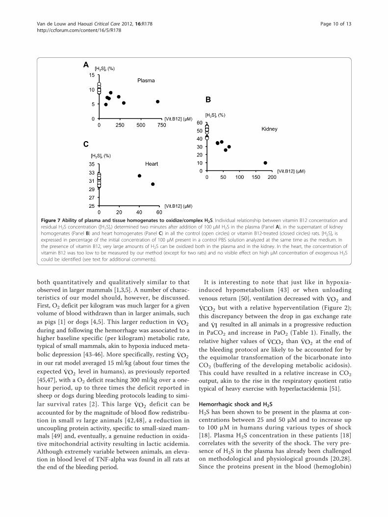
H2S concentrations in blood of 50,100 and 150 μΜdropped by 90, 92 and 75% respectively.Oxidation of 100 μM H2S by tissue homogenatesAs shown on Figure 7B, two minutes after adding 0.15 mlH2S to the supernatant of kidney homogenates to reach afinal concentration of 100 μM, residual H2S concentrationwas 50.2 ± 4.7 μM in control kidney homogenates and34.8 ± 12.9 μM in kidney homogenates of the animalswhich received vitamin B12 (P < 0.01). In the same condi-tions, residual H2S concentrations in heart homogenateswere 31.3 ± 0.9 and 30.5 ± 0.9 μM for control and vitaminB12-treated rats respectively (Figure 7C, NS). In vitaminB12-treated rats, hydroxocobalamin concentrations were47 ± 57 μM in kidney homogenates (Figure 7B), and
below the threshold of detection (30 μM) in heart homo-genates except for two animals (Figure 7C). Concentra-tions did not change in the PBS solutions.
Mortality and vitamin B12There was no difference in time to death (59 ± 26 vs 65 ±26 min) or mortality rates over time between vitamin B12and control rats (Figure 3).
Plasma TNF-alphaThere was very large intra-group variability but no sta-tistically significant difference in TNF-alpha plasmalevels between vitamin B12 treated and control rats (P =0.398, Table 1).
Figure 3 Survival rates. Panel A, survival rate (in %) in the control (open circles) and vitamin B12-treated rats (closed circles). Time zerocorresponds to the onset of the bleeding protocol. All rats survived the bleeding period (30 minutes), about 50% were still alive after 60minutes, and no rats survived after 120 minutes. There was no difference in the survival rates between the two groups (logrank test). Panel Bdisplays the individual survival times for the two groups. There was no significant difference in the mean survival time between control andvitamin B12 treated rats (65 ± 26 vs 59 ± 26 min).
Van de Louw and Haouzi Critical Care 2012, 16:R178http://ccforum.com/content/16/5/R178
Page 7 of 13

DiscussionIn major contrast to previous reports in humans [18]and in animal models [11], we did not observe anyincrease in blood H2S concentration in a model of lethaluntreated hemorrhagic shock in urethane anesthetizedrats, despite major O2 deficit, hyperlactacidemia and sys-temic inflammation. In addition, following the bleedingperiod, the plasma of every animal, as well as the super-natants from the heart and kidney kept a very high abil-ity of oxidizing/complex large (toxic) amounts of sulfide.Finally, following injection of a very large dose of vita-min B12, the ability to oxidize H2S by the plasma andthe kidney was enhanced in proportion to the local vita-min B12 concentration. However, the survival rate, O2
deficit or the various markers of the severity of theshock were not affected by the presence of μM levels ofhydroxocobalamin. These findings do not support thehypothesis that endogenous H2S does accumulate in theblood or in most tissues and contributes to the severityof hemorrhagic shock induced oxygen deficit [11,18].
Rat modelTo investigate the putative role of endogenous hydrogensulfide during hemorrhagic shock induced cellularhypoxia, we used the urethane-anesthetized rat as anexperimental model. Withdrawing 25 ml/kg of bloodwithin 30 minutes produced a dramatic reduction inABP, V̇O2 and V̇CO2 along with an increase in lacticacid and in the respiratory quotient ratio. This hemor-rhage protocol led to a fatal outcome in 50% of the ani-mals within one hour following the bleeding onset, whichparalleled the magnitude of O2 deficit and hyperlactaci-demia. All animals died within two hours. This relativelylow survival rate of hemorrhagic shock in rats comparedto larger [38,39] or non-anesthetized animals [40] is notunexpected. Indeed, not only anesthesia alone is likely toaffect the normal ability of the circulatory and respiratorysystems to respond to an acute reduction in volemia[31,41], but urethane, by itself, significantly blunts thenormal cardio-vascular regulation [42]. Nevertheless, thefatal outcome we observed in our study appears to be
Figure 4 Oxygen deficit in control vs treated rats. Panel A, time course of O2 deficit in four control (dashed lines) and six vitamin B12-treated(continuous lines) rats, from the onset of bleeding (time 0) to death. In all rats, O2 deficit accumulated continuously during the bleeding periodreaching 120 ml/kg at the end of the bleeding protocol (vertical arrow). Following the bleeding period, O2 deficit continued to accumulate in aramp-like fashion, while in the rats that survived much longer, O2 deficit remained constant. There was no significant difference in the timecourse of O2 deficit between control and vitamin B12-treated rats. Panel B, relationship between O2 deficit and plasma lactate levels at the endof bleeding periods in control (open circles) and vitamin B12-treated (closed circles) rats (r2 = 0.79).
Van de Louw and Haouzi Critical Care 2012, 16:R178http://ccforum.com/content/16/5/R178
Page 8 of 13

Figure 5 Absorbance of the plasma and H2S concentrations. Panel A, absorbance spectra of the plasma collected at the end of the overallbleeding period in control (open circles) and vitamin B12-treated (closed circles) rats. There was a clear peak of absorbance at 525 nm invitamin B12-treated rats corresponding to a vitamin B12 plasma concentration of 185 ± 216 μM/l. No peak was observed in any of the controlrats. At 670 nm, that is, the absorbance wavelength of the methylene blue, no peak was observed, neither in control nor in the vitamin B12-treated rats. Panel B, absorbance spectra, between 400 and 700 nm, of the plasma of the control rats, before (open triangles) and at the end ofthe bleeding period (closed squares). The observed absorbance values at 670 nm would theoretically correspond to a H2S concentration ofaround 4 μM/l in the dilute plasma (or 8 μM/l in the plasma, see text for additional comments), as illustrated using a control solution(phosphate-buffered saline, PBS) containing H2S (4 μM) (open diamonds). The lack of peak of absorbance in the plasma at 670 nm along withthe pattern of absorbance over the visible spectrum (continuous decrease of absorbance from 400 nm) strongly suggest that it is the turbidityof the medium which could account for this apparent presence of H2S in the plasma.
Figure 6 Plasma and H2S. Panel A, residual H2S concentration ([H2S]r), two minutes after addition of 100 μM H2S in the plasma of control(open circles) and vitamin B12-treated (closed circles) rats. [H2S]r is expressed in percentage of the concentration of 100 μM present in a controlPBS solution analyzed at the same time as the plasma. Within two minutes, H2S concentrations decreased by more than 90% in control andvitamin B12-treated rats respectively, with a significant difference between the two groups (*P < 0.05). Panel B, absorbance spectra of H2S addedto sham rat whole blood at 50, 100 and 150 μM and measured after two minutes. In the inset, the percentage of residual [H2S] ([H2S]r),corresponding to the absorbance at 670 nm, is shown for each initial concentration. Depending of the initial [H2S] ([H2S]i), exogenous H2Sconcentrations decreased between 92 and 75%.
Van de Louw and Haouzi Critical Care 2012, 16:R178http://ccforum.com/content/16/5/R178
Page 9 of 13
both quantitatively and qualitatively similar to thatobserved in larger mammals [1,3,5]. A number of charac-teristics of our model should, however, be discussed.First, O2 deficit per kilogram was much larger for a givenvolume of blood withdrawn than in larger animals, suchas pigs [1] or dogs [4,5]. This larger reduction in V̇O2
during and following the hemorrhage was associated to ahigher baseline specific (per kilogram) metabolic rate,typical of small mammals, akin to hypoxia induced meta-bolic depression [43-46]. More specifically, resting V̇O2
in our rat model averaged 15 ml/kg (about four times theexpected V̇O2 level in humans), as previously reported[45,47], with a O2 deficit reaching 300 ml/kg over a one-hour period, up to three times the deficit reported insheep or dogs during bleeding protocols leading to simi-lar survival rates [2]. This large V̇O2 deficit can beaccounted for by the magnitude of blood flow redistribu-tion in small vs large animals [42,48], a reduction inuncoupling protein activity, specific to small-sized mam-mals [49] and, eventually, a genuine reduction in oxida-tive mitochondrial activity resulting in lactic acidemia.Although extremely variable between animals, an eleva-tion in blood level of TNF-alpha was found in all rats atthe end of the bleeding period.
It is interesting to note that just like in hypoxia-induced hypometabolism [43] or when unloadingvenous return [50], ventilation decreased with V̇O2 and
V̇CO2 but with a relative hyperventilation (Figure 2);this discrepancy between the drop in gas exchange rateand V̇I resulted in all animals in a progressive reductionin PaCO2 and increase in PaO2 (Table 1). Finally, therelative higher values of V̇CO2 than V̇O2 at the end ofthe bleeding protocol are likely to be accounted for bythe equimolar transformation of the bicarbonate intoCO2 (buffering of the developing metabolic acidosis).This could have resulted in a relative increase in CO2
output, akin to the rise in the respiratory quotient ratiotypical of heavy exercise with hyperlactacidemia [51].
Hemorrhagic shock and H2SH2S has been shown to be present in the plasma at con-centrations between 25 and 50 μM and to increase upto 100 μM in humans during various types of shock[18]. Plasma H2S concentration in these patients [18]correlates with the severity of the shock. The very pre-sence of H2S in the plasma has already been challengedon methodological and physiological grounds [20,28].Since the proteins present in the blood (hemoglobin)
Figure 7 Ability of plasma and tissue homogenates to oxidize/complex H2S. Individual relationship between vitamin B12 concentration andresidual H2S concentration ([H2S]r) determined two minutes after addition of 100 μM H2S in the plasma (Panel A), in the supernatant of kidneyhomogenates (Panel B) and heart homogenates (Panel C) in all the control (open circles) or vitamin B12-treated (closed circles) rats. [H2S]r isexpressed in percentage of the initial concentration of 100 μM present in a control PBS solution analyzed at the same time as the medium. Inthe presence of vitamin B12, very large amounts of H2S can be oxidized both in the plasma and in the kidney. In the heart, the concentration ofvitamin B12 was too low to be measured by our method (except for two rats) and no visible effect on high μM concentration of exogenous H2Scould be identified (see text for additional comments).
Van de Louw and Haouzi Critical Care 2012, 16:R178http://ccforum.com/content/16/5/R178
Page 10 of 13

complex and/or catalyze very large amounts of sulfide[20], trivial levels of H2S, if any, are expected to befound in the plasma in baseline conditions, as shown byFurne et al. [28] and Whitfield et al. [20]. Whitfield etal. reported no measurable level of H2S [20] after addi-tion of 10 μM of H2S in rat blood before applying amethod similar to that used in the present study.Besides, the levels reported both in baseline conditionsand in shock [11,16,18] appear to be higher than thoseexpected to be found during severe H2S intoxication[52], wherein mitochondrial activity is inhibited.The method used to determine H2S in previous studies
[11,16,18] was developed by Siegel et al. [36] and relieson the transformation of one molecule of H2S and twomolecules of N, N-dimethyl-p-phenylenediamine intoone molecule of methylene blue (MB). H2S concentrationcan then be determined by measuring the absorbance ofthe solution at 670 nm (methylene blue). One of the lim-its of this method is directly related to the fact that theabsorbance is proportional to the concentration of agiven molecule - which color is the complementary ofthe light wave absorbed - if and only if none of the inci-dent light is scattered by dispersed particles or molecules.The presence of a minimal level of turbidity can alter theabsorbance of light at any wavelength irrespective of theactual “color” of the plasma, unless a genuine peak ofabsorbance can be found between 600 and 700 nm(Figure 5). We found that, even after application of TCAto remove the proteins and multiple centrifugations, asignificant absorbance can be found at 670 nm. Using abroader spectrum of wavelengths, one can show that theBeer-Lambert law cannot be applied to identify smallconcentrations of H2S in plasma. This is also illustratedin Figure 5 where the spectrum of absorbance of theplasma after reaction with the reagents to form MB isvery different from that of a solution containing H2S atthe hypothetical concentration corresponding to a similarabsorbance. These data suggest that if the method devel-oped by Siegel is to be used in the plasma, determinationof H2S concentrations based on the absorbance of lightat only 670 nm can be misleading, yielding to erroneousfindings of H2S. This issue has already been highlightedby Hughes et al., who reported that the linear depen-dence of absorbance on the MB concentration was onlyvalid for concentrations of H2S much lower than thosereported in all these studies; these authors also recom-mended the use of the spectra of absorbance between550 and 700 nm [53]. Using a different method, based onthe monobromobimane derivatization [54], Tokuda et al.reported H2S plasma concentrations ranging at bestbetween 2 to 4 μM, consistent with the present results,and more importantly that H2S levels decreased, if any-thing, in endotoxic shock in mice [55]. Volatilization ofH2S observed by De Leon et al. [56] when samples are
left in open air is unlikely to have occurred (see Methodand Result sections); since we took the precaution toentirely fill all the vials and to cap them immediately, toprevent any significant volatilization. The procedure usedfor plasma H2S measurements (including centrifugation)was applied as well to PBS solutions containing a knownamount on H2S (concentration 100 μM), and the level ofH2S was not affected. In addition, we previously foundthat using this procedure, H2S concentration (in PBS orsaline solution) remained stable with a few percent dropin concentration over one hour [30]. DeLeon et al.reported similar results when they took the precaution toclose their chambers [56]. For the determination of theability of the plasma and tissues to oxidize exogenousH2S, PBS solutions containing the same initial amount ofH2S were analyzed at the very same time as the plasmaor supernatant; H2S concentrations were unchanged inPBS within the two-minute period we chose for ourdetermination and all results have been expressed in per-centage of the concentration of 100 μM present in thePBS solution analyzed at the same time and following thesame procedure.
Endogenous H2S in the tissues, vitamin B12 andhemorrhagic shockThere is a spontaneous oxidation/complexation of H2Sin the plasma and the supernatant of tissues, which wasenhanced by μM concentrations of vitamin B12. Thelatter was obtained following intraperitoneal injection ofvitamin B12 at a dose used during cyanide intoxication(106 times the normal daily intake), as previouslyreported [46,57,58]. The kidney and heart were chosenas they are among the most important organs exposedto the consequences of hemorrhage-induced ischemia.In addition, our previous study [30] showed that theability of tissue homogenates of these organs to oxidizeH2S was clearly enhanced in vitamin B12-treated rats. Inthat previous study, we found that vitamin B12 couldoxidize large amounts of H2S in direct relation to itsconcentration, likely due to the presence of oxidizedcobalt [30]. We could establish that the presence of10 μM vitamin B12 was able to oxidize 20% of a 100 μMsolution of H2S; at 50 μM, about 80% of the H2S was oxi-dized within five minutes. This is consistent with ourpresent results, where vitamin B12 concentrations in theplasma were found to be about 180 μM and coulddecrease the exogenous H2S levels to 7.7 ± 0.8 μM(significantly lower than control plasma; initial concen-tration 100 μM), after two minutes. Similarly, about50 μM of hydroxocobalamin were found in our kidneyhomogenates, which in turn decreased H2S concentra-tions by 65% (vs only 50% in control; initial concentration100 μM). For the heart and, very likely, for some othertissues, the spectrophotometric method of detection of
Van de Louw and Haouzi Critical Care 2012, 16:R178http://ccforum.com/content/16/5/R178
Page 11 of 13
vitamin B12 was not sufficient to demonstrate the pre-sence of vitamin B12 (the threshold is about 30 μM [30]).Incidentally, pM - and not μM - concentrations of hydro-xocobalamin are expected to be present in the body[59,60]; therefore, with the methodology used in the pre-sent study we were unable to demonstrate whether lowμM concentrations of vitamin B12 were able to oxidizeH2S at the concentrations likely to be present in theheart [28]. This is a very important point to consider asour present results did not show that vitamin B12 wasabsent from the heart or could not oxidize sulfide, butwithin the very poor resolution of our method, no reli-able conclusion could be drawn.Hypoxic conditions have been proposed to decrease
H2S oxidation resulting in the accumulation of H2S [23].This increase in H2S concentrations may, however,occur only if PO2 decreases to extremely low levels [24],similar to those expected to be found in the vicinity ofthe mitochondria, suggesting to Olson [23,61] that thesite of action for endogenous H2S can only be the mito-chondria. Studies trying to establish the actual amountof H2S present and endogenously produced revealedthat at best nM changes in H2S concentrations can beobserved [46]. We speculate that low levels of vitaminB12 could still be able to decrease such concentration ofH2S in our study [30].The present study did not address the effect of reperfu-
sion, wherein production of cytokines and oxidativestress are prominent. Patients or animal models whoshowed an increase in plasma H2S concentrations [16,18]were resuscitated, and the question of the putative role ofendogenous H2S will need to be tested during the criticalperiod of reperfusion.
ConclusionsThere is no evidence that H2S can accumulate in the highmicromolar range in the blood and tissues (extravascularcompartment) during a lethal form of hemorrhagicshock. The presence of cobalt (hydroxocobalamin) didnot affect any of the outcomes of the shock. These resultsimply that H2S in the blood cannot be used as a markerof hemorrhagic shock. The hypothesis that H2S couldaccumulate during hemorrhagic induced tissular hypoxiamust be reconciled with the ability of tissues to oxidizeH2S.
Key messages• Even during a severe form of hemorrhagic shock inthe rat, where a major O2 deficit is present, there isno evidence for an increase in H2S concentration inthe blood.• Injection of high doses of hydroxocobalamin,although enhancing the ability of blood and kidneys
to oxidize exogenous H2S in vitro, does not improvethe survival, O2 deficit, lactacidemia or TNF-alphalevels in this model of shock.
AbbreviationsABP: arterial blood pressure; BTPS: body temperature and pressure saturated;f: breathing frequency; I.P.: intraperitoneal; MB: methylene blue; MOF:multiple organ failure; NS: non-significant; PBS: phosphate buffer saline;STPD: standard temperature and pressure: dry; TCA: trichloroacetic acid; V̇ :inspiratory flow; V̇I : minute ventilation; V̇CO2 : carbon dioxide production;V̇O2 : oxygen uptake; VT: tidal volume
Authors’ contributionsAV and PH conceived of the study, performed the animal experiments,analyzed the data and drafted the manuscript. All authors read andapproved the final manuscript.
Competing interestsThe authors declare that they have no competing interests.
Received: 30 July 2012 Revised: 7 September 2012Accepted: 2 October 2012 Published: 2 October 2012
References1. Rixen D, Raum M, Holzgraefe B, Sauerland S, Nagelschmidt M,
Neugebauer EA: A pig hemorrhagic shock model: oxygen debt andmetabolic acidemia as indicators of severity. Shock 2001, 16:239-244.
2. Rixen D, Siegel JH: Bench-to-bedside review: oxygen debt and itsmetabolic correlates as quantifiers of the severity of hemorrhagic andpost-traumatic shock. Crit Care 2005, 9:441-453.
3. Siegel JH, Fabian M, Smith JA, Kingston EP, Steele KA, Wells MR, Kaplan LJ:Oxygen debt criteria quantify the effectiveness of early partialresuscitation after hypovolemic hemorrhagic shock. J Trauma 2003,54:862-880, discussion 880.
4. Dunham CM, Siegel JH, Weireter L, Fabian M, Goodarzi S, Guadalupi P,Gettings L, Linberg SE, Vary TC: Oxygen debt and metabolic acidemia asquantitative predictors of mortality and the severity of the ischemicinsult in hemorrhagic shock. Crit Care Med 1991, 19:231-243.
5. Crowell JW, Smith EE: Oxygen deficit and irreversible hemorrhagic shock.Am J Physiol 1964, 206:313-316.
6. Shoemaker WC, Appel PL, Kram HB: Role of oxygen debt in thedevelopment of organ failure sepsis, and death in high-risk surgicalpatients. Chest 1992, 102:208-215.
7. Dewar D, Moore FA, Moore EE, Balogh Z: Postinjury multiple organ failure.Injury 2009, 40:912-918.
8. Yao YM, Redl H, Bahrami S, Schlag G: The inflammatory basis of trauma/shock-associated multiple organ failure. Inflamm Res 1998, 47:201-210.
9. Jarrar D, Chaudry IH, Wang P: Organ dysfunction following hemorrhageand sepsis: mechanisms and therapeutic approaches (Review). Int J MolMed 1999, 4:575-583.
10. Jastrow KM, Gonzalez EA, McGuire MF, Suliburk JW, Kozar RA, Iyengar S,Motschall DA, McKinley BA, Moore FA, Mercer DW: Early cytokineproduction risk stratifies trauma patients for multiple organ failure. J AmColl Surg 2009, 209:320-331.
11. Mok YY, Atan MS, Yoke Ping C, Zhong Jing W, Bhatia M, Moochhala S,Moore PK: Role of hydrogen sulphide in haemorrhagic shock in the rat:protective effect of inhibitors of hydrogen sulphide biosynthesis. Br JPharmacol 2004, 143:881-889.
12. Mok YY, Moore PK: Hydrogen sulphide is pro-inflammatory inhaemorrhagic shock. Inflamm Res 2008, 57:512-518.
13. Wagner F, Asfar P, Calzia E, Radermacher P, Szabo C: Bench-to-bedsidereview: hydrogen sulfide - the third gaseous transmitter: applications forcritical care. Crit Care 2009, 13:213.
14. Collin M, Anuar FB, Murch O, Bhatia M, Moore PK, Thiemermann C:Inhibition of endogenous hydrogen sulfide formation reduces theorgan injury caused by endotoxemia. Br J Pharmacol 2005,146:498-505.
Van de Louw and Haouzi Critical Care 2012, 16:R178http://ccforum.com/content/16/5/R178
Page 12 of 13

15. Collin M, Thiemermann C: Hydrogen sulfide and sulfite: novel mediatorsin the pathophysiology of shock and inflammation. Shock 2005,24:595-596.
16. Li L, Bhatia M, Zhu YZ, Zhu YC, Ramnath RD, Wang ZJ, Anuar FB,Whiteman M, Salto-Tellez M, Moore PK: Hydrogen sulfide is a novelmediator of lipopolysaccharide-induced inflammation in the mouse.FASEB J 2005, 19:1196-1198.
17. Hui Y, Du J, Tang C, Bin G, Jiang H: Changes in arterial hydrogen sulfide(H(2)S) content during septic shock and endotoxin shock in rats. J Infect2003, 47:155-160.
18. Goslar T, Mars T, Podbregar M: Total plasma sulfide as a marker of shockseverity in nonsurgical adult patients. Shock 2011, 36:350-355.
19. Olson KR, Healy MJ, Qin Z, Skovgaard N, Vulesevic B, Duff DW, Whitfield NL,Yang G, Wang R, Perry SF: Hydrogen sulfide as an oxygen sensor in troutgill chemoreceptors. Am J Physiol Regul Integr Comp Physiol 2008, 295:R669-680.
20. Whitfield NL, Kreimier EL, Verdial FC, Skovgaard N, Olson KR: Reappraisal ofH2S/sulfide concentration in vertebrate blood and its potentialsignificance in ischemic preconditioning and vascular signaling. Am JPhysiol Regul Integr Comp Physiol 2008, 294:R1930-1937.
21. Peng YJ, Nanduri J, Raghuraman G, Souvannakitti D, Gadalla MM, Kumar GK,Snyder SH, Prabhakar NR: H2S mediates O2 sensing in the carotid body.Proc Natl Acad Sci USA 2010, 107:10719-10724.
22. Olson KR: Hydrogen sulfide and oxygen sensing: implications incardiorespiratory control. J Exp Biol 2008, 211:2727-2734.
23. Olson KR: Hydrogen sulfide is an oxygen sensor in the carotid body.Respir Physiol Neurobiol 2011, 179:103-110.
24. Van de Louw A, Haouzi P: Inhibitory effects of hyperoxia andmethemoglobinemia on H(2)S induced ventilatory stimulation in the rat.Respir Physiol Neurobiol 2012, 181:326-334.
25. Haouzi P: Sulfide and methemoglobinemia. Respir Physiol Neurobiol 2011,179:119-120.
26. Haouzi P, Bell H, Philmon M: Hydrogen sulfide oxidation and the arterialchemoreflex: effect of methemoglobin. Respir Physiol Neurobiol 2011,177:273-283.
27. Hegde A, Bhatia M: Hydrogen sulfide in inflammation: friend or foe?Inflamm Allergy Drug Targets 2011, 10:118-122.
28. Furne J, Saeed A, Levitt MD: Whole tissue hydrogen sulfideconcentrations are orders of magnitude lower than presently acceptedvalues. Am J Physiol Regul Integr Comp Physiol 2008, 295:R1479-1485.
29. Haggard HW: The fate of sulfides in the blood. J Biol Chem 1921,49:519-529.
30. Van de Louw A, Haouzi P: Ferric iron and cobalt (III) compounds to safelydecrease H2S in the body? Antioxid Redox Signal 2012.
31. Haouzi P, Bell H, Van de Louw A: Hypoxia-induced arterial chemoreceptorstimulation and hydrogen sulfide: too much or too little? Respir PhysiolNeurobiol 2011, 179:97-102.
32. Chai W, Wang Y, Lin JY, Sun XD, Yao LN, Yang YH, Zhao H, Jiang W,Gao CJ, Ding Q: Exogenous hydrogen sulfide protects against traumatichemorrhagic shock via attenuation of oxidative stress. J Surg Res 2011,176:210-219.
33. Morrison ML, Blackwood JE, Lockett SL, Iwata A, Winn RK, Roth MB:Surviving blood loss using hydrogen sulfide. J Trauma 2008, 65:183-188.
34. Ganster F, Burban M, de la Bourdonnaye M, Fizanne L, Douay O, Loufrani L,Mercat A, Cales P, Radermacher P, Henrion D, Asfar P, Meziani F: Effects ofhydrogen sulfide on hemodynamics, inflammatory response andoxidative stress during resuscitated hemorrhagic shock in rats. Crit Care2010, 14:R165.
35. Drabek T: Hydrogen sulfide-curiouser and curiouser! Crit Care Med 2012,40:2255-2256.
36. Siegel LM: A direct microdetermination for sulfide. Anal Biochem 1965,11:126-132.
37. Peto R, Pike MC, Armitage P, Breslow NE, Cox DR, Howard SV, Mantel N,McPherson K, Peto J, Smith PG: Design and analysis of randomizedclinical trials requiring prolonged observation of each patient. II. analysisand examples. Br J Cancer 1977, 35:1-39.
38. Taylor JH, Beilman GJ, Conroy MJ, Mulier KE, Myers D, Gruessner A,Hammer BE: Tissue energetics as measured by nuclear magneticresonance spectroscopy during hemorrhagic shock. Shock 2004, 21:58-64.
39. Drabek T, Kochanek PM, Stezoski J, Wu X, Bayir H, Morhard RC, Stezoski SW,Tisherman SA: Intravenous hydrogen sulfide does not induce
hypothermia or improve survival from hemorrhagic shock in pigs. Shock2011, 35:67-73.
40. Crippen D, Safar P, Snyder C, Porter L: Dying pattern in volume-controlledhemorrhagic shock in awake rats. Resuscitation 1991, 21:259-270.
41. Van der Linden P, Gilbart E, Engelman E, Schmartz D, de Rood M,Vincent JL: Comparison of halothane, isoflurane, alfentanil, and ketaminein experimental septic shock. Anesth Analg 1990, 70:608-617.
42. Armstrong JM, Lefevre-Borg F, Scatton B, Cavero I: Urethane inhibitscardiovascular responses mediated by the stimulation of alpha-2adrenoceptors in the rat. J Pharmacol Exp Ther 1982, 223:524-535.
43. Mortola JP: Implications of hypoxic hypometabolism during mammalianontogenesis. Respir Physiol Neurobiol 2004, 141:345-356.
44. Gautier H: Interactions among metabolic rate, hypoxia, and control ofbreathing. J Appl Physiol 1996, 81:521-527.
45. Frappell P, Lanthier C, Baudinette RV, Mortola JP: Metabolism andventilation in acute hypoxia: a comparative analysis in small mammalianspecies. Am J Physiol 1992, 262:R1040-1046.
46. Haouzi P: Ventilatory and metabolic effects of exogenous hydrogensulfide. Respir Physiol Neurobiol 2012.
47. Shepherd RE, Gollnick PD: Oxygen uptake of rats at different workintensities. Pflugers Arch 1976, 362:219-222.
48. Shepherd AP, Pawlik W, Mailman D, Burks TF, Jacobson ED: Effects ofvasoconstrictors on intestinal vascular resistance and oxygen extraction.Am J Physiol 1976, 230:298-305.
49. Janssen BJ, De Celle T, Debets JJ, Brouns AE, Callahan MF, Smith TL: Effectsof anesthetics on systemic hemodynamics in mice. Am J Physiol HeartCirc Physiol 2004, 287:H1618-1624.
50. Haouzi P, Bell HJ: Respiratory effects of changing the volume loadimposed on the peripheral venous system. Respir Physiol Neurobiol 2010,171:175-180.
51. Beaver WL, Wasserman K, Whipp BJ: A new method for detectinganaerobic threshold by gas exchange. J Appl Physiol 1986, 60:2020-2027.
52. Kage S, Kashimura S, Ikeda H, Kudo K, Ikeda N: Fatal and nonfatalpoisoning by hydrogen sulfide at an industrial waste site. J Forensic Sci2002, 47:652-655.
53. Hughes MN, Centelles MN, Moore KP: Making and working with hydrogensulfide: The chemistry and generation of hydrogen sulfide in vitro andits measurement in vivo: a review. Free Radic Biol Med 2009, 47:1346-1353.
54. Wintner EA, Deckwerth TL, Langston W, Bengtsson A, Leviten D, Hill P,Insko MA, Dumpit R, VandenEkart E, Toombs CF, Szabo C: Amonobromobimane-based assay to measure the pharmacokinetic profileof reactive sulphide species in blood. Br J Pharmacol 2010, 160:941-957.
55. Tokuda K, Kida K, Marutani E, Crimi E, Bougaki M, Khatri A, Kimura H,Ichinose F: Inhaled hydrogen sulfide prevents endotoxin-inducedsystemic inflammation and improves survival by altering sulfidemetabolism in mice. Antioxid Redox Signal 2012, 17:11-21.
56. Deleon ER, Stoy GF, Olson KR: Passive loss of hydrogen sulfide inbiological experiments. Anal Biochem 2012, 421:203-207.
57. Truong DH, Mihajlovic A, Gunness P, Hindmarsh W, O’Brien PJ: Preventionof hydrogen sulfide (H2S)-induced mouse lethality and cytotoxicity byhydroxocobalamin (vitamin B(12a)). Toxicology 2007, 242:16-22.
58. de La Coussaye JE, Houeto P, Sandouk P, Levillain P, Sassine A, Riou B:Pharmacokinetics of hydroxocobalamin in dogs. J Neurosurg Anesthesiol1994, 6:111-115.
59. Slot WB, Merkus FW, Van Deventer SJ, Tytgat GN: Normalization of plasmavitamin B12 concentration by intranasal hydroxocobalamin in vitaminB12-deficient patients. Gastroenterology 1997, 113:430-433.
60. Linnell JC, Mackenzie HM, Wilson J, Matthews DM: Patterns of plasmacobalamins in control subjects and in cases of vitamin B12 deficiency. JClin Pathol 1969, 22:545-550.
61. Olson KR: “Hydrogen sulfide oxidation and the arterial chemoreflex:effect of methemoglobin” by Haouzi et al. [Respir. Physiol. Neurobiol.(2011)]. Respir Physiol Neurobiol 2011, 179:121, author reply 119-120.
doi:10.1186/cc11661Cite this article as: Van de Louw and Haouzi: Oxygen deficit and H2S inhemorrhagic shock in rats. Critical Care 2012 16:R178.
Van de Louw and Haouzi Critical Care 2012, 16:R178http://ccforum.com/content/16/5/R178
Page 13 of 13