oxidative phosphorylation and mitochondrial · pdf fileoxidative phosphorylation and...
TRANSCRIPT
Physiol. Chem. &Physics 13 (1981)
OXIDATIVE PHOSPHORYLATION AND MITOCHONDRIAL PHYSIOLOGY: A CRITICAL REVIEW OF CHEMIOSMOTIC THEORY, AND REINTERPRETATION BY THE ASSOCIATION-INDUCTION HYPOTHESIS
- - GILBERT N. LING Department of Molecular Biology, Pennsylvania Hospital, Eighth and Spruce Streets, Phila- delphia, Pennsylvania 19107
Fur7damental assumptions o f the chen~iosmotic hypothesis o f Mitchell are examined. Cornparis017 o f these assumptions with experimental data accunzulated over the past f i f ty years leads to the cotzclusion that the hypothesis has not been supported. A review o f important findings concerning the physical state of the major ir~tracellular cation potassium shows clearly that this ion does not exist in a free state but is adsorbed on specific anionic sites. These findings refute the membrane-pump theory but add powerful support for the association-i~~ductior? hypothesis, on the basis o f which a new mechanisrn o f oxidative phosphorylation as well as a wide variety o f rnitochondrial behaviors are proposed and compared with experimet7tal data.
SUBJECT OUTLINE
I. INTRODUCTION
11. THE CHEMIOSMOTIC HYPOTHESIS
A. Generation of an electrical potential difference by postulated electrogenic pump vs. law of macroscopic electroneutrality
B. Proton gradient
C. Electrical potential gradient
1 . In vivo measurements a. Membrane disruption b. Electrode misplacement in the intermembrane space c. Inpocketing of mitochondrial membrane at microelectrode tip
2. In situ measurements
3. Effect of valinomycin on resistance
D. Is the mitochondrial inner membrane impermeable?
1. Is there enough phospholipid in the inner membrane to form a continu- ous bilayer barrier?
2. Is the mitochondrial inner membrane virtually impermeable to ions in general and H+ in particular?
a. H+ permeability

G. N. LING
b. Cation impermeability ( I ) Inner membrane permeability to K+ and Nu+. (2) Inner mem-
brane permeability to divalent cations. c. Anion impermeability
E. Functions of uncoupling agents and ionophores
F. ATP synthesis
G . Conclusions regarding the chemiosmotic hypothesis
111. THEORY O F THE LIVING CELL
A. Basic features of the membrane-pump theory ( I ) Water. (2) Ions. (3) Cell surface barrier. (4) Membrane pumps. (5) Cell
volume. (6) Resting potential.
B. Basic features of the association-induction hypothesis ( I ) Water. (2) Ions. (3) Cell surface barrier. (4) Pumps and solute exclusion.
(5) Cell volume. (6) Resting potential.
C. Discriminatory experimental evidence
1. Energy requirements of pumps
2. Membrane vs. cytoplasm as the seat of discrimination in solute distribu- tion
3. The adsorbed state of K+
4. Consequences of K+ binding in living cells
IV. THE SOURCE OF ENERGY FOR ATP SYNTHESIS
V. PROTEINS ACCORDING TO THE A1 HYPOTHESIS : HIGH- AND LOW- ENERGY STATES OF PROTEIN-ION-WATER SYSTEMS. PRIMARY INDUCTIVE EFFECT
A. Inductive effect as the basis of energy and information transfer over distance
1. Universal applicability of the inductive effect
2. Target groups of the inductive effect
a. The acid dissociation constant (or pK,) b. H-bonding strength c. Oxidation-reduction potential
3. The inducing "groups"
4. Additivity and reversibility of the inductive effect
B. c-Value concept, linear model, and theoretically calculated change in pref- erence for K+, Na+, H+, and NH4+
C. Complex interaction between biologically active agents
D. Cooperative behavior of living cells: interpretation based on the A1 hy- pothesis
1. Cooperative adsorption isotherm of Yang and Ling
G. N. LING
2. Oxygen dissociation of hemoglobin and related phenomena
a. Empirical analysis (Hill's equation) b. The AAKM theories c. The Yang-Ling model: comparison with other models
3. Three types of autocooperative adsorption on proteins
E. Autocooperativity in selective solute accumulation in living cells
F. Control by cardinal adsorbents of shifts between two cooperative states
VI. TENTATIVE MODEL O F ASSOCIATIVE-INDUCTIVE COUPLING MECHANISM FOR ELECTRON TRANSPORT AND OXIDATIVE PHOSPHORYLATION
A. The coupling mechanism hypothesis
B. Comparison with other models
1. Heme-heme interaction and the Bohr effect
2. Oxidation-reduction controlled autocooperative ion adsorption shifts
a. Cytochrome c b. Hemoglobin c. Pyridine nucleotides
VII. INTERPRETATION O F OTHER MITOCHONDRIAL PROPERTIES UNDER THE A1 HYPOTHESIS
A. Additional basic A1 concepts
1. Physical state of the bulk of cell water in living cells
2. Swelling and shrinking
3. Polarized water rather than membrane-lipid as seat of selective permea- bility
4. Resting potential as surface adsorption potential
B. Important findings in mitochondrial physiology and some new interpreta- tions
1. Swelling and its reversal by ATP a. Type I. Simple swelling and shrinkage b. Type 11. Dissociative swelling c. Type 111. Depolarizing swelling
2. Reappraisal of previous reports according to the A1 hypothesis a. ATP and swelling/shrinkage of fetal and adult rat liver mitochondria b. Mg2+ and C a w vs. swelling/shrinkage c. Passive osmotic swelling
3. Ion and substrate transport
4. Uncouplers, ionophores, Ca2+, Mg2+, ATP, and other agents as cardinal adsorbents
a. Mechanism of action of valinomycin on mitochondrial K+ accumula- tion

b. Ionophores, uncouplers, and other cardinal adsorbents: induction of c-value change as basis of their action on mitochondrial ion dis- tribution (I) Respiration and anaerobiosis. (2) "Ionophores." (3) Uncouplers.
(4) Thiol reagents. (5) ATP and ADP.
5. Synchronous oscillatory changes in mitochondrial swelling, ion uptake, and other properties
6. Mitochondria1 electrical potential

G . N. LING 3 3
I. INTRODUCTION
Living cells, the basic units of all life, are at once extremely complex and exquisitely coherent. Acquiring understanding of the liv- ing cell, small though it is, resembles solving a gigantic multidimensional crossword puzzle into which, instead of words, appropriate phy- sical mechanisms must be inserted. But in this effort, two main obstacles continue to face the cell physiologist. First, the correct physi- cal mechanisms must be known-yet many remain unknown. Second, the task is so vast that it requires division of labor among many scientists over long periods, with each spe- cialized investigation adopting a conceptual foundation dictated by what at the time ap- pears to be the most trustworthy evidence- recent as well as "classic."
A dominant theory of the physico-chemical nature of the living cell is the membrane or membrane-pump theory, proposed more than a century ago by W. Pfeffer.l For many good reasons this theory has been broadly ac- cepted. However, the development of modern physics as well as various findings made pos- sible by powerful new tools have led numbers of scientists to conclude that some of those once convincing reasons are in fact wrong or equivocal. New basic concepts concerning the structure and function of the living cell are being forged, including the association-induc- tion hypothesis (A1 hypothesis) first eluci- dated in 1962.2 Meanwhile, much progress was made in all disciplines concerned with
- cell physiology. Of the many pertinent areas of specialization, mitochondrial physiology is now one of the most significant because of both the considerable volume of outstanding work in that field and the central importance of oxidative phosphorylation to cell viability. But such advances have so far been viewed predominantly in accordance with the long- held membrane-pump theory. The present paper interprets some of the major recent developments in the physiology of the cell in
terms of the association-induction hypothesis. In particular, the work on mitochondria is given attention. (For additional background, see review, ref. 114.)
11. THE CHEMIOSMOTIC HYPOTHESIS
Since Mitchell first proposed the chemios- motic hypothesis in 1961, it has been re- viewed many times"-'%nd therefore will be described only briefly here. According to this hypothesis, the inner membrane of a mito- chondrion is impermeable to H + as well as to other ions. The respiratory chain is ar- ranged in three loops corresponding to the three coupling sites. By a special vectorial arrangement of the electron-carrying mole- cules, an H+-adsorbing reaction occurs on the inside surface of the inner mitochondrial membrane (i.e., the surface facing the mat- rix). A concomitant H+-releasing reaction occurs on the outside surface of that mem- brane. As a result, an H + gradient develops, with the higher concentration of H+ on the outside of the inner membrane. A reversible ATPase complex on the mitochondrial inner membrane is located in a region impermeable to water but accessible to OH- from. one side of the inner membrane and accessible to H + from the other side. Thus ATP hydrolysis would be reversibly coupled to the transloca- tion of OH- ions across the system with a stoichiometry of one OH- translocated per ATP molecule hydrolyzed. The proton gradi- ent provides energy for synthesis of ATP.
Mitchell noted, however, that the H+ gradient alone is not sufficient to supply the energy required for ATP synthesis. Hence the mechanism was further elaborated by hypothesizing the creation of an electrical po- tential across the inner membrane as a result of asymmetrical electron transport. Mitchell argued that "the sum of the electrical poten- tial difference and the osmotic pressure differ- ence" provides a "protomotive force" (PMF)

34 G. N. LING
for ATP synthesis and other energy-consum- electroneutrality. Under that precept it is not ing processes. possible to achieve a net separation of a
The major share of this PMF is attributed chemically detectable quantity of charges be- specifically to a postulated membrane poten- tween two macroscopic phases (i.e., inside tial of 210 to 270 mV, with the inside phase phase and outside phase of a cell or mito- negative; and a small contribution to the PMF chondrion) without creating enormous po- is attributed to a postulated pH differen~e.~ tential differences.
For example, a single isolated rat liver
A. Generation of an Electrical Potential Difference by Postulated Electrogenic Pump vs. Law of Macroscopic Electroneutrality
According to the chemiosmotic hypothesis, the mitochondrial inner membrane is a meta- bolically driven charge-separating machine- or, to use a term often employed by electro- physiologists, an electrogenic pump. Mitchells
calculated that the translocation of as much as one milli-equivalent (or 10-"ram ions) of net ionic charges from the inside phase to the outside phase across the mitochondrial membrane would create a macroscopic po- tential difference of only 100 mV-a startling conclusion!
Consider that in his noted treatise on thermodynamics, Guggenheim14 posed this question: How big a potential difference would be created across the surface of a spherical body with a radius equal to 1 cm (i.e., m) and loaded with a quantity of electrical charge of only 10-lo gram ions? This quantity is so minute that it is not mea- surable by ordinary chemical methods, but it is demonstrably equal to 10-lo X 0.965 X lO%oulombs, or 0.96 x coulombs.
Now, in a vacuum the permissivity (cO) is 1.1 1 X 1 0-lo coulombs/volt m. The voltage of the charged sphere, then, is
There we have a quantitative illustration of the law of the conservation of macroscopic
mitochondrion suspended in alightly buffered 0.25 M aqueous sucrose solution with a di- electric constant of about 100 and maintained in the State 4 "orthodox" conformation is roughly spherical and about 1 pm in diameter. If there were a net charge separa- tion of only 10-lo gram ions as in Guggen- heim's calculation, the electrical potential dif- ference would be
If there were a charge separation of gram ions, as assumed by Mi t~he l l ,~ the po- tential would be 10'"-tremendous com- pared to Mitchell's finding of 100 mV!
We see that there are at least two errors in Mitchell's calculation. The first is improper use of the thermodynamic equation for the chemical potential of a single ion in a macro- scopic phase, implying thdt electrical charges can move from one macroscopic phase to an- other without regard to the law of electro- neutrality. The second error arises from equating potential difference across each mit- ochondrion with potential difference across an aggregate of mitochondria in one liter of '
solution, thereby in effect artificially creating a mitochondrion of gigantic dimensions. This huge figment, of course, can have no real significance.
Steady electrical potential differences do in fact occur at the interface between two unlike phases; e.g., between solid and liquid, liquid and liquid, liquid and gas. The magni- tude of such a difference is usually determined by ( I ) the difference in entropy (or more

G . N. LING 3 5
exactly, in statistical mechanical terms, the "partition functions") of the charge-carrying particles (free electrons or ions) in the two phases, and (2) the kinetic energy of the particles at the given temperature. The phase that offers greater entropy acquires an excess of one of the charge-carrying particles, and as a result an electrical potential develops. This is the basic mechanism for surface ad- sorption potentials, diffusion potentials, and the like.
The "electrogenic potential," on the other hand, has a different genesis. It was postu- lated not according to a consistent logical system based on knowledge of the inanimate world but as a sort of a catch-all for the dis- crepancies between the potential predicted by conventional membrane-pump theory and the poteritial experimentally observed. Thus Ker- nan15 wrote in his review on the electrogenic pump: "The electrogenic pumping of ions may be recognized by a change of the mem- brane potential which cannot be accounted for in terms of the passive ion movement and which has some characteristics of metabolic processes." In short, one cannot refrain from viewing the hypothesis of an electrogenic pump as yet another ad hoc postulation, with a heavy vitalistic accent. ,
B. Proton Gradient
Data derived from in vitro studies with the aid of 5,5-dimethyl-2,3-oxazolidinedione (DMO) led Addanki et al.16 to the conclu- - sion that the change of pH gradient across the inner membrane of mitochondria at State 4 respiration and uncoupled by 2,4-dinitrophe- no1 is only 0.005 pH units. That finding was confirmed in essence by Rothenberg.17 The latter author concluded that in Mitchell's model the protomotive force must be due pri- marily to the membrane potential.
Of course, Mitchell's hypothesis was di- rected primarily at isolated mitochondria and chloroplasts. Yet if the chemiosmotic ap-
proach is to have physiological significance, it must take into consideration the vast differ- ences between the highly artificial 0.25 M
sucrose solution and the natural environment of the mitochondrion; i.e., the cytoplasm of the parent cell. Cytoplasm contains a high concentration of pH buffers including solutes such as the various phosphorylated inter- mediates and, more importantly, proteins and other macromolecules. A rise in proton con- centration demonstrable in a suspension of mitochondria in a lightly buffered 0.25 M
sucrose solution does not automatically pre- dict a similar proton concentration change when the mitochondria are in their natural environment with its inherently high buffer capacity.
Isolated mitochondria utilizing such sub- strates as succinate and p-hydroxybutyrate are also different from mitochondria in their natural environment. One major natural en- ergy source for mitochondria is glucose, and the tricarboxylic acid cycle enzymes essential for degradation of glucose are situated inside the mitochondrial matrix, within the inner membrane (ref. 18, p. 512). Like the elec- tron transport system, these enzymes (i.e., isocitrate, a-ketoglutarate, malate dehydro- genase) produce H+, which therefore would increase H+ activity at the inside of the inner membrane and thus reduce the gradient of protons created by unidirectional spilling to the outside of the mitochondrial inner mem- brane, as stipulated in the chemiosmotic hypothesis.
C. Electrical Potential Gradient
Since a sizeable pH gradient is unlikely to contribute to the protomotive force, an electrical potential difference must exist across the inner membrane that can by itself provide the energy needed for ATP forma- tion, ion transport, and transhydrogenation reactions. According to Mitchell, a potential difference of about 210 to 270 mV (inside

negative) would serve this p ~ r p o s e . ~ - ~ , ~ Electrical potential differences across the
inner membranes of isolated mitochondria have been measured by a variety of methods. Unfortunately, many of these measurements were indirect, and their validity relied upon the assumptions of either Mitchell's own hypothesis or some other hypotheses.
1. In Vivo Measurements
Obviously, the least disputable way of as- sessing the magnitude and polarity of this potential difference is to measure it directly. To do so, the inside of the mitochondrion must be electrically tapped, as done by Tup- per and TedeschilVn the late 1960s using essentially the Ling-Gerard glass capillary microelectrode technique2" and the giant chromosomes of fruit flies. Tupper and Tedeschi showed that the measured electrical potential difference was only 10 to 20 mV in magnitude and that the polarity was oppo- site to that hypothesized by Mitchell. Further, the potential was not affected by metabolic inhibitors or oxidative phosphorylation un- couplers. Also, they reported that resistance across the inner membrane was only 1 to 4
cm2. They concluded that electrical poten- tial cannot play a major role in oxidative phosphorylation.
In 1977, Maloff, Scordilis, Reynolds, and Tede~chi"~~9eported another series of elec- tric potential studies. This time the giant mitochondria from mice fed with cuprazone were used. Although grossly oversized, these mitochondria carry out normal oxidative phosphorylation (P :O ratio for succinate was about 1.6). Again an electrical potential dif- ference of the mitochondria of 15 mV was observed, again with a polarity opposite to that demanded by the chemiosmotic hypo- thesis. And again the resistance was about 2 n cm2, and the potential was indifferent to the presence of succinate, antimycin A, or FCCP.
The iconoclastic import of these findings, in effect torpedo~ng Mitchell's hypothesis, de- manded close scrutiny of their foundation. In- deed, strong criticisms, directed primarily at Tupper and Tedeschi lbere voiced, as fol- lows.
a. MEMBRANE DISRUPTION. Lieberman and S k u l a ~ h e v , ~ ~ l s o Skulachev,12 suggested that the microelectrode technique is unsuit- able for measuring the electrical potential in so small and fragile structure as the isolated mitochondrion. In support they pointed out that the inner mitochondria1 membrane has in fact a very high resistance, citing Mit- c h e l l ' ~ ~ as well as their own calculated value of lo7 to loD a cm2. They considered that the much !ower resistance measured by Tup- per and Tedeschi (1 to 4 a cm2) confirmed their view that the mitochondria when im- paled by the microelectrode became grossly damaged. Another kind of evidence cited in favor of Lieberman and Skulachev's inter- pretation came from Lassen et al., who found that impalement of Ehrlich ascites cells24 and giant red cells of the Conger eeI2%ften leads to rapid depolarization ( t% = 1 msec) and consequent loss of sensitivity to external K+ concentration.
Rebuttal: Maloff, Scordilis, and T e d e ~ c h i ~ ~ presented the following rebuttal: ( I ) In the studies by Lassen et a1.,24,25 the rapid decline in electrical potential must have been due to a technical defect because other investigators were able to demonstrate much more stable potentials in both Ehrlich ascites cells ( t H = , 10 sec)" and Conger eel red cells (seconds to 1 min).2Woreover, in the giant mito- chondria of cuprazone-fed mice the electrical =
potential does not decay at all but remains constant, as has been observed in many other types of small cells.22 (2) Equally convincing evidence that no serious damage was pro- duced by the impaling microelectrode was the finding by Maloff et aLZ2 that exposure to valinomycin changed the polarity of the elec- trical potential and conferred a K+ sensitivity

G. N. LING 37
on the electrical potential of the cuprazone- treated mitochondria. Still other data support- ing Tedeschi, Tupper, Maloff, and their co- workers' view will be discussed below (p. 37) in relation to measuring the electrical poten-
- tial of mitochondria in situ. b. ELECTRODE MISPLACEMENT IN THE
INTERMEMBRANE SPACE. sku lac he^^^ sug- gested that the microelectrode tip might not have penetrated the inner membrane but only the outer membrane.
Rcb~lttal: Maloff et a1.22 refuted that sug- gestion in several ways, only two of which need be mentioned: (I) Advancement of a microelectrode will register an unchanging potential as long as the microelectrode is within the mitochondrion. (2) Impalement of mitochondria freed from their outer mem- branes (i.e., mitoplasts) gives the same po- tential as that from intact mitochondria.
c. INPOCKETING OF MITOCHONDRIAL MEM- BRANE AT MICROELECTRODE TIP. Rothen- bergm suggested that the valinomycin-sensi- tive electrical potential measured by Maloff et al. might have been artificially created when the microelectrode tip still outside the inner membrane was pushed against it, cre- ating a selectively permeable membrane sur- rounding a KC1-filled microelectrode tip that would then function as a K + electrode.
Rebuttal: Maloff et aLZ6 pointed out that if the microelectrode tip, enclosed by a valino- mycin-treated inner membrane, did indeed function as a K+ electrode, the potential should not develop when the microelectrode is filled with 2 M NaCl instead of 2 M KC], since valinomycin is not an ionophore for Na+. The opposite, however, was observed to be the case. NaCl- or KC1-filled microelec- trodes measured the same potential differ- e n ~ e . ~ ~ (For a comprehensive recent review, see Tede~chi .3~)
2. In Situ Measurements
The conclusions of Tedeschi, Tupper, and Maloff are further strengthened by the ele-
gant studies of Giulian and D i a c u m a k u ~ . ~ ~ These authors were able to measure the elec- trical potential of cultured HeLa cells with the electrode tip just inside the cell surface and also following the tip's penetration into the Golgi apparatus, nucleus, and mitochon- drion. Using thorium dioxide as a marker, they clearly established the placement of the microelectrode tip in the mitochondrial mat- rix. Yet the potential measured between the inside of the mitochondrion and the cyto- plasm was some 15 mV, with the inside negative.
The low negative potential thus measured offers general confirmation of the in vitro measurements of Tupper, Maloff, Tedeschi, and their coworkers. It follows that the elec- trical potential cannot be the major source of energy for phosphorylation, ion transport, and transhydrogenation as postulated by the chemiosmotic hypothesis.
3. Effect of Valinomycin on Resistance
Another explosive observation by Maloff and coworkers2%as that even though valino- mycin created a K+-sensitive electrical po- tential difference having characteristics simi- lar to those seen across the surface of many living cells, the introduction of valinomycin in the presence of varying external K+ con- centration ranging from 1 mM to 160 mM did not create changes in the resistance of the mitochondria. Indeed, the electrical resistance of the mitochondrial inner membrane re- mained at a constant value of 2 Mn, equiva- lent to a specific conductance of 2 n cm2. On this ground alone, the chemiosmotic approach can be considered fatally flawed.
D. Is the Mitochondria1 Inner Membrane Impermeable?
We see that taken as a whole, the studies (cited above) by Tedeschi, Maloff, and their coworkers render the chemiosmotic hypoth- esis untenable. But what of still another basic

3 8 G. N. LING
assumption of the chemiosmotic hypothesis- the idea that the inner membrane of mito- condria is an ion-impermeable barrier? It is argued that under normal conditions, the postulated pH gradient and electrical poten- tial difference are maintained to promote the continual production of ATP. When an elec- trochemical ionophore such as valinomycin is introduced into the external K+-containing medium, it will ferry K+ into the cell, thereby discharging the pH and potential gradients. Other ionophores may discharge the H + gradient. With the gradient discharged, pro- duction of ATP stops. Thus accounted for is the uncoupling action of K+ plus valinomy- cin and other similar ionophore uncouplers. This tenet of the chemiosmotic hypothesis may be separated into the following specific components: (1 ) The inner membrane is pri- marily a continuous phospholipid bilayer. (2) That membrane is virtually impermeable to ions in general and H+ in particular. ( 3 ) Ionophores such as valinomycin, monactin, and the like affect oxidative phosphorylation by changing the permeabiIity of the inner membrane to the ions that it transports. Let us examine these propositions in turn.
1. Is There Enough Phospholipid in the Inner
Membrane to Form a Continuous Bilayer
Barrier?
The concept of the inner membrane as pri- marily a ' continuous sheet of phospholipid came originally from a postulation of Over- ton32 made at the turn of the century. How- ever, there is strong evidence that this basic postulation is not true.
A direct answer to the question of the existence of a continuous lipid bilayer is most readily obtained by analyzing the chemical composition of the inner (and outer) mito- chondrial membranes. Actually, these anal- yses have long been on record and the results are widely (see also ref. 18, p. 512). It is the outer membrane, long recog-
nized as offering no barrier to solute move ments, that is rich in phospholipids (50% ). The inner membrane, on the other hand, con- tains only 20% phospholipids, the rest of the solids being proteins. Moreover, these per- centages were derived from dry-weight analy- - sis. Since all proteins and phosopholipids hy- drate and some hydrate e x t e n ~ i v e l y ~ ~ - ~ ~ the total percentage of lipid in fresh living mito-
%
chondrial inner membrane must be consider- ably less than 20%. Still, a possibility r e mains. Could it be that' even this limited amount of lipid is sufficient to spread out into a continuous bilayer?
Gorter and Grendel137 showed that human red blood cells contain just enough lipid to form a continuous bilayer covering the sur- face of the cell. But the human red blood cell membrane is also one of the richest (47%) in lipid content (ref. 38, p. 343). It would appear, then, that the 20% total lipid con- tent of the mitochondria1 membrane would not be enough to form a continuous bilayer.
Fleischer and coworkers39 fixed beef-heart mitochondria in osmium tetroxide, then ex- tracted 95% or more of the lipid with organic solvents. If lipids really existed as a continu- ous layer in the inner membrane, after the lipid extraction the inner membrane would certainly have become much th'inner. In fact, however, the electron micrograph of the de- fatted membrane showed an unaltered "rail- road" structure with spacing similar to that in the unextracted control. This finding, sup- ported by similar results from other labora- . t o r i e ~ , ~ ~ ~ ~ l again indicates that a continuous lipid bilayer cannot be a part of the inner membrane structure.
Sjostrand and coworker^,^^*^^ using an improved method that prevents denaturation of proteins, concluded that the inner mem- brane of liver mitochondria does not contain continuous lipid layers.
Maloff, Tedeschi, and their cowork- e r ~" . ~ ~ ." demonstrated a constant and low membrane resistance indifferent to the pres-

G. N. LING 3 9
ence or absence of valinomycin and K+. This is in full agreement with the equally important findings of Stillman, Gilbert, and rob bin^.^^ Those authors used voltage-clamp methods to show that monactin, another K+-specific
- ionophore, has no effect whatsoever on the K + conductance of giant squid axon mem- brane. As yet unpublished work of Ling and Ochsenfeld showed similar lack of effect of valinomycin M) on K + permeability in frog muscles and ovarian eggs. Since it has been unequivocally established that valinomy- cin4z,4~ and m ~ n a c t i n ~ ~ - ~ V n s t a n t l y increase
K+ conductance across artificial phospho- lipid membranes by several orders of magni-
. tude (see also ref. 38), the findings of Maloff, Tedeschi, Stillman, and their coworkers, as well as Giulian and Diacumakos, leave little doubt that at the inner membrane of the mitochondria (or the outer surface of squid axon, frog muscle, frog egg, etc.) the diffu- sion barriers are not continuous lipid layers. These data argue against not only the chemi- osmotic hypothesis but also the basic lipid membrane concept on which the chemios- motic hypothesis is founded.
2. Is the Mitochondria1 Inner Membrane Virtually
Impermeable to Ions in General and H+ in
Particular?
I have already mentioned that the inner membrane conductance given by Lieberman and sku lac he^^^ was 107 to 10% cm2 based
- on Mitchell's data, whereas the experimen- tally measured c o n d u ~ t a n c e ' ~ ~ ~ ~ ~ 2 ~ was only 1 to 1 0 n cm2, a discrepancy of lo8 to lo8. Which of the two estimates is closer to the truth?
For answer, let us compare the values with those derived from the study of natural mem- branes. One of the biomembranes that exists primarily to serve the purpose of electrical insulation is the frog nerve myelin sheath; its resistance is 1 to 1.6 X lo5 0 cm2.j0 This is from two to four orders of magnitude lower
than that cited by Lieberman and sku lac he^^^ for the mitochondrial inner membrane even though myelin contains about 75% of its dry weight as phospholipid while only 20% of the dry weight of the mitochondrial inner membrane is phospholipid.
The living cell membrane of squid axon has a resistance of 500 to 1000 a cm2.50 However, the nodal membrane of frog myelin- ated nerve exhibits only 1 0 to 20 0 cm2 (ref. 50, p. 53). Glial cells in tissue culture have a membrane resistance of 3 to 1 0 0 cm2.51 The membrane resistance of the excitable face of electric eel is from 1 to 1 3 n cm2 and of the inexcitable face is only 0.1 to 0.4 n cm2." From these data it is clear that the measured mitochondrial inner mem- brane resistance of 1 to 1 0 n is entirely rea- sonable. On the other hand, there is n o known record of measured membrane resis- tance as high as that derived by Lieberman and Skulachev on the basis of Mitchell's data.
As for the question of how the estimate of such an enormously high resistance came to be made, the answer has already been partly given by Maloff et the estimate at issue was based on an estimate of H+ conductance alone. Therefore let us examine the evidence purporting to show that the mitochondrial in- ner membrane is all but impermeable to H+ -and even less permeable to other ions.
a. H + PERMEABILITY. Mitchell and M ~ y l e ~ ~ studied the H+ permeability of the inner' membrane of rat liver mitochondria. They found that the introduction of acid into a mitochondrial suspension is followed by a two-step p H change in the external medium. Initially a fast rise of p H is seen, which the authors attribute to the buffering capacity of the space between the inner and outer mem- branes. Then a slow rise of pH occurs, which they attribute to entry of H+ into the space enclosed by the inner mitochondrial mem- brane. They considered that the rate of the slow change reflect; H+ permeability of the

40 G . N. LING
inner membrane. Since the half-time of pH change of the slow titration was 1 min at 25OC, Mitchell and Moyle concluded that this indicated "the lowest natural membrane ion conductance" known to them (ref. 54, p. 599).
However, they were mistaken in their cal- culation. In a surface-limited unidirectional flux of a solute labeled i, the half-time of ex- change ( t , ) is related to the permeability constant of the solute (KO by the simple relation2:
TABLE I. K' and Na+ Permeability Constants of Three Types of Cells
-- .-
Human red blood cell "2.4 X lo-'" ",,,--I"
Squid axon (passive flux) "5.6 X lo-' "1.5 x lo-:' . Frog muscle b21.1 x lo-' '1.2 x lo-'
" From compilation by Jain;- Tables 5-9. V r o m Katz." " From Harris."
K , = V/A*(ln 2 ) / t M ) tion was derived not from direct permeability measurements but indirectly from swelling and shrinkage measurements.
where V/A is th'e volume/surface ratio of In the early days of cell physiology, when
the cell. In the case of H + permeability of an more. direct methods were not yet available,
isolated mitochondrion with a radius ( r ) 0.5 cell permeability was defined on the basis of
pm, the V/A ratio is equal to osmotic theory. That is, if a living cell re-
( J$)rr3/rrr2 = ($$)r = 6.6 X cm.
For comparison I have collected in Table I the permeability constants for K+ and Na+ in three types of cells: frog muscle, squid axon, and human red blood cells. I t is obvious from the table that the inner mitochondrial membrane cannot be considered to be unusu- ally impermeable to H+. This conclusion, de- rived from Mitchell and Moyle's own data, supports in fact the 1966 conclusion of Chance and Mela,57 as well as that of Press- man et al.," that the inner membrane is quite permeable to H+.
b. CATION IMPERMEABILITY. A search of the literature reveals an astonishing abun- dance of experimental evidence indicating that the inner mitochondrial membrane is quite permeable to other ions as well. True, some related literature asserts that certain ions, such as C1-, are impermeant. However, much of the evidence for the latter interpreta-
mained shrunken when immersed in a hyper- tonic solution of a particular dissolved sub- stance X, the cell membrane was considered to be impermeable to substance X. On the other hand, swelling of the cells was taken to indicate permeability of the cell membrane to the substance in question. Based on these criteria, a vast amount of permeability data, including the bulk of the work published by Overton," was collected. Indeed, it was on this basis that the plasma membrane was initially believed to be impermeable to so- dium. It was not until the early 1940s that the decisive experiments of H e p p e F and SteinbachGo disproved the notion of Na+ im- permeability of cell membranes. The more
- general implication of their findings was that osmotic swelling or shrinkage could no longer = be depended on to determine solute permea- bility. In 1965 PressmanG1 drove home the point by discovering a discrepancy between swelling and transport in mitochondria.
I . Inner membrane permeability to K + and Nu+. The bulk of the K + in isolated mito- chondria readily exchanges with 42K-labeled K + in the external medium, as shown by
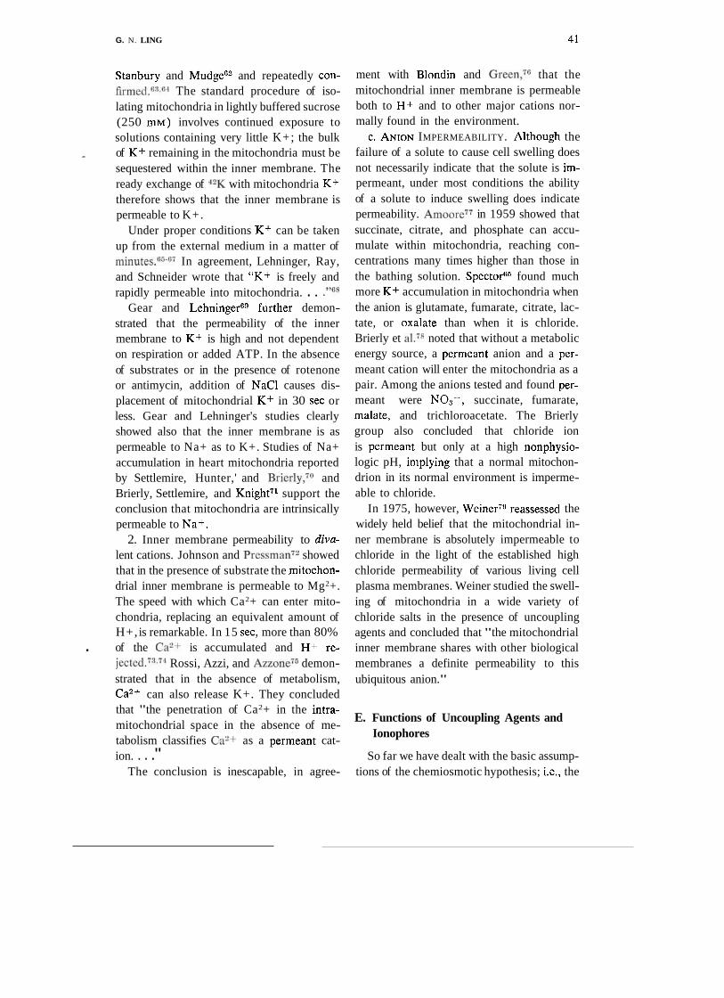
G. N. LING 4 1
Stanbury and Mudgew and repeatedly con- firmed.fi"G4 The standard procedure of iso- lating mitochondria in lightly buffered sucrose (250 m ~ ) involves continued exposure to solutions containing very little K+; the bulk
- of K+ remaining in the mitochondria must be sequestered within the inner membrane. The ready exchange of 4X with mitochondria K+ therefore shows that the inner membrane is permeable to K+.
Under proper conditions K+ can be taken up from the external medium in a matter of minutes.""-7 In agreement, Lehninger, Ray, and Schneider wrote that "K+ is freely and rapidly permeable into mitochondria. . . ."68
Gear and LehningerGVurther demon- strated that the permeability of the inner membrane to K+ is high and not dependent on respiration or added ATP. In the absence of substrates or in the presence of rotenone or antimycin, addition of NaCl causes dis- placement of mitochondrial K+ in 30 sec or less. Gear and Lehninger's studies clearly showed also that the inner membrane is as permeable to Na+ as to K+. Studies of Na+ accumulation in heart mitochondria reported by Settlemire, Hunter,' and Brierly,7O and Brierly, Settlemire, and Knight71 support the conclusion that mitochondria are intrinsically permeable to Na+.
2. Inner membrane permeability to diva- lent cations. Johnson and P r e s ~ m a n ~ ~ showed that in the presence of substrate the mitochon- drial inner membrane is permeable to Mg2+. The speed with which Ca2+ can enter mito- chondria, replacing an equivalent amount of H+, is remarkable. In 15 sec, more than 80%
. of the C a w is accumulated and H + re- j e ~ t e d . ~ " ? ~ Rossi, Azzi, and A ~ z o n e ~ ~ demon- strated that in the absence of metabolism, Ca2+ can also release K+. They concluded that "the penetration of Ca2+ in the intra- mitochondrial space in the absence of me- tabolism classifies C a w as a permeant cat- ion. . . ."
The conclusion is inescapable, in agree-
ment with Blondin and Green,7G that the mitochondrial inner membrane is permeable both to H+ and to other major cations nor- mally found in the environment.
C. ANION IMPERMEABILITY. Although the failure of a solute to cause cell swelling does not necessarily indicate that the solute is im- permeant, under most conditions the ability of a solute to induce swelling does indicate permeability. A m ~ o r e ~ ~ in 1959 showed that succinate, citrate, and phosphate can accu- mulate within mitochondria, reaching con- centrations many times higher than those in the bathing solution. SpectoF5 found much more K+ accumulation in mitochondria when the anion is glutamate, fumarate, citrate, lac- tate, or oxalate than when it is chloride. Brierly et al.7R noted that without a metabolic energy source, a permeant anion and a per- meant cation will enter the mitochondria as a pair. Among the anions tested and found per- meant were NO,-, succinate, fumarate, malate, and trichloroacetate. The Brierly group also concluded that chloride ion is permeant but only at a high nonphysio- logic pH, implying that a normal mitochon- drion in its normal environment is imperme- able to chloride.
In 1975, however, Weiner7"eassessed the widely held belief that the mitochondrial in- ner membrane is absolutely impermeable to chloride in the light of the established high chloride permeability of various living cell plasma membranes. Weiner studied the swell- ing of mitochondria in a wide variety of chloride salts in the presence of uncoupling agents and concluded that "the mitochondrial inner membrane shares with other biological membranes a definite permeability to this ubiquitous anion."
E. Functions of Uncoupling Agents and Ionophores
So far we have dealt with the basic assump- tions of the chemiosmotic hypothesis; i.e., the

42 G. N. LING
existence of a pH gradient with lower pH on shifted the emphasis to its role as an "iono- the outside, a large membrane potential with p h ~ r e . " ~ l the outside positive, and the impermeability Andreoli et al.,.'"n their study of the iono- of the mitochondrial inner membrane to ions phoric property of valinomycin across lipid in general and H + in particular. The evi- bilayer models, gave the rank order of pref- dence, examined above, does not support any erence as H + > Rb+ > K+ > Cs+ > Na+ of these assumptions. > Li+. Mitchell and coworkers, however, as
Why, then, does wide acceptance of the as a number the
chemiosmotic hypdhesis persist? The expla- chemiosmotic h~po thes i s~~ '~"~ee also ref. Y
nation is that at one time it seemed to offer 1 1, P. 58) believed that valinom~cin is a spe-
the most persuasive explanation for certain cific ion0phore K+ but an iOnO~hOre key observations concerning ( I ) uncoupling for H + . In support, it was pointed Out that agents and ionophores and ( 2 ) ion gradient- Andreoli et had studied H + transport at driven ATP synthesis. Next, therefore, let us a pH much lower than that the mitOchOn-
discuss those observations. drial environment. Moore and Pressmans4 studied the effect
Compounds like 2,4-dinitrophenol (DNP), of low concentrations of valinomycin on re- dicumarol, and carbonylcyanid p-trifluoro- spiring mi,tochondria suspended in sucrose methylphenylhydrazine (FCCP) inhibit mito- solution containing various concentrations of
chondrial phosphorylation of ADP to form KC1 up to 10 mM. They noted that valinomy- ATP without inhibiting the rate of respira- cin caused transport of K+ into the mito- tion. These "uncoupling" compounds also chondria with a concomitant release of H+. evoke ATPase activity in mitochondria, The following is a reviewer's account (ref. which are normally devoid of such activity. 1 1, p. 59) of this basic phenomenon from the
According to the chemiosmotic hypothesis, viewpojnt of the c ~ e m ~ o s m o t ~ c hypothesis: such uncouplers act by serving as specific car- terms of hypothesis, valinomy- riers for H + across the ~ h o s ~ h o l i ~ i d barrier tin, by transporting K+ across the membrane, of the mitochondrial inner membrane54 allows K+ to be taken up in for H+ which, as mentioned above, is postulated to ejected by the respiratory chain. . . .W ~h~~ be impermeab1e H+ and ions. By even though under the hypothesis valinomy- mediating H t movement across the inner tin is not to have H+-fransporting membrane, the uncOu~ler discharges the ability, and the membrane per se has no H+ protomotive force. Since in this theory the permeability, H+ for K+ exchange could protomotive force provides the energy for nevertheless occur across the inner mem- ATP ADP and Pf, the un- brane by a mechanism called "exchange dif- . coupler causes oxidative phosphorylation to fusion.,,* cease. In that view, uncou~lers Since each molecule of valinomycin has were indeed be pro- been shown to form a cavity in which exactly :
moting proton conductance across artificial one K + ion fits,39 and it is as part of this phospholipid membranes.12.38 valinomycin-K+ complex that K+ is trans-
Another group of compounds, valinomycin being a exercises a *Ussing's exchange diffusionss is limited to exchange strong effect on mitochondrial phosphoryla- of the sanle species of ion across the cell membrane, tion.80 However, discovery of the ability of therefore is quite distinct from what Mitchell con-
ceived as "exchange diffusion." It is of interest even val inom~cin to transport K+ (in preference though Ussing's exchange diffusion could not be
to Na+) across ~ h o s ~ h o l i ~ i d membranes46 experimentally verified in recent studies?"
G. N. LING 43
ported across a phospholipid membrane, it is not clear how valinomycin-mediated K + transport could be linked to a counter H + transport in a membrane supposedly imper- meable to H + and containing no H+-trans- porting ionophore.
Another contradiction is the requirement of inorganic phosphate (Pi) . As a rule, un- couplers elicit increased oxygen consumption in the absence of ADP. Moore and Press- mans-I and Azzone and Azzisi clearly showed that valinomycin in the presence of K+ elicits an increase in respiration. However, this is true only if P i is added, even though valino- mycin by itself induces K + and H + exchange. This relevant point apparently is ignored by the chemiosmotic hypothesis interpretation of the uncoupler action of valinomycin plus K+ mentioned above (p. 42) .
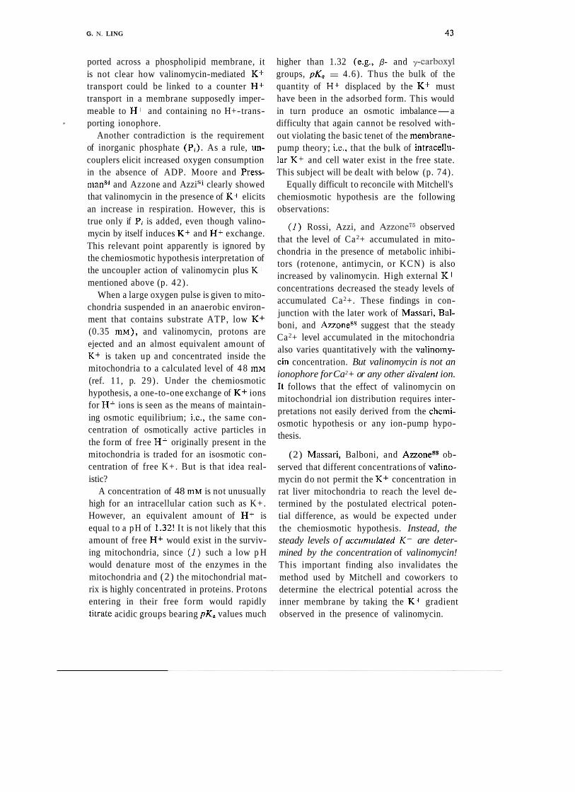
When a large oxygen pulse is given to mito- chondria suspended in an anaerobic environ- ment that contains substrate ATP, low K + (0.35 m ~ ) , and valinomycin, protons are ejected and an almost equivalent amount of K+ is taken up and concentrated inside the mitochondria to a calculated level of 4 8 mM (ref. 11, p. 29) . Under the chemiosmotic hypothesis, a one-to-one exchange of K + ions for H + ions is seen as the means of maintain- ing osmotic equilibrium; i.e., the same con- centration of osmotically active particles in the form of free H+ originally present in the mitochondria is traded for an isosmotic con- centration of free K+. But is that idea real- istic?
A concentration of 48 mM is not unusually high for an intracellular cation such as K+. However, an equivalent amount of H + is equal to a pH of 1.32! I t is not likely that this amount of free H + would exist in the surviv- ing mitochondria, since ( I ) such a low p H would denature most of the enzymes in the mitochondria and (2) the mitochondrial mat- rix is highly concentrated in proteins. Protons entering in their free form would rapidly titrate acidic groups bearing pK, values much
higher than 1.32 (e.g., P- and y-carboxyl groups, pK, = 4.6). Thus the bulk of the quantity of H+ displaced by the K + must have been in the adsorbed form. This would in turn produce an osmotic imbalance-a difficulty that again cannot be resolved with- out violating the basic tenet of the membrane- pump theory; i.e., that the bulk of intracellu- lar K + and cell water exist in the free state. This subject will be dealt with below (p. 74).
Equally difficult to reconcile with Mitchell's chemiosmotic hypothesis are the following observations:
( I ) Rossi, Azzi, and A ~ z o n e ~ ~ observed that the level of Ca2+ accumulated in mito- chondria in the presence of metabolic inhibi- tors (rotenone, antimycin, or KCN) is also increased by valinomycin. High external K+ concentrations decreased the steady levels of accumulated Ca2+. These findings in con- junction with the later work of Massari, Bal- boni, and Azzone" suggest that the steady Ca2+ level accumulated in the mitochondria also varies quantitatively with the valinomy- cin concentration. But valinomycin is not an ionophore for Ca2+ or any other divalent ion. I t follows that the effect of valinomycin on mitochondrial ion distribution requires inter- pretations not easily derived from the chemi- osmotic hypothesis or any ion-pump hypo- thesis.
(2) Massari, Balboni, and Azzoneg8 ob- served that different concentrations of valino- mycin do not permit the K+ concentration in rat liver mitochondria to reach the level de- termined by the postulated electrical poten- tial difference, as would be expected under the chemiosmotic hypothesis. Instead, the steady levels of accumcrlated K+ are deter- mined by the concentration of valinomycin! This important finding also invalidates the method used by Mitchell and coworkers to determine the electrical potential across the inner membrane by taking the K + gradient observed in the presence of valinomycin.

44 G. N. LING
F. ATP Synthesis "chemiosmotic" gradient maintained by an intact, membrane-enclosed structure.
Perhaps the most convincing support for Mitchell's hypothesis was provided by evi- dcnce that H+ and other ionic gradients G . Concll~sions Regarding the across membranes can provide energy for the Chemiosmotic Hypothesis synthesis of the high-energy phosphate bonds of ATP. Consideration began in 1966 with Jagendorf and Uribe," who reported that iso- lated spinach chloroplasts would synthesize A T P from ADP plus Pi, without illuniination or metabolism, if the chloroplasts simply were exposed first to an acid medium and then to an alkali medium. It seemed that the conse- quent proton gradient forced an H+-ATPase to work backward to synthesize ATP. This exciting finding was soon confirmed in other preparations. Reid, Moyle, and Mitche1l9O showed that in liver mitochondria a reverse change of base to acid causes A T P formation from ADP plus P,. Cockrell, Harris and Press- man" showed that in rotenone-treated rat liver mitochondria loaded with X + and sus- pended in a Kt- f ree sucrose niedium the addition of valinomycin brought about a rapid loss of K + and concomitant A T P synthesis. Rossi and Azzone" showed that if K+ ac-
From the above review it is clear that many new and old experimental results render the chemiosmotic hypothesis untenable. A new '
and different interpretation of the findings must be attempted in order to construct an alternative hypothesis compatible with data both in agreement with and at odds with the chemiosmotic hypothesis. The failure of that hypothesis is tightly interwoven with the fail- ure of the more general membrane-pump theory, which since 1877 has been the pre- vailing theory of cell physiology and the foun- dation for most ongoing biomedical investiga- tion. Accordingly, the remainder of this paper shall first review an alternative theoretical model of ion transport in the cell and then show how this model can be used to interpret much of the data collected with regard to mitochondria1 function.
cumulation was brought about by aerobic metabolism, treatment with rotenone also 111. THEORY OF THE LIVING CELL
brought about a slow leak of K + and con- comitant A T P synthesis. These and similar findings (see below) convinced many former skeptics that the chemiosmotic approach had validity.
But soon unexpected observations began to appear. In 1970, Kanazawa, Yamada, and TonomuraX3 observed formation of ATP from ADP and a phosphorylated Caw-dependent ATPase from frugrnented sarcoplasmic retic- ulum of muscle tissues (see also refs. 94 ,95) .
These latter findings, to be taken up again in Section IV below (p. 48) , heralded a revo- lution in our understanding of oxidative phos- phorylation. In anticipation, one notes that this central process of cell metabolism, ATP synthesis, does not rely on the existence of a
Since the publications of Claudem and later of Hogeboom, Schneider, and Palade," a bril- liant chapter has been added to the history of biochemical research concerning two key issues in cell physiology: oxidative metabol- ism and photosynthesis. The isolated mito- chondria and chloroplasts that perform these . specialized functions, however, are part and parcel of the cell. As such, they exhibit the properties of swelling, differentiated permea- bility, solute accumulation and exclusion, electrical potential, dependence on energy metabolism, and responsiveness to metabolic poisons and biologically active compounds. These properties, to all appearances, are similar to those of whole living cells.
It was natural that most investigators of

G. N. LING 45
mitochondria and chloroplasts took as a start- ing point the fundamental theory of the living cell at the time: the membrane or membrane- pump theory, a theory originally introduced by Pfeffer.l
A. Basic Features of the Membrane-Pump Theory
Essentially, the postulates of the mem- brane-pump theory may be summarjzed as follows:
I . Water. The bulk of cell water is free.
2. Ions. The major intracellular cation K+ is free in a dilute aqueous solution.
3. Cell srirface barrier. The cell is sur- rounded by a plasma membrane, the major components 'of which are phospholipids. These form a continuous layer broken occa- sionally by islets ofCproteins that may or may not connect the two aqueous phases separated by the membrane. The phospholipid layer is the seat of selective permeability for solutes and water.
4. Membrane pumps. In the plasma mem- brane are found a large variety of so-called pumps that regulate, usually with a high de- gree of specificity, the steady-state levels of permeant solutes in the cell. Energy needed for the pumping may be supplied by hydroly- sis and by liberation of the energy conserved in the high-energy phosphate bonds of ATP.
5. Cell volume. The cell behaves like an - osmometer. Its swelling and shrinking depend
on the presence of an intact cell membrane. Free K + and other ions and solutes provide just enough osmotic pressure to counter- balance that created by free Na+ and other ions and solutes in the external medium.
6. Resting potential. The electrical poten- tial difference measured between the inside and outside of a living cell is regarded as a "membrane potential" and is determined pri- marily by K + (and N a + ) ionic concentra-
tion gradients according to Donnan's theory of membrane equilibrium or its variant, the Hodgkin-Katz-Goldman model. Electrical potentials that cannot be explained by the Donnan theory are often attributed to special "electrogenic" pumps (as mentioned above, p. 35).
B. Basic Features of the Association- Induction Hypothesis
The membrane-pump theory, however, is by no means the only theory of the living cell. One alternative, the sorption theory, was pro- posed by the Russian investigator A. S. Tros- chin" (see also Nosonov, ref. 99) . Other concepts of the living cell were advanced by Ernst.loo Another and more complete theo- retical structure, known as the association- induction hypothesis (AT hypothesis), was first presented in an elementary form by Ling in 1 9511°1 and 19521°2 and in a more com- plete version in 1962' (for later reviews, see refs. 103-106). Over the years, as experi- mental data and interpretive concepts accum- ulated and were systemized, the A1 hypothesis has taken on the force of a formalized theory. But although more formal and more general, the current version of the A1 hypothesis still departs radically from the membrane-pump theory in describing the living cell. The basic A1 tenets are as follows:
I. Water. The bulk of cell water exists in a physical state different from that of normal free liquid water. This state is characterized by dynamic polarized multilayers formed by interaction with a matrix of extended and more or less parallel polypeptide chains in which the repeating sequences of CONH groups are directly exposed to the bulk water.
2. Ions. The bulk of intracellular K+ is adsorbed on P- and y-carboxyl groups of in- tracellular proteins.
3. Cell sc~rface 6arrier. The surface barrier
46 G. N. LING
is semipermeable. It consists primarily of polarized water, but includes surface proteins that offer fixed ionic and other sites essential for selectivity in solute permeability.
4 . Pumps and solute exclusion. Pumps in epithelial cell systems (e.g., frog skin, toad bladder, intestinal epithelium) would involve, as a rule, the entire cell--endowed with asymmetrical surfaces. Maintenance of the steady level of solutes in resting cells in gen- eral is not due to continually operating mem- brane pumps but reflects the combination of two basic mechanisms. The first mechanism is selective adsorption on macromolecular sites that tend to increase intracellular con- centrations to above those in the external medium (e.g., K + ) . The second mechanism is reduced solubility in the polarized-multi- layered cell water that tends to decrease intra- cellular concentrations of solutes to below those in the external medium (e.g., Na+). The larger and more complex the solute, the lower its equilibrium level in the cell water.
5. Cell volume. Maintenance of cell vol- ume as a rule is not directly dependent on an intact cell membrane, nor does it depend much on the small amount of free ions in the cell. Instead, it primarily reflects the reduced water activity in the state of polarized multi- layers and the reduced solubility of the major external solute, Na+.
6. Resting potential. The resting potential is not a Donnan or membrane potential but a surface adsorption potential. Its magnitude is determined by the nature and density of fixed anionic sites on the cell surface and the nature and concentration of the external ions adsorbed at these sites.
C. Discriminatory Experimental Evidence
It is obvious from the above that in many ways the membrane-pump theory and the A1 hypothesis are irreconcilably different.
A great deal of experimental work aimed
at providing decisive proof for one or the other approach has been and continues to be reported. Since many of the earlier findings have been r e v i e ~ e d , ~ ~ l - ' ~ ~ and since an up- dated and more comprehensive treatment in the form of a monograph is forthcoming, I shall limit myself here to brief discussion of several key pieces of evidence in support of the A1 hypothesis.
1. Energy Requirements of Pumps
Under controlled conditions, the Na+ pump in musclk, though but one of many energy-requiring pumps postulated by mem- brane-pump theory, would consume 15 to 30 times the total energy available in the The essence of this finding has been con- firmed by studies of cells other than muscle cells.107,10s Accordingly, three remedial hy- potheses (exchange diffusion, sarcoplasmic reticulum sequestration, and a nonenergy- consuming pump) were advanced to save the Na pump; but all were subsequently dis- proved.86
The energy impasse is eliminated by the A1 hypothesis. Since the resting cell exists in a metastable equilibrium state, no con- tinual energy expenditure is required for maintenance of the asymmetrical solute dis- tribution. Furthermore, the maintenance of cell ion levels does not depend on the rate of a "pumped" flux, as shown in muscle de- pleted of ATPIO%nd in lymphocytes at vary- ing temperatures.ll0
2. Membrane vs. Cytoplasm as the Seat of Dis-
crimination in Solute Distribution
Sheaths of intact squid axon membrane without cytoplasm, capable of .normal elec- trical activitieslll and ATP-dependent Na+ efflux112 can be prepared. Sacs of these sheaths with the ends tied should offer an un- usually favorable preparation for a decisive test of the Na+-pump concept. However, re- peated attempts to demonstrate actual pump-

G . N. LING 47
ing of K+ or Na+ against an electrochemical gradient in the presence of ATP produced no positive results.106
On the other hand, frog muscle cells devoid of a functional cell membrane pump are capa- ble of accumulating K+ and excluding Na+ in ways similar to cells with intact mem- branes.lI3 Similarly, '.'whitew erythrocyte ghosts without cytoplasm, but with intact membranes and K,Na-activated ATPase, do not transport K+ or Na+ against concentra- tion gradients."-'JlWnly "red" ghosts that retain considerable amounts of cytoplasmic proteins1lR,ll7 are able to transport limited amounts of K+ and Na+ against concentra- tion gradients.l15Jls
These findings again show that it is the cytoplasmic protein-water system and not the membrane that is responsible for K+ accumu- lation and Na+ exclusion.
3. The Adsorbed State of Kt
Since its inception in 1951, the A1 hy- pothesis has suggested that the p- and 7-
carboxyl groups represent the seat of selective K+ adsorption in living c e l l ~ . ~ J ~ l In voluntary muscle cells, more than 60% of these groups are found in the
Now, myosin in muscle cells, rather than being evenly distributed, is localized exclu- sively in the A bands.120J21 The work of Hodge and Schmidt122 shows that uranyl ion, the cationic electron microscope stain, binds to the p- and y-carboxyl groups of the aspar- - tic and glutamic acid residues of protein respectively. The observation that uranium stains primarily the A bands strongly sup- ports the A1 hypothesis, which predicts that the bulk of intracellular cations must be localized in the A bands and in other cyto- logical structures that appear dark after uran- ium staining. In other words, if one could somehow visualize K+ in the living, resting muscle cell, the picture would most likely resemble the electron micrograph of a fixed
muscle cell preparation stained with uranium. This theoretical expectation has recently
been confirmed by a series of observations by Edelmann. He first replaced the K+ in living frog muscle with the electron-dense cesium ion-a stoichiometric exchange leaving the muscle cells functionally intact.123 He then applied a simple but highly effective new freeze-drying t e ~ h n i q u e l ~ " ~ ~ ~ to the muscle cells. The specimen was infiltrated with Spurr medium at low temperature, and the sections were dry-cut. Figure 1A is an electron micro- graph of muscle fixed in glutaraldehyde and stained with uranium only in the conven- tional manner (Edelmann, unpublished). Fig- ure 1B, on the other hand, is Edelmann's unfixed and unstained frog muscle cell loaded in the living state with Cs+.124-126 Figures 1A and 1B match each other in almost all details. Figures 1C and I D show muscles loaded with thallium (TI+) rather than Cs+. Figures 1 E and I F are respectively a Cs+-loaded sec- tion that has been washed and a normal K+- loaded section. The dark areas in Figs. lB, lC, and 1D mark the cytological structures that selectively adsorb Cs+ (and hence K + ) in the resting state.
Compelling as they are, before these find- ings can be accepted as confirming the pre- dictive value of the A1 hypothesis, two ques- tions must be resolved.
( I ) Could the Cs+ seen in the region of the A band merely be free counterions hover- ing in the vicinity of fixed negative charges rather than adsorbed? A negative answer is provided by demonstration that the alkali- metal ion uptake in frog muscle is ion-specific, indicating expression of short-range attributes detectable only by direct contact, and is not just valence-specific, as would have been the case if Cs+ or K+ existed as free coun- terions.l1° Thus Cs+ is only one-third as ef- fective as K+ in displacing adsorbed 42K- labeled K+ from muscle cells, and such ion specificity persists in muscle cell preparations
48 G. N. LING
devoid of an intact cell membrane or of pos- tulated membrane pumps.127
(2) Could artifacts of the special tech- nique used be responsible for the observed results? This possibility is ruled out by studies in which four different techniques were used, including ( a ) autoradiography of dried sin- gle muscle fibers at 25OC carried out by ~ i~~;127 ,128 ( b ) autoradiography of frozen
fresh single muscle fibers at - 1 90°C carried out by Edelmann;12"c) dispersive X-ray microprobe analysis on single frog muscle cells by Edelmann12"confirmed using iso- lated single myofibrils from honeybee thorax muscle130), and (d) laser microprobe mass spectrometry (LAMA) and X-ray micro- analysis demonstration of selective K+ and Cs+ uptake over Na+ in the A-bands of freeze-dried embedded muscle section by Edelmann.131
Two of these methods--dispersive X-ray microprobe analysis and LAMA-also iden- tified K+ localized in the A bands without the use of Cs+ or T1+ surrogates. Thus selec- tive K+ adsorption in living muscle cells, cen- tral to the A1 hypothesis, has been substan- tiated thoroughly and unequivocally in recent years.
4. Consequences of K+ Binding in Living Cells
To repeat, the membrane-pump theory and the A1 hypothesis, internally consistent as they are, cannot be reconciled with each other. Free K+ in living cells is an indispensa- ble and integral part of the membrane-pump theory. Confirmation that the bulk of cell K+ is in an adsorbed state has disallowed the conventionally accepted state of osmotic equilibrium, since K+ makes up the bulk of the solutes found in living cells. By the same token, the membrane and Donnan theories of cellular potential are also invalidated, since they require that all muscle-cell K+ must be free to account for a resting potential of some 90 mV.55J32
Common sense-though admittedly not
rigorous proof-at one time led Monod and J a ~ o b ' ~ V o suggest that "anything found to be true of E. coli must also be true of ele- phants." Is what is true of cells also true of cell organelles? I have shown that whole striated muscle cells selectively accumulate K+ over Na+, and that this phenomenon is due to selective adsorption of K+ (and exclu- sion of both K+ and Na+ from cell water). ?'he mitochondria are intrinsic to these and other living cells. So it is not surprising that mitochondria have been shown to also selec- tively accumulate K+.134,135 Indeed, even fragments of mitochondria accumulate K+ over Na+, as demonstrated by Gamble136 in 1957. Thus common sense would dictate a great unlikelihood that mitochondria would achieve a similar selective K+ accumulation by resorting to postulated ion pumps of the conventional or chemiosmotic type.
1V. THE SOURCE OF ENERGY FOR ATP SYNTHESIS
Mitchell's chemiosmotic hypothesis is a modern extension of the membrane-pump theory. Beginning in microbiology, its generalization to mitochondria and chlo- roplasts and to other intact cells has been vigorously pursued (for examples, see refs. 13, 137; for critique, see ref. 114). The fundamental difficulties described above and elsewhere that argue against the mem- brane-pump theory114 also argue against the chemiosmotic hypothesis. It thus becomes timely to attempt an interpretation of mito- , chondrial behavior in general and of oxida- tive phosphorylation in particular in terms of thc association-induction hypothesis.
In the chemiosmotic model, ATP forma- tion is due to a "protomotive" force arising in part from a H+ gradient across the inner membrane of mitochondria and chloroplasts. One influential set of observations that per- suaded many scientists to accept the chemios- motic approach began with Jangendorf and Uribe's8Veport, mentioned earlier, to the ef-

G. N . LING 49
FIGURE 1. Electron micrographs of frog sartorius muscle. (A) Muscle fixed in glutaraldehyde only and stained with uranium by conventional procedure. ( B ) EM of section of freeze-dried Cs+-loaded muscle, without chemical fixation or staining. (C) TI+-loaded muscle without chemi- cal fixation or staining. (D) Same as C after exposure of section to moist air, which causes the hitherto even distribution of thallium to form granular deposits in the A band. (E) Section of central portion of B after leaching in distilled water. (F) Normal "K-loaded" muscle. A: froni Edebnar~n, unplcblished. B to F: f rom Edelmann,'z" by pernlission of Plzysiol. Clrerii. Plrys.
fect that spinach chloroplasts synthesize ATP if exposed first to an acid and then to an alka- line solution. This was soon followed by re- ports from Reid et al." and Cockrell et al.?!)l who demonstrated ATP synthesis associated with ionic gradients in liver mitochondria. Similar observations were made with red blood ~ e l l s . ~ " ~ ' ~ T h e n rabbit muscle sarco- plasmic reticulum vesicles preloaded with Caw and incubated in the presence of EGTA were found to show rapid release of Ca2+ accompanied by ATP s y n t h e s i ~ . l ~ ~ - ~ ~ ~ These findings also appeared to demonstrate that ATP was formed using energy derived from dissipation of the ionic gradient across the membrane. But against that view, Kana- zawa et a1.93 in 1970 showed that sarcoplas- mic reticulum (SR) fragments "phosphory- latedux with ATP could later transfer their phosphate groups to ADP, thus synthesizing ATP. Boyer et a1.1GJ44 showed that a small amount of the "phosphorylated" enzyme
could be formed when the SR was not loaded with Ca2+. K a n a ~ a w a l ~ ~ and Masuda and de Meis l"~ '~~ showed that vesicles were "phos- phorylated" in the absence of a C 2 + concen- bi- tration gradient. Clearly ~ a n a z a h and CO-
workers1" by 1972 had s u c c 5 i ~ m o - ---- strated the formation O ~ - A T P from ADP and 7
P;lir'ikeBbGke"of ion gradients either &' tFE<tep generating the phosphoenzyme or - in -- b
tliesubsequent step generating ATP. --. _ In 1975 Knowles and R a ~ k e r l ~ ~ confirmed
this finding by demonstrating the synthesis of ATP from ADP and P, in purified Ca2+- ZWf T--.--- -- - *"--- - .- =
ase lom the SR without a Ca2+ gradi- a
<rif-Knowles and Racker proposed that "the energy for ATP formation is derived from the interaction of ---,-....- Ca2+ with -- the protein." h
A parallel observation was made in the
'? Quotation marks around "phosphorylated," etc., signify a lack of knowledge whether a covalent bond is formed between Pi and protein. P i adsorp- tion, for example, may be an alternative.

G . N. LING
synthesis of ATP without an ionic gradient ___-- ---4-
and concluded that "binding of sodium ion- C___ __ _ __.. . ----- - --
to B low-affinity site on phosphoenzyme _ _ - - --< - - - - - formed from inorganic phosphate-is.sufficient
same year by Taniguchi and Post149,150 using / quires Mg2+ and K+.14sJ50 On the other
- - toinduce a conformational change in the ac- tive Enter which permits transfer of the phos- $ate group to adknisi,:e diphqsphate." For --- later reference it is to be noted that "phos- phorylation" by P, of both Ca2+-activated ATPase and K+,Na+-activated ATPase re-
-..-- purified Na+,K+-activated ATPase from guinea pig kidney. They demonstrated the
btead, a different ion is needed for the task- Ca2+-activated ATPase and Na+ for Na+, n K+-activated ATPase.
These revolutionary findings cast a new and different light on the subject of ATP syn- thesis. The emphasis is shifted from ionic or *
"osmotic" gradients, central to the mem- brane-pump theory, to ion adsorption on pro- teins, central to the A l hypothesis. Indeed, the findings lead to revision of a paramount
hand, Mg2+ inhibits ATP synthesis from ADP and the "phosphorylated" enzyme. In-
FIGURE 2. Relation between Taft's induction constant or and acid dissociation constant pK, of a-substituted acetic acid (XCHzCOOH), 6-substituted propionic acid (XCH2CHEOOH), a- substituted methyl-ammonium ion (XCH2NHs+) and 6-substituted ethyl-ammonium ion (XCH?- CH?NHaf ). In these formulae, X represents the substituent, which varies. Abscissa represents the or of each substituent indicated in graph. Ordinate gives the acid dissociation constant of that particular substituted compound as it is indicated on the graph. Frorn Ling,'" by permission of Tex. Rep. Biol. Med.

G. N. LING 5 1
question regarding mitochondria1 oxidative phosphorlylation : How does the mitochon- drion, without the aid of a biochemist, carry out ,two-step manipulations equivalent to laboratory procedures? The first step involves exposing the ATPase to Mg2+ (and some- times K + ) and Pi, thus producing the "phos- phoenzyme." The second step involves re- moving the Mg2+ or K+ already present and replacing it with a different cation, either Ca2+ or Na+ as the case may be. Additionally, to- day we must ask how adsorption of Ca2+ or Na+ creates the phosphate transfer from the ATPase to ADP to synthesize ATP?
V. PROTEINS ACCORDING TO THE A1 HYPOTHESIS: HIGH- AND LOW-ENERGY STATES OF PROTEIN-ION-WATER SYSTEMS. PRIMARY INDUCTIVE EFFECT
In recent years, the terms "high-energy state" and "low-energy state" have become widely applied to biological systems although the precise meanings of those terms are often not explicitly stated. The following is a brief review of the concept of the high-energy liv- ing state according to the A1 hypothesis.
As an analogy, consider a chain of soft- iron nails loosely joined end to end with pieces of string. If a strong magnet is brought close to one of the terminal nails, magnetiza- tion of this nail will cause the next nail in the sequence to be attracted to the opposite end of the first nail; the second nail will, in turn, attract and magnetize the third nail, and so on. If iron filings are placed in the area
. surrounding the chain, they will be picked up by the magnetized nails in a more or less or- dered manner. Thus approximation of the strong magnet will propagate magnetic polar- ization of a number of the nail elements ac- companied by an increase in the magnetic interaction not only between the magnet and each individual nail but also between the nails and the iron filings around them. Along with
a gain of magnetic energy in this system, there is also a loss of entropy; i.e., a more ordered state is created from a more random state.
Another model would be a chain of elec- trical insulators placed end to end. If an elec- trically charged rod is brought close to one of the terminal insulators, electrical polariza- tion or induction will propagate through the chain of insulators much as the magnetiza- tion propagated through the soft nails in the example above. In this case, it is electrical polarization and energy from an electrostatic interaction that are enhanced. The magnetic- ally polarized nails or the electrically polar- ized insulators represent a higher energy state, much like the high tide brought to a still higher energy state by the synergistic action of sun and &oon alignment. When the large magnet or the electrifying rod is removed, or when the sun and moon are no longer aligned, the magnetic poles, the electrons, or the water, as the case may be, revert to their normal low- energy, high-entropy state and in so doing can perform work with the released energy. It is also worth noting that in the polarized high-energy state the electron density is higher at some sites than during the unper- turbed state but lower at other sites. The key issue is the pertcrrbation of the entire linked system from its low-energy equilibrium posi- tion.
According to the A1 hypothesis, the unique partial resonance of the polypeptide chain in proteins enhances propagation of an elec- trical polarization or inductive effect through this chain. The energy elevation, as in the model system of nails in a sea of iron filings, is not limited to elements of the protein itself but critically involves all the other polarizable elements of which the proteins are a part. Thus propagated high- and low-energy states refer not to a single component but to all the closely associated components of living cells: water, proteins, and ions existing as a cooper- ative unit.
But induction is not the only basic mecha-

TABLE 11. Relation Between Inductive Index and Ionization Constants of Oxygen Acids u w
(1) Landolt-Bmatein. 1%0 Z ~ h h m m t e m d Padtionen, Seehate Auflap. Band II, Teil 7. S. 842-45. (2) Pauling. 1949 General Cha&ry, p. 3%. (3) Crofp and Korolnpoff, 1953 1. Am. CLem. Soc.. 75, 3379,
From Chiang and Tai,lb8 by permission o f Scierltia Sinica.
pK. 25. (1)
1.2 3.9; -3.7(2) 3.8 4.3 4.9
4.7 5.2 9. 7(30e); lO(2) 8.5(2)
10.3
11.7(30e)
9.1 9.2
11.2 9.9
- 9.1
10.0 10.7 12.7(-20') 10.3(18')
13.8(-20') 11.7(18')
l 1 for-OX
305.7 298.4 297.5 288.1 280.0
275.6 275.6 270.9 263.2 257.8
252.4 232.0 230.9 230.8. 222.6
221.7 220.0 214.5 210.4 210.2
191.1 157.1 154.9 145.2 89.8
10'1 forax
455.4 389.9 319.5 311.2 257.6
243.2 237.6 225.7 192.6 127.7
62.7 -2.3
367.8 332.8 317.9 302.2 297.4
280.3
PK. 25' (1) Compound H-OX Compound H-OX
--8(2) --3(2)
2.3; 2.0(23') 0.8 1.6
7.4(27'); 7.0(2) 8.5; 8.7(2) 9.7; 11.0(2) 8.3
12.4
>I5 > 15
- -3(2) --0.5 1.6-1.7 1.9; 2.0 1.9; 1.9(2)
--
a
% 5 2 - 2
-
a
&
H-OCIO, H-OCIOt H-OCIO H-OIO, H-OIO(OH),
H a l H-OBr H-OI H-OIO, OH), H-OIO,~OH),
H-OIO,:(OH) H-OIO,
H-OSO,H H-OSeO,H H-OS,O;- H-OSO,- H-OSO(0H)
H-OScO(0H)
3 2
10'1
-- 269.8 257.6 245.7 245.3 242.9
242.5 242.0 228.3 228.3 191.3
162.8
-- 251.9 217.0 214.0 210.7
205.2 203.2 202.0 151.0 150.5 --
85.1 83.0
H-OSe0,- H-OTeO(OH) H-OTe(OH), H-OS0,-
H-OSe0,- H-OTe0,- H-OTeO-(OH),
4 O
t 2 0 E
P 2
pK, 25' (1) Compound H-OX
1.0(18') H-OCOCOOH 1.5(18') H-OCOOH 2.3 H-OCHO
H-OPO,' H-OA&(OH) H-OAsO,' H-OAsO;
H-OPO(OH)-OPO(OH)2 H-OPO(OH)-OPO,-(OH) H-OPO(OH)-OPO,--0P0,-(OH) H-OPO(OH), H-OPH(O)(OH)
H-OAsO(OH , H-OPO(Me)~OH) H-OPH,(O) H-OPO(Mc), H-ONO
H-OAsO(Mc H-OPO,--Obbi-OPO,-(O~) H-OP0,--0P0,-(OH H - O P O z - - O ~ z - - ~ P ~ 3 ~ H-OPO,-(OH)
H-OPO --OPOIa H-OAS~OH H-OPO -(I-?' H-OAdi(2H) H-OAsO,-(Me)
H-ONH,
2.1 2.0
2.2 2.3(3) i.0; 2.0(2) 3.1(3) 3.2(20°); 3.2(30°)
6.1 6.3 6.6 8.9 7.2
-
9.6; 8.4(18', 3) 9.4 6.6 7.0 7.1(3)
8.0 12.0(2) 13.5 11.5(18')
>15(32')
260.8 233.1 228.8
231.9 195.9 168.3
2.5 7.8 7.0(18')
8.0(18') 7.8
10.3
n
$
" 6
-
- 2
.
g
H-OCOCOO- H-OCOCII,CH,SiMe,
H-OCOMc H-OCOCH,SiMe, H-OSi(OH), H-OGc(OH), H-OCOO-
H-OSiO-(OH),
H-OBO H-OB(OH), H-OAI(OH), H-OB(OH)-C,H,
H-OB(OH)-CH2C,H, H-OB(OH~H,CH,C,H! H-OB(OH)-Bu-n H-OBO-(OH) H-&~o-(OH)
H-OBO; H-OGaO,'

G . N. LING 53
nism involved in propagated high- and low- energy biological states. Equally basic is elec- tron density-dependent preferential ion ad- sorption at charged sites, tracing to the differ- ences in electron density between polarized and non-polarized states. Both these mecha- nisms will now be discussed.
A. Inductive Effect as the Basis of Energy and Information Transfer Over Distance
After the major discovery of ATP and its pivotal role in cell metabolism and function, ATP was believed to contain a high-energy bond in which an unusually large amount of potential energy was stored. Since the 1950s it has become clear that this concept is not correct. The enthalpy of hydrolysis of the ATP phosphate group is not unusually high;'" the free energy of hydrolysis of ATP is largely due to the liberation of H+ in an environment maintained at physiological p H as well as to other extraneous factor^.^"'-^^'
That A T P may serve a physiological func- tion without undergoing hydrolysis has been repeatedly suggested. Riseman and Kirk- wood,'" Botts and Morales,ljG and Ling in 1952102 believed that ATP adsorbed on mus- cle protein affected the protein conformation by a direct electrostatic repulsive effect medi- ated through space. The view that I finally adopted, however, is different; stress is placed on the inductive effect of adsorbed ATP medi- ated through the protein m o l e c ~ l e . ~ Indeed, the inductive effect permits (1 ) determination of the secondary and higher protein structure and (2) the properties of protein molecules due to their primary structure as well as their environment and past history.2~104~106~157,158 The mode of action of the inductive effect falls into two categories: inductive effect over a short range, referred to as the direct F-effect (describing the combined inductive or I effect and the direct electrostatic D-effect); and an indirect F-effect involving a propagated me-
chanism, or domino effect, reaching over longer distances.
1. Universal Applicability of the Indnctive Effect
The inductive effect, formally presented by L e ~ i s , ' " ~ ~ ~ ~ ' is the basis of a major aspect of the structure-activity relations in organic chemistry, often termed "linear free-energy" re la t i~ns . l~ ' -~" The work of HammettlG3 and Taft'mJGG was closely associated with this ef- fect and as a rule their equations and con- stants describe relations between substituents and alterations in the equilibrium and kinetic properties of close and distant functional groups. In 1963 Chiang and Tailos succeeded in transforming these empirical rules into equations that yield precise quantitative pre- dictions based on independent physical con- stants of atomic electronegativity, bond length, and molecular structure.
The Chiang-Tai theory made it feasible to calculate the entire constellation of properties of even very large and complex molecules from their molecular structure and thus repre- sents a major step beyond the limited con- ventional empirical approach. The Chiang- Tai theory also made it possible to advance the A1 hypothesis beyond the mere deduction that primary structure determines all proper- ties of a protein m o l e ~ u l e . ~ J ~ At last struc- ture and properties of all side chain, back- bone, and other functional groups actually could be calculated according to the aggre- gate inductive effects. The precise molecular structures of numerous pure proteins have now been determined, creating an opportu- nity for major advances in protein chemistry.
2. Target Groups of the Inductive Effect
The inductive effect is universal in that it can be applied to all properties and behaviors of complex molecules. T o illustrate its role under the A1 hypothesis in determining the properties of proteins, three examples are now discussed: the acid dissociation constant

FIGURE 3. Relations between inductive index and ionization constants of oxygen acids and metallic hydroxides. Oxygen acids and the theoretically calculated inductive indices are those listed in Table 11. Acid dissociation constants were collected by Chiang and ~ a i from the litera- ture. From Clliang and Tai,I6' by permission of Sci. Sin.
(or pK,), H-bonding strength, and oxidation- reduction potential.
a. THE ACID DISSOCIATION CONSTANT
(pK,). It is well known that although acetic acid and trichloroacetic acid possess the same carboxyl group, substitution of the more elec- tronegative chloride atoms for the three H- atoms causes a drastic change in the pK value of the carboxyl group from 4.7 to less than 1 . This is the classic example of the inductive effect.
Figure 2 plots the pK, values of various substituted fatty acids against the Taft induc- tion constants of each of the substitutions and exhibits the standard linear relation. The value and scope of the Chiang-Tai theory can be seen from the four graphed cases, chosen from the much more extensive and equally accurate data the authors presented in their 1963 paper.lG8 Table I1 presents ( I ) the acid dissociation constants of 73 oxygen acids, organic and inorganic, and (2) their theoret-
ically calculated "induction indices" derived from the literature. Figure 3 from Chiang and Tai,168 shows the linear relation between these two parameters for all 73 acids.
b. H-BONDING STRENGTH. That the proton- donating power and proton-accepting power of H-bond groups are determined by the in- duction effect is well k n o ~ n . ~ J ~ ~ J ~ ~ J ~ ~ Figure 4 illustrates how the H-bond strength, like the acid dissociation constant, is determined by Hammett'sl" inductive constants for para- , substitutes of aromatic molecules in a pre- dictive manner.
c. OXIDATION-REDUCTION POTENTIAL. Fig- ure 5, also from Chiang and Tai,le8 shows the linear relation between thiol and sulfide reac- tivity obtained from the literature and those authors' theoretically calculated induction in- dices. Thiol reactivity reflects the oxidation- reduction potential of tHe sulfhydryl group, which is also inductively controlled.

C. N. LING 55
3. The Inducing "Groupsn
The linear energy relation most extensively studied is that between covalently linked groups. However, because of its basically electrostatic nature, the inductive effect should extend to other closely associated groups; e.g., groups held together by ionic
. bonds or H-bonds. However, for an adsorbed entity to have a significant effect, the adsorp- tion must involve more than a trivial amount of energy. To illustrate this point, Table I11 (reproduced from an earlier publication2) shows the much greater enthalpy of ATP ad- sorption compared to the enthalpy of ATP hydrolysis. The table also shows the very strong coulombic energy between K+ and negatively charged carboxyl groups as part of the "absolute" enthalpy of adsorption of the K+ ion.
One example of a strong inductive effect exercised by a dissociable ion is the proton of a carboxyl group. The inductive constant of a dissociated COO- group is totally dif-
u HAMMETT
FIGURE 4. Relation between Hammett's o-constant - and the free energy of dimerization of para-substi- tuted acetophenoneoximes. Structural formula of para-substituted acetophenoneoximes appears in right upper corner. Abscissa represents Hammett's u-constant for each substituent indicated as X in the structural formula and more explicitly indicated in the graph. The free energy of dimerization of each substituted compound was calculated from Reiser's data and on the assumption that only dimers but no higher polymer existed. Frorn Ling,'" by permission of Tex. Rep. Biol. Med.
ferent from that of an undissociated COOH group. Other examples showing that H- bonded groups can exert a highly significant inductive effect have been reviewed else- where.15i
4. Additivity and Reversibility of the Inductive
Effect
The effects produced by two or more in- ducing groups on the same target group are additive. Also, such effects are completely reversible; that is, as A affects B, B affects A in a reciprocal manner. Although each in- ducing group tends to have a quantitatively different effect, in general the inducing group can either draw electrons toward itself or donate electrons. These basic tra.its are fun- damental to assessment of complex multi- component interactions, as discussed later below (p. 8 1 ) .
B. c-Value Concept, Linear Model, and Theoretically Calculated Change in Preference for K + , Na+, H+, and NH4+
The preceding discussion of the inductive effect prompts this question: What is the sig- nificance of the pKa values of, for example, the p- and y-carboxyl groups of a protein other than as indicators of their affinity for protons? The answer, according to the A1 hypothesis, is that variations in pKa values are a partial expression of the electron den- sity of these acidic groups. This is significant indeed, since it is the electron density that determines protein specificity in ionic adsorp- tion as well as in maintenance and conforma- tional change. Before this concept can be fully presented, it is necessary to introduce a new parameter, the c-value.
The familiar pKa value is not an indepen- dent variable. It expresses a specific interac- tion between an acidic group and a proton. The c-value, on the other hand, is the under- lying parameter that gives rise to the pKa value. The c-value, given in Angstrom units,
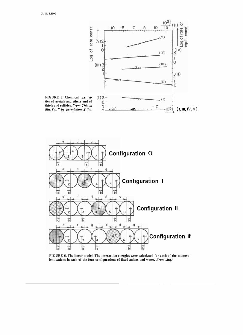
G . N. LING
FIGURE 5. Chemical reactivi- ties of acetals and ethers and of thiols and sulfides. Frorn Chiang
I
and Sin. Tai,'"' by permission of ,312. 1 O -20 -15 -lo 1031 ( 1,111, Iv, v )
Configuration 0
Configuration I
Configuration II
Configuration I l l
FIGURE 6. The linear model. The interaction energies were calculated for each of the monova- lent cations in each of the four configurations of fixed anions and water. From Ling.'

FIGURE 5. Chemical reactivi- ties of acetals and ethers and of thiols and sulfides. From Chiang
I and Sin. Tai,'" by permission of Sci. (I);; O -20 -15 -lo - 1031 ( 1, 111, IV, v 1
Configuration 0
Configuration I
Configuration II
Configuration Ill
FIGURE 6. The linear model. The interaction energies were calculated for each of the monova- lent cations in each of the four configurations of fixed anions and water. From Ling.'

58 G. N. LING
from these data, the average energy of asso- alkali-metal ions in addition to H+ and NH,+ ciation. This enables calculation of theoreti- as a function of the c-value. Note how the H+ cal curves that show either the adsorption preference becomes greatly increased with the energy (Fig. 7) or the selectivity coefficient increase in anion site polarizability. (Fig. 8 ) , measured against K+, of the five We now know that alkali-metal ions are
FIGURE 7. Relation between calculated association energy AE of various cations and c-value of the anionic group. Polarizability of anionic site, a, is 0.876 x cmS. From Ling.?
FIGURE 8. Relation between selectivity ratios of various cat- ions and c-value. The K + ion is taken as unity and selectivity ratios are calculated from asso- ciation energies. From Ling.'

G. N. LING 59
adsorbed in the pattern of one ion to one site in the sulfonate and carboxyl types of ion-exchange resins (Ling and Brady, unpub- lished). Figure 9 (adapted from Bregmanli8) bears a resemblance to Fig. 8, but in Fig. 9
. the abscissa represents the percentage of the cross-linking agent, divinyl benzene (DVB) . An increase of pK, value with increasing DVB has indeed been observed by Gregor and co- workers.17Vigure 9 is essentially a plot of cation selectivity coefficients against the p K , or c-value, as in Fig. 8. The general similarity between the theoretical data of F.ig. 8 and the experimental data of Fig. 9 is obvious. Still, for emphasis, two features of the theoretical curves (Figs. 7, 8 ) are worth mentioning:
(I) Small changes in c-value could involve large changes in, even a reversal of, selectivity between a pair of ions (e.g., between K+ and Na+, between NH,+ and Na+, etc.). The reason for the low preference for Na+ at low c-values is that at equilibrium Na+ exists in a higher conformation (i.e., it is more hy- drated-see Fig. 6 ) . The reason for the high preference for Na+ at high c-values is that Na+ then exists at a lower conformation (i.e., dehydrated). This relation is important to remember because, according to the A1
hypothesis, it explains the different effects of Na+ and K+ on enzyme activity in general and on ATP synthesis in particular (see p. 67 below).
(2) The NH,+ group may represent a prototype of the positively charged C-amino group of lysine side chain or the positively charged guanidyl group of an arginine side chain.
C. Complex Interaction Between Biologically Active Agents
The additive nature of the inductive effect presents theoretical models that yield complex multicon~ponent interaction patterns of bio- logical activity, of which enzyme activity is a well-known example. F.igure 10 shows theo- retical curves compared to actual experimen- tal observations reported in the literature.lo3 From these examples one sees that an agent promoting a particular effect in one direction may inhibit the same effect at a different con- centration or upon variation in the concen- tration of a second agent. Simple nomencla- ture such as "uncouplers" or "inhibitors," useful and adequate under one set of condi- tions, may not apply under other conditions- in fact, may describe an effect totally opposite to that seen under the first conditions. [Note that complex patterns of interaction have been reviewed and theoretically derived elsewhere (see ref. 2, chapts. 7, 14)].
2.5 D. Cooperative Behavior of Living Cells: Interpretation Based on the A1
3.0 o 4 8 12 16 2 0 24 Hypothesis
Percent D V B Many physiological manifestations of liv-
FIGURE 9. ~ ~ l ~ ~ i ~ ~ selectivity coefficients of HT, ing cells exhibit all-or-none characteristics, NH,+, and alkali-metal ions compared to K + in as exemplified by muscle contraction. nerve sulfonate ion exchange resin with varying percent- excitation, and egg activation. u ~ ~ ~ ~ t ~ ~ ~ - age of the cross-linking agent divinyl benzene (DVB). Redrawn from Bregman'" by permission tion" is a similar well-known all-or-none of Ann. NY Acad. Sci. property of proteins.
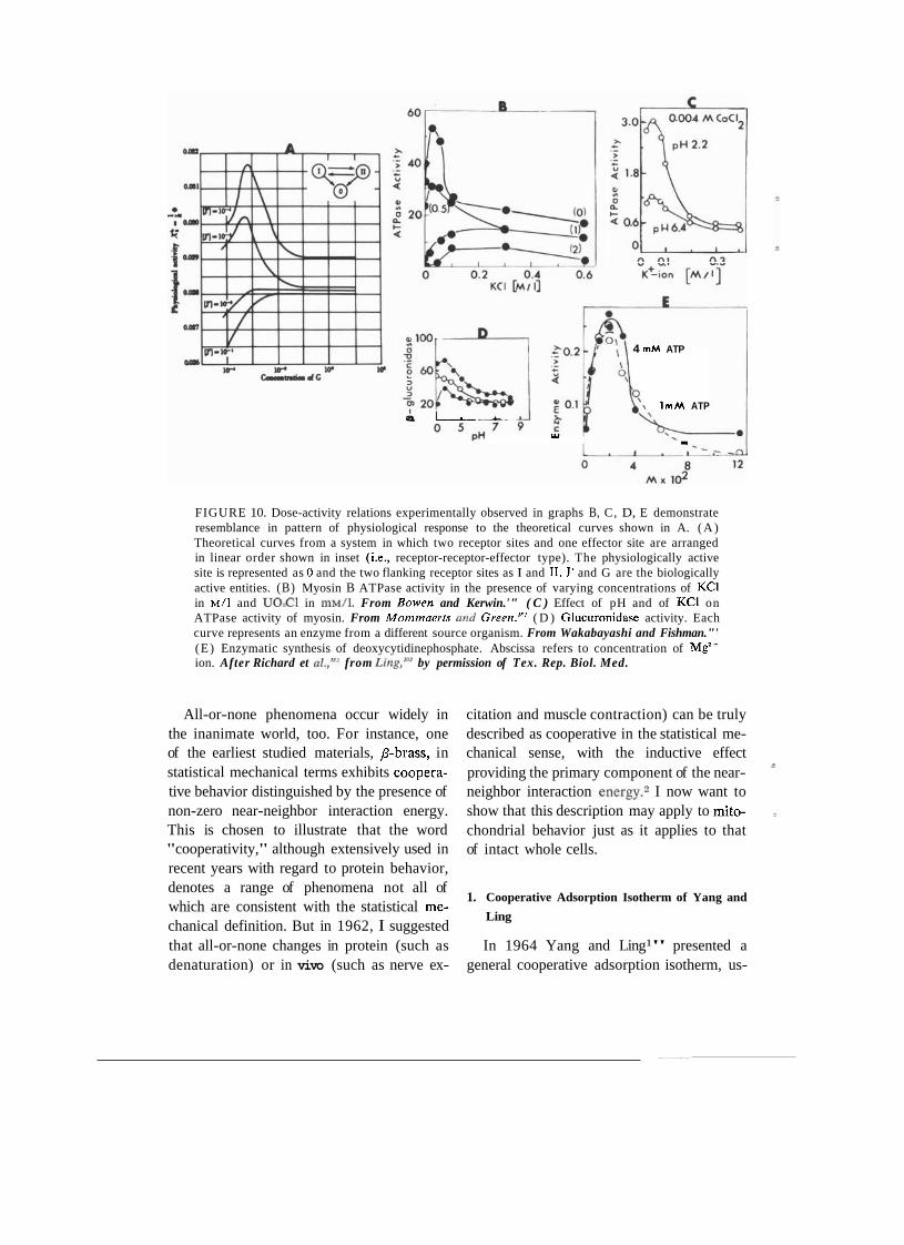
4 m M ATP
\ I m M ATP a I.---- %
Y1 .
FIGURE 10. Dose-activity relations experimentally observed in graphs B, C, D, E demonstrate resemblance in pattern of physiological response to the theoretical curves shown in A. ( A ) Theoretical curves from a system in which two receptor sites and one effector site are arranged in linear order shown in inset (i.e., receptor-receptor-effector type). The physiologically active site is represented as 0 and the two flanking receptor sites as I and 11. J' and G are the biologically active entities. (B) Myosin B ATPase activity in the presence of varying concentrations of KC1 in ~ / 1 and U02CI in ~ M / I . From Bowen and Kerwin.'" ( C ) Effect of pH and of KC1 on ATPase activity of myosin. From Mommaerrs and Greer~.'" ( D ) Glucuronidase activity. Each curve represents an enzyme from a different source organism. From Wakabayashi and Fishman."' (E) Enzymatic synthesis of deoxycytidinephosphate. Abscissa refers to concentration of Mg'+ ion. After Richard et from Ling,'o3 by permission of Tex. Rep. Biol. Med.
All-or-none phenomena occur widely in citation and muscle contraction) can be truly the inanimate world, too. For instance, one described as cooperative in the statistical me- of the earliest studied materials, p-brass, in chanical sense, with the inductive effect statistical mechanical terms exhibits coopera- providing the primary component of the near- tive behavior distinguished by the presence of neighbor interaction energy.2 I now want to non-zero near-neighbor interaction energy. show that this description may apply to mito- This is chosen to illustrate that the word chondrial behavior just as it applies to that "cooperativity," although extensively used in of intact whole cells. recent years with regard to protein behavior, denotes a range of phenomena not all of
1. Cooperative Adsorption Isotherm of Yang and which are consistent with the statistical me-
Ling chanical definition. But in 1962, I suggested that all-or-none changes in protein (such as In 1964 Yang and Ling1" presented a denaturation) or in vivo (such as nerve ex- general cooperative adsorption isotherm, us-
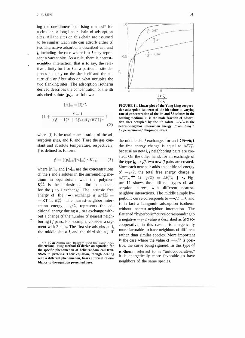
G. N. LING 6 1
ing the one-dimensional Ising method* for a circular or long linear chain of adsorption sites. All the sites on this chain are assumed to be similar. Each site can adsorb either of two alternative adsorbents described as i and
. j, including the case where i or j may repre- sent a vacant site. As a rule, there is nearest- neighbor interaction, that is to say, the rela-
' tive affinity for i or j at a particular site de- pends not only on the site itself and the na- ture of i or j but also on what occupies the two flanking sites. The adsorption isotherm derived describes the concentration of the ith adsorbed solute [pi],,, as follows:
where [f] is the total concentration of the ad- sorption sites, and R and T are the gas con- stant and absolute temperature, respectively. [ is defined as follows:
where [pilPx and [p,],, are the concentrations of the i and j solutes in the surrounding me- dium in equilibrium with the polymer. K z i is the intrinsic equilibrium constant for the j to i exchange. The intrinsic free energy of the j+i exchange is AF;& z
-RT In The nearest-neighbor inter- action energy, -y/2, represents the ad- ditional energy during a j to i exchange with- out a change of the number of nearest neigh- - boring i-j pairs. For example, consider a seg- ment with 3 sites. The first site adsorbs an i, the middle site a j, and the third site a j. If
[ pi 'ex
[P i 3.1 FIGURE 1 1 . Linear plot of the Yang-Ling coopera- tive adsorption isotherm of the ith solute at varying rate of concentration of the ith and jth solutes in the bathing medium. x i is the mole fraction of adsorp- tion sites occupied by the ith solute. -7/2 is the nearest-neighbor interaction energy. From Ling,'" by permission of Pergamon Press.
the middle site j exchanges for an i (ijj+iij) the free energy change is equal to AFP-,"~, because no new i, j neighboring pairs are cre- ated. On the other hand, for an exchange of the type jjj +- jij, two new ij pairs are created. Since each new pair adds an additional energy of -y/2, the total free energy charge is AF,O'& + 2(-y/2) = ~Fp-2 + y. Fig- ure 11 shows three different types of ad- sorption curves with different nearest- neighbor interactions. The middle simple hy- perbolic curve corresponds to -y/2 = 0 and is in fact a Langmuir adsorption isotherm without nearest-neighbor interaction. The flattened "hyperbolic" curve corresponding to a negative -y/2 value is described as hetero- cooperative; in this case it is energetically more favorable to have neighbors of different rather than similar species. More important
*In 1958 Zimm and Braggls4 used the same one- is the case where the value of -y/2 is posi- dimensional king method to derive an equation for tive, the curve being sigmoid. In this type of the specific phenomenon of helix-random coil tran- isotherm. referred to as "autocooDerative." sition in proteins. Their equation, though dealing with a different phenomenon, bears a formal resem-
it is energetically more favorable to have
blance to the equation presented here. neighbors of the same species.
62 G. N. LING
2. Oxygen Dissociation of Hemoglobin and Re- al.lg2 the four subunits of the hemoglobin lated Phenomena molecules are assumed to exist in one of two
states. The affinity of all four units in one One of the most familiar autocooperative
state is uniformly different from that of the adsorption isotherms is the oxygen-dissocia-
four units in the other state. tion curve of hemoglobin. Since the discovery that the binding of oxygen by hemoglobin fol- lows a sigmoid-shaped curve, three types of quantitative or semiquantitative analyses have been made. Let us examine these in turn.
a. EMPIRICAL ANALYSIS (HILL'S EQUA- TION). In 1910, A. V. Hillls6 presented the following empirical equation that, because of its simplicity, has been the most extensively used to describe sigmoid adsorption isotherms and related phenomena:
X = Kxn/(l + K P ) (4)
c. THE YANG-LING MODEL: COMPARISON :
WITH OTHER MODELS. The cooperative iso- therm described by Eq. 2 has been applied to the binding of oxygen to the hemoglobin molecule,193 yielding the Yang-Ling analyti- cal model. While a detailed comparison of this theory with those mentioned above is be- yond the scope of the present paper, a few key differences are pointed out to facilitate development of its main theme.
( 1 ) The AAKM theories were all proposed specifically to deal with hemoglobin as a tet- ramer containing four subunits, each with one binding site. Equation 2 suffers no such r e where 2 is the mole fraction of heme sites striction. It deals with all types of proteins binding oxygen, x is the concentration of oxy- that contain a single chain or any number of gen in the surrounding medium, and K is the subunits, and each chain or subunit may con-
equilibrium constant for oxygen binding. Usu- tain one or more binding sites (see p. 63 ally log F/(1 - Z) is plotted against log x: below).
log[F/(l - F)] = nlogx + nlogK. (5)
It has long been recognized that n deter- mines the degree to which the oxygen binding curves take on a sigmoid configuration. Since Eq. 5 is empirical, n has no specific physical meaning. WymanlS7 believed that "n is closeIy related to the average free energy of interac- tion of the sites." Wyman did not, however, provide an explicit quantitative formalization of this relation.
(2) The AAKM theories neither consider a nearest-neighbor interaction nor provide a mechanism for it. Equation 2, as part of the A1 hypothesis, offers a specific mechanism.
( 3 ) The AAKM theories offer no explicit interpretation of the Hill coefficient n, where- as Eq. 2 does provide an explicit explanation of n as follows:
From Eq. 2, the mole fractions of sites adsorbing the ith solute (xi) and jth solute (Fj) are
b. THE AAKM THEORIES. More recent an- - [pilat~
alytical theories include those of Adair,1ss.1s9 XI=--- - $5 Atkinson et al.,lgO and Koshland et aI.lg1 In [Fl
each of these theories an assumption is {l + r - 1
made that the successive binding constants [ (5 - I ) 2 + 45ex~(y/RT)I* (6) for oxygen on the four heme sites in a hemo- globin molecule are not the same but increase and by some unspecified mechanism. On the other
- - hand, in the symmetrical model of Monod et xj = 1 - xt.

G. N. LING 63
Thus
(7)
Figure 12 shows that this equation accurately describes the oxygen binding of horse hemo- globin studied by Lyster (Ling, refs. 104, 194). In a plot of ln[Ti/l - xi] against In[pi]es/[pj],s) at the locus at which half the sites are occupied by i, the slope of Eq. 7 is exp(-y/2RT). In fact, a straight line drawn through the log-log plot at the half-saturation point can be derived from Eq. 1 as
-
Xi [piles In = nln -- + nlnKZi.
Xi [piles (8)
In the case where i represents oxygen mole- cules and j is unspecified, Eq. 8 becomes
This is formally identical to the Hill equation in a log-log plot. Here, however, the Hill co- efficient n is an explicit function of -Y/2:
3. Three Types of Autocooperative Adsorption on
Proteins
Figures 13 and 14 show two additional . types of in vitro adsorption isotherms: the binding of the cation dodecyltrimethylam- monium bromide (DTAB) on bovine serum albumin (Fig. 13) from Few et al.ls6 and the binding of phenol on collagen (Fig. 14) in the form of dried cowhide powder from Kiintzel and Schwank.le6 In each case, as in Fig. 11, the points represent experimental
data and the solid curves are theoretical according to Eq.2.
In the case of oxygen binding on hemo- globin, the binding sites are the heme pros- thetic groups, one oxygen to each heme in each of the four subunits of the hemoglobin molecules. In the case of bovine serum al- bumin, the binding sites are the ,f3- and 7-
carboxyl groups; the maximum number of DTAB ions bound roughly equals the total number of p- and y-carboxyl groups of the protein known from chemical ana1y~es. l~~ In the case of phenol binding, the maximum number of phenols bound approaches the total number of backbone NHCO groups of the protein.193JQ6
The oxygen binding on hemoglobin repre- sents ligand binding on prosthetic groups; DTAB binding to carboxyl side chains of bovine serum albumin unravels the tertiary structure of the protein; phenol binding on collagen peptide groups unravels the secon-
X , I- X,
FIGURE 12. Log-log plot of data of Lyster on oxygen uptake by horse hemoglobin at pH 7.0, 19°C. From Lirzg,'"' by permission o f Znf. Rev. Cytol.

wtihin the same protein chain, as in the case of DTAB and phenol binding.
10' - E. Autocooperativity in Selective Solute
XDTAB Accumulation in Living Cells - DTAB
1-X DTAB According to the A1 hypothesis, autoco- n= 9.3
operative adsorption-desorption behavior is a
lo-' - general and fundamental property of proto- = plasmic proteins and may be expected to play
viscosity a major role in other biological phenomena
1 o - ~ including the selective accumulation of sol-
10-3 lo-2 utes in the living cell as well as in its orga-
DTAB CONCENTRATION (MI nelles (mitochondria, nuclei, e t ~ . ) . ~ With that in mind, let us return to the familiar
FIGURE 13. Binding of dodecyltrimethylammo- oxygen-dissociation curve of red blood cells.
nium bromide bv bovine serum albumin. Data from Oxygen is a present in the In Few et al.'"T,)l.,,,, refers to the mole fraction of sites arterial blood, the oxygen in the plasma occupied by DTAB. Frorn Ling.'" amounts to about 0.44 ml per 100 ml, while
dary structure. In spite of the great diversity of the particular sites involved they all ex- hibit autocooperative site-to-site interaction, describable by a positive value for ---,/2. As such, they show the most significant and important traits of all autocooperative inter- actions: the tendency to exist in an all-or- none" manner in either one of two alternative I
discrete states. Another highly significant fea- tore of autocooperative adsorption is the X phenol phenomenon of a "threshold" or critical I -)(phenol ligand concentration. Thus the twofold in- crease in the concentration of DTAB from 5 x M to 10 x M changes the lo-' protein conformation from one in which the p- and -,-carboxyl groups are locked in their native configuration (probably mostly in the form of salt linkages) to one in which these anionic groups bind cationic DTAB.lg5 Simi- larly, a small change of the phenol concen- tration switches cowhide collagen backbone lo-' I NHCO groups from H-bonding within the [phenol], chain to H-bonding to phenol molecules.19G (moles/liter) Clearly, cooperativity is not limited to four
FIGURE 14. Binding of phenol by cowhide colla- or any other number of identical or similar gen. Data of Kiintze, and Schwank.lw From Lirtg,,'P3
subunits but occurs between neighboring sites ljy permissjorl of Fed. Proc.

in the red blood cell the oxygen amounts to about 42 n ~ l per 100 ml of cell water. There- fore, there is nearly a 100-fold selective ac- cumulation of oxygen in red blood cells.
The inset of Fig. 15 shows a plot of the - in vivo oxygen-binding curve of human red
blood cells. Despite minor differences, this curve and the in vitro binding curve of hemo-
' globin shown in the large graph of Fig. 15 are quite similar. Indeed, this is accepted knowl- edge; oxygen binding to an intracellular pro- tein, hemoglobin, is the basis of selective oxygen accurnulation in red blood cells.
Now, another solute found in the blood cells is K+. Its concentration in the plasma is about 5 mM while that in erythrocytes, as well as in other ,cells, is many times higher (about 150 m ~ ) . As far back as 1910, Ben- jamin Moore of Oxford University1" pointed out this parallelism and suggested selective K+ binding onto intracellular protein, rather than absolute membrane impermeability and permanent entrapment-as was then widely thought to be the cause of selective accumu- lation of K+ in living cells. The powerful ar- ray of evidence cited earlier (e.g., Fig. 1 ) leaves little doubt that similar to oxygen ac-
cumulation in red blood cells, K+ accumu- lation is also a matter of selective adsorption on (the p- and y-carboxyl groups of) intra- cellular proteins. Figure 15 shows that similar to oxygen uptake in intact red blood cells, equilibrium levels of K+ in intact frog mus- cle cells also showed a sigmoid-shaped curve and a positive nearest-neighbor interaction energy (-y/2) quite close to that of oxygen binding in h e m ~ g l o b i n . ~ " ~ ~ ~ Indeed the sig- rnoid K+ uptake curve as well as the con- comitant mirror image Na+ uptake curve has been observed in a variety of living cells in- cluding frog voluntary m u ~ c l e * ~ ~ ~ " (see Fig. 15), guinea pig smooth muscle of Taeniae ~ o l i , * ~ ~ canine carotid arteries,201 rabbit myo- m e t r i ~ m , ~ " ~ Ehrlich ascites cells (Reisin and Gulati, unpublished), and human lympho- c y t e ~ . ~ ~ V u s t as relatively small changes in the external oxygen concentration can shift the oxygen uptake via its adsorption on hemo- globin, small concentration changes of K+ (or, more precisely, the K+/Na+ ratio) shift the alkali-metal ion uptake between alternate adsorption states.
A frequent question is, "If indeed myosin or other intracellular proteins adsorb K+ or
FIGURE 15. Equilibrium K+- ion concentration in frog sar- torius muscle in solutions with low K+-ion concentrations but high Na+-ion concentrations. In- set shows oxygen uptake by hu- man erythrocytes (broken line with filled circles) and by myo- globin (solid line). Fronz Ling,I9' by permission o f Fed. Proc. Data of inset from Eastman et al."'
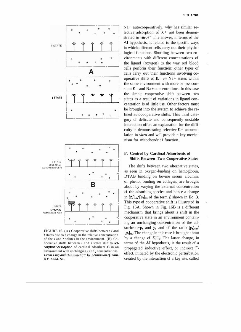
G . N. LING
j STATE lrrrrr.
i STATE (CARDINAL
ADSORBENTOFF)
(CARDINAL ADSORBENT ON)
FIGURE 16. (A) Cooperative shifts between i and j states due to a change in the relative concentration of the i and j solutes in the environment. (B) Co- operative shifts between i and j states due to ad- sorption/desorption of cardinal adsorbent C in an environment with unchanging i and j concentrations. From Ling and O~hsenfe ld , '~~ by permission of Ann. NY Acad. Sci.
Na+ autocooperatively, why has similar se- lective adsorption of K+ not been demon- strated in vitro?" The answer, in terms of the A1 hypothesis, is related to the specific ways in which different cells carry out their physio- logical functions. Shuttling between two en- :
vironments with different concentrations of the ligand (oxygen) is the way red blood cells perform their function; other types of
'
cells carry out their functions involving co- operative shifts of K+ + Na+ states within the same environment with more or less con- stant K+ and Na+ concentrations. In this case the simple cooperative shift between two states as a result of variations in ligand con- centration is of little use. Other factors must be brought into the system to achieve the re- fined autocooperative shifts. This third cate- gory of delicate and consequently unstable interaction offers an explanation for the diffi- culty in demonstrating selective K+ accumu- lation in vitro and will provide a key mecha- nism for mitochondria1 function.
I?. Control by Cardinal Adsorbents of Shifts Between Two Cooperative States
The shifts between two alternative states, as seen in oxygen-binding on hemoglobin, DTAB binding on bovine serum albumin, or phenol binding on collagen, are brought about by varying the external concentration of the adsorbing species and hence a change in [pi],,/[p,],, of the term shown in Eq. 3. This type of cooperative shift is illustrated in Fig. 16A. Shown in Fig. 16B is a different
-
mechanism that brings about a shift in the cooperative state in an environment contain- :
ing an unchanging concentration of the ad- sorbent~ p, and pj and of the ratio [pJ,,/ [p,],,. The change in this case is brought about by a change of K ; ' ~ . The latter change, in terms of the A1 hypothesis, is the result of a propagated inductive effect, or indirect F- effect, initiated by the electronic perturbation created by the interaction of a key site, called
G . N. LING 67
the cardinal site, with a cardinal adsorbent. This phenomenon is analogous to the effect of the magnet or of the electrically charged rod upon the chain of nails or the insulators, respectively, as previously discussed.
Figure 16 illustrates a step-by-step propa- gated indirect F-effect initiated by the cardi- nal adsorbent (C). Whereas the diagram illus- trates the stepwise mechanism of the all-or- none transition from one to the other state, the result also brings on a change in the re- activity of all the functional groups of the side chains due to the difference in the net electron-donating and electron-withdrawing effects of an a-a+ entity compared to those of the b-b+ entity where a+b+ are proton- donating groups or molecules and a-b- are proton-accepting groups or molecules. These affected side chains could include SH groups, with altered chemical reactivity, or P- and y- carboxyl groups, with altered c-values, as examples of targets of the inductive effect. Although the normal physiological function of hemoglobin in red cells require no cardinal site-mediated cooperative transition, the oxy- genation-deoxygenation curve has been known to be influenced by 2,3-diphosphoglycerate and inositol hexaphosphate204 and has been quantitatively described by a set of equations derived from the basic Eq. 3.205 Similarly, the adsorption of K+ and Na+ by frog muscle cells can be controlled by the cardiac glyco- side ouabain, again quantitatively described by a similar set of equations derivable from Eq. 3.?06 One difference between hemoglobin
- and myosin in this parallel behavior is that the myosin system is much less stable. The
. process of isolating myosin from its compli- cated "environment" has led to an even lower energy state and a loss of its specificity in K+ and Na+ adsorption. Thus the direct demon- stration of this specificity in ion adsorption has been possible only by using the freeze- drying techniques developed by Edelmann (see p. 47 above) and applied to the intact muscle cell.
VI. TENTATIVE MODEL OF ASSOCIATIVE-INDUCTIVE COUPLING MECHANISM FOR ELECTRON TRANSPORT AND OXIDATIVE PHOSPHORYLATION
Thanks to the work of Kanazawa, Kana- zawa and Boyer, Taniguchi and Post, and Knowles and Racker (see Section IV above) ATP formation from Pi and ADP has been shown in model enzyme systems to involve two steps. First, "phosphorylation" of the ATPase by Pi is achieved by interaction with Mg2+ and/or K+. Second, phosphorylation of ADP by the "phosphoenzyme" is achieved by interaction with Ca2+ and/or Na+. To ini- tiate ATP production these two steps are manipulated by equilibration of the enzyme and Pi in a Mg2+- and/or K+-containing medium, followed by dialyzing or otherwise removing the Mg2+ and/or K+ and trans- ferring the "phosphoenzyme" to the second ionic environment containing Ca2+ and/or Na+. Thus production of ATP in vitro in- volves a sequential exposure of the protein enzyme to two different ionic environments.
A. The Coupling Mechanism Hypothesis
To account for these phenomena, I intro- duce here a detailed hypothesis resting on the assumption that in the first environment the ATPase adsorbs Mg2+ and/or KC, and in the second environment the enzymes shift to adsorb Ca2+ and/or Na+. Since there are no living beings in living cells to manipulate the ionic environment of the mitochondria, and since mitochondria do not migrate from one ionic environment to another like circulating erythrocytes, clearly a mechanism different from manipulation or migration must exist if the change from adsorption of Mg2+ and/or K+ to that of Ca2+ and/or Na+ is necessary to bring about the phosphorylation of ADP and the production of ATP. I now suggest such a mechanism, which may be formally hypothesized as follows:
68 G . N. LING
Post~~late I. The generation of ATP is the consequence (as noted above) of two dis- crete steps: "phosphorylation" of ATPase by Pi promoted by adsorption of Mg2+ and/or K+ on the enzyme-and phosphorylation of ADP by the "phosphoenzyme" when the en- zyme adsorbs Ca2+ and/or Na+.*
Postc~late 2. Since free Mg2+, Ca2+, K+, and Na+ are available and more or less con- stant in the natural environment of the mito- chondria, the shift in adsorption from Mg2+ and/or K+ to Ca2+ and/or Na+ is brought about by an autocooperative all-or-none shift of c-value ensemble and a consequent change of the relative preference of the adsorption for Mg2+ over Ca2+ or for K+ over Na+. With reference to Eqs. 2 and 3, such a change in these relative preferences is achieved by a change in 6, not through altering the ratio of the concentration of the two alternative ionic species (K+ vs. Na+, Ca2+ vs. Mg2+) but through changing K7Gi, the intrinsic equi- librium constant for the jth (Mg2+ or K + ) to the ith (Ca2+ or Na+) ionic adsor- bent (see p. 80 below for further discussion.)
Postulate 3. The respiratory chain units are cooperatively linked, through operation of the indirect F-effect, to the ATPase at each cou- pling site. As discussed earlier (p. 63), autocooperative linkage can occur among sites on the same protein chain and among closely associated sites on different protein chains (e.g., cooperative interaction among the four subunits of a hemoglobin molecule). This postulate offers the propagated inductive effect as a bridge between the respiratory chain centers and the ATPase site.
Postulate 4. The autocooperative linkage between the respiratory chain center and the ATPase allows the c-value ensemble of the
*The specific ions mentioned in these pos- tulates are used to illustrate the principles involved. In other systems, different ions may be operative. Thus in spinach chloroplasts, H + rather than K + would more likely play the equivalent role.
ATPase to be controlled by the electron state of the respiratory chain center: when the re- spiratory chain center is in the reduced state, the c-value ensemble of the ATPase has a discretely different set of values. At one set of c-values, Mg2+ and/or K+ would be prefer- entially adsorbed; at the other set, Ca2+ and/ or Na+ would be adsorbed. In other words, the associated pyridine nucleotide, ubiqui- none, and the iron in the iron sulfur proteins and in the cytochromes act as "cardinal adsor- bents" on the cooperatively linked ATPase much as oxygenation and deoxygenation of the heme iron in a hemoglobin molecule can allosterically affect the pK, value of distant anionic groups [the alkaline Bohr effect (see p. 70 below)]. Thus each cycle of oxidation and reduction of the respiratory chain center is potentially capable of generating one ATP n~olecule.
In summary, the coupling mechanism hy- pothesis holds that the coupling between "electron transport" and "phosphorylation" is through cooperative shifts, mediated by the induction effect, between two different states of the ATPase system, permitting sequential formation of enzyme phosphate complex (EP) and then of ATP from ADP and EP. Selective ionic adsorption plays a key role in the mecha- nism. This hypothesis has its roots in ( I ) the universality of the inductive effect in chemis- try, (2) the strong evidence that cellular cat- ions exist in an adsorbed state, and ( 3 ) the observed indications that selective cation ad- sorption on proteins in living cells ,is con- trolled by cardinal adsorbents. Additional :
support can be found in the behavior of sev- eral other model systems (see below).
The proposed coupling model represents a special aspect of the basic concept of the liv- ing cell presented in the A1 hypothesis. As such, this coupling mechanism has little in common with chemiosmotic coupling mecha- nisms, which are based entirely on the lipid membrane concept. However, the present model, at least in formal terms, bears some
G. N. LING 69
resemblance to Slater's chemical intermediate hypothesismi and even the conformation hy- potheses of BoyerzOhnd Green.i6 Kell's re- v i e ~ ~ ~ ! ' d e m o n s t r a t e s an open-mindedness regarding new and different ideas and holds the promise of more unity and coherence in the field of cell physiology.
B. Comparison with Other Models
While the usefulness and validity of the present model remain to be decided by future experimental studies, our knowledge to date should allow us to determine whether oxida- tion and reduction of prosthetic groups can indeed influence selective ionic adsorption, since it has already been established that such adsorption leads to ATP systhesis. This oxi- dation-reduction mechanism applies espe- cially to the two heme proteins, hemoglobin and cytochrome c. Unlike cytochrome c, hemoglobin does not undergo the transition from F e w to Fez+. However, it is also well known that the oxidation-reduction equili- brium of hemoglobin (Fez+) and methemo- globin (Fe3+) bears a close resemblance to oxygenation-deoxygenation equilibrium (ref. 194, p. 188).
1. Heme-Heme Interaction and the Bohr Effect
In his 1979 review on hemoglobin, Per- utzz10 stated that the regulation of heme's oxygen affinity by the structure of globin could in principle be accomplished by induc- tive effects (ref. 210, p. 383). Nevertheless, he reiterated his mechanical interpretation of heme-heme interaction. Much of Perutz's argument in favor of the mechanical model was based on studies of hemoglobin crystals and evidence that structures similar to those seen in the crystalline state are maintained in solution. Space does not permit an exhaustive analysis of alternative theories; however, con- trary to Perutz's analysis, the long-range ef- fect could be modulated primarily through an inductive mechanism, with mechanical steric factors playing an auxiliary role. Supporting that view are the following observations:
( I ) In solution, hemoglobin does not main- tain the kind of mechanical rigidity needed for a purely mechanical interpretation of heme-heme and other interactions. Ease of oxygen uptake and release of hemoglobin both in solution and as part of the living erythrocyte are absolutely essential and well known by observation. The behavior of he- moglobin crystals does not allow such ready uptake and release. When deoxyhemoglobin crystals combine with oxygen, the protein crystals break up;211 crystals of oxyhemo- globin firmly retain oxygen when in a low-
oxygen environment (ref. 187, p. 280). (2) The mechanical feature of a rigid T-
structure in the deoxygenated state and a relaxed R-structure in the oxygenated state is not compatible with the lower entropy asso- ciated with the oxygenated state rather than the deoxygenated state.212
( 3 ) The rigid, impervious deoxygenated state of hemoglobin is not readily reconcilable with the binding of as many as 10 molecules of bromthymol blue to each chain of the de- oxygenated state but not to chains of the oxygenated ~ t a t e .~ l3
(4) Reactivity of the sulfhydryl groups is strongly influenced by whether the hemo- globin is in the oxygenated or deoxygenated state, much more reactivity being exhibited in the oxygenated state. Yet a simple me- chanical steric hindrance cannot easily ex- plain why only oxygenated hemoglobin reacts with mercuric benzoate and neither oxygen- ated nor deoxygenated hemoglobin react with i~doacetate."~ A combination of steric hin- drance and a difference in the induction- dependent oxidation-reduction potentials of these reagents seems to provide a more com- plete explanation (see Fig. 5).
2. Oxidation-Reduction Controlled Autocoopera-
tive Ion Adsorption Shifts
The observations cited above, in short, make it seem that cooperative transitions be- tween oxygenated and deoxygenated states mediated by the inductive effect are more ca-
70 G. N. LING
pable of reconciling findings that cannot be readily explained by the mechanical steric in- terpretation alone. Oxygenation. and deoxy- genation, like oxidation and reduction, can in- deed control the transitions between the two cooperative states, and in each state, as ob- served, many if not all of the functional groups exhibit different properties. It should next be asked, then, whether these transitions also involve shifts in the states of ion binding? In answer, one can cite observations of cyto- chrome c, hemoglobin, and pyridine nucleo- tides.
a. CYTOCHROME C. In this, the most ex- tensively studied cytochrome of the respira- tory chain, Margoliash et aL215s216 have dem- onstrated that ferrocytochrome binds only cations, including Mg2+, whereas ferricyto- chrome binds only anions, including C1-, Pt, and ADP. Thus oxidation-reduction changes clearly can alter ion binding in an all-or-none manner.
b. HEMOGLOBIN. Although hemoglobin is not a part of the electron transport system, its similarity of behavior makes it another useful model of the cytochromes. It has long been known that the effects of KC1 and NaCl on the in vitro oxygen-dissociation constant of hemoglobin are different.217*21s Tosteson and c o ~ o r k e r s , ~ ~ ~ o r example, have shown that in the red cells of patients with sickle-cell anemia, deoxygenation produces not only sickling but a profound change in the intra- cellular levels of K+ and Na+. Since it is well established that sickle-cell anemia involves only one gene and one faulty amino acid resi- due in the p-chain of hemoglobin, the Toste- son findings suggest two conclusions. First, hemoglobin carries sites which, under physi- ological conditions, adsorb K+ selectively. Second, the faulty amino acid in the p-chain permits deoxygenation of the heme group, altering the c-value of the sites that adsorb K+ such that a value is reached at which the preference for K+ over Na+ is reduced.
Although the oxygenation-deoxygenation
process differs from oxidation-reduction in degree, it is clear that oxygenation also in- volves a strong electrostatic effect and so is likely to produce similar short- and long- range induction effects. With this parallel in mind, the familiar Bohr effect and reverse - Bohr effect are seen to be outstanding con- firmation that prosthetic group reaction may lead to changes in the pK, value (and hence - the c-value) of acidic groups. In the case of lamprey hemoglobin,220 the large and steep Bohr effect is compatible with autocoopera- tive interaction even among the anionic groups, most likely p- and v-carboxyl as al- ready demonstrated in the case of bovine serum albumin interacting with DTAB (Fig. 13).
c. PYRIDINE NUCLEOTIDES. The most direct evidence that the oxidation-reduction state of the mitochondria1 electron transport system can determine cooperative adsorption of cat- ions essential for ATP synthesis has been provided by Lehninger et a1.221 in both rat liver and heart mitochondria as well as in Ehrlich ascites cells. Those workers showed that the mitochondria take up Ca2+ and hold it as long as the pyridine nucleotide is in the reduced state; when the nucleotide is oxidized, Ca2+ is promptly released.
VII. INTERPRETATION OF OTHER MITOCHONDRIAL PROPERTIES IN TERMS OF THE A1 HYPOTHESIS
A. Additional Basic A1 Concepts
The preceding discussions have demon- strated how the series of experimental find- - ings initiated by Jangendorf and Uribe'ssD studies of chloroplasts in spinach provided what was considered definitive suppor,t for the chemiosmotic hypothesis. Such interpreta- tion was then called into question by the revolutionary findings of Kanazawa and others. A coupling mechanism based, under the A1 hypothesis, on ionic association and
G. N. LING 71
the inductive effect as well as oxidation and reduction of the respiratory chain, was pro- posed above to better account for the obser- vations.
The A1 hypothesis is sufficiently powerful, however, to allow cogent reinterpretation of other significant findings that until now have been interpreted almost entirely on the basis of modified versions of the membranepump theory. These findings have to do with the mechanism of action of uncouplers and iono- phores, with steady oscillatory swelling and shrinkage, and with mitochondria1 electrical potentials. Hence certain additional funda- mental concepts of the A1 hypothesis must be discussed.
1. Physical State of the Bulk of Cell Water in
Living Cells
According to the A1 hypothesis, the bulk of cell water exists as polarized multilayers in a matrix of extended protein chains contain- ing an alternating array of negatively charged CO groups and positively charged NH groups. Water in the state of polarized multilayers differs from normal liquid water-as found, for example, in isotonic Ringer's solution- but also differs profoundly from ice. In an ice crystal (i.e., Ice I ) , individual water mole- cules are fixed in positions that can be re- vealed by "instantaneous" photography. Neither normal liquid water nor water in t5e state of polarized multilayers can reveal much long-range structure in an instantaneous photograph. However, normal liquid water
- and water in the state of polarized multilayers are also profoundly different from each other in that if many serial photographs are taken from a fixed position, the photograph of liquid water will remain free of structure while water in the state of polarized multi- layers will give sharper and sharper images as the number of exposures is increased. Thus water in the state of polarized multilayers can be said to possess structure-not the usual kind familiar to crys,tallographers, to be sure,
but dynamic structure, perhaps more like a sea-scape as seen by impressionist paint- ers.35,41,222,223
This theory of cell water has received ex- perimental c o n f i r m a t i ~ n . ~ ~ ~ . ~ ~ ~ I t has been shown that when the CO and NH groups of a protein molecule are locked in internal H- bonds (e.g., a-helix, p-structure), the water surrounding the molecule is minimally af- fected by the protein. On the other hand, if because of structural changes or the presence of denaturants that disrupt secondary struc- ture, the CO and NH groups in these proteins are directly exposed to bulk-phase water, the properties of the water will be altered. In this altered state, water partially excludes Na+, sucrose, and glycine, much as living cells do. Furthermore, the apparent equilibrium dis- tribution coefficient, p, between polymer- oriented water and external normal water decreases with increasing size and complexity of the solute molecules, showing a pvalue close to unity for small and simple molecules like methanol but lower for larger molecules like sucrose or i n~ l in ,~~%uch as has been demonstrated for living ~ e l l s . ~ ~ T h e s e find- ings support the A1 hypothesis concerning the mechanism for the partial exclusion of Na+ and other solutes in whole living cells or iso- lated parts of living cells. They also offer in- direct evidence in support of another tenet of the A1 hypothesis, according to which hormones, drugs, and other biologically ac- tive agents can manipulate cell properties by controlling shifts of protein conformation from *helical or other close structures, which accommodate water largely in the free state, to the extended open structure, which orients deep layers of water with reduced solubility as well as permeability to Na+, sucrose, and other solu tes.lO2J04
2. Swelling and Shrinking
According to the A1 hypothesis, the exis- tence of the bulk of cell K+ in an adsorbed and thus largely osmotically inactive form re-

72 G. N. LING
moves about half the osmotic activity of the living cell compared with that of the essen- tially isotonic solution of dissociated NaCl in the tissue fluid. This consideration alone ren- ders untenable the idea that living cells be- have like osmometers, as assumed by the chemiosmotic hypothesis.
Indeed, it has been shown that the linear relation between cell volume and external ("impermeant") solute concentration is largely fortuitous and due to the limited range of solubility of sucrose, salts, etc. in water used to prepare the hypertonic solution. When the much larger range of water activity pro- vided by exposing the cell indirectly (via an air phase) to different concentrations of sul- furic acid and sodium chloride are applied in cell shrinkage studies, the swelling-shrinkage curve is no longer linear as required by the osmometer concept. Rather, that curve as- sumes the familiar S-shape typical of polar- ized multilayer adsorption.226 Osmotic pres- sure, after all, is not an actual pressure but an expression of the reduced water activity in the cell; it is a "pressure" not exerted solely by free intracellular ions but also to a large extent by the long-range effect of the ex- tended protein chains.
In support of this view, it has been found that ( I ) muscle cells swell in hypotonic solu- tion to the same degree whether or not the membrane is intact;227 (2) as shown by Troschinm and other Soviet scientist^,^^^^^^^^^^ cells shrink in a concentrated solution of per- meable solutes (e.g., urea, alanine); and (3) cells swell in hypotonic salt solutions and shrink in hypertonic solutions, greatly resem- bling the behavior of dialysis bags filled with solutions of polymers that contain oxygen atoms spaced two water molecules apart as in an extended polypeptide These polymers are like urea-denatured proteins- able to polarize water in multilayers. When the polymer solution is enclosed in standard dialysis tubing and exposed to dilute or con- centrated salt solutions, the sacs will swell or
shrink even though the wall of the dialysis tubing is obviously freely permeant to the salt ions.z30 On the other hand, if the dialysis bag
is filled with a solution of a polymer that does not polarize water in multilayers, the sac will only swell and will not shrink permanently.
But polarization and orientation of water represents only one aspect of the swelling- shrinkage problem. To maintain cell volume, - often there are also restraining forces against expansion. The A1 hypothesis proposes that the restraining forces may reside among the salt-linkages between protein c h a i n ~ . ~ , ~ ~ l It is interesting to note that in recent years the im- portance of salt linkage has gained more and more recognition, resulting to some degree from the development of precise X-ray crys- ta110graphy.~~~
According to the A1 hypothesis, a high con- centration of KC1 causes swelling of muscle cells with or without an intact membrane227 o-wing to dissociation of the restraining salt- l i n k a g e ~ . ~ J ~ ~ . ~ ~ * Thus
where f + represents fixed cations (e.g., E-
amino, guanidyl groups) and f- represents fixed anions (e.g., p- and y-carboxyl groups). The preference of the fixed anion f- at its specific c-value for K+ over Na+ and the preference of the fixed cation f + for C1- rather than account for the effective- ness of KC1 in causing swelling and the in- effectiveness of NaCl or K,S04 at the same concentrations.
3. Polarized Water Rather than Membrane Lipid - as Seat of Selective Permeability
In 1973, I presented evidence that a mem- brane containing polarized water in multi- layers (i.e., hydrated cellulose acetate mem- brane) much h o r e accurately reproduces the selectivt: permeability properties of the living cell surface than does a pure lipid layer as
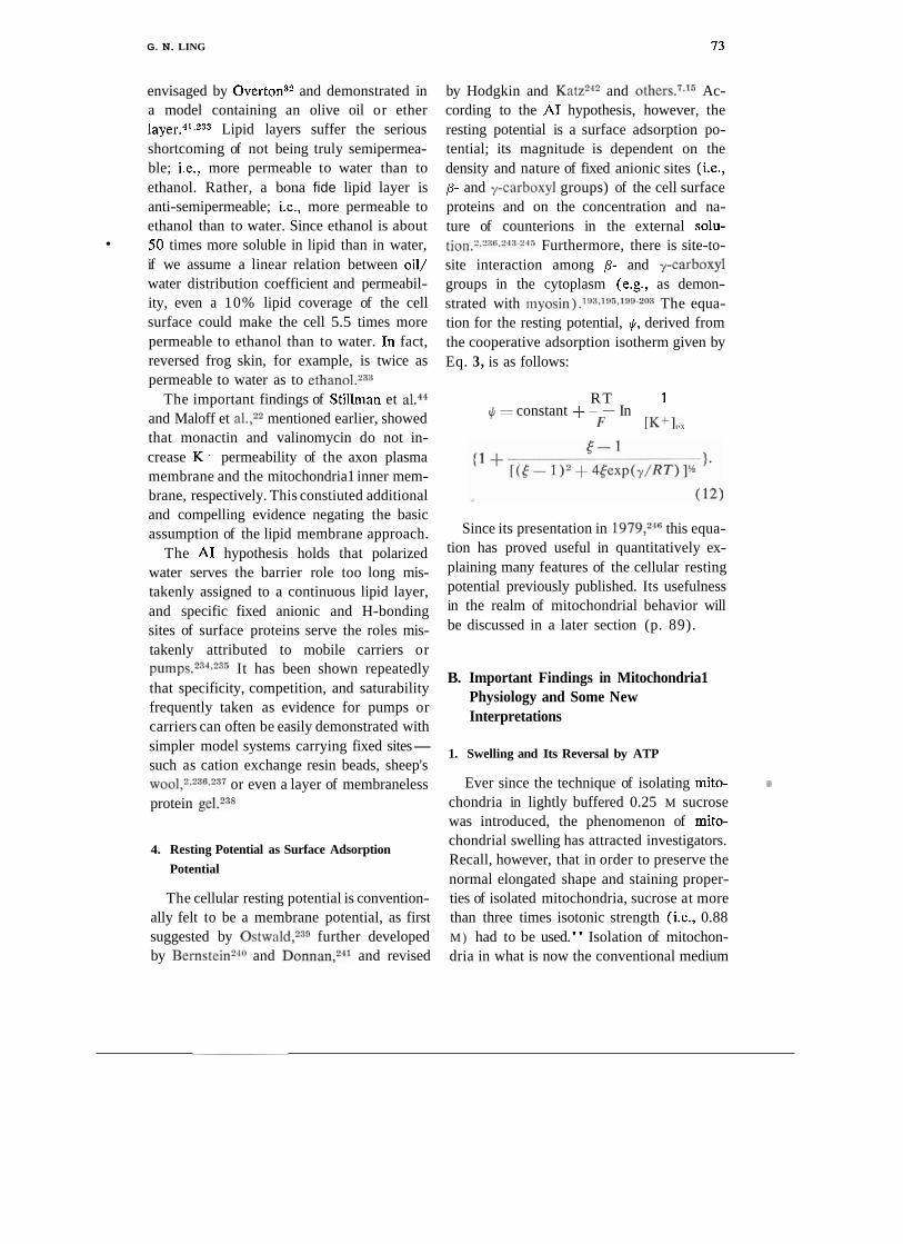
G. N. LING 73
envisaged by Overton" and demonstrated in a model containing an olive oil or ether layer."s2" Lipid layers suffer the serious shortcoming of not being truly semipermea- ble; i.e., more permeable to water than to ethanol. Rather, a bona fide lipid layer is anti-semipermeable; i.e., more permeable to ethanol than to water. Since ethanol is about 50 times more soluble in lipid than in water, if we assume a linear relation between oil/ water distribution coefficient and permeabil- ity, even a 10% lipid coverage of the cell surface could make the cell 5.5 times more permeable to ethanol than to water. In fact, reversed frog skin, for example, is twice as permeable to water as to
The important findings of Stillman et a1.44 and Maloff et a1.,22 mentioned earlier, showed that monactin and valinomycin do not in- crease K+ permeability of the axon plasma membrane and the mitochondria1 inner mem- brane, respectively. This constiuted additional and compelling evidence negating the basic assumption of the lipid membrane approach.
The A1 hypothesis holds that polarized water serves the barrier role too long mis- takenly assigned to a continuous lipid layer, and specific fixed anionic and H-bonding sites of surface proteins serve the roles mis- takenly attributed to mobile carriers or p ~ m p s . ~ ~ * * ~ " It has been shown repeatedly that specificity, competition, and saturability frequently taken as evidence for pumps or carriers can often be easily demonstrated with simpler model systems carrying fixed sites- such as cation exchange resin beads, sheep's ~ o o 1 , ~ . ~ ~ ~ * ~ ~ ~ or even a layer of membraneless protein gel.238
4. Resting Potential as Surface Adsorption
Potential
The cellular resting potential is convention- ally felt to be a membrane potential, as first suggested by O ~ t w a l d , ~ ~ ~ further developed by Bern~tein'~" and D ~ n n a n , ~ ~ ~ and revised
by Hodgkin and Katz242 and ~ t h e r s . ~ J ~ Ac- cording to the A1 hypothesis, however, the resting potential is a surface adsorption po- tential; its magnitude is dependent on the density and nature of fixed anionic sites (i.e., p- and y-carboxyl groups) of the cell surface proteins and on the concentration and na- ture of counterions in the external solu- tion.2*"K2-l"24s Furthermore, there is site-to- site interaction among p- and y-carboxyl groups in the cytoplasm (e.g., as demon- strated with myosin).1m~1m~1m-203 The equa- tion for the resting potential, $, derived from the cooperative adsorption isotherm given by Eq. 3, is as follows:
RT 1 = constant +- In ---
F [K+I,,
Since its presentation in 1979,246 this equa- tion has proved useful in quantitatively ex- plaining many features of the cellular resting potential previously published. Its usefulness in the realm of mitochondrial behavior will be discussed in a later section (p. 89).
B. Important Findings in Mitochondria1 Physiology and Some New Interpretations
1. Swelling and Its Reversal by ATP
Ever since the technique of isolating mito- 7
chondria in lightly buffered 0.25 M sucrose was introduced, the phenomenon of mito- chondrial swelling has attracted investigators. Recall, however, that in order to preserve the normal elongated shape and staining proper- ties of isolated mitochondria, sucrose at more than three times isotonic strength (i.e., 0.88 M ) had to be used." Isolation of mitochon- dria in what is now the conventional medium

74 G. N. LING
of 0.25 M sucrose is clearly a compromise between convenience and best preservation. Brenner-IIolzach and R a a f l a ~ b ~ ~ ~ noted in 1954 that spontaneous swelling of mitochon- dria occurs when their ATP content falls be- low a certain level. Seven years later Nakao et al.2-'8 noted that as the ATP concentration in human erythrocytes declines, the biconcave cells first develop crenation and then, when the ATP content reaches a still lower and critical concentration, the red cells swell into a spherical shape. In isolated mitochondria as in erythrocytes, then, it is the concentra- tion of ATP and not its rate of hydrolysis that determines shrinkage or swelling. In other words, it is not hydrolysis of ATP re- leasing a packet of energy believed to reside in a "high-energy phosphate b ~ n d , " l ~ ~ - l ~ * but rather the presence of ATP per se, that de- termines the normal mitochondrial or cellular shape and size. The view that ATP itself maintains the shape and structure of cells and its constituent parts by its adsorption on cell proteins was first expressed by Ling in 1952.1°2
However, to account for the mitochondrial findings under the A1 hypothesis, a further revision of accepted membrane-pump and chemiosmotic concepts needs to be consid- ered. For example, there is ooly one type of swelling and shrinkage according to the mem- brane-pump theory. It posits that the activity of water is higher in a hypotonic solution than in an isotonic solution but lower in a hyper- tonic solution. As a result, water moves from a hypotonic solution into the cell or from the cell into a hypertonic solution to reach ther- modynamic equilibrium. Does this thesis hold? Let us compare it with that of the A1 hypothesis, which proposes three types of swelling/shrinkage mechanisms active in liv- ing cells and subcellular organelles, with con- sequent changes in cell and/or organelle volume, as follows: (I) Simple changes due to equilibration with water that has altered activity. (2) Changes due to dissociation or
association of macromolecular salt-linkages. ( 3 ) Changes due to polarization or depolariz- ation of water. Let us examine these in turn.
a. TYPE I. SIMPLE SWELLING AND SHRINK- AGE. For nearly a century the majority of biologists have believed that maintenance of
- osmotic equilibrium is governed by a semi- permeable membrane and free intracellular !
K+ and other solutes that balance the osmo- tic activity of free Na+, C1-, and other solutes in the external medium. However, compelling evidence indicates that K+ is in fact adsorbed within the cell (see p. 47 above). Cells with- out an intact membrane continue to swell normally in hypotonic solution. Addition of permeant solutes such as urea nevertheless causes cell shrinkage. Clearly, then, the os- mometer concept is no longer tenable. Far more convincing is the A1 view that main- tenance of cell volume in an isotonic NaCl or sucrose solution represents a condition of balance among several opposing trends of water movement. The circumstances are:
First, certain intracellular proteins exist in an extended conformation, forming a matrix throughout the cells. In this conformation, these proteins maintain the bulk of cell water in a state of polarized multilayers. Water in this state has reduced activity. If proteins and polarized water were the only constituents of the cell, and if the cell were immersed in dis- tilled water, the cell would swell because the activity of the distilled water is greater than that of the polarized water; water would natu- rally move toward the phase with less water activity.
Second, if the external medium were a - sodium chloride solution (0.1 M) , the ac- tivity of the extracellular water would be lower than that of distilled water, and conse- quently, water would begin to move from the hypothetical water-filled cell toward the ex- ternal solution.
Third, as water moved out of the cell, Naf
G. N. LING 75
and C1- would move into it. If the cell water were simply normal liquid water, NaCl would reach the same concentration within the cell as in the external solution. When this condi- tion would be reached, intracellular water ac- tivity would again be lower than extracellular activity owing to the presence of water-polar- izing proteins, and again the cell would swell.
* However, if the cell water were not normal liquid water but existed in the state of polar- ized multilayers, the extent of water move- ment would be different. In that state, water has reduced solubility for Na+ and C1-; i.e., the equilibrium distribution coefficient* for Na+ as chloride, of q ~ , , , ~ , , would be below unity, say 0.1. Under this condition, water would reach a different equilibrium level be- tween the cell and its environment. The total reduction of water activity in the cell, then, would be due to the combined effects of first, the extended protein chains; second, the re- duced levels of Na+ and C1- in the cell water; and third, the tension or mechanical pressure due to cohesive salt-linkages and H-bonds among the protein chains.
Under the A1 hypothesis, therefore, simple swelling and shrinking imply that neither the mechanical tension nor the q-value for NaCl or other solutes present in the external solu- tion has changed during the process.
b. TYPE 11. DISSOCIATIVE SWELLING. In a previous section, mention was made of salt- linkage participation in the maintenance of cell volume by restraining the cell from gain- ing more water. Salt-linkages can be dissoci- ated by salt ions, which would cause swelling. However, this dissociative action is limited to
*Note that the q-value, which refers only to solutes in the cell water o r gel phase in comparison to the external concentration, is different from the p-value, the apparent equilibrium distribution co- efficient, which may include adsorbed o r other non- dissolved components.
salt ions that can compete successfully against the affinity of the fixed anions for the fixed cations and vice Swelling of nor- mal frog muscle in isotonic KC1 involves no depolarization of cell water exemplifying Type IIA
Furthermore, under the A1 hypothesis the relative affinity of dissociating cations is not constant but varies with the c-value of the anion groups in a manner shown in Figs. 7 and 8. Since the c-value is not a constant but can be controlled by very low concentrations of cardinal adsorbent, which modulates the affinity of fixed ion pairs relative to that for free competing ions. Swelling of ATP- depleted frog muscle in isotonic NaCl (or Ringer solution) involves depolarization of cell water exemplifying Type IIB swelling.
c. TYPE 111. DESORPTIVE SWELLING. In both simple swelling and dissociative swell- ing, no fundamental disturbance occurs in the physical state of the intracellular adsorbed ion (e.g., K + ) .
An entirely different kind of swelling oc- curs if the water remains polarized while K+ becomes desorbed. Under this condition the total osmotic activity of the cell will exceed that of an isotonic Ringer solution. As a re- sult the cell will swell.
Type I11 swelling may occur during the early stage of cell damage and ATP depletion and is usually a transient phenomenon.
2. Reappraisal of Previor~s Reports According to
the A1 Hypothesis
a. ATP AND SWELLING/~HRINKAGE OF
FETAL AND ADULT RAT LIVER MITOCHON- DRIA. P ~ l l a k ~ ~ ~ isolated mitochondria from the livers of fetal and adult rats, placed the material in concentrations of sucrose ranging from 50 to 500 mM, and recorded the optical extinction at the wavelength of 545 nm until constant readings were obtained. Liver mito- chondria from 5-day prenatal rats showed no swelling or shrinkage throughout the range of

76 G . N. LING
concentrations. Mitochondria from adult rats showed pronounced shrinkage, whereas those from the 3-day and I-day prenatal rats showed moderate shrinkage. The virtually total insensitivity of the 5-day prenatal rat liver n~itochondria to a 10-fold increase in external sucrose concentration could mean only that the mitochondrion is totally perme- able to sucrose and-equally important-that it did not exclude sucrose. Otherwise, the mitochondria would have shrunk as men- tioned earlier.
Prior to Pollak's study, Nakazawa et a1.250 had shown that the ATP and ADP contents of total rat liver mitochondria are very low dur- ing gestation but steadily increase from five days before birth to adulthood. Addition of ATP to the mitochondrial suspension greatly improves the respiratory control. Further- more, the addition of ATP and Mg" enabled respiring fetal mitochondria to release H+ in response to the addition of Ca2+, as occurs in adult mitochondria.
P01lak~~l further demonstrated that both ATP-induced shrinkage of fetal liver mito- chondria and enhancement of respiratory control depend not on hydrolysis of ATP but on the presence per se of ATP, which inter- acts with the mitochondrial inner membrane. In 1973 Stoner and S i r ~ i k * ~ ~ demonstrated that contraction induced in bovine heart mito- chondria by ATP, ADP, and pyrophosphate has nothing to do with their hydrolysis, since they are equally or more active in the presence of oligomycin plus cyanide. These authors concluded that "inner membrane contraction occurs as a result of adenine nu- cleotide binding. . . ."
To explain the data, a suggestion made several years ago2,226 is reiterated here. In adult resting cells, ATP is a m.ajor cardinal adsorbent that maintains certain matrix as well as membrane proteins in an extended water-polarizing conformation. Loss of ATP
from the cell causes Type 111 and/or Type IIB swelling; gain of ATP reverses it.
b. Mg" AND Ca2+ vs. SWELLING/~HRINK- ACE. It is possible that ATP is not the only cardinal adsorbent controlling water polariza- = tion? Mg" may be a co-cardinal adsorbent working synergistically with ATP. In the event that Mg" is competitively displaced - from its site by CaZ+, the effect of Mg + ATP may then be reversed. This possibility is sug- gested by findings reported by Hunter et a1.253 Those authors found that mitochondrial swelling induced by exposure to hypotonic solutions does not involve uncoupling of oxi- dative respiration. From this they concluded that "our results on hypotonically swollen mitochondria dismiss the long-held view that swollen mitochondria must be damaged and uncoupled. . . ." This type of swelling may be considered Type I.
One also recalls that in 1955 Hunter and Ford2" had noted likewise that simple os- motic swelling did not interrupt normal oxi- dative phosphorylation. Apparently no coop- erative changes of the c-value ensemble or of the state of water occur. Hunter et al. had found that the addition of Ca2+ produce an increase of sucrose-available space from about 85% to nearly 100%. Furthermore, this increase is not specific to sucrose but ap- plies to choline and glucose as well. All these responses can be promptly reversed by the addition of ATP and Mg2+. Again, I suggest that addition of Ca2+, like depletion or lack of ATP, causes Type IIB swelling; ATP and ' Mg2+ reverse this swelling by causing Type IIB shrinkage, producing long-range water . polarization, and restoring low q-values for sucrose as well as other normally excluded solutes. Again, this interpretation is generally consistent with that of Hunter et who concluded that the effects of ATP and Mg2+ require neither electron flow nor energy but rather the mere accessibility to Ca2+ of some internal sites.

G. N. LING
c. PASSIVE OSMOTIC SWELLING. While sub- strate oxidation or external ATP are essential for swelling, such as that induced by Ca2+ and a variety of other agents studied by Lehninger and many others.ls Brierly et a1.i8
, found that isolated heart mitochondria will swell even when substrate or ATP is absent; however, they will also swell in the presence of a permeant anion and a permeant cation. In terms of the A1 hypothesis, permeant anions, defined primarily on the basis of their capacity to induce swelling, are anions prefer- entially adsorbed by fixed cationic sites on cell proteins. Their presence therefore greatly en- hances the probability of Type I1 swelling because they displace salt-linkages (see Eq. 11) .
In contrast, salts of the same cation and the more weakly adsorbed anion C1- may not be able to effect any swelling. As an ex- ample, the fact that mitochondria can undergo swelling in KNO, or NaNOa but not in KC1 or NaCl suggests that NOa- is more strongly adsorbed than C1-. However, Brierly and J a r k o w i t ~ ~ ~ " have also shown that mitochon- dria will not swell in 100 mM K or NaN03 if the pH is neutral but will swell in a pH of 8. This also agrees with the present interpre- tation. Salt-linkages are formed between p- and y-carboxyl groups on the one hand and cu-amino, histidine, a-amino, and guanidyl groups on the other. A change of even one pH unit causes de-ionization of a-amino and histidine, the H + competing with Na+ and K+ and thus weakening the ability of NaNO:3 and KNO:I to disrupt the salt-linkages.
Type I1 swelling as illustrated here is of the , simple kind. A more complicated type, which
depends on the action of cardinal adsorbents, is discussed in a later section.
3. Ion and Substrate Transport
Extensive investigation has been accorded the transport of ions, substrates, and other solutes in and out of living cells. Recent evi- dence (reviewed above, p. 38) indicates
that Overton's postulation of the continuing lipid layer to serve as a selective permeability barrier is erroneous. The A1 hypothesis, how- ever, offers a feasible alternative: water polar- ized by macromolecules has been shown to reproduce more accurately the permeability properties of living cells.2" Such a model is entirely in harmony with the findings that monactin has no effect on K+ permeability of squid axon plasma membrane," and valino- mycin has no effect on K+ permeability of the mitochondria1 inner membrane22 or the cell surface of frog muscles and ovarian egg cells (Ling and Ochsenfeld, unpublished data). The cell membrane contains primarily protein and water. Phospholipid serves a sec- ondary though important role, that of stabiliz- ing the s t r ~ c t u r e ; ~ ~ and except in special cases, as in the myelin of myelinated nerves, the phospholipid does not form a continuous bar- rier. Polarized water has the distinguishing feature of possessing nearly normal solubility and permeability for small molecules, such as water itself or methanol, but progressively lower solubility and permeability for larger and more complex molecules and hydrated ions.226.25G,25i
While surface proteins offer extended poly- peptide chains to polarize deep layers of water, they also offer side chains that can adsorb ions and other solutes. Counterions enter exchange-resin beads and cells not by going between the fixed anionic sites but by a route involving, first, adsorption onto these sites and, second, libration around the sites and eventual desorption and entry.2.2x,2xs243.m Solutes too large and com- plex to enter through the polarized water may enter or leave by an adsorption-desorption mechanism on the inner membrane protein sites. The kinetics of this system are similar to those dkscribed for enzyme activity by Michaelis and M e n t ~ n ; ~ ~ ~ i.e., the adsorption- desorption mechanism exhibits the competi- tion and saturability conventionally regarded as demonstrating a carrier mechanism.

78 G . N. LING
Other fascinating findings from Pollak's laboratory illustrate the usefulness of the association-induction model. For instance, Sandor and Pollak2" showed that the initial rate of entry as well as the levels of 14C- labeled ATP in adult liver mitochondria are inhibited by atractyloside. In contrast, the ini- tial entry rate of fetal mitochondria is con- siderably faster than that in the adult and is totally indifferent to atractyloside. After a peak uptake of 14C-labeled ATP was reached at about 1 min, the level of labeled ATP began a decline, falling to nearly half peak level at the end of 5 min. While the accumulated level of [l4C]ATP was declining it also was developing a responsiveness to atractyloside. Sandor and Pollak interpreted their data to in- dicate the rapid, atractyloside-insensitive en- try of ATP into the permeable fetal mitochon- dria. But once inside, "ATP interacts with the inner mitochondria1 membrane to transform its configuration and permeability character- istics . . . resulting in a diminished matrix space so that labeled ATP may be regarded as literally squeezed out together with water as the matrix becomes very condensed. . . ."
This view of course is in full harmony with my interpretation, given above, of Pollak's studies on reversal of Type IIB swelling. Here a new and intriguing feature is presented- atractyloside further reduced the level of 14C- labeled ATP 2 min after exposure to non- labeled ATP. It is not likely that this could have been due to simple interference from an adenine nucleotide "translocase" operating at the inner membrane. Were that the case, atractyloside would have had no effect on the level of 14C-labeled ATP since the latter was already in the mitochondria and could not leave. A useful future experiment would be to expose fetal mitochondria to 14C-labeled ATP in both the presence and absence of atractyloside to determine with greater cer- tainty whether initial exposure to and accu- mulation of ATP indeed render atractyloside sensitive to further ATP influx.
Sandor and Pollak's e x p e r i m e n t a t i ~ n ~ ~ ~ clearly showed that atractyloside sensitivity is dependent on prior interaction of the fetal mitochondria with ATP, suggesting that maintenance of the polarized water surface barrier, as well as the presence of specific ATP binding sites, require that certain cardi- C
nal sites be occupied by ATP.
FIGURE 17. Diagram of cooperative transition in- duced by a cardinal adsorbent. Top figure shows !he * variation of the free energies of adsorption with changes of c-value analog of the C=O groups and c'-value analog of the NH group. Figures below demonstrate stepwise displacement of b+ and b- ( N H and C=O groups on the lower polypeptide) by a + and a- in consequence of interaction with cardinal adsorbent, C, at extreme left. Overall result of cooperative transition is dissociation of the two peptides (or uncoiling of a helix). Frorn Ling,"' by permission o f In!. Rev. Cytol.
G . N. LING 79
It is also interesting to recall some much earlier observations. Lehninger et aLB8 in 1957 reported that ATP-induced contraction of mitochondria swollen in thyroxine is most effective in the presence of KCl. McFarlane and SpencerGG and Gamble'" also observed that both ATP and K+ are essential for the prevention of swelling. These various findings agree with the theoretical model presented in Fig. 17 and introduced more than a decade agolo4 in which adsorption of K+ on p- and y-carboxyl groups parallels alteration of the protein backbone from extended conforma- tion and long-range polarization of water. Further, in a study of intact frog muscle cells, Ling and Och~enfe ld '~~ demonstrated a quali- tative correlation between selective K+ ad- sorption and the degree of sucrose and Na+ exclusion by cell water.
4. Uncouplers, Ionophores, Ca2+, Mg2+, ATP, and
Other Agents as Cardinal Adsorbents
Why do many chemical agents, although different in type and reactive with differert components of the mitochondria, produce essentially the same physiological effect? This question was first raised by Hunter and FordZ5"n 1955 and again 20 years later by Siliprande et a1.,261 who noted the almost iden- tical effects produced by inorganic phosphate, the sulfhydryl-oxidizing agent diamide, and the divalent-ion "ionophore," A23187. On the other hand, certain agents that could be expected to produce a specific effect not only fail to do so at times but may even produce an opposite effect! Thus oligomycin, an in- hibitor of oxidative phosphorylation, can at certain concentrations actually stimulate oxi- dative phosophorylation in both the forward and reverse directi~n.~" Valinomycin, a spe- cific K+ ionophore that uncouples oxidative phosphorylation in the presence of K+, may stimulate oxidative pho~phory la t ion .~~~ Yet valinomycin in the total absence of K+ or any other ion inhibits electron transport in iso-
lated chloroplasts.2G4~26Woreover, valinomy- cin, which does not combine with or transport Ca2+ or any other divalent ion across experi- mental phospholipid membranes, neverthe- less promotes accumulation in mitochondria of CaS+ in competition with K+.7Wonactin, a specific K + ionophore, does not increase K+ conductance in squid axon membranes44 nor does valinomycin in the presence of K+ increase K+ conductance in isolated liver mi toch~ndr i a .~~
To date, attempts to account for these puzzling phenomena have stemmed chiefly from the prevailing view in the field of mito- chondrial physiology, a view based on Over- ton's lipoidal theory of the cell membrane. Specifically, the inner membrane is supposed to be a continuous sheet of phospholipid serv- ing as a selective permeability barrier. Let us, however, review the data in the light of the totally different assumptions of the A1 hypothesis.
a. MECHANISM OF ACTION OF VALINO- MYCIN ON MITOCHONDRIAL K+ ACCUMULA- TION. According to conventional membrane- pump theory, the major intracellular cations, K+ and Na+, are in the free state. According to the A1 hypothesis, some solutes in the cell are in the free state-but others are not. The equilibrium level of the solutes in the free state, as a rule, is less than or equal to the concentration of the solute in the external medium according to the value of q. The re- maining fraction exists in one or more ad- sorbed states, the equilibrium level of each being described primarily by the parameter 8 and to a lesser extent by --,/2 (see Eq. 2 ) . Now, 8 = ([pi],,/[pj],,)K~,Oi. Thus the level of adsorbed ith solute [pilad depends on ( I ) the ratio of its concentration to that of the major competing ion, pj, in the external medium; and (2) Ky$, the intrinsic equili- brium constant for the i-to-j exchange ad- sorption.
With this dependency in mind, we can ex- tend the A1 hypothesis by suggesting that vir-
80 G . N. LING
tually all K+ in isolated mitochondria in an external medium containing a low concentra- tion of K+ is adsorbed on the p- and y-
carboxyl groups of mitochondrial proteins. Potentially these sites can also adsorb H+ , Rb+, Cs+, Li+, Ca2+, and Mg2+ as well as fixed cations that form salt-linkages. Whether these sites adsorb and thus accumulate K+ or some other ion depends on the c-value and on the proximity of the sites to one another.s6 The "ionophore" valinomycin may function as a cardinal adsorbent and affect the c-value of cation-adsorbing sites.
In retrospect, the theory that adsorption is the basis of selective K+ accumulation, pre- sented in 195126G and 1952,'02 was consid- ered favorably by Berger,135 who was then studying mitochondrial physiology under Lardy, who had expressed a similar view.134 Others, like Gamble,13G Judah et al.,267 Rossi et a l . , 7hnd ~ e r e i j o - ~ a n t a l o , ~ ~ ~ at various times also referred to ion binding. Neverthe- less, the majority of investigators continued to believe that K+, once inside the mitochon- drial inner membrane, must be in the free state or else the cell would suffer osmotic imbalance and shrink. That recurrent theme, although contrary to observation, worked against the concept of K+ adsorption from the first, just as it does now. Indeed, many years ago this argument against the notion of "bound K+" in whole cells was effectively supported by Hill,26%ho convinced many that cell K+ must be free.loO However, as mentioned above, in terms of the A1 hy- pothesis the loss of osmotic activity due to K4 adsorption is compensated for by the long- range polarization of water and thus presents no problem.
One crucial difference between the chemi- osmotic and A1 hypotheses with regard to valinomycin is that in the chemiosmotic view valinomycin can manipulate only the rate of K+ entry but not the final steady-state level of K+. The reason given is that the inner membrane by itself is K+-impermeable. In
the A1 model, on the other hand, valinomycin modulates the steady-state level in a quantita- tive manner. In 1972, Massari et aLSs showed that steady-state levels of K+ not only accu- mulate in the presence of various concentra- tions of valinomycin but indeed do vary with = changes in that concentration. These findings show that valinomycin does not just open a "closed door" to K+ but is related quantita- tively to accumulations of K+ and Ca2+.
Another possible interpretation is that val- inomycin might affect the steady-state level of K+ by manipulating the rate of operation of a K+ pump. However, membrane pumps as the basis of maintaining the steady-state level of K+ in whole resting cells have been dis- p r o ~ e n . ~ . " J ~ ~ Of a11 the pumps postulated for cells,1m the Na-K pump in muscle cells, under controlled conditions, alone would consume at least 15 to 30 times more energy than the total energy available to the whole The idea of additional pumps to control ion flux across the mitochondrial membranes, whose total surface area is 20 times greater than that of the plasma membrane surface, (ref. 270, p. 30), thus consuming many times more energy than the pump originally postulated to exist in the plasma membrane, is untenable.
Does valinomycin in truth interact with mitochondria by combining with cardinal sites? An interesting finding by GriffithZ7l offers some clues. He found that the mito- chondria of certain mutant yeasts lacked sen- sitivity to valinomycin with respect to K+ transport. Since a mutation of this kind is usually the result of an error in the DNA ' replication, and since DNA specifies protein sequence in the mutant, a part of the mito- - chondrial protein must be faulty. I suggest that this faulty or missing part contains the cardinal site that reacts with valinomycin. When Moore and Pressmans4 first reported on the mechanism of action of valinomycin on mitochondria, they concluded that "the triggering of K+ transport by valinomycin im- plies the existence of a mitochondrial recep-
G. N. LING 8 1
tor site. . . ." In terms of the A1 hypothesis, such a receptor site would be called a cardinal site. However, contrary to Pressman, in the A1 model the cardinal sites control K + dis- tribution not via a K-pump but by altering
- the affinity for adsorption of K+ on the other (non-cardinal) protein sites.158
b. IONOPHORES, UNCOUPLERS, AND OTHER %
CARDINAL ADSORBENTS: INDUCTION OF C-
VALUE CHANGE AS BASIS OF THEIR ACTION
ON MITOCHONDRIAL ION DISTRIBUTION. The variety of biologically active agents seems endless. In terms of the A1 hypothesis, this enormous variety resembles that found in the shapes of the "keys" described in Paul Ehrlich's lock-and-key analogy regarding drugs. Though greatly varied in shape, how- ever, the key can do only two things: open a door or close a. door. Cardinal adsorbents also have but two options: donate electrons or withdraw electrons. Thus these agents can be classified into electron-donating cardinal adsorbents (EDC) or electron-withdrawing cardinal adsorbents (EWC). But complexity is introduced by steric factors and the mul- tiplicity of attachment sites, which influence the specificity of agents in achieving the EDC or EWC effect. Another variable, of course, is the degree of intensity of the EDC and EWC actions. All these indicate that whereas the basic action may be simple, the actual observed effect may be extremely complex.
Earlier (p. 67) it was pointed out that a strong EDC in a cooperatively linked site tends to increase electron density (or the c-values of all the negatively charged sites) and also the positive charge density (or the . cr-values of all the positively charged sites). Hence as a first simple assumption we may expect that strong EDCs increase while strong EWCs decrease the c- and c'-values of many of the sites in the proteins of the mitochon- dria. But there are pitfalls in the application of this simple concept. For example:
( I ) One EDC displacing another EDC may create an electron-withdrawing effect if
the first EDC is a weaker electron donor than the second EDC. Conversely, one EWC dis- placing another EWC may produce an elec- tron-donating effect. Thus lack of awareness of a preexisting agent occupying the same cardinal site-which is often the case-could lead to conclusions opposite to the truth.
(2) A stronger EDC reacting with a cardi- nal site adjacent to an anionic site that is the first member of an anionic-cationic sequence may increase the c-value of all the anionic sites and the c'-value of all the cationic sites (see Fig. 18). On the other hand, if the cardi- nal site reacting with the stronger EDC is flanked by a cationic site which in turn is fol- lowed by a sequence of anionic and cationic sites, the result will be just the opposite: a decrease of the c-value of the anionic sites as well as of the c'-value of the cationic sites.
( 3 ) The multiplicity of and interactions among cardinal sites (Fig. 10) constitute an- other source of confusion. An agent may produce opposite effects at different concen- trations.
(4) Assessment of rates of fluxes vs. steady-state levels is tricky. Strictly speaking, changes in c-value can be assessed only on the basis of changes brought about in steady levels of ions accumulated in the mitochon- dria. Unfortunately, such data are relatively rare and often the c-values must be approxi- mated. Because of this ambiguity, c-value changes of bulk-phase sites may be confused with properties of the surface sites.
Because of such pitfalls, the following analyses, based on data collected by investi- gators with entirely different theoretical back- grounds, may need considerable future revision.
Indeed, the difficulties are so formidable, that one may well ask whether there is any point in making even a preliminary assess- ment of the properties of EDC or EWC? The answer is a strong affirmative, because the behavior of mitochondria in particular, and of living cells in general, exhibits a high de-

82 G. N. LING
gree of internal coherence. The best demon- stration of this is the oscillating response of isolated mitochondria, to be discussed later. For the moment, in order to determine if an agent is electron-withdrawing or electron- donating, we must rely on the following:
(i) Theoretical data (Figs. 7, 8) describing change in relative preference between a pair of adsorbed cations in response to the agent applied.
(ii) The reasonable assumptions that oxi- dation or respiration generally involves loss of electrons from the system and hence a c-value decrease, and that reduction of an- aerobiosis generally involves a gain of elec- trons and hence a c-value increase.
1 . Respiration and anaerobiosis. A cation- exchange resin can be made to replace all its counterions; it cannot be made to lose them altogether. As long as there are n moles of fixed anionic sites in the resin, n moles of
countercation must also be present. A living cell shares a number of important attributes with a simple ion-exchange resin bead, but in death-whether brought about by heat or by metabolic inhibition-all of the cell's accu- mulated ions may be lost. The A1 hypothesis has long argued that this is because living cells are neither cation-exchange resins nor anion- exchange resins but a mixed amphoteric ion- exchange system on flexible hai ins.^,^^^ In cell death, adsorbed ions are displaced by the formation of salt-linkages between the fixed anions and fixed cations that in the normal resting cell adsorb free cations and free anions, respectively. There are two reasons for the propensity of living cells to assume a final salt-linkage formation. The first is that the loss of entropy when a salt-linkage is formed is less than that when two free ions are adsorbed; however, if this were the only reason, no free ions would ever be adsorbed.
FIGURE 18. Effect of different concentrations of ouabain on equilibrium distributions of K+ ion in frog muscles. Each point is the average of 4 deteiminations. Standard errors not shown to avoid confusion. Numbers refer to equilibrium molar concentrations of ouabain in the external media. Frorn Ling and Bohr,'" by permission of Physiol. Chem. Phys.
G. N. LING 8 3
The second reason is that in the dying state, electrons flow from fixed cationic sites and other sources to fixed anionic sites as well as to backbone carbonyl groups (raising their c-value or. cr-value analogs), and the high
- c-value anionic sites then combine with high cr-value fixed cationic sites, forming tight salt-linkages. A similar high electron density of the carbonyl oxygen of the backbone pro- motes the formation of strong H-bonds with peptide NH groups of high positive charge.lo4 The internally neutralized amphoteric system is also seen in "dead" proteins, such as sheep's wool and hair.2
Mitochondria are not dead in any of the six metabolic states defined by Chance and W i l l i a m ~ ~ ~ ~ n d Chance.273 However, it is possible that in State 4 the c-value ensemble is fairly high, in agreement with the highly re- duced state of DPNH measured.272 Referring to Figs. 7 and 8, one can then predict sub- stantial retention of H + in State 4 even in a medium of very low H+ concentration (i.e., pH 7.4). In State 1, the respiratory carriers are in a somewhat more oxidized state than in State 4.272 Yet Gear and Lehninger,69 showed that the relative effectiveness of al- kali-metal ions in displacing K+ and H+ fol- lows the rank order Li+ > Na+ > K+ > Rb+-a preference corresponding to that of an anionic site of relatively high c-value (Figs. 7, 8 ) .
Under the A1 hypothesis, I suggest that the mitochondrion may be brought to the highest c-value when the entire respiratory chain is in the reduced state (high substrate concen- tration but no oxygen). The c-value increases - are accompanied by cr-value increases at the fixed cationic sites and extensive salt-linkage formation occurs, "squeezing out" both ad- sorbed cations (e.g., K+) from anionic sites and adsorbed anions from cationic sites (e.g., ATP), consistent with the data of Gamble and H e ~ s . ~ ~ ~ The contraction concomitant with salt-linkage formation (Type I1 shrink- age) is then seen as a strong increase of light-
scattering. On the other hand, respiration, which shifts the chain to a more oxidized state, by definition would lower the c-value. In the theoretically calculated curves (see ref. 2, Fig. 4.10) one notices that H + is greatly preferred over K+ at high c-value and that this preference reverses itself at low c-value. In agreement, it has been known for some time that respiration favors K+ retention in isolated m i t o c h ~ n d r i a . ~ ~ J ~ " l ~ ~ That is to say, respiration reduces the c-value to a level at which K+ preference is increased in relation to H+.
2. "lonophores." Valinomycin causes addi- tional uptake of K+ and release of H + in respiring rat liver m i toch~ndr i a .~~ As just dis- cussed, respiration tends to maintain the elec- tron ch,ain in an oxidized state, corresponding to a low c-value state. The introduction of valinomycin reduces the c-value still further to a level at which the preference of K+ over H + is further increased, and a K+-for-H+ exchange then follows. So it would seem that valinomycin is an EWC. Since A23187 has a similar effect on K+-for-H+ it may also be an EWC.
Pressman showed that gramicidin brings about a gain of both K+ and Na+ exchange for H + from a medium containing 1.5 mM K+ and 1.9 mM Na+.276 Consulting Figs. 7 and 8, one sees that a more moderate de- crease of the c-value than that brought about by valinomycin would bring about the uptake of both K+ and Na+. Gramicidin is thus a weaker EWC than valinomycin.
Pressman also described the response of the mitochondria to gramicidin when the oxy- gen was becoming exhausted: ". . . Upon approach of anaerobiosis (i) K+ ejection oc- curred first. . . (ii) followed by Na+ ejection, (iii) H + uptake and (iv) mitochondria1 con- traction," concomitant with H + ejection. Again referring to Figs. 7 and 8, one sees that the better part of this sequence of events can be explained by a steady increase in the c-value of the anionic sites. Why then was
84 G. N. LING
the H + accumulated at a high c-value also released with an increase of light-scattering after total anaerobiosis? I suggest that at such high c-values, salt-linkage formation and the concomitant Type TI shrinkage become ener- getically more favorable than continued H+ adsorption.
3. Uncouplers. It has long been known that 2,4-dinitrophenol (DNP) causes a loss of K+ and a gain of H+2m in respiring mito- ~ h o n d r i a . ~ ~ ~ ~ ~ ~ ' ~ ~ ~ ' " " That this K+ loss cum H + gain jnvolves an H+-for-K+ exchange caused by a DNP-induced increase in the preference for H+ over K+ is further sup- ported by the following observations. First, DNP inhibits H+ liberation in response to the addition of external K+.07 Second, in- creasing K+ and other alkali metal ions can prevent DNP-induced uptake of H+ in the rank order of effectiveness K+ > NH4+ > Na+.207 Third, while valinomycin causes K+ gain and H+ loss, DNP and valinomycin to- gether cause K+ release-a release that can be prevented by raising the external K+ con- c e n t r a t i ~ n . ~ ~ ~ From these findings it would seem that DNP has an effect opposite to that of respiration and valinomycin and must therefore primarily be an EDC.
In the same way, one can understand why DNP also prevents KC1-induced (Type 11) ~welling.~~"ut how does DNP increase oxy- gen consumption, uncouple oxidative phos- phorylation, and activate ATPase activity? At this stage, only general statements can be made in answer.
It has long been known that K+ is neces- sary for the respiratory activity of mitochon- dria. This requirement as first reported by Pressman and lard^*^^ was disputed.2iR However, later work including that of Krahl et a1.,25Wimmich and Rasmus~en ,*~~ and Gomez-Puyou et a1.281s282 has removed any doubt that the original conclusion of Press- man and Lardy was essentially correct. The A1 hypothesis suggests that rapidly oscillating cooperative adsorption and desorption of K+
accompany the propagated oxidation-reduc- tion cycles along the respiratory chain. Such rapidly oscillating cooperative adsorption- desorption depends on a delicate balance be- tween the c-value and the appropriate pair of counterions. DNP, like ADP acting as an : EDC, perturbs this state of balance and as a result the oxidation rate is increased. In general agreement with this suggestioi:, it will be recalled that DNP stimulates respi- ration only at low concentrations. At higher concentrations DNP actually depresses respi- ration.'"J." Similarly K+, which is necessary to increase DNP-induced oxygen consump- tion at low concentrations, inhibits respiration at high concentrations. These seemingly con- tradictory behavior patterns are to be ex- pected from the kind of complex electronic interactions illustrated in Fig. 10.
That continued respiratory activity depends on a delicately poised state of the respiratory chain proteins adsorbing K+ is also in har- mony with the following findings: ( I ) K+-de- pletion causes inhibition of r e s p i r a t i ~ n . ~ ~ ~ , ~ ~ ~ (2) Valinomycin and gramicidin, which tend to increase K+ in isolated mitochondria, in- crease (3) Nigericin, which causes K+ loss, inhibits respira- tion.28R.2sn (4) ADP-induced respiratory con- trol index increase depends on K+.281 LOSS of respiratory control parallels loss of K+ con- tent.282 (5) DNP added as a supplement to valinomycin causes loss of K+, after which respiration stops; addition of KC1 restores respiration.280
Other evidence supports the notion that in normal mitochondria the delicately balanced adsorption and desorption of K+ depends on , both Mg2+ and ATP acting as cardinal ad- sorbent~: (I) ATP, Mg, and diphosphopyri- dine nucleotide all restore mitochondria1 res- piration and oxidative pho~phoryla t ion .~~~ (2) Mg2+ is required for DNP-activated ATPase activity and oxidative phosphoryla- t i ~ n . ~ ~ " ( 3 ) Respiration of fetal rat liver mitochondria is not stimulated by ADP. The
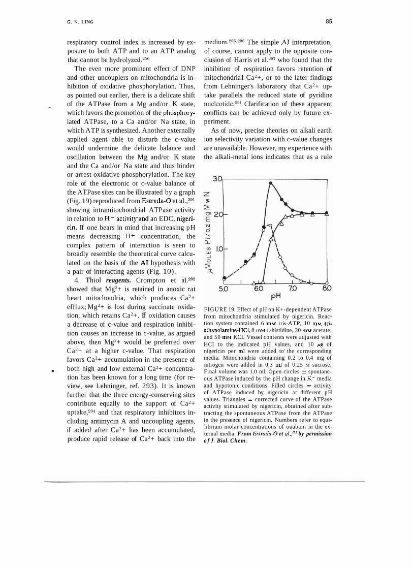
G. N. LING 8 5
respiratory control index is increased by ex- posure to both ATP and to an ATP analog that cannot be hydrolyzed.250
The even more prominent effect of DNP and other uncouplers on mitochondria is in- hibition of oxidative phosphorylation. Thus, as pointed out earlier, there is a delicate shift of the ATPase from a Mg and/or K state, - which favors the promotion of the phosphory- lated ATPase, to a Ca and/or Na state, in which ATP is synthesized. Another externally applied agent able to disturb the c-value would undermine the delicate balance and oscillation between the Mg and/or K state and the Ca and/or Na state and thus hinder or arrest oxidative phosphorylation. The key role of the electronic or c-value balance of the ATPase sites can be illustrated by a graph (Fig. 19) reproduced from Estrada-0 et al.,291 showing intramitochondrial ATPase activity in relation to H+ activitysand an EDC, nigeri- cin. If one bears in mind that increasing pH means decreasing H+ concentration, the complex pattern of interaction is seen to broadly resemble the theoretical curve calcu- lated on the basis of the A1 hypothesis with a pair of interacting agents (Fig. 10).
4. Thiol reagents. Crompton et al.292 showed that Mg2+ is retained in anoxic rat heart mitochondria, which produces Ca2+ efflux; Mg2+ is lost during succinate oxida- tion, which retains Ca2+. If oxidation causes a decrease of c-value and respiration inhibi- tion causes an increase in c-value, as argued above, then Mg2+ would be preferred over Ca2+ at a higher c-value. That respiration favors Ca2+ accumulation in the presence of
. both high and low external Ca2+ concentra- tion has been known for a long time (for re- view, see Lehninger, ref. 293). It is known further that the three energy-conserving sites contribute equally to the support of Ca2+ uptake,294 and that respiratory inhibitors in- cluding antimycin A and uncoupling agents, if added after Ca2+ has been accumulated, produce rapid release of Ca2+ back into the
m e d i ~ m . ~ " " , ~ ~ The simple A1 interpretation, of course, cannot apply to the opposite con- clusion of Harris et a1.2w who found that the inhibition of respiration favors retention of mitochondria1 Ca2+, or to the later findings from Lehninger's laboratory that Ca2+ up- take parallels the reduced state of pyridine n~c l eo t i de .~~ ' Clarification of these apparent conflicts can be achieved only by future ex- periment.
As of now, precise theories on alkali earth ion selectivity variation with c-value changes are unavailable. However, my experience with the alkali-metal ions indicates that as a rule
FIGURE 19. Effect of pH on K+-dependent ATPase from mitochondria stimulated by nigericin. Reac- tion system contained 6 mM tris-ATP, 10 mM tri- ethanolamine-HCI, 8 mM L-histidine, 20 mM acetate, and 50 mM KCI. Vessel contents were adjusted with HCI to the indicated pH values, and 10 pg of nigericin per ml were added to' the corresponding media. Mitochondria containing 0.2 to 0.4 mg of nitrogen were added in 0.3 ml of 0.25 M sucrose. Final volume was 1.0 ml. Open circles = spontane- ous ATPase induced by the pH change in K+ media and hypotonic conditions. Filled circles = activity of ATPase induced by nigericin at different pH values. Triangles = corrected curve of the ATPase activity stimulated by nigericin, obtained after sub- tracting the spontaneous ATPase from the ATPase in the presence of nigericin. Numbers refer to equi- librium molar concentrations of ouabain in the ex- ternal media. From Estrada-0 et al.,'P1 by permission o f J . Biol. Chem.
86 G. N. LING
the ions of lesser atomic weight tend to be preferred at higher c-values. Hence an as- sumption that Mg2+ over Ca2+ selectivity should increase with increased c-value seems reasonable and may be adopted for the pres- ent. Another line of evidence that Ca2+ is preferred at low c-values has been reported by Rossi et al.,'%ho showed that valinomy- cin increases Ca2+ uptake, and already pre- sented above (p. 83) is the argument that valinomycin may decrease the c-value. Some general support for the conclusion that Ca2+ is preferred at low c-value can also be derived from the more recent work of Siliprandi and ~ o w o r k e r s . ~ ~ ~ ~ ~ ~ Those authors showed that diamide causes Mg2+ loss and CaZ+ uptake in rat liver mit~chondria.~" This effect of diamide and other thiol oxidizing agents (e.g., tellurite and selenite) in enhancing the dele- terious action of Ca2+ on mitochondria is antagonized by a reduction of thiol groups (e.g., by dithioerythritol), which enhances the protective action of Mg2+ as ~e l l .~"S ince thiol oxiddtion, like respiration, removes elec- trons from the mitochondria, it may reason- ably be expected to lower the c-value also. The effect of diamide suggests that Mg2+ is preferred at a high c-value. A23187, like diamide, causes release of Mg2+14%nd would appear to act as an EWC, as already sug- gested (p. 83) . This agrees with Siliprandi's conclusion that diamide and A231 87 exhibit similar effects.298
Siliprandi et showed additionally that diamide is antagonized by carbonyl cyanide p-trifluoromethoxyphenylhydrazone (FCCP). If so, the primary action of this uncoupler on mitochondria would appear to be that of an agent increasing the c-value. This agrees with the suggestion (see p. 84 above) that DNP is an EDC, with the observation that both DNP and FCCP favor H+ uptake at the expense of K+,"O and with the earlier obser- vation that DNP and FCCP stop Ca2+ up- takeVzg5
5. ATP and ADP. Wehrle and Pedersen"O1
found that ATP greatly stimulates Ca2+ up- take by rat liver mitochondria. This makes ATP a c-value-reducing agent, or EWC, as would be expected from its ability to support K + accumulation in muscle.101~'02~15s That ATP can act as a c-value-reducing cardinal adsorbent is a matter of considerable signifi-
C
cance. First, it would explain why extraneous ATP and respiration often exert similar ef- fects. That these effects of ATP do not involve the hydrolysis of ATP can be deduced from the important work of Nakazawa et al.,'jo who showed that fetal liver mitochondria have no ATP and do not release H + on ex- posure to pulses of Ca2+. But addition of A T P with Mg2+, in the presence of both rotenone and oligomycin, brings about full liberation of H + during Ca2+ pulses as in mitochondria from the adult rat liver. This observation agrees with the thesis that H+ is preferred at a high c-value and Ca2+ at a low c-value and that ATP lowers the c-value so that Ca2+ can more effectively displace H+. That ATP, without undergoing hydroly- sis, can maintain the selective accumulation of K + over Na+ in whole living cells, of course, has been a main theme of the A1 hypothesis from the first.2~1n2Jn4~106
Clearly the various mitochondria1 studies cited in the present paper have expanded our understanding of how ATP acts as a cardinal adsorbent, in accordance with the A1 hy- pothesis. Let us, then, return now to the find- ing discussed earlier (p. 76) : that ATP, in the presence of oligomycin, can alter the os- motic response of mitochondria to sucrose. 2
This behavior, in terms of the A1 hypothesis, strongly suggests that ATP causes long-range polarization of water-and with it, a decrease in the q-value of sucrose. Can this suggested ATP action then be connected to the c-value- lowering effect? Again, the answer is affirma- tive, in agreement with previously published extensions of the A1 model. I suggested in 1964 and 1969 that the backbone CONH group tends to assume a-helical, nonwater-
G. N. LING 87
polarizing conformations when the side chains are strongly e lec t ron-d~na t ing . '~~J~~ Decreasing c-value from the position of a properly located cardinal site for ATP may indeed be expected to be the condition for long-range polarization of water.
Concerning ADP, several sets of indepen- dent evidence suggest that it may be electron- - donating and thus exert an effect opposite to that of ATP:
First, Azzone and Azzis7 showed that swelling of mitochondria under anaerobic conditions was reversed by either .4DP or DNP. Earlier (p. 84) the reason was pre- sented for suggesting that DNP is an EDC.
Second, Rossi and Azzoneg2 showed that upon the addition of rotenone, K+ accumu- lated aerobically in mitochondria in the pres- ence of valinomycin is slowly lost, and ADP accelerates this loss. Earlier (p. 83) valino- mycin was shown to be an EWC.
Third, Crompton et showed that ADP, like anaerobiosis, inhibits Mg2+ re- lease. (These authors offered an interpreta- tion different from the electron-donating effect proposed here, attributing their result indirectly to reduction of internal Pi.)
Fourth, Hoser et a1.m2 found that the addi- tion of ADP slowed Mg2+ release in a way similar to that brought about by nigericin. (These authors also explained this action as being due to internal Pd removal.)
If ATP and ADP indeed have opposite ef- fects on the c-value, this would offer an inter- esting mechanism for the reversible biological work performance controlled by ATP.2v10B
5. Synchronous Oscillatory Changes in Mitochon- . drial Swelling, Ion Uptake, and Other Properties
Since Lardy and Gravenm3 and Press- man30J simultaneously reported damped os- cillatory changes in mitochondria1 swelling and ion transport, there has been intense in- terest in the subject (for review, see ref. 304). To produce these oscillatory changes, the fol-
lowing factors, described in conventional terminology, are needed. ( I ) An energy source, which could be substrate plus 0 2 or ATP. (2 ) An alkali-metal ion. (3) "Perme- ant anions," such as Pi or acetate. ( 4 ) An "ionophore" or other agent that can increase the permeability of the alkali-metal ion. (5) Suitable pH.
The oscillatory changes at issue occur in cycles lasting one to several minutes under the proper conditions, as demonstrated for example by Chance and Y o ~ h i o k a ~ ~ V n pigeon heart mitochondria and apparently can go on indefinitely. The following synchro- nized changes have been demonstrated for each cycle, although not all in phase and not all in the same system: 305-308
( I ) Volume increase and decrease. (2 ) Uptake of alkali-metal ion and its
release. ( 3 ) Release of H+ and its reuptake. (4) Uptake of permeant anion and its re-
lease. (5) Oxidation of pyridine nucleotide fol-
lowed by its reduction. (6) Oxidation of cytochrome b followed
by its reduction.
The most extensively studied systems are rat liver and pigeon heart mitochondria in media containing valinomycin and potassium ph~sphate."~.~" Since it is now clear that valinomycin does not increase K+ permeabil- ity (p. 39) and that both "permeant" and "impermeant" anions are actually permeant, interpretation of the oscillatory phenomenon according to the A1 hypothesis is quite differ- ent.
Equation 2 describes the cooperative ad- sorption isotherm, which dictates that an all- or-none autocooperative transition occurs when the parameter 5 changes. Such change occurs in response either to a change in the ratio of two competing adsorbents or to a change in the intrinsic equilibrium constant Ky:i. That constant, in turn, is under the
88 G . N. LING
control of cardinal adsorbents.lm Ouabain at a concentration of 3.26 X lo-' M is capa- ble of converting frog muscle from an all Kf - adsorbing state to an all Na+-adsorbing state, provided the existing ratio of medium K/Na is close to 1/Kj-i.206 If, on the other hand, either K or Na is too high, 3.26 X M ouabain, or indeed any concentration of ouabain, would not be able to convert the cell from one state to the other.
Earlier (p. 83) it was suggested that the combination of oxidation of the respiratory chain plus valinomycin brings the c-value to a level that, if K+ is present in high enough concentration in the external medium, will bring about a K+-for-H+ exchange. If the external K+ is at a certain level but not too high, the K+/H+ exchange would be in a critical state; i.e., capable of changing from one state when K+ is adsorbed to another state when H+ is adsorbed, correlated with a cyclic change in the oxidation-reduction state of the mitochondria.
The observation that repeated oscillation also demands the presence of "permeant" anions suggests that the actual change in- volves not only K+-for-H+ exchange but also salt-linkage dissociation:
Salt-linkage dissociation in turn allows swelling, which is, of course, the Type I1 swelling described earlier (p. 75).
In agreement with this interpretation, Chance and Yoshioka306 have established that for continued oscillation (i.e., with mini- mal clamping factor), the external K+/H+ concentration ratio must fall within a narrow range (between 0.9 x lo4 to 1.5 x lo4) for the pigeon heart system in valinomycin-K+- containing solutions. A much higher ratio of alkali-metal ion/H+-about 3 X lo5-is re-
quired for rat liver mitochondria in the pres- ence of various nonactin homo log^.^^^
Significantly, it has been observed that ~ a l i n o m y c i n , 3 ~ ~ ~ ~ ~ ~ gramicidiq307 EDTA,309 monaz0mycin,3~0 and Sr2+ 311 a11 serve the same role in promoting oscillatory responses. These results agree with the basic A1 concept ' that reagents, alone or viaintermediates (e.g., Mg2+ vs. EDTA), act by shifting the c-value of the protein-water-ion systems.
The remarkable oscillatory phenomenon demonstrates nature's splendid coordination not only within each complex organelle but within the entire population of a complex mitochondrial suspension. Not only do the widely varied aspects of the protein-water-ion system undergo coordinate changes but these coordinated changes are perfectly synchro- nized in the time dimension also.
The A1 hypothesis has long postulated similar oscillating processes to underlie trans- epithelial solute transport as well as ciliary beating and other cyclic changes, just as in the present paper it has postulated similar though probably much faster cyclic changes as the basis of oxidative phosphorylation. Carrying that line of reasoning a step further, I now suggest that such a cyclic event may underlie the accumulation of Ca2+ phosphate in the form of hydroxyapatite in mitochondria when they are "maximally loaded" with Ca2+; i.e., in the presence of ATP.270~293~294 The event may be described thus:
( I ) Selective adsorption of Ca2+ on anionic sites displaces H+ and, along with Pi dissociates salt-linkages (Eqs. 13, 14). The adsorptions are favored by the lowered c- value due to ATP adsorption on cardinal sites.
(2) Ca2+ adsorption activates ATPase ac- tivity, leading to ATP hydrolysis.
( 3 ) With the opposing effect of ATP re- moved, water is depolarized and increase oc- curs in q-values of solutes within it.
(4) As a result, more Ca2+, Pi, and ATP enter mitochondrial water.

G . N. LING 89
(5) ATP becomes readsorbed, causing water polarization and decrease of q-value of Ca2+ and Pi.
( 6 ) The decrease of q-value causes pre- cipitation of calcium-phosphate.
(7) The decrease in c-value triggered by the readmitted or regenerated ATP causes the cycle to repeat itself. . A similar cyclic event may also be respon- sible for the precipitation of Ca2+ as oxalate in microsomal vesicles reported by Hassel- bach and M a k i n ~ s e . ~ ~ ~
6. Mitochondria1 Electrical Potential
According to a later version of the chemi- osmotic hyp~thesis ,~ the electrical potential difference across the mitochondria1 inner membrane constitutes the bulk of the proto- motive force that provides energy for ATP synthesis and other energy-requiring func- tions of mitochondria. In earlier portions of the present article, the evidence was broadly reviewed and the conclusion reached that the hypothesis is no longer tenable. Among the many reasons given was the crucial fact that the electriacl potential, as measured using the least disputed method (microelectrode re- cording), is far too small to match that minimally required by the chemiosmotic model. It was also shown that the method chosen by Mitchell and others to demonstrate a larger potential difference (measuring the K+ gradient in the presence of valinomycin) rests upon the view that the simple Nernst equation describes the electrical potential. This view was shown to be i n c o r r e ~ t . ~ ~
Moreover, it has been demonstrated that b
the steady-state level of K+ accumulation in mitochondria in response to valinomycin is not a constantss-as would be required if valinomycin alone renders an impermeable membrane permeable to K+-but varies quantitatively with the concentration of val- inon~ycin.~Vhis leaves little doubt that one cannot assess the potential by the K+ con-
FIGURE 20. Resting electrical potential of isolated giant mitochondria from cuprazone-fed mice in the presence and absence of valinomycin. Solid lines are theoretically calculated according to Eq. 12 (see text). The intrinsic equilibrium constant for ex- change of an unidentified cation (possibly H + ) and K + increased by a factor of 3.3 in response to valin- omycin treatment. The nearest-neighbor interaction energy, -7/2, remained at 0.201 Kcal/mole. The polarity of $ is that used by Maloff, et al. Dnta points from Maloff, Scordilis, Reynolds, and Tedeschi," by permission of I . Cell Biol.
centration gradient. In contrast, as mentioned previously, dependence of the steady-state level of K+ in mitochondria on valinomycin concentration agrees with the A1 hypothesis, which interprets this effect on the same basis as it interprets 2,3-diphosphoglycerate con- centration-dependent steady-state level of oxygen in hemoglobin.
In intact frog muscle cells, decisive experi- mental evidence indicates that it is the surface anionic sites, their autocooperative ionic ad- sorption specificity, the concentration of ex- ternal K+ and other monovalent cations that determine the magnitude of the resting po- tentia1.24"246 Bulk-phase selective adsorption of K+ has only an indirect relation to the resting potential. Figure 20 demonstrates that the A1 hypothesis can just as readily and qualitatively explain the electrical potential data from the giant mitochondria of cupra-

90 G. N. LING
zone-treated mouse liver as measured by Maloff et aLZ2 Here the experimental points were taken from Maloff et al. (Fig. 6); the solid lines are plots of the theoretical equa- tion (Eq. 12) that describes the mitochon- drial potential as a surface adsorption poten- tial. Magnitude as well as polarity of the potential depends on the c-value of the an- ionic sites on a microscopic surface of the inner membrane, and there is autocooperative interaction between these anionic sites. The data are adequately explained by assuming that the outer surface of the inner membrane is covered primarily with anionic sites, which in the absence of valinomycin have relatively less affinity for K+ when compared with an as yet undetermined cation, X+. Addition of valinomycin, which acts as a cardinal adsor- bent, changes the relative affinity of K+ vs. X + by a factor of only 3.33; i.e., K?,K is increased by this factor. The nearest- neighbor interaction energy remains un- changed at 0.20 Kcal/mole.
What then is the competing cation, X + ? Without additional data, we cannot be cer- tain. Maloff et al. used 1 mM 2(N-morpho- 1ino)ethane sulfonic acid (MES) (pK, 6.1 5) as a buffer but did not specify what was used to bring the pH to 7.4. A reasonable assump- tion is that this was potassium hydroxide (KOH) and that the K+ then introduced was added to K+ introduced as methane sul- fonate. If so, the X+ could only be H+. In that case K Y ~ - +,<+ would be K:?+,=+. The effect of valinomycin is to increase KEsIc from 1.5 X lo5 to 5.0 X lo5. These are very large numbers but not beyond the range of expected selectivity when the anionic sites have a fairly high polarizability. Chance and Yoshioka306 found that an ex- ternal K+/H+ concentration ratio of 1.2 X lo4 is required to maintain oscillations with minimal damping in valinomycin-treated pigeon heart mitochondria. Graven et a1.,307 on the other hand, found that in nonactin- treated rat liver mitochondria a ratio of alkali-
metal cation/H+ of 3 x lo5 was required. It was pointed out earlier that these ratios may be expected to be close to the K?+U+ values.
In frog muscle and other living cells, the resting potential is accurately described also by Eq. 12, with or without externally added 2
cardinal adsorbents. Thus ouabain at 3 x M has the effect of decreasing +
K"x~+,,~+ by a factor of about 10. The most remarkable aspect of the effect of valino- mycin on the mitochondria1 electrical poten- tial is that this agent increases K?fjK+. Thus in a limited way valinomycin seems to function like ATP.
The foregoing work was supported by NIH Grants 2-R01-CA16301-03 and 2-R01-GM11422- 13, and by Office of Naval Research Contract N00014-79-C-0126. I am most indebted to Dr. William Negedank and Dr. Douglas Kell, who have made many contributions to this article. Valu- able information provided by Dr. Efraim Racker is gratefully acknowledged, as well as manuscript preparation by Jean Brogan.
REFERENCES
1. W. Pfeffer. Osmotisc/re Untersuchungen. En- gelmann, Leipzig, 1877.
2. G. N. Ling. A Physical Theory o f the Living State: The Association-Induction Hypothe- sis. Blaisdell, Waltham, Massachusetts, 1962.
3. P. Mitchell. Nature, 191, 144 (1961). 4. P. Mitchell. Biol. Rev., 41, 445 (1965). 5. P. Mitchell. Chemiosmotic Corrpling in Oxi-
dative and Photosynthetic Phosphorylation. Glynn Res. Ltd., Bodmin, Cornwall, En- gland, 1966.
6. P. Mitchell. Fed. Proc., 26, 1370 (1967). - 7. P. Mitchell. Chemiosmotic Co~rpling and En- -
ergy Transduction. Glynn Res. Ltd., Bod- min, Cornwall, England, 1968. .
8. P. Mitchell. In Membranes and Ion Transport. Vol. 1, E . E. Bittar, Ed. Wiley-Interscience, New York, 1970, p. 1.
9. P. Mitchell. Anrzu. Rev. Biochern., 46, 996 (1977).
10. P. Mitchell and J. Moyle. Biochern. J . , 104, 558 (1967).
11. G. D. Greville. Curr. Top. Bioenerg., 3, 1 (1969).
12. V. P. Skulachev. Ibid., 4, 127 (1971).
G. N. LING 9 1
13. F. M. Harold. Bacterial. Rev., 36, 172 ( 1972). 14. E. A. Guggenheim. Thcrmodj~na~trics. An Ad-
vanced Treatment for Chernists and Pllysi- cists. 2nd Ed. North Holland, Amsterdam, 1950.
15. R. P. Kernan. See ref. 8, p. 399. 16. S. Addanki, F. D. Cahill, and J. F. Sotos. J.
Biol. Chetn., 243, 2337 (1968). 17. H. Rothenberg. Eur. J. Biochetn., 15, 22
(1970). - 18. A. L. Lehninger. Biochemistry. 2nd Ed. Worth, New York, 1975.
19. J. T. Tupper and H. Tedeschi. Science, 166, 1539 (1969).
20. G. N. Ling and R. W. Gerard. J. Cell. Cornp. Plrysiol., 34, 397 (1949).
21. B. L. Maloff, S. P. Scordilis, and H. Tedeschi. Science, 195, 898 (1977).
22. B. L. Maloff, S. P. Scordilis, C. Reynolds, and H. Tedeschi. J. Cell Biol., 78, 199 (1978).
23. E. A. Lieberman and V. P. Skulachev. Bio- clrim. Biophys. Acta, 216, 30 (1970).
24. U. V. Lassen. A. M. Nielson, L. Pape, and L. 0. Simonsen. J. Membr. Biol., 6 , 269 (1971).
25. U. V. Larsen, L. Pape, B. Vestergaard- Boginol, and 0. Bengtson. Ibid., 18, 125 (1 974).
26. B. L. Maloff, S. P. Scordilis, and M. Tedeschi. Science, 199, 568 (1978).
27. P. J. Sekiya. Gann, 53, 41 (1962). 28. L. C. Stoner and F. M. Kregenow. Biophys.
J., 16, 170a (1976). 29. H. Rothenberg. J. Bioenerg., 7, 61 (1975). 30. H. Tedeschi. Biol. Rev., 55, 171 (1980). 31. D. Giulian and E. G. Diacumakos. J. Cell
Biol., 71, 86 (1977). 32. E. Overton. Vierteljahresschr. Natrirforsch.
Ges. Zrrerich, 49, 88 (1899). 33. L. Ernster and B. Kuylenstierna. In Mem-
branes o f Mitochondria and Chloroplasts. E. Racker, Ed. Van Nostrand Rheinhold, New York, 1970, p. 191.
34. D. F. Parsons, G. R. Williams, W. Thompson, D. Wilson, and B. Chance. Round table dis- . cussion on mitochondria1 structure and compartmentation. In Proceedings of the Symposium on Mitoclrondrial Structure and . Function, Bari, Italy, May 23-26, 1966. J. M. Tager, S. Papa, E. Quagliarello, and E. C. Slater, Eds. Biochimica et Biophysica Acta Library, 1967.
35. G. N. Ling, Hydration of macromolecules. In Structure and Transport Processes in Water and Aqueous Solutions. A. Horne, Ed. Wiley-Interscience, New York, 1972, p. 20 1.
36. D. Chapman. (2. Rev. Biophys., 8, 185 (1975).
37. E. Gorter and F. Grendel. J. Exp. Med., 41, 439 (1925).
38. M. K. Jain. The Birnolecrtlar Lipid Mem- brane.Van Nostrand Rheinhold, New York, 1972.
39. S. Fleischer, B. Fleischer, and W. Stoeckenius. J . Cell Biol., 32, 193 ( 1967).
40. M. J. Markovitz and T. Perry. Bioclritn. Bio- phys. Acta, 183, 676 ( 1969).
41. G. N. Ling. Physiol. Chern. Plrys., 9, 301 (1977).
42. F. S. Sjiistrand and W. Bernhard. J. Ultm- strrrct. Res., 56, 233 ( 1976).
43. F. S. Sj8strand. Ibid., 64, 217 (1978). 44. I. M. Stillman, D. L. Gilbert, and M. Robbins.
Biochim. Bioplrys. Acta, 203, 338 ( 1970). 45. T. E. Andreoli, M. Tieffenberg, and D. T.
Tosteson. J. Gen. Pkysiol., 50, 2527 (1967). 46. A. A. Lev and B. P. Buzhinskii. Tsitolo,qi.ya,
9, 102 (1967). 47. S. Ciani, G. Eisenmann, and G. Szabo. J.
Membr. Biol., 1, 1 (1969). 48. G. Eisenmann, S. Ciani, and G . Szabo. Ibid.,
294. 49. H. K. Wipf, L. A. R. Pioda, Z. Stefanac, and
W. Simon. HeIv. Clritn. Acta, 51, 377 (1968).
50. A. L. Hodgkin. The Cotrdrrction o f the Ner- vous Itnpulse. Liverpool University Press, England, 197 1.
51. W. Hild and I. Tasaki. J . Nerrroplrysiol., 25, 277 (1962).
52. Y. Nakamura, S. Nakajima, and H. Grund- fest. J . Gen. Physiol., 49, 321 (1965).
53. R. D. Keynes and H . Martins-Ferreira. J . Physiol. London, 119, 3 15 (1953).
54. P. Mitchell and J. Moyle. Biochern. J., 104, 588 (1967).
55. B. Katz. Nerve, Mltscle and Synapse. McGraw- Hill, New York, 1966.
56. E. J. Harris. Trans. Faraday Soc., 46, 872 (1950).
57. B. Chance and L. Mela. Natrrre London, 212, 369 (1966).
58. B. C. Pressman, E. J. Harris, W. S. Jagger, and J. H. Johnson. Proc. Narl. Acad. Sci., 58, 1949 (1957).
59. L. A. Heppel, Am. J. Plrysiol., 128, 449 (1940).
60. H. B. Steinbach. J. Biol. Chem., 133, 695 (1940).
61. B. C. Pressman. Proc. Natl. Acad. Sci., 53, 1076 (1965).
62. S. W. Stanbury and G . H. Mudge. Proc. Soc. Exp. Biol. Med., 82, 675 (1953).
63. J. J. Diwan. Bioclrern. Biophys. Res. Com- mun., 50, 384 (1973).

92 G. N. LING
64. J. J. Diwan and P. H. Lehrer. Membr. Bio- chem., 1,43 (1978).
65. W. G. Spector. Proc. R. Soc. London, Ser. B, 141, 268 (1953).
66. M. G. McFarlane and A. G. Spencer. Bio- chem. J. , 54,569 (1953).
67. G. S. Christie, K. Ahmed, A. E. M. McLean, and J. D. Judah. Biocliim. Biophys. Acta, 94, 432 (1965).
68. A. L. Lehninger, B. L. Ray, and M. Schneider. J. Biophys. Biochem. Cytol., 5, 97 (1957).
69. A. R. L. Gear and A. L. Lehninger. J . Biol. Chem., 243, 3953 (1968).
70. C. T. Settlemire, G. R. Hunter, and G. P. Brierly. Biochim. Biophys. Acta, 162, 487 (1968).
71. G. P. Brierly, C. T. Settlemire, and V. A. Knight. Arch. Biochem. Biophys., 126, 276 (1968).
72. J. H. Johnson and B. C. Pressman. Ibid., 132, 139 (1969).
73. C. J. Ross and A. L. Lehninger. J . Biol. Chem., 239, 3971 (1964).
74. J. Bielauski and A. L. Lehninger. Ibid., 291, 4316 (1966).
75. C. Rossi, A. Azzi, and G. F. Azzone. Ibid., 242, 951 (1967).
76. G. A. Blondin and D. E. Green. Proc. Natl. Acad. Sci., 58, 612 (1967).
77. J. E. Amoore. Biochem. J . , 70, 718 (1959). 78. G. P. Brierly, M. Jurkowitz, K. M. Scott, and
J. Merda. J . Biol. Chem., 245,5405 (1970). 79. M. W. Weiner. Am. J . Physiol., 228, 122
(1975). 80. W. C. McMurray and R. W. Begg. Arch.
Biochem. Biophys., 84, 546 (1959). 81. B. C. Pressman. Ann. N Y Acad. Sci., 147,
829 (1969). 82. P. J. F. Henderson, J. D. McGiven, and J. B.
Chappel. Biochem. J . , 11 I , 521 (1969). 83. P. Mitchell. In Mitochondria: Structure and
Function. Symposium of the 5th Meeting of the Federation of European Biochemical Societies, Prague, 1969.
84. C. Moore and B. C. Pressman. Biochem. Bio- phys. Res. Commun., 15, 562 (1964).
85. H. H. Ussing. Physiol. Rev., 29, 127 (1949). 86. G. N. Ling, C. L. Walton, and M. R. Ling.
J . Cell. Physiol., 101, 261 (1979). 87. G. F. Azzone and A. Azzi. In Regulation of
Metabolic Process in Mitochondria. Bio- chimica et Biophysica Acta Spccial Issue, Vol. 7, Elsevier, Amsterdam, 1966, p. 332.
88. S. Massari, E. Balboni, and G. F. Azzone. Biochim. Biophys. Acta, 283, 16 (1972).
89. A. T. Jangendorf and H. Uribe. Proc. Natl. Acad. Sci., 55, 170 (1966).
90. R. A. Reid, J. Moyle, and P. Mitchell. Natnre, 212,257 ( 1966).
91. R. S. Cockrell, E. J. Harris, and B. C. Press- man. Ibid., 215, 1487 (1967).
92. E. Rossi and G. F. Azzone. Eirr. J . Biochem., 12, 319 (1970).
93. T. Kanazawa, S. Yamada, and Y. Tonomura. J . Biocltem. Tokyo, 68, 593 (1970). -
b,
94. H. Komai, D. R. Hunter, and J. Y. Takahashi. Biochem. Biophys. Res. Commun., 53, 82 (1973).
95. D. R. Hunter, H. Komai, and R. A. Howarth. Ibid., 56, 647 (1 974).
96. A. Claude. Cold Spring Harbor Symp. Quant. Biol., 9, 263 ( 1941 ).
97. G. H. Hogeboom, W. C. Schneider, and G. E. Palade. J. Biol. Chem., 172, 619 (1940).
98. A. S. Troschin. Problems o f Cell Permeability. Pergamon Press, New York, 1966.
99. D. N. Nosonov. Local Reaction o f Protoplasm and Gradual Excitation. Akad. Nauk. USSR, Moscow-Leningrad, 1959. English translation of this monograph available from the Office of Technical Services, US Dept. of Commerce, Washington, D.C., 1962.
100. E. Ernst. Biophysics of the Striated Muscle. Publishing House of the Hungarian Aca- demy of Sciences, Budapest, 1963.
101. G. N. Ling. Am. J . Physiol., 167, 806 (1951). 102. G. N. Ling. The role of phosphate in the
maintenance of the resting potential and selective accumulation in frog muscle cells. In Phosphorous Metabolism. Vol. 11, W. D. McElroy and B. Glass, Eds. Johns Hopkins University Press, Baltimore, 1952, p. 748.
103. G. N. Ling. Tex. Rep. Biol. Med., 22, 244 (1964).
104. G. N. Ling. In!. Rev. Cytol., 26, 1 (1969). 105. G. N. Ling, C. Miller, and M. M. Ochsenfeld.
Ann. NY Acad. Sci., 204, 6 (1973). 106. G. N. Ling. J. Mol. Cell. Biochem., 15, 159
(1977). 107. L. Minkoff and R. Damadian. Biophys. J . , 13,
1670 (1973). 108. A. W. Jones. Ph.D. Thesis. University of 2
Pennsylvania, Philadelphia, 1965. 109. G. N. Ling and M. M. Ochsenfeld. Physiol.
Chern. Phys., 8, 389 (1976). I..
110. W. G. Negendank and C. Shaller. J . Cell. Physiol., 103, 87 ( 1980).
11 1. I. Tasaki. Nerve Excitation. A Macromolecii- lar Approach. Charles C. Thomas, Spring- field, Illinois, 1968.
112. P. F. A. Baker, T. Hodgkin, and I. Shaw. J. Physiol., 164, 755 (1962).
113. G. N. Ling. Ibid., 280, 105 (1978).
G . N. LING 93
114. G. N. Ling and W. Negendank. Perspect. Biol. Med., 23, 215 (1980).
115. G. N. Ling and M. Tucker. In press. 116. G. N. Ling and M. Balter. Physiol. Chon.
Phys., 7, 529 (1975). 117. C. F. Hazlewood, 0. B. Singer, and P. T. Beall.
Ibid., 11, 181 (1979). 118. J. Friedman. Biochim. Biophys. Acm, 455,
989 (1976). 119. G. N. Ling and M. M. Ochsenfeld. J . Gen. - Physiol., 49, 819 (1966). 120. H. E. Huxley. Biochim. Biophys. Acta, 12,
387 (1953). 121. H. E. Huxley. J . Mol. Biol., 7, 281 (1963). 122. A. J. Hodge and F. 0. Schmidt. Proc. Natl.
Acad. Sci., 46, 186 (1960). 123. L. Edelmann. J . Microsc. Oxford, 112, 243
(1977). 124. L. Edelmann. Mikroscopie, 35, 31 (1979). 125. L. Edelmann. Physiol. Chem. Phys., 9, 313
(1977). 126. L. Edelmann. Microsc. Acta, Suppl., 2, 166
(1978). 127. G. N. Ling. Physiol. Chem. Phys., 9, 217
(1977). 128. G. N. Ling. Ibid., 319. 129. L. Edelmann. Ibid., 10, 469 (1978).. 130. A.Tigyi et al. In International Cell Biology
1980-81. S. Schweiger, Ed. Springer Ver- lag, New York, 198 1 , p. 925.
131. L. Edelmann. Physiol. Chem. Phys., 12, 509 (1980).
132. A. L. Hodgkin. Biol. Rev., 26, 339 (1951). 133. J. Monod and F. Jacob. Cold Spring Harbor
Symp. Quant. Biol., 26, 389 (1961). 134. H. Lardy. Symposium on Phosphorous Me-
tabolism. Vol. 1, W. McElroy and B. Glass, Eds. Johns Hopkins, Baltimore, 1951.
135. M. Berger. Biochim. Biophys. Acta, 23, 504 (1957).
136. J. L. Gamble. J . Biol. Chem., 228, 955 (1957). 137. H. R. Kabak. Ibid., 186, 882 (1974). 138. A. F. Lant, R. N. Priestland, and R. Whittam.
J . Physiol., 207, 291 (1970). 139. I. M. Glynn and V. L. Lewis. Ibid., 393.
i
140. M. Makinose and W. Hasselbach. FEBS Lett., 12, 271 (1971).
- 141. R. Panet and Z. Selinger. Biochim. Biophys. Acta, 255, 34 ( 1972).
142. S. Yamada and Y. Tonomura. J . Biochem. Tokyo, 574, 1091 (1973).
143. P. D. Boyer, R. L. Cross, 0. Chude, A. S. Dahms, and T. Kanazawa. In Biochemistry and Biophysics of Mitochondria1 Mem- branes. G. F. Azzone, E. Carafoli, A. L. Lehninger, E. Qualierello, and N. Sili- prandi, Eds. Academic Press, New York, 1972, p. 343.
144. T. Kanazawa and P. D. Boyer. I. Biol. Chem., 248, 3163 (1973).
145. T. Kanazawa. Seikagaku, 44, 323 (1972). 146. H. Masuda and L. De Meis. Biochemistry, 12,
4581 (1973). 147. L. De Meis, M. G. C. Carvalho, and M. M.
Sorensen. Concepts of Membrane Regula- tion and Excitation, Sr~pplement. M. Rocha e Silva and G. Suarez-Kurtz, Eds. Raven Press, New York, 1974, p. 7.
148. A. F. Knowles and E. Racker. J. Biol. Chem., 250, 1949 (1975).
149. K. Taniguchi and R. L. Post. Ibid., 3010. 150. R. L. Post, G. Toda, S. Kume, and K. Tani-
guchi. J . Supramol. Struct., 3 , 479 (1975). 151. R. J. Podolsky and M. F. Morales. J . Biol.
Chem., 218, 945 (1956). 152. R. J. Rutman and P. M. George. Proc. Natl.
Acad. Sci., 47, 1094 ( 1961). 153. R. C. Phillips, S. J. Phillips, and R. J. Rutman.
J . Am. Chem. Soc., 88,2631 (1966). 154. P. George, G. P. Witonsky, R. J. Tachtman,
W. C. Dorwart, W. Richman, L. Richman, F. S. Shurayk, and B. Lentz. Biochim. Bio- phys. Acta, 223, 1 (1970).
155. J. Riseman and J. G. Kirkwood. J . Am. Chem. SOC., 70, 2820 (1948).
156. J. Botts and M. F. Morales. J . Cell. Comp. Physiol., 37, 27 (1951).
157. G. N. Ling. Biopolyrners, I , 91 (1964). 158. G. N. Ling and M. M. Ochsenfeld. Ann. NY
Acad. Sci., 204, 325 (1973). 159. G. N. Lewis. I . Am. Chem. Soc., 38, 762
(1916). 160. G. N. Lewis. Valence and Studies of Atoms
and Molecules. Chemical Catalog, New York, 1923.
161. C. K. Ingold. Structure and Mechanism in Organic Chemistry. Cornell University Press, Ithaca, New York, 1953.
162. G. G. K. Branch and M. Calvin. The Theory o f Organic Chemistry, An Advanced Course. Prentice Hall, Englewood, New Jersey, 1941.
163. L. P. Hammett. Physical Organic Chemistrv. McGraw-Hill, New York, 1940.
164. H. H. Jaffe. Chem. Rev., 53, 191 (1953). 165. R. W. Taft. J . Am. Chem. Soc., 75, 4231
(1953). 166. R. W. Taft and I. C. Lewis. Ibid., 80, 2436
(1958). 167. H. B. Chapman and J. Shorter. Advances in
Linear Free Energy Relationship. Plenum, New York, 1972.
168. M. C. Chiang and T. C. Tai. Sci. Sin., 12, 785 (1963).
169. S. Mizushima. Structure of Molecules and
94 G. N. LING
Internal Rotation. Academic Press, New York, 1954.
170. W. Gordy. J . Chem. Phys., 4, 769 (1936). 171. W. Lardy and S. C. Stanford. Ibid., 9, 204
(1941). 172. G. Kortiim and J. O'M. Bockris. Textbook of
Electrochemistry. Elsevier, Amsterdam and New York, 1951.
173. K. Burton and H. A. Krebs. Biochenz. J. , 54, 94 (1953).
174. L. Levintow and A. Meister. J . Biol. Chern., 209, 265 ( 1 954).
175. G. E. Vladimirov, V. Vlassova, A. Y. Koloti- Iowa, S. N. Lyzlova, and N. S. Panteleyeva. Nature, 179, 1349 (1957).
176. L. B. Nanninga and W. F. H. M. Mommaerts. Proc. Natl. Acad. Sci., 43, 540 (1957).
177. S. Asakura. Arch. Bioclrern. Biophys., 92, 140 (1961).
178. J. I. Bregman. Ann. NY Acad. Sci., 57, 125 (1923).
179. H. P. Gregor, M. J. Hamilton, J. Becher, and F. Bernstein. J . Phys. CAem., 59, 874 (1955).
180. J. W. Bowen and T. D. Kerwin. Proc. Soc. Exp. Biol. Med., 89, 515 (1955).
181. W. F. H. M. Mommaerts and I. Green. J. Biol. Chem., 208, 833 (1954).
182. M. Wakabayashi and W. H. Fishman. Ibid., 236, 1150 (1961).
183. P. Richard, A. Baldesten, and L. Rutberg. Ibid.
184. B. H. Zimm and J. K. Bragg. J. Chem. Phys., 31,526 (1959).
185. G. N. Ling. The theory of the allosteric con- trol of cooperative adsorption and confor- mation changes: A molecular model for physiological activities according to the association-induction hypothesis. In Coop- erative Phenomena in Biology. G. Karre- man, Ed. Pergamon Press, New York, 1980, p. 39.
186. A. V. Hill. J . Physiol., 40, iv (1910). 187. J. Wyman. Adv. Protein Chem., 19, 223
(1964). 188. S. S. Adair. J . Biol. Chem., 63, 529 (1925). 189. S. S. Adair. Proc. R. Soc. London, Ser. A,
169, 292 (1925). 190. D. E. Atkinson, J. A. Hathaway, and E. C.
Smith. J . Biol. Chem., 240, 2682 (1965). 191. D. E. Koshland, G. Nemethy, and D. Filmer.
Biochemistry, 5, 365 ( 1966). 192. J. Monod, J. Wyman, and J. Changeux. J .
Mol. Biol., 12, 88 (1965). 193. G. N. Ling. Fed. Proc., 25, 958 (1966). 194. A. Rossi-Fanelli, A. Antonini, and A. Caputo.
Adv. Protein Chem., 19, 73 (1964). 195. A. V. Few, R. H. Ottevill, and H. C. Perreira.
Biochim. Biophys. Acta, 18, 136 (1955).
196. A. Kiintzel and M. Schwank. Collegium Darmstadt, 182, 178 ( 1908).
197. Eastman, Ceiling, and De Lauder. As quoted by R. G. McFarlane and H. T. Robb-Smith in Fu~rctions of the Blood. Academic Press, New York, 1961, Fig. 18.
198. B. Moore. Biochem; J., 3, 55 (1908). 199. G. N. Ling and G. Bohr. Biophys. J. , 10, 519
(1970). 200. I. L. Reisin and J. Gulati. Science, 176, 1137
(1972). 201. G. Karreman. Ann. NY Acad. Sci., 204, 393
(1973). 202. A. W. Jones. Physiol. Chem. Phys., 2, 151
(1970). 203. W. Negendank and C. Shaller. J . Cell. Phy-
siol., 98, 95 (1979). 204. R. Benesch and R. E. Benesch. Nature, 1721,
618 (1969). 205. G . N. Ling. Proc. Natl. Acad. Sci., 67, 296
(1970). 206. G. N. Ling and G. Bohr. Physiol. Chem.
Phys., 3, 573 (1971). 207. E. C. Slater. Nature, 172, 975 (1953). 208. P. D. Boyer. In Oxidascs and Related Redox
Systems. Vol. 2, T. E. King, H. S. Mason, and M. Morrison, Eds. Wiley, New York, 1965, p. 994.
209. D. B. Kell. Biochim. Biophys. Acta, 549, 55 (1979).
210. M. F. Perutz. Annu. Rev. Biochem., 48, 327 (1 979).
211. F. Haurowitz. 2. Physiol. Chern., 254, 266 (1938).
212. C. Manwell. Science, 127, 593 (1958). 213. B. Antonini, J. Wyman, R. Moretti, and A.
Rossi-Fanelli. Biochim. Biophys. Acta, 71, 124 (1963).
214. R. E. Benesch and R. Benesch. Biochemistry, 1, 735 (1962).
215. E. Margoliash, G. H. Barlow, and V. Byers. Nature, 228, 723 (1970).
216. S. Margolit and A. Schejter. Eur. J. Biochem., 46, 187 (1974).
217. R. Hill and H. P. Wolvekamp. Proc. R. Soc. _ London, Ser. B, 120, 484 (1936). C
218. A. Rossi-Fanelli, E. Antonini, and A. Caputo. Nature, 183, 627 (1959).
219. D. C. Tosteson. J . Gen. Physiol., 39, 55 (1965).
220. E. Antonini, S. Wyman, L. Belleli, N. Rumen, and M. Siniscalco. Arch. Biochem. Bio- phys., 105, 404 (1964).
221. A. L. Lehninger, A. Vercesci, and E. A. Raba- banmi. Proc. Natl. Acad. Sci., 75, 1690 (1978).
222. G. N. Ling. Ann. NY Acad. Sci., 125, 401 (1965).
223. G. N. Ling. The polarized multilayer theory

G . N. LING 95
of cell water and other facets of the associa- tion-induction hypothesis concerning the distribution of ions and other solutes in living cells. In The Aqueous Cytoplasm. A. D. Keith, Ed. Marcel Dekker, New York, 1979, p. 23.
224. G. N. Ling, M. M. Ochsenfeld, C. L. Walton, and T. J. Bersinger. Physiol. hem. Phys., 12, 3 (1980).
225. G. N. Ling, C. L. Walton, and T. J. Bersinger. - Ibid., 11 1.
226. G. N. Ling and W. Negendank. Ibid., 2, 15 (1970).
227. G. N. Ling and C. L. Walton. Science, 191, 293 (1976).
228. D. N. Nosonov and S. I. Ajzenberg. Biol. Zh., 6, 165 (1937).
229. I. Kemnev. Y e . Arkh. Anat. Gistol. Embr., 19, 145 (1938).
230. G. N. Ling. Physiol. Chern. Phys., 12, 383 (1980).
231. G. N. Ling and K. Peterson. Bull. Math. Biol., 39,721 (1977).
232. M. F. Perutz. Science, 201, 1187 (1978). 233. G. N. Ling. Biophys. J . , 13, 807 (1973). 234. E. Epstein and C. E. Hagen. Plant Physiol.,
27,457 (1952). 235. H. N. Christensen. Biological Transport. 2nd
Ed. W . A. Benjamin, Reading, Massachu- setts, 1975.
236. G. N. Ling. J. Gen. Physiol., 43, 149 (1960). 237. G. N. Ling and M. M. Ochsenfeld. Biophys.
J., 5, 777 ( 1965). 238. G. N. Ling and M. M. Ochsenfeld. Physiol.
Chem. Phys., 2, 189 (1970). 239. W. Ostwald. Z. Phys. Chem., 7, 71 (1890). 240. J. Bernstein. Pfluegers Arch. Gesamte Physiol.
Menscken Tiere, 92, 521 (1902). 241. F. Donnan. Chem. Rev., 1, 73 (1924). 242. A. L. Hodgkin and B. Katz. I . Physiol. Lon-
don, 108, 37 (1949). 243. G. N. Ling. Anion-specific and cation-specific
properties of the collodion-coated glass electrode and a modification. In Glass
c Electrodes for Hydrogen and Other Cations. G. Eisenman, Ed. Marcel Dekker, New York, 1967, p. 284.
244. G. N. Ling. Naturwiss. Rundsch., 20, 415 ( 1 967).
245. G. N. Ling. Bioelectrochern. Bioenerg., 5, 411 (1978).
246. G. N. Ling. Physiol. Chem. Phys., 11, 59 (1979).
247. 0. Brenner-Holzach and J. Raaflaub. Helv. Physiol. Pharmacol. Acta, 12, 242 (1954).
248. M. Nakao, T. Nakao, S. Yamazoe, and H. Yoshikawa. J. Biochem. Tokyo, 49, 497 (1961).
249. J. K. Pollak. Biochern. J., 150, 477 (1975). 250. T. Nakazawa, K. Asami, H. Suzuki, and 0.
Yukawa. J. Biochem. Tokyo, 73, 397 (1973).
251. J. K. Pollak. Biochern. Soc. Trans., 5, 341 (1977).
252. C. D. Stoner and H. D. Sirak. J . Cell Biol., 56, 51 (1973).
253. D. R. Hunter, R. A. Howarth, and J. H. Southard. J . Biol. Chem., 251,5069 (1976).
254. F. E. Hunter and L. Ford. Ibid., 216, 357 (1955).
255. G. P. Brierly and M. Jarkowitz. Biochem. Biophys. Res. Commun., 68, 82 ( 1976).
256. G. N. Ling, C. Miller, and M. M. Ochsenfeld. Ann. N Y Acad. Sci., 204, 6 (1973).
257. G. N. Ling and A. M. Sobel. Plrysiol. Chem. Phys., 7, 415 (1975).
258. G. N. Ling. Fed. Proc., 24, S-103 (1965). 259. L. Michaelis and M. L. Menten. Biochem. Z.,
49, 333 (1913). 260. K. Sandor and J. K. Pbllak. Biochem. Soc.
Trans., 4, 1 122 ( 1976). 261. D. Siliprande, A. Toninello, F. Zoccarato, and
A. Bindoli. FEBS Lett., 51, 15 (1975). 262. C. P. Lee and L. Emster. Eur. J . Biochem., 3,
391 (1968). 263. M. Hofer and B. C. Pressman. Biochemistry,
5, 3919 (1966). 264. K. K. Voegeli, D. O'Keefe, J. Whitmarsh, and
R. A. Dilley. Arch. Biochern. Biophys., 183, 333 (1977).
265. P. M. Sokolove and T. V. Marsho. FEBS Lett., 100, 179 (1979).
266. G. N. Ling. Am. J . Physiol., 167, 806 (1951). 267. J. D. Judah, A. E. M. McLean, K. Almed, and
G. S. Christie. Biochim. Biophys. Acta, 94, 441 (1965).
268. R. Cereijo-Santalo. See ref. 8, Vol. 2, p. 229. 269. A. V. Hill. Proc. R . Soc. London, Ser. B, 106,
445 (1930). 270. A. L. Lehninger. The Mitochondrion. W. A.
Benjamin, Menlo Park, California, 1964. 271. D. E. Griffith. In Mitochondria, Bioenergetics,
Biogenesis and Membrane Studies. L. Packer and A. Gomez-Puyou, Eds. Aca- demic Press, New York, 1976, p. 269.
272. B. Chance and G. R. Williams. Adv. Enzy- mol., 17, 65 (1956).
273. B. Chance. Fed. Proc., 23, 265 (1964). 274. J. L. Gamble and R. C. Hess. Am. J. Physiol.,
210,765 (1966). 275. D. R. Pfeffer and H. A. Lardy. Biochemistry,
15, 935 (1976). 276. B. C. Pressman. Proc. Natl. Acad. Sci., 53,
1076 (1965). 277. B. C. Pressman and H. A. Lardy. J. Biol.
Chem., 197, 547 (1952).
96 G. N. LING
278. L. J. Opit and J. B. Charnock. Biochim. Bio- phys. Acta, 110, 9 (1965).
279. A. Krall, M. Wagner, and D. Gazansky. Bio- chem. Biophys. Res. Commun., 16, 77 (1964).
280. G. A. Kimmich and H. Rasmussen. Biochim. Biophys. Acta, 131, 413 (1967).
281. A. Gomez-Puyou, F. Sandaval, M. Tuena, A. Pena, and E. Chavez. Biochem. Biophys. Res. Commun., 36, 315 (1969).
282. A. Gornez-Puyou, F. Bondival, E. Chavez, and M. Tuena. J . Biol. Chem., 245, 5239 (1970).
283. J. B. Chappell. Biocliem. J., 90, 237 (1964). 284. B. Chance, G. R. Williams, and G. Hollunger.
J . Biol. Chem., 278, 439 (1963). 285. H. N. Aithal and F. G. Toback. Lab. Invest.,
39, 186 (1978). 286. J. B. Chappel and A. R. Crofts. Biochem. J. ,
95, 393 (1965). 287. E. J. Harris. Ibid., 99, 200 (1966). 288. S. N. Graven, S. Estrada-0, and H. A. Lardy.
Proc. Natl. Acad. Sci., 56, 654 (1966). 289. N. Reed and J. N. Fain. J. Biol. Chem., 243,
6077 ( 1968). 290. N. M. Lee, I. Wiedemann, K. L. Johnson, D.
N. Skilleter, and E. Kun. Biochem. Bio- phys. Res. Comrnun., 40, 1058 (1970).
291. S. Estrada-0, S. N. Graven, and H. A. Lardy. J . Biol. Chem., 242, 2925 (1967).
292. M. Crompton, M. Capano, and E. Carafoli. Biochem. J. , 154, 735 (1976).
293. A. L. Lehninger. Adv. Enzymol., 29, 259 (1967).
294. C. S. Rossi and A. L. Lehninger. J. Biol. Chem., 259, 3971 (1964).
295. J. B. Chappell and A. R. Crofts. Biochem. J. , 95, 378 (1965).
296. Z. Drahda, E. Carafoli, C. S. Rossi, R. L.
Gamble, and A. L. Lehninger. I . Biol. Chem., 240, 2712 (1965).
297. E. J. Harris, M. Al-Shalkhaly, and H. Baum. Biochem. J. , 182, 455 (1979).
298. D. Siliprandi, A. Toninello, F. Zocarato, M. Rugolo, and N. Siliprandi. Biochem. Bio- phys. Res. Commun., 66, 956 (1975).
4 299. D. Siliprandi, A. Toninello, F. Zocarato, M. Rugolo, and N. Siliprandi. J. Bioenerg. Bio- membr., 10, 1 (1978).
300. E. J. Norris, G. Catlin, and B. C. Pressman. Biochemistry, 6, 1360 ( 1967).
301. J. P. Wehrle and P. L. Pedersen. J. Biol. Chem., 254,7269 (1979).
302. N. Hoser, R. Dargel, H. Dauczynski, and K. Winnefield. FEBS Left., 72, 393 (1976).
303. H. A. Lardy and S. N. Graven. Fed. Proc., 24,424 (1965).
304. B. C. Pressman. Ibid., 425. 305. Van D. Gooch and L. Packer. Biochim. Bio-
phys. Acta, 346, 245 (1974). 306. B. Chance and T. Yoshioka. Arch. Biochem.
Biophys., 117, 451 (1966). 307. S. N. Graven, H. A. Lardy, and A. Rutter.
Biochemistry, 5, 1735 (1966). 308. L. Packer, K. Utsumi, and M. Mustafa. Arch.
Bioclrem. Biophys., 117, 381 (1968). 309. A. B. Falcone and H. I. Hadler. Ibid., 125,
115 (1968). 3 10. S. Estrada-0 and C. Gomez-Lojero. Biochem-
istry, 10, 1598 (1971): 311. A. V. Gylkhandanyan, Yu. V. Evtodienko,
A. M. Zhabotinsky, and M. N. Kondra- shova. FEBS Lett., 66, 44 (1976).
312. W. Hasselbach and M. Makinose. Biocltem. Z., 333, 518 (1961).
(Received December 19,1980)