oxidation alters the architecture of the phenylalanyl-trna
TRANSCRIPT

Chapman University Chapman University
Chapman University Digital Commons Chapman University Digital Commons
Biology, Chemistry, and Environmental Sciences Faculty Articles and Research
Science and Technology Faculty Articles and Research
9-28-2021
Oxidation Alters the Architecture of the Phenylalanyl-tRNA Oxidation Alters the Architecture of the Phenylalanyl-tRNA
Synthetase Editing Domain to Confer Hyperaccuracy Synthetase Editing Domain to Confer Hyperaccuracy
Pooja Srinivas
Rebecca E. Steiner
Ian J. Pavelich
Ricardo Guerrera-Ferreira
Puneet Juneja
See next page for additional authors
Follow this and additional works at: https://digitalcommons.chapman.edu/sees_articles
Part of the Bacteriology Commons, Biochemistry Commons, Organic Chemistry Commons, Other
Biochemistry, Biophysics, and Structural Biology Commons, Other Chemistry Commons, and the Other
Microbiology Commons
Oxidation Alters the Architecture of the Phenylalanyl-tRNA Synthetase Editing Oxidation Alters the Architecture of the Phenylalanyl-tRNA Synthetase Editing Domain to Confer Hyperaccuracy Domain to Confer Hyperaccuracy
Comments Comments This article was originally published in Nucleic Acids Researchin 2021. https://doi.org/10.1093/nar/gkab856
Creative Commons License Creative Commons License
This work is licensed under a Creative Commons Attribution-Noncommercial 4.0 License
Copyright The authors
Authors Authors Pooja Srinivas, Rebecca E. Steiner, Ian J. Pavelich, Ricardo Guerrera-Ferreira, Puneet Juneja, Michael Ibba, and Christine M. Dunham

Nucleic Acids Research, 2021 1https://doi.org/10.1093/nar/gkab856
Oxidation alters the architecture of thephenylalanyl-tRNA synthetase editing domain toconfer hyperaccuracyPooja Srinivas 1,2,3, Rebecca E. Steiner4, Ian J. Pavelich1,3,5, Ricardo Guerrero-Ferreira6,Puneet Juneja6, Michael Ibba4 and Christine M. Dunham 1,3,*
1Department of Biochemistry, Emory University School of Medicine, Atlanta, GA 30322, USA, 2Molecular andSystems Pharmacology Graduate Program, Emory University, Atlanta, GA 30322, USA, 3Antibiotic ResistanceCenter, Emory University, Atlanta, GA 30322, USA, 4Department of Microbiology, The Ohio State University,Columbus, OH 43210, USA, 5Department of Chemistry, Emory University, Atlanta, GA 30322, USA and 6Robert P.Apkarian Integrated Electron Microscopy Core, Emory University School of Medicine, Atlanta, GA 30322, USA
Received July 29, 2021; Revised September 07, 2021; Editorial Decision September 08, 2021; Accepted September 16, 2021
ABSTRACT
High fidelity during protein synthesis is accom-plished by aminoacyl-tRNA synthetases (aaRSs).These enzymes ligate an amino acid to a cognatetRNA and have proofreading and editing capabili-ties that ensure high fidelity. Phenylalanyl-tRNA syn-thetase (PheRS) preferentially ligates a phenylala-nine to a tRNAPhe over the chemically similar tyro-sine, which differs from phenylalanine by a singlehydroxyl group. In bacteria that undergo exposure tooxidative stress such as Salmonella enterica serovarTyphimurium, tyrosine isomer levels increase due tophenylalanine oxidation. Several residues are oxi-dized in PheRS and contribute to hyperactive edit-ing, including against mischarged Tyr-tRNAPhe, de-spite these oxidized residues not being directly im-plicated in PheRS activity. Here, we solve a 3.6 Acryo-electron microscopy structure of oxidized S.Typhimurium PheRS. We find that oxidation resultsin widespread structural rearrangements in the �-subunit editing domain and enlargement of its editingdomain. Oxidization also enlarges the phenylalanyl-adenylate binding pocket but to a lesser extent. To-gether, these changes likely explain why oxidationleads to hyperaccurate editing and decreased misin-corporation of tyrosine. Taken together, these resultshelp increase our understanding of the survival of S.Typhimurium during human infection.
INTRODUCTION
Accurate protein synthesis is essential for cellular survivaland one important family of enzymes involved in this pro-cess are the aminoacyl-tRNA synthetases (aaRS). These en-zymes add an amino acid to cognate tRNAs at the 3′ termi-nal adenosine (A76) (1). The addition or charging of tRNAsis extremely accurate (∼10–4) to ensure that correct aminoacids are incorporated into proteins in a codon-specificmanner (2). To enable this high fidelity, aaRSs discriminatebetween chemically-similar amino acids and have the abilityto proofread and edit the incorrectly added amino acid (3).This proofreading capability allows for accurate translationto proceed.
aaRSs facilitate a two-step reaction to ligate an aminoacid on a tRNA (4). First, the aaRS charges an amino acidwith an adenylate group from ATP. Second, the amino-adenylate is ligated onto a tRNA. There are two structurallyunrelated classes of aaRSs that differ in the location of theligated aminoacyl group and in their editing functions (5,6).Class I synthetases aminoacylate the 2′-OH of the terminalA76 of a cognate tRNA (7,8), while Class II aaRSs gener-ally aminoacylate tRNA at the 3′-OH of A76 (9). Class IaaRSs contain highly conserved editing domains and per-form both pre-transfer editing, in which the misactivatedamino acid-adenylate is hydrolyzed, and post-transfer edit-ing, where the mischarged aminoacyl-tRNA is hydrolyzed(10–16). In contrast, class II aaRSs contain structurallydiverse editing sites (17–19) and primarily rely on post-transfer editing. The diversity in class II aaRSs impart dif-ferences in their response to environmental stressors, suchas increased thermal stability (20).
*To whom correspondence should be addressed. Tel: +1 404 712 1756; Fax: +1 404 727 2738. Email: [email protected] addresses:Puneet Juneja, Cryo-EM Facility, University of Iowa, Ames, IA 50011-1079 USA; Michael Ibba, Schmid College of Science and Technology, Chapman University,Orange, CA 92866, USA.Rebecca E. Steiner, College of Osteopathic Medicine, Lake Erie College of Osteopathic Medicine, Bradenton, FL 34211, USA.
C© The Author(s) 2021. Published by Oxford University Press on behalf of Nucleic Acids Research.This is an Open Access article distributed under the terms of the Creative Commons Attribution-NonCommercial License(http://creativecommons.org/licenses/by-nc/4.0/), which permits non-commercial re-use, distribution, and reproduction in any medium, provided the original workis properly cited. For commercial re-use, please contact [email protected]
Dow
nloaded from https://academ
ic.oup.com/nar/advance-article/doi/10.1093/nar/gkab856/6377402 by C
hapman U
niversity user on 04 October 2021

2 Nucleic Acids Research, 2021
Bacterial PheRS is a class II aaRS that functions as a het-erotetramer, composed of two � subunits and two � sub-units, (��)2 (21). An amino acid, ATP, and tRNA bindin the aminoacylation domain of the �-subunits, and liga-tion occurs to the 2′-OH of tRNAPhe, distinguishing PheRSfrom other class II aaRSs. The PheRS �-subunits con-tain the editing domain, where discrimination between Pheand Tyr occurs post-transfer via hydrolysis of misacylatedtRNAPhe to prevent misincorporation of Tyr at Phe codonsin the growing peptide chain. Tyr misincorporation ratesare high (21), but low error rates can be reconciled by post-transfer editing (8,9,22). One aspect of proofreading is therequirement for Tyr-tRNAPhe to translocate ∼30 A to theediting domain (22,23). Conserved residue His265 interactswith the aminoacyl group (22), and discrimination betweenPhe and Tyr occurs by �Glu334 recognition of the Tyr hy-droxyl (24). Conserved �Arg244 interacts with the tRNAPhe
backbone at C75, while two catalytic waters are adjacentto the aminoacyl group that assist with residues �Asn254and �Thr354 to hydrolyze the ester bond between Tyr andthe 2′-OH of tRNAPhe A76 (24). Residues �Gly318 and�Ala356 are also implicated in proofreading activity, as mu-tations in these residues prevent Tyr-tRNAPhe accommoda-tion into the PheRS editing site (25).
PheRS proofreading activity is of particular importancewhen bacteria are subjected to oxidative stress (26,27). Dur-ing oxidative stress, imbalances of reactive oxygen speciesdamage various cellular components, including both pro-teins and nucleic acids (28–33). Damages include oxida-tion of free amino acids that alters endogenous amino acidpools whose limitation affects translational fidelity (29).Specifically, oxidative stress increases concentrations of or-tho, para, and meta-Tyr isomers due to Phe oxidation, chal-lenging PheRS proofreading by increasing the likelihood oftRNAPhe misacylation, and subsequent mistranslation (29).
Salmonella enterica serovar Typhimurium is a pathogenthat infects the gastrointestinal (GI) tracts of humans andsurvives exposure to oxidative stress both in the GI tract andin the lysosomes of macrophages (27,34). Previous studiesshow that oxidized PheRS in Salmonella enterica serovarTyphimurium is hyperaccurate during oxidative conditions,increasing translational fidelity (25). Upon hydrogen perox-ide (H2O2) treatment, PheRS displays increased proofread-ing activity and lower misacylation rates without changesin canonical aminoacylation activity, protecting cells frommistranslation under adverse growth conditions. Severalresidues located throughout PheRS become oxidized re-sulting in changes to secondary structural features (25).Here, we solved a 3.6 A cryogenic-EM (cryo-EM) structureof the oxidized S. Typhimurium editing-deficient PheRS�G318W to assess structural changes that may contributeto increased proofreading. We report that PheRS oxidationinduces conformational rearrangements of both the � and� subunits providing a rationale for hyperaccuracy duringoxidative stress.
MATERIALS AND METHODS
PheRS expression and purification
Salmonella enterica serovar Typhimurium PheS and PheT�G318W proteins, encoding the �- and �-subunit, respec-
tively, were purified as previously described (25). Briefly,PheRS was affinity purified (TALON), fractions containingprotein were pooled, concentrated and dialyzed overnightin sample buffer (50 mM Tris pH 7.5, 100 mM KCl, 5 mMMgCl2, 3 mM 2-mercaptoethanol, 5% glycerol). For storageat −80◦C, PheRS was dialyzed 4 h in sample buffer contain-ing 50% glycerol.
Cryo-EM sample preparation
Purified PheRS was dialyzed at 4◦C for 4 h in a samplebuffer containing 5 mM H2O2. Three �L of oxidized PheRSat 1.5 mg/ml was incubated for 10 s at 4◦C on freshly glow-discharged grids (1.2/1.3 300 mesh Cu Quantifoil). Gridswere blotted for 3 s at 100% humidity, plunged into liquidethane with a Vitrobot Mark IV (FEI), and stored in liquidnitrogen.
Electron microscopy, image processing, and data analysis
A 1330 micrograph dataset was acquired using a Talos Arc-tica transmission electron microscope (ThermoFisher) op-erating at 200 keV with BioQuantum/K2 Summit directelectron detector (Gatan). Micrographs were collected ata pixel size of 1.04 A/pixel at a defocus range of −0.5to −3.5 �m. A total dose of 64.67 e/A2 per micrographwas fractioned into 48-frame movies with 12 s exposuretime.
Image processing was conducted in Relion 3.0 (35). Mo-tion correction and dose weighting was done using Mo-tionCorr2 (36), and contrast transfer function parameterswere estimated using Gctf (37). Laplacian of Gaussian al-gorithm was used to pick particles and generate templatesfor 2D-referenced based autopicking. Several rounds of2D-reference based autopicking were conducted, with in-correctly selected particles discarded after reference-free2D class averaging. An initial three-dimensional refinementwith C2 symmetry was conducted using a previously pub-lished PheRS structure (PDB code 4P71 (38)) low-pass fil-tered to 45-A in Chimera as a reference map (39) (Supple-mentary Figure S1). Three-dimensional refinement was fol-lowed by a 3D classification step, with poorly aligned par-ticles discarded. A total of 204 902 particles were finally se-lected and subjected to particle polishing, 3D refinement,CTF refinement, and post-processing to yield a final recon-struction of 3.6 A. Resolution estimations were calculatedfrom Fourier shell correlations (FSC) at 0.143 between thetwo independently refined half-maps. Local resolution wascalculated in Relion. The final map was refined and vali-dated in PHENIX (40) and modeled with PDB code 3PCO(14). Coot (41) and UCSF ChimeraX (42) were used formodel building.
Maps were analyzed using UCSF Chimera (39) andChimeraX (42). Global structural alignments and RMSDcalculations were conducted in ChimeraX, using the match-maker tool which creates a global sequence alignment thenfits aligned residue pairs (43). Distance calculations wereall conducted in ChimeraX, from the C� carbon of aresidue. Figures were created using UCSF Chimera andChimeraX.
Dow
nloaded from https://academ
ic.oup.com/nar/advance-article/doi/10.1093/nar/gkab856/6377402 by C
hapman U
niversity user on 04 October 2021

Nucleic Acids Research, 2021 3
RESULTS
Global structural impact of oxidation on PheRS �G318W
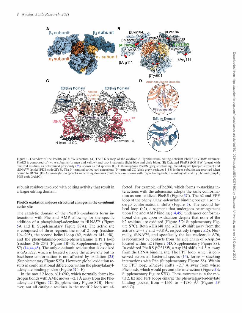
Treatment of S. Typhimurium wild-type (WT) and editing-deficient PheRS �G318W with H2O2 results in differentresidues becoming oxidized (25). The PheRS �G318W vari-ant has a 7.5-fold reduction in proofreading due to the pre-vention of Tyr-tRNAPhe binding in the editing site, likely bysteric hindrance (24). Both S. Typhimurium oxidized WTand PheRS �G318W show increased proofreading capac-ity, with PheRS �G318W displaying the most pronouncedeffect with a >3-fold increase in Tyr-tRNAPhe proofreading(25). Several PheRS �G318W residues were oxidized acrossboth the �- and �-subunits but outside of catalytic regions,and thus it is unclear how increased proofreading results. Todefine the structural changes that PheRS undergoes duringoxidative stress, we solved a 3.6-A resolution structure ofoxidized PheRS �G318W by single particle cryo- EM (Fig-ure 1A; Supplementary Table S1, Supplementary FigureS1). PheRS adopts a heterotetrametric conformation com-posed of two �-subunits and two �-subunits (��)2 with the�-subunit containing the aminoacylation catalytic domain(residues 108–307) and the �-subunit containing the editingdomain (residues 216–354) (14,44–47) (Figure 1C and D).Due to the resolution, it is not possible to resolve the oxi-dation of specific residues (Figure 1B), but mass spectrome-try revealed extensive oxidation under treatment conditions(25).
PheRS undergoes a conformational change upontRNAPhe binding (44,45), including the ordering of theN-terminal coiled-coil domain of the �-subunit (residues1–88) (Figure 1C) (48). This region is not resolved in ourstructure likely because this structure is in the absence oftRNAPhe (Figure 1A). S. Typhimurium PheRS is similarto the previously published Pseudomonas aeruginosa apo-PheRS structure (apo-PaPheRS) (PDB code 4P71; rootmean square deviation (RMSD) of ∼2.9 A comparing theC� of 790 residues) (38). S. Typhimurium PheRS and PaPheRS share high sequence identity between both subunits(51.5% for the �-subunits and 65.7% for the �-subunits)and adopt identical tertiary folds (Supplementary FiguresS2A and S3). For these reasons, and because our struc-ture is also apo, all future comparisons will be made toapo-PaPheRS unless otherwise noted.
Oxidation induces conformational changes in the PheRS�G318W catalytic domains
Different residues are oxidized in wild-type versus thePheRS �G318W editing-deficient variant, likely due to dif-ferences in the overall architecture (25). Six residues becomeoxidized in PheRS �G318W, which are located across theenzyme domains, with five residues in the �-editing domainand one residue in the �-aminoacylation domain (25) (Fig-ure 1B; Supplementary Table S2). Comparison of PheRS�G318W with apo-PaPheRS reveals that oxidized �Met280and �Lys282 undergo large changes in the position of theirbackbone (6.7 and 8.6 A, respectively) while their side chainconformation is minimally impacted (Figure 2B and C).These residues are in the �-catalytic domain (Figure 2A)located adjacent to the editing site (Figure 1D). The en-
tire �-strand surrounding the editing domain moves in to-wards the tRNA binding site in the �-subunit, likely con-tributing to the shift and conformational changes to edit-ing site residues. Other residues within the �-subunit be-come oxidized yet this has little to no impact on their con-formations. For example, ox-�Lys184, ox-�Tyr60 and ox-�Asn222 all have limited movement (Supplementary FigureS4), so it is unlikely that these rotamer changes play a signif-icant role in the hyperaccuracy phenotype. In contrast, ox-�Arg111 has a ∼180◦ rotation compared to EcPheRS com-plexed with Phe-AMP (Supplementary Figure S4F), and a16.6 A movement inwards towards the editing domain com-pared to apo-PaPheRS (Supplementary Figure S4E). How-ever, since �Arg111 is on the surface of the �-subunit, thisdifference may be a result from how apo-PaPheRS packsin the crystal lattice (Supplementary Figure S5). Together,these data demonstrate that oxidation has a differential ef-fect on local conformation of the PheRS residues, and oxi-dation of a given residue does not necessarily entail confor-mational change in that region.
PheRS oxidation causes structural rearrangements of its edit-ing domain
After PheRS misacylates tRNAPhe with a Tyr, Tyr-tRNAPhe
translocates to the editing domain in the �-subunit (Figure3A) (22,24). In the editing domain, many residues surroundTyr to recognize and edit including �Arg244, �Asn254,�His265, �Glu334 and �Thr354 (Figure 3B). The tRNAis typically stabilized by interactions of �Arg244 with nu-cleotide C75, while �Asn254 and �Thr354 are in proxim-ity to the aminoacyl ester bond and involved in substratebinding and hydrogen bonding with catalytic waters (Fig-ure 3B and C) (24). These residues were not demonstratedto be oxidized yet they display altered conformations (Fig-ure 3D). �Arg244, �Asn254 and �Thr354 all shift awayfrom the tRNA binding location at distances between ∼5.4and 7.2A (Figure 3D; Supplementary Figure S6A). No-tably, the �Arg244 flips ∼180◦ towards the editing site (Fig-ure 3D).
The changes in residues located in the PheRS editing do-main alter how these residues interact with Tyr and thetRNA backbone of C75. �Glu334 plays an important rolein tyrosine discrimination via the formation of hydrogenbonds with the hydroxyl group of Tyr as assessed by molec-ular docking studies (24). In our structure, �Glu334 shiftsaway from the editing site ∼5.3 A (Figure 3D). The �His265stabilizes substrate binding through �-stacking interactionswith the Tyr (Figure 3C) (23,24). Other well-conserved edit-ing site residues form an antiparallel �-sheet that upon ox-idation, appears to be disrupted (Figure 4A and B) (24).None of the editing site residues are oxidized but this globaloxidation disrupts the hydrogen bonding network of the �-sheet, shifting the �-sheet away from the editing site by ∼4.5A (Figure 4C; Supplementary Figure S6B) (38). This move-ment creates a larger binding surface (increases from ∼204A2 to ∼295 A2) (42) for the incorrect amino acid to be edited(Figure 4D and E). Despite the relatively moderate changesin the locations of the oxidized residues (Figure 2; Supple-mentary Figure S4), there is extensive rearrangement of �-
Dow
nloaded from https://academ
ic.oup.com/nar/advance-article/doi/10.1093/nar/gkab856/6377402 by C
hapman U
niversity user on 04 October 2021
4 Nucleic Acids Research, 2021
Figure 1. Overview of the PheRS �G318W structure. (A) The 3.6 A map of the oxidized S. Typhimurium editing-deficient PheRS �G318W tetramer.PheRS is composed of two �-subunits (orange and yellow) and two �-subunits (light blue and dark blue). (B) Oxidized PheRS �G318W (green) withoxidized residues, as determined previously (25), shown as red spheres. (C) T. thermophilus PheRS (grey) containing Phe-adenylate (purple, surface) andtRNAPhe (pink) (PDB code 2IY5). The N-terminal coiled-coil extensions (N-terminal CC (dark grey); residues 1–88) in the �-subunits are resolved whenbound to tRNA. (D) Aminoacylation (peach) and editing domains (dark blue) are shown with respective ligands, Phe-adenylate and Tyr, bound (purple,PDB code 2AMC).
subunit residues involved with editing activity that result ina larger editing domain.
PheRS oxidation induces structural changes in the �-subunitactive site
The catalytic domain of the PheRS �-subunits form in-teractions with Phe and AMP, allowing for the specificaddition of a phenylalanyl-adenylate to tRNAPhe (Figure5A and B; Supplementary Figure S7A). The active siteis composed of three regions: the motif 2 loop (residues194–205), the second helical loop (h2, residues 145–158),and the phenylalanine-proline-phenylalanine (FPF) loop(residues 246–254) (Figure 5B−E; Supplementary FigureS7) (14,44,45). The only �-subunit residue that is oxidizedis �Asn222, which is located outside the active site but itsbackbone conformation is not affected by oxidation (25)(Supplementary Figure S2B). However, global oxidation re-sults in conformational differences within the phenylalanyl-adenylate binding pocket (Figure 5C−E).
In the motif 2 loop, �His202, which normally forms hy-drogen bonds with AMP, moves ∼2.1 A away from the Phe-adenylate (Figure 5C; Supplementary Figure S7B). How-ever, not all catalytic residues in the motif 2 loop are af-
fected. For example, �Phe206, which forms �-stacking in-teractions with the adenosine, adopts the same conforma-tion as non-oxidized PheRS (Figure 5C). The h2 and FPFloop of the phenylalanyl-adenylate binding pocket also un-dergo conformational shifts (Figure 5). The second he-lical loop (h2), a segment that undergoes rearrangementupon Phe and AMP binding (14,45), undergoes conforma-tional changes upon oxidization despite that none of theh2 residues are oxidized (Figure 5D; Supplementary Fig-ure S7C). Both �His148 and �His149 shift away from theactive site ∼3.7 and ∼3.8 A, respectively (Figure 5D). Nor-mally, tRNAPhe, and specifically the last nucleotide A76,is recognized by contacts from the side chain of �Asp154located within h2 (Figure 5D; Supplementary Figure S8).In oxidized PheRS �G318W, �Asp154 shifts ∼4.5 A awayfrom the tRNA binding site. The FPF loop, which is con-served across all bacterial species (14), forms �-stackinginteractions with Phe (Supplementary Figure S8). Withinthe FPF loop, �Phe248 shifts ∼2.7 A away from wherePhe binds, which would prevent this interaction (Figure 5E;Supplementary Figure S7D). These movements in the mo-tif 2, h2 and FPF loops enlarge the phenylalanyl-adenylatebinding pocket from ∼1560 to ∼1980 A2 (Figure 5Fand G).
Dow
nloaded from https://academ
ic.oup.com/nar/advance-article/doi/10.1093/nar/gkab856/6377402 by C
hapman U
niversity user on 04 October 2021

Nucleic Acids Research, 2021 5
Figure 2. PheRS �Met280 and �Lys282 residues located outside the editing domain move substantially upon oxidation. (A) Overview of the �1 subunitmap and model (light blue) with the editing domain (dark blue) and oxidized residues (red spheres). The other PheRS subunits are shown as outlines. (B)Map and model of ox-�Met280 and ox-�Lys282 located in the editing domain. (C) Comparison of ox-�Met280 and ox-�Lys282 (corresponding residuein P. aeruginosa is Glu281) with P. aeruginosa apo-PheRS (yellow, PDB code 4P73) revealing significant movements of ∼6.7 and ∼8.6 A, respectively.
Upon oxidation, there is a 30% decrease in misacyla-tion of tRNAPhe with Tyr (25). Discrimination betweenPhe and Tyr is facilitated by �Glu210 within h2 which hy-drogen bonds to the bound amino acid to facilitate liga-tion (Figure5B and D; Supplementary Figure S8). In ourstructure, the position of �Glu210 moves to a position thatwould directly overlap with the hydroxyl group of Tyr (14)(Supplementary Figure S9) as compared to a Escherichiacoli PheRS structure containing Phe and AMP (PBD code3PCO). This new �Glu210 position may explain why mis-acylation decreases upon PheRS oxidation.
Similarities between oxidized S. Typhimurium PheRS andthe E. coli PheRS–Phe–AMP complex
Oxidation causes both larger editing and aminoacylationsites as compared to apo-PaPheRS as previously described(an increase of 204–295 A2 (Figure 4D and E) and an in-crease of 1560–1900 A2 (Figure 5F and G), respectively).Although all comparisons to this point have been of oxi-dized apo-PheRS �G318W to apo-PaPheRS, interestingly,specific regions of oxidized apo-PheRS are slightly moresimilar to the structure of EcPheRS complexed with lig-ands Phe and AMP (PDB code 3PCO (14), RMSD of ∼1.7A). While binding of Phe and AMP to EcPheRS increasesthe editing (∼272 A2) and Phe-adenylate binding pocket(∼1700 A2), these sites are still smaller than when PheRSis oxidized (Supplementary Figures S10D and S11B).
One of the most striking changes between oxidized apo-PheRS and EcPheRS bound to Phe and AMP is the place-
ment of �Met280 and �Lys282, residues that both becomeoxidized but are located outside the two catalytic sites (Fig-ure 2). Normally the backbone of these residues dramat-ically changes upon binding of Phe and AMP (changesof 9.4 A (�Met280) and 10.3 A (�Lys282)). Oxidation of�Met280 and �Lys282 also influences their positions how-ever, now these residues adopt similar positions as when Pheand AMP bind (Supplementary Figure S10A) (14). Thismovement places these residues closer towards the editingdomains where tRNA binds despite not containing any lig-ands.
Comparison of Phe-AMP bound EcPheRS with oxidizedapo-PheRS also reveals changes of editing site residuesthat appear to enlarge the binding pocket and change howoxidized PheRS would engage either the amino acid orAMP (Supplementary Figure S10). First, the side chainof �His265 flips away from the amino acid binding site(Supplementary Figure S10B), likely influencing recogni-tion of the incorrect Tyr. This flipping may occur as PheRStransitions to prepare for hydrolysis of incorrectly boundTyr. The �Arg244 sidechain, which normally recognizesthe tRNA backbone of nucleotide C75, is rotated ∼180◦away in the absence of tRNA (Figure 3C). Oxidation ofPheRS causes �Arg244 to more resemble EcPheRS-Phe-AMP despite lacking the tRNA (Supplementary FigureS10B). Lastly, oxidation cause the entire �-sheet compris-ing the editing domain to shift by ∼2.7–3.7 A and rotate∼45◦ thus placing it further away from the aminoacylated-tRNA binding site (Supplementary Figure S10C). Collec-tively, these changes create a larger editing site but with ac-
Dow
nloaded from https://academ
ic.oup.com/nar/advance-article/doi/10.1093/nar/gkab856/6377402 by C
hapman U
niversity user on 04 October 2021

6 Nucleic Acids Research, 2021
Figure 3. Localized active site conformational changes of the PheRS editing domain. (A) Oxidized S. Typhimurium editing-deficient PheRS �G318W�1-subunit with corresponding map (light blue) and active site residues in the editing domain shown as spheres (dark blue). The other PheRS subunitsare shown in outline. (B) Closeup of editing site residues with Tyr-tRNAPhe docked into the editing site (PDB code 3HFZ, purple). (C) Overview of theediting site residues (blue) and interactions with a Tyr-tRNAPhe CCA end (purple), and the two catalytic waters that carry out the reaction. (D) Residuesof the editing domain of the oxidized S. Typhimurium PheRS compared to the P. aeruginosa apo-PheRS (yellow, PDB code 4P73) undergo a significantshift, particularly �Arg244 which is shifted ∼7.2 A and flipped ∼180◦ inwards towards the editing site (green circle).
tive site residues positioned in conformations to allow forthe increase in editing activity of the oxidized PheRS.
DISCUSSION
To prevent protein mistranslation, aminoacyl-tRNA syn-thetases pair correct amino acids to their cognate tRNAswith high fidelity to ensure the correct addition of aminoacids into proteins. Environmental stress, including oxida-tive stress, alters cellular amino acid pools and this changecan affect the fidelity of gene expression. During oxidativestress, oxidation of Phe converts it to Tyr, increasing Tyrisomer levels and leads to an overall increase in tRNAPhe
misacylation (29). However, oxidation of PheRS compen-sates by increasing proofreading to reduce misacylated Tyr-tRNAPhe from participating in protein synthesis. SeveralPheRS residues are oxidized and these modifications arefound throughout PheRS and not localized solely at the ac-tive site (29). Since oxidation causes a hyperaccurate phe-notype but there is no oxidation at the active sites, this sug-
gests that allosteric changes may contribute to PheRS ac-quiring hyperaccuracy consistent with previous results thatdemonstrated global changes (25). Based upon these re-sults, we set out to determine the cryo-EM structure ofoxidized PheRS. Overall, we find that oxidation results instructural changes mainly in the �-subunit editing domainand to a lesser extent, the �-subunit aminoacylation activesite. The �-subunit editing domain is enlarged where mis-charged Tyr-tRNAPhe would bind and its surface area in-creases by 50%. Oxidization also enlarges the phenylalanyl-adenylate binding pocket in the �-subunit by 30%. Theseenlargements are predicted to contribute to faster and effi-cient binding, while additional conformational changes ofcatalytic residues suggest an improvement in Tyr hydrolysisrequired to ligate the correct Phe. These results shed light onhow S. Typhimurium is able to survive and grow in adverseenvironments.
Based on high structural homology between bacterialPheRS (49,50), it is likely that most bacterial PheRS en-zymes have a similar hyperaccuracy phenotype under ox-
Dow
nloaded from https://academ
ic.oup.com/nar/advance-article/doi/10.1093/nar/gkab856/6377402 by C
hapman U
niversity user on 04 October 2021

Nucleic Acids Research, 2021 7
Figure 4. Oxidization of PheRS �G318W opens the editing domain leading to hyperaccuracy. (A) Structure of oxidized S. Typhimurium PheRS �G318W�1-subunit with corresponding map density (blue) and catalytic residues that are implicated in editing activity shown as dark blue spheres, with othersubunits shown in outline. (B) Closeup of the catalytic region with Tyr overlaid from a T. thermophilus PheRS-Tyr structure (white, PDB code 3HFZ). (C)The catalytic residues �Tyr216, �Ala336, and �Phe388 of the oxidized S. Typhimurium PheRS undergo a shift averaging ∼4.5 A away from the editingdomain compared to the P. aeruginosa apo-PheRS (PDB code 4P73, yellow). (D) Editing domain of oxidized PheRS �G318W shown as surface (white)with Tyr-tRNAPhe docked in site (PDB code 3HFZ, purple). (E) Editing site residues of apo-PheRS shown as surface (white) with Tyr-tRNAPhe dockedinto the editing site (PDB code 3HFZ, purple).
idative stress. Although there is typically high homol-ogy among mitochondrial and bacterial synthetases (51),PheRS is an exception as the mitochondrial and bacte-rial synthetase are divergent (52). Mitochondrial PheRSis a monomer and lacks an editing domain found in thetetrameric bacterial PheRS (52), and as a consequence, isunable to proofread misacylated tRNAPhe (52). Althoughmitochondrial PheRS is oxidized during oxidative stress,this does not result in hyperaccuracy and oxidation can bereversed (25), restoring normal function.
While oxidation of other synthetases including ThrRSand AlaRS can occur, this does not result in hyperaccu-racy, in contrast to PheRS. ThrRS is known to misacylate
tRNAThr with Ser and contains a catalytic cysteine in theediting domain (53). Under oxidative conditions, this re-active cysteine is oxidized and thus prevents ThrRS edit-ing ability (54), the opposite phenotype to what is observedwith PheRS (25,53). AlaRS also relies on a critical cysteineresidue to carry out editing activity upon misacylation of aSer- or Gly-tRNAAla (55), but changes in AlaRS editing ac-tivity upon oxidation have not yet been reported. Changesin AlaRS editing activity are significantly more determinan-tal for bacteria viability compared to ThrRS (56). From afitness perspective, maintenance of AlaRS editing activityis more important for bacterial survival, suggesting possi-ble divergence in editing response under oxidation as com-
Dow
nloaded from https://academ
ic.oup.com/nar/advance-article/doi/10.1093/nar/gkab856/6377402 by C
hapman U
niversity user on 04 October 2021

8 Nucleic Acids Research, 2021
Figure 5. Conformational changes of the aminoacylation domain. (A) Structure of oxidized S. Typhimurium PheRS �G318W �1-subunit and map(orange). Aminoacylation active site residues shown as spheres, with other subunits shown in outline. (B) Active site residues in the phenylalanyl-adenylatebinding pocket (orange) with modeled AMP and Phe (PDB code 3PCO, white). The motif 2 loop (pink), helix 2 loop (h2, dark red), and FPF loop (purple)are shown. (C) The motif 2 loop shifts outwards by ∼2.1 A from P. aeruginosa apo-PheRS (PDB code 4P73, yellow). (D) The h2 of oxidized PheRS hasconformational rearrangements, 3.7 A and 3.8 A at �His148 and �His149, respectively, and 4.5 A at �Asp154, away from the phenylalanyl-adenylatebinding pocket compared to apo-PheRS (PDB code 4P73, yellow). (E) The FPF loop contains minor conformational changes, with 2.7, 2.6 and 2.9 Ashifts of the �Phe248, �Pro249 and �Phe250, respectively, away from the phenylalanyl-adenylate binding pocket compared to apo-PheRS (PDB code 4P73,yellow) (F) Enlargement of the aminoacylation domain shown with surface (white) of oxidized PheRS �G318W, with AMP and Phe groups docked in andshown with surfaces (PDB code 3PCO, grey). (G) Enlargement of the aminoacylation domain shown as surface (white) of the apo-PaPheRS (PDB code4P73, yellow), with AMP and Phe groups docked in shown as surfaces (PDB code 3PCO, grey).
Dow
nloaded from https://academ
ic.oup.com/nar/advance-article/doi/10.1093/nar/gkab856/6377402 by C
hapman U
niversity user on 04 October 2021

Nucleic Acids Research, 2021 9
pared to ThrRS. Under normal growth conditions, PheRSediting activity is not vital for bacterial viability, but be-comes required for growth under oxidative conditions (29).Translation accuracy under oxidative stress is vital for cel-lular survival, but among the class II aaRSs, only PheRSundergoes a gain of function change. The benefit of PheRShyperaccuracy under conditions that directly increase thelikelihood of mistranslation serves as a direct compensatorymechanism to decrease the likelihood of Tyr misincorpora-tion; while ThrRS and possibly AlaRS are susceptible tooxidative stress, the Ser or Gly amino acid pools are not ex-pected to change significantly, placing less selective pressureon these synthetases to compensate for misacylation underoxidative conditions.
Oxidation is a well characterized signal for proteins di-rectly involved in the oxidative stress response includingredox-operated switches that activate transcription of ox-idative response elements (57). However, these mechanismsare indirect and contribute to a regulatory pathway that al-lows for increased cell survival under adverse conditions.PheRS represents one of the few examples of a functionalprotein that self-regulates as a feedforward mechanism todirectly compensate against an adverse condition.
Responses to stress are typically analyzed at the levelof gene expression (58,59). With oxidative stress, severalgenes including oxyR, oxyS, or perR, are directly regulatedby oxidation of several cysteine residues allowing for tran-scription factor binding to promote the expression of pro-teins involved in metabolism or clearance of oxidative stress(60). The hyperaccuracy phenotype of PheRS under oxida-tive stress illustrates how important pathways in proteinsynthesis work to maintain proper gene expression. Thisensures successful expression of oxidative response genes,which relies on accurate amino acid additions to growingnascent chain on the ribosome. Oxidized PheRS hyperac-curacy ensures that correct gene expression is not compro-mised by elevated and potentially toxic Tyr isomer misin-corporation under oxidative stress. Hyperaccurate aminoa-cylation thus helps to increase survival, particularly for or-ganisms regularly present in oxidative environments likeS. Typhimurium, which can thrive in environments like amacrophage despite a host reactive oxygen species attack.Overall, the proper activation of stress responses, includingincreased proofreading activity of PheRS, increases S. Ty-phimurium survival and contributes to the overall fitness ofthe bacterium.
DATA AVAILABILITY
Atomic coordinates and maps for the reported cryo-EMstructures have been deposited with the Protein Data bankunder accession number 7N8Y and Electron MicroscopyData bank under accession number EMD-24249.
SUPPLEMENTARY DATA
Supplementary Data are available at NAR Online.
ACKNOWLEDGEMENTS
We thank Dunham lab member Dr Alexandra KuzmishinNagy for critical reading of the manuscript and Ha AnNguyen for technical support.
FUNDING
National Institutes of Health [R01GM065183 to M.I. andC.M.D., R01 GM093278 to C.M.D., 5T32 GM008602to P.S., T32 AI106699 to P.S.]; Burroughs Wellcome FundInvestigator in the Pathogenesis of Infectious Disease award(to C.M.D.); Robert P. Apkarian Integrated Electron Mi-croscopy Core (IEMC) at Emory University, which is subsi-dized by the School of Medicine and Emory College of Artsand Sciences. Funding for open access charge: National In-stitutes of Health.Conflict of interest statement. None declared.
REFERENCES1. McClain,W.H. (1993) Rules that govern tRNA identity in protein
synthesis. J. Mol. Biol., 234, 257–280.2. Cochella,L. and Green,R. (2005) Fidelity in protein synthesis. Curr.
Biol., 15, R536–R540.3. Jakubowski,H. and Goldman,E. (1992) Editing of errors in selection
of amino acids for protein synthesis. Microbiol. Rev., 56, 412–429.4. Schimmel,P.R. and Soll,D. (1979) Aminoacyl-tRNA synthetases:
general features and recognition of transfer RNAs. Annu. Rev.Biochem., 48, 601–648.
5. Delarue,M. (1995) Aminoacyl-tRNA synthetases. Curr. Opin. Struct.Biol., 5, 48–55.
6. Ahel,I., Korencic,D., Ibba,M. and Soll,D. (2003) Trans-editing ofmischarged tRNAs. Proc. Natl. Acad. Sci. U.S.A., 100, 15422–15427.
7. Eriani,G., Delarue,M., Poch,O., Gangloff,J. and Moras,D. (1990)Partition of tRNA synthetases into two classes based on mutuallyexclusive sets of sequence motifs. Nature, 347, 203–206.
8. Fraser,T.H. and Rich,A. (1975) Amino acids are not all initiallyattached to the same position on transfer RNA molecules. Proc. Natl.Acad. Sci. U.S.A., 72, 3044–3048.
9. Sprinzl,M. and Cramer,F. (1975) Site of aminoacylation of tRNAsfrom Escherichia coli with respect to the 2′- or 3′-hydroxyl group ofthe terminal adenosine. Proc. Natl. Acad. Sci. U.S.A., 72, 3049–3053.
10. Bullwinkle,T.J. and Ibba,M. (2014) Emergence and evolution. Top.Curr. Chem., 344, 43–87.
11. Kaiser,F., Krautwurst,S., Salentin,S., Haupt,V.J., Leberecht,C.,Bittrich,S., Labudde,D. and Schroeder,M. (2020) The structural basisof the genetic code: amino acid recognition by aminoacyl-tRNAsynthetases. Sci. Rep., 10, 12647.
12. Fukai,S., Nureki,O., Sekine,S.-i., Shimada,A., Tao,J., Vassylyev,D.G.and Yokoyama,S. (2000) Structural basis for double-sievediscrimination of L-valine from L-isoleucine and L-threonine by thecomplex of tRNAVal and valyl-tRNA synthetase. Cell, 103, 793–803.
13. Lincecum,T.L., Tukalo,M., Yaremchuk,A., Mursinna,R.S.,Williams,A.M., Sproat,B.S., Van Den Eynde,W., Link,A., VanCalenbergh,S., Grøtli,M. et al. (2003) Structural and mechanisticbasis of pre- and posttransfer editing by leucyl-tRNA synthetase.Mol. Cell, 11, 951–963.
14. Mermershtain,I., Finarov,I., Klipcan,L., Kessler,N., Rozenberg,H.and Safro,M.G. (2011) Idiosyncrasy and identity in the prokaryoticphe-system: crystal structure of E. coli phenylalanyl-tRNA synthetasecomplexed with phenylalanine and AMP. Protein Sci., 20, 160–167.
15. Silvian,L.F., Wang,J. and Steitz,T.A. (1999) Insights into editing froman Ile-tRNA synthetase structure with tRNAIle and mupirocin.Science, 285, 1074–1077.
16. Martinis,S.A. and Boniecki,M.T. (2010) The balance between pre-and post-transfer editing in tRNA synthetases. FEBS Lett., 584,455–459.
17. Dock-Bregeon,A.-C., Sankaranarayanan,R., Romby,P., Caillet,J.,Springer,M., Rees,B., Francklyn,C.S., Ehresmann,C. and Moras,D.(2000) Transfer RNA-mediated editing in threonyl-tRNA synthetase.The class II solution to the double discrimination problem. Cell, 103,877–884.
18. Beebe,K., Merriman,E., de Pouplana,L.R. and Schimmel,P. (2004) Adomain for editing by an archaebacterial tRNA synthetase. Proc.Natl. Acad. Sci. U.S.A., 101, 5958–5963.
19. Beuning,P.J. and Musier-Forsyth,K. (2000) Hydrolytic editing by aclass II aminoacyl-tRNA synthetase. Proc. Natl. Acad. Sci. U.S.A.,97, 8916–8920.
Dow
nloaded from https://academ
ic.oup.com/nar/advance-article/doi/10.1093/nar/gkab856/6377402 by C
hapman U
niversity user on 04 October 2021

10 Nucleic Acids Research, 2021
20. Smith,T.F. and Hartman,H. (2015) The evolution of Class IIaminoacyl-tRNA synthetases and the first code. FEBS Lett., 589,3499–3507.
21. Moor,N., Klipcan,L. and Safro,Mark G. (2011) Bacterial andeukaryotic phenylalanyl-tRNA synthetases catalyzemisaminoacylation of tRNAPhe with 3,4-dihydroxy-L-phenylalanine.Chem. Biol., 18, 1221–1229.
22. Roy,H., Ling,J., Irnov,M. and Ibba,M. (2004) Post-transfer editing invitro and in vivo by the � subunit of phenylalanyl-tRNA synthetase.EMBO J., 23, 4639–4648.
23. Kotik-Kogan,O., Moor,N., Tworowski,D. and Safro,M. (2005)Structural basis for discrimination of L-phenylalanine fromL-tyrosine by phenylalanyl-tRNA synthetase. Structure, 13,1799–1807.
24. Ling,J., Roy,H. and Ibba,M. (2007) Mechanism of tRNA-dependentediting in translational quality control. Proc. Natl. Acad. Sci. U.S.A.,104, 72–77.
25. Steiner,R.E., Kyle,A.M. and Ibba,M. (2019) Oxidation ofphenylalanyl-tRNA synthetase positively regulates translationalquality control. Proc. Natl. Acad. Sci. U.S.A., 116, 10058–10063.
26. Slauch,J.M. (2011) How does the oxidative burst of macrophages killbacteria? Still an open question. Mol. Microbiol., 80, 580–583.
27. Winter,S.E., Thiennimitr,P., Winter,M.G., Butler,B.P., Huseby,D.L.,Crawford,R.W., Russell,J.M., Bevins,C.L., Adams,L.G., Tsolis,R.M.et al. (2010) Gut inflammation provides a respiratory electronacceptor for Salmonella. Nature, 467, 426–429.
28. Ling,J. and Soll,D. (2010) Severe oxidative stress induces proteinmistranslation through impairment of an aminoacyl-tRNAsynthetase editing site. Proc. Natl. Acad. Sci. U.S.A., 107, 4028–4033.
29. Bullwinkle,T.J., Reynolds,N.M., Raina,M., Moghal,A., Matsa,E.,Rajkovic,A., Kayadibi,H., Fazlollahi,F., Ryan,C., Howitz,N. et al.(2014) Oxidation of cellular amino acid pools leads to cytotoxicmistranslation of the genetic code. eLife, 3, e02501.
30. Ghosh,R. and Mitchell,D.L. (1999) Effect of oxidative DNA damagein promoter elements on transcription factor binding. Nucleic AcidsRes., 27, 3213–3218.
31. Nawrot,B., Sochacka,E. and Duchler,M. (2011) tRNA structural andfunctional changes induced by oxidative stress. Cell. Mol. Life Sci.,68, 4023–4032.
32. Tanaka,M., Chock,P.B. and Stadtman,E.R. (2007) Oxidizedmessenger RNA induces translation errors. Proc. Natl. Acad. Sci.U.S.A., 104, 66–71.
33. Vogel,C., Silva,G.M. and Marcotte,E.M. (2011) Protein expressionregulation under oxidative stress. Mol. Cell. Proteomics, 10,M111.009217.
34. Steele-Mortimer,O. (2008) The Salmonella-containing vacuole:moving with the times. Curr. Opin. Microbiol., 11, 38–45.
35. Scheres,S.H.W. (2012) RELION: implementation of a Bayesianapproach to cryo-EM structure determination. J. Struct. Biol., 180,519–530.
36. Li,X., Mooney,P., Zheng,S., Booth,C.R., Braunfeld,M.B.,Gubbens,S., Agard,D.A. and Cheng,Y. (2013) Electron counting andbeam-induced motion correction enable near-atomic-resolutionsingle-particle cryo-EM. Nat. Methods, 10, 584–590.
37. Zhang,K. (2016) Gctf: real-time CTF determination and correction.J. Struct. Biol., 193, 1–12.
38. Abibi,A., Ferguson,A.D., Fleming,P.R., Gao,N., Hajec,L.I., Hu,J.,Laganas,V.A., McKinney,D.C., McLeod,S.M., Prince,D.B. et al.(2014) The role of a novel auxiliary pocket in bacterialphenylalanyl-tRNA synthetase druggability. J. Biol. Chem., 289,21651–21662.
39. Pettersen,E.F., Goddard,T.D., Huang,C.C., Couch,G.S.,Greenblatt,D.M., Meng,E.C. and Ferrin,T.E. (2004) UCSFChimera––a visualization system for exploratory research andanalysis. J. Comput. Chem., 25, 1605–1612.
40. Adams,P.D., Afonine,P.V., Bunkoczi,G., Chen,V.B., Echols,N.,Headd,J.J., Hung,L.-W., Jain,S., Kapral,G.J., Grosse Kunstleve,R.W.et al. (2011) The Phenix software for automated determination ofmacromolecular structures. Methods, 55, 94–106.
41. Emsley,P., Lohkamp,B., Scott,W.G. and Cowtan,K. (2010) Featuresand development of Coot. Acta Crystallogr., Sect. D: Biol.Crystallogr., 66, 486–501.
42. Goddard,T.D., Huang,C.C., Meng,E.C., Pettersen,E.F., Couch,G.S.,Morris,J.H. and Ferrin,T.E. (2018) UCSF ChimeraX: meetingmodern challenges in visualization and analysis. Protein Sci., 27,14–25.
43. Meng,E.C., Pettersen,E.F., Couch,G.S., Huang,C.C. and Ferrin,T.E.(2006) Tools for integrated sequence-structure analysis with UCSFChimera. BMC Bioinf., 7, 339.
44. Mosyak,L., Reshetnikova,L., Goldgur,Y., Delarue,M. andSafro,M.G. (1995) Structure of phenylalanyl-tRNA synthetase fromThermus thermophilus. Nat. Struct. Biol., 2, 537–547.
45. Goldgur,Y., Mosyak,L., Reshetnikova,L., Ankilova,V., Lavrik,O.,Khodyreva,S. and Safro,M. (1997) The crystal structure ofphenylalanyl-tRNA synthetase from Thermus thermophiluscomplexed with cognate tRNAPhe. Structure, 5, 59–68.
46. Kreutzer,R., Kruft,V., Bobkova,E.V., Lavrik,O.I. and Sprinzl,M.(1992) Structure of the phenylalnyl-tRNA synthetase genes fromThermus thermophilus HB8 and their expression in Escherichia coli.Nucleic Acids Res., 20, 4173–4178.
47. Khodyreva,S.N., Moor,N.A., Ankilova,V.N. and Lavrik,O.I. (1985)Phenylalanyl-tRNA synthetase from E. coli MRE-600: analysis of theactive site distribution on the enzyme subunits by affinity labelling.Biochim. Biophys. Acta, 830, 206–212.
48. Moor,N., Kotik-Kogan,O., Tworowski,D., Sukhanova,M. andSafro,M. (2006) The crystal structure of the ternary complex ofphenylalanyl-tRNA synthetase with tRNAPhe and aphenylalanyl-adenylate analogue reveals a conformational switch ofthe CCA end. Biochemistry, 45, 10572–10583.
49. Klipcan,L., Moor,N., Kessler,N. and Safro,M.G. (2009) Eukaryoticcytosolic and mitochondrial phenylalanyl-tRNA synthetases catalyzethe charging of tRNA with the meta-tyrosine. Proc. Natl. Acad. Sci.U.S.A., 106, 11045–11048.
50. Neubauer,C., Gao,Y.-G., Andersen,K.R., Dunham,C.M.,Kelley,A.C., Hentschel,J., Gerdes,K., Ramakrishnan,V. andBrodersen,D.E. (2009) The structural basis for mRNA recognitionand cleavage by the ribosome-dependent endonuclease RelE. Cell,139, 1084–1095.
51. Kartvelishvili,E., Peretz,M., Tworowski,D., Moor,N. and Safro,M.(2016) Chimeric human mitochondrial PheRS exhibits editing activityto discriminate nonprotein amino acids. Protein Sci., 25, 618–626.
52. Roy,H., Ling,J., Alfonzo,J. and Ibba,M. (2005) Loss of editingactivity during the evolution of mitochondrial phenylalanyl-tRNAsynthetase. J. Biol. Chem., 280, 38186–38192.
53. Wu,J., Fan,Y. and Ling,J. (2014) Mechanism of oxidant-inducedmistranslation by threonyl-tRNA synthetase. Nucleic Acids Res., 42,6523–6531.
54. Sankaranarayanan,R., Dock-Bregeon,A.C., Romby,P., Caillet,J.,Springer,M., Rees,B., Ehresmann,C., Ehresmann,B. and Moras,D.(1999) The structure of threonyl-tRNA synthetase-tRNA(Thr)complex enlightens its repressor activity and reveals an essential zincion in the active site. Cell, 97, 371–381.
55. Pasman,Z., Robey-Bond,S., Mirando,A.C., Smith,G.J., Lague,A. andFrancklyn,C.S. (2011) Substrate specificity and catalysis by theediting active site of alanyl-RNA synthetase from Escherichia coli.Biochemistry, 50, 1474–1482.
56. Kelly,P., Backes,N., Mohler,K., Buser,C., Kavoor,A., Rinehart,J.,Phillips,G., Ibba,M. and Merrikh,H. (2019) Alanyl-tRNA synthetasequality control prevents global dysregulation of the Escherichia coliproteome. mBio, 10, e02921–e02919.
57. Pomposiello,P.J. and Demple,B. (2001) Redox-operated geneticswitches: the SoxR and OxyR transcription factors. TrendsBiotechnol., 19, 109–114.
58. Murray,J.I., Whitfield,M.L., Trinklein,N.D., Myers,R.M.,Brown,P.O. and Botstein,D. (2004) Diverse and specific geneexpression responses to stresses in cultured human cells. Mol. Biol.Cell, 15, 2361–2374.
59. Majmundar,A.J., Wong,W.J. and Simon,M.C. (2010)Hypoxia-inducible factors and the response to hypoxic stress. Mol.Cell, 40, 294–309.
60. Mongkolsuk,S. and Helmann,J.D. (2002) Regulation of inducibleperoxide stress responses. Mol. Microbiol., 45, 9–15.
Dow
nloaded from https://academ
ic.oup.com/nar/advance-article/doi/10.1093/nar/gkab856/6377402 by C
hapman U
niversity user on 04 October 2021