ovesco research update 34 | research & clinical trials 1 · sneha singh, mbbs1, gerardo...
TRANSCRIPT
Ovesco Research Update 35 | Research & clinical trials 1
Ovesco Research Update | Research and clinical trials | Version 35 | 2020-06-30
June 2020 | First clinical cases with LiftUp® reveal promising results, proving it as valuable, safe and fast
The novel submucosal injection agent LiftUp was successfully used in a first clinical case series with 10 patients. Resection of colorectal polyps (median lesion size of 20.5 mm) was macroscopically complete in 100 %. R0 resection was achieved in 78 %. No adverse events were reported. M. Schaefer et al. reported on the first experience with the novel agent LiftUp for submucosal injection. 10 consecutive patients (median age 74.5 years; 6 male, 4 female) with colorectal polyps ≥ 15 mm were included. Lesions were localized in the rectum (n=5), caecum (n=2), sigmoid (n=1), transverse colon (n=1), and ascending colon (n=1) and the median lesion size was 20.5 mm (range: 15-30 mm). To assess endoscopic resection after LiftUp injection, EMR (8 patients) or ESD (2 patients) were performed. Median volume of LiftUp was 5.25 ml (range 2-12 ml) and median procedure time was 2.25 minutes for EMR and 5.5 minutes for ESD. In all 10 cases macroscopic complete resection was achieved and the R0 resection rate was 78 % (7/9). No adverse events occurred. The authors concluded that endoscopic resection with LiftUp is feasible, safe, and fast. However, larger studies are needed for better evaluation of efficacy and safety. First human in-vivo experience with novel agent for submucosal injection (LiftUp) Moritz Schaefer, Benjamin Meier, Andreas Wannhoff, Karel Caca. ePoster submitted at DDW 2020; No. 3351549.
June 2020 | BougieCap: case study of post-esophagectomy stricture in the high cervical esophagus successfully dilated to 12 mm with the BougieCap
The patient had experienced esophageal perforation due to bougienage with a through-the-scope balloon, which led to emergency thoracotomy with esophagectomy and gastric pull-up. A 5 mm stricture developing at the anastomotic site 15 months later was safely and effectively dilated with the BougieCap H. Ramrakhiani and G. Triadafilopoulos, Stanford University School of Medicine, Stanford, CA, USA, presented a case study on stricture dilation with the BougieCap. A 62-year old man had previously presented with chest pain, nausea, vomiting, and odynophagia. Diagnostic endoscopy with circumferential biopsies showed evidence of acute ischemic injury of the esophageal wall. Four weeks later, the ischemic area had turned into a stricture, extending 10 cm long from mid to distal esophagus and as narrow as 4 mm. A balloon dilation was undertaken with a through-the-scope balloon, which was complicated by an esophageal perforation. Emergency thoracotomy was necessary, associated with distal esophagectomy, gastric pull-up and placement of a feeding jejunostomy. 15 months later the patient presented with progressive difficulty in swallowing associated with 16 lb weight loss. Barium swallow examination revealed a tight stricture at the anastomosis with a narrowed lumen diameter of 5 mm. The stricture was located just distal to the upper esophageal sphincter. This time, the decision for bougienage with the BougieCap was made. Over the course of a few weeks, the patient underwent three upper endoscopies with BougieCap dilation of the stricture to 12 mm size with good results. At a follow-up examination 6 months later, the patient reported being able to eat a normal diet and had gained 10 lb. The authors discussed that currently available bougienage techniques for esophageal strictures (balloons and mechanical dilators) have limitations when used for the treatment of high cervical esophageal strictures or eosinophilic esophagitis-related tight esophageal strictures. The transparent BougieCap allows for visualization of mucosal disruption in real time during stricture dilation. Thus the authors conclude that the device may help to avoid overstretching and reduce the risk of complications. As the BougieCap is a relatively new device, prospective studies with larger numbers of patients and with long-term follow-up
are still outstanding. Negotiating Dire Straits with a BougieCap Ramrakhiani H, Triadafilopoulos G. Digestive Diseases and Sciences; https://doi.org/10.1007/s10620-020-06236-z
June 2020 | Conference Report of DDW 2020 Conference report of the Digestive Disease Week (DDW) 2020
▪ FTRD® System: EFTR enables new therapeutic options for the treatment of lesions and is a feasible and safe modality, which leads to a high R0 resection rate
▪ First clinical cases with LiftUp® reveal promising results, proving it as valuable, safe and fast
▪ OTSC® shows superiority & cost-effectiveness over standard therapy for first-line UGIB and shows high success rates in the management of non-acute, full-thickness gastrointestinal defects
The conference of the Digestive Disease Week (DDW) 2020 scheduled for May 2-5, 2020 in Chicago, had to be cancelled due to the current situation. However, many scientific publications on Ovesco products were submitted this year again. The most important abstracts on Ovesco products are summarized in the report attached below.
FTRD System
The FTRD System is a safe and useful methodforresection of colonic lesions including the appendix Y.Ichkhanian et al. presented the first multicenter international study assessing the outcome of using FTRD for endoscopic full-thickness resection of appendiceal lesions.To study the rate of appendicitis after lesion resection,56 consecutive patients between 11/2016 and 11/2019 underwent resection of colonic lesions involving the appendix. Most lesions were of Is 25 (45 %) and IIa 20 (36 %) Paris class and had a mean circumferential appendiceal orifice involvement of 66±29 %. The technical successratewas 95 % (53 patients) and in 47 cases successful EFTR was achieved. R0 resection was achieved in 92 % (49/53), antibiotics were administered in 81 % (43/53) for a median of 5 days. Follow-up colonoscopy showed no residual lesions in 19/53 patients. In 9 (19 %) out of 47 successful EFTR cases appendicitis was diagnosed. Three patients were treated with antibiotics alone, 6 underwent appendectomy. The authors concluded that the FTRD is safe and feasible for the resection of appendiceal lesions, with appendicitis occurringin about 20 % of cases. Endoscopic full-thickness resection of polyps involving the appendiceal orifice: first multicenter international study Yervant Ichkhanian, Mohammed Barawi, Bachir Ghandour, Talal Seoud, Shyam Thakkar,Truptesh H. Kothari, Krystle Bittner, Asad Ullah, Vivek Kaul,Shivangi Kothari, Edris Wedi, Patrick Aepli,Thomas E. Kowalski, Brianna Shinn, Reem Z. Sharaiha, Qais M. Dawod, SriHari Mahadev, Kaveh Hajifathalian,Gianluca Andrisani,Heinz Albrecht, Andrew Q. Giap, Shou J. Tang, Yehia M. Naga, Ayla S. Turan, Shai Friedland, Benjamin Tharian, Shayan S. Irani, Andrew S. Ross, Laith H. Jamil, Daniel Lew, Andrew Nett,Omid Sanaei, Nasim Parsa, Jad Farha, Manol Jovani, Vivek Kumbhari, Anthony N. Kalloo, Mouen A. Khashab
EFTR with the FTRD in the colorectum is safe and more efficient when compared to conventional endoscopic resection B. Meier et al. report from the German colonic FTRD registry in which the use of the FTRD for resectionof colorectal lesionsis analysed. Data from 1178 FTRD procedures provided by variousGerman endoscopy centers were collected between 09/2015 and 10/2019.Whereasthe R0 resection rate (overall 80 %) was higher in the rectum (83.6 %) than in the colon, the full-thickness resection rate (overall 89.9 %) in the rectum (83.3 %) was lower when compared to the colon.In 92 % of cases (1086/1178) follow-up data were available, endoscopic follow-up in 58 % (683/1178) with amean follow-up time of 22 weeks (median 14 weeks, range 1-202 weeks). Dislocation of the FTRD Clip
appeared in 69 % and in 31 % it was still in situ. Procedure-related adverse events were reported in 12.1 %, of which 2 % requiredsurgical treatment. The authors summarizedthat FTRD shows safe, effective, and fast results in endoscopic full-thickness resection of colorectal lesions. Efficacy and safety of endoscopic full-thickness resection with the FTRD in the colorectum: evaluation of a large PMCF analysis Benjamin Meier, Arthur Schmidt, Armin Kuellmer, Julius Mueller, Andreas Wannhoff, Klaus Weiss, Georg Huebner, Michael Repp, Alexander Meining, Benjamin Walter, Karel Caca.
EFTR is a more reliable in the therapy of colorectal neoplasia with high risk of incomplete removal when compared to endoscopic submucosal dissection (ESD) S. Suchaneket al. compared endoscopic full-thickness resection (EFTR) with the endoscopic submucosal dissection (ESD) in colorectal neoplasia (size ≤3 cm) therapy and primarily assessed R0 resection. The Bicentric prospective randomized study included 35 patients (71 %men, mean age 66 years) with T1 cancers (46 %), non-lifting adenomas (29 %) and local residual neoplasia (23 %).The 19 lesions (54 %) treated by EFTR were mostly localized in the right colon, while the 16 lesions treated by ESD were mostly localized in rectum. Technical success in the EFTR group was achieved in 90 %and in the ESD group in 94 % (p-value 0.999). R0 resection was successful in 17 patients (90 %) treated with EFTR and in 13 (81 %) patients treated with ESD.In 5/11 patients (46 %; EFTR) and in 6/10 patients (60 %; ESD) curative resection of malignant lesions was performed. Perforation occurred in 4 cases (11 %; all in ESD) of which 2 (6 %) needed surgical therapy and 2 (6 %) were treated endoscopically. The authors concluded that both EFTR and ESD are technically effective methods for the treatment of colorectal neoplasia with high risk of incomplete removal, but EFTR seems to be the more safe method comparing to ESD. Endoscopic full thickness resection versus endoscopic submucosal dissection in colorectal neoplasia therapy –bicentric prospective randomized study Stepan Suchanek, Premysl Falt, Ondrej Ngo, Nagyija Brogyuk, Renata Chloupkova, ondrejurban, Ondrej Majek, Miroslav Zavoral
EFTR+ EMR as hybrid technique enables new possibilities in resection of large colorectal lesions W. Yuen et al. reported on the comparison of using the FTRD alone and using a hybrid EFTR + EMR technique in consecutive patients with lesions unresectable by conventional EMR alone.Many large colorectal lesions can be targetedbycombining full-thickness resection (FTR) and endoscopic mucosal resection (EMR) by treating the laterally spreading components with EMR and invasive, non-liftingportions with FTR.Out of the 62patients (total) 33 were treated with FTR alone and 29 were treated with hybrid EMR + FTR.The mean lesion size for the hybrid group was 36 mm (15-60 mm) and 19 mm (7-40 mm) for the FTR group. Technical success for FTR was achieved in 55 out of 62 patients (89 %),of which 53 (96 %) had R0 resection margins. No difference in R0 resection rate appeared when comparing hybrid FTR + EMR (23/24, 96 %) to FTR (30/31, 97 %) alone. In 2 cases adverse events occurred: one patient developed acute appendicitis and one patient suffered an inadvertent perforation. The authors concluded that FTRDis a safe and effective method to resect large and complex colorectal lesionswith high technical and clinical success rates. Ahybrid EMR + FTR technique can expand the pool of resectable lesions. Hybrid technique using endoscopic mucosal resection and endoscopic full-thickness resection for large colorectal lesions: initial north American experience William Yuen, Sri Hari Mahadev, Poi Yu Sofia Yuen, Kristen Koller, Ashley A. Vareedayah, Gregory B. Haber
FTRD is an effective and safe method for lesions in the GI tract not amenable to traditional endoscopic resection A. Agnihotriet al. published a systematic review and meta-analysis to assess the efficacy and safety of the endoscopic full-thickness resection device (FTRD). Only studies with

Ovesco Research Update 35 | Research & clinical trials 2
Ovesco Research Update | Research and clinical trials | Version 35 | 2020-06-30
more than 9patients were searched in bibliographic databases (PubMed, Ovid, Scopus, EMBASE, Web of Science, Cochrane and Google scholar) and abstracts published in major gastroenterology conferences within the last two years were evaluated. Overall, 15 studieswith 1752 patients (mean age ranged from 59.7 to 70 years; 60 %male)with the following indications were included: recurrence or incomplete resection after prior polypectomy (50.7 %), primary non-lifting adenoma (17.8 %), known intramucosal cancer (9.9 %), submucosal lesions (9.2 %) and lesions involving appendicular orifice (6.6 %). Pooled technical success was achieved in 88.3 %of cases (95 %CI: 0.86-0.89, I2=0 %) and most adverse events (overall pooled rate 12.3 %) were minor. The authors confirmed previous study results by classifying the FTRD as effective and overall safe modality for endoscopic full-thickness resection. Safety and efficacy of endoscopic full-thickness resection device (FTRD) in the gastrointestinal tract: a systematic review and meta-analysis Abhishek Agnihotri, Mohammad Bilal, Shailendra Singh, Laura C. Horton, Corey S. Miller, Brian Chan, Douglas K. Pleskow, Mandeep Sawhney, Tyler M. Berzin, Jonah Cohen
FTRD in therapy for lesions in the upper GI tract is associated with a high clinical success rate and low risk of recurrence K. Hajifathalian et al. presented aninternational multi-centerretrospective study in which 56 patients from 13 centers (average age 61±14 years) with mesenchymal neoplasms including gastrointestinal stromal tumour (n=23, 41 %), adenomas (n=7, 13 %), and hamartomas (n=6, 11 %) were included. The average lesion size was 14 mm (range 3 to 33 mm), of which 47 (84 %) were localizedin the stomach, and 8 (14 %) in the duodenum. The technical success rate with the FTRD was 93 %. In 43 patients (77 %) complete resection and in 9 patients (16 %) partial resection was achieved. R0 resection was successfully performed in38 (68 %) cases and 12(21 %) cases of adverse events were reported. Follow-up endoscopy of31 cases (55 %) showed no residual or recurrent lesionin 30 patients (97 %), in one patient (3 %) residual adenoma was found.Based on the results, the authors concludedthat FTRD in the upper GI tract shows high technical success rates, as well as an acceptable risk of complication and histologically complete resection. Full-thickness resection device (FTRD) for treatment of upper gastrointestinal tract lesions: the first international experience Kaveh Hajifathalian, Yervant Ichkhanian, Qais M. Dawod, Alexander Meining, Arthur Schmidt, nicolas glaser, Kia Vosoughi, David L. Diehl, Ian S. Grimm, Theodore W. James, Adam W. Templeton, Jason B. Samarasena, Nabil E. Chehade, John Lee, Kenneth J. Chang, Meir Mizrahi, Mohammed Barawi, Shayan S. Irani, Shai Friedland, Mohammad A. Al-Haddad, Thomas E. Kowalski, George Smallfield, Gregory G. Ginsberg, Norio Fukami , Shou J. Tang, Franklin Kasmin, Martin Goetz, Stefan Seewald, Vivek Kumbhari, Saowanee Ngamruengphong, Saurabh Mukewar, Kartik Sampath, SriHari Mahadev, David L. Carr-Locke, Mouen A. Khashab, Reem Z. Sharaiha RESECT+
First case-series to use LiftUp for human in-vivo study M. Schaefer et al. reported on the first experience with the novel agent LiftUp for submucosal injection. 10 consecutive patients (median age 74.5 years; 6 male, 4 female) with colorectal polyps ≥15 mm were included. Lesions were localized in the rectum (n=5), caecum (n=2), sigmoid (n=1), transverse colon (n=1), and ascending colon (n=1)and the median lesion size was 20.5 mm (range: 15-30 mm). To assess endoscopic resectionafter LiftUp injection, EMR (8 patients) or ESD (2 patients) were performed. Median volume of LiftUp was 5.25 ml (range 2-12 ml) and median procedure time was 2.25 minutes for EMR and 5.5 minutes for ESD. In all 10 cases macroscopic complete resection was achieved and the R0 resection rate was 78 %(7/9). No adverse events occurred. The authors concluded that endoscopic resection with LiftUp is feasible, safe, and fast. However, larger studies are
needed for better evaluation of efficacy and safety. First human in-vivo experience with novel agent for submucosal injection (LiftUp) Moritz Schaefer, Benjamin Meier, Andreas Wannhoff, Karel Caca
OTSC System
OTSC as first-line therapy for severe non-variceal UGI hemorrhage is recommended as the treatment of choice when compared to standard endoscopic hemostasis D. Jensenet al. presented two randomized controlled trials (RCT) on the first-line treatment of UGIB where Doppler probe assisted (DEP) or OTSChemostasis is comparedwith STANDARD endoscopic hemostasis. Individual data from the twosuccessive RCT’s were used to analyze the rates of recurrent bleeding and severe complicationsfor patients with bleeding peptic ulcers (PUB’s) or Dieulafoy’s lesions (DL). 69patients were treated with DEP, 24 with OTSC and 101 with STANDARD endoscopic treatment.All cases were randomized concerning the presence of stigmata of hemorrhage –majorSRH (spurting arterial bleeding, non-bleeding visible vessel, or adherent clot) or lesser SRH (oozing bleeding or flat spot). The rates of 30-day rebleeding for OTSC were 4.17 %andmore than double for DEPin10.14 %. STANDARD endoscopic hemostasis had a rebleeding rate of 25.7 %, which undermines the significant superiority of DEP/OTSC. The authors concluded that regarding the higher rates of rebleeding and severe complications for the STANDARD treatment group, the DEP or OTSC are the new initial treatments of choice for severe NVUGIH from PUB’s or DL’s. What treatment to use for severe non-variceal UGI hemorrhage: STANDARD endoscopic hemostasis, Doppler guided hemostasis, or over-the-scope-clip (OTSC)? Dennis M. Jensen, Thomas O. Kovacs, Rome Jutabha, Kevin A. Ghassemi, MarcKaneshiro, Mary Ellen Jensen, Jeffrey Gornbein
OTSC as first-line therapy for NVUGIB is associated with lower costs and higher effectiveness in QALY when compared to standard therapy J. Yu et al. evaluated costs and effectiveness in quality adjusted life years (QALY) for first-line therapy of severe NVUGIB with the OTSC System, the doppler probe assisted hemostasis (DEP) and standard therapy based on two randomized controlled trials (RCT). The three therapies were compared for lesions with major SRH (spurting bleeding, visible vessel, adherent clot) and lesser SRH (oozing bleeding, flat spot). 98 % of lesions included in the study were peptic ulcer disease (PUB) or Dieulafoy’s (DL), while 2 % were Mallory Weiss tears. Cost research included procedure and hospitalization costs, as well as equipment costs. QALY was evaluated based on literature review. Analysis revealed that for high risk-stigmata OTSC costs less and shows better effectiveness in QALY results. Main reason for this is, that due to low rebleeding rates, fewer reintervenitons are necessary. For lesser risk stigmata, DEP and OTSC showed same effectiveness in QALY, while DEPtherapy ($6147) costs $18 less than OTSC ($6165). Standard therapy had higher costs and lower QALY rates when compared to OTSC/DEP for both stigmata. The authors concluded that the OTSC System is the therapy of choice for severe NVUGIB, especially for high-risk stigmata. For lesser-risk stigmata, DEP is preferred as it hasa small advantage in cost-effectiveness. Clipping over the-scope is cost effective for first line therapy of severe non-variceal UGI bleeding lesions with major stigmata Jessica X. Yu, W A. Russell, Dennis M. Jensen, Roy M. Soetikno
OTSC application is a valuable tool as part of combination of endoscopic and cystoscopic treatment for closure of colovesicular fistula S. Siegal et al. reported on a male patient with diverticulitis presented with fecaluria, pneumaturia and urine per rectumand who was unable to undergo surgery. The workup revealed a colovesicular fistulawhich was treated with
acombined approach of endoscopic and cystoscopic closure. For an easier fistula tract identification, initial cystoscopy was performed. Wire access through the bladderwas placed to guide the therapeutic endoscope to the fistula in an endoscopic rendezvous maneuver. After successful deployment of the over-the-scope clip,the bladder mucosa was ablated by cystoscopic laser.Fluoroscopyshowed thatcomplete closure of the fistula was achieved. The authors summarized that combined endoscopic and cystoscopic maneuvers are safe and reliableand are useful for patients to avoid surgical procedures. Combined endoscopic and cystoscopic closure of colovesicular fistula Steve R. Siegal, John Knoedler, Jeffrey S. Scow, Eric Pauli
OTSC is an effective alternative with high success rates to surgical intervention for the management of non-acute, full-thickness gastrointestinal defects D. Morrell et al. published a systematic review and meta-analysis to investigate the use of OTSC fornon-acute full-thickness gastrointestinal defects (FTGID).Databank research including MEDLINE and the Cochrane Library was performed for 43 studies in which the OTSC system was the primary treatment modality. These data were analyzed using a general variance-based approach and successful management was defined as radiologic 6 or clinical evidence of complete defect closure after conclusion of thefollow-up period. 691 patients, of which most had gastrocutaneousfistulae (74), enterocutaneous fistulae (45), gastrogastric fistulae (39), and gastric sleeve leaks (29) were evaluated. The pooled success rate for OTSC treated full-thickness GI defects was 67 % (95 %CI: 59-75 %). Analysis of subgroup studies of fistulas (n=410) and leaks (n=210) showed 52 % and 77 % pooled success. As conclusion, the authors see the OTSC as promising endoscopic treatment modality for non-acute GI defects. High rates of successful management as well as high effectiveness is expected when using the OTSC for defects usually treated with potentially morbid surgical interventions. Over-the-scope clip management of non-acute, full-thickness gastrointestinal defects: a systematic review and meta-analysis David Morrell, Christopher Hollenbeak, Eric Pauli
OTSC is a safe endoscopic method or hemostasis inacute variceal bleeding especially as a rescue treatment* G. Sirinet al. presented a study to evaluate the use of the OTSC System as first-line treatment or rescue therapy for endoscopic hemostasis in patients with severe variceal bleeding. Out of 21 consecutive patients (14 male) with a mean age of 38, 14 patients (rescuetreatment group) had previously been treated unsuccessfullywith conventional endoscopic methods and 7 patients (first-line therapy group) have not had endoscopic treatment before. In the rescue treatment group,5 patients had anastomotic varices, 5 had fibrotic esophageal varices,and 4 had fundal varices.In first-line therapy group,2 patients had anastomotic varices, 2 had fibrotic esophageal varices and 3 had fundal varices. Immediate hemostasis was achieved in all patients. Three patients with fundal varices suffered rebleeding, 2 in primary hemostasis group, and 1 in rescue therapy group, which could be treated successfully with synthetic glue injection. No adverse events related to OTSC application occurred. The authors concluded that the OTSC seems to be a safe and useful method for hemostasis in acutevariceal bleeding, especially as rescue treatment. *Note: Usage of the OTSC System Set is contraindicated for the treatment of bleedings of esophageal varices.
Is over-the-scope-clip an effective rescue therapy for serious variceal bleeding? Goktug Sirin, Ali Erkan Duman, Hasan Yilmaz, Altay Celebi, Sadettin Huelague
May 2020 | OTSC®: 100 % clinical success rate without any complications in patients with perforated peptic ulcer
106 patients with perforated peptic ulcer were treated with OTSC (n = 26) or conservative therapy (n = 80). In

Ovesco Research Update 35 | Research & clinical trials 3
Ovesco Research Update | Research and clinical trials | Version 35 | 2020-06-30
the OTSC group, technical and clinical success was achieved in 100 % of patients without any complications, the median operation time was 10 min. In the control group, the clinical success rate was 57.5 %, the mortality rate was 13.8 % and subsequent surgeries were required in 30 % of patients. J. Wei et al., Endoscopic Center, The First Affiliated Hospital of Fujian Medical University, Fuzhou, China, conducted a retrospective study on 106 patients with perforated peptic ulcer. 26 of those patients were treated with OTSC and 80 were treated with pharmacotherapies as the initial choice. No significant differences in age (p = 0.073), gender composition (p = 0.182), Boey score (p = 0.847) or MPI (Mannheim Peritonitis Index) score (p = 0.113) were noted between the two treatment groups. The mean lesion size of perforation was 5.0 ± 1.0 mm in the OTSC group and lesions were located to duodenal (n = 18), gastric (n = 7), and anastomotic (n = 1) regions. In the control group, the sites of perforation were not specified in up to 70 % of patients who were examined radiologically. The sites of perforation were located in the duodenal bulb (n = 17) and stomach (n = 7) in the patients who underwent laparotomy. The technical success rate in the OTSC group was 100 %. None of the patients experienced any complications associated with OTSC placement. The mean procedure time was 10.0 ± 2.5 min. The clinical success rate was 100 % (26 / 26) in the OTSC group and 57.5 % (46 / 80) in the control group. Subsequent surgery for diagnosis and/or treatment of recurrent ulcer after discharge from hospital including abdominal laparotomy and peritoneal lavage were not required for any of the patients in the OTSC group but were required for 30.0 % (24 / 80) of patients in the control group (p < 0.001). All patients in the OTSC group were discharged from the hospital. However, the mortality rate was 13.8 % (11 / 80) in the control group. The causes of death were uncontrolled sepsis followed by advanced multiple organ failure (8 / 11), heart failure (2 / 11), and gastrointestinal bleeding (1 / 11). The time to oral feeding was significantly shorter in the OTSC group (3.5 days, IQR 2.0 – 5.25) compared with the control group (7.0 days, IQR 5.0 – 9.0; p < 0.001). However, no significant difference was noted in the length of hospital stay (p = 0.439) or antibiotic use (p = 0.237). The authors discussed that the advantage of OTSC therapy for perforated peptic ulcer therapy is that the procedure enables clear location of the perforation, accurate evaluation of lesion sizes, and reliable assessment of the patients’ response to therapy. The authors concluded that the OTSC-based endoscopic treatment of perforated peptic ulcers is associated with a very high clinical success rate and low complication rate. Over-the-scope-clip applications for perforated peptic ulcer Wei J, Xie XP, Lian TT, Yang ZY, Pan YF, Lin ZL, Zheng GW, Zhuang Z. Surgical Endoscopy 2019; 33:4122-4127. https://doi.org/10.1007/s00464-019-06717-x
May 2020 | Ovesco FTRD® is recommended in the “ASGE guideline for endoscopic full-thickness resection and submucosal tunnel endoscopic resection” for the treatment of subepithelial tumors and epithelial neoplasia with associated fibrosis
The document reviews current techniques and devices used for EFTR, clinical applications and outcomes. The Technology Committee of the American Society for Gastrointestinal Endoscopy (ASGE) prepared an ASGE guideline for endoscopic full-thickness resection and submucosal tunnel endoscopic resection, aiming to review current techniques and devices used for EFTR and to review clinical applications and outcomes. The document also contains a number of links to videos illustrating different techniques of exposed and non-exposed EFTR. The authors state that with the development of reliable endoscopic closure techniques and tools, endoscopic full-thickness resection (EFTR) is emerging as a therapeutic alternative to surgical resection for the treatment of
subepithelial tumors that involve the muscularis propria and for epithelial neoplasia with significant fibrosis. EFTR techniques are classified in two groups: “exposed” and “non-exposed” EFTR. In exposed EFTR, the full-thickness resection is undertaken first, with subsequent closure of the defect, the temporary exposure of the peritoneal cavity to the intestinal lumen being the basis for the term “exposed”. Further classification of the exposed techniques is made into tunneled and non-tunneled techniques. In non-exposed EFTR, a secure serosa-to-serosa apposition is achieved before full-thickness resection of the isolated lesion. Ovesco’s FTRD as the first FDA-cleared device for non-exposed EFTR in the colon is described with detailed technical data, a video illustration and a summary of some studies evaluating the device. A few studies directly comparing different resection and closure techniques are also presented. The authors summarize that exposed and non-exposed EFTR techniques are emerging as less invasive alternatives to surgical resection of subepithelial tumors that involve the muscularis propria or mucosal neoplasia with associated fibrosis. However, the degree to which endoscopic resection can be used in place of oncologic surgical resection remains to be determined by additional data on variable lesion types, sizes, locations, and stages evaluated in future studies. Continued refinement of techniques and dedicated tools may expand the use of EFTR and make it accessible to more endoscopists. ASGE guideline for endoscopic full-thickness resection and submucosal tunnel endoscopic resection Aslanian HR, Sethi A, Bhutani MS, Goodman AJ, Krishnan K, Lichtenstein DR, Melson J, Navaneethan U, Pannala R, Parsi MA, Schulman AR, Sullivan SA, Thosani N, Trikudanathan G, Trindade AJ, Watson RR, Maple JT for the ASGE Technology Committee VideoGIE. 2019 Jun 29;4(8):343-350. doi:10.1016/j.vgie.2019.03.010.
April 2020 | OTSC® Proctology: rectovaginal fistula repair with the OTSC Proctology System is feasible, safe and effective
16 patients with rectovaginal fistulas, 13 of those having undergone previous surgical interventions, were treated with the OTSC Proctology. The healing rate at a median follow-up of 10.2 months was 43.7 %. Complications were pain in 4 patients and spontaneous clip detachment in 2 patients. No patients reported postoperative incontinence. Y Tong et al., Michallon University Hospital, Grenoble, France, published a non-randomized prospective monocentric study evaluating feasibility, safety, and effectiveness of the OTSC Proctology for rectovaginal fistula repair. The OTSC Proctology is a minimally invasive technique avoiding sphincter damage, and has the advantage of maintenance of compression force on the internal fistula opening. 16 consecutive patients with a median age of 40.1 years were enrolled in the study. The causes of rectovaginal fistula were adverse events following proctectomy or pelvic radiotherapy (n = 6), obstetric trauma (n = 5), inflammatory bowel disease (n = 3), and unknown (n = 2). 13 patients had undergone previous surgical interventions before the clipping procedure, thereof 7 patients up to three surgical interventions and 6 patients more than 3 surgical interventions. The fistula tract was drained by loose setons in 6 patients and 11 patients had a temporary diverting stoma at time of the clipping procedure. The placement of the OTSC Proctology was technically successful in all cases. Healing was defined as an absence of symptoms of leakage reported by the patient, and confirmed by clinical examination, radiological contrast control through the lower limb of the colostomy if present, and examination under general or epidural anesthesia. Successful primary healing of the fistula was observed in 7 patients (43.7 %) at 8 months and this result persisted at a median follow-up of 10.2 months (range 8 – 36 months). Short-term postoperative complications were rare: one patient reported pain due to a perianal abscess that required an immediate removal of the clip and drainage. Two cases
of spontaneous clip detachment were observed at 1-month follow-up. Chronic pain and ulceration around the clip led to surgical removal of the clip in three further patients. Most recurrences appeared shortly after the procedure, with a median delay of 45 days (range 16 – 217). No patients reported postoperative incontinence. The authors concluded that the OTSC Proctology is feasible, safe and effective for rectovaginal fistula repair. Further trials are needed, incorporating comparisons to well-established techniques, a longer follow-up period, and a larger cohort. Short-term outcomes of the over-the-scope clip proctology system for rectovaginal fistula repair: a prospective study Tong Y, Trilling B, Sage P, Girard E, Faucheron J. Techniques in Coloproctology 2019; 23:245-249. https://doi.org/10.1007/s10151-019-01948-5
March 2020 | EMR+ enables a grasp-and-snare technique and consequently facilitates en-bloc resection of larger lesions compared to conventional EMR
In lesions > 2 cm, EMR+ outdoes its advantages: comparative trial demonstrates en-bloc resection rate of 86.36 % in 3 cm lesions with EMR+ vs. 18.18 % with conventional EMR For larger sessile or laterally spreading gastrointestinal polyps ≥ 2 cm, en-bloc resection with the conventional EMR technique is technically very difficult. EMR with a piecemeal technique can be performed but this is associated with higher recurrence rates. The classical EMR technique can be improved with a new technique using an external additional working channel (AWC) that is mounted on a standard endoscope similar to the setup used with the FTRD® System. The technique is called EMR+. Knoop RF et al., Department of Gastroenterology and Gastrointestinal Oncology, University Medical Center, Goettingen, Germany, evaluated the EMR+ technique prospectively in flat lesions comparing it with a conventional EMR. The study was conducted in an ex vivo animal model with porcine stomachs placed into the EASIE-R simulator. Prior to intervention, standardized lesions were set by coagulation dots, measuring 1 cm (n = 12 per group), 2 cm (n = 22 per group), 3 cm (n = 22 per group), or 4 cm (n = 20 per group). Overall, 152 procedures were performed. In lesions of 1 cm, both EMR and EMR+ were very reliable with en-bloc resection rates (R0) of 100 %. In 2 cm lesions, EMR+ en-bloc resection rate was significantly higher (p = 0.02), conventional EMR already dropped to 54.55 % en-bloc resection rate, while EMR+ still achieved an en-bloc resection rate of 95.44 %. Conventional EMR did not provide sufficient resection rates for lesions of 3 cm or even 4 cm (18.18 % and 0 %). EMR+ still achieved very satisfying results in 3 cm lesions (86.36 %, p < 0.01) but also relevantly decreased at 4 cm (60.00 %, p < 0.01). From 3 cm on, EMR+ was significantly faster than conventional EMR. In 3 cm lesions, procedural time was 5 min with EMR+ vs. 12.5 min with conventional EMR, p < 0.01. In 4 cm lesions, procedural time was 5.5 min with EMR+ vs. 15 min with conventional EMR, p < 0.01. In lesions of all sizes, the resection area was significantly larger in the EMR+ groups. In 1 cm lesions, the median resection area was 4.44 cm2 for EMR+ vs. 3.14 cm2 for conventional EMR, p = 0.012. In 2 cm lesions, it was 5.94 cm2 for EMR+ vs. 3.30 cm2 for conventional EMR, p < 0.01. At 3 cm it was 9.62 cm2 for EMR+ vs. 1.50 cm2 for conventional EMR, p < 0.01. In 4 cm lesions, EMR+ median resection area was 13.37 cm2 vs. 4.02 cm2 for conventional EMR, p = 0.03. A perforation rate of 15 % (3/20) was observed in the 4 cm-group treated with EMR+. The authors summarized that EMR+ enables a grasp-and-snare technique and consequently facilitates en-bloc resection of larger lesions compared to conventional EMR. In lesions > 2 cm, EMR+ outdoes its advantages, especially concerning en-bloc resection rate. At 3 cm, EMR+ reaches its best discriminatory power. At 4 cm, also EMR+ comes to its inherent limits and the risk of perforations rises. Then, ESD or surgery should be considered. The authors concluded that EMR+ could help to close a therapeutic gap

Ovesco Research Update 35 | Research & clinical trials 4
Ovesco Research Update | Research and clinical trials | Version 35 | 2020-06-30
in interventional endoscopy with manageable technical complexity, time and costs. Endoscopic mucosal resection with an additional working channel (EMR+) in a porcine ex vivo model: a novel technique to improve en-bloc resection rate of snare polypectomy Knoop RF, Wedi E, Petzold G, Bremer SCB, Amanzada A, Ellenrieder V, Neesse A, Kunsch S. Endoscopy International Open 2020; 08: E99-E104. https://doi.org/10.1055/a-0996-8050
March 2020 | OTSC® beyond standard use: new pictorial publication which explains the use of OTSC to salvage in challenging clinical situations
Because of its tissue apposition capabilities and easy en face application, OTSC is used as a single therapy or as part of a multimodal combination approach to treat complex defects of the luminal gastrointestinal tract Moenkemueller K et al., Department of Gastroenterology, Otto-von-Guericke University, Magdeburg, Germany, published an atlas of examples of challenging clinical situations treated with the OTSC, aiming to describe its use beyond traditional indications. Special emphasis is laid on key technical aspects and potential limitations of the device in the context of complex endoluminal gastrointestinal disorders. Diagnostic pictorial material of complex clinical conditions, pictorials of the endoscopic view before, during and after therapy, graphics showing the steps of OTSC application, and a supplementary video provide an overview on the topic as well as detailed information on the different clinical pictures and therapeutic steps. Topics treated in the publication include OTSC application for bleeding ulcers, located in difficult positions, for prophylaxis and management of perforations associated with endoscopic resection of submucosal tumors, for closure of complex gastrointestinal wall defects such as gastro-pleural and entero-enteric fistulas, for esophageal stent fixation, for acquisition of a full-thickness specimen for diagnostic purpose and many more. The authors emphasize, that although using the OTSC clip is generally straightforward, it is essential that the therapeutic endoscopist has a clear understanding of the advantages and potential limitations of this device. The Use of the Over the Scope Clips Beyond Its Standard Use: A Pictorial Description Moenkemueller K, Martínez-Alcalá A, Schmidt AR, Kratt T. Gastrointest Endoscopy Clin N Am 30 (2020) 41-74. https://doi.org/10.1016/j.giec.2019.09.003
March 2020 | OTSC® is associated with better procedural risk profile when compared to standard surgical correction in patients with non-resolving enterocutaneous fistula
In 7/10 patients with enterocutaneous fistula, OTSC application resulted in complete fistula closure. While NSQIP predicted rates of mortality, any complication, and median length of stay were 1.1 %, 34.5 %, and 9.5 days, respectively; patients successfully closed with OTSC experienced 0 mortalities, 0 complications, and had a median length of stay of 4 days. J. Roy et al., Department of Surgery, University of South Alabama, Mobile, AL, USA, described their technical approach for OTSC closure of enterocutaneous fistulas (ECF) and the outcomes of ECF patients at their tertiary care hospital treated with OTSC between 07/2015 and 10/2017. Enterocutaneous fistulas represent one of the most protracted and difficult problems in colorectal surgery with substantial morbidity and mortality. Closure rates without operative intervention in the era of advanced wound care and parenteral nutrition are markedly variable in reports, with most studies demonstrating closure rates in 30 – 35 % of cases. Conventional surgical intervention consists of extensive lysis of adhesions, bowel resection, and complex abdominal wall reconstruction and represents a potentially
perilous endeavor. Mortality rates for ECF patients in different studies vary considerably but are as high as 10 – 20 %. Besides, ECF treatment is related to substantial costs for health care systems. For the actual study, 10 patients (5 female, 5 male, average age 54.5 years, range 32 – 76) met the inclusion criteria. 7 patients had acute ECF (≤ 30 days old), 3 suffered from chronic ECF (> 30 days old). None of the patients had undergone prior abdominal or pelvic radiation. Median NSQIP-predicted risks for surgical correction of the fistula were 1.1 % for mortality, 34.5 % for periprocedural complications and 9.5 days for the length of stay. Technical success was achieved in 9/10 OTSC applications. The overall clinical success rate for fistula closure was 70 %. Closure of acute fistulas resulted in an 86 % success rate (6/7), while closure of chronic fistulas resulted in a 33 % (1/3) success rate. There were no mortalities or operative complications associated with OTSC application, the median length of stay was 4 days. The authors concluded that the OTSC is an effective adjunctive measure to improving rates of successful ECF closure. OTSC treatment of non-resolving ECFs can substantially decrease morbidity, hospitalization time, need for major surgical intervention, and possibly mortality. Endoscopic technique for closure of enterocutaneous fistulas Roy J, Sims K, Rider P, Grimm L, Hunter J, Richards W. Surg Endosc. 2019 Oct;33(10):3464-3468. https://doi.org/10.1007/s00464-018-06646-1
February 2020 | Conference Report of the American College of Gastroenterology 2019 Annual Scientific Meeting-ACG 2019
▪ OTSC®: Besides a RCT showing OTSC’s superiority in NVUGIB treatment, many case reports were presented documenting successful OTSC use in critical clinical cases
▪ FTRD®: Successful R0 resection of difficult adenomas, subepithelial lesions and early carcinomas located in the upper and lower GI tract by EFTR with the FTRD System
▪ BougieCap: The BougieCap allows for direct visual control and optical feedback during bougienage procedures
The American College of Gastroenterology 2019 Annual Scientific Meeting (ACG) 2019 was held on October 25-30, 2019, in San Antonio, TX USA. Ovesco products were presented during various plenary and poster sessions throughout the conference. A summary of those presentations can be found below:
OTSC System Besides a RCT showing OTSC’s superiority in NVUGIB treatment, many case reports were presented documenting successful OTSC use in critical clinical cases
OTSC treatment of NVUGIB is associated with 85 % lower rebleeding rate when compared to standard hemostasis methods Jensen DM et al., CURE Digestive Diseases Research Center, Los Angeles, CA, United States presented the results from their recent Randomized Controlled Trial (RCT) that was conducted at 2 academic medical centers which evaluated the outcomes of patients treated with OTSC for initial endoscopic treatment of severe non-variceal upper gastrointestinal bleeding (NVUGIB) to patients treated with standard hemostasis RX methods (e.g. hemoclips and/or multipolar probe with epinephrine pre-injection). 49 patients who met clinical and esophagogastroduodenoscopy (EGD) criteria for PUB’s or UGI Dieulafoy’s lesions (with major SRH or spots with arterial blood flow) were randomized in a 1:1 allocation to either standard RX (as described in above RCT) or OTSC. All patients received high dose PPI’s after randomization and were followed prospectively for 30 days. The proportion of patients with 30-day rebleeding was significantly higher with standard RX – 28 % (7/25) - compared to OTSC RX – 4.2 % (1/24). The OTSC rebleeding rate was 85 % lower than standard group (relative risk 0.149 with 95 % confidence intervals of 0.006,
0.863). The number needed to treat (NNT) was 4.2. In patients with severe UGI bleeding from PUB’s or Dieulafoy’s lesions, primary endoscopic treatment with OTSC resulted in significantly lower rates of rebleeding and severe complications and fewer RBC transfusions than standard endoscopic hemostasis. Results appear to relate OTSC’s superior ability to obliterate arterial blood flow underneath SRH and thereby reduce lesion rebleeding. Randomized Controlled Trial (RCT) of Over-the-Scope Clip (OTSC) as Initial Endoscopic Treatment of Severe Non-Variceal Upper Gastrointestinal Bleeding (NVUGIB) Jensen DM1,2,3, Kovacs TOG1,2, Ghassemi KA1, 2, Kaneshiro M1,2, and Gornbein, J4 1CURE Digestive Diseases Research Center, Los Angeles, CA, United States. 2David Geffen School of Medicine at UCLA and Ronald Reagan UCLA Medical Center, Los Angeles, CA, United States. 3West Los Angeles Veterans Administration Medical Center, Los Angeles, CA, United States. 4Department of Medicine – GIM - DOMSTATS, UCLA, Los Angeles, CA, United States
The OTSC as valuable tool for salvage hemostasis in duodenal ulcer Meir Mizrahi, MD et al, University of South Alabama College of Medicine, Mobile, AL, presented a case where traditional hemostasis methods failed, and an OTSC was used as salvage/rescue hemostasis therapy. The patient was a 45-year-old Asian male with a history of cirrhosis secondary to alcohol presented with jaundice and melena with a hemoglobin of 5.9 gm/dl. EGD showed severe duodenitis. A side viewing duodenoscope showed a large, cratered duodenal bulb ulcer with a visible vessel. Epinephrine was injected and 5 hemoclips were placed. Despite this, there was still active oozing of blood. All the hemoclips were removed with snare. OTSC System was loaded onto a standard gastroscope and advanced to the lesion. Target tissue was sucked into the cap and OTSC clip was released by turning the hand wheel. The clip closed itself and firmly anchored the tissue resulting in complete hemostasis. Proton pump inhibitors were continued and follow up endoscopy at 1 month showed healed ulcer site with clip in place. While OTSC have been well established in leaks, perforations and fistulas, their role in UGI hemorrhage is evolving. Several randomized trials comparing OTSC with conventional endoscopic hemostasis therapies are ongoing and results are awaiting, however our case reiterates that OTSC platform should be available and be part of endoscopic hemostasis armamentarium. Dominant Claw Control: Role of OTSC Clip as Salvage Hemostasis Therapy Gilad Shapira, MD1, Manoj Kumar, MD2, Yazan Fahmawi, MD2, Meir Mizrahi, MD3 1University of South Alabama College of Medicine, Mobile, AL; 2University of South Alabama, Mobile, AL; 3University of South Alabama College of Medicine, Spanish Fort, AL
Clip retention time of OTSC is median 8.9 months, clip location and indication for placement are key factors in time to clip loss Xiao Jing Wang, MD et al, Mayo Clinic, Rochester, MN presented their findings from a retrospective review of OTSC usage from 1/1/2011 to 1/1/2019. The primary endpoint was clip retention time (time when the clip was placed to time the clip was last seen or was physically removed). Secondary endpoints were rate of complications and factors that influence clip retention time and clinical success. A total of 358 OTSC were placed in 299 patients (55.8 % male, n = 184) with mean age 64 + 15.3 years. Clip placement was clinically successful in 81 % of cases (n = 265). Median time to clip loss as assessed by Kaplan-Meier curve was 267 days (95 % CI 196 - 406). Adverse events occurred in 34 clip placements (10.7 %) and included bleeding (n = 8, 24.2 %), perforation (n = 5, 15.2 %), ulceration (n = 3, 9.1 %), migration (n = 15, 45.5 %), and infection (n = 1, 3.0 %). Univariate analysis revealed that age, gender, type of OTSC clip used, and performing endoscopist did not impact clip retention rate. A size 14 clip had marginally higher rates of retention compared to others. Clips placed in the rectum or esophagus were lost earlier compared to those in the small/large intestine and stomach. Clips placed for fistulas and those associated with adverse
Ovesco Research Update 34

Ovesco Research Update 35 | Research & clinical trials 5
Ovesco Research Update | Research and clinical trials | Version 35 | 2020-06-30
event were associated with earlier clip loss. In evaluation of clinical success, clips placed for fistulas had a relative risk of clinical failure of 2.08 (95 % CI 1.29 - 3.36) compared to all other indications. OTSC are retained for a median of 8.9 months with clip location and indication for placement being key factors in time to clip loss. Clip It and Forget It? A Natural History of 358 Over-the-Scope Clips Xiao Jing Wang, MD1, Daniel Gonzalez Izundegui, MD1, Sneha Singh, MBBS1, Gerardo Calderon, MD2, Oscar Garcia Valencia, MD3, Andrew C. Storm, MD4 1Mayo Clinic, Rochester, MN; 2Indiana University Hospital, Rochester, MN; 3University of Miami, Rochester, MN; 4Mayo Clinic College of Medicine and Science, Rochester, MN
Post-operative duodenal-enteric fistula in patient with multiple gunshot wounds successfully closed with OTSC David H. Kruchko, et al, Advocate Lutheran General Hospital, Chicago, IL presented a case where an OTSC was used successfully for immediate closure of a duodenal-enteric fistula caused by a gunshot wound, which saved the patient from undergoing additional surgery. A 41-year-old man presented with multiple gunshot wounds to the abdomen. An emergent exploratory laparotomy was performed, with partial small bowel resection for gunshot wounds to the jejunum and ileum. His postoperative course was complicated by an intra-abdominal abscess. A sinogram revealed a duodenal-enteric fistula with a fluid collection, requiring Jackson Pratt drain placement and antibiotic therapy. Given the lack of improvement, the patient was taken for EGD. A 10 mm fistulous tract was identified at the junction of the 2nd and 3rd portion of the duodenum. A 12/6t OTSC was deployed over a pediatric colonoscope. The scope was passed to the level of the fistula, with fluoroscopy for localization. Using suction, the fistula was pulled into the cap of the OTSC device and the clip was successfully deployed, closing the fistula. Clip placement was confirmed via fluoroscopy. The next day, upper gastrointestinal stomach X-Ray was completed and did not show extravasation to suggest a leak at the duodenum. His diet was advanced and after a prolonged hospital course, he was discharged. Endoscopic Closure of Post-Operative Duodenal-Enteric Fistula Following Repair of Abdominal Gunshot Wound David H. Kruchko, DO1, Natasha Shah, MD2, Ryan T. Hoff, DO2, Asif Lakha, MD2 1Advocate Lutheran General Hospital, Chicago, IL; 2Advocate Lutheran General, Hospital, Park Ridge, IL
Boerhaave’s Syndrome successfully repaired by placement of 2 OTSC clips and esophageal stent Eric Then, MD et al, St. Barnabas Hospital, Bronx, NY, presented a case of Boerhaave’s Syndrom successfully repaired by using 2 OTSC clips and placement of an esophageal stent. Patient is a 65-year-old male with a medical history of chronic obstructive pulmonary disease who presented the emergency department with chest pain, shortness of breath and productive cough after an episode of retching 2 days prior. Notable workup in the emergency department included a chest x-ray which showed left lower lobe pneumonia, pneumothorax, and a left pleural effusion. A chest tube was then placed and a computed tomography angiography of the chest was also done. This showed pneumomediastinum with air around the mid to distal esophagus. A barium esophagram was subsequently done which showed contrast leak at the distal esophagus. An upper endoscopy was then performed and showed a large linear perforation near the gastroesophageal junction. This was closed with 2 OTSC clips in addition to placement of an overlapping partially covered stent. In order to ensure complete closure an esophagram was done with radioisotope. This showed complete closure of the perforation, with no evidence of leakage of the radiotracer. After the procedure the patient was able to tolerate oral feedings with no clinical sequelae. During his hospital stay the patient completed treatment with antibiotics, his chest tube was successfully removed and he was subsequently discharged home. Surgical repair is currently the treatment of choice in BS. Present day, endoscopic measures are challenging this consensus. More recently cases of BS have
been successfully treated with the use of endoscopic suturing with esophageal stent placement. Our case was successfully treated by using 2 OTSC and placement of an esophageal stent. Successful Endoscopic Repair of Boerhaave’s Syndrome: Changing the Narrative Eric O. Then, MD1, Michell Lopez, MD1, Andrew Ofosu, MD2, Vijay Gayam, MD3, Vijay S. Are, MD4, Vinaya Gaduputi, MD, FACG5 1St. Barnabas Hospital, Bronx, NY; 2Brooklyn Hospital Center, Brooklyn, NY; 3Interfaith Medical Center, Brooklyn, NY; 4Stormont Vail Health System, Topeka, KS; 5SBH Health System, Bronx, NY
Staple-Line Leak Following Sleeve Gastrectomy successfully closed with OTSC Augustine Tawadros, MD et al, Robert Wood Johnson Medical School, Rutgers University, New Brunswick, NJ, presented a case of endoscopic management with an OTSC for a staple-line leak following gastrectomy. A 38-year-old woman, with limited past medical history, presented 6 weeks after SG with a staple-line leak. Her immediate post-op period had been complicated by a peri-splenic abscess, which was managed with IV antibiotics (ABX) and percutaneous drainage (PD). Upper GI series (UGIS) at this time did not reveal a SLL. After clinical and radiographic resolution, the drain was removed. Several days later, the patient presented with nausea, vomiting, and non-bloody watery diarrhea. She was found to be febrile and tachycardic; labs, including a test for Clostridium difficile, were negative. A repeat CT scan confirmed an upper abdominal fluid collection. Subsequent UGIS revealed a SLL in the proximal stomach with a suture line fistula. After several weeks of NPO status, ABX and PD failed to lead to leak closure, the patient underwent endoscopy with successful placement of an gastric OTSC clip. Repeat UGIS confirmed fistula closure. After several weeks of additional conservative management, the PD was removed. Several months later, the patient remains stable. Endoscopic Management With an Over-the-Scope Gastric Clip of a Staple-Line Leak Following Sleeve Gastrectomy Augustine Tawadros, MD1, Kevin Skole, MD2, Wai Y. Chau, MD3 1Robert Wood Johnson Medical School, Rutgers University, New Brunswick, NJ; 2University Medical Center of Princeton at Plainsboro, Plainsboro Township, NJ; 3University Medical Center of Princeton at Plainsboro, Plainsboro, NJ
Successful closure of Boerhaave with use of OTSC alone Muaataz Azawi, MD et al, Elmherst Medical Center, Astoria, NY, reported a case of a 57-year-old male who presented with trauma in setting of alcohol intoxication. In the ED patient was found to have a left hemopneumothorax found in CT scan. The patient was admitted for lactatemia and chest tube management. Afterwards patient developed sepsis due to empyema which prompted him to undergo for VATs procedure. Patient remained septic and CT with contrast revealed an esophageal leak and Boerhaave’s syndrome was suspected. GI was consulted and patient underwent EGD. A 8 mm perforation was found in the left latero-posterior portion of the esophagus consistent with Boerhaave syndrome. To repair the defect, the tissue edges were approximated using suction and one 11 mm OTSC was successfully placed (Bear claw). Thereafter patient underwent g-tube and j-tube placement. Next, the patient was discharged home and was seen in the clinic, where the g-tube and j-tube was removed. The current management of BS includes conservative, endoscopic, and surgical treatments. The survival rates for each treatment are 75 %, 100 %, and 81 %, respectively. The estimated mortality rate of patients with BS is 20 % – 40 %. Endoscopy can be utilized to confirm the diagnosis of BS, particularly in equivocal cases. Endoscopic treatment with stenting can be effective and less invasive. In a systematic review that, endoscopic stenting had a success rate of 81 % but endoscopic intervention was required in 17 % and surgical reintervention in 10 % patients. More recently, the use of OTSC has been reported in treatment of boerhaave’s alone or in combination with local therapies or esophageal stent. These endoscopic interventions are
typically performed within several days of the perforation. Here we report successful closure of boerhaave with use of OTSC alone after 14 days from perforation. Boerhaave's Syndrome Was Treated With Over-the-Scope Clip OTSC Closure (Bear Claw Closure): A Rare Case Report Muaataz Azawi, MD1, Soohwan Chun, MD2, Raghav Bansal, MD3 1Elmhurst Hospital Center, Astoria, NY; 2Elmhurst Hospital Center, Elmhurst, NY; 3Icahn School of Medicine at Mount Sinai - Elmhurst, Elmhurst, NY
Gastric perforation by foreign body successfully managed by removal of foreign body with endoscopic forceps and perforation closure with OTSC Andy Tien, MD et al, Kaiser Permanente Los Angeles Medical Center, Los Angeles, CA, presented a case of non-surgical endoscopic management of a sealed nail perforation in the gastric antrum. A 48-year-old homeless male with a history of multiple abdominal surgeries due to gunshots and stab wounds complicated by abdominal hernias presented with 1 week of abdominal pain after drinking an open bottle of soda. Vital signs were stable on presentation. Labs were unremarkable. Computed tomography (CT) revealed a needle-like foreign body in the antrum of the stomach that appeared to project beyond the lumen without signs of pneumoperitoneum free perforation or fluid. An upper endoscopy was performed to further characterize the foreign body. Upon entering the stomach, a 2 mm round metallic circular object was visualized along the lesser curvature of the antrum, suggestive of a nail embedded into the gastric antrum. General surgery was consulted given concern for perforation, however, the patient was deemed a high-risk surgical candidate due to hostile abdominal scarring. The decision was made for endoscopic repair of the nail perforation. On repeat endoscopy, the nail was again visualized in the lesser curve of the antrum; rat-tooth forceps were used to remove a 2.8 cm rusty nail which was safely pulled through an overtube. The scope was reinserted with an OTSC attached, which was then successfully deployed at the site of perforation. Patient tolerated the procedure well with no signs of peritonitis and follow-up CT four days post-procedure showed the clip in place. There are limited cases in the literature of endoscopic intervention in a sealed nail perforation in the stomach in an adult. We found that an OTSC clip was effective in sealing a small perforation without evidence of peritonitis on follow-up examinations. Coordination with the surgical service is important in endoscopic repair of perforations. Overall, endoscopic removal of a sealed nail perforation is a viable option for high-risk patients. Non-Surgical Endoscopic Management of a Sealed Nail Perforation in the Gastric Antrum Andy Tien, MD, MS, Elizabeth Dong, MD, Marianne Fahmy, MD Kaiser Permanente Los Angeles Medical Center, Los Angeles, CA
Successful endoscopic closure of pyelo-duodenal fistula using the OTSC System Erica Duh, MD et al, University of California Irvine Medical Center, Orange, CA, presented a case of successful endoscopic closure of a pyelo-duodenal fistula using the OTSC System. A 62-year-old female presented with sharp, severe abdominal pain. She denied back pain, hematuria, fever, and dysuria. She had no significant past medical history and did not take any medications. CT abdomen and pelvis demonstrated a large right-sided staghorn calculus extending into the renal pelvis. The patient underwent percutaneous nephrolithotomy, which was complicated by perforation of the renal pelvis and abdominal compartment syndrome. Subsequently, a proximal ureteral stricture was refractory to stenting and balloon dilation. CT urography demonstrated a PDF. After placement of a metallic ureteral stent and nephrostomy tube, a nephrostogram was negative for extravasation, so the patient underwent EGD to evaluate for the resolution of the PDF. During the procedure, iohexol contrast and methylene blue were inserted via nephrostomy tube, and fluoroscopic imaging showed contrast in the right kidney and bladder without extravasation. However, on endoscopy, methylene blue extravasation led to

Ovesco Research Update 35 | Research & clinical trials 6
Ovesco Research Update | Research and clinical trials | Version 35 | 2020-06-30
identification of two mucosal defects in the anterior duodenal sweep. The larger defect was closed using the helix tissue retractor and OTSC System (12/6 t-type). The smaller defect was closed with 2 hemostasis clips. Follow-up renal scan and CT imaging showed no evidence of contrast extravasation. The OTSC is an innovative endoscopic technique most often used for hemostasis and for treatment of endoscopic or surgical complications (e.g., fistula, leakage, or perforation). Most reports document successful OTSC use for fistula closure including gastrocutaneous and esophageal fistulas. Our case is, to our knowledge, the second ever documented PDF closure using the OTSC System. Successful Endoscopic Closure of a Pyeloduodenal Fistula Using the Over-the-Scope Clip System Erica Duh, MD1, Meredith Clary, MD2, Jason Samarasena, MD, FACG1, Ralph Clayman, MD3, Kenneth J. Chang, MD, FACG2 1University of California Irvine Medical Center, Orange, CA; 2University of California Irvine, Orange, CA; 3University of California Irvine, Irvine, CA
Duodeno-pleural fistula secondary to transarterial chemoembolization successfully treated with OTSC Peter Natov, BSc et al, Center and Tufts University School of Medicine, Boston, MA presented a case of endoscopic closure of a duodeno-pleural fistula secondary to transarterial chemoembolization, A 49-year-old woman with a history of chronic hepatitis B cirrhosis and HCC presented to the emergency room with right-sided chest pain. She was diagnosed with HCC 16 years ago in China and underwent partial hepatectomy and chemotherapy. Two years prior to current presentation, MRI demonstrated a 2.0 x 2.7 x 3.3 cm liver lesion consistent with recurrent HCC. TACE was performed. She subsequently reported abdominal pain. Endoscopy discovered a duodenal ulcer, and imaging noted a developing phlegmon and free air. Management with bowel rest and parenteral nutrition led to clinical improvement without the need for surgery. On this presentation, she described new onset pleuritic chest pain and dyspnea. Imaging revealed a large loculated right pleural effusion with gas foci and visible extraluminal enteric contrast surrounding the duodenal bulb, suggestive of an empyema secondary to a duodeno-pleural fistula. Pleural fluid analysis showed a pH < 6.8, high amylase and lipase levels, and cultures of multiple enteric organisms. Fistula closure was attempted endoscopically. A 4 mm opening was found in the duodenal bulb. The mucosa was denuded with argon beam plasma coagulation and an 11/6t OTSC successfully closed the defect. Extravasated contrast was not seen on imaging two days later. Endoscopic Closure of a Duodeno-Pleural Fistula Secondary to Transarterial Chemoembolization Peter S. Natov, BSc1, Sukeerti Kesar, MD2, Robert F. Yacavone, MD3 1Tufts Medical Center and Tufts University School of Medicine, Boston, MA; 2Tufts Medical Center, Boston, MA; 3Tufts University Medical Center, Boston, MA
Gastro-colo-cutaneous fistula with OTSC closed with hemoclips and OTSC Hassaan Zia, MD et al, University of Oklahoma Health Sciences Center, Oklahoma City, OK, presented a case of endoscopic closure of a gastro-colo-cutaneous fistula with an OTSC System. A 55-year-old male with Stage IV gastroesophageal junction neuroendocrine carcinoma with ongoing chemoradiotherapy had a PEG tube placed for dysphagia. There were no post procedural complications. Two months post procedure, patient presented with pancytopenia and a two-week history of persistent diarrhea, malodorous peristomal drainage and weight loss. Computerized tomography (CT) imaging with intravenous (IV) contrast confirmed the presence of a gastro-colo-cutaneous fistula with internal bolster located in the transverse colon. Due to immunocompromised status, endoscopic closure was pursued with a colonoscopy and esophagogastroduodenoscopy (EGD). A pediatric colonoscope with distal cap attachment was used, an internal bolster with colo-cutaneous fistula with no cologastric fistula was seen. The site was tattooed with 6 mL of spot (carbon black). The gastrostomy tube was removed, and three hemostatic clips were used for closure.
No air leak was noted over the exterior abdominal wall. This was followed by EGD, the residual gastrocolic fistula was closed by a OTSC 10/3t. A CT scan with oral contrast demonstrated no residual leak/fistula. Total parenteral nutrition (TPN) was initiated. Patient continued to do well, and post-procedural hospital stay was uneventful. Endoscopic Closure of GastroColoCutaneous Fistula: A Rare Complication of Percutaneous Endoscopic Gastrostomy Tube Placement Hassaan Zia, MD, Maham Hayat, MD, Ralph Guild, MD University of Oklahoma Health Sciences Center, Oklahoma City, OK Two patients, in which the OTSC System was successfully used as salvage therapy for refractory NVUGIB after failed conventional management Shivantha Amarnath, MD et al, Staten Island University Hospital, Northwell Health, Staten Island, NY, presented a case series of OTSC as salvage therapy after failed conventional management. 85-year-old female with Pulmonary Hypertension and Atrial Fibrillation was admitted for hypovolemic shock from hematemesis. Endoscopy revealed a large posterior duodenal bulb ulcer with adherent clot for which epinephrine injection was used. The ulcer’s size and location prompted angiography and coil embolization of the gastroduodenal artery (GDA). Patient was in hypovolemic shock again due to a massive bleed. She was high risk for surgical intervention due to pulmonary hypertension, and repeat endoscopy was performed for hemostasis. An 11/6t-type OTSC clip was successfully deployed at a large vessel oozing blood, after removal of a large adherent clot by snare. No further intervention was needed afterward. 76-year-old male with extensive cardiac history was admitted with burns and inhalation injury from a house fire, requiring tracheostomy and gastrostomy placement. During hospitalization, patient had multiple episodes of melena and required blood transfusion. Endoscopy revealed two large anterior and posterior duodenal bulb ulcers, with a visible vessel, managed by electrocautery. Due to persistent bleeding, he underwent coil embolization of the GDA. Two days later, patient had recurrent bleeding. Repeat endoscopy with OTSC was used for successful hemostasis of a large visible vessel in the posterior duodenal bulb. Traditional endoscopic management carries a 20 % rebleeding risk and successful secondary hemostasis drops from 90 % to 75 %. Anatomic variation in blood supply to the duodenal bulb, either from branches of celiac axis or superior mesenteric artery may be the reason why our patients failed angiographic coil embolization of the GDA. To our knowledge, we report the first two cases where OTSC was used for successful salvage of NVUGIH that failed initial endoscopic and angiographic interventions. Over-the-Scope Clip as Salvage Therapy for Refractory Non-Variceal Upper Gastrointestinal Hemorrhage After Failed Conventional Management: A Case Series Shivantha Amarnath, MD, BSc (Hons) Biology, MRSB1, Jobin Philipose, MD2, Jeffrey Abergel, MD1, Hafiz Khan, MD2 1Staten Island University Hospital, Northwell Health, Staten Island, NY; 2Staten Island University Hospital, Staten Island, NY
FTRD System Successful R0 resection of difficult adenomas, subepithelial lesions and early carcinomas located in the upper and lower GI tract by EFTR with the FTRD System
Esophagus dilation was performed to enable insertion of colonic FTRD in the stomach for resection of suspected GIST Meir Mizrahi, MD et al, University of South Alabama College of Medicine, Mobile, AL, presented a case of endoscopic full-thickness resection (EFTR) of stomach lesion using colonic FTRD using a novel technique for upper esophagus dilation to assist in the insertion of colonic FTRD through the mouth. A 49-year-old female was referred for endoscopic resection of submucosal lesion suspected for gastric stromal tumor (GIST) prior to bariatric gastric bypass surgery. EGD revealed the lesion in the gastric body. Endoscopic ultrasound (EUS) confirmed it as well demarcated hypoechoic lesion measuring 0.8 cm x 0.8 cm
arising from the intersection of submucosal layer and muscularis layer without adjacent lymphadenopathy. FTRD under general anesthesia was planned. EFTR technique: To assist the insertion of colonic FTRD, upper esophagus was dilated with savory dilator size 51 French for 1 minute, then with 55 French for 1 minute and then with 60 French for 5 minutes. Pediatric colonoscope installed with FTRD was gently advanced into the stomach lumen. Circumferential marking of the lesion was done using FTRD marking probe. Favorable positioning was achieved. FTRD grasper was used to grasp and mobilize the tissue into the cap. Clip was applied and snare was hooked to high frequency generator performing high frequency snare resection. Endoscope was removed along with resection specimen. Histopathology revealed fibrotic nodule with complete resection (R0). Endoscopy at 3-month revealed no recurrence. Overcoming the Challenge of Full Thickness Resection of Gastric Lesions Using Colonic Full Thickness Resection Device (FTRD): Novel Technique Yazan Fahmawi, MD1, Manoj Kumar, MD1, Meir Mizrahi, MD2 1University of South Alabama, Mobile, AL; 2University of South Alabama College of Medicine, Spanish Fort, AL
Meta-analysis including 480 patients with FTRD treatment demonstrates pooled R0 resection rate of 81.12 % and technical success rate of 90.39 % Meir Mizrahi, MD et al, University of South Alabama College of Medicine, Mobile, AL, presented a meta-analysis findings from a study conducted to confirm efficacy and safety of the FTRD System. A comprehensive literature review was performed. Studies with < 5 cases were excluded. Rates of histologic complete resection (R0), technical success, and complications were extracted. Pooled estimates and the 95 % CI were calculated depending on heterogenicity. Heterogenicity was assessed using I2 statistics. Nine studies (5 retrospective and 4 prospective) including 480 patients with 489 lesions were included. Indications were difficult adenomas (355), early carcinoma (82), and subepithelial lesions (SEL) (47). Locations of the lesions: stomach (5), duodenum (20), proximal colon (220), distal colon (98), and rectum (141). 57 % of patients were male with median of age 66.5 years (range 20 - 92). Mean size of the lesions was 15.36 mm (range 1 - 40). Median procedure time was 46.56 minutes (range 3 - 190). Mean hospital stay was 3.23 days (range 0 - 11). Pooled overall R0 resection rate and technical success rate were 81.12 % (95 % CI: 77.63 - 84.6; I 56.7 %) and 90.39 % (95 % CI: 87.76 - 93.00; I 10.2 %), respectively. The pooled R0 resection rate was 81.59 % (95 % CI: 76.96 - 85.63) for difficult adenomas, 83.47 % (95 % CI: 73.19 - 91.00) for early carcinomas, and 81.59 % (95 % CI: 68.25 - 91.05) for SELs. For locations, R0 resection rate were 78.42 % (95 % CI: 72.17 - 85.50), 76.97 % (95 % CI: 62.56 - 87.88), and 80.59 % (95 % CI: 67.56 - 90.09) for proximal, distal colon, and rectum, respectively. The complications were as follows: minor bleeding 2.89 (95 % CI: 1.60 - 4.76), major bleeding 0.88 (95 % CI: 0.26 - 2.15), perforation 2.23 % (95 % CI: 1.12 - 3.95), postpolypectomy syndrome 1.26 % (95 % CI: 0.48 - 2.68), local trauma 0.9 % (95 % CI: 0.27 - 2.19), and other complications 1.91 % (95 % CI: 0.90 - 3.54). Of 50 patients with peri-appendicular lesion, the rate of appendicitis was 14.29 % (95 % CI: 6.38 - 26.23). The rate of complications that required surgery was 1.55 (95 % CI: 0.66 - 3.06). No procedural death related was found. There was no correlation between mean lesion’s size and complications rates. Efficacy and Safety of Endoscopic Full-Thickness Resection Device (FTRD) for Gastrointestinal Lesions Full-Thickness Resection: A Meta-Analysis Study Yazan Fahmawi, MD1, Manoj Kumar, MD1, Gilad Shapira, MD2, Nicholas Ludvik, MD2, Meir Mizrahi, MD3 1University of South Alabama, Mobile, AL; 2University of South Alabama College of Medicine, Mobile, AL; 3University of South Alabama College of Medicine, Spanish Fort, AL
Single Center Experience with FTRD System shows difficulties in endoscope advance / suction of fibrotic lesions, but high R0 resection rate Meir Mizrahi, MD et al, University of South Alabama College of Medicine, Mobile, AL, presented data from a retrospective

Ovesco Research Update 35 | Research & clinical trials 7
Ovesco Research Update | Research and clinical trials | Version 35 | 2020-06-30
case series of 17 patients who underwent EFTR with FTRD their institution. Complete resection (R0), technical success, and clinical outcomes were reviewed. The indications for EFTR were recurrent/residual adenoma (12 cases), primary non-lifting adenoma (2 cases), submucosal lesions resection (2 cases), and early carcinoma (1 case). Eleven patients were males and six were females. The median age was 60 years (range 32 - 84). Locations of the lesions were: (3) in the cecum, (8) in the ascending colon, (2) in the transverse colon, (2) in the hepatic flexure, (1) in the rectum, and (1) in the stomach. Technical success rate was 13/17 (76 %). Three of the unsuccessful attempts were due to inability to advance the endoscope to the cecum in one case and inability to pull up fibrotic lesions in the other two cases. The fourth unsuccessful case was due to device malfunction as the lesion was snared but the clip was not deployed. The mean maximum diameter of lesions after resection was 18.3 mm (range 13 - 22). R0 resection rate was 14/14 (100 %). Microscopic full-thickness resection was achieved in 43 % (6/14). The median procedure time was 67 minutes (range 31 - 140). Endoscopic Full-Thickness Resection of Gastrointestinal Lesions With the Over-The-Scope Device: A Single Center Experience Yazan Fahmawi, MD1, Manoj Kumar, MD1, Baret Bercier, MD2, Rufaat Mando, MD3, Meir Mizrahi, MD4 1University of South Alabama, Mobile, AL; 2University of South Alabama College of Medicine, Mobile, AL; 3University of South Alabama College of Medicine, Mando, AL; 4University of South Alabama College of Medicine, Spanish Fort, AL
Successful R0 resection of residual sigmoid carcinoid tumor by FTRD Pratik Patel, MD et al, East Carolina University - Vidant Medical Center, Greenville, North Carolina presented a technically and histologically successful full-thickness resection (FTR) of a sigmoid goblet cell carcinoid tumor. A 61-year-old male presents for screening colonoscopy and was found to have an 8 mm submucosal mass in the distal sigmoid. Pathology of biopsies showed hyperplastic colonic mucosa with marked reparative stromal and epithelial changes and mixed features of prolapse. He was referred for EUS of the submucosal lesion, which revealed hypoechoic submucosal lesion in distal sigmoid measuring 7.4 x 4.7 mm. FNA was attempted but unsuccessful as the mass was small and mobile. Therefore, decision was made to perform endoscopic mucosal resection (EMR). Pathology on EMR of sigmoid lesion revealed goblet cell carcinoma. Deep and lateral margins were positive. Chromogranin A level was mildly elevated. He was referred to surgical oncology who recommended 3-month surveillance colonoscopy given low Ki index. CT Scan of Abdomen and Pelvis as well as Octreotide scan were both negative for distant metastases. At 3-month follow up colonoscopy, the decision was made to use Full-Thickness-Resection-Device (FTRD) of the sigmoid lesion given positive margins. Pathology of FTRD was consistent with neural sheath tumor and deep and lateral margins were negative. Repeat colonoscopy revealed well healed scar at site of FTRD and cold biopsies around the scar were benign. He will follow up in 3 years for repeat colonoscopy. Full-Thickness-Resection-Device for the Removal of a Submucosal Sigmoid Carcinoid Tumor Pratik Patel, MD1, Ethan Phan, MD1, Narasimha Swamy Gollol Raju, MD1, Prashant Mudireddy, MD2 1East Carolina University, Greenville, NC; 2Brody School of Medicine, Greenville, NC
Successful R0 resection of duodenal neuroendocrine tumor using the FTRD Device Andrew Groff, BS et al, Penn State University Milton S. Hershey Medical Center, Hershey, PA, presented a successful R0 resection of a T2, grade 2 neuroendocrine tumor using the FTRD. A 68-year-old-male with history of Barrett’s esophagus was found to have a low-grade neuroendocrine tumor of the duodenal bulb on surveillance upper endoscopic screening. Computed tomography (CT) imaging of the abdomen revealed a hypervascular duodenal bulb mass, without obvious extension to the wall, lymphadenopathy, or evidence of metastasis. Endoscopic ultrasound (EUS) with fine needle aspiration (FNA),
revealed a 1.2 cm mass arising from the submucosal layer with pathology indicating a low-grade neuroendocrine tumor. Due to the small tumor size, EFTR by FTRD was planned. Upon advancing the endoscope with the FTRD System attached, a non-bleeding neuroendocrine tumor was visualized. The mass was carefully pulled into the cap and after banding, ligation and cutting; there was a full-thickness en-bloc resection with all layers of the duodenum apparent. Pathology of the specimen documented a grade 2 neuroendocrine tumor with clean margins, confirming R0 resection. The patient was discharged the same day without associated complications. The FTRD System offers a novel endoscopic treatment approach to tumors extending beyond the submucosa that require full-thickness resection and would otherwise require higher risk surgical intervention. The technology allows for safe tissue closure, valid histologic evaluation of the en-bloc specimen, and minimal thermal injury, thus reducing the morbidity and cost otherwise associated with surgery. A Minimally Invasive Approach to Resection of a Duodenal Neuroendocrine Tumor Using the Full-Thickness-Resection-Device Leonard Walsh, MD1. AAndrew Groff, BS2, Matthew Moyer, MD, MS1 1Penn State University Milton S. Hershey Medical Center, Hershey, PA; 1Penn State University Milton S. Hershey Medical Center and College of Medicine, Hershey, PA
Successful full-thickness resection of a recurrent colonic adenoma using the FTRD Pradeep Yarra, MD et al, University of Kentucky, Lexington, KY presented a case of complete adenoma resection using the FTRD System. A 66-year-old male with a past medical history of CAD s/p CABG, hypertension, hyperlipidemia, liver cirrhosis and diabetes mellitus presented for surveillance colonoscopy. Three months prior to this he had a piece meal EMR of a 22 mm flat Tubulo-villous Adenoma in the proximal ascending colon. After this, a colonoscope was passed under direct vision. EFTR device was passed and when it reached the proximal ascending colon, the previously placed tattoo was seen. A postpolypectomy scar was found between the tattoos. This was a non-lifting lesion and was granular lateral spreading and 22 mm in size. Thermal marking was placed and the FTRD clip was now passed. FTRD clip was deployed and using a snare within cap technique a full-thickness resection of 22 mm was performed. EFTR as a Means for Complete Resection for Adenomas Pradeep Yarra, MD , Praneeth Kudaravalli, MD , Moamen Gabr, MD, MSc University of Kentucky, Lexington, KY; University of Kentucky College of Medicine, Lexington, KY
Meta-analysis including 559 patients with colorectal lesions resected by EFTR shows R0-resection rate of 81 % and overall combined rate of any complication of 9.2 % Chandra S. Dasari, MD et al, Kansas City VA Medical Center, Kansas City, KS presented findings from their FTRD meta-analysis. An electronic database search was conducted in PubMed/MEDLINE, Embase, Google Scholar and Cochrane databases to identify studies that used EFTR for colorectal lesions using the Full-Thickness-Resection-Device (FTRD). The primary outcome was the rate of technical success and R0 resection. Secondary outcomes included complications (bleeding, perforation and post polypectomy syndrome) and the total procedure time. Pooled rates were reported with 95 % Confidence Interval (CI) with heterogeneity (I2). We used a random effects model to calculate pooled rates and used the I-square statistic to quantify heterogeneity. A total of 7 studies were included in the final analysis - 559 patients with 563 lesions removed using FTRD; mean age 67 years, males 59.3 % and mean lesion size of 15.7 mm. Mean follow up of the studies was 4.0 months. The colorectal lesions resected by EFTR included: 32 % cancers, 0.01 % neuroendocrine tumors and 67 % adenomas. The pooled overall technical success was 91 % (95 % CI 88 % - 93 %, I2 = 0 %) with a R0 resection rate of 81 % (95 % CI 74 % - 86 %, I2 = 62 %). The pooled rates of immediate bleeding, perforation, and post polypectomy syndrome were 5 %, 3 % and 2 %
respectively. The overall combined rate of any complication (bleeding, perforation and post polypectomy syndrome) was 9.2 % (6.1 % - 13.5 %). The mean total procedure time was 47.76 min (95 % CI 40 - 55, I2 = 84 %). EFTR using FTRD System appears to be an effective and safe technique for the resection of non-lifting colorectal lesions, with a > 90 % technical success rate, R0 resection rate of > 80 % and an overall complication rate of < 10 %. Efficacy and Safety of Endoscopic Full-Thickness Resection (EFTR) of Colorectal Neoplasms Using the Full-Thickness-Resection-Device (FTRD): A Systematic Review and Pooled Analysis Chandra S. Dasari, MD1, Venkat Nutalapati, MD1, Abhiram Duvvuri, MD2, Viveksandeep Thoguluva Chandrasekar, MD3, Afeerah Malik3, Divyanshoo Kohli, MD3, Prateek Sharma, MD, FACG4 1Kansas City VA Medical Center, Kansas City, KS; 2University of Kansas Medical Center, Kansas City, KS; 3Kansas City VA Medical Center, Overland Park, KS; 4Kansas City VA Medical Center, Kansas City, MO
BougieCap The BougieCap allows for direct visual control and optical feedback during bougienage procedures
Patient with prolonged history of esophageal stricture and complicated treatment experienced significant improvement of symptoms and no complications with BougieCap treatment George Triadafilopoulos, MD, FACG et al, Stanford University School of Medicine, Mountain View, CA presented a case successful esophageal stricture dilation using the BougieCap. A 62-year-old male presented with dysphagia. He had a prior history of peptic stricture at the gastroesophageal junction, treated with multiple balloon dilations that was complicated by an esophageal perforation. Patient underwent an emergent thoracotomy, esophagectomy, and gastric pull-up with placement of a feeding jejunostomy. Four months after surgery, he noted recurrence of dysphagia, with food getting lodged in the area below his throat and upper chest. Barium swallow revealed a tight stricture at the anastomosis that narrowed the lumen down to 5 mm in size. Upper endoscopy confirmed a stricture located 18 cm from the incisors. Using an endoscopic BougieCap the stricture was dilated serially from 7 to 12 mm in size with good results. Expected mucosal disruption was identified at the end of the procedure. On follow up, patient reported significant improvement in his symptoms without any complications. Use of Savary-Gilliard Dilators for stricture dilation provides only haptic control and often requires the use of fluoroscopic guidance. Our case highlights the use of a novel device - the BougieCap - for treatment of esophageal strictures which allows for direct visual control and optical feedback during the procedure. Ability to visualize the tension on the tissue while passing the stricture with the cap avoids overstretching and may help reduce the risk of complications, allowing for a more effective procedure. Moreover, by eliminating the need to switch instruments, the use of BougieCap can reduce procedure times. The Bougie Cap: A Novel Device for Treatment of Esophageal Strictures Hannah Ramrakhiani , George Triadafilopoulos, MD, FACG Palo Alto High School, Palo Alto, CA; Stanford University School of Medicine, Mountain View, CO
For questions and further information: Ovesco Endoscopy USA, Inc. 120 Quade Drive Cary, NC 27513 U.S.A. [email protected]
January 2020 | EFTR is a fast, safe and effective option for rectal NETs < 20 mm
In 40 patients with rectal neuroendocrine tumors (NETs), EFTR resulted in 100 % macroscopically and histologically complete resection. Median procedure time was 18.5 minutes. No major adverse events occurred. B. Meier et al., Department of Gastroenterology,

Ovesco Research Update 35 | Research & clinical trials 8
Ovesco Research Update | Research and clinical trials | Version 35 | 2020-06-30
Ludwigsburg Hospital, Ludwigsburg, Germany, conducted the first larger study evaluating endoscopic full-thickness resection (EFTR) of rectal neuroendocrine tumors (NETs) with the FTRD® device. Rectal NETs are subepithelial tumors which bear potential for malignancy in about a quarter of cases. In consequence, depending on tumor size and risk factors, endoscopic or surgical resection is necessary. Hitherto, the optimal endoscopic approach is not defined. For the study, the German FTRD registry, an online database comprising data on FTRD employment in 31 German endoscopy centers, was searched for cases of rectal NETs. 40 cases of rectal NETs were identified and analyzed retrospectively. The median lesion size was 8 mm (range 3 – 25 mm); small lesions (< 10 mm) accounted for 67.5 % (27/40), medium size (10 – 20 mm) for 30 % (12/40), and large lesions (> 20 mm) for 2.5 % (1/40). 15 % (6/40) were recurrent NETs and had been pretreated with EMR (4/6) or multiple forceps biopsies (2/6). Resection of the lesion using the FTRD device was successful in all cases. The median procedure time was 18.5 minutes (range 7 – 60 minutes). Resection was macroscopically and histologically complete in all cases. Full-thickness resection was achieved in 38/40 cases (95.0 %). Histological examination of the resected specimen revealed in 70 % (28/40) low grade NETs without lympho-vascular infiltration (G1, L0, V0) and without other risk factors. In 30 % (12/40) histology showed granulation tissue or scarring. No major adverse events occurred. Procedure-related minor adverse events were observed in 12.5 % (5/40). In four patients, minor periprocedural bleeding was seen and managed endoscopically by coagulation and/or injection. In one case, an FTRD snared rupture occurred during resection. The procedure was completed using a conventional resection snare. Endoscopic follow-up was available in 80 % (32/40) and performed over median 12 weeks (range 1 – 49 weeks). In no case, evidence of residual or recurrent tumor was found. The authors concluded that EFTR with the FTRD is safe and effective for resection of smaller rectal NETs. It is associated with short procedural time, low complication rate and simplicity of the technique. EFTR with FTRD seems to be preferable to EMR/ESD for small and medium size rectal NETs, however, prospective comparative trials are needed to clearly define the role of EFTR in the treatment of rectal NETs. Full-thickness resection of neuroendocrine tumors in the rectum JMeier B, Albrecht H, Wiedbrauck T, Schmidt A, Caca K. Endoscopy. 2020 Jan; 52(1):68-72. https://doi.org/10.1055/a-1008-9077
January 2020 | Systematic review and meta-analysis of OTSC® therapy for digestive bleeding, covering 851 cases shows consistently high rates of definitive hemostasis, technical and clinical success
In 851 patients from 21 studies, definitive hemostasis was reached in 87.8 %, technical success in 97.8 %, and primary clinical success in 96.6 %. V.T. Chandrasekar et al., Department of Gastroenterology and Hepatology, University of Kansas School of Medicine, Kansas City, Kansas, United States, conducted a comprehensive electronic database search for articles using OTSC for hemostasis aiming to determine technical and clinical success rates in achieving hemostasis as well as the rate of re-bleeding. All articles describing the use of OTSC for GI bleeding were reviewed. Case reports and smaller case series of fewer than five patients were excluded. The primary outcome was the rate of definitive hemostasis after primary technical and clinical success and without re-bleeding during follow-up. A total of 21 studies comprising overall 851 patients met the inclusion criteria. 62.2 % of patients were males, median patient age was 69.7 years. 687 patients (80.7 %) were treated for upper GI bleeding and the remaining 164 patients (19.3 %) were treated for lower GI bleeding. OTSC was utilized as first-line treatment in 645 patients (75.8 %) and
as second-line treatment in 206 patients (24.2 %). The definitive hemostasis rate was 87.8 % (95 % CI: 83.7 % – 92 %) during a median follow-up of 56 days. The rate of definitive hemostasis was 86.6 % (95 % CI: 81.9 % – 91.3 %) for upper GI bleeding and 89.5 % (95 % CI: 85.2 % – 93.8 %) for lower GI bleeding. The technical success rate was 97.8 % (95 % CI: 96.7 % – 98.9 %) and the primary clinical success rate was 96.6 % (95 % CI: 95.1 % – 98.2 %). Re-bleeding occurred in 10.3 % of patients (95 % CI: 6.5 % – 14.1 %). The failure rate of OTSC as first-line treatment was 9 % (95 % CI: 5.2 % – 12.8 %) and 26 % (95 % CI: 16.1 % – 36.0 %) when used as second line treatment. Only two adverse events requiring intervention were reported in 851 patients. In one patient with bleeding duodenal ulcer perforation occurred during OTSC placement which required surgery and another patient developed duodenal obstruction 1.8 months after OTSC placement; the obstruction could be resolved by three balloon dilations. The authors concluded that this systematic review evaluating OTSC treatment of gastrointestinal bleeding showed high rates of definitive hemostasis, technical success, and primary clinical success, along with low re-bleeding rates. More randomized-controlled trials were desirable. Efficacy and safety of Over-The-Scope-Clips for gastrointestinal bleeding: a systematic review and meta-analysis. Chandrasekar VT, Desai M, Aziz M, Patel HK, Gorrepati VS, Jegadeesan R, Rai T, Sathyamurthy A, Murino A, Hassan C, Repici A, Sharma P.. Endoscopy 2019; 51: 941-949. https://doi.org/10.1055/a-0994-4361
January 2020 | The OTSC Proctology® is a safe, effective, minimally invasive, and sphincter-sparing instrument for the treatment of complex cryptoglandular fistulas
In 96 patients with complex anorectal fistulas, OTSC Proctology application lead to an overall healing rate of 65 % and a success rate of 79 % for first-line fistula therapy. Prosst RL, Proctological Institute Stuttgart, Germany, and Joos AK, Center for Rectal and Colonic Diseases Mannheim, Germany, published follow-up data on 96 consecutive patients who underwent treatment of complex anorectal fistulas with the OTSC Proctology. A total of 100 OTSC Proctology procedures were performed in 96 patients (median age 50 years, range 20-80 years, 64 male, 32 female) with 55 transsphincteric, 38 suprasphincteric, 2 extrasphincteric and 5 rectovaginal fistulas. All but 11 fistulas (8 Crohn’s disease, 3 ulcerative colitis) were of cryptoglandular origin. In 73 cases, the clip was used as first-line therapy, whereas in 27 cases, OTSC Proctology was applied in recurrent fistulas. Minimally invasive surgery with the OTSC Proctology was performed according to a standardized technique: the fistula tract was debrided using a special fistula brush, and the clip was applied on the internal fistula opening. The median operation time was 32 min (range 17 – 66 min). There were no major intraoperative technical problems. The transanal clip procedure was fast and easy to perform in experienced hands. In 93 % of procedures, the clip reliably closed the internal fistula opening already after the first application. Clip malplacement with inadequate fistula closure requiring clip removal and application of a new clip occurred in only 7 patients. All patients found the postoperative pain to be well controlled with standard pain medication. Patients were discharged home on the third and fourth postoperative days. 6-months follow-up data was available for 99 clip applications. On examination 6 months after surgery, 65 % of patients had no clinical signs or symptoms of their previous fistula and were considered to be healed. The healing rate for first-line fistula therapy was 79 %, whereas in recurrent fistulas, the success rate was 26 %. The healing rates of transsphincteric, suprasphincteric, extrasphincteric, and rectovaginal fistulas were 61, 74, 100, and 20 %, respectively. OTSC Proctology was successful in 45 % of fistulas associated with inflammatory bowel disease. The authors discussed that with an overall healing rate of
65 % and a success rate of 79 % for first-line fistula treatment, the clip achieves results at least as good as established surgical techniques like the advancement flap procedure and the fistulectomy with primary sphincter reconstruction. Moreover, this is done without traumatizing the sphincter muscle and without the risk of fecal incontinence, even if surgery fails. When compared to another sphincter-preserving minimally-invasive technique, the fistula plug, which renders a maximal healing rate of 50-60 %, OTSC Proctology obviously achieves better results. The authors concluded that the OTSC Proctology provides convincing results as first-line treatment for complex cryptoglandular fistulas. It is a safe, effective, minimally invasive, and sphincter-sparing procedure with postoperative pain comparable to other types of fistula surgery. Short-term outcomes of a novel endoscopic clipping device for closure of the internal opening in 100 anorectal fistulas. Prosst RL, Joos AK. Tech Coloproctol 2016; 20:753-758.
December 2019 | Conference Report: United European Gastroenterology Week (UEGW) 2019
• OTSC® shows superiority over conventional therapy in high-risk ulcer bleeding and variceal bleeding • First clinical cases with stentfix OTSC® yield promising results, proving the tools as valuable, safe and effective • EFTR is a feasible and safe treatment of T1 colorectal cancer, which delivers optimal histology for risk assessment and leads to a high R0 resection rate
The 27th United European Gastroenterology Week (UEGW) was held on October 19-23, 2019, in Barcelona, Spain. Several workshops, talks and posters presented original research with Ovesco technology and procedures. OTSC System OTSC as first-line therapy for high-risk GI ulcer bleeding is associated with shorter procedure time and less rebleeding when compared to case-match controls with conventional therapy R. Oleas et al., Instituto Ecuatoriano de Enfermedades Digestivas (IECED), Guayaquil, Ecuador, presented a case-match control study assessing the OTSC as first-line therapy in comparison to a combined therapy with conventional hemoclips and adrenaline injection in the management of high-risk bleeding peptic ulcers. The following bleeding ulcers were considered as high-risk ulcers: those located in a major arterial territory, those having an endoscopically visible large-caliber artery > 2mm, and those with a fibrotic ulcer base and high-risk endoscopic stigmata (Forrest classification types I and II). 95 consecutive patients (mean age 60.9 ± 19.1, 32.6 % female) presenting with high-risk ulcer GI-bleeding between 05/2014 and 09/2018 were included, 46 received an OTSC as primary therapy and 49 matched cases received the combined conventional therapy. Most lesions were gastric ulcers (71.6 %). 6 cases of rebleeding occurred: 2 in the OTSC group and 4 in the combined therapy group (p=0.444). The median procedure time was 11 min (10-15) for OTSC and 20 min (15-40) for combined therapy (p<0.001). The authors concluded that the OTSC is safe and effective for first line single therapy of high-risk bleeding peptic ulcers. It should be considered the treatment of choice in patients with high-risk bleeding peptic ulcers. Over-the-scope clip as first-line therapy in the management of high-risk bleeding peptic ulcers: a case-match control study Robles-Medranda C, Alcivar-Vasquez J, Oleas R, Baquerizo-Burgos J, Olmos JI, Rubio-Cordova M, Puga-Tejada M, Pitanga-Lukashok H, Guayaquil, Ecuador.
OTSC is a safe and effective alternative to sclerotherapy in the management of actively bleeding fibrotic esophageal varices R. Oleas et al., Instituto Ecuatoriano de Enfermedades Digestivas (IECED), Guayaquil, Ecuador, reported on the use of the OTSC in the management of actively bleeding

Ovesco Research Update 35 | Research & clinical trials 9
Ovesco Research Update | Research and clinical trials | Version 35 | 2020-06-30
fibrotic esophageal varices. Repetitive endoscopic band ligations are associated with fibrosis of the esophageal wall. Fibrosis of the esophageal wall, however, impairs band ligation in case of rebleeding. Out of 95 patients presenting with actively bleeding esophageal varices between 09/2016 and 01/2019, 5 patients could not be treated with band ligation due to fibrosis of the esophageal wall. Thus, the OTSC was deployed over the bleeding fibrotic varix as rescue therapy. The cirrhosis etiology was alcohol in one patient and NASH in 4 patients. 2 patients had received 2 previous band ligations, 2 patients had received 3 previous band ligations and 1 patient had received a single previous band ligation. 4 patients were staged as Child-Pugh B and 1 patient as Child-Pugh C. During follow-up endoscopy, 2 patients were submitted to further band ligation for variceal eradication. Neither re-bleeding nor mortality did occur. The authors concluded that the OTSC is a safe and effective alternative in the management of fibrotic esophageal varices. Randomized controlled trials are required to validate these data. Over-the-scope clip as a rescue therapy for fibrotic bleeding esophageal varices: A single-center experience Oleas R, Alcivar-Vasquez J, Alvarado-Escobar H, Puga-Tejada M, Robles-Medranda C, Guayaquil, Ecuador.
Endoscopic clipping is a therapeutical option for complications after esophago-gastric surgery A. Meining, University Hospital Wuerzburg, Germany, held a talk on therapeutic options for complications after esophago-gastric surgery. When diagnosis of complications is made early and perforation/leakage is small, endoscopic clipping can be a valuable treatment option. The ESGE Guidelines recommend the following endoscopic treatment options for early diagnosed gastric perforations, as long as the patient shows no signs of sepsis: Endoclips for small defects, OTSC for large defects if available, when OTSC is not available, endoloop in combination with clips is recommended. If endoscopic treatment fails or the patient develops signs of sepsis/peritonitis, surgical management is recommended. A. Meining presented a clinical case of a patient with leak after oesophagectomy, closure of the leak was attempted with stents, hemoclips, glueing, and OTSC. Mucosal incision prior to placing another OTSC was performed. This led to a successful treatment as it reduced the tension on the tissue and therefore closed the leak. Crash Course: Complications after oesophago-gastric surgery: What to do? Endoscopic clipping is still an option Meining A, Wuerzburg, Germany.
OTSC for prophylactic closure of the mucosal defect after ESD for SNADET-treatment is associated with higher rates of complete closure, lower rates of delayed adverse events and shorter procedure time when compared to conventional clips and laparoscopic hand-suturing O. Dohi et al., Kyoto Prefectural University of Medicine, Kyoto, Japan, presented a study comprising consecutive patients with superficial non-ampullary duodenal epithelial tumor (SNADET), who were treated by Endoscopic submucosal dissection (ESD) and prophylactic defect closure using conventional clips, laparoscopic hand-saw suturing, or OTSC. Inclusion criteria were SNADETs larger than 10 mm without submucosal invasion with clinically low risk for lymph node metastasis. ESD was performed using a needle-type knife (Flush Knife) or a scissor-type knife (Clutch Cutter). The primary end point was to evaluate the en-bloc resection rate and intraoperative complication rate (perforation and bleeding) for comparison of Flush Knife and Clutch Cutter. The secondary endpoint was to evaluate the rate of delayed complications for comparison of the three prophylactic closure techniques. A total of 77 lesions in 75 patients (male/female 54/23; median age 67 years) were resected by ESD. 37 ESD procedures were performed using the Flush Knife and 40 procedures using the Clutch Cutter. There were 14, 13, and 50 cases using conventional clip, laparoscopic hand-sewn suturing and OTSC for prophylactic closure of the mucosal defect after ESD, respectively. R0-resection rate was 83,8 % using the Flush Knife and 97,5 % using the Clutch Cutter, the difference did not reach statistical significance.
The rates of complete closure were 78.6 % with conventional clip, 92.3 % with hand-sewn laparoscopic suture and 98.0 % with OTSC (p=0.13). The procedure time with OTSC was significantly shorter than that with the conventional clip and the laparoscopic suturing (p<0.01). Adverse events such as delayed bleeding and delayed perforation occurred in 15.4 % and 7.7 % with conventional clip, in 2.3 % and 0 % with laparoscopic suturing, and in 0 % and 2.3 % with OTSC, respectively (p=0.56 and p=1). The authors concluded that ESD using Clutch Cutter and prophylactic defect closure with OTSC seems to be the best alternative for minimally invasive treatment of SNADET. Clinical impact of endoscopic submucosal dissection using clutch cutter with over-the-scope clip closure for superficial non-ampullary duodenal epithelial tumor Dohi O, Yoshida N, Ishida T, Takayama S, Nakano T, Majima A, Inoue K, Kamada K, Takagi T, Konishi H, Naito Y, Itoh Y, Kyoto, Japan.
100 % technical and clinical success rate with OTSC for EUS related iatrogenic duodenal perforation H. El Bacha et al., Ibn Sina Hospital, University Mohamed V, Rabat, Morocco, presented a retrospective study of patients with EUS related iatrogenic perforation. The study included all consecutive patients with EUS related perforation observed in the gastroenterology and hepatology unit between 2011 and 08/2018, who were diagnosed immediately and received conservative endoscopic management. Endoscopic management consisted of immediate OTSC-clipping. Patients with primary surgical management and conservative non-interventional management, as well as perforation resulting from an endoscopic intervention were excluded. 13 perforations in 8504 EUS procedures had occurred (0.15 %). One was a large duodenal tear requiring immediate surgery, one other was misdiagnosed and early discharged, but readmitted 24 h later with peritonitis which lead to emergency surgery. These two patients were excluded from the study. A total of 11 patients were included (all women, mean age 75, range 68-88). Perforations were located in the superior flexure of the duodenum in 9/11 cases (81 %), in the descending part of the duodenum in 1/11 cases (9 %), and in the inferior duodenal flexure in 1/11 cases (9 %). Defect size ranged from 10-15 mm. OTSC clipping was technically and clinically successful in all cases. 3/11 patients (27 %) had a stay in the intensive care unit for less than 72 h, total hospital stay ranged from 3- 22 days. The authors concluded that duodenal perforation is a potentially serious adverse event of diagnostic EUS, but conservative endoscopic treatment with OTSC represents a feasible, efficient and safe treatment that can prevent surgery in most instances. Over the scope clips for EUS duodenal perforation El Bacha H, Rabat, Morocco, Prat H, Paris, France.
stentfix OTSC The stentfix OTSC System is a viable approach to avoid migration of a fully covered self-expandable metallic stent in the treatment of trachea-esophageal fistula M. Conio et al., General Hospital, Sanremo (IM), Italy, presented a clinical case, where the stentfix OTSC System was used. In the treatment of tracheoesophageal fistulas, the placement of fully covered self-expandable metallic stents (FC SEMS) is currently one of the most commonly used strategies. The main limitation of FC SEMS is migration, occurring in at least one-third of patients and requiring further endoscopic interventions. For the anchorage of esophageal stents, external or internal fixation by clips is employed. The stentfix OTSC is a new designed version of the OTSC with a rounded clip preloaded on an applicator cap especially designed for stent fixation. It is intended for use in the gastrointestinal tract for the fixation of metallic stents. A 53-year old man with locally advanced pharyngeal squamous cell carcinoma was treated with pharyngolaryngectomy and adjuvant radiotherapy and received endoscopic percutaneous gastrostomy (PEG) and tracheostomy. One year thereafter, he developed complex hypopharyngeal stricture with severe dysphagia. Multiple mechanical and pneumatic dilatation sessions led to the occurrence of a trachea-esophageal fistula. For fistula
treatment, a 16 mm FC SEMS was placed. Two weeks later, dysphagia occurred due to stent migration. The migrated stent was removed and another stent was placed and anchored with the stentfix OTSC System to treat the fistula. For treating the hypopharyngeal stenosis a similar second stent was placed. No adverse events occurred. Three months later, the patient can swallow a semiliquid diet. The authors concluded, that the stentfix OTSC System is a viable approach to avoid stent migration. Its efficacy needs to be fully explored. A new endoscopic device to prevent fully covered metal stent migration Conio M, Savarese MF, Crespi M, De Ceglie A, Sanremo, Italy.
OTSC Proctology The OTSC Proctology is a valuable tool within the great variety of approaches for the treatment of cryptoglandular fistulas J. Warusavitarne, St. Mark’s Hospital Harrow, London, UK, held a talk about the treatment of cryptoglandular fistulas. There is a whole toolbox of endoscopicand surgical approaches and even stem cell therapy available for the treatment of cryptoglandular fistulas. The different techniques are used to either lay open the track, disconnect the track from the gut, excise or obliterate the track, fill the track, or correct the immunopathology. Important factors to consider when treating a fistula are the questions, whether the fistula is intersphincteric or transphincteric, how many tracts the fistula does have, the size of the internal opening, the degree of muscle damage from repeated sepsis, if there is faeces or air coming from the opening and the amount of discharge. The approach has to be tailored to the patient after careful consideration of all influencing details. The OTSC Proctology was presented as valuable tool within the great variety of approaches for the treatment of cryptoglandular fistulas with high success rates reported in clinical case series. Cryptoglandular fistula: What is new? Warusavitarne J
FTRD® System Dutch nationwide prospective cohort study shows 88.8 % R0 resection rate and 2.4 % major adverse events in EFTR of T1 colorectal cancer L. Zwager, Amsterdam UMC, University of Amsterdam, Amsterdam, the Netherlands, presented a prospective multicenter cohort study evaluating technical and clinical success rates and safety of EFTR for T1 colorectal cancers (T1 CRC). Consecutive patients of 21 Dutch hospitals were included between 09/2015 and 04/2019. Inclusion comprised all scheduled T1 related procedures, both, primary treatment for lesions with optical diagnosis of T1 CRC and secondary treatment after previous (potentially) incomplete resection of T1 CRC. Technical success was defined as en-bloc resection with no macroscopic evidence of residual lesion judged by the endoscopist. Clinical success was defined as R0 resection with tumor-free lateral and deep resection margins and possibility of discrimination between high-risk vs. low-risk T1 CRC. A lesion was defined as high-risk if one of the following risk-factors was present: poor differentiation, lymphatic or vascular invasion, deep submucosal invasion (≥ 1000 µm) or incomplete resection (R1/Rx resection). Besides, adverse events were evaluated. 247 procedures were included. Indications for EFTR were primary resection for suspected T1 in 81 cases and re-resection after previous incomplete resection of T1 CRC in 166 cases. Technical success was achieved in 211/247 cases (85.4 %), histopathology could not be obtained in 15/247 cases (6.1 %) because the lesion could either not be reached or not be retracted into the cap. In the remaining 232 cases amenable to EFTR, the R0 resection rate was 88.8 % (206/232). Final histopathology confirmed residual adeno-carcinoma in 33.2 % (n=77/232). Discrimination between high-risk and low-risk carcinoma was achieved in 97.4 % (75/77). Low-risk T1 CRC was identified in 22.1 % (n=17/77) and high-risk T1 CRC in 75.3 % (n=58/77). Additional surgery was performed in 41.4 % (24/58) of the high-risk cases, of which 21/24 (87.5 %) had no residual cancer or lymph node metastasis. Endoscopic surveillance strategy was initiated in 46.6 % (27/58). The overall adverse event rate was 8.5 % (n=21/247), with emergency surgery

Ovesco Research Update 35 | Research & clinical trials 10
Ovesco Research Update | Research and clinical trials | Version 35 | 2020-06-30
in 2.4 % (6/247) for 2 immediate and 4 delayed perforations. In conclusion, the authors rated EFTR a feasible and safe treatment for T1 CRC, both as primary treatment and secondary treatment after previous incomplete resection. EFTR delivers optimal histology for risk assessment and leads to a high R0 resection rate, avoiding surgery in most cases. Endoscopic full-thickness resection is feasible for T1 colorectal cancers – a Dutch nationwide prospective cohort study Zwager L, Amsterdam, Bastiaansen BAJ, Amsterdam, van der Spek BW, Alkmaar, Heine GDH, Alkmaar, Bronzwaer MES, Amsterdam, Haasnoot KJC, Alkmaar, van der Sluis H, Zwolle, Perk L, the Hague, Boonstra JJ, Leiden, Rietdijk ST, Amsterdam, Schwartz MP, Amersfoort, Wolters HJ, Groningen, Weusten BLAM, Nieuwegein, Gilissen LPL, Eindhoven, ten Hove WR, Leiden, Nagengast WB, Groningen, Bekkering FC, Capelle aan den Ijssel, Terhaar sive Droste JS, Hertogenbosch, Fockens P, Amsterdam, Dekker E, Amsterdam, the Netherlands.
EFTR of adenomas involving the appendiceal orifice is associated with an intermediate risk for developing appendicitis and consecutive risk for appendectomy B. Walter et al., University Hospital Ulm, Ulm, Germany, presented the results of a retrospective analysis of 38 patients from 2 centers with adenomas involving the appendiceal orifice treated with the colonic FTRD. Objective of the study was to evaluate post treatment complications in acute and long term follow up (appendicitis, mucocele). All patients had received prophylactic antibiotic treatment for an average of 3.7 days in a row started preinterventionally. No acute severe events were reported for the EFTR procedure. Within follow-up, symptoms of appendicitis occurred in 9 patients (23.7 %). In 3 cases, conservative treatment was sufficient, 5 patients were transferred to appendectomy. No development of a mucocele was reported during long term follow-up (median follow-up time 11 months, range 6-32 months). The authors concluded that EFTR of adenomas involving the appendiceal orifice is associated with an intermediate risk for developing appendicitis and consecutive risk for appendectomy. Patients must be thoroughly informed about the risks. Intermediate risk of appendicitis following full-thickness resection of adenomas arising from the appendiceal orifice – a retrospective analysis Walter B, Ulm, Germany, Wannhoff A, Ludwigsburg, Germany, Schmidbaur S, Ulm, Germany, Meier B, Ludwigsburg, Germany, Meining A, Würzburg, Germany, Caca K, Ludwigsburg, Germany.
Spanish multicenter study proves EFTR as safe and feasible for various colorectal lesions Uchima H et al., University Hospital Doctoor Josep Trueta de Girona, Girona, Spain, from the Endoscopic Resection Working Group of the Spanish Society of Digestive Endoscopy, presented a multicenter study evaluating efficacy and safety of the FTRD System in colorectal lesions. Consecutive patients of 10 Spanish endoscopy centers treated with the colonic FTRD were assessed. 71 EFTR were scheduled. Indications for EFTR were: recurrent lesions with non-lifting sign (46.47 %), not pretreated lesions with non-lifting sign (23.94 %), residual lesions with non-lifting sign (11.26 %), appendicular lesions (2.8 %), suspected T1 lesions (7 %), suspicious scar (4.2 %) and subepithelial lesion (4.2 %). In 3 patients passage of the sigmoid was not possible with the FTRD cap. In the other 68 patients, technical success rate was 85.2 %, the en-bloc resection rate was 83.8 %. Final histology revealed LGD adenoma (40 %), HGD adenoma (23 %), intramucosal adenocarcinoma (4.47 %), SSP (5.87 %), T1sm1 (2.9 %), advanced adenocarcinoma < sm2 (13 %), scar tissue (6 %) and others (2.8 %). In one case, the OTSC clip was not deployed resulting in an intraprocedural perforation. There were 2 cases of delayed perforation and 1 case of delayed bleeding. 10 patients underwent surgery: 3 for perforation, 1 for intraappendicular lesion and 6 for advanced adenocarcinoma. During follow-up, 3 residual/recurrent adenomas were treated endoscopically, all of which showed benign histology. The authors concluded that EFTR is a safe and feasible
technique for colorectal lesions. Evaluation of the insertion with a long cap (e.g. “prOVE” cap) and traction of the lesion prior to EFTR is highly recommended. Safety and feasibility of endoscopic full-thickness resection in colorectum using over the scope clip. A multicenter Spanish experience Uchima H, Girona, Barcelona and Badalona, Barquero D, Barcelona, Esteban Lopez-Jamar JM, Madrid, Espinos JC, Terrassa, Marin-Gabriel J, Madrid, Roson P, Malaga, Fernandez Cadenas F, Oviedo, Palacio Galan MA, Oviedo, Puig I, Manresa, Rodriguez Sanchez J, Ciudad Real, Fraile Lopez M, Oviedo, Ortega Alonso A, Malaga, Arribas-Anta J, Madrid, Mel L, Madrid, Sabado F, Madrid, Garcia Lledo, Madrid, Fernandez-Simon A, Barcelona, Mata A, Barcelona, Albeniz E, Pamplona, Spain.
Cases of intended EFTR, where part of the lesion was trapped inside the OTSC, are evaluated H. Uchima et al., University Hospital Doctor Josep Trueta de Girona, Girona, Spain, reported on 10 EFTR cases, where the lesion was trapped in the OTSC clip and could not be resected properly. Locations of EFTR were appendix (n=1), stump (n=1), right colon (n=1), transverse colon (n=2), left colon (n=2), sigma (n=2), and rectosigmoid junction (n=1). Mean diameter of the lesions was 19 mm. Indications for EFTR were: recurrent lesion with non-lifting sign (6 cases), native lesion with non-lifting sign (n=1), residual lesion with non-lifting sign (n=2), and appendicular lesion (n=1). In 8 cases, partial resection of the lesion was performed, in 2 cases only biopsies were taken. Final histology revealed LGD in 4 cases, HGD in 2 cases, intramucosal adenocarcinoma in 1 case, SSP in 2 cases and advanced adenocarcinoma > sm2 in 2 cases. 3 patients underwent surgery (appendicular lesion and advanced adenocarcinomas), 3 residual lesions were treated endoscopically and in 4 cases the scar showed no residual tissue. The authors concluded that in some cases of intended EFTR, where part of the lesion was trapped inside the OTSC, residual lesion may be easily treated endoscopically or must not be treated at all when histology shows scar without residual dysplasia. EFTR with OTSC in colorectum: what happens when the lesion is trapped in the over-the-scope-clip and is not resected Uchima H, Girona and Barcelona, Barquero D, Barcelona, Esteban Lopez-Jamar JM, Madrid, Espinos J, Terrassa, Marin-Gabriel J, Madrid, Roson P, Malaga, Fernandez Cadenas F, Oviedo, Palacio Galan MA, Oviedo, Puig I, Manresa, Rodriguez Sanchez J, Ciudad Real, Fraile Lopez M, Oviedo, Ortega Alonso A, Malaga, Arribas-Anta J, Madrid, Mel L, Madrid, Sabado F, Madrid, Garcia Lledo J, Madrid, Fernandez-Simon A, Barcelona, Mata A, Barcelona, Eduardo A, Pamplona, Spain.
For questions and further information: Ovesco Endoscopy AG Scientific Information Service Friedrich-Miescher-Straße 9 D-72076 Tübingen [email protected]
December 2019 | New randomized-controlled trial: OTSC® in first line has superior ability to reduce rebleeding in NVUGIB when compared to standard treatment
A randomized controlled trial presented at the American College of Gastroenterology (ACG) meeting shows the rebleeding rate after first-line OTSC treatment of NVUGIB to be 85 % lower than after standard treatment. At this year’s ACG Annual Scientific Meeting, taking place on October 25-30 in San Antonio, TX, USA, Dr. D. Jensen UCLA School of Medicine, Los Angeles, CA, presented a RCT evaluating OTSC as first-line endoscopic treatment of severe non-variceal upper gastrointestinal bleeding (NVUGIB). The RCT was conducted at two academic medical centers. Patients with peptic ulcer bleeding or Dieulafoy’s lesions, who met emergency endoscopy criteria were included in the study and randomized in a 1:1
allocation to either standard treatment (conventional clips and/or multipolar electrical probe with epinephrine pre-injection) or OTSC. All patients received high dose PPIs after randomization and were followed for 30 days. Overall 49 patients could be included in the study, 24 patients were allocated in the OTSC arm and 25 patients in the standard treatment arm. The two groups were well matched regarding demographics, risk factors, lesion type and stigmata of hemorrhage. During the 30 day follow up, rebleeding occurred in one patient in the OTSC group (1/24, 4.2 %) and 7 patients in the standard treatment group (7/25, 28 %; p=0.024). The rebleeding rate was 85 % lower in the OTSC group than in the Standard treatment group (relative risk 0.149, 95 % confidence intervals of 0.006 and 0.863). The number needed to treat (NNT) was 4.2. Severe complications did not occur in the OTSC group (0/24, 0 %), but did occur in 4 patients in the standard treatment group (4/25, 16 %, p=0.041). Angiographic embolization was not necessary in the OTSC group, but was necessary in 2 patients (8 %) of the standard treatment group (p=0.157). The mean number of red blood cell transfusions (± standard deviation) after randomization was 0.4 ± 0.2 in the OTSC group and 0.76 ± 1.64 in the standard treatment group, a Kruskal-Wallis test performed for statistical comparison. In summary, patients with severe NVUGIB, who received primary endoscopic treatment with the OTSC, experienced significantly lower rates of rebleeding, significantly less severe complications, and needed fewer red blood cell transfusions than patients treated with standard endoscopic hemostasis. The authors concluded that the OTSC has a superior ability to obliterate arterial blood flow underneath stigmata of hemorrhage and thereby reduce lesion rebleeding. Randomized Controlled Trial (RCT) of Over-the-Scope Clip (OTSC) as Initial Endoscopic Treatment of Severe Non-Variceal Upper Gastrointestinal Bleeding (NVUGIB). Jensen DM, Kovacs TOG, Ghassemi KA, Kaneshiro M, Gornbein J. Los Angeles, CA, USA. ACG 2019, Annual Scientific meeting & Postgraduate Course, Oct 25-30, 2019, San Antonio, TX, USA.
December 2019 | Conference Report of DGVS and DGAV 2019
The 74th annual conference of the German Society of Gastroenterology, and Digestive and Metabolic Diseases with Endoscopy Section (DGVS) took place together with the 13th autumn conference of the German Society of General Surgery and Visceral Surgery (DGAV) on October 2-5, 2019 in Wiesbaden, Germany. Ovesco products were presented in talks, posters, meetings, video forums, workshops and hands-on training sessions.
FTRD® System Endoscopic full-thickness resection with the FTRD System is highly cost-effective when compared to surgical and endoscopic alternatives.
Additional costs per R0 resection in relation to EFTR are +27,000 € for the surgical alternative and +4,800 € for EMR/ESD A. Kuellmer, University Hospital Freiburg, presented a cost-effectiveness study of the endoscopic full-thickness resection (EFTR) of difficult-to-remove lesions with the FTRD System in comparison to available alternative therapies. The data for analysis was drawn from the patients of the WALL-RESECT study and a hypothetically designed control group. The control group was generated by a systematic survey of the treating physicians of all participating centers of the study (University Hospital Freiburg, Hospital Ludwigsburg, Evangelical Hospital Duesseldorf, Elisabeth-Hospital Essen, University Hospital Ulm, University Hospital Wuerzburg, Hospital Augsburg, Hospital Dortmund, Hospital Krefeld, and Hospital Boeblingen-Sindelfingen) and comprised various alternative therapies (ESD, EMR, surgical resection). Costs and revenues were determined from the DRG-Report 2019. The defined effectiveness parameter was the primary endpoint of the WALL-RESECT study, the R0 resection. For the control group, the R0 resection rates published in current
Ovesco Research Update 33

Ovesco Research Update 35 | Research & clinical trials 11
Ovesco Research Update | Research and clinical trials | Version 35 | 2020-06-30
literature were used. ICER (Incremental Cost-Effectiveness Ratio) and ACER (Average Cost-Effectiveness Ratio) served as cost effectiveness criteria. Data was presented from the payers’ point of view and from the care providers’ point of view. In the control group, 106 patients underwent surgical resection (59 %), 45 patients underwent EMR (25 %) and 29 patients underwent ESD (16 %). Case costs for the different procedures were 2,758 € for EFTR, 8,895 € for surgical resection, 1,712 € for endoscopic alternatives (EMR/ESD), and 5,828 € for the whole group of all alternative therapies (“casemix-alternative”). From the care providers’ point of view, the ACER for EFTR is 3,582 €, for surgical resection 8,924 €, for EMR/ESD 2,985 € and for the casemix-alternative 7,195 €. The ICER in relation to EFTR, meaning the additional costs per R0 resection, are +27,066 € for the surgical alternative, +4,788 € for the endoscopic alternative and +76,748 € for the casemix alternative. The authors concluded that endoscopic full-thickness resection with the FTRD System is highly cost-effective when compared to surgical and also endoscopic alternative therapies. Endoscopic full-thickness resection in the colorectum with the FTRD System – a cost-effectiveness analysis. (Die endoskopische Vollwandresektion im Kolorektum mit dem FTRD System – eine Kosteneffektivitätsanalyse.) Kuellmer A, Behn J, Beyna T, Schumacher B, Meining A, Messmann H, Neuhaus H, Albers D, Birk M, Probst A, Faehndrich M, Frieling T, Goetz M, Caca K, Thimme R, Schmidt A. Freiburg, Ludwigsburg, Duesseldorf, Essen, Ulm, Wuerzburg, Augsburg, Dortmund, Krefeld, Boeblingen-Sindelfingen.
Costs for EFTR are significantly lower by 5,094 € when compared to surgical segment resection M. Rathmayer, Inspiring-health GmbH, Munich, presented first results of a study, which compares the costs for open-surgical and laparoscopic procedures with EFTR. In addition, an analysis of cost-covering by the G-DRG System was performed. Performance numbers were queried at the German Federal Statistical Office (DeStatis). Healthcare costs will be represented by the respective DRG revenues of 2019. For calculation of the actual costs, cost data of hospitals shall be used, which participate in the cost calculation of the Institute for Remuneration System in Hospitals (Institut fuer Entgeltsystem im Krankenhaus - InEK). The proof of cost coverage shall be provided by the comparison of the actual costs of the hospitals with the standard deviation of the respective DRG as published by InEK. 2016, EFTR was performed in 166 hospitals 763 times (OPS 2016: 5-452.25; from 2017: 5-452.65); in 2017, the FTRD System was used in 234 hospitals 1,096 times. In comparison, in 2016 and 2017, 5,922 and 8,184 open surgical and laparoscopic segment resections of the colon with anastomosis were performed (OPS 2019: 5-455.01 and 5-455.02). EFTR is represented in the G-DRG System by DRG G47B. The average costs are 3,058 € and are remuneration with 3,069 €. Open-Surgical and laparoscopic segment resection has an average cost of 8,138 € and a remuneration of 8,164 € (DRG G18C). In summary, the number of deployments of the FTRD System increased by 44 % from 2016 to 2017 to 1,096 cases in 2017. The costs for the health care system are significantly lower by 5,094 € when compared to surgical segment resection. A cost-cost comparison of cases in hospitals, which participate in the InEK-calculation, will show, whether this advantage can also be proved on case level. In addition, the cost coverage in the G-DRG-System 2019 in these hospitals will be evaluated and compared with the cost data from 2018. Economic analysis of the endoscopic full-thickness resection (Ökonomische Analyse der endoskopischen Vollwandresektion). Rathmayer M, Schmidt A, Schepp W, Heinlein W, Albert JG, Gundling F. Munich, Freiburg, Stuttgart.
Pooled analysis from 5 hospitals shows 77.9 % R0 resection rate and 1.3 % rate of major complications for EFTR of various lesions in the whole GI-tract B. Lewerenz, Hospital Bogenhausen, Munich, presented a
study comprising data of 232 patients from 5 different hospitals in Southern Germany (focal care and maximum care hospitals), in whom an EFTR was performed between 11/2014 and 08/2019. Technical success rate, resection status, and procedural complications were retrospectively analysed. EFTR was performed in 232 patients on the basis of the following indications: 69.4 % adenomas with negative lifting sign, thereof 22.8 % relapse adenomas, 28.0 % residual adenomas after uncomplete resection and 18.5 % native adenomas. In 21.1 % of cases, a proven colonic or gastric adenocarcinoma existed or was suspected to a high degree, in 0.9 % of cases there was a DALM (Dysplasia associated lesion or mass) and in 8.2 % a submucosal tumor or NET (neuro-endocrine tumor). In one case, biopsy was performed on suspicion of M. Hirschsprung. Localisation of EFTR was in 25.4 % in the ascending colon, in 25.0 % in the rectum, 14.7 % in the sigmoidal colon, in 11.6 % in the transverse colon, in 8.2 % in the descending colon, in 8.2 % in the cecum, in 3.9 % at the appendiceal orifice, in 1.7 % at a colonic surgical anastomosis, in 0.9 % in the stomach and in 0.4 % in the duodenum. The median size of the resected lesions was 16.22 mm (range 3 – 50 mm). EFTR could not be performed in 8 cases (3.4 %) due to impossibility of endoscope advancement with FTRD cap and in 29 cases (12.5 %) resection was technically not successful (n=1 marking of the lesion not possible, n=7 tissue cannot be fixed, n=8 suction in FTRD cap not sufficient because of severe scarring, n=1 clip malfunction, n=3 tissue avulsion by grasper, n=9 macroscopically diagnosed residual adenoma after EFTR). Thus, the technical success-rate was 84.8 %, the R0 resection rate was 77.9 %. Post-interventionally, in 3 cases (1.3 %) major complications occurred (1 perforation, 2 acute appendicitis). The authors concluded that EFTR as minimally invasive method is highly successful in the resection of various lesions in the whole gastrointestinal tract with an overall very low procedural complication rate. Technical success, resection status, and procedural complication rate of the endoscopic full thickness resection with the “full-Thickness Resection Device” (FTRD) – results of a pooled analysis from various hospitals. (Technischer Erfolg, Resektionsstatus sowie prozedurale Komplikationsrate der endoskopischen Vollwandresektion mittels „Full-Thickness Resection Device“ (FTRD) – Ergebnisse einer gepoolten Analyse aus verschiedenen Kliniken). Krutzenbichler I, Dollhopf M, Lewerenz B, Lilje T, Fuchs M, Herrmann S, Vitali F, Rath T, Kaiser C, Eigler A, Diepolder H, Schepp W, Gundling F. Munich, Kaufbeuren, Erlangen.
The FTRD System is a safe and effective instrument for resection of adenomas with high-grade dysplasia and of small early-stage adenocarcinomas P. Stathopoulos, University Hospital Marburg, presented monocentric data supplying information on the deployment of the FTRD in the colon and rectum. Between 11/2016 and 08/2019, 50 patients (32 % female, median age 66 ± 13 years) were treated with the FTRD System. 23 lesion (60.5 %) were located in the colon and 15 lesions (29.5 %) in the rectum. Median size of the lesions was 18.9 mm (range 3-50 mm). Indication for EFTR was in 21 cases a relapse/residual adenoma, in 10 cases an adenoma with negative lifting-sign, in 12 cases an incompletely resected early-stage carcinoma or adenoma with high-grade dysplasia, in 6 cases a rectal NET, in 1 case a submucosal tumor, and in 1 case the suspicion of M. Hirschsprung. All lesions could be reached with FTRD cap. In 2 lesions, a hybrid technique was performed (EMR +EFTR) due to the big size of the lesions (lesion size 40 and 50 mm, respectively). The median procedural time was 50 min (10 – 110 min). Resection was technically successful in 94 % (47/50, in 2 cases insufficient suction into FTRD cap due to severe scarring, in one case cutting of the loop at the side of clip). The R0 resection rate was 76 % (38/50). In 11 patients, histological examination proved the existence of an adenocarcinoma (R0 in 6/11 cases, 55 %). 2 of the adenocarcinomas were diagnosed as sm2 stage, 8 of the adenocarcinomas as sm3 stage and one as T2 stage, 4 patients underwent oncological surgical revision. In 7 cases, complications occurred: 4 bleedings which could be managed conservatively, 2 perforations (1 treated
surgically, 1 closed endoscopically with OTSC) and 1 Subileus (treated with balloon dilation and decompression tube). In 34 patients, follow-up data was available, in 2 cases a residual/relapse adenoma was found, both could be curatively resected by de novo EFTR. The authors concluded that EFTR represents a safe and effective method, which usefully complements the present endoscopic set of instruments. According to the authors, EFTR plays a particular role in the resection of adenomas with high-grade dysplasia and of small early-stage adenocarcinomas. Endoscopic full-thickness resection (eFTR) of colorectal lesions – a retrospective analysis of 50 consecutive cases. (Endoskopische Vollwandresektion („endoscopic full-thickness resection”, eFTR) für kolorektale Läsionen – eine retrospektive Analyse von 50 konsekutiven Fällen.) Stathopoulos P, Gallmeier E, Gress TM, Denkert C, Denzer U. Marburg.
Medium risk of appendicitis after EFTR of adenomas near the appendiceal orifice S. Schmidbaur, University Hospital Ulm, reported on a study examining how high the risk for post-interventional appendicitis is after full-thickness resection at the appendiceal orifice. Data of patients of 3 centers (Hospital Ludwigsburg, Hospitals of the Neumarkt district, and University Hospital Ulm), in which an EFTR of adenomas near the appendiceal orifice had been performed between 2014 and 2018, was retrospectively evaluated. Patients which had undergone appendectomy prior to EFTR were excluded from the study. 50 patients (16 male, median age 65.8 years, range 46-83 years) fulfilled the inclusion criteria. All patients received peri-interventional antibiosis for median 3.7 days. Intra-interventionally, no major complications occurred. During in-hospital and further follow-up (4 months), 7 patients developed signs of appendicitis (14 %). In 4 patients, the signs arose within 10 days after the intervention, in the remaining 3 patients about 1 month after intervention. In 4 cases, conservative therapy was possible, in 3 patients, surgical appendectomy was necessary. In summary, 57 % of patients with appendicitis could be treated conservatively. Development of mucocele was not observed in any patient. The authors concluded that a medium risk of appendicitis must be presumed for EFTR of adenomas near the appendiceal orifice. Thus, it is necessary to sufficiently inform the patients about the risk and the eventual necessity for surgical appendectomy. Possibly, the risk can be minimized by longer-time post-interventional antibiosis. Further studies on this would be desirable. Moderately increased risk of appendicitis after eFTR of adenomas near the appendiceal orifice – a retrospective analysis. (Moderat erhöhtes Appendizitisrisiko nach eFTR von Adenomen nahe des Appendixabgangs – eine retrospektive Analyse.) Walter B, Schmidbaur S, Wannhoff A, Meier B, Meining A, Caca K. Ulm, Ludwigsburg, Wuerzburg.
Application-oriented lectures at the conference underline the establishment of the instrument in everyday clinical practice Various other lectures, held e.g. by B. Schuhmacher, Elisabeth Hospital Essen, K. Caca, Hospital Ludwigsburg, and S. Hollerbach, AKH Celle, explained application-oriented and practically the deployment of the FTRD in everyday clinical practice. By means of clinical examples, photos, videos and illustrations, the correct deployment and avoidable mistakes were illustrated and appropriate indications were explained as well as therapy alternatives. Besides, data from actual studies evaluating the deployment of the FTRD in the upper GI tract was presented. Full-thickness resection in the colorectum. Why do we need endoscopic FTRD? (Vollwandresektion im Kolorektum. Warum brauchen wir die endoskopische FTRD?) Hollerbach S, Celle. Endoscopic Full Thickness Resection (EFTR). Caca K, Ludwigsburg. Endoscopic Full-Thickness Resection in the duodenum (Endoskopische Vollwandresektion im Duodenum).

Ovesco Research Update 35 | Research & clinical trials 12
Ovesco Research Update | Research and clinical trials | Version 35 | 2020-06-30
Caca K, Ludwigsburg. Submucosal tumors in the upper gastrointestinal tract. Tunnel techniques and full-thickness resection – the solution for many clinical conditions? (Subepitheliale Tumoren im oberen Gastrointestinaltrakt. Tunneltechniken und Vollwandresektion – die Lösung für vieles?) Schumacher B, Essen. Tumor resection by tunneling and by full-thickness resection in the upper GI-tract (Tumorresektion durch Tunnel und per Vollwandresektion im oberen GI Trakt). Caca K, Ludwigsburg. RESECT+ Deployment of an additional working channel leads to the development of new techniques from EMR and ESD, which show significant advantages regarding resectable lesion size, procedural time and complication rate
ESD+ is in ex-vivo model related to significantly shorter procedural time (24.5 vs. 32.5 min) and causes significantly less muscular damage (1/32 vs. 6/32, p=0.04) than conventional ESD S. Kunsch, University Medicine Goettingen, presented a study at the ex-vivo model, which compares the new resection technique ESD + with the conventional ESD technique. The study was performed in the ex-vivo model of a porcine stomach in the EASIE-R-Simulator. Lesions of 3 cm and 4 cm were manually placed and afterwards resected by ESD or ESD+, respectively. Overall 64 interventions (32 ESD and 32 ESD+; each (n=16) 3 cm and (n=16) 4 cm lesions, thereof again half (n=8) antegrade and the other half retrograde) were performed. En-bloc resection was reached in all resections. Procedural time was significantly shorter with ESD+ than with conventional ESD (median 24.5 min vs. 32.5 min). Particularly in 3 cm and 4 cm lesions resected in retroflexion, this advantage could be seen. Perforations did not occur, however, in the ESD+ group, there was significantly less muscular damage than in the ESD group (1/32 vs. 6/32; 3.2 % vs. 18.7 %; p=0.04). In conclusion, the authors rated ESD+ a promising new resection method, which shows significant advantages concerning procedural time and complication risk when compared to conventional ESD. Endoscopic submucosal dissection with an additional working channel (ESD plus) as new method improving the traditional ESD in the es vivo model. (Endoskopische Submukosadissektion mit einem zusätzlichen Arbeitskanal (ESD plus) als neue Methode zur Verbesserung der traditionellen ESD im Ex-vivo-Model.) Knoop RF, Wedi E, Ellenrieder V, Neesse A, Kunsch S, Goettingen.
Lesions of 3 cm or 4 cm size can be resected en-bloc by EMR+ (86.36 % and 60 % en-bloc resection rate, respectively), this is insufficient up to not possible by conventional EMR (18.8 % and 0 % en-bloc resection rate, respectively) R.F. Knoop, University Medicine Goettingen, presented a study evaluating the EMR+ technique in the ex-vivo model. EMR+ in contrast to EMR is performed using an additional working channel (AWC), which enables the additional use of a grasping instrument. The study was performed prospectively in the ex-vivo model of a porcine stomach in the EASIE-R-Simulator. Lesions (overall n=152) of 1 cm, 2 cm, 3 cm, and 4 cm in size were manually placed by coagulation. In 1 cm-lesions, EMR and EMR+ achieved the same en-bloc resection rate of 100 %. In 2 cm-lesions, the en-bloc resection rate dropped to 54.55 % with EMR, while EMR+ still achieved 95.44 %. Conventional EMR did not achieve sufficient results in 3 cm and 4 cm lesions (18.8 %; 0 %). However, EMR+ still achieved an en-bloc resection rate of 86.36 % in 3 cm lesions and dropped to 60 % in 4 cm lesions. In conclusion, the authors rated the EMR+ technique a promising new resection technique with the potential to improve the safe en-bloc resection rate of lesions ≥ 2 cm with reasonable technical and financial effort. EMR+: Presentation of a new endoscopic resection method in the ex-vivo model. (EMR+: Vorstellung einer neuen endoskopischen Resektionsmethode im Ex-vivo
Model.) Knoop RF, Wedi E, Ellenrieder V, Neesse A, Kunsch S, Goettingen. OTSC® System The clinical application of the OTSC System is trained in workshops and hands-on training sessions
Hands-on training of hemostasis techniques in small groups attracted lively interest On Wednesday, hands-on trainings sessions of endoscopic-interventional techniques took place. Training on two different topics was offered: first hemostasis and clips, and second stent placement. In the beginning, a short lecture gave an overview over the respective topic, then hands-on training of the respective procedures was performed in small groups. Within this part, also possible complications of the hemostasis procedures were presented and acceptable therapeutic solutions worked out. The mainly practical instruction in small groups on the basis of examples from everyday endoscopic practice found widespread appeal among the very interested endoscopists.
DEGEA offers instruction on leakage closure and variceal bleeding at the training model The German Society for Endoscopy Professionals (Deutsche Gesellschaft für Endoskopiefachberufe – DEGEA) offered several workshops on Saturday, including one workshop with the topic: “What is when… difficult situations and prevention of complications”. In the theoretical part, S. Loeffler, ovesco Endoscopy AG, Tuebingen, spoke about perforation closure, foreign body removal and stent fixation. In the practical part, various techniques were trained under instruction by different tutors at the training model; besides others, the OTSC System and endoclips were employed for leakage closure and variceal bleeding. The program was well attended by experienced colleagues and beginners.
Endoscopic closure techniques are highly effective for the management of acute perforations A. Schmidt, University Hospital Freiburg, reported on perforation management in endoscopy. First, he defined the term perforation and differentiated it from leakage/insufficiency and fistula development on the basis of clinical characteristics. Then he explained that a paradigm change has taken place within the last years in the management of free abdominal air due to the introduction of highly effective endoscopic closure techniques. The presence of extraluminal air does not automatically imply the need for surgical therapy any more. Based on the recommendations of the ESGE Position Statement 2014, he explained general measures in case of acute perforation and criteria for decision making between endoscopic and surgical treatment. He emphasized, how important it is for therapeutical success, to keep the time between discovery of the perforation and start of therapy as short as possible. After that, he presented common endoscopic closure techniques. These included through-the-scope clips, OTSC clips, stents (SEPS, SEMS) and sponge/drainage for endoscopic vacuum therapy. Endoscopic suturing techniques were not part of the lecture. Depending on defect size, localization and local expertise, the different endoscopic closure techniques are employed for perforation management. Herein, inter-disciplinary peri-interventional patient care is of great importance. In cases of late diagnosis or not certain/ not possible closure, or clinical worsening, surgical therapy is still the treatment of choice. Perforations in endoscopy – do I still need my surgeon? (Perforationen bei der Endoskopie – brauche ich meinen Chirurgen noch?) Schmidt A, Freiburg.
For questions and further information: Ovesco Endoscopy AG Scientific Information Service Friedrich-Miescher-Straße 9 D-72076 Tübingen [email protected]
November 2019 | EFTR is a useful tool for staging of early colorectal cancer and simultaneously can be sufficient treatment for low-risk lesions
In 156 patients with histologic evidence of adenocarcinoma, EFTR was technically successful in 92.3 % and R0 resection was achieved in 71.8 %. Exact risk stratification was possible in 99.3 %. Severe procedure-related adverse events were recorded in 3.9 % of patients. A. Kuellmer et al., University Medical Center Freiburg, Germany, conducted a multicenter, retrospective study aiming to evaluate efficacy, safety and clinical value of EFTR for early colorectal cancer. Current international guidelines recommend endoscopic resection for T1 colorectal cancer with low-risk histology features and oncologic resection for those at high risk of lymphatic metastasis. Exact risk stratification is therefore crucial to avoid under-treatment as well as over-treatment. Records of 1234 patients undergoing EFTR for various indications at 96 centers were screened for eligibility. A total of 156 patients with histologic evidence of adenocarcinoma were identified. This cohort included 64 cases undergoing EFTR after incomplete resection of a malignant polyp (group 1) and 92 non-lifting lesions (group 2). Technical success was achieved in 144/156 patients (92.3 %). Mean procedural time was 42 minutes. R0 resection was achieved in 112/156 patients (71.8 %). A subgroup-analysis was performed assessing the R0 resection rate for subgroup 1 (previous incompletely resected malignant polyps, n=64) and subgroup 2 (non-lifting lesions, n=92). In group 1, a R0 resection rate of 87.5 % and in group 2 a R0 resection rate of 60.9 % had been reached (p<0.001). Severe procedure-related adverse events (all perforations) were recorded in 6 cases (3.9 % of patients). One perforation was diagnosed immediately during the intervention and closed successfully with an over-the-scope clip. Five secondary perforations were observed after 1-8 days and required surgical intervention. Discrimination between high-risk versus low-risk tumor was successful in 155/156 cases (99.3 %). In group 1, 84.1 % were identified as low-risk lesions, whereas 16.3 % in group 2 had low-risk features. In total, 53 patients (34 %) underwent oncologic resection due to high-risk features. 98 patients (62 %) were followed endoscopically. The authors concluded that in colorectal cancer, EFTR is technically feasible, effective and safe. It allows exact histological risk stratification and can avoid surgery for low-risk lesions. Prospective studies are required to further define indications for EFTR in malignant colorectal lesions and to evaluate long-term outcome. Endoscopic full-thickness resection for early colorectal cancer. Kuellmer A, Mueller J, Caca K, Aepli P, Albers D, Schumacher B, Glitsch A, Schaefer C, Wallstabe I, Hofmann C, Erhardt A, Meier B, Bettinger D, Thimme R, Schmidt A. Gastrointest Endosc 2019; 89(6):1180-89.
November 2019 | Resection techniques EFTR, EMR and ESD chosen as one of the top 3 endoscopic advances of 2018
The Gastrointestinal Endoscopy Editorial Board chooses resection techniques namely EMR/submucosal dissection and full-thickness resection as one of the top 3 endoscopic advances of 2018. Votes from each individual of the 9-member board were tallied to identify a consensus list of 10 areas of endoscopic research they considered a top priority based on the criteria of significance, novelty, impact on national health and impact on global health. With 7 votes, gastrointestinal resection techniques including EFTR were listed as number 3 of this list. To identify major new advances in gastrointestinal endoscopy in 2018, the American Society for Gastrointestinal Endoscopy’s (ASGE) Gastrointestinal Endoscopy (GIE) Editorial Board reviewed original research
Ovesco Research Update 35 | Research & clinical trials 13
Ovesco Research Update | Research and clinical trials | Version 35 | 2020-06-30
articles pertaining to GI endoscopy published during 2018 in Gastrointestinal Endoscopy and 10 other leading medical and gastroenterology journals. These journals were selected on the basis of impact factor and included medical journals (Annals of Internal Medicine, Journal of the American Medical Association, Lancet, New England Journal of Medicine), gastroenterology journals (American Journal of Gastroenterology, Clinical Gastroenterology and Hepatology, Gastroenterology, Gut, Hepatology), and specific endoscopic journals (endoscopy, GIE). Reviewing the titles, each member of the 9-member editorial board independently identified 10 areas of endoscopic research they considered a top priority based on the criteria of significance, novelty, impact on national health, and impact on global health. The list from each member was compiled, and the votes were then tallied to identify the consensus “Top 10 topics in GI endoscopy in 2018”. The list is arranged in order of priority based on the number of votes for each topic: adenoma detection (9 votes), bariatric endoscopy (8 votes), EMR/submucosal dissection/full-thickness resection (7 votes), artificial intelligence (7 votes), expandable metal stents for palliation of biliary obstruction (6 votes), pancreatic therapy with lumen-apposing metal stents (6 votes), endoscopic reprocessing (6 votes), Barrett’s esophagus (6 votes), interventional EUS (5 votes), and GI bleeding (5 votes). Acknowledging there may still be some bias and subjectivity, the board trusts that the list of the top 10 advances will guide educators by defining new endoscopic techniques that need to be propagated in clinical practice, and focus investigators on priority areas for research. Gastrointestinal Endoscopy Editorial Board top 10 topics: advances in GI endoscopy in 2018. Cohen J, Desilets DJ, Hwang JH, Baig KRKK, Leung FW, Maranki JI, Okolo PI, Swanstrom LI, Chak A. Gastrointestinal Endoscopy 2019; 90(1):35-43; DOI: https://doi.org/10.1016/j.gie.2019.03.020
October 2019 | EMR+: preclinical evaluation of the novel technique for endoscopic en-bloc resection proves it to be effective, safe, and easy and fast to perform
The novel EMR+ technique uses an external additional working channel (AWC®) and a poloxamer agent for submucosal injection (LiftUp®). A preclinical study evaluated the technique in the EASIE-R1 Simulator (explanted pig stomach, 22 lesions) and subsequently in 13 in vivo resections in 3 domestic pigs. In vivo, resection was macroscopically complete in 92.3 %. Median procedure time was 5 min and median size of the resected specimens was 35 mm x 35 mm x 11 mm. Adverse events did not occur except from one minor periprocedural bleeding managed by coagulation. B Meier et al., Ludwigsburg Hospital, Ludwigsburg, Germany, presented a preclinical study evaluating a modified EMR technique (EMR+) using an additional working channel (AWC, Ovesco Endoscopy) and a new agent for submucosal injection (LiftUp, Ovesco Endoscopy). The EMR+ technique is a modified grasp and snare technique and allows for en-bloc resection of larger polyps, which could be related with lower recurrence rates when compared to piecemeal endoscopic mucosal resection (EMR) and with lower complication rates when compared to endoscopic submucosal dissection (ESD). After the technique was established, 22 ex vivo resections were performed (EASIE-R1 Simulator, explanted pig stomach). Primary aim of this setting was to develop the concept of EMR+ (e.g. positioning of instruments, injection technique, resection technique). The procedure time was measured from the beginning of submucosal injection to the extraction of the resected specimen and lasted in median 7 min (range 5-11 min). Median size of the resected specimens was 30 mm x 26 mm x 11 mm. Afterwards, the technique was transferred and evaluated in 13 EMR+ resections in in vivo porcine models (stomachs of 3 domestic pigs). In vivo, median procedure time was 5 min (range 3-12) and median size of resected specimens was 35 mm x 35 mm x 11 mm. Major adverse events did not occur. During one resection (7.7 %), minor periprocedural bleeding occurred which could be managed by coagulation.
The authors concluded that EMR+ showed to be effective and safe in the porcine model and allowed for en-bloc resection of lesions up to 40 mm. Further evaluation of the novel technique in clinical comparative trials is needed. Novel technique for endoscopic en-bloc resection (EMR+) – Evaluation in a porcine model. Meier B, Wannhoff A, Klinger C, Caca K. World J Gastroenterol 2019; 25(28): 3764-3774
September 2019 | OTSC® Proctology: Pilot RCT shows efficacy and safety in achieving permanent closure of cryptoglandular anal fistulas
When compared to fistulectomy and primary sphincter reconstruction, the OTSC Proctology showed comparable results (93 % primary healing rate) but a significantly reduced number of hospitalization days and medications after treatment. Mascagni D et al., Department of Surgical Sciences, Sapienza University, Rome, Italy, published a randomized controlled trial comparing the OTSC Proctology with standard fistulectomy and primary sphincter reconstruction for the treatment of anorectal low trans-sphincteric fistula. Between 02/2012 and 03/2013, patients affected by trans-sphincteric anal fistula were consecutively enrolled in the trial. Patients were randomized to receive fistulectomy with primary sphincter reconstruction of OTSC Proctology. Demographic characteristics, comorbidities, previous anorectal treatments, and recurrent fistula data were acquired. Post-operative therapy data and Wexner scores (30 and 60 dd) were acquired during follow-up. 30 consecutive patients were included in the study.15 patients underwent the OTSC Proctology procedure and 15 patients underwent standard fistulectomy. The primary healing rate was 93.3 % in the OTSC group, in one patient a relapse fistula developed after 30 days which had to be re-treated surgically. No relapse fistula occurred in the fistulectomy group. The mean postoperative stay was 1.3 days in the OTSC group and 3.6 days in the fistulectomy group. The mean medications required for complete healing was 3.2 (range 2-5) in the OTSC group and 8.9 (range 7-12) in the fistulectomy group. The Wexner Score at 30 days was 0 (range 0-0) in the OTSC group and 3 (range 0-6) in the fistulectomy group. After 60 days, the Wexner score was 0 in both groups. After 6 months, all patients underwent MRI examination, which showed complete healing in all patients. In the 3 years of follow-up, none of the patients showed recurrence of the fistula or incontinence. The authors concluded that the OTSC Proctology is an effective and safe tool for achieving permanent closure of the internal opening of anal fistulas, with excellent results in terms of pain, postoperative incontinence, healing time, and days of hospitalization. OTSC® Proctology vs. fistulectomy and primary sphincter reconstruction as a treatment for low trans-sphincteric anal fistula in a randomized controlled pilot trial. Mascagni D, Pironi D, Grimaldi G, Romani AM, LA Torre G, Eberspacher C, Palma R, Sorrenti S, Pontone S. Minerva Chir. 2018 Feb 1. doi:10.23736/S0026-4733.18.07617-4. [Epub ahead of print]
September 2019 | The BougieCap provides visual control for effective and safe dilation of esophageal strictures and prevents complications from overdilation
In 50 patients with complex benign esophageal stenoses, endoscopic bougienage with the BougieCap was successful in 96 %. Symptoms of dysphagia decreased significantly after bougienage (59.0 points at Day 0 vs. 28.6 points at Day 14; p<0.001). No severe adverse events occurred. B Walter et al., University Clinic Ulm, Germany, presented a multicenter study (Ulm, Southampton and Essen) evaluating dilation of benign esophageal stenoses with the BougieCap. The BougieCap allows, in contrast to Savary bougies, direct visual control of the process without the need
for x-ray. 50 patients (25 f, 25 m, median age 67.1 ± 16.8 years) with benign stenosis of the esophagus and clinically apparent symptoms of dysphagia were included. Genesis of the stenosis was peptic (n=23), radiation (n=13), anastomotic (n=6), caustic ingestion (n=4), Post-ESD (n=2), EoE (n=1) and unknown (n=1). Dilation was successful in 96 % of cases (48/50). In two cases with narrow and longer strictures (one peptic, one post-radiochemotherapy) located in the lower esophagus (length 40 mm, diameter 2 mm and 3 mm, respectively), bougienage failed due to high resistance, which caused buckling of the endoscope in the pharynx. Bougienage was aborted and balloon dilation was performed successfully. The mean number of sequential bougienages was 2.3 ± 0.7. A stabilizing guidewire was used in 10 cases, 8 of which were with a pediatric scope. Severe complications did not occur. In two cases, a BougieCap was lost in the stomach; no clinical discomfort of complications resulted. The BougieCaps evacuated spontaneously with the stool. Mean dysphagia-associated symptoms, using the DHI (Dysphagia Handycap Index), decreased from 59.0 (moderate dysphagia) to 28.6 points (mild dysphagia) over the short-term follow-up period of 14 days (p<0.001). The authors concluded that endoscopic treatment of benign esophageal stenoses with the BougieCap allows direct visual control of the dilation process and of beginning mucosal lacerations. Thus, in contrast to the conventional blind method, overdilation and re-traumatization are reduced and the dilation process can be performed with better adaptation to the stenosis. Usage of a guidewire is reasonable and necessary in special cases (i.e. very high-grade stenosis, usage of a pediatric gastroscope). The BougieCap – a new method for endoscopic treatment of complex benign esophageal stenosis: results from a multicenter study. Walter B, Schmidbaur S, Rahman I, Albers D, Schumacher B, Meining A. Endoscopy 2019; DOI: https://doi.org/10.1055/a-0959-1535.
August 2019 | gastroduodenal FTRD®: Full-thickness resection of gastric SETs enables definitive histological diagnosis, better than conventional biopsy
Gastric EFTR in 29 patients with gastric SET led to en-bloc resection in 89.7 %, 76 % R0 resection and final histological diagnosis in all resected specimens. The only periprocedural complication of minor bleeding was managed endoscopically in all cases. Dr B. Meier et al.., Department of Gastroenterology, Ludwigsburg Hospital, Ludwigsburg, Germany, initiated the RESET trial in March 2017, a prospective observational multicenter pilot trial evaluating efficacy and safety of clip-assisted endoscopic full-thickness resection (EFTR) for gastric subepithelial tumors (SET). Gastric SET up to 15 mm were included in the trial. Primary endpoint was complete en-bloc resection. Secondary endpoints were R0 resection, full-thickness resection, adverse events and recurrence rate at 3-months follow-up. 29 patients underwent gastric EFTR. Histology after conventional biopsy prior to EFTR was only able to define histological tumor type in 31.2 %. Complete en-bloc resection with the gFTRD System was achieved in 89.7 %. Histology of the specimen defined the histological tumor type in 100 %. 76 % of all SET was resected histologically complete (R0), requiring no further diagnostic intervention; full-thickness-resection specimens were obtained in 65.5 %. In the remaining cases resection reached deep into the wall. In 31 % minor bleeding was observed and managed endoscopically in the same session. Follow-up was available in 79.3 % and showed OTSC clip detachment in 78.3 % and the OTSC clip in position in 21.7 %. No signs of residual or recurrent tumors were observed after 3 months. The authors concluded that EFTR of gastric SET with the gFTRD is safe and effective. It provides a definitive diagnosis (including sufficient risk stratification in case of GIST or NET) in contrast to conventional biopsy. R0 resection is achieved in most cases and might obviate the
Ovesco Research Update 32
Ovesco Research Update 35 | Research & clinical trials 14
Ovesco Research Update | Research and clinical trials | Version 35 | 2020-06-30
need for further surveillance endoscopies. Endoscopic full-thickness resection of gastric subepithelial tumors with the gFTRD-system: a prospective pilot study (RESET trial). Meier B, Schmidt A, Glaser N, Meining A, Walter B, Wannhoff A, Riecken B, Caca K. Surgical Endoscopy https://doi.org/10.1007/s00464-019-06839-2
August 2019 | Conference Report of the Digestive Disease Week 2019
The 50th Digestive Disease Week (DDW) 2019 took place on May 18 – 21, 2019 in San Diego, CA, USA. Ovesco products were presented in talks, posters, state-of-the-art-lectures and debates. National and international multicenter studies confirm that the FTRD® allows for fast, safe, and effective resection of difficult colorectal and upper GI lesions FTRD System U.S. multicenter FTRD study shows R0 resection rate of 79 % in patients with difficult colonic lesions Y. Ichkhanian et al., Baltimore, Maryland, USA, performed a retrospective study at 26 U.S. tertiary-care centers assessing feasibility, efficacy and safety of the FTRD for the resection of colonic lesions. Patients who underwent EFTR using the FTRD for lower GI lesions between 10/17 and 01/19 were included. Outcomes were technical success (defined as en-bloc resection) and R0 histologic margin. Chi square test was used to assess the association between lesion type, size, and location with the two outcomes. A total of 95 patients (mean age 65.5 years, 38.9 % female) underwent resection of colonic lesions using the FTRD. Inclusion criteria were the following lesions of the lower gastrointestinal tract: difficult adenomas (defined as non-lifting, recurrent, residual or involving appendiceal orifice/ diverticular opening) (n=63), low-risk adenocarcinomas (n=21), and sub-epithelial tumors (n=11). Lesion location was in the proximal colon in 58 (61 %) patients, followed by distal colon in 17 (18 %), and rectum in 20 (21 %). 65 patients (70 %) received propofol sedation, 21 patients (23 %) received general anesthesia. Prophylactic antibiotics were used in 32 patients (33.8 %). Mean pre-resection lesion diameter and total procedure time were 15.5 ± 6.4 mm (range 3-30 mm) and 59.7 ± 31.8 min, respectively. 71 patients (76.3 %) were discharged post-procedurally. Technical success was achieved in 80 (84.2 %) patients. R0 resection was achieved in 75 patients (79 %). A total of 5 (5.3 %) adverse events occurred, 3 AEs were mild (3.6 %) (1 minor bleeding and 2 iatrogenic strictures), and 2 AEs (2.1 %) were severe (1 appendicitis, 1 perforation). The authors concluded that EFTR is a feasible, safe and effective technique for EFTR of difficult colonic lesions. Surgical management can be avoided in the vast majority of cases. Non-exposure full-thickness resection of colonic lesions in the U.S.: The FTRD experience. Ichkhanian Y¹, Vosoughi K1, Sharaiha RZ2, Hajifathalian K2, Tokar JL3, Templeton AW4, James TW5, Grimm IS5, Mizrahi M6, Samarasena JB7, Chahade NEH7, Lee J7, Chang KJ7, Barawi M8, Irani SS9, Friedland S10, Korc P11, Aziz AAdam A12, Al-Haddad MA13, Kowalski TE14, Novikov AA14, Diehl DL15, Smallfield G16, Ginsberg GG17, Oza V18, Pannu D18, Fukami N19, Pohl H20, Lajin M21, Kumta NA22, Tang SJ23, Amateau SK24, Ngamruengphong S1, Kumbhari V1, Brewer Gutierrez OI1, Khashab MA1. ¹Baltimore, 2New York, 3Philadelphia, 4Seattle, 5Chapel Hill, 6South Alabama, 7Orange, 8Detroit, 9Indianapolis, 10Stanford, 11Newport Beach, 12Chicago, 13Indianapolis, 14Philadelphia, 15Danville, 16Richmond, 17Philadelphia, 18Florence, 19Scottsdale, 20New York, 21Michigan, 22New York, 23Jckson, 24Minneapolis
EFTR of colonic lesions with evidence of adenocarcinoma allows exact histological risk stratification and avoidance of surgery in low-risk lesions
A. Küllmer et al., University of Freiburg, Freiburg, Germany, presented a retrospective study at 96 German centers which evaluated EFTR with the FTRD for risk stratification of early colorectal cancer. 156 patients with evidence of adenocarcinoma were included. 64 patients underwent EFTR after incomplete resection of a malignant polyp (group 1) and 92 patients had non-lifting lesions. Technical success was achieved in 144/156 (92.3 %), mean procedural time was 42 minutes. R0 resection was achieved in 112/156 (71,8 %). Subgroup analysis showed a R0 resection rate of 87.5 % in Group 1 and 60,9 % in Group 2 (p<0.001). Severe procedure-related adverse events were recorded in 3,9 %. Discrimination between high- vs. low-risk tumor was successful in 155/156 cases (99.3 %). 84,1 % of Group 1 were identified as low-risk lesions, whereas 16,3 % in group 2 had low-risk features. In total 53 patients (34 %) underwent oncologic resection due to high risk features whereas 98 patients (62 %) were followed endoscopically. The authors concluded that EFTR in early colorectal cancer is technically feasible and safe. It allows exact histological risk stratification to avoid surgery for low-risk lesions. Endoscopic full-thickness resection in colorectal cancer. experience. Küllmer A1, Mueller J1, Caca K2, Aepli P3, Dakkak D4, Schumacher B4, Glitsch A5, Schäfer C6, Wallstabe I7, Hofmann C8, Erhardt A9, Meier B2, Bettinger D1, Thimme R1, Schmidt A1
1Freiburg, 2Ludwigsburg, 3Luzern, 4Essen, 5Greifswald, 6Neumarkt, 7Leipzig, 8Mainz, 9Wuppertal EFTR with the FTRD is a fast, safe and effective option for rectal neuroendocrine tumors B. Meier et al., Ludwigsburg hospital, Ludwigsburg, Germany, presented a study evaluating FTRD resection of rectal neuroendocrine tumors (NET). Rectal NET are subepithelial tumors with potential for malignancy. Prevalence is rare but increasing over the last decades. Between 09/2015 and 05/2017, data of 40 cases of rectal NET were collected. Lesions were located in the lower (13/40), middle (24/40) and upper (3/40) rectum and had a median size of 8 mm (SD 4.43, range 3-25 mm). In 15 % (6/40) rectal NET were reported as recurrent NET. All lesions could be reached and resected with FTRD. The median procedure time was 18.5 minutes (range 7-60 min). All lesions could be resected macroscopically and histologically complete. Histology after EFTR showed low-grade NET (G1) without lymphovascular infiltration (L0, V0) and without other risk factors in 70 % (28/40). In 30 % (12/40) histology revealed granulation tissue or scarring. Procedure-related adverse events were observed in 12.5 % (5/40). In four cases (10 %) minor periprocedural bleeding was observed and managed endoscopically (coagulation and/or injection). In one case (2.5 %) a technical problem was observed (rupture of the FTRD snare). Endoscopic follow-up was available in 80 % (32/40) and conducted after a median time of twelve weeks (range 1-49 weeks) after resection. In 72 % (23/32) the OTSC had spontaneously detached and in 28 % (9/32) the clip was still in situ. No evidence of a residual or recurrent lesion could be found. The authors concluded that EFTR with the FTRD is a fast, safe and effective option for rectal NET < 20 mm. IT should be considered as first-line therapy for rectal NET < 20 mm without risk factors. Full thickness resection of neuroendocrine tumors in the rectum. Meier B1, Albrecht H2, Wiedbrauck TH3, Schmidt A4, Caca K1
1Ludwigsburg, 2Neumarkt, 3Duisburg, 4Freiburg 86 % R0 resection rate achieved in large colorectal lesions resected with FTRD alone or hybrid technique A. Vareedayah et al., NYU Langone Health, New York, New York, USA, performed a retrospective study on consecutive patients treated with the FTRD device at 9 North American centers. A total of 64 patients was included. The primary indication for EFTR was non-lifting adenoma (37 patients, 58 %). Other indications included suspected high-grade dysplasia/cancer, lesions in the
appendiceal orifice and subepithelial lesions. The mean size of the resected lesions was 24 mm. The FTRD could not be advanced to the lesion in 4 patients. 56/60 lesions (93 %) were successfully removed by FTRD alone (when <2 cm) or a combination of FTRD and EMR (when >2 cm). R0 (including clear vertical margin for hybrid procedures) was achieved in 48/56 patients (86 %). Complications occurred in 3 patients. Perforation in one patient was treated endoscopically. Rectal bleeding in one patient did not require intervention. One patient required surgery for appendicitis 72 h after the procedure. The authors concluded that EFTR is a safe and effective method for resection of large colorectal lesions. Initial north American experience with endoscopic full-thickness resection of colorectal lesions: a multicenter retrospective cohort study. Vareedayah A1, Yuen PYS1, Skinner M1, Koller K1, Alkaade S2, Diehl DL3, Al-Haddad MA4, Templeton AW5, Hwang JH6, Stavropoulos SN1, Cohen J1, Mendoza Ladd AH7, Grajales-Figueroa G8, Mahadev S9, Haber GB1 1New York, 2St. Louis, 3Danville, 4Indianapolis, 5Seattle, 6Redwood City, 7El Paso, 8Mexico City, 9New York
First international experience of FTRD application in the upper GI tract shows high technical and clinical success rates K. Hajifathalian et al., Cornell, NYC, New York, United States, presented an international multicenter retrospective study including patients who had an endoscopic resection of an upper GI tract lesion using the FTRD System between 08/2017 and 11/2018. 54 patients from 11 centers with endoscopic resection of an upper GI tract lesion using FTRD were included. The most common lesions were mesenchymal neoplasms (n=22), followed by ectopic pancreas or scar tissue (n=12), adenomatous polyps (n=7), hamartomatous polyps (n=6), adenoma with high-grade dysplasia or adenocarcinoma (n=3), carcinoid tumor (n=3), and hyperplastic polyp (n=1). 1 lesion was located in the esophagus, 10 in the cardia/fundus, 15 in the stomach body, 20 in the antrum, and 8 in the duodenum. The average size of lesions was 14 mm (SD 8 mm). Deployment of FTRD was technically successful in 92 % of the patients (n=50) leading to complete and partial resection of the target lesion in 41 (76 %) and 9 (16 %) patients, respectively. Histological margin of resection was reported in 47 patients, and in these FTRD led to R0 resection in 74 %(n=35), R1 resection in 21 % (n=10), and Rx in 4 % (n=2). Thus a R0 resection could be achieved in 35 patients (65 %) out of the 54 patients the procedure was attempted. Out of 4 patients with adenoma with high-grade dysplasia or adenocarcinoma, 3 had an R0 resection. FTRD was complicated by intra-procedural minor bleeding in 5 patients (11 %) and major bleeding in 4 patients (9 %), all of which were controlled endoscopically. 9 patients (17 %) were taking antithrombotic medication at the time of the procedure. Previous biopsy or attempted resection by hot snare, EMR or ESD had been performed in 32 patients (59 %). The authors concluded that these results suggest a high technical and clinical success rate with low risk of early recurrence and an acceptable complication rate for FTRD in the upper GI tract. Full-thickness resection device (FTRD) for treatment of upper gastrointestinal tract lesions: the first international experience. Hajifathalian K1, Dawod QM1, Issa D1, Meining A3, Schmidt A4, Vosoughi K2, Ichkhanian Y2, Ngamruengphong S2, Kumbhari V2, Samarasena JB5, El Hage Chehade N5, Tang SJ6, Kasmin F7, Templeton AW8, Fukami N9, Goetz M10, Sampath K1, Glaser N4, Mahadev SH1, Mukewar S1, Call-Locke DL1, Hwang JH11, Sharaiha RZ1, Khashab MA2
1New York City, 2Baltimore, 3Ulm, 4Freiburg, 5Irvine, 6Jackson, 7New York City, 8Seattle, 9Scottsdale, 10Tuebingen, 11Stanford Hybrid EMR-EFTR is associated with 76 % negative vertical margins and low complication rate P.Y.S. Yuen et al. presented a comparative study, comparing consecutive patients treated with hybrid EMR-EFTR due to large colorectal lesions (n=17) with patients

Ovesco Research Update 35 | Research & clinical trials 15
Ovesco Research Update | Research and clinical trials | Version 35 | 2020-06-30
who underwent EFTR alone due to colorectal lesions (n=14). Mean lesion size in the EFTR group was 16.8 mm, mean lesion size in the hybrid group was 33.6 mm. Mean procedure time was 67 minutes in the EFTR group and 100 minutes in the hybrid group. Histology confirmed R0 resection in 93 % (13/14 patients) in the EFTR group and negative vertical margins in 76 % (13/17 patients) of lesions in the hybrid group. Two of four in the hybrid group with positive vertical margins were due to technical difficulty (snare malfunction and lesion not entrapped by snare) and subsequently removed with EMR. Complications were relatively scarce. In one patient the snare was inadvertently closed, with tissue resection prior to clip deployment resulting in a wall defect. One patient, who did not take the antibiotic medication as prescribed, developed appendicitis, which required surgery 72 hours after the procedure. The authors concluded that Hybrid EMR-EFTR for colorectal lesions is a safe and effective method for resection of lesions that are otherwise too large for EFTR alone. There were no adverse events related to deployment of the clip into tissue with EMR defect. This approach is an alternative to ESD or surgery. A novel hybrid technique using endoscopic mucosal resection (EMR) and endoscopic full-thickness resection (EFTR) for large colorectal neoplasms unresectable by EMR alone Yuen PYS1, Vareedayah AA1, Skinner M1, Hoerter NA1, Koller K1, Mahadev SH2, Haber GB1 1,2New York City
OTSC® treatment of high-risk peptic ulcer bleeding is proven to be safe, effective and cost-effective when compared to standard treatment OTSC System Treatment of recurrent peptic ulcer bleeding with the OTSC is associated with on average $2160 savings per patient when compared to standard TTS clipping J. X. Yu et al., University of Michigan, Ann Arbor, Michigan, USA, presented a study evaluating the cost-effectiveness of OTSC clips as compared to through-the-scope clips. A decision tree was used to model the costs, effectiveness (quality-adjusted life years) and rates of persistent/recurrent bleeding were compared in OTSC versus standard therapy for the treatment of peptic ulcer bleeding. The costs were estimated based on 2016 cmS reimbursement rates. Cost-effectiveness of the modalities was determined by the incremental cost-effectiveness ratio. The initial procedure cost was estimated to be the cost of an EGD with hemostasis and the cost of either an OTSC or 2 TTS was estimated using actual costs from a large health care system in the US. The authors assumed that the patients who were successfully treated incurred the cost of an admission for gastrointestinal bleeding with less than major comorbidity or complication. Patients who did not have clinical success with either the OTSC or standard therapy would incur the cost of an admission for gastrointestinal bleeding with major comorbidity or complication. The primary outcome of interest was the total cost. Sensitivity analyses were performed to ensure the robustness of the results. The total cost to treat a patient with recurrent bleeding was $8368.56 using the over the scope clip and $10,528.55 using TTS. Thus, the use of OTSC clips, on average, resulted in $2160.00 savings per patient. The findings can be regarded as robust as sensitivity analyses showed that OTSC remains cost effective if the rate of further bleeding after OTSC remains lower than 55 % or remains higher than 17 % with standard therapy using TTS clips. The authors concluded that a strategy to treat recurrent peptic ulcer bleeding using the OTSC is associated with both a higher efficacy and a lower cost. Gastroenterologists should consider using the over the scope clip rather than standard therapy when the risk of rebleeding after standard therapy is higher than 55 %. Over the scope clips for recurrent peptic ulcer bleeding is cost effective as compared to the through the scope clips Yu J X¹, Kaltenbach T R2, Keyes E2, Soetniko R M2 ¹Ann Arbor, ²San Francisco
State-of-the-art lecture on the OTSC for severe upper GI bleeding J. Hochberger, Vivantes hospital Friedrichshain, Academic Teaching Hospital of Humboldt University Charité, Berlin, Germany, held a state-of-the-art lecture on the OTSC treatment of severe upper GI bleeding. The OTSC is a Nitinol macro clip which provides controlled mechanical tissue compression. It has the working principle of a surgical stapler, therefore, tissue perfusion is preserved after clip application. The application of the clip is easy and similar to a band ligation device. The lesion is targeted, the OTSC cap is brought in connection to the tissue, the target tissue is suctioned into the cap and the OTSC clip is released by turning the hand wheel. In acute bleeding, suction is often enough to pull the target tissue into the application cap. For fibrotic tissue or tangential application, a forceps or OTSC Anchor can be used for transferring the target tissue into the cap. For this maneuver, the OTSC Anchor is positioned and tissue is fixated with the anchor, the OTSC cap is aligned to the lesion by pulling the Anchor and advancing the endoscope. Thereby, the tip of the OTSC Anchor can be mobilized into the cap, the anchor spikes may remain external. In the next step, the clip is released. After clip application the OTSC Anchor is detached from the tissue. The OTSC bench data was collected on hemostasis (GIE 2012; 75: 152-9). OTSC showed a persistent pressure increase after application in comparison to the sloping pressure curve achieved with conventional clips. With OTSCs, a significantly lower number of clips was needed for effective hemostasis and a significantly shorter time to effective hemostasis was needed with OTSC vs 2 TTS clips. In summary, the OTSC is easy to apply, application is fast, one single OTSC is sufficient in most cases. The OTSC provides a strong and reliable mechanical closure with maintained tissue perfusion. Early clip loss is rare with OTSC. Its special clinical strength are chronic peptic ulcers with fibrotic base. Limitations for the OTSC are rare, application is limited when access to the bleeding vessel with the OTSC and clip housing is insufficient. This can be the case when there is a stenosis between endoscope and target area, then prior dilation is necessary. Another reason for insufficient access is a lateral position of the bleeding source, in this case, traction into the housing is necessary, use with a side viewing endoscope is not possible. Another limitation for OTSC application is diffuse tumor bleeding, in such a case, spray, injection and multiple conventional clips must be used. Different types of over-the-scope clips have been developed. The OTSC t has teeth with small spikes, it provides compression plus anchoring. The OTSC a has round teeth, it provides mainly compression. The OTSC gc has prolonged teeth with spikes for gastric wall closure. There are three different hood sizes (8.5-11 mm, 10.5-12 mm, 11.5-14 mm) and 2 different cap depths (3 mm and 6 mm) on the market. The OTSC t (traumatic) is used for stomach and chronic duodenal ulcers at the level of the bulb. The OTSC a (atraumatic) is used for the small intestine and colon parts with thin wall. Recent studies with the OTSC are ▪ A Meta-Analysis of 20 studies (n = 510 patients)
regarding OTSC hemostasis showing a high technical and clinical success rate of 93.0 % and 87.5 % (Weiland T et al., MinInvTh 2019)
▪ The multicenter FLETRock trial (n=118) of the OTSC as first line therapy revealing a total success rate of 92.5 %. The re-bleeding rate and re-bleeding associated mortality determined in comparison to the prognostic Rockal score was significantly reduced (Wedi E et al., Surg.End. 2018)
▪ The randomized controlled STING trial (n=66) proving that OTSC clipping is significantly superior to former standard therapy techniques in the treatment of severe recurrent UGIB (Schmidt A et al., Gastro 2018)
▪ A large multicenter cohort study (n=286) on OTSC first-line treatment revealing superior technical and clinical success rates of 97.9 % and 96.4 % (Manta R et al., End Int open 2018).
In conclusion, the OTSC should be used as primary tool in all high-risk patients (Rockal 7 +), high-risk defined e.g. for patients under anticoagulation, for patients with
hemoglobin < 10, for patients with cirrhosis and ulcer bleeding, for patients with a spurting ulcer that can easily be faced with an OTSC, and for patients with ulcers with sclerosed ulcer base. Besides, the OTSC should be used in all re-bleeders, if the lesion can be reached with OTSC and clip housing. Over-the-scope clip for severe upper GI-bleeding – Time for a change in practice? Hochberger J1, Wedi E2 1Berlin, 2Göttingen The OTSC as first-line single therapy is as safe and effective as combined therapy for the management of high-risk bleeding peptic ulcers C. Robles-Medranda et al., Guayaquil, Guayas, Ecuador, reported on an analysis of data on consecutive patients who presented with high-risk ulcer GI bleeding between 05/2014 and 09/2018. High-risk upper GI bleeding was considered as those ulcers located in a major arterial territory, if the lesion had an endoscopically visible large-caliber artery (>2 mm), if there was a fibrotic ulcer with high-risk endoscopic stigmata (Forrest classification types I and II). 95 patients were included, 46 received an OTSC as primary therapy for HR bleeding ulcers and 49 matched cases received TTS hemostatic clips in combination with epinephrine injection (combined therapy). The mean age was 60.9 ± 19.1, 32.6 % female. Most lesions were gastric ulcers (71.6 %). The median number of OTSC used was 1 (1-3), whereas for combined therapy was 2 (1-8) TTS clips. Six cases of rebleeding (6.3 %) were observed: two in the OTSC group and four in the combined therapy group (p=0.444). Two cases of the OTSC group (4.3 %) had rebleeding after 48 hours of the procedure; meanwhile, one case of rebleeding was observed in the combined therapy group at the same period and was treated with APC (p=0.520). Three cases in the combined therapy group had rebleeding in less than 48 hours after the procedure (p=0.088), two treated with an OTSC and one with APC. The median procedure time was 11 (10-15) mins for OTSC and 20 (15-40) for combined therapy (p<.001). The authors concluded that the OTSC as first-line single therapy is as safe and effective as combined therapy for the management of high-risk bleeding peptic ulcers; improving the procedure time. Over-the-scope clip as first-line therapy in the management of high-risk bleeding peptic ulcers: a case-match control study. Robles-Medranda C1, Alcivar-Vasquez JM1, Baquerizo-Burgos ROJ1, Ignacio Olmos J1, Rubio-Cordova M1, Pitanga Lukashok H1
1Guayaquil, Guayas, Ecuador The OTSC is more effective in obliterating arterial blood flow in severe NVUGIB than standard visually guided endoscopic hemostasis D.M. Jensen et al., David Geffen School of Medicine at UCLA, Santa Monica, California, USA, held a state-of-the-art lecture on the treatment of patients with severe non-variceal upper gastrointestinal bleeding (NVUGIB). Recurrent NVUGIB after standard visually guided endoscopic hemostasis is common in high-risk patients. A recent randomized controlled trial (Gastro 2017; 152:1310-18) found the 30-day rebleeding rate to be 26.3 % (20/76) with visually applied MPEC and/or standard hemoclips with or without epinephrine. The rebleeding rate was reduced to 11.1 % (8/72) when blood flow was monitored with Doppler endoscopic probe (DEP) and used as a guide for hemostasis. However, when residual arterial blood flow was not obliterated, the rebleeding rate was very high – 88.8 % (8/9 patients). D.M. Jensen et al performed a prospective cohort study with OTSC in 20 patients with severe NVUGIB as primary hemostasis with DEP monitoring before and after hemostasis. 19 patients had bleeding peptic ulcers (12 duodenal, 7 gastric) and 1 Dieulafoys lesion. Results were compared to previously studied patients from the Gastro RCT and to results of a cohort study of DEP in patients with peptic ulcer bleeding before and after visually guided hemostasis (GIE 2016; 83: 129-36). Residual arterial blood flow detection after OTSC and DEP

Ovesco Research Update 35 | Research & clinical trials 16
Ovesco Research Update | Research and clinical trials | Version 35 | 2020-06-30
guided complete hemostasis were similar (5 % - 1/20 vs. 0 % - 0/63 respectively) but were significantly lower than standard visually guided therapy – 24.2 % (23/95) in the cohort study. Low 30 day rebleeding rates were seen after OTSC or successful DEP hemostasis (5 % - 1/20 vs. 0 % - 0/63 respectively) which were significantly lower than standard visually guided hemostasis – 26.3 % (20/76) in the Gastro RCT. Compared to standard through the scope hemoclips, the OTSC was able to imbed in fibrotic based chronic ulcers, grasp a greater volume of tissue with the stigmata of hemorrhage in the center, and more effectively obliterate blood flow underneath NVUGI lesions. The authors concluded that OTSC was more effective in obliterating arterial blood flow in severe NVUGIB lesions than standard visually guided endoscopic hemostasis. Residual arterial blood flow highly correlated with lesion rebleeding rates. The OTSC as primary treatment of NVUGIB lesions has the potential of significantly reducing rebleeding rates compared to other, standard visually guided hemostasis techniques. A new RCT has been planned by the study group to compare OTSC with standard hemostasis in patients with severe NVUGIB. Why over-the-scope clip is potentially more effective than standard endoscopic hemostasis as primary treatment of severe non-variceal upper gastrointestinal bleeding Jensen DM2,1,3, Kovacs TO2,1, Ghassemi KA2,1, Kaneshiro M2,1, Dulai G2,1, Machicado GA2,1
1Santa Monica, 2Los Angeles, 3Los Angeles, CA, USA Case control study shows decreased rebleeding rates with OTSC in high-risk peptic ulcer bleeding when compared to conventional endoscopic treatment G. Ermerak et al., Liverpool Hospital, Sydney, Australia, presented a case control study comparing patient outcomes including risk of re-bleeding and mortality in patients with bleeding peptic ulcer disease (PUD) undergoing conventional endoscopic intervention versus OTSC application at initial endoscopy. 16 cases of bleeding PUD managed with primary OTSC application over a period of 2 years were identified from a prospectively maintained database of GI bleeding at a large tertiary center. Age and sex matched controls undergoing endoscopic intervention with conventional hemostatic treatment were used from the same database. Indications for primary OTSC use included a bleeding vessel >4mm (n=12), concurrent dual antiplatelet or anticoagulant use (n=3), likely concurrent perforation (n=2), difficult access to the bleeding site (n=1) or failure of other interventions at the initial endoscopy (n=1). Cases treated with OTSC were more likely to be hospital inpatients (12 vs 4, P=0.005), hypotensive (Median SBP 100 vs 118, P=0.04), tachycardic (Median HR 101 vs 95, P=0.02) and have a greater proportion of Forrest I lesions (12 vs 4). There was a trend towards decreased re-bleeding within 30 days in the OTSC group (1 vs 5, P=0.07). The OTSC rebleed case required angioembolisation. All control rebleeds were managed endoscopically. 1 had OTSC salvage therapy. 30-day readmission, angioembolisation or mortality were not significantly different between the two groups. The authors concluded that despite the presence of more high risk features the patients treated with primary OTSC application in this series had a trend towards reduced rebleeding rates and similar other outcomes when compared to conventional endoscopic therapy. Over the Scope Clips for primary therapy of bleeding upper gastrointestinal ulcers: a retrospective case control study Ermerak GG1, Behary J2, Koo JH1,3, Levy MT1,3, Abi-Hanna D1, Edwards PD1, Bassan MS1,3
1,2,3 Sydney, Australia After OTSC closure of iatrogenic colonic perforations only 7.5 % of patients required surgical intervention D. Horenkamp-Sonntag et al., Technicians’ Health Insurance, Hamburg, Germany, performed a nationwide evaluation of the use of OTSC in the colon in hospital reality using administrative codes in a large health insurance data base with about 10 Mio insured patients. OTSC in the colon was administered in 500 patients (mean
age 66 years, 61 % males) in 212 hospitals. Application in combination with polypectomy was the predominant indication (62.2 %) whereas perforation during colonoscopy (16.0 %) and colonic bleeding (9.8 %) were less common indications. Various clinical settings (e. g. closure of anastomotic leaks, fistulas etc.) were applied in 12.0 % of patients. After closure of iatrogenic perforations by OTSC, only 7.5 % of cases required early surgical intervention. Operative procedures beyond 30 days after OTSC application were predominantly due to underlying diseases (colorectal cancer, diverticulitis etc.). The authors concluded that OTSC application in the colon is predominantly employed in the context of polypectomy and iatrogenic perforations. OTSC use seems to be safe also in the colon, and in the case of iatrogenic perforation, use of OTSC is an attractive alternative to surgical closure. Efficacy and safety of over-the-scope clips (OTSC) application in the colon: evidence generated from administrative data for an innovative endoscopic procedure Horenkamp-Sonntag D1, Liebentraut J1, Engel S1, Koop H2
1Hamburg, 2Berlin Successful closure of PEG-related fistula achieved with OTSC M. A. Al Samman et al., Alpert Medical School of Brown University, Providence, Rhode Island, United States, presented a case of an elderly patient with a non-healing draining fistula from previous feeding tube placement. The 86-year-old female patient was a high-risk surgical candidate, therefore, endoscopic closure of the fistula using an OTSC was planned. OTSC placement was performed uneventful and resulted in immediate closure of the fistula. Follow-up for several months did not reveal any further leak. A new scope for the management of gastrocutaneous fistulas using over the scope clips Al Samman MA1, Gao T1, Feller E1, Shah SA1 1Providence, Rhode Island, USA
Experimental study suggests that the OTSC is feasible for duodenal C-SEMS fixation Migration of duodenal covered self-expandable metallic stents (C-SEMS) is the main cause of stent dysfunction in patients with malignant gastric outlet obstruction. The ideal method to prevent migration has not been clarified. Y. Hori et al., Nagoya City University Graduate School of Medical Sciences, Nagoya, Japan, evaluated over-the-scope clip, suture and clip methods for fixation of duodenal C-SEMS based on pathological findings. Gripping force of each device and invasion depth were assessed. The OTSC and suturing systems had a significantly higher mean gripping force compared with the clipping system (OTSC vs clip: 13.2 vs 1.0 N, p < 0.001; suture vs clip: 8.5 vs 1.0 N, p < 0.001). OTSC compression was stronger when compared with suturing (OTSC vs suture: 13.2 vs 8.5 N, p= 0.006). The submucosal layer, but not the muscle layer, was compressed more widely and deeply by OTSC compared with clips based on pathological findings by hematoxylin and eosin staining. The authors concluded that both OTSC and suturing methods used for duodenal C-SEMS fixation were feasible compared with the clipping method. The pathological evaluation of invasion depth indicated that OTSC may be safe even for preventive use. The study suggests that these methods can be applied clinically for duodenal C-SEMS fixation. Feasibility and safety of duodenal covered self-expandable metallic stent fixation Hori Y1, Hayashi K1, Naitoh I1
1Nagoya, Japan
RESECT+ New demilune shaped device (Coag Dissector) for ESD allows for rapid and safe dissection H. Neumann et al., University Medical Center Mainz, Mainz, Germany, presented a study evaluating the efficacy and learning curve of a newly developed demilune shaped device for endoscopic submucosal dissection (ESD). The demilune device allows for fast submucosal cutting due to its special design which allows rapid movements above the
muscle layer. In addition, the device allows for selective grasping of the vessels thereby enabling ad hoc hemostasis. Ex vivo porcine models were utilized in an advanced endoscopic simulator of interventional endoscopy. Artificial lesions, each 2x2 cm, were created at the fundus, corpus and antrum. ESD was performed after marking of the lesions with the ESD instrument, followed by lifting of the mucosa with submucosal injection of colored saline. Afterwards, circular incision of the lesions was performed with the new ESD instrument. For resection, the submucosa was lifted with a distal clear cap and cut with the new Demilune device. Resection specimens were retrieved to evaluate if all marks were included (R0). Average size of the removed lesions was 30 mm. En-bloc resection rate was 100 % and R0 resection rate was 95 %. Mean total procedure time was 25 minutes and not dependent on the location. No perforations occurred despite the rapid dissection speed through the submucosa. Satisfaction of the endoscopist and the supporting nurse staff was high through all cases. The authors concluded that the new demilune shaped device for ESD allows for rapid and safe dissection of the submucosa due to its inherent design. Further studies are now focusing on in vivo studies to confirm these initial results. Evaluation of a new demilune shaped device for endoscopic submucosal dissection Neumann H1, Grimminger PP2, Zimmermann T1,
Thieringer F1, Rahman KF1, Galle PR1, Kneist W2
1,2Mainz, Germany
EMR+ allows for fast enbloc resection of lesions up to 30 mm B. Meier and K. Caca, Ludwigsburg Hospital, Ludwigsburg, Germany, reported on the ex-vivo and later in-vivo evaluation of the EMR+ technique in pigs’ stomachs. The stomachs were adjusted in a special simulation model (EASIE R) to be accessible to endoscopy. Imaginary lesions were created with a template (circular, 30 mm) by coagulation. An additional working channel (AWC) was mounted on a gastroscope and used for a resection snare. The conventional working channel of the scope was used for an anchor device. For submucosal injection the LiftUp agent was used. After injection, the anchor device was used simultaneously with the snare to facilitate resection. 22 resections were performed and evaluated ex vivo, 13 resections were performed in vivo. Ex-vivo results were the following: Median size of enbloc resection specimen was 30 x 26 x 11 mm. Median procedure time (time from injection to extraction of the resection specimen) was 7 minutes. No perforations occurred. In-vivo results were the following: Median size of enbloc resection specimen was 35 x 35 x 11 mm. Median procedure time was 5 minutes. 92.3 % (12/13) of lesions could be resected macroscopically complete. No major adverse events were observed. In one case (7.3 %) a minor periinterventional bleeding occurred and was managed by coagulation. The authors concluded that EMR+ allows for fast enbloc resection of lesions up to 30 mm. The technique needs to be further evaluated in vivo and in actual mucosal neoplasia. A novel technique for endoscopic enbloc resection for lesions up to 30 mm (EMR+) Meier B1, Caca K1
1Ludwigsburg, Germany For further information: ovesco endoscopy AG Friedrich-Miescher-Strasse 9 72076 Tuebingen Germany [email protected]
August 2019 | Conference Report of the German Coloproctology-Congress 2019
The 45th Coloproctology-Congress of the German Society for Coloproctology (Deutsche Gesellschaft für Koloproktologie – DGK) together with the Association of Coloproctology Professionals (BCD) and the Surgical Working Group for Coloproctology of the German Society
Ovesco Research Update 35 | Research & clinical trials 17
Ovesco Research Update | Research and clinical trials | Version 35 | 2020-06-30
for General and Visceral Surgery (CACP of DGAV) took place on April 14 – 16, 2019 in Munich, Germany. Crohn’s disease und stoma were central topics of the congress. Almost 1250 participants and 45 exhibiting companies attended. As one highlight of the congress, a prize winners meeting was held on the last day of the congress, during which PD Dr. S. Blaumeiser and colleagues presented a multicentric long-term study with the OTSC Proctology. The OTSC Proctology is safe, effective and continence preserving Multicentric data on the OTSC Proctology confirms good healing rates (87 %), low risk of complications and maintenance of continence in all cases Blaumeiser S. et al. presented a multicenter-analysis evaluating the application of the OTSC Proctology regarding surgical practicability and long-term results. Data of all patients treated with the OTSC Proctology between 11/2012 and 04/2018 in 4 centers were retrospectively analyzed. Permanent fistula closure was defined as primary endpoint. Secondary endpoints were the surgical procedure, intra- and postinterventional complications, relapse rate and incontinence. Overall 62 patients (m:f = 42:20, median age 50 years, range 25 – 84 years) were included in the study. The fistula was cryptoglandular in 87 % of cases. 27 patients were presented with a low transsphincteric fistula, 19 patients with a high transsphincteric fistula, 5 patients with an intersphincteric fistula, 9 patients with a suprasphincteric fistula and 2 patients with an extrasphincteric fistula. 34 % of fistulas were relapse fistulas after other surgical therapy. Pre-interventional drainage was performed in all fistulas, median seton drainage time was 20 weeks. Healing after clip application was observed in 54/62 patients (87 %). In 2 patients, a relapse fistula developed after presumed healing, in 2 patients a fistula persistence was observed after clip removal, in 3 patients the clip detached before fistula healing and in 1 patient the clip had to be removed prematurely due to pain. The average procedure time was 32 minutes; in all hospitals, the same standardized procedure was performed: (1) debridement, (2) rinsing of the fistula tract, (3) cutting out of the anoderm before fistula closure, (4) excision of the outer fistula orifice. Complications occurred in 6 patients: in 1 case pain after clip application, in 5 cases abscess/halt and in 1 case transmural clip migration. The follow-up time was 26 weeks on average. In 39 cases the clip was removed during follow-up, in 8 cases spontaneous clip loss was found during follow-up. Impairment of continence was not observed in any of the patients. The authors conclude, that the OTSC Proctology is a technically simple, fast and safe option for the treatment of cryptoglandular anal fistulas. Good healing rates are achieved, the risk of complications is low, and the method does not carry the risk of incontinence. Clinical experience with the OTSC Proctology: a multicenter-analysis. (Klinische Erfahrung mit dem OTSC Proctology: eine Multicenter-Analyse) S. Blaumeiser¹, J. Lutonsky², M. Maurus³, U. Nitsche¹, W. Kauer¹ ¹Munich, ²Schneeberg, ³Immenstadt. For further information: ovesco endoscopy AG Friedrich-Miescher-Strasse 9 72076 Tuebingen Germany [email protected]
July 2019 | EMR+ achieves significantly higher R0 resection rate and requires significantly lower procedure time when compared to conventional EMR
In lesions of 3 cm, EMR+ reaches its best discriminatory power: R0 resection rate of 86.36 % was achieved compared to R0 resection rate of 18.18 % with conventional EMR On April 4-6, 2019, ESGE Days (European Society of
Gastrointestinal Endoscopy) took place in Prague, Czech Republic. At the conference, Knoop RF et al., University Medicine Goettingen, presented a prospective trial systematically comparing EMR+ to the gold standard of classical EMR. Standardized lesions, measuring 1 cm, 2 cm, 3 cm or 4 cm were set in an ex-vivo animal model with pig stomachs placed into the EASIE-R simulator, a well-established endoscopic model. Overall 152 procedures were performed. In lesions of 1 cm, both EMR and EMR+ were very reliable with a R0 resection rate of 100 %. In 2 cm lesions, EMR already dropped to 54.55 %. Classical EMR did not provide sufficient resection rates for lesions with 3 cm or even 4 cm (18.18 % and 0 %). EMR+ still presented very satisfying results in 3 cm lesions (86.36 %) but also relevantly decreased at 4 cm (60.00 %). From 3 cm on, EMR + was significantly faster than conventional EMR. A perforation rate of 15 % was observed in the 4- cm-group treated with EMR+. The authors summarized that EMR+ enables a grasp-and-snare technique and consequently facilitates en-bloc resection of larger lesions compared to conventional EMR. In lesions > 2 cm, EMR+ outdoes its advantages, especially concerning the rate of R0 resections. At 3 cm, EMR+ reaches its best discriminatory power. At 4 cm, also EMR+ comes to its inherent limits and the risk of perforations rises. Then, ESD or surgery should be considered. The authors concluded that EMR+ could help to close a therapeutic gap in interventional endoscopy with manageable technical complexity, time and costs. The use of an additional working channel (AWC) in endoscopic mucosal resection (EMR+) compared to conventional EMR Knoop RF, Wedi E, Ellenrieder V, Neesse A, Kunsch S. Endoscopy 2019; 51(04): S39-S40
July 2019 | Meta-analysis comprising 475 patients demonstrates success of OTSC® as first and second line therapy for non-variceal gastrointestinal bleeding
93 % primary hemostasis was achieved with primary OTSC therapy (288 patients), 91 % with rescue OTSC therapy (187 patients) Ofosu A et al., The Brooklyn Hospital Center, Clinical Affiliate of the Mount Sinai Hospital, Brooklyn, NY, USA, performed a systematic review and meta-analysis to evaluate primary hemostasis rates and re-bleeding rates of the OTSC for primary therapy and rescue endoscopic interventions in patients with non-variceal gastro-intestinal bleeding (NVGIB). A total of 16 studies which involved 475 patients were included. 288 patients were treated with OTSC as primary therapy while 187 patients were treated with OTSC as rescue therapy. Primary hemostasis rate achieved with primary endoscopic therapy with OTSC was 0.93 (95 % CI: 0.89 – 0.96). Similarly, primary hemostasis rate achieved with rescue endoscopic therapy with OTSC was 0.91 (95 % CI: 0.84 – 0.95). Re-bleeding rates after primary endoscopic therapy with OTSC was 0.21 (95 % CI:0.08 – 0.43) and 0.25 (95 % CI:0.17 – 0.34) with rescue therapy. There was a decreased risk of re-bleeding in patients treated with OTSC as primary therapy versus rescue therapy RR = 0.52 (95 % CI: 0.31-0.89). The authors concluded that this meta-analysis demonstrates success on the use of OTSC as primary and rescue therapy in the management of NVGIB. Further studies are however needed. Over-the-scope-clips as primary and rescue therapy for non-variceal gastrointestinal bleeding: a systematic review and meta-analysis Ofosu A, Ramai D, John F, Barakat M, Sunkara T, Sharma S, Gaduputi V, Adler DG, Reddy M. Minerva Gastroenterol Dietol. 2018 Nov 7. doi: 10.23736/S1121-421X.18.02513-8. [Epub ahead of print]
June 2019 | FTRD®: Multicenter Italian experience confirms effectiveness and safety of the FTRD for several high-risk indications
in the colorectum
Technical success (lesion reached and resected) was achieved in 94.4 %. R0 resection was achieved on lateral and deep margins in 90 % and 92 %, respectively. Major complications (requiring surgical treatment) occurred in 1.8 %. Andrisani G et al., University Campus Bio-Medico, Rome, Italy, reported on 110 patients (61m/49f, mean age 68±11 years, range 20-90) from 12 Italian endoscopy centers who underwent EFTR using the FTRD System. Indications were: residual/recurrent adenoma (39), incomplete resection at histology (26), non-lifting lesion (12), adenoma involving the appendix (2) or diverticulum (2), subepithelial lesions (10), suspected T1 carcinoma (16), and diagnostic resection (3). Technical success (lesion reached and resected) was achieved in 94.4 % of cases. R0 resection was achieved on lateral and deep margins in 90 % and 92 %, respectively. Mean size of specimens was 20 mm (range 6-42). In residual/recurrent adenomas, final analysis revealed: low-risk T1 (11), adenoma with low-grade dysplasia (LGD) (24) adenoma with high-grade dysplasia (HGD) (3), scar tissue (1). Histology reports of R1 resections were: adenoma with LGD (6), adenoma with HGD (1), low-risk T1 (6) and high-risk T1 (1), scar tissue (12). Non-lifting lesions were diagnosed as: adenoma with HGD (3), low-risk T1 (7) and high risk T1 (2). Adverse events occurred in 12 patients (11 %), 2 patients (1.8 %) required further surgical treatment: In one patient, stenosis of the sigmoid colon occurred after clip deployment and had to be finally treated surgically after post-dilation perforation. One of 2 patients with resection involving the appendix developed acute appendicitis and underwent surgery. Three-months follow-up was available in all patients and revealed residual disease in 7 patients (6.4 %). The authors concluded that EFTR using the FTRD System is a feasible, effective and safe technique for the treatment of different high-risk colorectal lesions. Colo-rectal endoscopic full-thickness resection (EFTR) with the over-the-scope device (FTRD): A multicentre Italian experience. Andrisani G, Soriani P, Manno M, Pizzicannella M, Pugliese F, Mutignani M, Naspetti R, Petruzziello L, Iacopini F, Grossi C, Lagoussis P, Vavassori S, Coppola F, La Terra A, Ghersi S, Cecinato P, De Nucci G, Salerno R, Pandolfi M, Costamagna G, Di Matteo FM.
Digestive and Liver Disease 2019; 51:375-381.
June 2019 | Conference of DGE-BV 2019 with FTRD® Lunch Symposium
The 49th Conference of the German Society for Endoscopy and Imaging Procedures (DGE-BV) 2019 took place together with the learned societies of CAES, CATC, DEGEA, DEGUM, DGBMT, DGD, ÖGGH and bng on March 28 – 30, 2019 in Stuttgart, Germany. Ovesco products were presented in nine workshops on three different topics: “Hemostasis techniques”, “Management of complications: perforations and post-operative leakages” (OTSC®) and “ESD techniques” (RESECT+). Additionally, a workgroup meeting of young endoscopists on OTSC® and RESECT+ took place. Besides the FTRD® Lunch Symposium on the topic “EFTR with the FTRD® – where are we today?”, several talks and posters discussed products of Ovesco. Advancement of ESD and EMR is subject of current endoscopic research – at Ovesco, too! RESECT+ The novel high viscosity injection solution LiftUp® for ESD leads to a long-lasting lifting effect and could make the ESD procedure more effective Endoscopic submucosal dissection (ESD) is an established procedure for endoscopic treatment of early-stage neoplasms. A crucial step of the complex procedure is the submucosal injection of an ideally highly viscous injection solution. E. Wedi et al., University Hospital Goettingen, Germany, presented the results of a prospective, randomized
Ovesco Research Update 31

Ovesco Research Update 35 | Research & clinical trials 18
Ovesco Research Update | Research and clinical trials | Version 35 | 2020-06-30
preclinical study comparing the novel copolymer injection fluid LiftUp to normal saline solution (NaCl 0.9 %) and hydroxyethyl starch (HAES 6 %). A total of 60 standardized ESD procedures were performed in artificial lesions, each 3 x 3 cm in size, in an ex vivo porcine model (n=20 per injection solution). All 60 lesions were successfully resected using the standard ESD technique. R0 resection was achieved in 95 % (n=19) with LiftUp, in 100 % (n=20) with HAES and in 80 % (n=16) with NaCl. LiftUp had no procedure related perforations, one perforation occurred in the HAES group, and two perforations in the NaCl group. Adequate lifting was achieved in 16/20 cases (80 %) using LiftUp, in 6/20 cases (30 %) using HAES and in 6/20 cases (30 %) using NaCl. General ESD procedure time was shorter in the LiftUp group than in the other two groups, the difference, however, did not reach statistical significance. The authors concluded that LiftUp appears to be a safe alternative to established fluids for ESD. It had a significantly improved lifting effect and required significantly less injected volume compared to well-established lifting solutions. With LiftUp, the ESD procedure could become more effective. A novel high viscosity injection solution (LiftUp) for Endoscopic Submucosal Dissection (ESD). A prospective comparison study with two established lifting solutions (Eine neue hochvisköse Injektionslösung (LiftUp) für die Endoskopische Submukosa Dissektion (ESD). Eine prospektive Vergleichsstudie mit zwei etablierten Injektionslösungen) E. Wedi¹, C. Jung¹, J. Hochberger2, J. Maiss3, C.-N. Ho4, G. Conrad4, U. Baulain5, V. Ellenrieder¹, P. Koehler5 ¹Goettingen, ²Berlin, ³Forchheim, 4Tuebingen, 5Mariensee The EMR+ technique allows bi-manual operation by triangulation R.F. Knoop et al., University Hospital Goettingen, Germany, presented the results of a preclinical study comparing efficacy and safety of a modified EMR technique (EMR+) using an Additional Working Channel (AWC®) to the established ESD procedure. In total 40 standardized lesions (3 x 3 cm) were manually placed in ex-vivo porcine models secured in an EASIE-R simulator. 20 lesions were resected using the novel EMR+ technique and 20 lesions by ESD. Median procedure time was significantly shorter in the EMR+ group (median 10.5 min, range 4.4 – 24 min) than in the ESD group (median 32 min, range 14 – 61.6 min; p<0.0001). The median resection area per minute was significantly larger in the EMR+ group (median 53.2 cm2/min, range 15.6 – 370.4 cm2/min) than in the ESD group (median 28.1 cm2/min, range 12.7 – 64.4 cm2/min, p<0.0001). The median size of the resected specimen was smaller in the EMR+ group (median 6 cm2, range 2.5 – 30 cm2 vs. constantly 9 cm2). The rate of en-bloc resections was significantly lower in the EMR+ group (38 %) vs. the ESD group (95 %, p<0.0001). Two perforations occurred in the EMR+ group, in the ESD group no perforations occurred. The authors concluded that the EMR+ technique using the AWC enables the transformation of a standard one-channel endoscope into a double-channel tool, which makes bi-manual operation by triangulation possible. The technique is a cost-effective option to overcome limitations of the conventional EMR as well as the ESD technique. Comparison study evaluating Endoscopic Submucosal Dissection and a modified EMR technique using an Additional Working Channel (EMR+) (Vergleichsstudie zwischen der Endoskopischen Submukosadissektion und einer modifizierten EMR-Technik mit einem zusätzlichen Arbeitskanal (EMR+)) R.F. Knoop¹, C. Jung¹, C.-N. Ho², G. Conrad², J. Maiss³, U. Baulain4, V. Ellenrieder¹, P. Koehler4, E. Wedi¹ ¹Goettingen, ²Tuebingen, ³Forchheim, 4Mariensee The BougieCap leads to high dilation rate in benign esophageal stenosis and to significant improvement of the clinical symptoms of dysphagia BougieCap Bougienage with the BougieCap leads to significant improvement of dysphagia-associated symptoms (DHI
score regression from 51.6 to 27.9) B. Walter and colleagues, University Hospital Ulm, Germany, presented the first prospective multicenter-study evaluating application of the BougieCap for endoscopic treatment of benign esophageal stenosis. Use of the BougieCap allows in contrast to conventional Savary-Bougies a direct optical control of the dilation process, x¬¬¬ ray imaging is not necessary with the BougieCap. 50 patients (25 m/ 25 f, median age 67.1 ± 16.8 years) with benign esophageal strictures and clinically apparent dysphagia symptoms were included in the study. Etiology of the stenoses was peptic (n=23), radiation-induced (n=13), post-surgery (n=6), consequence of alkali burn (n=4), condition after ESD (n=2), eosinophilic esophagitis (n=1) and unknown (n=1). The pre-interventional diameter of the stenoses was median 7.5 mm (± 2.4 mm). Bougienage was successful in 96 % of cases (n=48). The average number of bougienage procedures per patient was 2.3 (± 0.7). A guide wire was used in 10 cases, in 8 of these a pediatric endoscope was in use, in the 2 remaining cases a standard gastroscope. In 2 cases, bougienage had to be discontinued because passage of the endoscope through the pharynx into the esophagus was not possible. In those two cases no guidewire had been used. Dysphagia symptoms were regredient from a median DHI score of 51.6 (±14.4) before bougienage to a score of 27.9 (± 9.3) 14 days after bougienage (Mann-Whitney: p<0.0001). No major complications occurred. In 2 cases a BougieCap was lost in the stomach, which did not lead to clinical symptoms. Results of a prospective multicenter study evaluating endoscopic treatment of benign esophageal stenoses (BougieCap study) (Ergebnisse einer prospektiven multizentrischen Studie zur endoskopischen Behandlung gutartiger Ösophagusstenosen (BougieCap Studie)) B. Walter¹, S. Schmidbaur¹, I. Rahman², B. Schumacher³, D. Albers³, A. Meining¹ ¹Ulm, ²Southampton/UK, ³Essen The FTRD is used for risk stratification of colorectal carcinomas FTRD System Full-thickness resection of colorectal cancer with the FTRD System allows exact histologic risk stratification and spares patients with “low-risk” features (62 % of the cohort) a surgical intervention A. Kuellmer et al., University Hospital Freiburg, Germany, presented a retrospective multicenter study assessing efficacy, safety and clinical value of the FTRD System for colorectal cancer. Data of 1234 patients treated with the FTRD System for various indications at 96 endoscopic centers were screened for eligibility. 156 patients with histological evidence of an adenocarcinoma were identified. This cohort comprised 64 patients undergoing FTRD resection after incomplete resection of a malignant polyp (group 1) and 92 non-lifting lesions (group 2). Endpoints of the study were: technical success, R0 resection rate, adverse events, successful discrimination of “low-risk“ and “high-risk” tumors, as well as the need for surgical oncological resection. Technical success was achieved in 144/156 patients (92.3 %). Mean procedural time was 42 min. R0 resection was achieved in 122/156 cases (71.8 %). Subgroup analysis showed a R0 resection rate of 87.5 % in group 1 and of 60.9 % in group 2 (p<0.001). Severe procedure-related adverse events were recorded in 3.9 % of patients. Discrimination between high-risk versus low-risk tumor was successful in 155/156 patients (99.3 %). In group 1, 84.1 % were identified as low-risk lesions, whereas 16.3 % in group 2 had low-risk features. In total 53 patients (34 %) underwent oncologic resection due to high-risk features whereas 98 patients (62 %) were followed endoscopically. The authors concluded, that endoscopic full-thickness resection with the FTRD for colorectal cancer is feasible, effective and safe. It allows exact histological risk stratification and can avoid surgery for patients with “low-risk” tumors. Endoscopic full-thickness resection of colorectal carcinomas with the FTRD-System - a retrospective multicentric study
(Endoskopische Vollwandresektion von kolorektalen Karzinomen mit dem FTRD-System - eine retrospektive multizentrische Studie) A. Kuellmer¹, J. Mueller¹, K. Caca², P. Aepli³, D. Albers4, B. Schumacher4, A. Glitsch5, H. Albrecht6, I. Wallstabe7, C. Hofmann8, A. Erhardt9, B. Meier², D. Bettinger¹, R. Thimme¹, A. Schmidt¹, and the FTRD study group ¹Freiburg, ²Ludwigsburg, ³Luzern/Switzerland, 4Essen, 5Greifswald, 6Neumarkt i.d.OPf, 7Leipzig, 8Mainz, 9Wuppertal Increased risk of appendicitis following FTRD resection of adenomas arising from the appendiceal orifice A. Wannhoff et al., Ludwigsburg Hospital, Germany, presented a study assessing the risk of post-interventional appendicitis after FTRD resection at the appendiceal orifice. Data of all patients at the Ludwigsburg Hospital or the University Hospital Ulm, who had undergone full-thickness resection with the FTRD System at the appendiceal orifice between 2014 and 2018, was retrospectively analysed. Available follow-up data was analysed regarding development of appendicitis. Patients with appendectomy before FTRD application were excluded from the study. Overall 44 patients (median age 68 years, range 47-85 years, n=25 female) matched the inclusion criteria. EFTR was successfully performed in all patients. During follow-up (median follow-up time 21 weeks, range 0-126 weeks) acute appendicitis occurred in 9 patients (23.7 %). 5 patients developed appendicitis within 10 days after resection, the remaining patients more than a month after the intervention. Six patients received appendectomy and 3 patients conservative treatment. All patients recovered completely. The authors concluded that acute appendicitis can develop after full-thickness resection with the FTRD at the appendiceal orifice. Development of appendicitis can occur early after the intervention or after a period of longer latency. During informed consent discussion, patients should be informed about the appendicitis risk and a possible need for surgical treatment. Evaluation of the risk of appendicitis following FTRD resection of adenomas arising from the appendiceal orifice (Untersuchung des Appendizitis-Risikos nach FTRD-Resektion am Appendixabgang) A. Wannhoff¹, B. Walter², T. Kreutzer¹, S. Schmidbaur², B. Meier¹, A. Meining², K. Caca¹ ¹Ludwigsburg, ²Ulm Application of the FTRD System can avoid surgical revision in many patients with colorectal relapse adenoma A. Schmidt, University Hospital Freiburg, Germany, held an expert review on the treatment of colorectal relapse adenomas. Non-pedunculated adenomas are mostly resected by EMR. Meta-analyses show that the risk of a relapse adenoma following this resection method is about 15 %. Piece-meal resection is a significant risk factor for recurrence of the adenoma. The risk of recurrence is about 20 % after piece-meal resection, while it is 3 % after en-bloc resection (p<0.0001). The ESD technique carries a risk of recurrence of about 4.8 %. Especially problematic for the follow-up resection is the fact that the previous resection results in scarring of the resection area, the relapse adenoma shows a non-lifting sign in most cases. In non-lifting lesions, EMR resection is usually not possible, ESD resection is time-consuming and technically extremely difficult when the lesion is located beyond the rectum. Besides, the risk of perforation is high when using the ESD technique. Another problem in the therapy of relapse adenomas is the fact that only 62 % of patients undergo endoscopic follow-up examination, this results in non-discovery of residual lesions and relapse adenomas or large size of residual and relapse lesions at a late follow-up date. The FTRD System is very suitable for therapy of colorectal relapse adenomas. On the one hand it can be used within the entire colon and rectum, on the other hand, also non-lifting lesions can be resected with the FTRD System. Several mono- and multicenter clinical studies, among
Ovesco Research Update 35 | Research & clinical trials 19
Ovesco Research Update | Research and clinical trials | Version 35 | 2020-06-30
them the prospective multicenter study WALL RESECT incorporating 181 patients, showed technical success rates of about 90 %, R0 resection rates of about 80 % and rates of major complications of about 2 %. FTRD application is related to short procedural times and is relatively simple. FTRD usage is limited in large lesions, the optimal lesion size for this method is ≤ 2 cm. In larger lesions often hybrid techniques are possible (i.e. EMR + FTRD). In conclusion, application of the FTRD represents for many patients with colorectal relapse adenoma the avoidance of revision surgery. Colorectal relapse adenomas: always FTRD? (Rezidiv-Adenome im Kolorektum: Immer FTRD?) A. Schmidt¹, ¹Freiburg Lunch Symposium on the FTRD System at the Conference of the DGE-BV 2019 During the 49th conference of the German Society for Endoscopy and Imaging Procedures (DGE-BV), which took place on March 28th – 30th, 2019 in Stuttgart, experts evaluated and discussed in a lunch symposium the current status of the clinical application of the FTRD System. From the point of its introduction into clinical use until now, the FTRD System has become a well-established instrument for colonic full-thickness resection and can be used safely and effectively in hospitals of all levels of care Prof. Dr. Karel Caca, Ludwigsburg: EFTR with the FTRD System is considered the standard procedure for resection of colonic lesions, which are not resectable or not safely resectable using conventional techniques K. Caca, Ludwigsburg hospital, Germany, presented several recent studies on FTRD application. Especially in German-speaking countries the FTRD System is already in wide clinical use and has established as an efficacious and safe procedure. For resection of colorectal lesions, which are not resectable (i.e. non-lifting adenomas) or not safely resectable (i.e. lesions at diverticula) using conventional methods, EFTR with the FTRD System nowadays is considered the method of choice. For large, partly non-lifting lesions, hybrid-techniques in combination with EMR or ESD are used. The FTRD is also increasingly used in the upper gastrointestinal tract. Here, it is currently preferably used for resection of relapse duodenal adenomas with non-lifting sign and for resection of submucosal tumors. Furthermore, full-thickness resection with the FTRD allows for definite diagnosis of gastric and duodenal lesions. In conclusion, Caca stated that endoscopic full-thickness resection with the FTRD System has developed into a standard procedure in interventional endoscopy. Limitations of FTRD application are size and localization of the lesion, as well as availability of endoscopic equipment and expertise. Caca emphasized the importance of expertise and close cooperation with surgical colleagues. Is the FTRD nowadays a standard in endoscopic resection procedures? A positioning statement (Ist das FTRD heute ein Standard bei endoskopischen Resektionsverfahren? Eine Positionsbestimmung) K. Caca¹ ¹Ludwigsburg PD Dr. Arthur Schmidt, Freiburg: prospective data of the German colonic FTRD registry confirm efficacy and safety of the colonic FTRD in broad clinical application A. Schmidt, University Hospital Freiburg, Germany, presented the final evaluation of the German colonic FTRD registry. Between 09/2015 and 12/2018, prospective data from 64 endoscopic centers in Germany have been collected in the registry in order to assess safety and efficacy of the colonic FTRD in routine clinical use in hospitals of all levels of care. Data from overall 1176 patients (median age 69 years, range 11 – 93 years, 39.7 % female) have been evaluated. Primary endpoints were technical success (successful clip application and resection with the FTRD System) and R0 resection rate. Secondary endpoints were rate of full-thickness resection, procedural time, complications, and follow-up findings. Indications for
FTRD application were: non-lifting relapse adenomas (n=454), native non-lifting adenomas (n=223), adenoma at diverticula (n=10), adenoma at appendiceal orifice (n=87), follow-up resection of malignant polyp (n=171), early carcinoma (n=64), submucosal tumor (n=74), diagnostic FTRD without lesion (n=20) and others (n=73). Location of lesion and intervention was in 52.1 % rectal, in 14.2 % sigmoidal, in 5.3 % in the descendent colon, in 12.8 % in the transverse colon, in 18.3 % in the ascendant colon, in 19.2 % coecal, and in 5.2 % in other locations (right flexure, appendix, terminal ileum, recto-sigmoidal transition, left flexure, coecal pole). Average lesion size was 15 mm (range 2 – 56 mm, lesions with very large diameter were resected by hybrid-technique. Median procedure time was 35 min (range 2 – 203 min), technical success rate was 89.5 %, the rate of full-thickness resection was 87.3 % and of R0 resection 77.3 %. Minor or moderate complications (no need for surgery) occurred in 14.5 % of cases, major complications (need for surgery) in 1.8 %. Follow-up data after median 4.8 months was available for 595 patients. Rx/R1 situation was diagnosed in 43 patients (7.2 %), suspected local recurrence of initially diagnosed R0 resected lesion in 38 patients (6.4 %). Most of these patients underwent EFTR once more (33.3 %) or EMR/ESD (30.8 %). (note: the listed data is preliminary, outstanding information on interventions is still delivered by users. Therefore, some values might still change. Final data can be extracted from the following publication) For summary, Schmidt presented a pooled-proportion analysis of the registry data in comparison with recent meta-analysis data of EMR/ESD of large adenomas (>2 cm). With regard to technical success rate and R0 resection rate, the EFTR is in the range of the ESD, significantly higher than the success rates of the EMR. In regard to complication rates the following applies: the EFTR corresponds in peri-interventional bleeding rates to the conventional techniques, the perforation rate of EFTR is about as high as with EMR, which is significantly lower than with ESD. The need for surgery because of complications is with EFTR technique approximately as high as with the conventional resection techniques, however, this is frequently owed to a wrong sequence of the interventional steps. Schmidt concluded, that the German colonic FTRD registry confirms that the colonic FTRD is safe and efficacious; results of the registry equal those of clinical studies, for example of the WALL RESECT study, a prospective multicenter study. Hence, the colonic FTRD has found broad-based appliance with comparable quality to large centers. German colonic FTRD registry – final evaluation of 1176 cases (Deutsches colonic FTRD Register – Abschlussauswertung über 1176 Fälle) A. Schmidt¹ ¹Freiburg Prof. Dr. Peter Bauerfeind, Zuerich: FTRD application in the upper gastrointestinal tract requires more experience, but is feasible, effective and safe P. Bauerfeind, city hospital Triemli, Zuerich, Switzerland, presented on the basis of exemplary clinical cases the application of the FTRD in the upper gastrointestinal tract. In three cases, the FTRD was used in the duodenum, in one case in the distal esophagus and in another case in the stomach. Histological examination of the full-thickness resectate of patient 1 showed a well differentiated neuroendocrine tumor (NET G1) with infiltration of the muscular layer (ca. 4 mm DM), the resection margins were tumor-free. Patient 2 had a tubulovillous adenoma with focal high-grade dysplasia at the duodeno-jejunal transition, full-thickness resection was successful, no complications occurred, histological examination showed adenoma-free and dysplasia-free lateral and basal margins. In patient 3, a duodenal relapse adenoma with non-lifting sign was found, also in this case full-thickness resection with the FTRD was successful. Due to the lack of time, Bauerfeind then directly mentioned patient 5, this patient presented with a GIST at the gastral basis with Rx/R0 situation. Follow-up resection with FTRD was
effective and without complications. FTRD application in the upper intestinal tract is in some points technically more challenging then application in the lower GI-tract: for one thing, the pylorus represents a difficult-to-pass narrow and has to be dilated, on the other hand the mucosal layer of the stomach is very motile, which can cause difficulty especially in submucosal and small lesions in identifying whether the target tissue is sucked in correctly into the cap of the endoscope. Before application of the FTRD in the duodenum, the papilla should be reliably identified. The risk of post-intervention hemorrhage is increased in the upper gastrointestinal tract when compared to the lower GI-tract, it occurs in about 10 % of cases. This should be addressed in the informed consent discussion. Visible vessel endings should always be obliterated after FTRD application. Close monitoring of the patient after the intervention allows timely endoscopic re-intervention in case of hemorrhage. Despite these technical specialties, also for the upper gastrointestinal tract the FTRD is a very promising instrument, first clinical applications show very good results. Indications for FTRD application are in a particular way submucosal tumors (NET, GIST…) up to 1.5 cm, adenomas and relapse adenomas with or without non-lifting sign and adenomas in patients at high surgical risk. Besides, the FTRD is very well suitable for follow-up resections of endoscopically pre-treated lesions with R1-finding. FTRD in the upper gastrointestinal tract: duodenum and stomach (FTRD im oberen Verdauungstrakt: Duodenum und Magen) P. Bauerfeind¹ ¹Zuerich, Switzerland For further information: Ovesco Endoscopy AG Friedrich-Miescher-Strasse 9 D-72076 Tuebingen [email protected]
May 2019 | Systematic review and meta-analysis of OTSC® literature data covering 1868 patients confirms high technical and clinical success rates
Pooled durable clinical success proportions of OTSC use were 87.5 % in hemorrhage, 81.4 % in acute leaks/perforations and 63.0 % in chronic leaks/fistulae Since its market launch in 2007, the OTSC System has been the object of intensive clinical research. Weiland T et al, Tuebingen, performed a systematic collection of all data for post-market clinical follow-up (PMCF) aiming to systematically assess efficacy and safety of the OTSC. The PMCF database was systematically searched for clinical data on OTSC therapy of GI hemorrhage (H), acute leaks/perforations (AL) and chronic leaks/fistulae (CL). Major outcomes were successful clip application and durable hemostasis/closure of defects. 457 publications were reviewed. 58 articles comprising 1868 patients fulfilled criteria to be included in the analysis. These consisted of retrospective analyses, prospective observational trials, one randomized-controlled trial (STING) and one quasi-controlled study (FLETRock). The pooled proportion analysis revealed high overall proportions of technical success: H – mean 93.0 % [95 % CI 90.2 – 95.4], AL-mean 89.7 % [95 % CI 85.9 – 92.9] and CL – mean 83.8 % [95 % CI 76.9 – 89.7]. Pooled durable clinical success proportions were: H – mean 87.5 % [95 % CI 80.5 – 93.2], AL – mean 81.4 % [95 % CI 77.0 – 85.3] and CL – mean 63.0 % [95 % CI 53.0 – 72.3]. By pooling all clinical data gained, the authors concluded that OTSC application in GI hemorrhage and closure of GI lesions is safe and effective in real clinical use. Efficacy of the OTSC System in the treatment of GI bleeding and wall defects: a PMCF meta-analysis. Weiland T, Rohrer S, Schmidt A, Wedi E, Bauerfeind P, Caca K, Khashab MA, Hochberger J, Baur F, Gottwald T, Schurr MO. Minimally Invasive Therapy & Allied Technologies, DOI: 10.1080/13645706.2019.1590418.

Ovesco Research Update 35 | Research & clinical trials 20
Ovesco Research Update | Research and clinical trials | Version 35 | 2020-06-30
https://doi.org/10.1080/13645706.2019.1590418
April 2019 | FTRD® resection in the colorectum ensures local radical excision where other endoscopic techniques do not suffice and reduces the need for surgery
Prospective data from 51 FTRD-procedures shows technical success in 88 % (45/51) and a R0-resection rate of 89 % (40/45). EFTR-specimens, obtained for indeterminate previous T1 colorectal carcinoma resection, were free of residual carcinoma in 25 of 26 cases (96 %) Van der Spek B et al., Department of Gastroenterology and Hepatology, Northwest Hospital group, Alkmaar, The Netherlands, published a single-center case series evaluating resection of colorectal lesions with the FTRD device. Between 07/2015 and 10/2017, 51 EFTR procedures were performed in 48 patients (63 % men, median age 69). Indications for EFTR were non-lifting adenoma (n = 19), primary resection of malignant lesion (n = 2), resection of scar tissue after incomplete endoscopic resection of low-risk T1 colorectal carcinoma (n = 26), adenoma involving a diverticulum (n = 2) and neuroendocrine tumor (n = 2). Two lesions were treated by combining endoscopic mucosal resection and EFTR. Technical success was achieved in 45 of 51 procedures (88 %). Five resections were macroscopically incomplete and in one case no specimen could be obtained due to inability to mobilize the lesion into the cap. Mean estimated lesion size was 12.2 mm (range 2 – 30). Mean maximum resection specimen diameter was 23 mm (range 11 – 45). Histopathology confirmed full-thickness resection in 43 of the remaining 45 specimens (96 %) and radical resection (R0) in 40/45 procedures (89 %). EFTR-specimens, obtained for indeterminate previous T1 colorectal carcinoma resection, were free of residual carcinoma in 25 of 26 cases (96 %). In six patients (13 %) a total of eight adverse events occurred within 30 days after EFTR. Four of these patients had minor bleeding not necessitating blood transfusion. One patient suffered major bleeding needing blood transfusion. One perforation occurred, which was corrected endoscopically. No emergency surgery was necessary. Six patients needed additional surgery because of either high risk for lymph node metastases (n = 4; three patients with T2-T3 CRC and one patient with lymphatic invasion in T1 CRC), technical failure of EFTR (n = 1) or endoscopically untreatable adenoma recurrence at surveillance (n = 1). The authors concluded that this study confirmed safety and efficacy of the FTRD device for the resection of colorectal lesions. They proposed a clinical algorithm for EFTR case selection. EFTR allows en-bloc and transmural resection where other advanced endoscopic techniques are unsuitable and reduces the need for surgery in selected cases. Endoscopic full-thickness resection in the colorectum: a single-center case series evaluating indication, efficacy and safety Van der Spek B, Haasnoot K, Meischl C, Heine D Endoscopy International Open 2018; 06:E1227-E1234
April 2019 | First case series with the AWC (Additional Working Channel) shows bimanual instrumentation to facilitate endoscopic resection of large GI lesions
The AWC was used in 8 patients with GI lesions 17-45 mm (average 35.9 mm) in size for EMR with modified grasp-and-snare technique (4 cases) or ESD (4 cases). R0-resection was achieved in 6 cases. Complications were acute arterial bleeding post-EMR in two cases treated by endoscopic clipping. Walter B et al., Department of Gastroenterology, InExEn, University Hospital Ulm, Ulm, Germany reported on the use of the AWC (Additional Working Channel) in eight patients with large, flat lesions or early stages of cancer in the upper or lower gastrointestinal tract. Endoscopic en-bloc resection of large, flat GI lesions is challenging. No bimanual tasks are possible using standard endoscopes. Dual-channel endoscopes are not
available everywhere and have a small distance between the channels. The AWC can be fixed to the tip of a standard gastroscope or pediatric colonoscope, a second endoscopic tool can be inserted through the AWC and used for a distinct tissue traction and counter-traction during endoscopic resection, as the distance between working channels can be adjusted. Eight patients with large, flat lesions or early stages of cancer in the upper or lower gastrointestinal tract were treated with endoscopic mucosal resection (EMR) with a modified grasp-and-snare technique (4 cases) and endoscopic submucosal dissection (ESD, 4 cases) using the AWC. Mean procedure time (scope-in to scope-out) was 68.5 minutes. R0-resection was achieved in all of the four cases treated by ESD and in two cases treated by EMR. The remaining two cases were treated by piecemeal EMR with positive lateral margins. Complications were acute arterial bleeding post-EMR in two cases treated by endoscopic clipping. No delayed bleeding, no perforation and no further severe adverse events occurred. The authors concluded that the AWC enables endoscopic resection of large lesions in the upper and lower gastrointestinal tract. Benefits are its suitability for EMR and ESD, no need for a dual-channel endoscope and an adjustable distance of working channels. Improved endoscopic resection of large flat lesions and early cancers using an external additional working channel (AWC): a case series. Walter B, Schmidbaur S, Krieger Y, Meining A. Endoscopy International Open 2019;07: E298-E301.
March 2019 | FTRD® is feasible and safe in T1 early rectal cancer: a case series and video report
6 consecutive T1-ERC patients underwent FTRD application which resulted in complete endoscopic resection in all cases. No complications nor disease recurrence were observed during the 1-year follow-up period. Soriani P et al., IRCCS Policlinico San Donato, San Donato Milanese, Milan, Italy performed a study aiming to assess the feasibility and long-term clinical impact of endoscopic treatment with the FTRD in patients with high-risk malignant rectal polyps = T1 early rectal cancer (T1-ERC). According to Ueno et al. histopathological criteria for high risk malignant polyps are defined as follows: low tumor differentiation grade (G3), Haggitt’s levels (pedunculated polyps) 3-4, Kikuchi’s levels (sessile polyps) sm3, width of submucosal invasion ≥ 4000 µm, depth of submucosal invasion ≥ 2000 µm, positive tumor budding, distance from the excision margin < 1 mm, presence of vascular invasion. 6 consecutive patients (5 men, mean age 63 years, range 51-78 years), who had undergone a rectal EMR within the previous 1-3 months and thereby received the histopathological diagnosis of high risk malignant polyp, were treated with the FTRD System. Previous endoscopic mucosal excision had been incomplete in all cases. Three patients were unfit for surgery (ASA IV), the other three patients refused surgery. The endoscopic full-thickness resection was technically feasible in all cases within 8-15 minutes. No immediate or late complications occurred. All patients could be discharged home within the following 24 hours. The histopathologic analysis performed on the en-bloc-resected specimen demonstrated a complete endoscopic resection in all patients. During follow-up, all patients underwent an oncologic work-up with endoscopy, CT scan, and rectal EUS every 6 months. All patients were in oncologic remission after a median follow-up of 12 months (range 12-18 months) without any radio- or chemotherapy. One patient died from cardiac failure at the 8th month of follow-up after showing no sign of disease recurrence at the 6-month oncological work-up. The authors concluded that this study provides initial evidence in favor of the FTRD System as therapeutic option for T1 early rectal cancer in patients either unfit for surgery or refusing the standard surgical approach. A full-length demonstrative video as well as high resolution endoscopic images displaying the original rectal lesions, the EFTR procedures, and the follow-up have been
published at Endoscopy International Open: https://doi.org/10.1055/s-0043-118657. Endoscopic full-thickness resection for T1 early rectal cancer: a case series and video report. Soriani P, Tontini GE, Neumann H, de Nucci G, De Toma D, Bruni B, Vavassori S, Pastorelli L, Vecchi M, Lagoussis P. Endoscopy International Open 2017; 05:E1081-E1086. Risk factors for an adverse outcome in early invasive colorectal carcinoma. Ueno H, Mochizuki H, Hashiguchi Y et al. Gastroenterology 2004; 127: 385 – 394.
March 2019 | Application of the FTRD® in duodenal lesions is feasible, efficacious and safe
EFTR of duodenal lesions in 20 patients showed technical success in 17/20 (85.0 %), a R0 resection rate of 12/17 (70.6 %) and only minor peri-procedural bleeding in 3/20 cases (15.0 %). No major bleedings or perforations occurred. Bauder M et al., Department of Gastroenterology and Oncology, Ludwigsburg Hospital, Ludwigsburg, Germany, performed a study investigating the safety and efficacy of FTRD application in the duodenum. Between 03/2014 and 06/2017, a total of 20 patients underwent EFTR of a duodenal lesion. Indication for EFTR was: adenomas (n=13, seven treatments naïve, six pretreated), subepithelial tumors (n=5) and T1 adenocarcinoma (n=1). The FTRD could be advanced to the lesion in 19/20 cases (95 %). In one case advancing the FTRD through the pylorus was not possible despite balloon dilatation. Overall technical success was 17/20 (85.0 %). In two cases the FTRD clip was deployed correctly, but the integrated snare could not be closed because of device dysfunction. Both lesions were then resected with a standard snare above the FTRD clip after extraction of the device. In both cases there was no macroscopic evidence of residual adenoma. However, R0 resection could not be confirmed in both cases. R0 resection rate in the technically successful FTRD applications was 12/17 (70.6 %). Minor bleedings occurred at the first post-interventional day in 3/20 patients (15.0 %). No major bleedings or perforations occurred. During follow-up after 3 and 12 months, there were two recurrent adenomas that were successfully re-resected by FTRD. The authors concluded that the FTRD indicates good technical efficacy and safety for resection of duodenal non-ampullary adenomas and subepithelial tumors. It offers the possibility of re-resections at the same site. Especially in pretreated or difficult lesions, such as non-lifting adenomas, EFTR should be considered. Endoscopic full-thickness resection of duodenal lesions-a retrospective analysis of 20 FTRD cases. Bauder M, Schmidt A, Caca K. United European Gastroenterol J 2018 Aug;6(7):1015-1021. February 2019 | Large multicenter study with 286 patients confirms safety and efficacy of the OTSC® for hemostasis of high-risk lesions
96.4 % hemostasis rate is reported from 286 emergency endoscopies for either upper or lower gastrointestinal bleeding in whom the OTSC was used as first-line therapy. Manta R et al., Digestive Endoscopy Unit, S. Agostino-Estense Hospital, Modena, Italy, published a study comprising data from all patients with upper or lower gastrointestinal bleeding from eleven Italian tertiary endoscopic referral centers who underwent OTSC placement as first-line treatment between 2014 and 07/2017. The decision to use an OTSC as first-line endoscopic treatment depended on the endoscopist’s evaluation. Briefly, all high-risk patients and/or those with high-risk bleeding lesions were considered for the OTSC approach. Patients on antithrombotic therapy and those with relevant comorbidities (heart, kidney, and hepatic
Ovesco Research Update 30

Ovesco Research Update 35 | Research & clinical trials 21
Ovesco Research Update | Research and clinical trials | Version 35 | 2020-06-30
impairment) were considered as high-risk. Bleeding lesions were classified as high-risk when at least one of the following conditions was present: lesion with bleeding artery or vessel larger than 2 mm in diameter visible, lesion deeply penetrating, excavated or fibrotic in which the presence of a microperforation could not completely be ruled out or thermal therapy could increase risk of perforation, and lesion not suitable for safe treatment by other endoscopic devices. Overall 286 patients with either UGIB (n=214) or LGIB (n=72) were included. 112 patients (39.2 %) were receiving antithrombotic therapy at the time of the intervention. Technical success and primary hemostasis rates were 97.9 % and 96.4 %, respectively. Early re-bleeding occurred in 4.4 %, more frequently in those with antithrombotic therapy, and no late re-bleeding was observed. Following a successful primary hemostasis, only 5.2 % of patients needed blood transfusions, the median hospital stay was 4 days (range 3 -11). 18 patients with either technical failure (n=6) or re-bleeding (n=12) underwent radiological or surgical approaches. Overall, bleeding–related deaths occurred in 5 patients (1.7 %), including 3 patients with technical procedural failure and 2 in the re-bleeding group. The authors conclude that the data from this large multicenter study show that OTSC placement is an effective first-line treatment for hemostasis in high-risk patients with lesions in the upper or lower gastrointestinal tract. First-line endoscopic treatment with over-the-scope clips in patients with either upper or lower gastrointestinal bleeding: a multicenter study. Manta R, Mangiafico S, Zullo A, Bertani H, Caruso A, Grande G, Zito FP, Magniavillano B, Pasquale L, Parodi A, Germana B, Bassotti G, Monica F, Zilli M, Pisani A, Mutignani M, Conigliaro R, Galloro G. Endoscopy International Open 2018;06:E1317-E1321.
February 2019 | OTSC® is safe and effective in pediatric patients for acute GI bleeding throughout the GI tract
Case series of 11 OTSC applications in 10 pediatric patients shows 100 % technical success with immediate hemostasis and no complications. Tran P et al., UT Southwestern Medical Center, Children’s Health – Children’s Medical Center Dallas, TX, USA, published the center’s experience utilizing OTSCs for nonvariceal gastrointestinal bleeding in pediatric patients. Overall 10 patients (median age 14.7 years, range 3.9 – 16.8 years, median weight 39 kg, range 17.4 – 85.8 kg) underwent 11 endoscopic procedures utilizing the OTSC System for hemostasis. Upper GI bleeding due to stomach or duodenal ulcer was seen in 4 patients, 2 of these had ulcer disease of the stomach and duodenum, respectively, secondary to nonsteroidal anti-inflammatory drug use. 1 patient had peptic ulcer disease of unknown etiology and 1 had duodenal ulcers secondary to active Helicobacter pylori infection. Upper intestinal bleeding was found in 2 other patients, 1 with postpolypectomy bleeding in the stomach and 1 with biliary sphincterotomy. Lower intestinal bleeding was seen in the remaining 4 patients. 1 patient had an ulcer located in the sigmoid colon presumed to be secondary to intestinal ischemia, 1 had postpolypectomy bleeding in the sigmoid colon, and 2 patients had anastomotic ulcers, 1 at an ileoileal and 1 at an ileocolonic anastomosis. 4 patients (40 %) had OTSC placed as first-line intervention. Placement of the OTSC was technically successful in all patients resulting in immediate hemostasis. No complications occurred. The two patients with anastomotic ulcers have continued to have clinical bleeding resulting in chronic anemia. One of these patients continues to require monthly iron infusions and the other remains transfusion dependent. The remaining 8 patients have had no evidence of recurrent bleeding at follow-up (median follow-up time 32.9 months, range 21.2 – 39.4 months). The authors concluded that the OTSC System is a reliable and effective tool for active GI bleeding or high-risk lesions and should be considered for high risk or urgent/emergent cases of bleeding in children.
Over the Scope Clips for Treatment of Acute Nonvariceal Gastrointestinal Bleeding in Children Are Safe and Effective. Tran P, Carroll J, Barth BA, Channabasappa N, Troendle DM. JPGN 2018;67: 458-463.
February 2019 | FTRD® is rated a highly effective, time-sparing and safe alternative to ESD for the treatment of non-lifting colorectal lesions and/or scars from R1 resection
Italian single-center study of 20 consecutive patients with non-lifting lesions, adenoma recurrence/relapse of previous endoscopic resections, and scars from incomplete endoscopic resection reports on 100 % full-thickness resection and no major bleeding or perforation. Andrisani G et al., University Campus Bio-Medico, Rome, Italy, published a single center study evaluating consecutive patients with superficial colorectal neoplasms, who underwent endoscopic full-thickness resection (EFTR) using the FTRD device. Inclusion criteria were non-lifting lesions, adenoma recurrence/relapse of previous endoscopic resections with a negative lifting sign, and scars from incomplete endoscopic resection with a positive deep margin (R1 resection). Between 1/2016 and 9/2016, 20 patients (12 m/ 8f, mean age 67, range 51-79 years) met the inclusion criteria. Indications included scars of incomplete resections of polypectomy/mucosectomy (7/20), non-lifting lesions (9/20), and adenoma recurrence/relapse (4/20). The lesions were located in the rectum (n=10), descending colon/sigmoid (n=5), transverse colon (n=1), ascending colon (n=3) and caecum (n=1). Technical success, defined as full-thickness resection, was obtained in 100 % and was subsequently histologically confirmed. Among the 7 resection scars treated with FTRD, all were histologically negative for neoplasia. Among the 9 non-lifting lesions, seven were histologically diagnosed as T1/G1/sm1, one as T1/G1/sm2, and one as T1/G1/sm3; this patient underwent a surgical resection. Mean size of the resected lesions was 26 mm, ranging from 10 to 42 mm. No major immediate or delayed bleeding was observed. There was no perforation or need for emergency surgery. One (5 %) patient developed abdominal pain, fever and leukocytosis after the intervention. Perforation or abscess was excluded by CT scan; the patient was treated conservatively with medical therapy. The endoscopic follow-up after 3 months showed spontaneous OTSC dislocation in 100 % of patients and the scar histology was negative for neoplasia. The authors concluded that EFTR is a feasible and effective technique and a valid alternative to EMR and ESD in the management of recurrent adenomas, no-lifting lesions and scars of R1 resections. Endoscopic full-thickness resection of superficial colorectal neoplasms using a new over-the-scope clip system: A single-centre study. Andrisani G, Pizzicannella M, Martino M, Rea R, Pandolfi M, Taffon C, Caricato M, Coppola R, Crescenzi A, Costamagna G, Di Matteo FM. Digestive and Liver Disease 2017; 49:1009-1013.
January 2019 | OTSC®: A meta-analysis of 1517 cases over 9 years confirms its outstanding clinical effects for rescue therapy
OTSC use in patients with refractory gastrointestinal diseases achieved an overall clinical success rate of 78 %, 85 % for bleeding, 85 % for perforation, 52 % for fistula, 66 % for anastomotic dehiscence, and 95 % for other conditions. Overall OTSC-associated complications were 1.7 %, severe OTSC-associated complications 0.59 %. Kobara H et al., Departments of Gastroenterology und Neurology, Faculty of Medicine, and Gastroenterological Surgery, Faculty of Medicine, Kagawa University, Takamatsu, Japan, published a meta-analysis clarifying the current status and limitations of OTSC according to different indications of GI refractory disease, including refractory bleeding, perforation, fistula, and anastomotic
dehiscence. An extensive literature search identified studies reporting on 10 or more cases, in which the OTSC System was applied. A total of 1517 cases described in 30 articles were retrieved. The clinical success rates and complications were calculated overall and for each indication. The average clinical success rate was 78.3 % (n = 1517) overall, 84.6 % for hemorrhage (n = 559), 84.6 % (n = 351) for perforation, 51.5 % (n = 388) for fistula, 66 % (n = 97) for anastomotic dehiscence, and 95.1 % (n = 122) for other conditions, respectively. The authors rated these results, despite the lower performance of the OTSC System for fistula, as more than satisfactory when considering that there are no other effective endoscopic methods currently available and these refractory conditions hitherto required surgical interventions. With respect to safety, the overall OTSC-related complication rate was 1.7 % (26/1517 cases), the incidence rate of severe complications that required surgery was 0.59 % (9/1517 cases). The authors concluded that the OTSC system serves as a safe and effective device for GI refractory diseases, which hitherto required surgical interventions. Over-the-scope clip system: A review of 1517 cases over 9 years. Kobara H, Mori H, Nishiyama N, Fujihara S, Okano K, Suzuki Y, Masaki T. J Gastroenterol Hepatol 2018 Aug 2 doi:10.1111/jgh.14402
January 2019 | Multicentric analysis: OTSC® is highly effective as first- and second-line treatment for NVUGIB in high-risk patients with cardiovascular disease and complex, large ulcers
In a multicentric cohort of 100 consecutive patients with mean age 72 y, 51 % severe cardiovascular comorbidity, and 73 % on antiplatelet or/and anticoagulation therapy presenting with non-variceal upper GI bleeding from ulcers with median size of 3 cm, OTSC therapy led to 94 % primary hemostasis and 86 % long-term clinical success. Non-variceal upper gastrointestinal bleeding (NVUGIB) is a common clinical problem with high rates of morbidity and a mortality rate between 5-10 %. An aging patient population with a high prevalence of cardiovascular comorbidity has led to increase in the incidence of NVUGIB in patients older than age 70 years. A high comorbidity rate has also been identified as an independent risk factor for complications and mortality after NVUGIB. Wedi E et al., Department of Gastroenterology und GI Oncology, University Medical Centre Goettingen, Germany presented prospectively collected multicentric data from 02/2009 to 09/2015 from all patients who underwent emergency endoscopy for high-risk NVUGIB in two academic centers and were treated with OTSC as first-line (n=81) or second-line therapy (n=19). 100 consecutive patients (mean age 72 years, range 27-97) were included in the study. 51 % had severe cardiovascular comorbidity (ischemic heart disease, congestive heart failure, hypertension, valvular heart disease, peripheral arterial occlusive disease and atrial fibrillation) and 73 % were on antiplatelet or/and anticoagulation therapy, other comorbidities in the patient cohort included kidney disease (n=20), former or recent malignancy (n=23), respiratory disease (n=16), liver disease (n=6) and diabetes mellitus (n=26). The median size of the treated ulcers was 3 cm (range 1-5 cm), the patients in 85 % of cases were classified as ASA 3 to 5 (ASA 3: 40 %, ASA 4: 40 %, and ASA 5: 5 %). In 94 % of patients (n=94) primary hemostasis with OTSC was achieved. The primary endpoint with successful initial hemostasis and no early (≤ 24 h) or delayed (≤ 30 days) re-bleeding was achieved in 86 % of patients. Mean number of OTSCs placed on the initial endoscopic exam was 1 (range 1-3). Long-term hemostasis (6 months) was achieved in 86 %. The authors concluded that in this cohort the OTSC System was demonstrated to be a safe and effective first-and second-line treatment for NVUGIB in high-risk patients with cardiovascular disease and complex, large ulcers. Use of the over-the-scope-clip (OTSC) in non-variceal upper gastrointestinal bleeding in patients with severe

Ovesco Research Update 35 | Research & clinical trials 22
Ovesco Research Update | Research and clinical trials | Version 35 | 2020-06-30
cardiovascular comorbidities: a retrospective study Wedi E, von Renteln D, Gonzalez S, Tkachenko O, Jung C, Orkut S, Roth V, Tumay S, Hochberger J Endoscopy International Open 2017; 05: E875–E882 | http://dx.doi.org/10.1055/s-0043-105496
December 2018 | Significant reduction of rebleeding rates in patients with high-risk NVUGIB by OTSC® OTSC use decreased the rebleeding rate in high-risk (RS ≥ 8) patients with statistical significance compared to the rates reported by the Rockall study (0 % vs. 53 %, p < 0.01). Also in intermediate-risk (RS = 4 – 7) patients rebleeding was reduced (0 % vs. 24 %, p = 0.08). Asokkumar et al., Singapore General Hospital, Singapore, studied rebleeding and mortality rates of patients treated with OTSC for high-risk adverse outcome (HR-AO) non-variceal upper gastrointestinal bleeding (NVUGIB). The Rockall data and a historic cohort of the same institution (52 patients with peptic bleeding) were used for comparison. 18 patients with 19 bleeding lesions were included: 9 (47 %) duodenal ulcers, 4 (21 %) Dieulafoy’s lesions, 3 (16 %) gastric ulcers, and 3 (16 %) bleedings after gastric biopsy, gastric polypectomy and endoscopic ultrasound-guided fine-needle aspiration of peri-gastric mass. OTSC was applied as first-line treatment in 10 (53 %) and as second-line treatment in 9 (47 %) lesions. Complete hemostasis was achieved in all patients. There were no complications associated with OTSC placement. OTSC use significantly decreased (0 % vs. 53 %, p < 0.01) and reduced (0 % vs. 24 %, p = 0.08) the rebleeding rate in high-risk (RS ≥ 8) and intermediate-risk (RS = 4 – 7) Rockall score patients as compared to the rates reported by the Rockall study, respectively. When compared to the institution’s prior study, a decrease in the rebleeding rate was found with OTSC (0 % vs. 21 %, p = 0.06) in the intermediate-to-high risk Rockall score patients (RS ≥ 4). There was no difference in mortality rates as compared to both control studies. The authors concluded that use of OTSC is safe, efficacious and appears superior to standard treatment for HR-AO NVUGIB. OTSC should be considered as first-line treatment for HR-AO bleeding. Use of over-the-scope-clip (OTSC) improves outcomes of high-risk adverse outcome (HR-AO) non-variceal upper gastrointestinal bleeding (NVUGIB). Asokkumar R, Soetniko R, Sanchez-Yague A, Wie LK, Salazar E, Ngu JH. Endoscopy International Open 2018; 06: E789-E796 C.
December 2018 | Conference Report United European Gastroenterology Week (UEGW) 2018 • RESECT+: additional working channel (AWC®) and temperature-dependent agent for submucosal injection (LiftUp®) enable fast endoscopic en-bloc resection of specimens up to 30 mm • BougieCap: prospective multicenter study shows 96 % successful bougienage and no complications • FTRD®: endoscopic full-thickness resection of rectal neuroendocrine tumors is feasible, safe and effective and allows for definite diagnosis and treatment in the same session • OTSC®: large systematic review (2462 patients) shows 77-96 % clinical success by OTSC in various indications without the need for further intervention
The 26th United European Gastroenterology Week (UEGW) was held on October 20-24, 2018, in Vienna, Austria. Several workshops, talks and posters presented original research with Ovesco technology and procedures. Hands-on training sessions in the ESGE learning area with the OTSC System attracted lively interest. RESECT+ Additional working channel (AWC) effectively supports endoscopic resection of large lesions in the upper and lower GI tract
B. Walter et al., Department of Internal Medicine I, University Hospital Ulm, Germany, presented first experiences using the additional working channel (AWC). The device can be fixed at the tip of a standard gastroscope or pediatric colonoscope. The distance of the two working channels can be adjusted by the endoscopist. Via the AWC a second endoscopic tool can be inserted and used for bi-manual handling. ESD and EMR with a modified ‘grasp-and-snare’ technique was performed, EMR in 4 patients (1 with lesion in the upper GI tract, 3 with lesions in the lower GI tract), and ESD in 4 patients (2 with lesions in the upper GI tract, 2 with lesions in the lower GI tract). Mean procedure time was 68.5 min. Reported complications were acute arterial bleeding post EMR in two cases treated in the same session. No delayed bleeding or perforation were reported. Passage with the AWC-equipped endoscope was possible in all cases. The authors concluded that the AWC effectively supports endoscopic resection of large lesions in the upper and lower GI tract. Potential benefits are its suitability for EMR and ESD, no need for dual-channel endoscope and an adjustable distance or working channels. EMR+: the new technique allows for fast endoscopic en-bloc resection of lesions up to 30 mm B. Meier and K. Caca, Department of Internal Medicine, Klinikum Ludwigsburg, Germany presented preclinical data on a new EMR technique (EMR+). This technique allows for en-bloc resection of specimen > 20 mm, which are usually resected in piecemeal EMR or by ESD, which however is time-consuming and associated with a higher risk for complications. EMR+ was developed and evaluated in an ex vivo porcine stomach. The stomach was adjusted in a special simulation model to be accessible to endoscopy. An additional working channel (AWC) was mounted on a standard gastroscope and used for a resection snare. The conventional working channel of the scope was used for an anchor device. For submucosal injection a newly developed agent with a temperature-dependent viscosity (LiftUp) was used. The agent has liquid consistency at room temperature, which allows submucosal injection. At body temperature, the agent gels and forms a stable cushion within seconds, which provides stable resection conditions (no deminishing over time, re-injections are not necessary). The effectiveness and safety of this agent has already been shown in vivo in domestic pigs. Imaginary lesions of 30 mm were marked by coagulation. After injection, the anchor device was used for tissue lifting simultaneously with the snare to facilitate resection. After the resection technique was established, 22 resections were performed and evaluated. The median size of the en-bloc resection specimens was 30 x 26 x 11 mm (max. 40 x 33 x 14 mm). The procedure times were between 6-7 minutes. No perforations occurred. The authors concluded that the EMR+ technique allows for fast en-bloc resection and obtains resection specimens of 30 mm. BougieCap Endoscopic treatment of benign stenosis using the BougieCap enables direct visual control of the bougienage B. Walter et al., Department of Internal Medicine I, University Hospital Ulm, Germany, presented a prospective interventional study on patients with a benign oesophageal stenosis and with clinical symptoms of dysphagia treated with the BougieCap at three endoscopy units in Germany and UK. 50 patients (m/f 25/25) underwent the procedure, mean age was 67.1 years (±16.8). Etiology of strictures was peptic (n=23), radiation (n=13), anastomosis (n=6), caustic ingestion (n=4), post ESD (n=2), EoE (n=1) or unknown (n=1). Successful dilatation with the BougieCap was possible in 96 % (n=48). On average 2.3 (±0.7) BougieCaps of subsequent sizes were used per patient. A stiff guide-wire was used in 10 cases to aid with bougienage, using a pediatric scope in 8 cases and a standard gastroscope in 2 cases. In two cases with a narrow stricture and no usage of guide wire treatment failed as a result of high resistance at the site of stricture causing buckling of the endoscope in the pharynx. Symptoms of dysphaghia (as assessed per Dysphagia Handicap Index
score) decreased significantly after bougienage in short-time follow-up (14 days post-interventional). No severe complications were reported. Adverse events were loss of 2 BougieCaps in the stomach causing no symptoms. The authors concluded that endoscopic treatment of benign stenosis using the BougieCap enables direct visual control of the bougienage procedure and therefore of mucosal damage within the area of strictures. This might help to adapt endoscopic treatment even more precisely to the stricture. Symptoms of dysphagia are improved in short-term follow-up. Additional wire guidance is reasonable. FTRD System 3-year multicenter UK experience: EFTR highly successful in the treatment of colonic lesions not previously amenable to endoscopy I. Rahman et al., Department of Gastroenterology University Hospital Southampton, UK, presented data from the UK FTRD registry. Registry data from 04/2015 – 01/2018 comprised 52 cases of FTRD application in 8 centers. Patients had a median age of 72 years (39-93). The target lesion could be reached with the FTRD mounted on top of the endoscope in 51/52 patients (98 %). 1 caecal lesion could not be reached due to sigmoid diverticulosis. Median total procedure time was 45 minutes (10-150). Median FTRD insertion time was 5 minutes (1-100). Median specimen size was 22 mm (10-30). Technical success was achieved in 88 % (45/51). Technical difficulty was experienced in 9 cases: In 6 cases snare closure was not possible, in 3 cases the lesion slipped from the grasper on clip deployment. R0 resection was achieved in 74 % (38/49), for two patients, histological data was incomplete. Residual/recurrent lesions at follow-up were found in 7 % (2/30). Complications occurred in 3 patients; 1 acute appendicitis at day 6 after resection of appendix base adenoma, 1 arterial fibrillation and hypotension, and 1 rectal bleeding. There were no cases of perforation or fistula. The authors concluded that treating colonic lesions with the FTRD shows high success rates and low complication rates, making EFTR a viable alternative to surgery. Pooled analysis from all studies that report on FTRD use (532 patients): 77.5 % R0-resection rate, 5.4 % complication rate A. Wannhoff et al., Department of Internal Medicine, Klinikum Ludwigsburg, Germany, reported on a study analyzing all so far published data with the FTRD System (published studies and relevant congress abstracts). A total of 18 studies were included, 9 of them published as a full-text and 9 as congress abstracts, which comprised a total of 532 patients from 7 countries. The target lesion was reached with the FTRD mounted on top of the endoscope in 522 (98.1 %) patients and technical success was achieved in 486 (91.4 %) patients. The full-thickness resection was histologically confirmed in 326 of 401 (81.3 %) patients, in the remaining 131 no data on this endpoint was reported. The R0 resection rate was 77.5 % and achieved in 383 of 494 patients for which data on resection margins was reported. Technical problems were mostly related to the resection snare, which occurred in 34 cases. In most of this cases a successful resection however was achieved by use of a conventional resection snare following clip application with the FTRD. Complications included minor bleeding and post-polypectomy syndrome in 14 (2.6 %) patients each. Severe bleeding occurred in 2 (0.4 %) patients and perforations were reported in 13 (2.4 %) patients. A surgical intervention due to a FTRD related complication was necessary in 9 (1.7 %) patients. The authors concluded that the FTRD system provides high efficacy in the colorectum. The complication rate is low and most complications can be managed conservatively or endoscopically. EFTR with the FTRD for rectal NET is feasible, safe and effective and allows for definite diagnosis and therapy at once B. Meier and K. Caca, Department of Internal Medicine, Klinikum Ludwigsburg, Germany, presented a study evaluating EFTR for rectal neuroendocrine tumors. All cases of rectal NETs in the German FTRD registry, which
Ovesco Research Update 29
Ovesco Research Update 35 | Research & clinical trials 23
Ovesco Research Update | Research and clinical trials | Version 35 | 2020-06-30
comprises data of FTRD procedures of 31 German centers, were retrospectively analyzed. 40 patients (19 male, 21 female, median age 58 years, range 28-81) met the inclusion criteria. Lesions were located in the lower (n=13), middle (n=24) and upper rectum (n=3). Median size of the lesions was 8.4 mm (3-25). Biopsies were taken before EFTR in 19 patients and EMR had been performed in 10 patients prior to EFTR, histology had shown well differentiated NET in all cases. However, in all cases resection status was unclear or incomplete. 6 NET (15 %) were recurrent NET and had been treated previously (multiple forceps biopsies or snare resection). Mean procedure time of EFTR was 23 minutes (range 7-60 minutes). A full-thickness resection specimen could be obtained in all cases. R0-resection was achieved in all cases. However, in 7 cases (28 %) a NET could no longer be proven. Adverse events occurred in 5 cases (12.5 %), 4 patients suffered peri-interventional bleeding, which could be managed endoscopically in all cases, in 1 patient a technical problem occurred (rupture of the FTRD snare, resection was performed with a conventional snare). Follow-up data was available for 32/40 patients. Mean follow-up time was 17 weeks (1-45 weeks). Residual or recurrent tumors were not found during follow-up. The authors concluded that EFTR of rectal NET < 20 mm is feasible, safe and effective and allows diagnosis/risk stratification and therapy (R0 resection) at once. The technique should be considered as first-line therapy. OTSC System Lively interest in Hands-On Trainings with the OTSC System The European Society of Gastrointestinal Endoscopy (ESGE) offered an ESGE Learning Area to all delegates of the UEGW to provide a platform for live encounter and interaction among aspiring endoscopists and renowned experts in the field. In the ESGE Learning Area, three 90-minute Hands-On Trainings with the OTSC System were offered. All Hands-On Trainings were fully booked. Besides, a talk on the OTSC System was held in the ESGENA Lunch Session (A. Caputo: “Advantages of the OTSC System in the treatment of UGIB”) and the exhibition of Ovesco products attracted lively interest. Large systematic review shows 77-96 % clinical success of OTSC in various indications without the need for further intervention N. Bartell et al., Department of Gastroenterology and Hepatology, University of Rochester, United States, reported on a systematic review with the OTSC System. The study evaluated a large body of literature to determine the overall efficacy and safety of OTSC. 81 case series/ retrospective reviews/ prospective studies (Group A with a total of 2285 patients) and 157 case reports (Group B with a total of 177 patients) were included. In Group A, technical success of OTSC placement was 95.3 %, with a clinical success of 77.2 %. Indications for OTSC placement were fistula closure (30.6 %), bleeding (28.9 %), perforation closure (16.3 %), leaks (15.1 %), EFTR (8.4 %) and stent fixation (0.7 %). Complete luminal obstruction (n=1) was the only reported adverse event across all studies. 24/81 papers reported the need for surgery despite OTSC placement (90/673 patients, 13.4 %). Indications for OTSC placement in Group B were fistula closure (37.9 %), perforation closure (33.9 %), bleeding (14.1 %), EFTR (7.9 %) and leaks (6.2 %). Pooled technical success in this group was 99 % and clinical success was 96.0 %. 7/177 (4 %) patients required surgical intervention despite OTSC placement. Complete luminal obstruction in 1/177 patients and small bowel fixation with pneumoperitoneum in 1/177 patients were the only OTSC related adverse events reported. The authors concluded that the OTSC is a safe and effective, surgery-sparing endoscopic tool in today’s GI practice with 77-96 % of patients achieving clinical success without the need for further intervention. Technical success of > 95 % has been reported across all indications. OTSC for high-risk peptic ulcer bleeding: one and done
in 75 % S. Gölder et al., Department of Internal Medicine III, Klinikum Augsburg, Germany, presented a study evaluating the use of OTSC for the treatment of high-risk peptic ulcer bleeding (HRUB). Between 4/2014 and 03/2018, 100 patients with peptic ulcer bleeding (Forrest Ia-IIb), in the stomach of the duodenum were treated with OTSC. The OTSC was used as first-line procedure in 66 patients. Successful primary hemostasis could be achieved in 89.4 %. The OTSC was used as secondary treatment after failure of an initial endoscopic treatment in 34 patients. OTSC clipping led to successful primary hemostasis in 94.1 %. Recurrent bleeding occurred in n=9 for primary OTSC (15.3 %) and in n=7 patients with secondary OTSC (21.9 %) (p=0.812). No treatment beside the single OTSC clip was necessary in 75.8 % (n=50) in the primary-OTSC arm and in 73.5 % (n=25) in the secondary-OTSC arm, respectively. OTSC failure occurred more often in large ulcers (> 3 cm, p=0.03), in the duodenal bulb (p=0.03) and in ulcers with negative helicobacter test (p=0.045). The patients with OTSC failure received more blood transfusions (p=0.002). No statistical difference was found for the Rockall score (median 7.5), the Glasgow Blatchford score (median 15.5), NSAID use or anticoagulation. The authors concluded that the OTSC has a high rate of bleeding control in first- and second line treatment of peptic ulcer bleeding. Potential risk factors for treatment failure are location in the duodenal bulb, longer ICU stay, higher amount of transfusions and a higher reimbursement per case. For questions and further information: Ovesco Endoscopy AG Scientific Information Service Dorfackerstraße 26 D-72074 Tübingen [email protected]
November 2018 | Korean multicenter study confirms efficacy and safety of OTSC® for GI fistulas, leaks and perforations All 19 patients were treated successfully with the OTSC System. In 74 % of cases, complete healing of the leakage was achieved using OTSC alone Lee HK et al., Department of Internal Medicine, Hanyang University College of Medicine, Seoul, Korea, performed a prospective multicentre multicenter study at seven centers in Korea aiming to examine the therapeutic success rate of endoscopic treatment of gastrointestinal perforations, fistulas and anastomotic leakages using the OTSC Ssystem in Korean patients. A total of 19 patients were included, with gastrointestinal leakages from gastrojejunostomy sites (n=3), esophago-jejunostomy sites (n=3), esophago-gastrostomy sites (n=4), esophago-colonostomy sites (n=1), jejuno-jejunal sites (n=1), endoscopic full thickness resection site closures (n=2), Boerhaave’s syndrome (n=1), esophago-bronchial fistulas (n=2), gastro-colonic fistula (n=1), and colono-pseudocyst fistulas (n=1). The size of the leakage ranged from 5 to 30 mm (median diameter 10 mm). The median procedure time was 16 min. All cases were technically successful. Complete healing of the leakage was achieved in 14 of 19 patients (74 %) using OTSC alone. There were no complications associated with the OTSC procedures. The authors concluded that the OTSC System is a safe and effective method for the management of gastrointestinal defects, especially in cases of anastomotic leakage after surgery. Efficacy of the Over-the-Scope Clip System for Treatment of Gastrointestinal Fistulas, Leaks, and Perforations: A Korean Multi-Center Study. Lee HK, Cho JY, Cho JH, Park JJ, Kim CG, Kim SH, Han JH (2017). Clin Endosc 2017, Aug 29. [Epub ahead of print] https://doi.org/10.5946/ce.2017.027 Print ISSN 2234-2400 • On-line ISSN 2234-244.
November 2018 | Visually controlled dilatation
with the BougieCap is effective and prevents complications due to overdilatation Multicenter study shows high technical success rate and significant improvement of dysphagia symptoms. B Walter et al. presented a multicenter study (Ulm, Southampton and Essen) evaluating dilatation of benign esophageal stenoses with the BougieCap. The BougieCap allows, in contrast to Savary bougies, direct visual control of the process without the need for x-ray. 50 patients (25 f, 25 m, median age 67.1 ± 16.8) with benign stenosis of the esophagus and clinically apparent symptoms of dysphagia were included. Cause of the stenosis was peptic (n=23), radiation (n=13), anastomotic (n=6), caustic ingestion (n=4), Post-ESD (n=2), EoE (n=1) and unknown (n=1). Dilatation was successful in 96 % of all cases (48/50). In eight cases a pediatric gastroscope with guidewire was used. In two cases a standard gastroscope with guidewire was used. In the two cases, passage of the stenosis was not possible, no attempt with guidewire had taken place. BougieCap (median 2.3 ± 0.7) of different sizes were used per session. Dysphagia symptoms were reduced from a median DS value of 3.0 ± 0.6 before dilatation to 1.6 ± 0.7 after dilatation (Mann-Whitney, p < 0.0001). No major complications occured. In two cases, a BougieCap was lost in the stomach; no clinical discomfort of complications resulted. The authors stated that endoscopic treatment of benign esophageal stenoses with the BougieCap allows direct visual control of the dilatation process and of beginning mucosal lacerations. Thus, in contrast to the conventional blind method, overdilatation and re-traumatization are reduced and the dilatation process can be performed with better adaptation to the stenosis. Usage of a guidewire is reasonable and necessary in special cases (i.e. very high-grade stenosis, usage of a pediatric gastroscope). The BougieCap: a new method for endoscopic treatment of esophageal strictures*. Walter B, Schmidbaur S, Rahman I, Schumacher B, Albers D, Meining A. *UEG Week, Vienna, Austria, October 20-24, 2018.
November 2018 | Conference Report DGVS / DGAV The 73th annual conference of the German Society of Gastroenterology, and Digestive and Metabolic Diseases with Endoscopy Section (DGVS) took place together with the 12th autumn conference of the German Society of General Surgery and Visceral Surgery (DGAV) on September 12-15, 2018 in Munich, Germany. Ovesco products were presented in talks, posters, research, innovation and video forums and hands-on training sessions. Dr med. Edris Wedi (University Hospital Goettingen) received the DGVS endoscopy research award and the award of the Olympus Europe foundation 2018 for his work. FTRD® is described as effective and safe resection device for lesions otherwise difficult to treat endoscopically. colonic FTRD Meta-analysis of all to date published data (777 patients) regarding FTRD application in the colorectum shows 78 % R0 resection rate and < 1 % surgery because of complications A Wannhoff et al. presented a pooled analysis of all published data (full texts and conference contributions) evaluating FTRD application in the colorectum. 21 studies comprising overall 777 patients were included. The target lesion was reached in 746 (96 %) cases. Resection was technically successful in 684 (88 %) cases. Main reason for technical failure were problems with the snare in 35 cases, in 29 of these cases resection succeeded with subsequently introduced conventional snare. Histological examination confirmed full-thickness resection in 326 of 401 (81.3 %) cases and R0 resection in 383 of 494 (78 %) cases; respective information was not available for the remaining cases. Complications occurred in overall 8 %, thereof 13 post-polypectomy-syndroms (1.7 %), 16 minor haemorrhage (2 %), 2 major haemorrhage (0.3 %), 12 perforations (2 %, partially due to wrong order of

Ovesco Research Update 35 | Research & clinical trials 24
Ovesco Research Update | Research and clinical trials | Version 35 | 2020-06-30
operational steps), and 5 appendicitis (0.6 %); surgery because of complications was necessary in < 1 % of patients. The authors concluded that FTRD application in the colorectum is safe and the target lesion can be successfully resected in the majority of cases. Effektivität und Sicherheit des Full-Thickness Resection Device (FTRD) im Kolorektum: Ergebnisse einer gepoolten Analyse bisher veröffentlichter Daten (Efficacy and safety of the Full-Thickness Resection Device (FTRD) in the colorectum: Results of a pooled analysis of to date published data). Wannhoff A, Meier B, Caca K, Ludwigsburg. University Hospital of Erlangen achieves 85 % R0 resection rate of adenomas and early adenocarcinomas with FTRD T Rath and colleagues presented the experiences gathered at the University Hospital of Erlangen with the FTRD System. Between 06/2015 and 09/2017, the FTRD was applied in 14 patients (7 m, 6 f, median age 64.5 ± 6.1y) with colorectal adenomas and early T1 adenocarcinomas. The lesions had a median size of 16 ± 4.7 mm and were located in the rectum (n=6), caecum (n=2), ascending colon (n=2), left flexure (n=1), and right flexure (n=3). The technical success rate was 100 %. The procedural time was 72 ± 40 min. R0 resection was achieved in 85 % of cases (11/13). Histological examination of the specimen yielded the following findings: adenoma with low-grade intraepithelial neoplasia (n=6), adenoma with high-grade intra-epithelial neoplasia (n=4), fibrotic area without dysplasia (n=2), adenocarcinoma (n=2). No complications occurred. In one patient, a relapse lesion was found at the resection site during follow-up, which could be resected once again with the FTRD. In conclusion, the authors rated the endoscopic full-thickness resection with the FTRD a procedure with few complications and high technical success and R0 resection rate. Endoskopische Vollwandresektion mittels FTRD für die Resektion von kolorektalen Adenomen und frühen T1 Adenokarzinomen: eine Single Center Erfahrung (Endoscopic full thickness resection using the FTRD for resection of colorectal adenomas and early T1 adenocarcinomas: A single centre experience). Vitali F, Naegel A, Siebler J, Neurath M, Rath T, Erlangen. colonic FTRD is suitable for therapy of early colorectal carcinoma S Herrmann et al. presented the experiences of the Clinical Center in Neuperlach, Munich, with the FTRD System. Between 01/2015 and 04/2018 indication for eFTR was present in 30 patients. In 11 patients malign histology was previously known (8 adenocarcinomas, thereof 2 verified by biopsy, 6 pre-treated with resulting Rx/R1 situation, 3 NETs). The remaining 19 patients showed residual or recurring adenoma, non-lifting sign or difficult localisation. Technical success was achieved in 25 of 30 patients (83.3 %). In 5 patients, the procedure had to be discontinued due to insufficient mobilisation of the lesion into the cap (n=4) or due to failure of the snare (n=1). 4 of the 5 technically unsuccessful procedures took place in the first 12 patients. The R0 resection rate in technically successful procedures was 92 % (23/25). Resected lesions were 27.8 ± 6.4 mm in size. 12 of the 25 resected specimen proved to be malignant, 11 (91.7 %) of those were resected in R0. Oncological surgery was necessary in 3 patients. Thus, colonic FTRD was the curative treatment in 84 % (21/25) of patients. Complications occurred in 3 cases: post-polypectomy-syndrome (n=1), clip failure (n=1), and perforation (n=1). All complications could be managed conservatively. In 1 patient, a relapse polyp was found during follow-up after 3 months (SSA without dysplasia). In conclusion, the authors stated that the future primary application field of the FTRD could be small pretherapeutically verified carcinomas, because eFTR yields a save R0 resection option and enables definitive evaluation of sm-invasion for histologically based therapy stratification. Endoscopic full-thickness resection (eFTR): Effektivität der eFTR für komplexe kolorektale Läsionen, insbesondere als Therapie des kolorektalen Frühkarzinoms (Efficacy of eFTR for complex
colorectal lesions, especially for the treatment of early stage colorectal carcinomas). Herrmann S, Götzberger M, Blöchinger M, Dollhopf M, Ulm. Retrospective multicenter study shows 71 % R0 resection using the FTRD in different conventionally-not-resectable lesions I Krutzenbichler and colleagues presented “real life” data gathered in the Clinical Center in Munich evaluating FTRD application in various cases. Overall, data from 61 procedures in 59 consecutive patients undergoing eFTR was retrospectively analysed. Indications for eFTR were: 25 % adenocarcinoma in colon and stomach, 11 % flat adenoma with non-lifting sign, 12 % relapse adenoma and 11 % neuroendocrine tumors. The size of the resected lesions was 20.2 ± 5.5 mm. The primary technical success was 70.5 %. Minor peri-interventional bleeding occurred in 4.9 %. Post-interventionally, further complications occurred in 4 cases (3 bleedings, 1 gangrenous appendicitis treated by emergency ileocecal resection). The R0 resection rate was 71 %, the full thickness resection rate was 80.36 %. The authors concluded, that eFTR shows a high success rate in resecting different lesions across the entire colon and a low rate of procedural complications. The FTRD is regarded as alternative to surgery for lesions that cannot be resected with conventional methods (EMR/ESD). Anwendung der endoskopischen Vollwandresektion mithilfe des „full thickness resection device“ (FTRD) bezüglich technischer Erfolgsrate sowie prozeduraler Komplikationen – eine Single Centre Studie (Application of the endoscopic full-thickness-resection using the “full thickness resection device” (FTRD) regarding technical success rate and procedural complications – a single-center study). Krutzenbichler I, Fuchs M, Lewerenz B, Leimbach T, Nehrlich A, Schepp W, Gundling F, München. FTRD resection at the appendiceal origin can spare over 80 % of patients a surgical procedure T Kreutzer and colleagues presented a study evaluating the risk of post-interventional appendicitis following FTRD application at the appendiceal origin. All patients of the Clinical Center Ludwigsburg and the University Hospital Ulm undergoing endoscopic full-thickness resection at the appendiceal area using the FTRD between 2014 and 2018 were analysed retrospectively. The available follow-up data was analysed in regard of the development of appendicitis. Patients that had undergone an appendectomy prior to FTRD application were not included in the study. Overall 38 patients (65.8 % female, median age at FTRD application 68 years (47-85)) met the inclusion criteria. FTRD application was successful in all cases. During follow-up (average of 21 weeks, range 0-126 weeks) 9 patients (23.7 %) developed acute appendicitis. In 5 patients the appendicitis occurred within 10 days after FTRD application, in the 4 remaining cases more than a month after the procedure. In 6 cases, an appendectomy was performed, the remaining 3 patients were treated conservatively. The authors concluded that about a fifth of all patients undergoing FTRD application at the appendiceal origin developed acute appendicitis. The complication may occur early after FTRD application or with greater latency. Patients should be informed about the risk of appendicitis development before FTRD application at the appendiceal origin. Untersuchung des Appendizitis-Risiko nach endoskopischer Vollwandresektion von Adenomen im Bereich der Appendix mit dem FTRD System (Evaluation of the risk of appendicitis following endoscopic full-thickness resection of adenomas close to the appendix using the FTRD System). Kreutzer T, Walter B, Schmidt A, Meier B, Wannhoff A, Schmidbaur S, Meining A, Caca K, Ludwigsburg/Ulm. gastric FTRD RESET study: reliable dignity determination of gastric SETs using gFTRD Meier B and colleagues presented a multicenter prospective pilot study evaluating the use of the gFTRD for endoscopic full-thickness resection of sub-epithelial tumors (SETs) of the stomach. Gastric SETs are rare, mostly
benign and usually coincidentally found during gastroscopy. Superficial biopsy is often insufficient for reliable histological assessment. Endoscopic resection with standard methods (EMR/ESD) is often not possible and associated with an increased risk for complications. The study assessed feasibility, efficacy and safety of endoscopic full-thickness resection using the gFTRD for resection of gastric SETs in 29 patients. Lesions up to 15 mm in size were included. In 77 % of cases initial histology could not provide a reliable dignity determination of the SET. With full-thickness resection, the dignity of all SETs could be reliably determined. Average lesion size was 11 mm (range 5 – 15 mm). Median procedure time was 36.3 min (24 – 90 min). 76 % (22/29) of the specimen were resected in R0, 65.5 % (19/29) in full-thickness. In 31 % of cases peri-interventional minor bleeding occurred, which could be directly treated endoscopically. In the follow-up examination after 3 months, clips were already dislocated in 81 % of the cases, there was no evidence for relapse or residual lesions in any case. The authors concluded, that endoscopic full-thickness resection with the gFTRD is a safe and effective procedure, which enables in contrast to conventional biopsy a reliable dignity determination of gastric SETs. Sufficient risk stratification (in case of GIST/NET) is possible. Besides, sufficient therapy by R0 resection is achieved in most cases. Endoskopische Vollwandresektion subepithelialer Tumoren des Magens mit dem gFTRD-System – Eine prospektive Pilotstudie (RESET Studie) (Endoscopic full thickness resection of sub-epithelial tumours of the stomach with the gFTRD-system – A prospective pilot study (RESET study)). Meier B, Schmidt A, Meining A, Caca K, Ludwigsburg, Freiburg, Ulm. OTSC® System – presented studies confirm superiority of the OTSC in acute gastrointestinal haemorrhage Marburg: OTSC highly effective for the treatment of acute ulcer bleeding A Waldthaler presented retrospective data gathered in the University Hospital of Giessen and Marburg evaluating different endoscopic modes of therapy for non-variceal upper gastro-intestinal bleeding (NV-UGIB). Between 09/2016 and 1/2018, 131 patients (median age 68 years, 77 male) with NV-UGIB were treated. In 68 patients, the bleeding required intervention at the time of examination. Cause of hemorrhage was a peptic ulcer in 47 cases (69.1 %; 31 duodenum, 13 stomach, 1 cardia, 2 anastomosis), a Mallory-Weiss syndrome in 7 cases (10,3 %), tumor bleeding in 6 cases (8.8 %), angiodysplasia in 5 cases (7.4 %), and other causes in 3 cases (4.4 %). Primary endoscopic therapy consisted of a combination approach using injections and hemoclipping (n=15), injections (n=10), hemoclipping (n=9), OTSC (n=12, thereof 8 for duodenal ulcer) and thermal coagulation (n=1). 9 of the 68 treated patients suffered from recurrent ulcer bleeding (6 from a duodenal ulcer, 2 from anastomosis, 1 patient with Mallory-Weiss syndrome), none of these had received OTSC as primary therapy (rebleeding rate primary OTSC vs primary other treatment 0 % vs 8 %; p=0.001). 4 of the 6 patients suffering rebleeding from duodenal ulcer were treated with OTSC. The two remaining patients received a combination therapy consisting of injection and hemoclipping, both patients developed a second rebleeding which in turn was treated using an OTSC Clip. The authors concluded that therapy of acute ulcer bleeding with the OTSC proves to be highly efficient as primary and secondary therapy. They enhanced the fact that none of the patients included in the present study, which received an OTSC, developed recurrent bleeding. Advantages of OTSC treatment especially arose in the therapy of duodenal ulcer not only in cases of recurrent bleeding but also as primary therapy. OTSC Therapie der nicht varikösen oberen gastrointestinalen Blutung im Klinikalltag – eine retrospektive Analyse (OTSC treatment of non-variceal upper gastrointestinal bleeding in hospital routine – a retrospective analysis). Lerner C, Waldthaler A, Wißniowski TT, Bauer C, Grote T, Gallmeier E, Gress TM, Denzer U, Marburg.
Ovesco Research Update 35 | Research & clinical trials 25
Ovesco Research Update | Research and clinical trials | Version 35 | 2020-06-30
Augsburg: closure of ulcer bleedings with high rrisk of recurrence: one and done in 75 % S Gölder et al. presented a retrospective study comprising all patients with high-bleeding-risk ulcers (Forrest Ia-IIb), treated with OTSC at the Augsburg Hospital. A total of 100 patients with peptic ulcer, primarily or secondarily treated with OTSC, were included (n=25 with gastric ulcer, n=75 with duodenal ulcer, primary OTSC treatment n=66, secondary OTSC treatment n= 34). Primary hemostasis by OTSC without further endoscopic treatment was achieved in 92 patients (92 %, n=60 primary therapy, n=32 secondary therapy). In 8 cases hemostasis could not be achieved with one single OTSC clip. In 17 cases recurrent bleeding occurred 1-12 days after initially successful hemostasis (n=10 primary therapy, n=7 secondary therapy). The group of patients with unsuccessful OTSC treatment showed significantly larger ulcers (median size 3 cm, IQR 2 – 3, 13; p=0.03), more frequent bleeding in the duodenal bulb (22 vs. 2, p=0.033), more frequent negative H.p. status (p=0.045) and significantly higher number of transfused ECs (p=0.002). No significance was reached regarding the Rockall score (median 7.5, p=0.69) nor regarding the Glasgow-Blatchford score (median 15.5, p=0.15). Also, NSAID or anticoagulant treatment was not significantly different between the groups (p=0,53 and p=0,44, respectively). The authors concluded, that OTSC Clip application for peptic ulcer bleeding shows high clinical success rates as primary and secondary therapy. Possible risk factors for therapy failure are ulcer size, localization of the bleeding source in the duodenal bulb, negative H.p. status and increased demand for transfusion. Over the Scope Clip (OTSC) bei Magen- und Duodenalulcera mit hohem Blutungsrisiko – One and done? (Over-the-Scope Clip (OTSC) for gastric and duodenal ulcers with high bleeding risk – one and done?) Gölder S, Neuhaus L, Stückle J, Ebigbo A, Braun G, Probst A, Weber T, Freuer D, Messmann H, Augsburg, Deutschland. Analysis of the STING treatment cases: haemorrhage treatment with OTSC in comparison to standard therapy not only cost-effective, but cost-cutting A Küllmer et al. presented results of a study based on data gathered during a prospective randomized study (STING), exploring whether OTSC treatment is more cost-effective than conventional clips due to the higher success rate, despite of the higher price per clip. Two parameters for cost effectivity were calculated: (1) ICER (Incremental Cost Effectiveness Ratio): defines additional expenses for additional clinical results, meaning ∆costs of both alternatives divided by ∆clinical effect. (2) ACER (Average Cost Effectiveness Ratio): costs arising from a specific clinical result. The clinical status that had to be achieved was similar to the primary outcome of the STING study: successful hemostasis without any recurrent bleeding. The parameters for the total procedure, including costs for accommodation etc. were calculated as well as the costs for the endoscopic treatment only. The overall costs of standard treatment approaches were 13,025.95 €, versus 12,776.19 € for OTSC treatment; costs for the endoscopic procedure alone were 2,100.03 € (standard therapy) versus 1,960.17 € (OTSC-therapy). The ICER regarding the overall treatment was -589.01 € and -329.86 € for the endoscopic treatment. The ACER for the overall costs was 30,721.58 € for standard therapy and 15,066.26 € for OTSC therapy. ACER for the endoscopic procedure showed 4,952.90 € and 2,311.52 € for standard and OTSC treatment respectively. As a conclusion, OTSC therapy of recurrent ulcer bleeding was rated cost-effective and cost-cutting when compared to standard approaches. OTSC- versus Standard-Therapie der Rezidiv-Ulkusblutung: eine Kosteneffektivitätsanalyse (OTSC versus standard treatment of recurrent ulcer bleeding: an analysis of cost effectiveness). Küllmer A, Behn J, Glaser N, Thimme R, Caca K, Schmidt A, Freiburg Ludwigsburg, Deutschland. Cross-sector routine data from social health insurance confirms safety and efficacy of colonic OTSC
D Horenkamp-Sonntag et al., German Technicians’ Health Insurance, Hamburg, presented a study based on cross-sector routine data gathered by social health insurance (>10 million insured parties), examining OTSC application in the colon. Indication, patient characteristics, outcome and complications were assessed in the actual care setting. 348 patients (median age 67 years, 60 % male) were subject to colonic OTSC (OPS-Code 5460s3). Using further codes from different performance sectors, suspected indications were identified: (iatrogenic) perforation (n=58), polypectomy (n=210), bleeding (n=34) and others (n=46). A total of 16 patients (4.6 %) underwent an additional endoscopic intervention within 10 days of the initial procedure, 43 patients (12.4 %) within 100 days of the initial procedure. 12 patients (3.4 %) received abdominal surgery within 10 days after OTSC procedure, 41 patients (11.8 %) within 100 days of the procedure. Surgery after more than 30 days after OTSC application was mostly due to treatment of the underlying disease (carcinoma, diverticulitis etc.). Overall 9 patients (2.6 %) deceased within 100 days after the intervention. The authors concluded that, in the actual care setting, OTSC is mostly applied for polypectomies and iatrogenic perforations. The presented data supports first findings indicating that OTSC application in the colon is safe and helps to prevent surgery due to iatrogenic complications. Sind OTS-Clips am Kolon effektiv und sicher? Evidenz-Generierung von endoskopischen Innovationen durch GKV-Routinedaten (Are OTS-Clips in the colon effective and safe? Evidence generation of endoscopic innovations with health insurance routine data. Horenkamp-Sonntag D, Liebentraut J, Engel S, Knoop H, Hamburg bzw. Berlin, Deutschland. OTSC as part of combination therapy of esophageal perforations and anastomotic insufficiencies following oncological resections C Jung et al. presented a retrospective evaluation of all patients, that had been treated since 2014 at the University Hospital Goettingen for iatrogenic esophageal perforation (IEP) or post-surgical anastomotic insufficiency (PAI) with the EndoVac system, with esophageal stents and OTSCs. A total of 21 patients were recorded, 4 out of these with iatrogenic esophageal perforation and 17 with PAI. 12/17 PAI patients had received a preoperative radio/chemotherapy (5 CROSS, 1ICF, 1 FLOT+RTC, 2 FLOT, 1 RTC, 1 GASTRIPEC, 1 unknown). Overall 8 patients received a fully-covered esophagus stent as primary therapy whereas 13 patients received an EndoVac as primary therapy. Complementary therapy was necessary in 6 patients (28.6 %) (2 stent + EndoVac, 1 EndoVac + Stent, 1 EndoVac + stent + fibrin, 1 stent + EndoVac + OTSC, 1 stent + OTSC). In overall 16/21 patients (76.2 %) complete restoration of the anastomosis was achieved. In 5 cases, continuity could not be restored, 2 of the patients died, 3 patients received a cervical drainage. The authors concluded that the group of patients examined was heterogenic and showed complex disease courses. The concept of combination therapy using EndoVac, esophageal stent, OTSC and endoscopic debridement seems to be promising. Further large scale studies are necessary to reliably describe the efficacy of this approach. Multimodale endoskopische Behandlung von Ösophagusperforationen und postoperativen Anastomoseninsuffizienzen nach onkologischen Resektionen. Was ist die richtige Strategie? (Multimodal endoscopic treatment of esophageal perforation and post-surgical anastomosis insufficiency following oncological resection. Which is the correct strategy?) Jung C, Kunsch S, Müller-Domieden A, Gaedcke J, Schüler P, Seif Amir Hosseini A, Ghadimi M, Ellenrieder V, Wedi E, Göttingen, Deutschland. remOVE – registry study on clip removal and first multicenter case series evaluating the BougieCap remOVE System – endoscopic removal of OTSC and FTRD clips is effective and safe M Bauder and colleagues presented multicentric
prospective registry data regarding application of the remOVE System. Data on 119 patients from 63 centers were submitted. Main indications for clip removal were: necessity of local re-therapy (62/119), local clip-associated complications (27/119), and ineffective clip placement (16/119). Cutting of the clip through both bows was successful in 89.1 % of cases, endoscopic retrieval of both clip fragments was possible in 82.4 %. Uncovering the clip from granulation tissue before application of the remOVE System was necessary in 23 cases. Average procedure time was 62 min, whereby a correlation to the thickness of the nitinol scaffold of the clip was seen (statistically significant between OTSC 11 and FTRD). Complications occurred in 3.4 % (4/119). These were in all cases minor bleedings, which could be managed endoscopically. The authors concluded that removal of OTSC and FTRD clips using the remOVE System is effective and safe. Prospektive multizentrische Registerstudie zur Entfernung von OTSC und FTRD-Clips mit einem Gleichstrom-Schneideinstrument (Prospective multicenter registry study evaluating removal of OTSC and FTRD clips with a DC cutting instrument) Bauder M, Wannhoff A, Meier B, Caca K, Ludwigsburg. BougieCap – visually controlled dilatation with the BougieCap is effective and prevents complications due to overdilatation B Walter et al. presented a multicenter study (Ulm, Southampton and Essen) evaluating dilatation of benign esophageal stenoses with the BougieCap. The BougieCap allows, in contrast to Savary bougies, direct visual control of the process without the need for x-ray. 50 patients (25 f, 25 m, median age 67.1 ± 16.8) with benign stenosis of the esophagus and clinically apparent symptoms of dysphagia were included. Genesis of the stenosis was peptic (n=23), radiation (n=13), anastomotic (n=6), caustic ingestion (n=4), Post-ESD (n=2), EoE (n=1) and unknown (n=1). Dilatation was successful in 96 % of all cases (48/50). In eight cases a pediatric gastroscope with guidewire was used. In two cases a standard gastroscope with guidewire was used. In the two cases, passage of the stenosis was not possible, no attempt with guidewire had taken place. The number of subsequent endoscopic bougienages was median 2.3 ± 0.7. Dysphagia symptoms were regressive from a median DS value of 3.0 ± 0.6 before dilatation to 1.6 ± 0.7 after dilatation (Mann-Whitney, p < 0.0001). Severe complications did not occur. In two cases, a BougieCap was lost in the stomach; no clinical discomfort of complications resulted. The authors stated that endoscopic treatment of benign esophageal stenoses with the BougieCap allows direct visual control of the dilatation process and of beginning mucosal lacerations. Thus, in contrast to the conventional blind method, overdilatation and re-traumatization are reduced and the dilatation process can be performed with better adaptation to the stenosis. Usage of a guidewire is reasonable and necessary in special cases (i.e. very high-grade stenosis, usage of a pediatric gastroscope). The BougieCap: a new method for endoscopic treatment of esophageal stenosis Walter B, Schmidbaur S, Rahman I, Schumacher B, Albers D, Meining A. RESECT+ – presented studies on first (pre-)clinical data confirm beneficial effort of the additional working channel (AWC®) and a new injection solution (LiftUp®) for optimized endoscopic resection EMR+ – new endoscopic resection technique for en-bloc resection of lesions up to 30 mm B Meier und K Caca presented a preclinical study evaluating a new endoscopic resection technique, which allows en-bloc resection of large lesions. The endoscopic mucosal resection (EMR) is regarded as standard procedure for endoscopic resection of mucosal intestinal neoplasms. However, when the lesion size surpasses 20 mm, the en-bloc resection rate is below 40 %. For lesions > 20 mm with urgent need of en-bloc/R0 resection, the endoscopic submucosal dissection is on hand, which albeit proves to be very sophisticated and time-consuming and associated to a higher complication rate. Submucosal

Ovesco Research Update 35 | Research & clinical trials 26
Ovesco Research Update | Research and clinical trials | Version 35 | 2020-06-30
injection is a crucial part of both techniques, with longer duration of the intervention, however, the so created cushion is more and more absorbed. The authors reported on a new procedure technique (EMR+). For the EMR+ technique, a standard endoscope with additional working channel (AWC) is used. Through the working channel of the endoscope, a grasping anchor is conducted, a resection snare through the additional working channel. The grasping anchor is led through the resection snare. For submucosal injection of the lesion, a new polymer injection solution (LiftUp) is used. After injection, the target lesion is lifted with the anchor, the snare is conducted over the lesion and the tissue below the lesion is cut. For the preclinical study, EMR+ was performed in two sessions with 11 resections each in ex-vivo porcine models. The en-bloc specimen had an average size of approx. 30 x 26 x 11 mm (maximal 40 x 33 x 14 mm). The overall procedure time was in average 6-7 minutes. Perforations did not occur. The authors rated the EMR+ as technically easy and fast technique for the en-bloc resection of lesions up to 30 mm in size. Neue endoskopische Resektionstechnik zur enbloc Resektion für Läsionen bis 30 mm (EMR+) (New endoscopic resection technique for lesions up to 30 mm (EMR+)) Meier B, Caca K, Ludwigsburg. First clinical data shows that the AWC (additional working channel) makes en-bloc resection of large polyps in the upper and lower GI tract possible B Walter and colleagues presented first clinical results with the AWC (additional working channel). The AWC, through which an additional instrument can be introduced to allow bi-manual working, can be fixed at the tip of a standard endoscope. In contrast to the double-channel endoscope, the distance between the working channels can be adjusted individually. In 4 patients, an ESD was performed and in 4 patients an EMR in a modified “grasp-and snare-technique”. For this technique, the target lesion is lifted by submucosal injection and subsequently with an OTSC anchor, which was previously conducted through a snare (EMR+) or the target lesion is held in tension with a grasper during the cutting process (ESD+). Lesions for ESD+ were located in the stomach (n=2, size 17 and 37 mm, respectively) and rectum (n=2, size 33 and 37 mm, respectively). Lesions for EMR+ were located in the stomach (n=1, size 31 mm) and colon (n=3, size 42 mm and two times 45 mm). En-bloc resection was successful in 6 cases, 1 EMR in the colon was achieved in two fragments and 1 EMR in the colon in 3 fragments. Median procedure time was 68.5 min. Complications occurred in the form of acute arterial bleeding directly after EMR in 2 cases. No perforations or latent haemorrhage occurred. The authors concluded, that the AWC makes en-bloc resection of large lesions possible. Advantages are the usability for ESD as well as EMR, the possibility of bi-manual working without 2-channel endoscope and the individual adjustment of the channel distance. Verwendung eines zusätzlichen externen Arbeitskanals (AWC) zur verbesserten endoskopischen Großflächenresektion (Usage of an additional external working chanel (AWC) for improved endoscopic resection of large areas). Walter B, Schmidbaur S, Hann A, Meining A, Ulm. LiftUp – first preclinical data shows that this high-viscosity injection solution creates a stable and long-lasting cushion thereby increasing safety and efficacy of EMR and ESD LiftUp is a polymer injection solution consisting of surface-active mass polymers, which is used for submucosal injection of early neoplasms in the course of endoscopic submucosal dissection (ESD). E Wedi et al. presented a prospective randomised study comparing LiftUp, NaCl 0.9 % and hydroxyl-ethyl-starch (HES 6 %) in an EASIE-R model. Overall 60 standardised ESD procedures were performed (n=20 per injection solution) in artificial lesions of 3 x 3 cm size in the corpus of a pig’s stomach. ESD technique led to successful resection of all 60 lesions. R0 resection was achieved with LiftUp in 95 % (n=19), with HES in 100 % (n=20), and with NaCl in 80 % (n=16). Adequate mucosal lifting was reached in 80 % (n=16) with
LiftUp, in 30 % (n=6) with HES and in 30 % (n=6) with NaCl (p<0.0002). Three perforations occurred, one in the HES-group and 2 in the NaCl-group. The authors rated the LiftUp injection solution a safe alternative for HES and NaCl. A particularly advantageous characteristic of LiftUp is the creation of a stable submucosal cushion, which retains for hours. Thereby, the ESD procedure as well as presumably the EMR procedure could become more safe and effective. Evaluierung einer neuen submukosalen, hochviskösen Injektionslösung (LiftUp) für die Endoskopische Submukosa Dissektion (ESD) am EASIE-R Model: Eine prospektiv randomisierte Vergleichsstudie (Evaluation of a new high-viscosity solution (LiftUp) for submucosal injection in the course of endoscopic submucosal dissection (ESD) in the EASIE-R model: a prospective randomised comparison study). Wedi E, Köhler P, Hochberger J, Dammer SS, Maiss J, Kunsch S, Ho N, Conrad G, Baulain U, Ellenrieder V, Jung C, Göttingen. New demilune ESD-device (Coag Dissector) allows for rapid, effective and safe endoscopic submucosal dissection Endoscopic submucosal dissection (ESD) has been established as an effective treatment option for early gastrointestinal cancer. To date, various devices for ESD are available. H Neumann and colleagues presented a prospective preclinical study evaluating the efficacy and learning curve of a new demilune device for ESD, which potentially allows for fast submucosal cutting above the muscular layer due to its special design. In addition, the device can be opened like scissors therefore also acting for hemostasis. The study was performed in two steps. First, ex vivo porcine models were utilized in an advanced endoscopic simulator or interventional endoscopy. After the initial learning curve, the study was repeated in living pigs under general anesthesia. For both study arms, artificial lesions, each 25 x 25 mm in size, were created in the fundus, corpus and antrum of the stomach. ESD was performed after marking of the lesions with the ESD instrument, followed by lifting of the mucosa with submucosal injection of colored saline. Afterwards, circular incision of the lesions was performed with the new ESD instrument. For resection, the submucosa was lifted with a distal clear cap and cut with the new demilune device. Resection specimen were retrieved to evaluate if all marks were included (R0). Average size of removed lesions was 30 mm. En-bloc resection rate was 100 % and R0 resection rate was 95 %. Mean total procedure time was 25 minutes and not dependent on the location or if the resection was performed in ex vivo models or in vivo. No perforations occurred during the study despite the rapid dissection speed through the submucosa. Satisfaction of the endoscopist and the supporting nurse staff was high throughout all cases. The authors concluded that the new demilune device for ESD is safe and efficient and allows for rapid dissection of the submucosa due to its inherent design. Preliminary report of a new demilune device for rapid endoscopic submucosal dissection (ESD). Neumann H, Zimmermann T, Grimminger P, Rahman F, Thieringer F, Galle PR, Kneist W, Mainz. For questions and further information: Ovesco Endoscopy AG Scientific Information Service Dorfackerstraße 26 D-72074 Tübingen [email protected]
October 2018 | Successful application of OTSC® in GI bleeding under antithrombotic / anticoagulant therapy 100 % primary hemostasis rate and improved management of rebleeding with OTSC used as first line therapy Lamberts R and colleagues, HELIOS Park-Hospital, Department II for Internal Medicine, Leipzig, Germany, conducted a retrospective study examining success rates in hemostasis of acute upper and lower gastrointestinal
bleeding with the OTSC System as first or second line therapy in patients taking different regimens of antithrombotic and/or anticoagulant therapy. Overall 75 consecutive patients (mean age 71.7, 55 men, 20 women) with active gastrointestinal bleeding were analysed. 34 patients (45.3 %) were under antiplatelet monotherapy, 10 patients (13.3 %) under dual antiplatelet therapy, 13 patients (17.3 %) under inhibitors of plasmatic coagulation, and 18 patients (24.0 %) no antithrombotic/anticoagulant therapy. OTSC was the first-line treatment in 45 (60 %) patients, in 30 patients (40 %) it was used in second line after preceding hemostasis attempts with conventional clips, adrenalin injection, fibrin glue and/or APC therapy. Key outcomes measured were: success rate with the OTSC therapy, rebleeding episodes, their management and the influence of antithrombotic or anticoagulant therapy. Application of the OTSC resulted in immediate hemostasis in all 75 patients (100 % primary success rate). In 26 patients (34.7 %) a rebleeding episode was noted. In the group of first-line OTSC treatment the rebleeding rate was 28.9 % (13/45) compared to 43.3 % (13/30) in the group of second line OTSC treatment. In 23 patients rebleeding could be treated by further endoscopic interventions. Only 3 patients had to undergo radiological or surgical treatment because of final failure of endoscopic therapy attempts. In the rebleeding group the use of antiplatelet therapies was higher (73.1 % vs 48.9 %). The authors concluded that primary OTSC application should be the treatment of choice in this high-risk patient population. Repeated endoscopic treatments to achieve definitive hemostasis may be justified and show promising results. Use of over-the-scope clips (OTSC) for hemostasis in gastro-intestinal bleeding in patients under antithrombotic therapy. Lamberts R, Koch A, Binner C, Zachaeus M, Knigge I, Bernhardt M, Halm U (2017). Endoscopy International Open 2017; 05: E324-E330.
October 2018 | FTRD® offers endoscopic approach in the management of non-lifting and submucosal colorectal lesions and avoids surgical interventions 87.9 % R0-resection of non-lifting and submucosal colonic lesions with FTRD Aepli P et al., Gastroenterology and Hepatology Unit, Luzerner Kantonsspital, Lucerne, Switzerland, and Division of Gastroenterology/Hepatology, Kantonsspital St. Gallen, St. Gallen, Switzerland, reported on the clinical experience of the two tertiary referral centers with FTRD procedures. 33 consecutive patients with colonic neoplasms (21 colon, 12 rectum) were scheduled for EFTR using the FTRD device between 05/2015 and 11/2016. Indications were residual adenoma with non-lifting sign after previous polypectomy (n=18), non-lifting adenoma without previous polypectomy (n=4), staging following resection of a malignant polyp (n=4), adenoma at appendiceal orifice (n=2), primary EFTR of polyps suspected to be malignant (n=2), adenoma involving a diverticulum (n=1), non-lifting adenoma recurrence after EFTR (n=1), and incomplete resection of neuroendocrine tumor G1 (n=1). 31 resections were successfully performed. In one case the target lesion could not be reached because of sigmoid stenosis due to diverticulosis, the other failure was due to snare malfunction. Resection was en bloc and histologically complete (R0) in 87.9 % (29/33) of patients. The mean diameter of resected specimen as assessed by the pathologist was 2.7 cm (range 18-43 mm). Three post-procedure bleedings and one perforation were seen. The authors concluded that the FTRD System offers an additional endoscopic approach in the management of non-lifting colorectal lesions and helps to avoid surgical interventions. Endoscopic full thickness resection (EFTR) of colorectal neoplasms with the Full Thickness Resection Device (FTRD): Clinical experience from two tertiary referral centers in Switzerland. Aepli P, Criblez D, Baumeler S, Borovicka J, Frei R (2017.)

Ovesco Research Update 35 | Research & clinical trials 27
Ovesco Research Update | Research and clinical trials | Version 35 | 2020-06-30
United European Gastroenterology Journal 0(0) 1-8.
October 2018 | OTSC® safe and effective for treatment of leak at the tip of the “J” ileal pouch 66.6 % of patients with leaking from the “J” of the tip of an ileo-anal pouch anastomosis were spared surgery by use of the OTSC The tip of the “J” ileal pouch is the vulnerable location for leak after restorative proctocolectomy, which has normally been treated with surgery. Lian L and Shen B, Interventional IBD Center, Digestive Disease and Surgery Institute, Cleveland Clinic, Cleveland, OH, USA, described the first case of endoscopic treatment of the leak at the tip of the “J” with OTSC in 2014. Since then, OTSC therapy has become the first-line approach for this lesion in the Cleveland Clinic. Recently, Kochhar GS and Shen B, same affiliation, published a cohort study comprising 12 consecutive patients with a leak at the tip of the “J” from the Center’s prospectively maintained Pouch Registry. In all patients, OTSC was used for leak closure. All 12 patients had successful deployment of OTSC during endoscopy. No excessive bleeding or perforation was observed. Eight patients (66.6 %) achieved complete closure of the leak documented by endoscopy confirmed with guidewire and/or contrasted pouchogram, with 6 requiring a single endoscopic session and 2 undergoing a repeat session. Four patients (33.3 %) had a persistent leak and required surgical intervention, of whom 1 developed abscess in the pre-spine region 14 days after the endoscopic procedure and underwent pouch revision surgery. The authors concluded that leaking from the tip of the “J” in patients with ileo-anal pouch anastomosis can be effectively and safely treated with the over-the-scope clipping system. Endoscopic treatment of leak at the tip of the “J” ileal pouch. Kochhar GS, Shen B (2017). Endoscopy International Open 2017; 05: E64-E66.
September 2018 | Clinical experience with the remOVE System, a bipolar cutting device for OTSC® removal Bauder M et al., Department of Gastroenterology and Oncology, Ludwigsburg Hospital, Ludwigsburg, Germany, presented a clinical study evaluating OTSC removal with the remOVE System. OTSC (or FTRD) removal may be indicated when OTSC/FTRD-associated complications occur, when the clip was misplaced, re-therapy after full-thickness resection is needed or an OTSC-fixed stent has to be removed. The remOVE System is a bipolar grasping device with which short direct current (DC) impulses can be applied to cut the OTSC at two opposing sites. The DC impulses are delivered by a special electric generator connected to the grasping device. The bipolar grasper can be advanced through a 2.8 mm endoscope working channel. Its tip consists of three electrodes that are brought in contact with the thinnest parts of the nitinol clip. Application of DC impulses then selectively heat up and melt the nitinol. As soon as the clip is cut or the contact to the nitinol is lost during the cutting process, an integrated safety feature automatically stops the current flow. In the next step, OTSC fragments are extracted with a standard forceps. For extraction, a plastic cap at the tip of the endoscope is used to avoid tissue damage. Data of all consecutive patients with indication for OTSC removal were collected and analysed retrospectively. Between 12/2012 and 02/2016, a total of 42 OTSC removals in the upper (n=25) and lower (n=17) gastrointestinal tract have been performed. Overall technical success, defined as cutting the OTSC at two opposing sites and extraction of both fragments, was achieved in 92.9 % (39/42) of all cases. Successful fragmentation of the OTSC was achieved in 97.6 % (41/42). Minor bleedings were rare and could be managed endoscopically in all cases. There were no perforations and no major or delayed bleedings.
The authors concluded that endoscopic OTSC removal with the remOVE System is feasible, safe and effective. The technique can be applied in the upper and lower gastrointestinal tract. Endoscopic removal of over-the-scope clips: Clinical experience with a bipolar cutting device. Bauder M, Meier B, Caca K, Schmid A (2017). United European Gastroenterol J 2017 Jun; 5(4):479-484.
August 2018 | Systematic review: leaks and fistulae after laparoscopic sleeve gastrectomy successfully closed by OTSC® in 86.3 % A systematic literature review evaluating efficacy and safety of the OTSC System in the management of leak and fistula after laparoscopic sleeve gastrectomy (LSG) was published by Shoar S et al., Bariatric and Metabolic Institute, department of surgery, The Brooklyn Hospital Center, Icahn School of Medicine at Mount Sinai, Brooklyn, NY, USA. A total of 10 eligible studies including 195 patients with post-LSG leaks/fistulae were identified. The time interval between LSG and leak/fistula ranged from 1 day to 803 days. Most of the leaks/fistulae were located at the proximal staple line, and had a size from 3 to 20 mm. Time between leak diagnosis and OTSC clipping ranged from 0 to 271 days. Details for endoscopic management of post-LSG leak/fistula by OTSC were available for nine studies (73 patients). Of the 73 patients with post-LSG leak treated with OTSC, 63 patients had an overall successful closure (86.3 %). Number of the deployed OTSC was reported by six studies (53 patients). Of these, 33/53 patients (63.5 %) required one clip for closure of the lesion, 14 patients (36.9 %) required one or more clips and 5 patients (9.6 %) required two clips. Regarding OTSC-related complications, OTSC migration was reported in one patient (1.4 %), stenosis in one patient (1.4 %), and tear in one patient (1.4 %). In conclusion, the authors stated that the OTSC System is a promising endoscopic approach for management of post-LSG leaks in appropriately selected patients. Unfortunately, most studies are series with a small sample size, short-term follow-up, and mixed data of concomitant procedures with OTSC. Further studies should distinguish the net efficacy of the OTSC system from other concomitant procedures in treatment of post-LSG leak. Efficacy and Safety of the Over-the-Scope Clip (OTSC) System in the Management of Leak and Fistula After Laparoscopic Sleeve Gastrectomy: a Systematic Review. Shoar S, Poliakin L, Khorgami Z, Rubenstein R, El-Matbouly M, Levin JL, Saber AA (2017). Obes Surg 2017 Sep;27(9):2410-2418. August 2018 | Multicenter experience comparing simple suction and OTSC® Twin Grasper® Kobara H and colleagues, Department of Gastroenterology and Neurology, Faculty of Medicine, Kagawa University, Kagawa, Japan performed a retrospective study examining 58 consecutive patients undergoing OTSC placement for gastrointestinal defect closure in 5 medical centers. The overall rates of technical success, clinical success, complications and procedure time were analysed as major outcomes. Subsequently, 56 patients, excluding 2 cases that used the Anchor device, were divided into two groups: 14 cases of simple suction (SS-group) and 42 cases using the OTSC Twin Grasper (TG-group). Secondary evaluation was performed to clarify the predictors of OTSC success. Overall clinical outcomes demonstrated efficacy and safety of the OTSC System and were as follows: technical success rate (TSR) 89.7 %, clinical success rate (CSR) 84.5 %, complications 1.8 %, and median procedure time 8 minutes (range 1-36 min). Significant differences were observed between the two groups in terms of the mean procedure time (5.9 min vs 14.1 min). The clinical success rate of the SS- and TG-groups among cases with a maximum defect size ≤ 10 mm and immediate or acute refractory bleeding was 100 %, which suggests that SS is a better method than TG in terms
of time efficacy. The clinical success rate in the SS-group (78.6 %), despite the technical success of the SS method (100 %), tended to decrease due to delayed leakage compared to that in the TG group (TSR 88.1 %, CSR 88.1 %), indicating that the OTSC Twin Grasper may be desirable for leaks and fistulae with defects of the entire layer. The authors concluded, that the OTSC System is a safe and effective therapeutic option for gastrointestinal defects. Individualized selection of the suction method based on particular clinical conditions may contribute to the improvement of OTSC success. Outcomes of gastrointestinal defect closure with an over-the-scope clip system in a multicenter experience: An analysis of a successful suction method. Kobara H, Mori H, Fujihara S, Nishiyama N, Chiyo T, Yamada T, Fujiwara M, Okano K, Suzuki Y, Murota M, Ikeda Y, Oryu M, AboEllail A, Masaki T (2017). World J Gastroenterol 2017 March 7; 23(9): 1645-1656.
July 2018 | Closure of acute GI defects with OTSC® successful in more than 75 % of patients of an unselected cohort Raithel M and colleagues, Department of Medicine II, University Erlangen, Germany presented data from three tertiary referral centers with 24-h emergency endoscopy (Erlangen, Wuerzburg, Fuerth) on patients receiving OTSC therapy for acute gastrointestinal wall defects. Unselected consecutive patients presenting with acute non-surgical perforations, postoperative anastomotic leaks or inadvertent postoperative perforations underwent attempted OTSC placement as primary closure method after interdisciplinary consensus. Their clinical data and intervention characteristics were evaluated in an intention to treat analysis during a 24-month period to assess closure rates, 30-day mortality, hospitalization and comorbidity. In total, 34 patients were included with 22 non-surgical perforations and 12 postoperative leaks or perforations. 5 GI defects were located in the oesophagus, 14 in the stomach, 4 in the duodenum, 2 in the jejunum, 1 in the ileum, 2 in the colon, and 6 in the rectum. Definitive closure of the perforations and leaks was achieved in 26 patients (76.5 %). The closure rate among non-surgically caused perforations was 72.7 % and among acute postoperative GI wall defects 83.3 % in total. Successful closure of the GI wall defect resulted in a significantly shorter hospital stay (8 vs 18 days, p = 0.03). In the group with OTSC failure, 6 of 8 patients (75 %) required immediate surgery. In the group with successful OTSC closure, comorbidity rate was significantly higher (19/26 patients vs 4/8 patients in the group with OTSC failure; p = 0.005). Three deaths occurred in the group with successful OTSC closure due to comorbidity, while one death in the OTSC failure group was related to a refractory perforation. Favourable indications and locations for a successful OTSC procedure were identified as PEG complications, and endoscopic or postoperative leaks of stomach, colon or rectum. The authors concluded, that OTSC was effective for closure of acute GI wall defects in more than 75 % of patients in an unselected cohort. Outcome, comorbidity, hospitalization and 30-day mortality after closure of acute perforations and postoperative anastomotic leaks by the over-the-scope clip (OTSC) in an unselected cohort of patients. Raithel M, Albrecht H, Scheppach W, Farnbacher M, Haupt W, Hagel AF, Schellerer V, Vitali F, Neurath MF, Schneider. HT (2016) Surg Endosc 2017 Jun; 31 (6): 2411-2425.
July 2018 | Single center experience: OTSC® especially valuable in treating defects of the upper gastrointestinal tract Mizrahi I and colleagues, Digestive Disease Institute, Cleveland Clinic Florida, Weston, FL, USA, reported their institutional experience with OTSC for patients with GI defects. Gastrointestinal tract defects are associated with high patient morbidity and pose a clinical and technical
OTSC®
Update 28
Ovesco Research Update 35 | Research & clinical trials 28
Ovesco Research Update | Research and clinical trials | Version 35 | 2020-06-30
challenge for surgeons and gastroenterologists. Defects such as anastomotic leak and fistula are reported in up to 13 % of patients following upper GI tract surgery for weight loss. Even higher rates are reported after colorectal surgery for malignant and inflammatory indications. The conventional treatment of the defects mentioned above is surgery, often including stoma creation or percutaneous drainage with the obvious related morbidity. For the study, prospective data from all patients treated with OTSC in the institution were analyzed. Primary outcome was the clinical success of the OTSC for the individual indication. During the study period, 51 patients (28 females, mean age 54.9 ± 19.4 years) were treated with OTSC, 21 patients had defects in the lower GI tract (LGI) and 30 patients in the upper GI tract (UGI). The most common indication for OTSC was anastomotic leak (n=24; UGI = 12, LGI =12), followed by chronic fistula (n=17; UGI = 8, LGI = 9), acute perforation (n=4), acute bleeding (n=4) and stent anchoring (n=2). OTSC was the primary therapy in all patients with bleeding and stent anchoring, in all other patients alternative endoscopic or surgical attempts had been made prior to OTSC. Technical success was achieved in 98 % of all patients. Clinical success rates for UGI perforation, bleeding, and stent anchoring indications were 75, 75, and 50 %, respectively. Clinical success rate for the treatment of anastomotic leaks was 59 % (UGI 66 % vs LGI 33 %). A lower success rate was noted for chronic fistulae (UGI 62 % vs LGI 0 %). The authors conclude that the OTSC is a reasonable minimally invasive option for managing patients with various GI defects before a more invasive operative approach is attempted. It appears that the OTSC is especially valuable in the treatment of acute defects of the upper gastrointestinal tract. The Clinical Utility of Over-the-Scope Clip for the Treatment of Gastrintestinal Defects. Mizrahi I, Eltawil R, Haim N, Chadi SA, Shen B, Erim T, DaSilva G, Wexner SD (2016). J Gastrointest Surg 2016; 20: 1942-49.
July 2018 | Large single center experience presented: Establishment of the OTSC® clip in daily endoscopic routine Honegger C and colleagues, Division of Gastroenterology and Hepatology, University Hospital Zurich, Zurich, Switzerland, presented data on 262 OTSC placements in a total of 233 interventions. Since 2009, the placement of OTSC has been established at the University Hospital Zurich for the entire spectrum of indications. OTSC has become a device of daily practice. A retrospective study now presents data of all patients treated with the OTSC device at the institution, focussing on indications, anatomic site of OTSC deployment, complications, and immediate and 30-day success rates. Patient age ranged from 14 to 93 years with a median of 61 years. 51.5 % were male. Immediate success of OTSC treatment was observed in 87.1 % of all sessions (203/233). The success rates per indication were as follows: spontaneous bleeding 84.8 % (28/33); iatrogenic bleeding 100 % (20/20); acute perforation 90.3 % (65/72); prophylaxis for perforation 100 % (24/24); anastomotic leakage 61.1 % (11/18); fistulae 80.7 % (46/57); diameter reduction of the gastro-jejunal anastomosis 100 % (6/6); and stent fixation 100 % (3/3). At 30-day follow-up, the overall success rate was 67.4 % (157/233). The success rates per indication were as follows: spontaneous bleeding 69.7 % (23/33); iatrogenic bleeding 90 % (18/20); acute perforation 86.1 % (62/72); prophylaxis for perforation 100 % (24/24); anastomotic leakage 33.3 % (6/18); fistulae 29.8 % (17/57), diameter reduction of the gastro-jejunal anastomosis 83.3 % (5/6); and stent fixation 66 % (2/3). The authors concluded that the treatment with an OTSC is safe and feasible in clinical routine, with high immediate success rates with sustained clinical success at 30-day follow-up. Establishment of Over-The-Scope-Clips (OTSC) in daily endoscopic routine.
Honegger C, Valli PV, Wiegand N, Bauerfeind P, Gubler C (2016). United European Gastroenterol J. 2017 Mar;5(2):247-254.
June 2018 | 100 % long-term success with OTSC® in acute GI defects, 73 % in chronic defects Gianfranco G. et al., Interventional Endoscopy Unit, Hospital Privé des Peupliers, Paris, France, presented a retrospective study from a prospectively-maintained database evaluating immediate and long-term success rates of OTSC deployment in acute and chronic gastrointestinal pathologies. Between 01/2012 and 12/2015 a total of 51 OTSCs were delivered in 45 patients (35 female, average age 56 years, range 24-90) due to GI defects resulting from a diagnostic or interventional endoscopic procedure (acute setting group; n=15) or due to fistula following abdominal surgery (chronic setting group; n=30). All procedures were carried out in a private endoscopic service. Technical success was always achieved in the acute setting group with an excellent clip adherence and a clinical long-term success rate of 100 % (15/15, median follow-up 9 months, range 1-24 months). Considering the chronic setting group (OTSC treatment after an average period of 146.6 days (range 5-880 days) after primary surgery), technical success was achieved in 50 % of patients (15/30). Long-term clinical success in patients with succeeded primary fistula closure by OTSC was 73.3 % (11/15; mean follow-up 23 months, range 1-34 months). Two minor complications occurred. A total of three patients died due to causes not directly related to clip deployment. The authors concluded that OTSC deployment is an effective and minimally-invasive procedure for GI defects in acute settings. It avoids emergency surgical repair and it allows, in most cases, completion of the primary endoscopic procedure. OTSC should be incorporated as an essential technique of today’s modern endoscopic armamentarium in the management of GI defects in acute settings. OTSCs were less effective in cases of chronic defects. Closure of gastrointestinal defects with Ovesco clip: long-term results and clinical implications. Donatelli G, Cereatti F, Dhumane P, Vergeau BM, Tuszynski T, Marie C, Dumont JL, Meduri B (2016). Therap Adv Gastroenterol. 2016 Sep;9(5):713-21.
June 2018 | Breaking news: Ovesco OTSC® Clip superior to standard hemostatic therapy in randomized-controlled trial OTSC has long been described in the scientific literature as a highly effective device for the treatment of upper GI hemorrhage. Now a randomized-controlled trial at 9 academic referral centers (in Germany, Switzerland, and Hong Kong) has proven OTSC to be superior to standard methods. The trial, published by Dr. Arthur Schmidt, Ludwigsburg, Germany, enrolled 66 patients with recurrent bleeding and randomized them to receive either OTSC therapy or standard techniques (a combination of 2 methods from through the scope clipping, injection or electrical coagulation). Persistent bleeding after per-protocol hemostasis was observed in 42.4 % of patients in the standard therapy group and 6.0 % in the OTSC group (P=.001). Further bleeding occurred in 57.6 % in the standard therapy group and 15.2 % in the OTSC group (absolute difference, 42.4 %; 95 % CI 21.6-63.2; P=.001). Over the Scope Clips are More Effective Than Standard Endoscopic Therapy for Patients With Recurrent Bleeding of Peptic Ulcers. Schmidt A1, Gölder S2, Goetz M3, Meining A4, Lau J5, von Delius S6, Escher M7, Hoffmann A8, Wiest R9, Messmann H2, Kratt T2, Walter B4, Bettinger D10, Caca K11. Author information 1Department of Gastroenterology, Klinikum Ludwigsburg, Ludwigsburg, Germany; Department of Medicine II, Medical Center, Faculty of Medicine, University of Freiburg, Germany. 2Department of Gastroenterology, Klinikum Augsburg,
Augsburg, Germany. 3Interdisciplinary Endoscopy, University of Tübingen, Tübingen, Germany. 4Department of Gastroenterology, University of Ulm, Ulm, Germany. 5Department of Surgery, University of Hong Kong. 6Department of Gastroenterology, Klinikum Rechts der Isar, TU München, München, Germany. 7Department of Gastroenterology, Robert Bosch Krankenhaus Stuttgart, Stuttgart, Germany. 8Department of Gastroenterology, Horst Schmidt Kliniken Wiesbaden, Wiesbaden, Germany. 9Department of Gastroenterology, Inselspital Bern, Bern, Switzerland. 10Department of Medicine II, Medical Center, Faculty of Medicine, University of Freiburg, Germany; Berta-Ottenstein-Programme, Faculty of Medicine, University of Freiburg. 11Department of Gastroenterology, Klinikum Ludwigsburg, Ludwigsburg, Germany. Gastroenterology. 2018 May 24. pii: S0016-5085(18)34570-0. doi: 10.1053/j.gastro.2018.05.037. [Epub ahead of print].
May 2018 | Single-center study confirms safety and efficacy of the FTRD® in the colorectum Valli and colleagues, Division of Gastroenterology and Hepatology, Zurich University Hospital, Switzerland, reported about their data on the colonic FTRD® between June 2012 and October 2016. Full-thickness resection in the colorectum (52) and off-label use in the upper GI (8) were performed in 60 patients with the following indications: recurrent and primary non-lifting adenomas, combined procedure of EMR and EFTR in large polyps (EFTR for non-lifting area), primary and re-resection of T1 tumors not suitable for surgical resection, submucosal lesions and adenomas at difficult locations (appendix base, diverticulum). The overall technical success rate was 97 % (58/60), the overall R0 and full-thickness resection rates were 79 % and 88 % respectively. Clinical success was shown to be even better (88 %) based on follow-up histology. In median a procedure took 60 minutes with a mean resection size of 24 mm. The adverse event rate was 7 %. One patient (2 %) developed appendicitis after resection at the appendix base and needed surgical appendectomy. All other complications (minor bleeding, perforation due to accidental lack of clip deployment) were treated endoscopically. The data corresponds with the results of the Wall Resect trial. The authors conclude that EFTR with the colonic FTRD is safe and feasible with respective prior training. It enables endoscopic resection of all gut layers with low risk of severe adverse events. EFTR with the FTRD shows also an alternative procedure to surgery for lesions that were previously not endoscopically resectable. Safe and successful resection of difficult GI lesions using a novel single-step full-thickness resection device (FTRD®). Valli P.V., Mertens J., Bauerfeind P. Surg Endosc. 2017 Jun 29. doi: 10.1007/s00464-017-5676-9.
May 2018 | Conference Report The 48th Conference of the German Society for Endoscopy and Imaging Procedures (DGE-BV) took place together with the learned societies of CAES, CATC, DEGEA, DEGUM, DGBMT, DGD, ÖGGH and bng on March 15 – 17, 2018 in Munich, Germany. Ovesco products were presented in six workshops on two different topics (hemostasis techniques held by M. Mühleck and S. Loeffler, respectively, and management of complications held by C. Hamperl and S. Loeffler, respectively). Additionally, several talks and posters discussed products of Ovesco.
FTRD® System Diagnosis of amyloidosis with FTRD full-thickness rectal tissue sampling
Ovesco Research Update 35 | Research & clinical trials 29
Ovesco Research Update | Research and clinical trials | Version 35 | 2020-06-30
A. Braun and H. Dawson, Gastroenterology and Endoscopy, SRO Langenthal, Switzerland and Institute of Pathology, University of Bern, Switzerland, presented their study on amyloidosis diagnosis with the FTRD System. This is the first description of FTRD use in this indication. Amyloidosis is a heterogeneous group of diseases with accumulation of abnormal protein, known as amyloid fibrils, which build up in interstitial tissue, leading to manifold clinical problems. The GI tract is also affected. Diagnosis must be confirmed by biopsy and histological examination and samples must contain submucosal vessels and muscularis propria. There is currently no reliable minimally invasive sampling technique. The study investigated feasibility, performance and safety of endoscopic full-thickness resection with the FTRD System in the rectum for gastrointestinal amyloidosis diagnosis. Between 2015 and 2017, full-thickness excision of rectal wall with the FTRD System was performed in 12 patients (5 female, median age 73 years (29-81)) with suspected amyloidosis. Sigmoidoscopy was performed under light sedation in all patients. Biopsies were taken from the upper third of the rectum 14 – 18 cm ab ano. FTRD application and full-thickness resection were successful in all cases. Maximal procedure time was 20 minutes. No adverse events occurred and the clinical course was uneventful in all cases. Clear diagnosis was possible in all histological examinations. In 7 of the 12 patients (m=5, f=2) amyloid fibril accumulation was found in small submucosal vessels and in the muscularis propria layer, confirming the diagnosis of amyloidosis. The authors conclude that FTRD application for diagnosis of gastrointestinal amyloidosis is a safe and very effective method and mitigates the diagnostic challenges that amyloidosis can pose. Amyloidose-Diagnostik durch Full-Thickness Resection Device (FTRD). Braun A, Langenthal and Dawson H, Bern. Neoplasms, which were not accessible for EMR, in over 90 % resected with the FTRD System A. von Helden and colleagues, Community Hospital Bonn, presented a case series on endoscopic full-thickness resection (EFTR) with the FTRD System in colorectal neoplasms with difficult localisation or extensive fibrosis. In 27 patients minor adenomas were found, which were not accessible for endoscopic mucosal resection (EMR) due to localisation (5 x appendix, 1 x diverticulum) or extensive fibrosis in consequence of prior attempts of endoscopic resection or chronic inflammatory bowel disease. 74 % of the reported lesions were located in the right colon, 15 % in the left colon, and 11 % in the rectum. All interventions were performed with the FTRD System on standard colonoscope with peri-interventional antibiotics (2g Ceftriaxon) and under analgo-sedation. Technical success was achieved in 25/27 patients (92,59 %), the target lesion could not be reached with FTRD cap in two cases due to distal stenosis. The size of the resected specimen was median 27 mm (12 – 33 mm). Histopathological examination showed 2 carcinomas (8 %; 1 x curative, 1 x elective oncological resection due to high-risk histological findings), 6 high-grade intra-epithelial neoplasms (24 %), 11 low-grade intra-epithelial neoplasms (44 %) and 6 serrated adenomas (24 %). R0-resection-rate was 76 % (19/25). Two perforations occurred (8 %), one was treated by emergency surgery. In one patient with native appendix, appendicitis developed after two days and required ileocoecal resection. Relevant haemorrhage was not observed. 30-days mortality rate was 0 %. In summary this study confirms that application of the FTRD System makes endoscopic resection of neoplasms with difficult localisation and extensive fibrosis possible. Technical problems arise from stenosis distal to the target lesion. The authors warn against using the device in cases with native appendix. Endoskopische Vollwandresektion mit dem FTRD System: Effektivität der Methode bei 27 Patienten mit kolorektalen Neoplasien, die einer Mukosaresektion nicht zugänglich waren. Von Helden A, Sido B, Hildenbrand R, Dumoulin FL, Bonn. FTRD application in adenomas with non-lifting sign,
submucosal localisation or progressed histology: analysis from 3 hospitals H. Albrecht and colleagues presented an analysis of data from patients from 3 hospitals, who had been admitted for EFTR due to adenomas in the lower gastrointestinal tract. The respective adenomas showed non-lifting sign, submucosal localisation or suspected (pre-) malign histology. Aim of the study was to describe the histological findings of the resected lesions, the resulting proceeding and the effects on the clinical course of the patient. Between 11/2014 and 02/2017, a total of 55 patients were admitted to the three centers for FTRD application due to above-mentioned indication. Two of the adenomas initially deemed suitable for FTRD therapy presented endoscopically too big and were subsequently treated by primary surgery. Four lesions could not be resected because of inaccessible localisation or lacking retrieval. The other 49 lesions were diagnosed as relapse adenoma or adenoma with non-lifting sign (n=21), high-grade intra-epithelial neoplasm and/or intra-mucosal adenocarcinoma (n=21), submucosal lesion / NET (n=6) and metastasis of malign melanoma (n=1). The resected specimen had a median size of 2.5 ± 2 cm. R0-resection was achieved in 38/49 (77.6 %), 11 lesions were incompletely resected (R1 or R2). In 8 cases (8/49 = 16.3 %), surgical revision was necessary. In three cases (3/49 = 6.1 %) the full-thickness specimen showed early infiltration of lymphatic vessels, these patients also underwent surgery. The following complications occurred (n=4): one Hb-relevant haemorrhage, which could be managed endoscopically, one perforation, which could be managed without surgery by OTSC application, in one case not enough tissue could be drawn into the cap, and in one case resection was incomplete because of snare dislocation during resection. In summary theses data show that surgery could be avoided by FTRD application in more than two thirds of the patients. The authors recommend, however, that the indication for EFTR for big lesions (>4 cm) and in case of evidence of mucosal carcinoma in biopsies should be rather strict. Endoskopische transmurale Vollwandresektion (EFTR) am unteren Gastrointestinatrakt (GIT): Welche Patienten profitieren? Albrecht H, Neumarkt i.d.OPf., Raithel M, Nagel A und Braun A, Erlangen, Stegmaier A, Schwabach, und Schaefer C, Neumarkt i.d.OPf. Successful eFTR at the appendiceal basis with the FTRD System C. Schaefer and colleagues reported on a 66-year old female patient, who was admitted for EMR attempt of a coecal polyp at the appendiceal basis. Colonoscopy showed the aforesaid adenomatous polyp located at the appendiceal cavity of the coecum. Histological examination yielded the diagnosis of a tubular adenoma without evidence of dysplasia. Submucosal injection and following resection by EMR was not successful due to the location of the polyp. A second attempt of resection by eFTR with the FTRD System was performed and succeeded. The resected specimen was sized 3x1x1 cm. Histological examination showed a partial appendix, at the appendiceal basis a 9 x 8 mm sized polyp with R0 resection status. The patient received peri-interventional antibiotic prophylaxis with Cefuroxim and Metronidazol. The post-interventional clinical course was uneventful, step-wise return to normal diet was tolerated without problems and the patient could be discharged after two days. Erfolgreiche endoskopische Appendektomie mittels EFTR. Schaefer C, Michalek M, Albrecht H, Hemmel M, Reitinger S, Neumarkt i.d.OPf., und Evert M, Universität Regensburg. OTSC® System 100 % hemostasis with OTSC reported for first-line emergency treatment of acute hemorrhage A. Braun and S. Peter, SRO Langenthal, Switzerland, presented personal data on OTSC application for emergency treatment of acute hemorrhage. Between 2011 and 2017, 48 patients (29 female, median age 75.5 years (61-92)) each received one OTSC clip for first-line treatment of acute gastrointestinal hemorrhage.
Bleeding was located in the upper GI tract in 34 cases (14 Forrest Ia, 15 Forrest Ib, and 5 Forrest IIa) and in the lower GI tract in 14 cases (4 Forrest Ia, 7 Forrest Ib and 3 Forrest IIa). Patients with upper GI bleeding received peri-interventional PPI medication (80mg i.v. bolus, 320 mg i.v. / 24 h). OTSC application and primary hemostasis were successful in all cases. Maximal procedure time was 20 minutes. No relapse hemorrhage occurred. 26 patients (15 F Ia, 9 F Ib) received follow-up endoscopy on day 1 to 4, which showed the clip in situ and no bleeding stigmata. The other 22 patients received no follow-up examination. The clinical course was uneventful in all cases. The authors concluded that OTSC application for emergency endoscopic treatment of acute hemorrhage is safe and very effective, and related to short procedural time. Endoskopische Behandlung von akuten Blutungen mit einem over-the-scope clip (OTSC). Braun A, Peter S, Langenthal, Schweiz.
April 2018 | Diagnosis of amyloidosis with FTRD® full-thickness rectal tissue sampling A. Braun and H. Dawson, Gastroenterology and Endoscopy, SRO Langenthal, Switzerland and Institute of Pathology, University of Bern, Switzerland, presented their study on amyloidosis diagnosis with the FTRD System at the 48th DGE-BV Conference (DGE-BV: German Society for Endoscopy and Imaging Procedures) in March 2018. This is the first description of FTRD use in this indication. Amyloidosis is a heterogeneous group of diseases with accumulation of abnormal protein, known as amyloid fibrils, which build up in interstitial tissue, leading to manifold clinical problems. The GI tract is also affected. Diagnosis must be confirmed by biopsy and histological examination and samples must contain submucosal vessels and muscularis propria. There is currently no realiable minimally invasive sampling technique. The study investigated feasibility, performance and safety of endoscopic full-thickness resection with the FTRD System in the rectum for gastrointestinal amyloidosis diagnosis. Between 2015 and 2017, full-thickness excision of rectal wall with the FTRD System was performed in 12 patients (5 female, median age 73 years (29-81)) with suspected amyloidosis. Sigmoidoscopy was performed under light sedation in all patients. Biopsies were taken from the upper third of the rectum 14 – 18 cm ab ano. FTRD application and full-thickness resection were successful in all cases. Maximal procedure time was 20 minutes. No adverse events occurred and the clinical course was uneventful in all cases. Clear diagnosis was possible in all histological examinations. In 7 of the 12 patients (m=5, f=2) amyloid fibril accumulation was found in small submucosal vessels and in the muscularis propria layer, confirming the diagnosis of amyloidosis. The authors conclude that FTRD application for diagnosis of gastrointestinal amyloidosis is a safe and very effective method and mitigates the diagnostic challenges that amyloidosis can pose. Amyloidose-Diagnostik durch FTRD (Full-Thickness Resection Device). Braun A and Dawson H (2018).
April 2018 | 100 % hemostasis with OTSC® reported for first-line emergency treatment of acute hemorrhage The 48th Conference of the German Society for Endoscopy and Imaging Procedures (Deutsche Gesellschaft für Endoskopie und Bildgebende Verfahren, DGE-BV) took place on March 15-17, 2018 in Munich, Germany. Dr. A. Braun, SRO Langenthal, Switzerland, presented personal data on OTSC application for emergency treatment of acute hemorrhage. Between 2011 and 2017, 48 patients (29 female, median age 75.5 years (61-92)) each received one OTSC clip for first-line treatment of acute gastrointestinal hemorrhage. All patients had shown acute hemoglobin decrease and secure bleeding signs such as hematemesis, melena or hematochezia. Bleeding was located in the upper GI tract in 34 cases (14 Forrest Ia, 15 Forrest Ib, and 5 Forrest IIa)

Ovesco Research Update 35 | Research & clinical trials 30
Ovesco Research Update | Research and clinical trials | Version 35 | 2020-06-30
and in the lower GI tract in 14 cases (4 Forrest Ia, 7 Forrest Ib and 3 Forrest IIa). Patients with upper GI bleeding received peri-interventional PPI medication (80mg i.v. bolus, 320 mg i.v. / 24 h). For placement of the OTSC in some cases an endoscopic forceps was used to grasp tissue. No further local therapies were applied. All OTSC applications were performed by one single endoscopist. OTSC application and primary hemostasis were successful in all cases. Maximal procedure time was 20 minutes. No relapse hemorrhage occurred. 26 patients (15 F Ia, 9 F Ib) received follow-up endoscopy on day 1 to 4, which showed the clip in situ and no bleeding stigmata. The other 22 patients received no follow-up examination. The clinical course was uneventful in all cases. The author concludes that OTSC application for emergency endoscopic treatment of acute hemorrhage is safe and very effective. Primary hemostasis is achieved in a large fraction of patients, which makes reduction of the mortality rate of acute gastrointestinal hemorrhage possible. OTSC application is related to short procedural time. Endoskopische Behandlung von akuten Blutungen mit einem over-the-scope clip (OTSC). Braun A (2018).
March 2018 | One step application of OTSC® for salvage hemostasis and simultaneous perforation closure El Douaihy Y et al., Department of Internal Medicine, Staten Island University Hospital - Northwell Health System, Staten Island, New York, USA, reported on a case of active bleeding from a gastroduodenal artery pseudoaneurysm and simultaneous perforation, which was treated by deployment of a single OTSC-Clip. The 61-year-old male patient with history of duodenal ulcer and angiographic embolization of a gastroduodenal artery pseudoaneurysm 6 months before presented to the emergency room for bright red blood per rectum with signs of upper GI bleed. In esophagogastroduodenoscopy two vessels were identified at the base of an ischemic ulcer correlating with the previous location. The endoscopist at the time elected to inject epinephrine and apply electrocautery which resulted in an arterial pulsatile bleed and a perforation. The field of vision was extremely compromised in addition to the difficult location. Mounting of a cap on the gastroscope to improve stability of the scope and applying point pressure to decrease bleeding, as well as vigorous water irrigation permitted the identification of the exact bleeding site. Then, an OTSC was deployed in a single attempt which resulted in immediate adequate hemostasis and closure of the perforation. The authors emphasize that deployment of the OTSC requires pin-point precision to achieve satisfactory hemostasis. They rate the use of the over-the-scope clip simple yet very effective. The device was not only a rescue tool for hemostasis from a recurrent actively bleeding GDA pseudoaneurysm, but also for simultaneous perforation closure. The video can be viewed directly from the GIE website or by using the QR code below.
Over-the-scope clip to the rescue of a bleeding gastroduodenal artery pseudoaneurysm El Douaihy Y, Kesavan M, Deeb L, Abergel J, Andrawes S (2016). Gastrointest Endosc. 2016 Jun 12. pii: S0016-5107(16)30236-X. doi: 10.1016/j.gie.2016.05.043.
February 2018 | OTSC® prevents rebleeding in over 70 % of high-risk GI bleeding cases J Brandler and colleagues, Department of Internal Medicine, Mayo Clinic, Rochester, Minnesota, USA, performed a study on 67 patients with gastrointestinal bleeding from high-risk lesions who were treated with the OTSC System. The definition of high-risk lesions was lesions situated in the area of a major artery and larger than 2 mm in diameter
and /or a deep penetrating, excavated fibrotic ulcer with high-risk stigmata, in which perforation could not be ruled out or thermal therapy would cause perforation, or lesions that could not be treated by standard endoscopy (epinephrine injections, hemoclips, coagulation). Between 12/2011 and 02/2015, data from 67 patients with high risk non-variceal gastrointestinal bleeding, of which 49 received OTSCs as primary and 18 as rescue therapy, was prospectively collected and retrospectively analysed. Clinical severity was determined based on the Rockall score and a modified Blatchford score. Out of 67 patients, 47 (70.1 %) remained free of rebleeding at 30 days after OTSC placement. No difference was found in the proportion of patients with rebleeding who received primary or rescue therapy (hazard ratio .639; 95 %CI .084 – 4.860; P=.6653). Only 9 rebleeding events were linked clearly to OTSCs and required intervention, indicating an OTSC success rate of 81.3 %. The authors concluded that OTSCs have a valuable role in managing and adverting high-risk radiologic or surgical interventions for non-variceal gastrointestinal bleeding, despite the presence of high risk of adverse outcome and severe prognostic scores. Efficacy of Over-the-Scope Clips in Management of High-Risk Gastrointestinal Bleeding Brandler J, Baruah A, Zeb M, Mehfooz A, Pophali P, Wong Kee Song L, AbuDayyeh B, Gostout C, Mara K, Dierkhising R, Buttar N (2017) Clin Gastroenterol Hepatol. 2017 Jul 26. pii: S1542-3565(17)30857-1. doi: 10.1016/j.cgh.2017.07.020.
November 2017 | 96 % hemostasis with OTSC® as first-line treatment in patients with gastrointestinal bleeding: an Italian multicentric study comprising 201 consecutive patients Mangiafico S et al., Azienda Ospedaliero, University of Modena, Italy, presented at the 25th UEG week (October 28 – November 1, 2017, Barcelona) data from 9 Italian tertiary referral centers comprising a large series of patients with non-variceal upper and lower gastrointestinal bleeding lesions in whom OTSC was used as first-line endoscopic treatment. Over a period of three years (01/2014 - 01/2017), data on 201 consecutive patients (mean age 68 years, range 28-89 years), who underwent emergency endoscopy for severe acute nonvariceal gastrointestinal bleeding and were treated with OTSC as primary first-line therapy, was prospectively collected and analyzed. 106/201 patients were treated with the a version of the OTSC system while in 95/201 patients the t clip was preferred. Indications for OTSC treatment included duodeno-jejunal ulcer Forrest 1a (n=29) and Forrest 1 b (n=35), gastric ulcer Forrest 1a (n=19) and Forrest 1b (n=28), Mallory Weiss (n=19), Dieulafoy's lesion (n=9), post gastric- ESD bleeding (n=14), post EMR bleeding (n=15), post ESD bleeding (n=12), traumatic rectal ulcer (n=2), colonic diverticulum (n=4), and surgical anastomosis bleeding (n=15). Technical success was achieved in all cases (100 %). Primary hemostasis was achieved in 193/201 patients (96 %). In the remaining 8 patients hemostasis was obtained with radiological vascular embolization (n=5) or surgery (n=3). Early rebleeding (within the first 24 hours) occurred in 9/201 patients (4 %) and it was treated with epinephrine injection with or without use of through the scope clips or radiological vascular embolization. No late rebleeding was observed in the series. The authors concluded that the use of OTSC as first-line therapy in acute high-risk gastrointestinal bleeding is safe and highly effective. High efficacy of OTSC as first-line endoscopic treatment in patients with gastrointestinal bleeding: an italian multicentric experience in a large cohort of patients. Mangiafico S, Russo S, Lupo M, Caruso A, Grande G, Zito F, Bertani H, Conigliaro R, Pisani A, Germaná B, Galloro G, Pasquale L, Mangiavillano B, Bassotti G, Mutignani M, Manta R (2017)
November 2017 | Recommendation for OTSC® as first-line therapy in non-variceal upper gastrointestinal bleeding Chan SM and Lau JYW, Prince of Wales Hospital, The Chinese University of Hong Kong, Hong Kong, China, published an editorial in Endoscopy International Open on the question: “Can we recommend OTSC as first-line therapy in case of non-variceal upper gastrointestinal bleeding?” The authors explicate that 8 to 15 % of patients with non-variceal upper GI bleeding (NVUGIB) continue to bleed after endoscopic hemostasis and acid suppression therapy. Further bleeding remains one of the most important predictors of mortality. These facts make research on methods to improve endoscopic hemostasis so important. The authors list several limitations to conventional hemostatic methods such as the impossibility to consistently seal larger vessels with thermocoagulation, the difficulty of tangential application of hemostatic clips, the frequent dislodgement of the clips and the difficulty of clip application in chronic ulcers with a fibrotic base. The authors argue that the Over-the-Scope-Clip, with a wider jaw and greater strength, has the advantages of a firm grip over a larger amount of tissue. Clip retention is almost universal. The editorial names the study from Wedi et al with 100 patients with NVUGIB and first-line OTSC management and a reported 94 % success rate for primary hemostasis. Besides, the study of Richter-Schrag et al is cited, including 100 patients with both NVUGIB and lower GI bleeding and showing similar results. However, the paper also names problems that can lower the success of OTSC hemostasis, namely tangential application or OTSC deployment with scope in retroflexion (when ulcers are located in the lesser curve or the posterior wall of the duodenal bulb). The text offers a solution to this problem: usage of a smaller OTSC and an anchoring device to puncture near the bleeding site to guide the OTSC. Second, pretreatment with adrenaline injection is recommended to improve visualization in case of actively bleeding ulcers. The authors narrate to eagerly await the publication of the STING trial, which randomized patients with refractory bleeding to OTSC or conventional treatment. They propose an RCT comparing OTSC as primary treatment to current standards. In summary, the editorial recommends the application of OTSC in patients with hemodynamic instability, comorbid illness, with active bleeding ulcers, large ulcers and ulcers at posterior duodenum and lesser curve. The authors speculate that the added cost in managing further bleeding after standard treatment likely outweighs the cost of OTSC. Can we now recommend OTSC as first-line therapy in case of non-variceal upper gastrointestinal bleeding? Chan SM, Lau JYW Endoscopy International Open 2017; 05: E883–E885
September 2017 | Prospective multicenter study confirms efficacy and safety of the FTRD® device for difficult-to-resect colorectal lesions Schmidt A, Beyna T, Schumacher B et al., affiliated to different German hospitals (Ludwigsburg, Freiburg, Dusseldorf, Essen, Ulm, Augsburg, Dortmund, Krefeld and Tuebingen) published a prospective multicenter study which evaluates efficacy and safety of the FTRD device for resection of colorectal difficult adenomas (non-lifting and/or at difficult locations), early cancers and subepithelial tumours (SET). Hitherto, small retrospective studies had shown feasibility of the EFTR technique in the before-mentioned indications. In total, 181 patients were recruited. The overall technical success rate was 89.5 %, the overall R0 resection rate 76.9 %. In 127 patients with difficult adenomas and benign histology, R0 resection rate was 77.7 %. In 14 cases, lesions harboured unsuspected cancer, another 15 lesions were primarily known as cancers. Of these 29 cases, R0 resection was achieved in 72.4 %; 8 further cases had deep
OTSC®
Update 27
Ovesco Research Update 35 | Research & clinical trials 31
Ovesco Research Update | Research and clinical trials | Version 35 | 2020-06-30
submucosal infiltration >1000 µm. Therefore, curative resection could only be achieved in 13/29 (44.8 %). In the subgroup with SET (n=23), R0 resection rate was 87.0 %. In general, R0 resection rate was higher with lesions equal to or smaller than 2 cm vs >2 cm (81.2 % vs 58.1 %, p=0.0038). Adverse event rate was 9.9 % with a 2.2 % rate of emergency surgery. Three month follow-up was available from 154 cases and recurrent/residual tumour was evident in 15.3 %. In summary, this first prospective multicenter study on the FTRD System demonstrates its efficacy for colorectal lesions that otherwise would have required more invasive techniques (mainly surgery), especially in lesions equal to or smaller than 2 cm with acceptable complication rates. The authors state that the curative resection rate for early cancers was too low though to recommend its primary use in this indication for now. They state that further comparative studies will show the clinical value and the longterm outcome in such lesions. Colonoscopic full-thickness resection using an over-the-scope device: a prospective multicentre study in various indications Schmidt A, Beyna T, Schumacher B, Meining A, Richter-Schrag HJ, Messmann H, Neuhaus H, Albers D, Birk M, Thimme R, Probst A, Faehndrich M, Frieling T, Goetz M, Riecken B, Caca K (2017) Gut 2017;0:1–10. doi:10.1136/gutjnl-2016-313677
July 2017 | Video case report: OTSC® hemostasis in patients with refractory bleeding due to chronic peptic ulcer Xiao X and Lau JY, Department of Surgery, Chinese University of Hong Kong, Hong Kong, published an article on VideoGIE, the official video journal of the American Society of Gastrointestinal Endoscopy, showing OTSC treatment in two patients with refractory peptic ulcer bleeding. The first patient was an 89-year-old woman admitted with fresh hematemesis and a haemoglobin of 4.8g/dl. Endoscopy revealed bleeding from a 2- cm chronic bulbar ulcer. She was treated by angiographic embolization to her right gastroduodenal artery (GDA). Three days later, she again experienced massive bleeding. A pulsatile vessel at the ulcer base was discovered and treatment with an OTSC clip induced. The cap was adjusted to encompass the vessel, and a trip string was pulled to deploy the OTSC. The patient was discharged 4 days later without further bleeding. Patient two was a 76 year old man presenting with fresh melena and a haemoglobin of 7.5 g/dl. He reported on a history of recurrent bleeding from a chronic gastric ulcer. Additionally, he had previously been on warfarin therapy for the treatment of deep vein thrombosis complicated by pulmonary embolism. Endoscopy revealed bleeding from a chronic ulcer at the ankle incisura of the stomach. The first attempt to stop the bleeding with heaterprobe and hemoclips failed. Then an OTSC anchor device was used to target the ulcer base and deploy an OTSC clip without suction. Complete hemostasis was achieved and the patient had an uneventful recovery. The authors concluded that OTSC is useful in the treatment of chronic peptic ulcerations with refractory bleeding. The anchor device was rated a helpful tool, which allows accurate targeting of the bleeding artery. Over-the-scope clip treatment of refractory peptic ulcer bleeding Xiao X, Lau JY (2016) Gastrointest Endosc. 2016 Feb;83(2):458-9. doi: 10.1016/j.gie.2015.05.040. https://www.youtube.com/watch?v=G6u_szn_Yqc&feature=youtu.be 309 June 2017 | 80 % success in endoscopic closure of post- surgical gastrointestinal leaks R Manta et al., Niguarda-Ca Granda Hospital, Milan, Nuovo S. Agostino Hospital, Modena, Nuovo Regina Margherita Hospital, Rome, Baggiovara Hospital Modena and Federico II University of Naples, Naples, all Italy, published a large case series on patients with post-surgical
gastrointestinal leaks managed with endoscopy as initial approach. A total of 76 patients underwent endoscopic treatment for a leak either in the upper (47 cases) or lower (29 cases) gastrointestinal tract. The first attempt for leak closure was the application of one or more OTSC clips. Fibrin glue was used as an adjuvant treatment to close the gap between two OTSCs where needed. A covered self-expanding metal-stent (SEMS) was applied when the closure was considered incomplete at endoscopy. When dehiscence characteristics were not fitting for OTSC positioning, a SEMS was directly used. Endosponge was the first line therapy, when an abscess cavity was present beyond the anastomotic leak. Leak closure was successful in 39 patients with upper GI-leaks (83 %) and 22 patients with lower GI leaks (75.9 %), accounting for an overall 80.3 % success rate. Leak closure failed in 15 (19.7 %) patients, and the surgical approach was successful in all 14 patients who underwent re-intervention, whilst one patient died due to sepsis at day 7 post-op. The authors conclude that an endoscopic approach is successful and safe in the majority of patients with anastomotic gastrointestinal leaks. Therefore, endoscopic treatment should be attempted before resorting to more invasive, costly and risky re-interventions. Endoscopic management of patients with post-surgical leaks involving the gastrointestinal tract: A large case series. Manta R, Caruso A, Cellini C, Sica M, Zullo A, Mirante VG, Bertani H, Frazzoni M, Mutignani M, Galloro G, Conigliaro R (2015) United European Gastroenterology Journal 0(0) 1–8 DOI: 10.1177/2050640615626051
March 2017 | High-risk GI bleeding: primary hemostasis in first-line OTSC® treatment in 95 % HJ Richter-Schrag and colleagues, Center of Interdisciplinary Gastrointestinal Endoscopy and Department of General and Visceral Surgery, University of Freiburg, Germany, performed a retrospective study evaluating rebleeding, primary failure and mortality of patients, in whom OTSCs were used as first-line and second-line endoscopic treatment (FLET, SLET) of upper and lower gastrointestinal bleeding (GIB). All patients with upper and lower GIB who underwent FLET and SLET with OTSCs between 04/2012 and 05/2016 were included. In addition, patients with upper GIB were categorized by complete Rockall risk score, and the data were used to calculate predictors of OTSC success and mortality. A total of 93 patients (58 males, median age 72, range 19-98) with altogether 100 severe acute GIB lesions fulfilled the inclusion criteria. One patient had 3 OTSC applications, and five other patients had 2 OTSCs on different lesions. First-line OTSC treatment was performed in 61 cases and second line OTSC treatment in 42 cases. The mean hospital stay was 19.8 d (range 1-79). Primary hemostasis was achieved in 88 % of cases (88/100). Clinical success (no in-hospital rebleeding) was achieved in 78 % of cases (78/100). Primary failure was significantly lower when OTSCs were applied as FLET compared to SLET (4.9 % vs 23 %, P=0.0008). Patients with Rockall scores ≥ 7 had a significantly higher in-hospital mortality compared to those with scores < 7 (35 % vs 10 %, P=0.034). No significant differences were observed in patients with scores < or ≥ 7 in rebleeding and rebleeding-associated mortality. The authors concluded that the reduction of primary failure in endoscopic treatment of severe acute gastrointestinal bleeding was best achieved when OTSC was used for first line treatment. In this series, first line OTSC treatment seemed to be a predictor of successful reduction of rebleeding rates. First-line endoscopic treatment with over-the-scope clips significantly improves the primary failure and rebleeding rates in high risk gastrointestinal bleeding: A single-center experience with 100 cases. Richter-Schrag HJ, Glatz T, Walker C, Fischer A, Thimme
R (2016) World J Gastroenterol 2016 Nov 7; 22(41): 0000-0000. ISSN 1007-9327 (print) ISSN 2219-2840 (online)
December 2016 | Experience with the FTRD® System in Halle, Germany: FTRD® broadens endoscopic therapeutic spectrum and reduces surgery rate The 25th annual meeting of the Society for Internal Medicine in Sachsen-Anhalt (25. Jahrestagung der Gesellschaft für Innere Medizin Sachsen-Anhalt) took place on November 18-19, 2016 in Halle (Saale), Germany. Ohse C et al., hospital Martha-Maria Halle-Doelau, Germany, presented in a poster their experience with the FTRD device. This poster received the Poster Champ Award at the meeting. The FTRD System is used at the hospital since January 2015. Until October 2016, 31 endoscopic full-thickness resections with the FTRD in 30 patients were performed. Indications were: non-lifting adenoma (n=9), subepithelial tumor (n=7), adenoma at appendiceal orifice (n=1), adenoma at appendiceal orifice after Stapler-appendectomy (n=1), hybrid EMR-EFTR in large adenoma with non-lifting parts (n=2), follow-up resection of a carcinoma in situ after piece-meal-EMR (n=1), Tis-carcinoma (n=2), T1sm1 carcinoma (n=2), T1sm2 carcinoma (n=1), T1sm3 carcinoma (n=3), T2 carcinoma (n=2). The lesions were located in the rectum (n=10), sigmoidal colon (n=3), left flexure (n=2), transverse colon (n=3), right flexure (n=3), ascendant colon (n=4), and cecum (n=6). The median size of the resected specimen was 19 mm (range 13-35 mm) Resection of the lesions with the FTRD succeeded in 100 % (31/31). En-bloc-R0-resection was achieved in 94 % (29/31). One patient (3.2 %) suffered a post-polypectomy syndrome after resection of an adenoma at the appendiceal orifice, this complication could be managed conservatively. No other complications occurred. In 5 patients, correct oncological follow-up resection was undertaken due to the T-stage (3xT1sm3, 2xT2). During 3-months follow-up, which could be carried out in 26 patients, a relapse adenoma was found in one patient and treated with a second EFTR. The authors concluded that EFTR with the FTRD System broadens the therapeutic spectrum of lesions in the lower gastrointestinal tract and helps to avoid surgery in selected patients. Endoskopische Vollwandresektion (EFTR - 'endoscopic full-thickness resection') mit dem FTRD-System (Full-Thickness-Resection-Device): Dölauer Daten Ohse, Geelhaar H, Hopf K, Buchmann J, Krummenerl P (2016) Poster auf der Tagung der Gesellschaft für Innere Medizin Sachsen-Anhalt, 18/19.11.16 in Halle (Saale)
November 2016 | Large single-center experience with 101 OTSC® applications in patients with severe hemorrhage, perforations and fistulae: 89 % overall primary clinical success Wedi E and colleagues, Strasbourg University Hospitals, Strasbourg, France and St. Bernward Academic Teaching Hospital, Hildesheim, Germany, and Icahn School of Medicine at Mount Sinai, New York, United States, and Boston Children’s Hospital, Boston, United States, conducted a retrospective study to investigate efficacy and clinical outcome of patients treated with an OTSC clip for gastrointestinal (GI) emergencies and complications. From 02/2009 to 10/2012, 84 patients were treated with 101 OTSC clips. 41 patients (48.8 %) presented with severe upper-GI bleeding, 3 (3.6 %) patients with lower-GI bleeding, 7 patients (8.3 %) underwent perforation closure, 18 patients (21.4 %) had prevention of secondary perforation, 12 patients (14.3 %) had control of secondary bleeding after endoscopic mucosal resection or endoscopic submucosal dissection (ESD) and 3 patients (3.6 %) had an intervention on a chronic fistula. In 78/84 patients (92.8 %), primary treatment with the OTSC was technically successful. Clinical primary success was achieved in 75/84 patients (89.28 %). In detail OTSC application lead to a clinical success in 35/41 (85.36 %) patients with upper GI
OTSC®
Update 26
OTSC®
Update 25
Ovesco Research Update 35 | Research & clinical trials 32
Ovesco Research Update | Research and clinical trials | Version 35 | 2020-06-30
bleeding and in 3/3 patients with lower GI bleeding. Technical success of perforation closure was 100 % while clinical success was seen in 4/7 cases (57.14 %) due to attendant circumstances unrelated to the OTSC. Technical and clinic success was achieved in 18/18 (100 %) patients for the prevention of bleeding or perforation after endoscopic mucosal resection and ESD and in 3/3 cases of fistula closure. Two application-related complications were seen (2 %).In conclusion, this study confirms the high value of the OTSC for the treatment of severe gastrointestinal bleeding, fistula closure and the non-surgical management of perforations. One hundred and one over-the-scope-clip applications for severe gastrointestinal bleeding, leaks and fistulas Wedi E, Gonzalez S, Menke D, Kruse E, Matthes K, Hochberger J (2016) World J Gastroenterol. 2016 Feb 7; 22(5): 1844-1853.
October 2016 | New patient series with the FTRD® System in Switzerland: EFTR efficacious and safe
The annual congress of the Swiss Society for Gastroenterology (SSG) this year took place in Interlaken, Switzerland on September 22-23, 2016. Clinical experience with the FTRD from two tertiary referral centers in Switzerland was presented by P. Aepli and colleagues, canton hospitals of Lucerne and St. Gallen, Switzerland. Nineteen consecutive patients with colonic polyps were treated with the FTRD during a one-year period (05/15 - 05/16). Thirteen procedures were performed in the colon and six in the rectum. Indications were adenoma recurrence or residual adenoma with non-lifting sign after previous polypectomy (n=7), staging resection following presumed incomplete polypectomy of early carcinoma (n=6), treatment-naïve adenoma with non-lifting sign (n=5), and one adenoma located at the appendiceal orifice (n=1). In only one case (polyp at appendiceal orifice) the lesion could not be reached due to diverticulous narrowing of the sigmoid. En bloc and histologically complete (R0) resection was achieved in 17 of 18 cases (94.4 %), complete full-thickness resection succeeded in 15 of 18 cases (83.3 %), i.e. 11/12 (91.7 %) in the colon and 4/6 (66.7 %) in the rectum. The mean diameter of resection specimen was 2.6 cm (range 1.8-3.2 cm). Two post-procedure minor bleedings were seen (one requiring re-colonoscopy and adrenalin injection). During a one-month follow-up no further complications were seen. The authors concluded, that treatment of non-lifting polyps ≤ 30 mm with the FTRD device was efficacious and sufficiently safe. Full thickness resection was evaluated an adjunctive colonoscopic technique, which offers minimally invasive treatment to a group of patients that would otherwise undergo surgery. Full thickness resection device (FTRD): A novel tool for colonoscopic adenoma resection. First clinical experience from two tertiary referral centers in Switzerland. Aepli P, Frei R, Borovicka J, Criblez D (2016) Swiss Med Wkly. 2016 Sep; 146 (Suppl 218): 2S
September 2016 | Complete endoscopic resection of early colorectal cancer with FTRD®: case study of a high-risk anticoagulated patient A case report on the use of the FTRD System for R0-resection of early colorectal cancer was published by P. Lagoussis et al., Polyclinic San Donato, San Donato Milanese, Italy. Colonoscopy was performed due to hematochezia in a 78-year-old man with a history of coronary artery disease and recent pulmonary embolism, therefore under anticoagulant therapy. A 3- cm non-pedunculated colorectal polyp was observed and resected by en-bloc endoscopic mucosal resection. Histology revealed an adenocarcinoma (pT1 G2 Sm3) with a positive resection margin (0.7 mm) and deep submucosal invasion (1.4 mm). Total body computed tomography and rectal endoscopic ultrasound showed no lymphatic or metastatic disease. Because of the patient’s comorbidities and anticoagulant treatment, endoscopic resection with the FTRD System was considered the appropriate therapeutic option to achieve R0-resection. After antibiotic prophylaxis with an intravenous
cephalosporin and the last dose of low-molecular-weight heparin being administered 12 hours before, the procedure was carried out. The lateral margins of the scarred resection site were marked with argon plasma coagulation (APC). The FTRD was mounted on the tip of an operative gastroscope. Through a tissue anchor, the whole scarred lesion was pulled into the cap and the clip was deployed. The pseudopolyp thereby created was resected using the preloaded snare and a standard electrosurgical setting. The procedure took about 8 minutes and there were no complications. Low-molecular-weight heparin was re-introduced 24 hours thereafter and the patient was discharged. Histological examination of the full-thickness specimen 15 mm in size revealed no remnant dysplasia. This outcome was confirmed in the biopsy samples taken from the rectal scar 3 months later. Endoscopic ultrasound and CT further confirmed the absence of lymphatic or metastatic disease, therefore a chemotherapy was relinquished. In summary, full-thickness resection with the FTRD System was a feasible and safe treatment for early colorectal cancer in this high risk patient, where standard surgery would carry considerable risks and require aggressive strategies. Over-the-scope clip-assisted endoscopic full-thickness resection after incomplete resection of rectal adenocarcinoma Lagoussis P, Soriani P, Tontini GE, Neumann H, Pastorelli L, de Nucci G, Vecchi M (2016) Endoscopy. 2016; 48: E59-E60. doi: 10.1055/s-0042-100197.
September 2016 | Endoscopic full-thickness resection of gastric submucosal tumors: Use of OTSC® significantly associated with shorter hospital stays Yang F et al., Endoscopic Center, Shengjing Hospital of China Medical University, Shenyang, China, conducted a retrospective cohort study to identify factors that impact the procedure and treatment outcomes for endoscopic full-thickness resection (EFTR) of gastric submucosal tumors (SMTs). Gastric SMTs include gastrointestinal stromal tumors leiomyomas, schwannomas, malignant lymphomas, lipomas, carcinoids, lymphangiomas, and hemangiomas. They are usually detected incidentally during upper gastrointestinal endoscopy, and have an estimated prevalence of 0.4 %. For the study, the medical records of all patients with gastric SMTs who underwent EFTR procedures in Shengjing Hospital between June 2012 and April 2014 were reviewed. EFTR procedures had been performed as follows: the tumor was completely removed endoscopically, including surrounding mucosa, muscularis propria, and serosa, without injury to the tumor capsule. The post-resection gastric defect was closed immediately using metallic clips or an OTSC system. In total, 41 patients (13 males and 28 females, mean age 53.9 ± 14.1 years) were treated. All patients underwent endoscopic ultrasound (EUS) before EFTR, the mean tumor size based on EUS was 16.3 ± 5.9 mm. Of the 41 SMTs, 1 was located in the anterior wall of the antrum, 2 in the greater curvature of the antrum, 6 in the anterior wall of the corpus, 6 in the greater curvature of the corpus, 3 in the lesser curvature of the corpus, 9 in the posterior wall of the corpus and 13 in the fundus of the stomach. EFTR of the gastric SMTs was successfully performed in all 41 patients. The final pathologic analyses revealed R0 Resection in all cases. An OTSC system was used in 6 patients, metal clips were used in 35 patients. Maximum tumor size on EUS and tumor location in the greater curvature were significantly associated with the length of the procedure (P=0.000 and P=0.026, respectively). A pneumoperitoneum occurred in 26 patients during EFTR. There were no cases of bleeding, peritonitis, or abdominal abscesses. A pneumoperitoneum was more likely to occur during EFTR in tumors with a larger EUS size (P=0.017). The use of the OTSC system was significantly associated with shorter hospital stays (P=0.047) and a higher cost of defect closure (P=0.001). The authors conclude that endoscopic full-thickness resection is an effective and safe treatment for patients with gastric submucosal tumors.
Factors associated with endoscopic full-thickness resection of gastric submucosal tumors Yang F, Wang S, Sun S, Liu X, Ge N, Wang G, Guo J, Liu W, Feng L, Ma W (2015) Surg Endosc. 2015; 29(12): 3588-3593.
August 2016 | Post-ESD duodenal ulcer closure with OTSC® vs conventional clips: Significantly less adverse events and shorter closure times with OTSC® Mori H and colleagues, Departments of Gastroenterology and Neurology, Faculty of Medicine, Kagawa University, Japan, compared in a retrospective study safety of closure methods, closure times and medical costs between two groups of patients who had post-endoscopic resection (ER) artificial ulcers in the duodenum. Nineteen patients with duodenal adenoma, early duodenal cancer, and subepithelial tumors underwent resection of the lesion by endoscopic submucosal dissection (ESD) between 09/2009 and 09/2014. Ulcer floor closure was achieved either by conventional clips (in 12 patients) or over-the-scope clips (in 7 patients). The closure method was selected at the discretion of each operator (i.e. three endoscopists, each having performed > 100 cases of gastric ESD). Delayed bleeding was observed in three patients from the conventional clip group, in the OTSC group no delayed bleeding occurred (p=0.049). No perforations were observed in either group. The mean procedure time for ulcer closure was 33.26 ± 12.57 min with conventional clips and 9.71 ± 2.92 min with OTSC clips (p=0.0001). The resection diameters were 18.8 ± 1.30 mm and 22.9 ± 1.21 mm for the conventional clip group and the OTSC group, respectively (p=0.039). In conclusion, the authors state that if the post-endoscopic resection ulcer is over 20 mm, the OTSC closure should be selected with regard to safety and reliable closure. Suitable closure for post-duodenal endoscopic resection taking medical costs into consideration Mori H, Ayaki M, Kobara H, Fujihara S, Nishiyama N, Matsunaga T, Yachida T, Masaki T (2015) World J Gastroenterol 2015; 21 (17): 5281-5286 ISSN 1007-9327 (print) ISSN 2219-2840 (online)
August 2016 | OTSC® safe and effective for closure of gastric access in NOTES appendectomy and other procedures In NOTES (natural orifice transluminal endoscopic surgery), surgical procedures in the abdominal cavity are performed with an endoscope passed through a natural orifice, which allows to decrease the number of trocars placed through the abdominal wall or eliminating them completely. However, obtaining reliable closure of access points at the completion of a NOTES procedure is currently the most significant limitation and one of the main factors that will determine the future of NOTES in clinical use. Magdeburg R and Kaehler G, Department of Surgery, Medical Faculty Mannheim, University of Heidelberg, Germany, conducted a retrospective study on prospectively collected data of patients who underwent transgastric flexible endoscopic NOTES with final gastric access closure by OTSC clip application. Between 04/2010 and 03/2014, a total of 43 patients (mean age 34 years) had received an OTSC clip for gastric access closure after a transgastric NOTES procedure. Indications have been acute appendicitis (n=36), prophylactic bilateral salpingo-oophorectomy (n=6), and uterus myomatosus (n=1). In all 43 cases, the endoscopic access to the abdominal cavity succeeded without any difficulty. After performing the operation, closure of the access by OTSC application was also possible in all cases. Overall, three adverse events occurred: in two patients (4.7 %) clinical signs of acute gastrointestinal bleeding appeared, gastroscopically there was no demand for action in one case and in the other case the bleeding could be stopped by hemoclip application. The third patient (2.3 %) suffered insufficiency of the gastric closure with local peritonitis; this patient received laparoscopic suturing and the abdominal cavity was cleaned with liquid solution. With antibiotic
Ovesco Research Update 35 | Research & clinical trials 33
Ovesco Research Update | Research and clinical trials | Version 35 | 2020-06-30
therapy, no further problems occurred. In summary, this retrospective study showed in more than 40 consecutive patients that OTSC application is safe and effective for closure of gastrotomy after NOTES procedures, however, further investigation in clinical settings is absolutely necessary to establish clear indications and guidelines for the use of transgastric NOTES. Natural orifice transluminal endoscopic surgery in humans: feasibility and safety of transgastric closure using the OTSC system Magdeburg R, Kaehler G (2015) Surg Endosc DOI 10.1007/s00464-015-4163-4, Epub March 24, 2015 July 2016 | >90 % success rate in closure of post-ESD gastric ulcers by combined use of a single OTSC® and supplemental through the scope clips A five to seven day hospital stay is usually needed after endoscopic submucosal dissection (ESD) of gastric tumor because of the risk of delayed perforation or bleeding. S. Maekawa et al., Department of Gastroenterological Medicine, Niigata Rosai Hospital Japan, evaluated in a prospective study the efficacy of a new technique for post-ESD ulcer closure by combined use of a single over-the-scope clip (OTSC) and additional through-the-scope clips (TTSCs). Twelve patients (average age 71 years) with early gastric cancer or gastric adenoma underwent the surgical procedure. En-bloc and margin-free resection of the lesion by ESD was achieved in 100 %. Mean size of post-ESD ulcer was 54.6 mm. The mean time for the closure procedure was 15.2 min, and the success rate was 91.7 % (11/12). Mean numbers of OTSC and TTSCs were 1 and 5.8 per patient, respectively. No complications (ex. delayed perforation, postoperative bleeding and gastric stenosis) occurred. The patients with successful ulcer closure could be discharged the day after ESD. In conclusion, the authors considered the method of post-ESD ulcer closure using a combination of a single OTSC and TTSCs a safe and effective technique for shortening the period of hospitalization and reducing treatment cost. Further investigations are needed to establish the appropriate role of the closure method. Complete closure of artificial gastric ulcer after endoscopic submucosal dissection by combined use of a single over-the-scope clip and through-the-scope clips (with videos) Maekawa S, Nomura R, Murase T, Ann Y, Harada M (2015) Surg Endosc 29:500-504 DOI 10.1007/s00464-014-3725-1
July 2016 | OTSC® Proctology: 70 % closure rate in recurrent complex anal fistulae in retrospective analysis R. Mennigen, et al., Department of General and Visceral Surgery, University Hospital Muenster, Germany, conducted a retrospective study to evaluate the efficacy of the OTSC Proctology in patients with multiple previous fistula operations, Crohn’s disease, or anovaginal fistulae. Only patients with refractory anal fistulae were included, in which alternative surgical approaches had failed or were not feasible any more, e.g. due to scarring. By this policy, only the most problematic and refractory anal fistulae were selected. Between 10/2012 and 06/2014, five male and five female patients, with a median age of 41 years (range 26-69 years) met the inclusion criteria. The etiology of the fistula was cryptoglandular in four patients, and perianal Crohn’s disease in six patients (including one patient with an anovaginal fistula). The surgical procedure was technically successful in all patients. There were no intraoperative or postoperative complications. Permanent fistula closure was achieved in seven out of ten patients (70 %) within a median time of 72 days (range 31–109 days). Median total follow-up time was 230.5 days (range 156–523 days). There were three failures (30 %), including two cryptoglandular and one Crohn’s disease-associated fistula. In all three cases, the OTSC was lost spontaneously on days 22, 23, and 40, respectively, and persistence of the fistula was diagnosed thereafter. In three of the seven patients with successful closure, the OTSC was removed after complete healing of the fistula, because of
slight anal discomfort or soiling. These symptoms disappeared completely after clip removal. There was no postoperative incontinence. The authors conclude that the OTSC Proctology system is a safe and effective method for the closure of even complex and recurrent fistulae. It will compete with established surgical procedures in the management of transsphincteric or suprasphincteric fistulae. The OTSC proctology clip system for the closure of refractory anal fistulas Mennigen R, Laukötter M, Senninger N, and Rijcken E (2015) Endoscopy. 2015 0;47(S 01):E115-E116. Epub 2015 Mar 11
June 2016 | OTSC® vs. cSEMS for intestinal leakage: clipping associated with significantly higher clinical success rates A retrospective study comparing over-the-scope clips (OTSC) and covered self-expanding metal stents (cSEMS) for upper gastrointestinal perforation or leakage was conducted by Prof. Dr. med. H. Farnik, university hospital Frankfurt am Main, and colleagues in four German tertiary endoscopic centers (Frankfurt, Tübingen, Jena, Dortmund). Technical success, outcome (e.g. duration of hospitalization, in-hospital mortality), and complications were assessed and analyzed with respect to etiology, size and location of leakage. Between 2006 and 2013, overall 106 patients underwent endoscopic treatment for postoperative leakage, endoscopic perforation or spontaneous rupture of the upper gastrointestinal tract. Of these, 72 (69 %) were treated by cSEMS and 34 (31 %) by OTSC. OTSC was preferred in small-sized lesions and in perforation caused by endoscopic interventions, cSEMS in patients with concomitant local infection or abscess. For cSEMS vs. OTSC, mean treatment duration was 41.1 vs. 25 days (p<0.001), median leakage size was 10 mm (range 1-50 mm) vs. 5 mm (range 1-30 mm), and complications were observed in 68 % vs. 8.8 % (p<0.001), respectively. Clinical success for primary interventional treatment was observed in 29/72 (40 %) vs. 24/34 (70 %, p = 0.006), and clinical success at the end of follow-up was 46/72 (64 %) vs. 29/34 (85 %) for patients treated by cSEMS vs. OTSC; p = 0.04. In conclusion, cSEMS and OTSC are rather complementary means than to be mutually exchangeable. The authors suggest, that, due to its low complication profile and high effectivity rates, the OTSC should be the first choice in all cases when it is technically feasible and the diameter of the lesion is not too large. In patients with larger defects and already infection accompanying the leak, cSEMS placement might be preferred. Indication for ‘Over the Scope’ (OTS)-Clip vs. Covered Self-Expanding Metal Stent (cSEMS) Is Unequal in Upper Gastrointestinal Leakage: Results from a Retrospective Head-to-Head Comparison Farnik H, Driller M, Kratt T, Schmidt C, Fähndrich M, Filmann N, Königsrainer A, Stallmach A, Heike M, Bechstein WO, Zeuzem S, Albert JG (2015) PLoS One. 2015 Jan 28;10(1):e0117483. doi: 10.1371/journal.pone.0117483. eCollection 2015.
May 2016 | 100 % defect closure rates with OTSC® after EFTR of gastric tumors and no complications Guo J and colleagues, Shengjing Hospital of China Medical University, Shenyang, China, reported on their experience of defect closure with OTSC after endoscopic full-thickness resection (EFTR) of gastric subepithelial tumors. Between October 2013 and March 2014, 23 patients underwent EFTR of a gastric subepithelial tumor ≤ 2 cm originating from the muscularis propria by intentional transection of the gastric wall. Defect closure was achieved by tissue approximation with an OTSC clip. Endoscopic follow-up was performed at 1 week, 1 month and 6 months after operation to check OTSC closure. The full-thickness resection rate was 100 % (23/23), the success rate of defect closure was also 100 % (23/23), and
the average time of defect closure was 4.9 min (range 2-12 min). No post-operative complications such as bleeding and perforation were seen, and no premature OTSC detachment was found. The authors conclude that OTSC placement is a simple, convenient, safe and effective way of defect closure after EFTR of gastric subepithelial tumors. Endoscopic full-thickness resection with defect closure using an over-the-scope clip for gastric subepithelial tumors originating from the muscularis propria Guo J, Liu Z, Sun S, Liu X, Wang S, Ge N, Wang G, Qi Y (2015) Surg Endosc. doi: 10.1007/s00464-015-4076-2
April 2016 | Technical success rates and long-term clinical outcomes of fistula closure with OTSC® Dr. R. Law and Dr. L.M. Wong Kee Song, Division of Gastroenterology and Hepatology, Mayo Clinic, Rochester, MN, USA together with Dr. S. Irani, Digestive Disease Institute, Virginia Mason Medical Center, Seattle, WA, USA and Dr. T.H. Baron, Division of Gastroenterology and Hepatology, University of North Carolina at Chapel Hill, NC, USA investigated long-term clinical outcomes of fistula closure with OTSC. The authors note that most of the literature focuses on short-term success, so they conducted a two-center, review of all patients at Mayo Clinic, Rochester and Virginia Mason Medical Center, Seattle, who were treated with OTSC for closure of chronic fistulas from October 2011 to September 2012. The retrospective study includes 47 unique patients (24 men, 23 women) at a mean age of 57 years. Fistula locations were the small bowel (18), stomach (16), colo-rectum (10) and esophagus (3). Previous percutaneous endoscopy gastrostomy/jejunostomy and prior bariatric procedure were the cause of these fistulas in 10 cases respectively. Patients were treated with the gc (gastric closure) OTSC clip for gastric indications and t (traumatic) clips for all other indications. A guidewire, tissue graspers and tract ablative therapies (i.e., argon plasma coagulation or cytology brush) were used where deemed beneficial by the endoscopist. Initial technical success rate, measured by lack of contrast extravasation after OTSC placement, was 89 % (42/47). At a median of 39 days (range: 26-86 days), however, fistulas recurred in 19 patients (46 %). The OTSC was still present adjacent to these fistulas in 16 cases (84 %) at repeat intervention. The authors cite current literature reporting similar technical success rates to their own findings, but note that reported long-term success varies widely, from 74 % in a systematic meta-analysis by Weiland et. al. to three studies reporting success rates of 38, 50 and 67 % respectively. Since nearly 2/3 of fistula recurrence in their study was discovered over 4 weeks after initial technical success, the authors conclude that longer term follow-up is necessary to properly assess success rates. They call for future studies to determine which specific types of fistula are most likely to respond well to OTSC closure and which assistant therapies might facilitate long-term treatment success. Immediate technical and delayed clinical outcome of fistula closure using an over-the-scope clip device. Law R, Wong Kee Song LM, Irani S, Baron TH. Surg Endosc. 2015 Jul;29(7):1781-6. doi: 10.1007/s00464-014-3860-8.
March 2016 | OTSC® helps to avoid emergency laparotomy, according to review on endoscopic treatment of iatrogenic gastrointestinal perforations The “Deutsches Aerzteblatt” (German journal of physicians) reports on a review conducted by Schmidt and colleagues assessing endoscopic treatment of iatrogenic gastrointestinal perforations. Emergency laparotomy after iatrogenic gastrointestinal perforation can often be avoided by application of modern endoscopic occlusion techniques. The review presents different strategies for management of iatrogenic perforations. These include supportive therapies such as broad-spectrum antibiotic treatment or decompression of high intraabdominal pressure. For endoscopic closure of the perforations different techniques
Ovesco Research Update 35 | Research & clinical trials 34
Ovesco Research Update | Research and clinical trials | Version 35 | 2020-06-30
are displayed, namely standard and OTSC-clips and, for oesophageal perforations, covered stents. Standard through-the-scope (TTS-) clips are used for small colonic perforations and slit-shaped gastric perforations < 10 mm. However, these clips normally only grasp mucosa and submucosa, closure of larger lesions especially in oesophagus and stomach cannot be accomplished by standard clips. Over-the-scope clips (OTSC) on the contrary also grasp deeper layers of the gastrointestinal wall. This enables closure of 20 -30 mm lesions. Besides, deployment of the OTSC is a time saving one-step action in contrast to the sequential occlusion with standard clips. Mainly retrospective clinical studies can be found assessing the use of OTSC-clips for iatrogenic gastrointestinal perforations. These studies examined between 3 and 48 patients with a follow-up of one week to 92 weeks. Technical success rates reported were between 50 and 100 %. Clinical success rates vary because of heterogeneous size and localisation of the perforations and time of diagnosis. The prospective, multicentric CLIPPER - study comprises 36 patients with iatrogenic gastrointestinal perforations < 30 mm. Technical and clinical success rates reached 92 and 89 %, respectively. A systematic review conducted by Weiland et al. in 2013 reports on a technical success rate of 80-100 % and a clinical success rate of 60-100 %. Authors of recently published retrospective multicentre studies with 106 and 48 patients recount similar results. Based on the named results the occlusion with OTSC-Clips in stomach and colon found entry into the recommendations of the current position paper of the European Society of Gastrointestinal Endoscopy (ESGE). The endoscopic treatment of iatrogenic gastrointestinal perforation Schmidt A, Fuchs KH, Caca K, Küllmer A, Meining (2016) Dtsch Arztebl Int 2016; 113: 121–8. DOI: 10.3238/arztebl.2016.0121
March 2016 | Use of OTSC® system is safe and efficient in endoscopic full-thickness-resection in the upper and lower digestive tract Use of OTSC system is safe and efficient in endoscopic full-thickness-resection in the upper and lower digestive tract Fähndrich M and Sandmann M, Department of Gastroenterology, Klinikum Dortmund, report about a study to evaluate the efficacy and safety of the over-the-scope clip (OTSC) system for endoscopic full-thickness-resection (EFTR) of endoluminal gastrointestinal tumors. The retrospective, observational, open-label, single-arm, consecutive case study from May 2010 to May 2014 included 17 patients (8 men and 9 women with a median age of 57,65 years). Six patients suffered from carcinoids, located in stomach, duodenum or rectum, seven patients from R1 situations after conventional endoscopic polypectomy with low risk for colorectal cancer of the colon, three from adenoma relapses in the colon and one patient from a submucosal lesion of the stomach. The mean diameter of the lesions was 22.7 mm with a range of 10 - 25 mm. In 16 cases, a combination of the OTSC System and the Inoue Cap for EFTR was used and in one case a new, dedicated full-thickness resection device (FTRD). All cases were performed using OTSCs (12 /6gc or 14/6t) or FTRD clips. The application of the clips was successful in 16/17 cases, only in one case the clip did not deploy correctly. An endoscopic successful resection was accomplished in all remaining patients (16/16, 100 %). On final pathology, a full-thickness resection was achieved in 69 % (11/16) and a deep muscle margin of resected specimen (DMR) was accomplished in the remaining 31 % (5/16). Also a Complete resection (R0) was achieved in all patients. The authors discuss that the OTSC System is safe and feasible for EFTR and eDMR of gastrointestinal lesions up to 25 mm in diameter. In their opinion the technique does not replace existing methods but may become a useful addition of the interventional endoscopy and an effective and valid alternative to surgical resection. As another advantage of OTSC, the article describes that the dynamic
clip does not induce strangulation necrosis (like endoloops may do). The report concludes that the use of the over-the-scope clip system in endoscopic full-thickness-resection is a safe and efficient addition for patients and endoscopists. Endoscopic full-thickness resection for gastrointestinal lesions using the over-the-scope clip system: a case series Fähndrich M, Sandmann M (2014) Endoscopy. 2015 Jan;47(1):76-9. doi: 10.1055/s-0034-1377975. Epub 2014 Sep 15.
February 2016 | Complete closure of artificial gastric ulcer after endoscopic submucosal dissection by combined use of a single over-the-scope clip and through the scope clips Maekawa S, Nomura R, Murase T, Ann Y and Harada M from the Department of Gastroenterological Medicine, Japan Labour Health and Welfare Organization Niigata Rosai Hospital, Japan, reported about a prospective study to evaluate the combined use of a single OTSC and multiple TTSCs in closure of artificial gastric ulcer after ESD. From June 2013 to March 2014 nine patients were included with early gastric cancer and three patients with gastric adenoma with a mean age of 71 ± 8 years. The tumors were 3 cm or less in diameter and did not involve the cardiac or pyloric region. Mean size of the artificial ulcer after ESD was 54.6 mm. The average operating time of post-ESD artificial gastric ulcer was 15.1 minutes. 1 OTSC and a mean of 5.8 TTSCs per patient were used. The success rate of complete defect closure was 91.7 % (11/12). No complications like delayed perforation, postoperative bleeding or gastric stenosis occurred. The OTSC dropout rate was 0 % on the day following ESD and 8.3 % two months later. The authors argue that ESD-associated complications are common in the treatment of gastrointestinal tract tumors. Studies have shown that postoperative bleeding occurs in 6-16 % of patients after gastric ESD. Maekawa at al. see an advantage of the OTSC System at this point. Beside the closure of fistulas, perforation sites, leaks and severe bleeding of the GI tract the OTSC could improve therapeutic options in endoscopy. Often it has been difficult to close large artificial ulcers only with TTSCs. But the combined use of a single OTSC with its closure power and easy handling showed excellent results in this study. The new closure technique allowed the authors to discharge their patients from hospital after only 2 days instead of the traditional 5-7 days. In conclusion, Maekawa and colleagues consider this new closure method of artificial gastric ulcers after ESD safe and feasible. It is useful for shortening the period of hospitalization and reducing treatment cost compared to traditional therapy with through-the-scope clips only. Complete closure of artificial gastric ulcer after endoscopic submucosal dissection by combined use of a single over-the-scope clip and through the scope clips Maekawa S, Nomura R, Murase T, Ann Y, Harada M. (2014) Surg Endosc. 2015 Feb; 29(2):500-4. doi: 10.1007/s00464-014-3725-1. Epub 2014 Jul 23.
February 2016 | OTSC® as successful last resort before surgery for challenging bleeding lesions Endoscopedia, the official blog of “GIE: Gastrointestinal Endoscopy” recently published a video on OTSC use by Dr. James Y. W. Lau. The video, called “Over-the-scope clip treatment of refractory peptic ulcer bleeding”, presented two cases in which OTSC was used to treat chronic peptic ulcerations with refractory bleeding after failed angiographic embolization and endoscopic treatment respectively. Dr. Lau concluded that, “Endoscopists should consider the use of OTSC when tackling challenging bleeding lesions especially when other standard treatments have failed and certainly before referring your patients to surgery.”
February 2016 | Use of Over-The-Scope Clip for treatment of refractory upper gastrointestinal
bleeding: high technical and clinical success rates reported Chan SM, Chiu PWY, Teoh YB and Lau JYW from the Department of Surgery, Institute of Digestive Disease, Prince of Wales Hospital, Chinese University of Hong Kong, China, reported about a prospective case series to evaluate the safety and efficacy of the Over-The-Scope Clip in patients with refractory GI bleeding. The case series from included nine patients (4 men, 5 women) with a median age of 72.5 years (range 39 - 91 years), suffering from bleeding gastric ulcers (n=2), bleeding duodenal ulcers (n=5), bleeding gastrointestinal stromal tumor in the stomach (n=1), and bleeding from ulcerative carcinoma of the pancreas (n=1). Median diameter of the ulcers was 2.5 cm (1-4 cm). Six of the nine patients underwent previous endoscopic hemostasis attempts. A total of 10 OTSCs were applied in the nine patients. The technical success rate of OTSC was 100 % (10/10). Endoscopic hemostasis was achieved in all patients. No local complications occurred. The clinical effectiveness was 77.8 % (7/9), while two patients with specific conditions developed rebleeding after OTSC application due to chronically fibrotic ulcers because of residual tumor infiltration and previous radiotherapy. Chan and colleagues discuss that in 8/10 patients, the bleeding was located in difficult positions, where application of conventional clips would have been complicated as the endoscope approach to the ulcer would have been at a steep angle. The OTSCs allowed a larger amount of tissue to be captured for compression compared to common clips while avoiding the possibility of thermal injury with its high risk of perforation, as can happen with thermal hemostasis methods. As numerous methods of endoscopic hemostasis have been developed, the authors recommend considering the OTSC System in refractory gastrointestinal bleeding before conventional clips, surgery or angiographic embolization. Use of Over-The-Scope Clip for treatment of refractory upper gastrointestinal bleeding: a case series Chan SM, Chiu PWY, Teoh YB, Lau JYW Endoscopy. 2014 May;46(5):428-31. doi: 10.1055/s-0034-1364932. Epub 2014 Feb 6.
January 2016 | OTSC® as successful treatment of massively bleeding jejunal varix, which had resisted previous interventions S Kothari, T Kothari and V Kaul of the Center for Advanced Therapeutic Endoscopy, Division of Gastroenterology and Hepatology at the University of Rochester/Strong Memorial Hospital in Rochester, NY, USA presented a case of successful treatment of massive gastrointestinal bleeding from a jejunal varix with OTSC after several other treatment options had failed. The 67-year old male patient had a medical history of coronary artery disease, chronic renal insufficiency and Laennec’s cirrhosis before he was admitted for a laparoscopic left radical nephrectomy for renal cell carcinoma. Afterwards, the patient suffered from several complications, including superior mesenteric vein thrombosis, melena with a significant drop in hematocrit and clinical signs of bleeding, which led to identification and unsuccessful treatment of several possible bleeding sites. The patient underwent anticoagulation, a tagged red blood cell scan, angiography, coil-embolization, repeat mesenteric angiography and repeat (push) enteroscopy. The patient also received a total of 38 units of packed red cells, 13 units of thawed plasma, 9 units of fresh frozen plasma, 3 units of platelets and 2 units of cryoprecipitate. Due to multiple comorbidities, he was deemed as a high-risk patient unfit for surgery. Finally, a tortuous, varix-like, prominent blood vessel with a central small ulceration, bleeding actively, was identified in the proximal jejunum. Ethanolamine injection into the varix did not achieve hemostasis. Finally, a size 12/6t OTSC clip was applied over the actively bleeding jejunal varix using a pediatric colonoscope. Instant and complete hemostasis was achieved with this single clip. No additional transfusions were required and his hematocrit stabilized over the next few days. Due to his overall poor prognosis and multiple comorbidities, the patient’s family opted for “comfort measures only” and he passed away several days later. The

Ovesco Research Update 35 | Research & clinical trials 35
Ovesco Research Update | Research and clinical trials | Version 35 | 2020-06-30
authors emphasize the fact that they were able to quickly and effectively treat a massively bleeding jejunal varix, which had resisted multiple evaluations and courses of treatment. They deem the OTSC device a major advance in endoscopic management of high-risk patients in a variety of challenging clinical settings, especially in case of poor candidates for surgical intervention. They also note that endoscopic perforation management with the OTSC clip may avoid the cost and orbidity of surgery and other interventions. Statement by Ovesco Endoscopy: the treatment of jejunal varix hemorrhage is not a common indication for the use of OTSC and there is limited experience with such application. The Over-The-Scope-Clip Device: An Indispensable Tool in Interventional Endoscopy: A Case Series. Kothari S, Granato cm; Sharma S, Kothari T, Fagan N, Adamcewicz M, Wang G, Ullah A, Kaul V. Program No. P234. ACG 2015 Annual Scientific Meeting Abstracts. Honolulu, HI: American College of Gastroenterology.
January 2016 | Novel remOVE DC ClipCutter for endoscopic removal of OTSC®s: a retrospective case series Dr. Schmidt and colleagues from the Department of Gastroenterology and Oncology, Klinikum Ludwigsburg, Germany, report about a new method of removing OTSCs using a bipolar DC impulse cutting device. The retrospective study includes 11 consecutive patients who underwent endoscopic OTSC removal between December 2012 and November 2013. The clips were located all along the digestive tract, in the esophagus, gastric antrum, pylorus, pyloric-jejunal anastomosis, descending duodenum, sigmoid colon and rectum. By that time, the DC ClipCutter was applied under compassionate use conditions, given the lack of suitable other methods. The clip time in situ ranged between 31 and 469 days (mean clip time in situ: 138 days) before extraction. The DC ClipCutter is an endoscopic, bipolar instrument device connected to an electrical generator (DC Impulse) producing direct current impulses which are applied at two opposite sides of the clip. The maximum strength of 140 A selectively heats up the nitinol and separates it thermally. The mean procedure time for clip removal in this case series was 47 minutes (range 35 - 75 minutes), given the specific circumstances of compassionate ruse with a pre-series system. Cutting of the clip was successful in 100 % of cases (11/11). All clip fragments were successfully removed in 10/11 patients (91 %). In one patient a single clip fragment could not be removed due to deep ingrowth into the duodenal wall. The fragment was left in place and had not caused any complications in follow-up visits three months later. During clip cutting, no local or systemic complications were observed. Minor bleeding occurred in two patients after clip removal and was treated with epinephrine injection. One patient had a superficial mucosal tear with minor bleeding which presented during extraction of a clip fragment. The bleeding could be managed by application of two standard clips. OTSCs have been demonstrated to be effective tools for endoscopic treatment of gastrointestinal perforation, leakages and fistulas and are also used for full-thickness resection. They usually dislodge from the tissue and are expelled after weeks or months. Although OTSCs are made of biocompatible material and suitable as implants, active removal can be indicated in special situations. The remOVE technique is described as safe and effective with a high overall success of 91 %. In conclusion, Dr. Schmidt et al. expect the remOVE System to be a valuable tool for OTSC removal for emergency situations, e.g. inadvertent misplacement of the clip as well as for elective OTSC removal, where clip extraction is indicated. Endoscopic removal of over-the-scope clips using a novel cutting device: a retrospective case series Schmidt A, Riecken B, Damm M, Cahyadi O, Bauder M and Caca K Endoscopy 2014, DOI: 10.1055/s-0034-136549
December 2015 | OTSC® is a safe and efficient technique in treatment of colorectal postsurgical leaks and fistulas Bonino MA, Verra M, Salvai A, Bullano A, Rapetti L, Arezzo
A and Morino M, Department of Surgical Sciences, University of Turin, Italy, reported at the 20th National Congress of Digestive Diseases in Napoli, Italy, March 19th-22nd 2014, about a prospective study including 26 patients treated with OTSC for postsurgical leaks and fistulas of the colorectum. Anastomotic leakage is a feared and serious complication in colorectal surgery associated with increased morbidity and mortality. The prevalence in literature ranges from 1 to 39 %, whereas clinically relevant leaks commonly occur in 3-6 %. Out of 26 consecutive patients treated with OTSC in this study, 10 patients suffered from acute and 16 patients from chronic leaks (fistulas). The mean defect diameter was 8.7 mm. 14 cases were complicated by recto-vaginal, recto-vesical or colo-cutaneous fistula. In 3 cases OTSC was used to complete endoscopic vacuum-assisted closure of large defects. The overall success rate was 77 % (20/26): 90 % (9/10) in acute and 69 % (11/16) in chronic cases. No OTSC-related complications occurred in the study. Further surgery was required in two cases. The authors conclude that the endoscopic OTSC closure of colorectal postsurgical leaks is a safe technique with a high success rate in acute and chronic cases, including the treatment of fistulas. Efficacy of the Over-the-Scope Clip (OTSC) for treatment of colorectal postchirurgical leaks and fistulas. Bonino MA, Verra M, Salvai A, Bullano A, Rapetti L, Arezzo A and Morino M (2014) 20th National Congress of Digestive Diseases, Napoli, Italy, March 19-22 2014
November 2015 | Promising case series on novel OTSC® removal device Dr. A. Schmidt, Dr. B. Riecken, Dr. M. Damm, Dr. O. Cahyadi, Dr. M. Bauder and Dr. K. Caca, Department of Gastroenterology and Oncology, Ludwigsburg Hospital, Germany, reported the results of a case series of OTSC clip removal with a specifically developed cutting device (future trade name: remOVE, courtesy of Ovesco). The authors note that over-the-scope clips (OTSC) have proven their efficacy in an ever-growing variety of indications. Still, there is a lack of follow-up studies on the course of the clip once it is deployed in the gastrointestinal tract. Clinical experience shows that clips usually fall off after several weeks or months, depending on the amount of tissue grasped. Since OTSC clips are fully biocompatible, they may stay in place indefinitely. However, there are a few situations which call for active removal. In these situations, clinicians have to rely on techniques that lack proof of safety and efficacy. In the case series, the prototype of a specifically developed bipolar cutting device for OTSC removal was used.
A total of 11 patients (male: 7, female: 4) between 43 and 73 (median age: 62) were treated with the device under the “compassionate use” statute. Average procedure time was 47 minutes (range: 35-75 minutes) and cutting of the clip was successful in all cases (100 % success rate). Fragments were removed successfully in all but one case (91 %), where a fragment was deeply grown into the duodenal wall. In a follow-up after 3 months, this fragment had not caused any complications. Indications for clip removal included intermittent epigastric/abdominal pain, the need for a repeat biopsy at the resection site and patients’ wishes. The cutting device consists of a grasper connected to a generator. Two electrodes in the grasper tips conduct a direct current impulse, which heats up the clip and thermally cuts the material. The current has a maximum strength of 140 ampere and maximum duration of 100 milliseconds. After successful cutting, the device stops automatically, and there is no relevant current pathway through the patient’s tissue. During the procedures, the clip was grasped at its thinnest visible part and cut by applying 1 to 4 direct current impulses. Then the clip was cut again at the opposite site, creating two clip fragments, which were then removed from the tissue with standard forceps. To avoid tissue damage during extraction of sharp fragments, a soft clear plastic cap was used (prototype accessory, courtesy of Ovesco). After
removal of all fragments, the OTSC site was inspected for bleeding or perforation. Minor bleeding was observed in two patients and treated immediately. One patient had a superficial mucosal tear with minor bleeding in the lesser curvature, caused by fragment removal without the use of a cap. In all subsequent patients, the cap was used for fragment extraction. The authors stress that the removal of an OTSC clip is only necessary in a minority of cases. Even if it does not fall off on its own, the clip is designed as a durable implant. The few indications for endoscopic clip removal include wrongly placed clips, accidental clipping of the retraction device to the tissue, local complications due to the clip (e.g. luminal obstruction, ulcers), scheduled removal of esophageal stents fixed with an OTSC, and the need for a repeat biopsy of the OTSC site. Additionally, patients may insist that symptoms are caused by the clip’s presence. According to the group, recommended removal times have yet to be established. However, they assume that for repeat biopsy, a good time span is 3 months, while for esophageal stent removal they favor 6-8 weeks. Longer periods of time are correlated with deep ingrowth into the intestinal wall, which impedes removal. They also caution that clips placed for perforation closure may not be removed too early to prevent re-perforation. In the case series, clips had been placed at least 4 weeks prior, while the clip in-situ for the longest time (15 months) was difficult to remove and had the longest procedure time (75 minutes). Overall, the authors see the new device for OTSC clip removal as an indispensable addition to the clinician’s repertoire since it proved safe and effective in all treated patients. Endoscopic removal of over-the-scope clips using a novel cutting device: a retrospective case series. Schmidt A, Riecken B, Damm M, Cahyadi O, Bauder M, Caca K. Endoscopy. 2014 Sep;46.9:762-6. doi: 10.1055/s-0034-1365493 182
September 2015 | OTSC® as effective treatment of GI fistulae: abscess drainage increases healing rates to 88 % Dr. P. Mercky, Dr. J.-M. Gonzalez, Dr. E. Aimore Bonin, Dr. O. Emungania, Dr. J. Brunet, Dr. J.-C. Grimaud and Dr. M. Barthet of the Departments of Gastoenterology and Digestive Surgery, North Hospital, Méditérannée University, Marseille, France, presented the results of a retrospective study in two teaching hospital centers. The study encompassed 30 patients (12 male, 18 female) of 23 to 75 years of age (mean age: 48) suffering from GI fistulae (upper GI: 24; lower GI: 6). 60 % of fistulae (18 patients) occurred after laparoscopic sleeve gastrectomy (LSG), the other 12 patients suffered from fistula at mixed locations (rectovaginal, urethrorectal, rectovesical, gastrogastric, gastrocutaneous, esophagojejunal fistulae and one colorectal anastomotic leak). Fistula orifice sizes ranged from 3 mm to 20 mm (mean: 7.2 mm) and mean time between fistula diagnosis and OTSC placement was 12.4 months (8 days to 10 years). 18 patients (60 %) had previously undergone endoscopic or surgical treatment attempts of their fistula in another center. The OTSC clip was placed successfully in all cases. and 16 patients (53 %) recovered without further intervention. Others required secondary treatment. Overall final success rate in the whole group was 70 %. Regarding efficacy, the paper notes several points: In patients with a previously drained abscess, the success rate was significantly higher (88.2 % healing vs. 53.8 %). The highest primary efficacy, however, could be reported for fistulae related solely to LSG (88.9 %). Efficacy was related to operator experience since the overall success rate of the high-volume center was higher than that of the low-volume center (74 % vs. 60 %). The authors note that OTSC placement allows closure of much larger fistulae (<30 mm) in a single procedure than standard clips, and that the procedure is similar to the well-established technique of band ligation, which helps the operators’ learning curve. They also state that it enables treatment of difficult-to-treat fistulae, e.g. those at the lower extremity of the staple line after LSG. Judging from the eight

Ovesco Research Update 35 | Research & clinical trials 36
Ovesco Research Update | Research and clinical trials | Version 35 | 2020-06-30
patients who did not benefit from OTSC placement, the authors speculate that previous radiotherapy (and resulting microvascular lesions) as well as cardiovascular risk factors might impede fistula closure with OTSC. Overall, they deem the OTSC system a safe and effective treatment for GI fistulae, both as a primary and secondary option. They also emphasize that the treatment of serious and difficult-to-treat leakages after LSG especially benefitted from the use of OTSC. Usefulness of over-the-scope clipping system for closing digestive fistulas. Mercky, P, Gonzalez, JM, Aimore Bonin E, Emungania O, Brunet J, Grimaud JC, Barthet M. Dig Endosc. 2015 Jan;27(1):18-24. doi: 10.1111/den.12295. 185 / 238
August 2015 | Clinical experience with OTSC® shows high success rate for recurrent bleeding and complex resections E. Wedi and J. Hochberger of the Department of Hepato-Gastroenterology at the University Hospital of Strasbourg, France, reported on clinical experiences with the over-the-scope clip system and its application aids such as the OTSC Twin Grasper and OTSC anchor for coarse tissue. They present a review of 14 clinical studies on OTSC use and add their own experiences with 84 patients (101 OTSC applications). All Strasbourg patients suffered from recurrent bleeding/lesions of perforations and fistulae or post-operative leakage. 78 out of 84 cases (92.85 %) could be treated successfully. The report notes that misapplication and complications are rare (<3 % according to the literature) when using OTSC. If they occur, they include narrowing of the organ lumen in case of small passageways and comprehensive aspiration of tissue. The authors remark that this should be kept in mind, especially in narrow sections of the esophagus or bowel. They also maintain, however, that OTSC is often a good option for achieving quick closure in case of acute perforation or severe bleeding. Possible complications might then be compensated in a subsequent procedure, e.g. through dilation of a stenosis. They also report isolated cases of a grasper getting caught in the OTSC clip. However, if the clip is deployed before the grasper is pulled back fully into the cylinder, this complication is extremely unlikely in clinical practice. The authors report that a device for cutting the clip for later removal as well as other instruments based on the OTSC concept show promising results in experiments. The paper concludes that OTSC is an asset in interventional endoscopy, especially in case of complex endoluminal resections. According to the literature, OTSC is especially useful for closure of perforations of up to 1.3 cm (and much larger in individual cases) and bleeding lesions with a high risk of recessive bleeding, e.g. in anti-coagulated patients or treatment of acute Forrest Ia/b hemorrhage. Chronic fistulae, which have limited chances of successful treatment, regardless of treatment method, due to insufficient circulation in scarred and calloused tissue, remain a challenge, even with OTSC. Caution is also in order when closing no longer fresh postoperative leakage or perforations since these may require sufficient external drainage. Klinische Erfahrungen mit dem Over-the-Scope Clip (OTSC) Wedi E, Hochberger J (2014) Endo-Praxis, 30.1, 14–17 dx.doi.org/10.1055/s-0034-1370894 179
June 2015 | Prophylactic OTSC® application for prevention of complications after colorectal ESD A team from Kagawa University, Japan, around Dr. S. Fujihara, Department of Gastroenterology and Neurology, together with Dr. Kazi Rafiq, Department of Pharmacology, conducted a study exploring prophylactic closure after colorectal endoscopic submucosal dissection (ESD) using the OTSC clip as a preventative measure.
The authors note that in spite of ESD’s increasing prevalence, the technique is not widely used in treatment of large superficial colorectal neoplasms. They assume that this is due to the technical skill required and higher incidence of complications, like inflammation and especially perforation (reported at a 1.4–10.4 % incidence in previous studies) as well as postpolypectomy syndrome and transmural burn syndrome. They also note the risk of delayed perforation after ESD, possibly linked to excessive coagulation in the muscular layer, which sometimes even requires emergency surgery (incidence at 0.3–0.7 % in previous studies). The study included 68 patients (39 male, 29 female) whose colorectal tumors (mean tumor size 35.4 mm) were treated with ESD. Prophylactic closure was performed on patients with excessive coagulation in the muscularis propria or larger resection size (n=27). Closure was performed either with conventional clips (n=18) or OTSC (n=9). OTSC was used for large mucosal defects (>30 mm), in case of flexure of the colon, excessive coagulation in the muscularis propria and when closure could not be achieved with conventional clips. OTSC closure required more time than conventional closure since in 5 cases tumors were located at a sharp bend in the sigmoid colon and required the use of the Twin Grasper. The median of clips needed was 8 for conventional clips (range 4–12) and 1 (range 1–3) for OTSC. The study showed the efficacy of endoscopic closure after ESD in preventing local peritoneal inflammation and abdominal symptoms without any adverse effects. It did, however, not demonstrate a reduction of perforation and post-operative bleeding. The authors point out the limited sample size of their study and lack of randomization. They call for more randomized prospective studies with more patients to confirm their encouraging results. Efficacy and safety of over-the-scope clip: including complications after endoscopic submucosal dissection Nishiyama N, Mori H, Kobara H, Rafiq K, Fujihara S, Kobayashi M, Oryu M, Masaki T World J Gastroenterol. 2013 May 14;19(18):2752-60 doi: 10.3748/wjg.v19.i18.2752 242
May 2015 | Conference Report: German Society for Endoscopy and Imaging Procedures (DGE-BV) The conference, held from March 26–28, 2015 in Munich, featured papers on fistula closure and perforation management (even after EVT or stent therapy) with OTSC®, full-thickness resection with FTRD® at success rates of well over 80 %, treatment of therapy-resistant complicated fistulae with OTSC® Proctology, and one presentation on the new remOVE System for OTSC clip removal (currently in development). Conference Report | talks, posters and videos 45th Conference of the German Society for Endoscopy and
Imaging Procedures (DGE-BV)
Munich, Germany, March 26 – 28, 2015
45. DGE-BV-Kongress, München, 26. – 28. März 2015
Deutschen Gesellschaft für Endoskopie und Bildgebende
Verfahren e.V.
Chairman: Prof. Dr. Hans-Dieter Allescher, Garmisch-Partenkirchen
Ovesco products were presented in four workshops on two different topics (hemostasis techniques, held by E. Wedi and A. Nägel respectively, and management of complications led by T. Lankisch and J. Bernhardt respectively). Additionally, several talks, posters and videos discussed products by Ovesco.
OTSC System
Improved fistula closure with OTSC: treatment of fistulae with highly fibrotic openings A Meining, University Hospital Ulm, together with M. Bajbouj, Technical University Munich, well as H. Feußner und D. Wilhelm, Technical University Munich, presented a video of a 41-year-old female patient who had developed a large fistula into the bronchial system after resection of the esophagus with gastric interposition due to lye ingestion. Because of coarse, scarred mucosa at the fistula opening in the gastro-esophageal junction it was impossible to permanently and securely anchor an endoscopic clip.
Overstenting treatment was also unsuccessful, and the patient had to be tube fed for several weeks. In order to ensure secure anchoring of the OTSC clip, the mucosa around the fistula opening was incised in a diameter of about 15 mm (corresponding to the opening of the OTSC applicator) with an HF knife. In the uncovered submucosa, the clip could be securely fastened and applied around the fistula with the mobilized mucosal edges covering the opening of the fistula. Administration of a contrast agent confirmed fistula closure, the patient suffered no more aspirations, and endoscopic follow-up after three months confirmed treatment success. The group recommends incision of the mucosa before ap- plication of the OTSC clip in case of coarse and chronically indurated mucosa tissue in order to improve clip anchoring. Erfolgreicher Verschluss einer großen ösophago-bronchialen Fistel durch mukosale Inzision vor OTSC-Klipp-Platzierung A. Meining, Ulm; M. Bajbouj, H. Feußner, D. Wilhelm, München
OTSC as treatment option for fistulae, even after EVT or stent therapy M. Laukötter, T. Vowinkel, D. Palmes, N. Senninger und R. Mennigen, University Hospital Münster, presented two cases in which patients suffered from leaking anastomoses after sleeve gastrectomy. In the first case, three separate insufficiencies with abscess formation manifested along the staple line in a 50-year-old male. He was treated with endoscopic vacuum therapy (EVT) and a total of 25 polyurethane sponges. The group contrasted this with the case of a 45-year-old woman, who had been unsuccessfully treated with stents after an anastomosis fistula. After the stent had been extracted, one OTSC clip was applied. Treatment duration was eight days. In both cases, no complications occurred during or after and treatment was successful. The group recommends the OTSC system for fistulae in fresh lesions free from inflammation as well as for rescue treatment after EVT or stent therapy failed. Endoskopisches Komplikationsmanagement nach Sleeve-Magen-Resektion. M. Laukötter, T. Vowinkel, D. Palmes, N. Senninger und R. Mennigen, Münster.
OTSC as important treatment option for management of perforations A. Meining, University Hospital Ulm, emphasized the important role of timing (acute or chronic), pathogenesis and etiology, size and location of a perforation for treatment. He favors the OTSC clip as the best treatment option for perforations during ESD since it is easy to place and offers a higher success rate than other, smaller clips. All in all, he sees OTSC as a useful therapy tool, even for late complications (e.g. after unsuccessful stent placement) and for fistula closure. Rationales Handeln nach Perforation: Clippen, Stenten oder Sponge? A. Meining, Ulm.
“Live Demonstration on Stage” on correcting OTSC clip placement During a live demonstration using a porcine stomach model, K. Caca, Ludwigsburg hospital, showed possible mistakes in placing OTSC clips and offered suggestions for preventing or correcting these errors. He also demonstrated OTSC removal with the remOVE System. OTSC-Clip/Clip Fehlplatzierung. K. Caca, Ludwigsburg; R. Landschoof, Düsseldorf.
FTRD (Full-Thickness Resection Device)
FTRD for endoscopic full-thickness resection in case of NET P. Klare, B. Neu, M. Bajbouj, R.M. Schmid and S. von Delius, TU Munich, together with R. Burlefinger, Maria Theresia Hospital, Munich, und K. Specht, TU Munich, presented a video the treatment of a 50-year-old male with a neuroendocrine tumor (NET). During a screening procedure, the asymptomatic patient had a rectal polyp removed with a snare. Histological examination identified a NET with a proliferation rate < 2 percent, resection was deemed incomplete, a rectal endosonography and PET-CT revealed no pathologic findings. Six weeks later, an endoscopy revealed a scar at the
OTSC®
Update 20
Ovesco Research Update 35 | Research & clinical trials 37
Ovesco Research Update | Research and clinical trials | Version 35 | 2020-06-30
resection site, and it was decided to perform an endoscopic full-thickness resection using the FTRD System. The edges of the target area were marked and the tissue was pulled completely into the 23 mm long FTRD cap with the FTRD grasper. Then the FTRD Clip was deployed and full-thickness resection was performed using the integrated HF snare. No complications occurred during the procedure and the patient was fit to leave the hospital the next day. The group stated that NET < 1 cm in size with a low or moderate proliferation rate (G1 and G2) may be resected endoscopically, but that complete resection is a prerequisite for successful treatment. In case of incomplete resection, they deem endoscopic full-thickness resection a minimally invasive treatment option, which enables definitive histological examination. Endoskopische Vollwandresektion nach R1-Resektion eines neuroendokrinen Tumors im Rektum unter Verwendung eines neuen Over-the-scope Device. P. Klare, R. Burlefinger, B. Neu, M. Bajbouj, K. Specht, RM Schmid und S. von Delius, München.
Experiences and Recommendations regarding full-thickness resection using the FTRD System In his presentation, K. Caca, Ludwigsburg hospital, presented clinical data from Ludwigsburg and Zurich of 38 patients treated using FTRD. In 36 patients, the target lesion could be reached with the FTRD, in 31 of these cases, treatment was technically successful. Average size of the resected tissue was 23.6 mm (12-40 mm) and histological examination confirmed full resection (R0) in 80.5 % of cases and full-thickness resection 86.1 % of cases. Average duration of hospital stay was 4 days (1-12). Caca recommends identifying and marking the target lesion without mounted FTRD, marking the lesion with a clip next to the lesion site if necessary, and then rinsing before using the FTRD system. If finding the lesion proves difficult, he recommends fluoroscopy using a TTS balloon (15–20 mm) if appropriate, verifying air insufflation, repeatedly pulling back the endoscope and repositioning the patient. After the resection is performed, the endoscope should be reintroduced without mounted attachment to inspect the resection site. Routine second-look endoscopy the next day is unnecessary if not contraindicated. After 8 to 12 weeks, however, another endoscopy, possibly including clip removal, is advised, although the clip is fully biocompatible and, based on experience, 2/3 of all clips will already have fallen off by then. Limitations to the procedure are a maximum lesion size of 2.5-3 cm (although the largest lesion treated in this trial was 4 cm) and impaired vision because of the size of the cap. Since the rectum is closely attached to the para rectal tissue in the small pelvis, the procedure often results in “deep resection” rather than full-thickness resection. Caca assesses the FTRD system as valuable and currently the only instrument for full-thickness resection in the lower gastrointestinal tract. He sees FTRD as an effective therapy option for the main indication of non-lifting neoplasia regarding both diag-nosis and treatment. Endoskopische Vollwandresektion im Kolon mit FTRD. K. Caca, Ludwigsburg.
„Live Demonstration on Stage“ on FTRD usage Conference chairman H.-D. Allescher, Garmisch-Partenkirchen hospital, presented the use of FTRD for full-thickness resection on a porcine gastric model in a special 3D demonstration. 3D Bildgebung im klinischen Einsatz. Live-Demonstartion on Stage. Sonographie, Laparoskopie und Endoskopie. Vorsitz: H.-D. Allescher, Garmisch-Partenkirchen. F. Hage- müller, Hamburg, U. Beilenhoff, Ulm, H. Feußner, München und A Melzer, Dundee/Schottland.
OTSC Proctology OTSC Proctology deemed effective for closure of therapy-resistant complicated anal fistulae R. Mennigen, University Hospital Münster, presented his center’s experience in using OTSC Proctology. Technical success rate for fistula closure with OTSC Proctology was 100 percent in 10 patients (5 male, 5 female) with a median age of 41 years (26 to 69). All patients had been treated unsuccessfully before (e.g. mucosa flap, Anal Fistula Plug), and some had undergone several unsuccessful procedures.
4 patients suffered from crypto-glandular fistulae, and in 6 patients fistulae were associated with Crohn’s disease. In 70 percent of patients permanently successful closure and healing was achieved. In 3 patients fistulae recurred (2 cryptoglandular, one patient with Crohn’s disease), and the clip dislodged spontaneously after over three weeks. In 3 patients the clip was surgically removed after successful healing of the fistula. Dr. Mennigen deemed the OTSC Proctology system a safe and effective treatment option, even for recurrent complicated anal fistulae. Verschluss von komplizierten therapierefraktären Analfisteln mit dem OTSC Proctology System. R. Mennigen, M. Laukötter, T. Vowinkel, N. Senninger und E. Rijcken, Münster.
remOVE (System for OTSC clip removal, in development)
remOVE prototype for treatment of severe dysphagia due to magnetic implant A new, promising treatment approach for reflux disease is to augment the lower esophageal sphincter with a magnetic system (LINX® Reflux Management System) consisting of a band of interlinked magnetic beads implanted laparoscopically. A video was presented by M. Bauer, M. Kranzfelder, D. Wilhelm, R. Schirren, A. Jell, H. Friess and H. Feußner, TU Munich, together with A. Meining, University Hospital Ulm. The video showed a patient suffering from esophageal erosion and resulting dysphagia caused by the LINX® system. Since there were no other therapy options available, the team was able to treat the patient with the Ovesco clip cutter (trade name remOVE system, market launch planned for 2015), which is not yet available for routine treatment, within “compassionate use”. They were able to perform a full endo- scopic removal, leading to complete remission of symptoms. Perforation eines magnetischen Antirefluxsystems nach intraösophageal–Ösophaguserosion durch ein LINX® system. M. Bauer, M. Kranzfelder, D. Wilhelm, R. Schirren, A. Jell, H. Friess, H. Feußner, München und A. Meining, Ulm.
May 2015 | OTSC® Proctology evaluated favorably at two medical conferences in March 2015 Clinicians presented new data on fistula closure with the OTSC Proctology at the 41st Conference of Coloproctolo- gists as well as at the 45th Conference of the German Soci- ety for Endoscopy and Imaging Procedures (DGE-BV), both held in Munich, Germany: OTSC Proctology proves to be effective, especially in initial intervention Dr. Lothar Duschka, department of surgery/coloproctology at DKD Helios hospital Wiesbaden, Germany, presented his experience in treating 44 patients using OTSC Proctology. Patient median age was 47 years (24 to 72) with 14 female and 30 male patients. Operating times were between 16 and 95 minutes. Apart from 25 trans-sphincter fistulae, inter-sphincter (3), supra-sphincter (2), recto-vaginal (4) and anal fistulae (8) were included as well as one recto-vesical and one pouch fistula. During initial intervention, successful closure was achieved in 78 percent of cases. With recurrent fistulae, the success rate was 42 percent. In patients with chronic inflammatory bowel diseases, 64 percent of fistulae were closed successfully. Dr. Duschka praised minimal preparation time at the site of the fistulae when using OTSC Proctology as well as the fact that special variants could often be treated successfully (e.g. 50 percent of recto-vaginal fistulae and the one pouch fistula). He also emphasized a positive learning curve for the procedure, increasing success rates over time. Das OTSC-Verfahren und seine (Miss)-Erfolge im klinischen Alltag Al-Haidary J, Zieker D, Borschitz T, Rimpel J, Duschka L Wiesbaden (41. Koloproktologen-Kongress) OTSC Proctology deemed effective for closure of therapy-resistant complicated anal fistulae Dr. Menningen, department of general and visceral surgery, university hospital Münster, Germany, presented his
center’s experience in using OTSC Proctology. Technical success rate for fistula closure with OTSC Proctology was 100 percent in 10 patients (5 male, 5 female) with a median age of 41 years (26 to 69). All patients had been treated unsuccessfully before (e.g. mucosa flap, Anal Fistula Plug), and some had undergone several unsuccessful procedures. 4 patients suffered from cryptoglandular fistulae, and in 6 patients fistulae were associated with Crohn’s disease. In 70 percent of patients permanently successful closure and healing was achieved. In 3 patients fistulae recurred (2 cryptoglandular, one patient with Crohn’s disease), and the clip dislodged spontaneously after over three weeks. In 3 patients the clip was surgically removed after successful healing of the fistula. Dr. Menningen deemed the OTSC Proctology system a safe and effective treatment option, even for recurrent complicated anal fistulae. Verschluss von komplizierten therapierefraktären Analfisteln mit dem OTSC Proctology System Mennigen R, Laukötter M, Vowinkel T, Senninger N, Rijcken E Münster. (45. DGE-BV-Kongress)
April 2015 | Study identifies OTSC® as effective and safe endoscopic therapy for acute gastrointestinal bleeding In an observational retrospective case series, Dr. Matthew Skinner, Dr. Juan P. Gutierrez, Dr. Helmut Neumann, Dr. C. Mel Wilcox, Dr. Chad Burski and Dr. Klaus Mönkemüller of the Basil I. Hirschowitz Endoscopic Center of Excellence, Department of Gastroenterology, University of Alabama, Bir- mingham, USA, evaluated the efficacy and safety of OTSC clip placement in patients with upper gastrointestinal blee- ding after traditional endoscopic methods had failed. The study was conducted at a large tertiary care hospital, comprising 12 patients (8 male, 4 female), mean age of 59 (range: 29–86) with ongoing upper gastrointestinal bleeding despite two or three previous sessions of endoscopic thera- py. Patients had a mean ASA score of 3 (range: 2–4), mean hemoglobin of 7.2 g/dL (range: 5.2–9.1), and shock was present in 75 % of patients. They had all received packed red blood cells (mean 5.1 units, range 2–12). Bleeding was due to duodenal ulcer (6), gastric ulcer (2), Dieulafoy lesion (2), anastomotic ulceration (1) and Mallory-Weiss tear (1). Hemostasis was achieved in all patients, but rebleeding oc- curred in two patients 1 day and 7 days* after OTSC place- ment. There were no complications associated with OTSC application. The OTSC System was loaded onto a standard gastro- scope and introduced into the upper gastrointestinal tract under standard direct visualization. The bleeding lesion was located and the gastroscope was maneuvered towards it. Once the OTSC cap was positioned on top of the lesion, full suction was applied to tissue, so that the lesion was ful- ly engulfed inside the transparent cap before the clip was released. In two patients with post-bulbar ulcers, a wire or a wire placed inside a feeding tube was advanced into the distal duodenum and the scope advanced alongside the wire. This measure helped to prevent small-bowel luminal occlusion, which has previously been reported as a major adverse event. The authors conclude that the OTSC System provides tis- sue apposition far superior to traditional clipping and can function as a “rescue therapy” in patients with severe non- variceal upper gastrointestinal bleeding in whom prior endo- scopic therapies have failed, avoiding more invasive proce- dures such as embolization or surgery. They point out that the OTSC System appears promising for the treatment of bleeding lesions with large-diameter visible vessels or those located in awkward positions, such as the greater curvature of the stomach or the posterior duodenal wall, which may not always be amenable to treatment with standard endo- scopes and endoscopic devices. * Any bleeding occurring more than 72 hours after therapy is usually regarded as a new incident. Over-the-scope clip placement is effective rescue therapy for severe acute upper gastrointestinal bleeding Skinner M, Gutierrez JP, Neumann H, Wilcox C, Burski C, Mönkemüller K Endoscopy International Open 2014; 02: E37–E40 171

Ovesco Research Update 35 | Research & clinical trials 38
Ovesco Research Update | Research and clinical trials | Version 35 | 2020-06-30
March 2015 | JFHOD Congress, France: OTSC® in hemostasis – high success rate in anti-coagulated patients Paris, March 19–22: The JFHOD congress – Journées Francophones d‘Hépato-Gastroenterologie et d‘Oncologie Digestive 2015 – the major French clinical congress on gastroenterology, hepatology and GI oncology was held under the presidency of Prof. Jean-Christophe SAURIN. The group around Prof. J. Hochberger presented their data on OTSC in the treatment of severe gastrointestinal hemorrhage. Dr. E. Wedi, Dept. Of Gastroenterology and Digestive Endoscopy, University Hospital, Strasbourg, France summarized cases admitted to emergency care due to severe GI bleeding. All patients had Hb <7g/dl upon admission. Median age 73 yrs (29–97). 80 % were under concomitant anti-coagulation or antiplatelet therapy. In 87.2 % (48/55) bleeding was from a gastroduodenal peptic ulcer, and the remaining from various other causes. OTSC clips were placed. The overall clinical success rate without recurrence was 83.6 % (46/55). In 32.7 % OTSC was placed as a salvage treatment due to prior ineffectiveness of other techniques. 12.7 % (n=7) had to undergo surgical treatment. Out of these 7 patients 4 died, giving a total mortality rate of 7.2 % in this highly challenging case series. The report by Wedi et al. underlines that patients with a high Rockall score can profit from OTSC treatment, especially patients with anticoagulative or antiplatelet therapy.
March 2015 | German Congress of Coloproctology: further clinical data on OTSC® Proctology Munich, March 12–14, 2015: The annual congress of the German Society for Coloproctology took place in Munich under the presidency of Prof. Dr. Dr. h. c. W. Hohenberger, Erlangen. Dr. L. Duschka and colleagues from the department of colorectal surgery and proctology, DKD Helios hospital, Wiesbaden reported in a plenary lecture about their clinical experience in using OTSC Proctology. In their abstract (Coloproctology (2015); 1:76) they summarize the data of 22 patients, treated between March and August 2014. The majority had trans-sphincteric fistulas (n=18), followed by inter-sphincteric, rectal, recto-vaginal and pouch fistula (one case each). 13 patients had prior fistula surgery and 8 patients suffered from IBD. Post-surgical follow-up was 3–9 months. 68 % of the patients had healing of their fistula, 32 % had recurrence. The authors found that patients without prior history of fistula surgery had a higher probability for healing. They conclude that selection of patients is important to optimize the clinical result. Das OTSC-Verfahren und seine (Miss)-Erfolge im klinischen Alltag Al-Haidary J, Zieker D, Borschitz T, Rimpel J, Duschka L Deutsche Klinik für Diagnostik, Wiesbaden Coloproctology, February 2015; 37(1):76 41. Deutscher Koloproktologen-Kongress
February 2014 | Two Studies show efficacy of anchoring esophageal SEMS (self-expanding metal stents) with OTSC® In a retrospective study of 13 patients, Dr. Irani, Dr. Gluck, Dr. Gan, Dr. Ross and Dr. Kozarek of the Department of Gastroenterology, Virginia Mason Medical Center, Seattle, Washington, together with Dr. Baron, Division of Gastroenterology & Hepatology, Mayo Clinic Rochester, Rochester, Minnesota, explored the efficacy of OTSC for securing self-expanding metal stents (SEMS) in patients who had experienced stent migration before. Indications included post-operative leaks in 4 patients (2 esophagogastric anastomotic leaks, 2 fistulae after bariatric surgery), 1 perforation, 3 benign esophageal strictures (2 peptic, 1 anastomotic), and 5 malignant esophageal strictures. The patients (8 male, 4 female) had a median age of 67 years with a range from 40 to 89 years. Before anchoring the stent with OTSC, three patients had a new SEMS deployed, in the other ten cases the migrated stent
was merely repositioned. A standard upper endoscope was advanced to the proximal end of the stent and suction was applied, attempting to position tissue and stent in such a way that upon deployment half of the OTSC would grasp the stent and the other half the esophageal wall. Clip placement was successful in all cases with a median procedure time of 3.5 minutes (range of 2 to 5.5 minutes). Cutting and OTSC/SEMS removal was 6 minutes on average. Migration occurred in two patients, both with benign esophageal strictures at a mean of 32 days compared with a mean of 3.5 days for prior migrations without OTSC use. In 4 of 5 patients with malignant disease, the SEMSs remained in place indefinitely. Successful healing occurred in 11 of 13 patients (85 %) with a median stent dwell time of 57 days (2–226 days range). Of three patients with refractory benign esophageal strictures, one recurred after stent removal. The authors discuss several uses of esophageal stents, noting that fully covered SEMSs have high migration rates, which call for external or internal fixation. They note that in spite of improved stent fixation times from 26.4 to 12.5 minutes when using a suturing device, the use of OTSC is much more time efficient at a median of 3.5 minutes. Additionally, OTSC placement does not require the use of an overtube or double-channel upper endoscope and prevents additional costs of 700 to 800 USD for suturing. Taking into account the fact that all patients in this study had previously experienced stent migration, the reduction of stent migration from 100 % to 15 % through OTSC use seems promising. The authors note that prospective data is needed to define an optimal approach for OTSC use in SEMS placement. Dr. Mudumbi, Dr. Velazquez-Aviña of the Basil I. Hirschowitz Endoscopic Center of Excellence, Division of Gastroenterology and Hepatology, University of Alabama at Birmingham, United States cooperated with Dr. Baig and Dr. Mönkemüller, Department of Medicine, University of Erlangen, Nürnberg, Germany as well as Dr. Neumann, affiliated with both institutions, in a single-center, retrospective cohort study of SEMS anchoring with OTSC and subsequent removal of clip and stent with an inject-and-resect technique. The study covered a total of 12 patients (8 male, 4 female) at a median age of 57 years (range: 45–72 years). Indications included different nonstricturing benign or malignant esophageal diseases (tracheoesophageal fistula, postoperative leaks, and esophageal perforation). Application and initial anchoring of the OTSC clip was successful in all cases, in two cases clip dislodgment and subsequent stent migration was documented during follow-up. After complete healing, stent and clip were removed in six patients, while the stent was left indefinitely in four patients to treat their underlying condition. The authors emphasize the potential to significantly reduce stent migration rates currently at up to 40 % in esophageal stent placement by using OTSC as an anchoring device. The inject-and-resect technique used for removal was successful in all cases in which the underlying condition had been healed, although the authors caution that a possible risk of perforation is connected with anchoring the clip to deeper tissue. While the group believes benefit to be greater for nonstenosing diseases, they point out that patients with malignant stricture receiving chemo-radiation may also benefit from clipping, as a reduction in the size of the tumor may also lead to stent migration. They also mention ex-vivo trials showing that sutures are most resistant to tensile forces (average of 20.4 newton needed for stent removal) compared to clip-anchored stents (16 newton on average) and unanchored stents at 4.0 to 5.6 newton. The paper identifies clips as the least expensive device, but advises that costs associated with the possible use of multiple clips or stent migration should also be taken into account. In conclusion, the authors evaluate the use of OTSC for anchoring of fully covered SEMSs as an easy and safe avenue of treatment with the potential to significantly reduce stent migration rates and call for further studies to assess and refine the technique. * The fixation of stents is not a common indication for the OTSC System and there is only very limited experience. Preventing migration of fully covered esophageal stents with an over-the-scope clip device (with videos). Irani S, Baron TH, Gluck M, Gan I, Ross AS, Kozarek RA Gastrointest Endosc. 2014 May;79(5):844-51
Epub 2014 Jan 25 166 Anchoring of self-expanding metal stents using the over-the-scope clip, and a technique for subsequent removal Mudumbi S, Velazquez-Aviña J, Neumann H, Kyanam Kabir Baig KR, Mönkemüller K Endoscopy. 2014 Dec;46(12):1106-9 210 Epub 2014 Sep 30 Dr. Irani et al. videos: To watch videos of the procedures performed by the group around Dr. Irani, please visit the website of Gastrointestinal Endoscopy at http://dx.doi.org/10.1016/j.gie.2013.12.012 (behind a paywall)
January 2015 | OTSC® clip for closure of pancreatico-colonic fistulas – new case studies K. Ito, Y. Igarashi, T. Mimura et al., Division of Gastroenterology and Hepatology, Toho University Omori Medical Center, Tokyo, Japan, published a case study on the successful OTSC closure of a colonic fistula complicating severe pancreatitis. Dr. E.C. Gorospe, Dr. S. Desai, Dr. B. Al-Bawardy et al., Division of Gastroenterology and Hepatology, Mayo Clinic, Rochester, Minnesota, USA, describe the clip closure of a pancreatico-colonic fistula caused by severe necrotizing pancreatitis. Y. Koike, T. Kudo, T. Shigesawa et al., Department of Gastroenterology, Sapporo City General Hospital, Sapporo, Japan, presented the closure of a colonic fistula complicating a pancreatic pseudocyst. The Tokyo case was a 44-year-old male with hyperlipidemic acute pancreatits including an abscess in the left abdominal cavity and immense peripancreatic fluid collection. Conservative treatment resulted in middling success. A disruption on the tail of the main pancreatic duct was suspected and confirmed after 90 days and treated with an ENPD tube and a pancreatic stent, which proved ineffective. After conservative management options had been exhausted, surgical therapy was considered, but postponed due to presence of E. coli and MRSA in abscess culture. Finally, an OTSC clip was used on day 148 to endoscopically seal the fistula. A follow-up indicated complete sealing of the leak and improved healing. After the patient had been upgraded to a full diet, an endoscopic pancreatic stent was placed to deal with increased percutaneous drainage. Several follow-ups showed the success of the procedure with improvement of the abscess, clip in situ and healed perforation site with no signs of inflammation, ulceration or pancreatic duct disruption. Patient is well and now receives outpatient care for hyperlipidemia. The Mayo Clinic reported the case of a 69-year-old female with necrotizing pancreatitis, who had had 3 previous transgastric necrosectomies. When she was hospitalized 3 weeks after her last necrosectomy, there was reflux of fecal-like material into the debrided cavity as well as a sigmoid structure, likely caused by pancreatic necrosis and pancreatic secretions directly into the colon through fistulae. Two fistulae were located and closed from a colonic approach using OTSC clips. Closure was confirmed fluoroscopically and endoscopically. After 7 months, the patient remained asymptomatic. The team from Sapporo City General Hospital reported the case of a 53-year-old man with a history of alcohol-induced chronic pancreatitis. A fistula between a pancreatic pseudocyst and descending colon did not respond well to traditional endoscopic drainage and was reinfected. It was also believed to be the cause of repeated cyst infections. The fistula was finally closed using the OTSC clip and closure was confirmed via radiographic imaging. The patient’s pancreatic pseudocyst has decreased in size with no signs of reinfection. In cases where endoscopic drainage alone cannot ensure colonic fistula closure, the OTSC System is an interesting treatment option since it is less invasive than surgery. Severe Acute Pancreatitis with Complicating Colonic Fistula Successfully Closed Using the Over-the-Scope Clip System Ito K, Igarashi Y, Mimura T, KishimotoY, Kamata I, Kobayashi S, Yoshimoto K, Okano N Case Rep Gastroenterol. 2013 Jul 23;7(2):314-21 Print 2013 May 134 Over-the-scope clip closure of pancreaticocolonic
OTSC®
Update 19
Ovesco Research Update 35 | Research & clinical trials 39
Ovesco Research Update | Research and clinical trials | Version 35 | 2020-06-30
fistula caused by severe necrotizing pancreatitis Gorospe EC, Desai S, Al-Bawardy B, Baron TH, Buttar NS, Wong Kee Song LM Gastrointest Endosc. 2014 May;79(5):71 Epub 2013 Dec 12 158 Pancreatic pseudocyst with complicating colonic fistula successfully closed using the over-the-scope clip system Koike Y, Kudo T, Shigesawa T, Fujita Y, Endo A, Ono Y, Nakamura M, Nagasaka A, Nishikawa S Endoscopy. 2014;46 Suppl 1 UCTN:E178-9 Epub 2014 Apr 22 191
January 2015 | ASGE: Over-The-Scope Clipping device is safe and effective for management of GI defects ASGE – The American Society for Gastrointestinal Endoscopy issued a press release concerning a publication in its GIE-Gastrointestinal Endoscopy journal: „An international multicenter study reports that over-the-scope clip (OTSC) placement is a safe and effective therapy for the closure of gastrointestinal (GI) defects, which includes anastomotic leaks, fistulae and perforations. Clinical success was best achieved in patients undergoing closure of perforations or leaks when OTSC placement was used for primary or rescue therapy. The overall clinical success for the closure of perforations and leaks ranged between 90 percent and 73 percent; however, successful closure of fistulae was achieved in less than half of the patients. The type of defect (i.e. perforation or leak) is the best predictor of successful long-term closure. The study appears in the october issue of GIE: Gastrointestinal Endoscopy, the monthly peer-reviewed scientific journal of the American Society for Gastrointestinal Endoscopy (ASGE).“ For Immediate Release: Media Contact: Anne Brownsey [email protected] American Society for Gastrointestinal Endoscopy www.asge.org | www.screen4coloncancer.org AN OVER-THE-SCOPE CLIPPING DEVICE FOR ENDOSCOPIC MANAGEMENT OF GASTROINTESTINAL DEFECTS IS SAFE AND EFFECTIVE DOWNERS GROVE, Ill – October 23, 2014 – An international multicenter study reports that over-the-scope clip (OTSC) placement is a safe and effective therapy for the closure of gastrointestinal (GI) defects, which includes anastomotic leaks, fistulae and perforations. Clinical success was best achieved in patients undergoing closure of perforations or leaks when OTSC placement was used for primary or rescue therapy. The overall clinical success for the closure of perforations and leaks ranged between 90 and 73 percent; however, successful closure of fistulae was achieved in less than half of the patients. The type of defect (i.e. perforation or leak) is the best predictor of successful long-term closure. The study appears in the October issue of GIE: Gastrointestinal Endoscopy, the monthly peer-reviewed scientific journal of the American Society for Gastrointestinal Endoscopy (ASGE). Conventional treatment of GI defects is with surgical management, which is associated with significant morbidity and mortality. Technological advances in endoscopic devices have allowed for endoscopic closure of GI defects. Endoscopic therapies include placement of self-expandable metal stents (SEMSs) and application of clips and sealants, all of which have proven their utility in different clinical scenarios with varying degrees of success. The most common endoscopic approach for treatment of GI defects before the over-the-scope clip was the use of temporary SEMSs. A large case series reported a success rate as high as 75 percent with SEMSs; however, this practice was associated with a high rate of adverse events (46 percent). The OTSC provides more durable closure than standard clips because of its wider mouth and ability to grasp larger amounts of tissue. In addition, full-thickness closure is achievable because of greater compressive force. The current study is the largest to-date to assess outcomes of OTSCs in the management of GI fistulae, perforations and leaks. “The primary goal of this study was to describe a large, international, multi-center experience with OTSCs for the management of GI perforations, fistulae and anastomotic leaks and to
determine the overall success of GI defect closure. Secondary goals were to determine success rate by type of defect and type of therapy, primary vs. rescue, and to determine predictors of OTSC success,” said study lead author Yamile Haito-Chavez, MD, Johns Hopkins University, Baltimore, Maryland. “Our study found that long-term success was achieved in 60.2 percent of patients. The rate of successful closure of perforations was 90 percent, closure of leaks was 73.3 percent and closure of fistulae was 42.9 percent. Long- term success was significantly higher when OTSCs were applied as a primary therapy.” Methods A retrospective review of consecutive patients who underwent attempted OTSC placement (either as primary or rescue therapy) for the indication of GI leak, fistula or perforation at 16 academic centers in the United States, The Netherlands, Germany, Italy, and Chile was conducted between May 2006 and November 2012. Patients were identified by using endoscopic databases at each institution. Anastomotic leak was defined as disruption at a surgical anastomosis resulting in a fluid collection with or without evidence of extravasation of contrast medium on radiologic evaluation. Fistula was defined as abnormal communication between two epithelialized surfaces. Perforation was defined as an unintentional, acute iatrogenic, full-thickness defect in the GI tract. The main outcome measurement was the long-term success of the procedure. Results A total of 188 patients (108 fistulae, 48 perforations, 32 leaks) were included. Long-term success was achieved in 60.2 percent of patients during a median follow-up of 146 days. The rate of successful closure of perforations (90 percent) and leaks (73.3 percent) was significantly higher than that of fistulae (42.9 percent). Long-term success was significantly higher when OTSCs were applied as primary therapy (primary 69.1 percent vs. rescue 46.9 percent). On multivariate analysis, patients who had OTSC placement for perforations and leaks had significantly higher long-term success compared with those who had fistulae. In an accompanying editorial, Danny Cheriyan, MB, Bch, MRCPI and Robert Enns, MD, FRCP, Division of Gastroenterology, St. Paul’s Hospital, University of British Columbia, Vancouver, Canada, state “The future of the OTSC in mainstream endoscopy is promising. It is arguable that these clips should be available in every well-stocked unit, and because application and deployment are similar to those of a standard ligation bander, it would be appropriate for implementation into training and clinical practice in gastroenterology.
December 2014 | Full-thickness r esection of adenoma in colonic diverticulum using Ovesco FTRD® System PV Valli, M Kaufmann and P Bauerfeind, Dept of Gastroenterology and Hepatology, University Hospital Zurich and B Vrugt, Institute of Pathology, University Hospital Zurich in Switzerland published the first case where colonic adenoma located from a diverticulum, a rare finding, was treated using the FTRD full-thickness resection device. The patient was a 66-year old woman with extensive diverticulosis in the entire colon. Colonoscopy revealed a 10 mm lesion (pathological size: 13 mm) inside a diverticulum in the ascending colon. Using a standard colonoscope with Indian ink injection and a hemoclip, the diverticulum was marked before a therapeutic colonoscope, fitted with the FTRD System, was introduced and advanced to the adenoma, located 10 cm proximal to the hepatic flexure. Adenoma and inverted diverticulum were mobilized into the cap of the FTRD with grasping forceps and additional suction before the FTRD clip was placed. Then resection was performed above the clip with the electrical snare integrated in the FTRD device. Histopathology showed successful full- thickness resection of a tubular adenoma with low-grade dysplasia and the resected diverticulum. The patient received single-shot peri-interventional antibiotic prophylaxis and was kept overnight for observation. Free of pain, the patient was discharged the next day, and no signs of complication arose over a 3 month follow-up period. The authors consider the new FTRD System as a secure treatment option for the resection of high-risk polyps without the risk of leakage of bowel content into the peritoneal cavity
and see potential for use in an outpatient setting. To watch a video of the procedure, please visit the website of Gastroenterology journal at: x.doi.org/10.1053/j.gastro.2014.07.053 Endoscopic Resection of a Diverticulum-Arisen Colonic Adenoma Using a Full-Thickness Resection Device Valli PV, Kaufmann M, Vrugt B, Bauerfeind P Gastroenterology 2014, 147.5: 969-71 221
November 2014 | Clinical presentations confirm efficacy of OTSC® clipping at German Congress of Visceral Medicine in Leipzig Clinical presentations at German Congress on Visceral Medicine confirm efficacy of OTSC clipping and show clinical data on novel Ovesco products FTRD and remOVE Leipzig, September 17–20, 2014. The 69th annual congress of the German society for gastroenterology, digestive and metabolic diseases, DGVS, was held under the presidency of Prof. Dr. med. Peter R. Galle. A significant number of presentations provided clinical data on OTSC clipping and confirmed clinical efficacy and safety in the primary indications of the product, hemostasis, closure of acute lesions/perforations and closure of chronic lesions/fistulae (source: www.viszeralmedizin.com).
Conference report
Clinical presentations at German Congress on Visceral
Medicine confirm efficacy of OTSC clipping and show
clinical data on novel Ovesco products FTRD and remOVE.
Leipzig, September 17–20, 2014: The 69th annual congress
of the German society for gastroenterology, digestive and
metabolic diseases, DGVS, was held under the presidency
of Prof. Dr. med. Peter R. Galle.
A significant number of presentations provided clinical data
on OTSC clipping and confirmed clinical efficacy and safety
in the primary indications of the product, hemostasis,
closure of acute lesions/perforations and closure of chronic
lesions/fistulae (source: www.viszeralmedizin.com).
Reports on Ovesco OTSC
OTSC proves to be preferable treatment option for several indications. Glitsch A, Schreiber A, Boldt J, Keßler W, and Mayerle J, Greifswald, reported about a cohort of 46 patients treated with OTCS clips. The indications include postoperative anastomotic insufficiency (n=13), bleeding (n=7), perforations (n=15), pancreatic fistulae in the colon (n=4), fistulae in patients with inflammatory bowel disease (n=5) and OTSC use after ESD (n=2). Successful closure and complete healing was achieved for all but one indication. In the case of postoperative anastomotic insufficiencies 3 out of 13 insufficiencies could not be closed successfully (76.93 % success rate). These patients had to undergo further laparoscopic treatment. In all other cases (n=43), no complications were observed and thus no further treatment was necessary. The authors declare OTSC to be a procedure with significantly lower morbidity and mortality in comparison to conventional treatment options and emphasize that it spares patients elaborate and more complication-prone methods of treatment. Retrospektive Auswertung der OTSC Anwendung an der Universitätsmedizin Greifswald Glitsch A, Schreiber A, Boldt J, Keßler W, Mayerle J
OTSC for stopping acute bleeding in the gastrointestinal tract. Braun A, Freiburg, and Kirschniak A, Tübingen, presented data about a total of 16 patients (median age=75.5 years, R=61–92 years; m=9, f=7) over three years with acute bleeding, who were treated with OTSC application during emergency endoscopy. 8 procedures were performed in the upper and 8 in the lower gastrointestinal tract. Patients with upper GI bleeding were given a highly dosed proton pump inhibitor (80mg i.v. Bolus, 320 mg i.v./ 24h). Hemorrhages were classified as follows: Forrest Ia (n=7), Forrest Ib (n=7), and Forrest IIa (n=2). All patients suffered an acute drop in hemoglobin and showed definite signs of bleeding. No further local therapies were administered. All OTSC

Ovesco Research Update 35 | Research & clinical trials 40
Ovesco Research Update | Research and clinical trials | Version 35 | 2020-06-30
applications were performed by the same clinician and took 20 minutes or less. OTSC application and thus primary hemostasis war successful in all cases. None of the patient suffered recurrent gastrointestinal bleeding. 6 patients (4 FIa, 1 FIb, 1 FIIa) had a follow-up endoscopy between day 1 and 7; all clips were in-situ with no signs of bleeding. The other 7 patients were not reexamined due to good response to treatment. The authors see OTSC as a safe and very effective treatment option in emergency endoscopy. Primary hemostasis is possible for a large percentage of patients, which improves lethality, and examination time is low. Endoskopische Behandlung von akuten Blutungen mit einem Over-The-Scope Clip (OTSC) Braun A, Kirschniak A
Interdisciplinary treatment regime for thoracic anastomosis insufficiencies. Pauthner M, May A, Lorenz D, and Ell C, Offenbach, introduced the complication management regime for thoracic anastomosis insufficiencies (AI) at HSK Wiesbaden hospital. From 07/2000 to 12/2013, they counted 632 cases of resections in the esophagus, 557 of which included transthoracic esophageal resections with intrathoracic anastomosis of a gastric sleeve. 49 of these 557 patients (8.8 %) suffered from confirmed AI. Of these cases, 13 (26.5 %) were treated conservatively with a triple-lumen jejunal feeding and gastric decompression tube (TLT), 14 (28.6 %) were treated with a primary stent, in 7 patients an OTSC clip was placed endoscopically (14.3 %) and 2 (4.1 %) received transluminal vacuum therapy. 12 patients (26.5 %) had to undergo repeat thoracotomy, 7 of which had a stent placed during the procedure (14.3 %). Hospital lethality after all esophageal resections was 3.6 %, with only 2.3 % in the last 4 years. If gastric sleeve shows good blood circulation and the AI is small, either a clip or a TLT is placed. Routine check after 36–48h. Larger AIs are treated with stents; routine check after 24 h and placement of TLT to protect stent from bile. If pleural empyema occurs, a repeat thoracotomy is performed, including decortication and sewing-over of the AI as well as stent placement; stent is fixated with an absorbable suture. The authors report that this indicationspecific, standardized complication management regime reduced lethality after occurrence of thoracic AI from 14.3 % to 3.1 %. Therapie der Anastomoseninsuffizienz nach Ösophagus-Resektion – die viszeralmedizinische Herausforderung Pauthner M, May A, Lorenz D, Ell C
New treatment option for chronic, therapy-resistant esophageal-bronchial fistulae. Wedi E, Sportes A, and Hochberger J, Strasbourg, France, presented the case of a 68-year-old patient with a chronic esophageal-bronchial fistula. In early 2010 he presented with haemoptysis and an unidentified pulmonary lesion. Examination of the mediastinum and subsequent exploratory thoracotomy showed giant-cell granuloma with no indication of malignity. In December 2011 patient reported repeated incidents of coughing during food intake with recurrent bronchio-pulmonary infections, and an esophageal-bronchial fistula was diagnosed. Initial treatment included a fully covered Nitinol stent (23/18 mm wide, 12 cm long), which dislocated two days after food was reintroduced. In the following 1.5 years, a variety of endoscopic treatment options were explored (partially covered stent, fibrin glue, standard hemoclips, etc.), but all proved unsuccessful while a 5–7 mm wide fistula tract had formed. In collaboration with pulmonologists and thoracic surgeons, an experimental course of treatment was employed. Deep tissue in the fistula tract was excised using endoscopic submucosal dissection (ESD). Then a flexible bronchoscopy with APC and chafing of the fistula tract with a brush was performed. Finally, the fistula tract was closed with a 17.5 mm OTSC macro clip. Preliminary endoscopic-radiological follow-up over a 4 month period has shown no recurrence. The authors conclude that excision of the fistula and subsequent closure with an OTSC macro clip is a promising new treatment option, which should be further evaluated. Therapie-refraktäre ösophago-bronchiale Fistel – Was
tun, wenn alle Therapieoptionen versagen? Wedi E, Sportes A, Hochberger J
Report on Ovesco FTRD (Full-Thickness Resection Device), newly launched by Ovesco Preliminary clinical experience with the FTRD system in the lower gastrointestinal tract. Schmidt A, Damm M and Caca K, Ludwigsburg, together with Gubler C and Bauerfeind P, Zurich, Switzerland, reported their experience with endoscopic full-thickness resection in the lower GI tract of 21 patients from July 2012 to March 2014. Resection was always performed using the FTRD system mounted onto a standard endoscope. Indications included recurrent or incompletely resected adenoma with non-lifting sign (n=9), adenoma with high-grade prostate intraepithelial neoplasia (HGPIN) (n=1), andenoma on base of appendix (n=3), broad-based adenoma in patient with coagulation disorder (n=1), diagnostic (re-)resection in patients with T1 carcinoma (n=3), adenoma on diverticulum (n=1), a submucosal tumor (n=2), a diagnostic FTR for a patient with suspected Hirschsprung‘s disease (n=1). Lesions were located in the cecum (3), ascending colon (4), transverse colon (2), descending colon (4), sigma (2), recto-sigmoid (3), and rectum (3). Navigation to target lesion with FTRD mounted onto endoscope was possible in all but one case (95.2 % success rate). Once the lesion was reached, resection was technically successful in all cases and macroscopically complete in 19 out of 20. Histological findings confirmed complete full-thickness resection in 17/20 cases (85 %). No perforations or relevant bleeding occurred. Two patients developed postpolypectomy syndrome (PPCS) after resection in the cecum, which was treated conservatively and with success. The authors conclude that endoscopic full-thickness resection in the lower GI tract with the FTRD System is technically feasible, effective and safe. Larger studies are necessary for further evaluation of this technique. Endoskopische Vollwandresektion im unteren GI-Trakt mit dem FTRD® System: eine retrospektive Studie Schmidt A, Damm M, Gubler C, Caca K, Bauerfeind P
Report on OTSC Proctology Prospective study deems OTSC Proctology a very promising method for anorectal fistula closure. Prosst R presented the experiences of a prospective pilot study at St. Anna hospital, Stuttgart, and edz center of excellence in proctology, Mannheim, regarding the use of OTSC Proctology. The study included 20 patients (14 male, 6 female), aged 56.1 years on average (R 25–73 years). There were 14 transsphincteric and 6 suprasphincteric anorectal fistulae. Average procedure time for clipping of fistulae was 32 minutes (R 17 to 66 minutes). There were no intraoperative complications. Follow-up endoscopy after six months or more showed proper healing in 18 of 20 cases (90 % success rate). The clip had remained in-situ in 13 patients. The clips fell off spontaneously (10 to 4 days post-op) in three patients. The clip was surgically removed in two patients (clip dislocation, severely impaired wound healing). The fistula persisted/recurred in two of 20 cases (10 %). Reasons were spontaneous clip displacement on day 3 post-op and failure to heal. In the second case, the clip was removed and another loop was placed. The author pointed out that, since this is a new method, clinical experience is limited and efficiency with regards to ano‐/recto‐vaginal fistulae is unclear. The report claimed OTSC Proctology to be a very promising new treatment option with lower morbidity and fewer complications than established methods. It prevents traumatizing the sphincter, eliminates the risk of postoperative incontinence, and does not impede further treatment options. Additionally, patient satisfaction is high and the instrument is easy to use. OTSC ohne Endoskop: Anorektaler Fistelverschluss mittels OTSC Proctology Prosst R (cf. Prosst RL, Joos AK, Ehni W, Bussen D, Herold A Prospective pilot study of anorectal fistula closure with the OTSC | Proctology. Colorectal Dis. 2014 Sep 1. Epub)
Report on remOVE System
(product approval in Europe pending) Early clinical data on new bipolar DC cutting instrument for OTSC clips show efficacy and safety. Rische S, Schmidt A, Damm M, Cahyadi O, Bauder M, and Caca K, Ludwigsburg, summarized a retrospective study of compassionate use cases, which used a prototype of the remOVE System to remove OTSC clips in 13 patients. Clips remained in the upper and lower gastrointestinal tract for an average of 70 days (R 7–469 days). Fragmentation of clip was successful in all cases. Mean procedure time was 45 minutes (R 35–75 minutes). Endoscopic removal of clip fragments was possible in all but one case (92.31 % success rate). No serious complications were observed. The authors conclude that OTSC clip removal with the remOVE System prototype is easy, fast and safe, and is thus suited for elective procedures as well as endoscopic emergencies. Endoskopische Entfernung von Over-The-Scope Clips (OTSC) mit Hilfe eines bipolaren Schneideinstrumentes (DC ClipCutter): retrospektive Fallserie von 13 Patienten Rische S, Schmidt A, Damm M, Cahyadi O, Bauder M, Caca K
October 2014 | ESGE Position Statement on the treatment of endoscopic perforations: OTSC® endorsed as preferential device. Under the lead of GA Paspatis, Gastroenterology Department, Benizelion General Hospital, Heraklion, Greece, a group of experts analysed best clinical practice in the treatment of iatrogenic endoscopic perforations. Iatrogenic perforation of the gastrointestinal tract related to diagnostic or therapeutic endoscopy is a severe adverse event, associated with significant morbidity and mortality. In the Position Statement, ESGE aimed to define the main risk factors for iatrogenic perforations as well as clear diagnostic and therapeutic algorithms for their management. The authors allude to the OTSC Clip by Ovesco Endoscopy as a first line therapy for the use in different GI organs. For esophageal perforations that are associated with particularly high risk of secondary complications OTSC is recommended for lesions between 1 and 2 cm. In gastric perforations the authors cite the high therapeutic efficacy of OTSC published in the literature. OTSC is the device of choice for closing gastric defects larger than 10 mm. The use of conventional clips is not recommended in such lesions. Also combinations of such clips and endoloops is only appropriate in case no OTSC clip should be available. For colonic perforations the guideline states that OTSC is the preferrential device for larger lesions. Only small holes can be treated with conventional clips. For the complete list of recommendations by ESGE please check the resources linked at the bottom of this news message. The position paper is an official statement of the European Society of Gastrointestinal Endoscopy. ESGE guidelines are intended to be an educational device to provide infor- mation that may assist endoscopists in providing best care to patients. Diagnosis and management of iatrogenic endoscopic perforations: European Society of Gastrointestinal Endoscopy (ESGE) Position Statement Paspatis GA, Dumonceau JM, Barthet M, Meisner S, Repici A, Saunders BP, Vezakis A, Gonzalez JM, Turino SY, Tsiamoulos ZP, Fockens P, Hassan C Endoscopy. 2014 Aug;46(8):693-711 doi: 10.1055/s-0034-1377531 Epub 2014 Jul 21. Free paper (download German version) ESGE: www.esge.com/esge-guidelines.html
September 2014 | OTSC® clip for closure – new case studies on alimentary tract fistulas Dr. T. H. Kothari and Dr. G. Haber, Division of Gastroenterology, Lenox Hill Hospital, New York, USA published a three-case series on closure of fistulas in the alimentary tract, while Dr. T. Meister, Dr. J. Kuhlgatz and Dr. M. Floer, Helios-Albert-Schweitzer-Klinik, Northeim, Germany published a case report on closure of a postoperative enterocutaneous fistula. The first New York case was an 80-year-old female with dysphagia, history of aortic valve regurgitation, esophageal

Ovesco Research Update 35 | Research & clinical trials 41
Ovesco Research Update | Research and clinical trials | Version 35 | 2020-06-30
perforation due to TEE, repair with feeding jejunostomy and venting gastrostomy. Healing resulted in a gastrocutaneous fistula, which persisted for 9 months despite PPI therapy. Tissue was grasped with OTSC Anchor and pulled into the cap, then an OTSC clip was successfully deployed. Follow-up after 3 months showed that the fistula had fully healed. The second case was a 36-year-old female with a history of diverticulosis, presenting with passage of feculent material from her vaginal canal and recurrent urinary tract infections. CT scan revealed a fistula between sigmoid colon and vagina, but locating it gastroscopically was difficult. The OTSC clip was deployed, resulting in good tissue entrapment. The patient was symptom-free for several weeks. When symptoms recurred, surgery revealed an abscess communicating with the colovaginal fistula, which prevented healing. The third case was a 41-year-old female with colonic interposition after lye ingestion and PEG tube placement. After the tube was removed, gastrostomy tract did not close for several months and conventional methods of closure failed. The fistula tract presented with some exudate at the gastric orifice. After several attempts to draw sufficient tissue to the cap with the OTSC Anchor, the OTSC clip was successfully deployed. After a few weeks, patient started having secretions. It was hypothesized that the diameter of the fistula (> 1 cm) was to blame for inefficient healing. The German case report was about a 48-year-old female suffering from an enterocutaneous fistula for four months, leading to malnourishment. Prior attempts to close the fistula with fibrin glue had failed. The OTSC clip was placed onto the fistula opening under continuous suction and success fully deployed. Follow-up after 12 months showed continued success. The over-the-scope clip system – a novel technique for gastrocutaneous fistula closure: the first North Ameri- can experience Kothari TH, Haber G, Sonpal N, Karanth N Can J Gastroenterol. 2012 Apr;2 6(4):193-5 75 Over-the-Scope Clip (OTSC) application as rescue therapy for postoperative enterocutaneous fistula closure Meister T, Kuhlgatz J, Floer M Acta Chir Belg. 2014 Jan-Feb;114(1):87-9 186
August 2014 | Management of postoperative anastomotic defects: OTSC® System as preferred treatment option Two case reports recently published by Dr. Tontini and colleagues, Dept. of Medicine I, University of Erlangen-Nuernberg, Germany and by Dr. Chen and colleagues, Dept. of Gastroenterology, Riverside, University of California, USA illustrate the complete closure of larger anastomotic leaks with the OTSC System when other techniques have failed or deemed unsuitable. A 69-year old man had an Ivor-Lewis esophagectomy due to esophageal carcinoma. After the development of chest pain 6 days later a CT scan revealed an anastomotic dehiscence. During an EGD a metal stent was placed but a persistent defect was found 10 days later by a CT. An additional stent was placed overlapping the first stent. Because of a continued leakage another EGD was performed demonstrating a gastric conduit fistula. After application of conventional endoscopic clips the patient was discharged but presented with worsening symptoms. Another EGD showed a persistent fistula that was finally closed with an OTSC clip resulting in a complete healing. The other patient (71-year old woman) presented with hypotension, melena and low hemoglobin level 3 weeks after a Billroth I gastroenteral anastomosis. EGD showed an oozing bleeding and as well a defect at the anastomosis. In this case the dehiscence extended over half the circumference of the anastomosis. Since other techniques seemed inappropriate due too large leak, massive bleeding and difficult target position it was decided to use the Twin Grasper and an OTSC clip to close the defect. Complete closure was confirmed by a subsequent endoscopic examination. According to the authors, the OTSC clip should be considered as the first choice for sealing of intermediate leaks. Successful over-the-scope clip (OTSC) treatment for severe bleeding due to anastomotic dehiscence Tontini GE, Naegel A, Albrecht H, Vieth M, Vecchi M, Neurath
MF, Neumann H Endoscopy. 2013;45 Suppl 2 UCTN:E343-4 146 Over-the-scope clip for closure of persistent post-esophagectomy gastric conduit fistula Chen AI, Lim BS, Ma JS, Chaya CT Gastrointest Endosc. 2014 Apr;79(4):546 147
July 2014 | Retrospective study confirms safety and effectiveness of OTSC® in the endoscopic treatment of GI bleeding, perforation and fistula Dr. Vijay Jayaraman and colleagues, Cedars Sinai Medical Center, Los Angeles, recently presented a retrospective study on their experience with the OTSC System in the treatment of GI bleeding, fistula and perforation. Their case series consisted of 24 consecutive patients treated between January 2011 and April 2012 (mean age 70 years) included the following indications for OTSC placement (28 clips): postsurgical enterocutaneous fistula (n=10), spontaneous perforation (n=1), anastomotic leak (n=4), perforation after mucosal resection (n=3), prophylactic closure of mucosal defect after EMR (n=1), postpolypectomy bleeding (n=2), postendoscopic perforation (n=2), tracheoesophageal fistula (n=1) and leakage from a percutaneous jejunostomy site (n=1). Instruments or modalities used to grasp the tissue were dedicated devices (OTSC Twin Grasper and OTSC Anchor) in 16 and nondedicated devices (rat tooth/alligator forceps or suction alone) in 15. Median follow-up time was 2.9 months; mean defect size 10 mm (range 5–2 mm). The overall success rate was 61 %. In their experience the success rate of closure of an acute defect is higher compared to chronic fistula. 9 out of 24 lesions were chronic (>1 month) in this series which might explain the lower overall success rate in comparison to the literature (72–100 %). Furthermore, a trend towards higher success rate was noted in defects <10 mm compared to defects >10 mm. No patient reported any complications associated with OTSC placement. Endoscopic therapy is still the initial choice before any surgical intervention to manage GI bleedings, fistulae, perforations and leaks. As through the scope clips are limited by their smaller wing span and low force of closure leading to suboptimal results, the OTSC clip provides a safe and effective endoscopic alternative. Clinical Application and Outcomes of Over the Scope Clip Device: Initial US Experience in Humans Jayaraman V, Hammerle C, Lo SK, Jamil L, Gupta K Diagn Ther Endosc. 2013;2013:381873 137
June 2014 | Spanish researchers receive award for successful OTSC® case presented at the National Digestive Congress Spain, June 14–16, 2014 in Valencia D. López Peñas and colleagues, Servicios de A. Digestivo, Otorrinolaringología y Oncología médica, Hospital de Llerena, Spain received an award for their presentation at the Semana de las Enfermedades Digestivas (SED 2014) in Valencia. The researchers report on a successful closure of a pharyngo-cutaneous fistula after total laringectomy in a 58-year old patient. The 3-stage treatment of dilatation, subsequent percutaneous gastrostomy and closure with an OTSC clip implicated a substantial improvement of food supply and quality of life. Here you can see the presentation (in Spanish): http://www.ovesco.com/fileadmin/Downloads/Flyers_for_ events/2014_National_Digestive_Congress_Spain.pdf
June 2014 | Three case reports on surgery-sparing uses of the OTSC® clip in multiple indications Three different case reports lately published by Dr. V. Gómez et al., Dept. of Gastroenterology and Hepatology, Mayo Clinic, Jacksonville, USA, Dr. S. Singhal et al., Div. of Gastroenterology, The Brooklyn Hospital Center, New York, USA and Dr. J. Albert, Center of Internal Medicine, Johann
Wolfgang Goethe University Hospital, Frankfurt/Main, Germany illustrate the broad spectrum of indications for which placement of OTSC clips can be useful. The first case report describes the use of the OTSC System in the management of a Dieulafoy lesion. A 74-year old man suffered from a recurrent, obscure, life-threatening gastrointestinal bleeding. EGD revealed a non-bleeding Dieulafoy lesion at the lesser gastric curvature. Due to the large size and difficult position of the lesion, conventional through-the-scope clips were not used, but an OTSC clip was successfully deployed. In another case report a 61-year old woman presented for EGD for evaluation of dysphagia. Four arteriovenous malformations were found in the duodenum, which were cauterized. On withdrawing the endoscope, a 2- cm gastric perforation was identified on the lesser curvature. Using the suction technique an OTSC clip was applied to close the defect. The third paper presents the case of a patient with severe bleeding from a duodenal ulcer that could not be controlled by conventional clips and injection of fibrin glue. Angiographic placement of coils into the afferent vessel then stopped the bleeding. After 3 days a fistula penetrated into the dorsal duodenum leading to a peritoneal leakage. Successful closure of the fistula was achieved with an OTSC clip. All the authors agree that the OTSC System is an effective tool for endoscopic control of bleedings, perforations and fistulas. Novel treatment of a gastric Dieulafoy lesion with an over-the-scope clip Gómez V, Kyanam Kabir Baig KR, Lukens FJ, Woodward T Endoscopy. 2013;45 Suppl 2 UCTN:E71. 129 Endoscopic closure of gastric perforation using over-the-scope clip: a surgery-sparing approach Singhal S, Atluri S, Changela K, Gupta SS, Krishnaiah M, Anand S Gastrointest Endosc. 2014 Jan;79(1):23 150 Closure of an Ischemic Duodenal Fistula with an Over-The-Scope Clip Albert JG Video Journal and Encyclopedia of GI Endoscopy. 2013 June, pages 219–20 155
June 2014 | Prophylactic closure of large mucosal defects after colorectal ESD significantly reduces the inflammatory reaction and abdominal symptoms of patients with neoplasms Fujihara et al., Department of Gastroenterology and Neurology, Faculty of Medicine, Kagawa University, Kagawa, Japan, assessed the efficacy and safety of a prophylactic closure for large mucosal defects after colorectal ESD. From April 2010 to December 2012, 68 patients with colorectal tumors were treated with ESD. The prophylactic closure using a conventional clip and the over-the-scope clip (OTSC) system was indicated for patients with excessive coagulation in the muscularis propria or larger resection size. The closure group reduced the peritoneal inflammatory reaction and abdominal symptoms without increasing complications. The closure group also had a significantly lower WBC count (post operative day 1), CRP (post operative day 4) and abdominal pain after colorectal ESD compared to the non-closure group. Perforation occurred in 1 case, and postoperative bleeding in 2 cases, with only 1 bleeding case needing an emergency endoscopy in the non-closure group. One perforation case needed emergency surgery because the endoscopic treatment was ineffective. Without increasing adverse effects, the prophylactic closure efficiently reduced the inflammatory reaction and abdominal symptoms of colorectal ESD in patients with large superficial colorectal neoplasms. The efficacy and safety of prophylactic closure for a large mucosal defect after colorectal endoscopic submucosal dissection Fujihara S, Mori H, Kobara H, Nishiyama N, Kobayashi M, Rafiq K, Masaki T Oncol Rep. 2013 Jul;30(1):85-90 | [Epub 2013 May 14]4
129
May 2014 | Sleeve gastrectomy leaks: Closure
OTSC®
Update 17
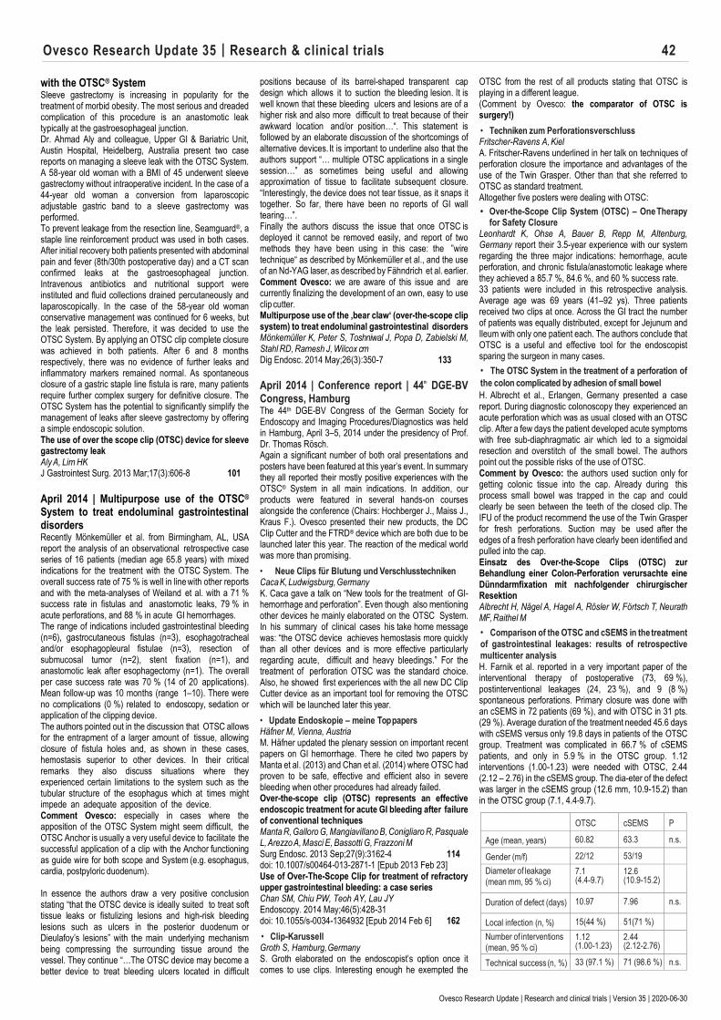
Ovesco Research Update 35 | Research & clinical trials 42
Ovesco Research Update | Research and clinical trials | Version 35 | 2020-06-30
with the OTSC® System Sleeve gastrectomy is increasing in popularity for the treatment of morbid obesity. The most serious and dreaded complication of this procedure is an anastomotic leak typically at the gastroesophageal junction. Dr. Ahmad Aly and colleague, Upper GI & Bariatric Unit, Austin Hospital, Heidelberg, Australia present two case reports on managing a sleeve leak with the OTSC System. A 58-year old woman with a BMI of 45 underwent sleeve gastrectomy without intraoperative incident. In the case of a 44-year old woman a conversion from laparoscopic adjustable gastric band to a sleeve gastrectomy was performed. To prevent leakage from the resection line, Seamguard®, a staple line reinforcement product was used in both cases. After initial recovery both patients presented with abdominal pain and fever (8th/30th postoperative day) and a CT scan confirmed leaks at the gastroesophageal junction. Intravenous antibiotics and nutritional support were instituted and fluid collections drained percutaneously and laparoscopically. In the case of the 58-year old woman conservative management was continued for 6 weeks, but the leak persisted. Therefore, it was decided to use the OTSC System. By applying an OTSC clip complete closure was achieved in both patients. After 6 and 8 months respectively, there was no evidence of further leaks and inflammatory markers remained normal. As spontaneous closure of a gastric staple line fistula is rare, many patients require further complex surgery for definitive closure. The OTSC System has the potential to significantly simplify the management of leaks after sleeve gastrectomy by offering a simple endoscopic solution. The use of over the scope clip (OTSC) device for sleeve gastrectomy leak Aly A, Lim HK J Gastrointest Surg. 2013 Mar;17(3):606-8 101
April 2014 | Multipurpose use of the OTSC® System to treat endoluminal gastrointestinal disorders Recently Mönkemüller et al. from Birmingham, AL, USA report the analysis of an observational retrospective case series of 16 patients (median age 65.8 years) with mixed indications for the treatment with the OTSC System. The overall success rate of 75 % is well in line with other reports and with the meta-analyses of Weiland et al. with a 71 % success rate in fistulas and anastomotic leaks, 79 % in acute perforations, and 88 % in acute GI hemorrhages. The range of indications included gastrointestinal bleeding (n=6), gastrocutaneous fistulas (n=3), esophagotracheal and/or esophagopleural fistulae (n=3), resection of submucosal tumor (n=2), stent fixation (n=1), and anastomotic leak after esophagectomy (n=1). The overall per case success rate was 70 % (14 of 20 applications). Mean follow-up was 10 months (range 1–10). There were no complications (0 %) related to endoscopy, sedation or application of the clipping device. The authors pointed out in the discussion that OTSC allows for the entrapment of a larger amount of tissue, allowing closure of fistula holes and, as shown in these cases, hemostasis superior to other devices. In their critical remarks they also discuss situations where they experienced certain limitations to the system such as the tubular structure of the esophagus which at times might impede an adequate apposition of the device. Comment Ovesco: especially in cases where the apposition of the OTSC System might seem difficult, the OTSC Anchor is usually a very useful device to facilitate the successful application of a clip with the Anchor functioning as guide wire for both scope and System (e.g. esophagus, cardia, postpyloric duodenum). In essence the authors draw a very positive conclusion stating “that the OTSC device is ideally suited to treat soft tissue leaks or fistulizing lesions and high-risk bleeding lesions such as ulcers in the posterior duodenum or Dieulafoy’s lesions” with the main underlying mechanism being compressing the surrounding tissue around the vessel. They continue “…The OTSC device may become a better device to treat bleeding ulcers located in difficult
positions because of its barrel-shaped transparent cap design which allows it to suction the bleeding lesion. It is well known that these bleeding ulcers and lesions are of a higher risk and also more difficult to treat because of their awkward location and/or position…“. This statement is followed by an elaborate discussion of the shortcomings of alternative devices. It is important to underline also that the authors support “… multiple OTSC applications in a single session…” as sometimes being useful and allowing approximation of tissue to facilitate subsequent closure. “Interestingly, the device does not tear tissue, as it snaps it together. So far, there have been no reports of GI wall tearing…”. Finally the authors discuss the issue that once OTSC is deployed it cannot be removed easily, and report of two methods they have been using in this case: the ”wire technique“ as described by Mönkemüller et al., and the use of an Nd-YAG laser, as described by Fähndrich et al. earlier. Comment Ovesco: we are aware of this issue and are currently finalizing the development of an own, easy to use clip cutter. Multipurpose use of the ‚bear claw‘ (over-the-scope clip system) to treat endoluminal gastrointestinal disorders Mönkemüller K, Peter S, Toshniwal J, Popa D, Zabielski M, Stahl RD, Ramesh J, Wilcox cm Dig Endosc. 2014 May;26(3):350-7 133
April 2014 | Conference report | 44
th DGE-BV Congress, Hamburg The 44th DGE-BV Congress of the German Society for Endoscopy and Imaging Procedures/Diagnostics was held in Hamburg, April 3–5, 2014 under the presidency of Prof. Dr. Thomas Rösch. Again a significant number of both oral presentations and posters have been featured at this year’s event. In summary they all reported their mostly positive experiences with the OTSC® System in all main indications. In addition, our products were featured in several hands-on courses alongside the conference (Chairs: Hochberger J., Maiss J., Kraus F.). Ovesco presented their new products, the DC Clip Cutter and the FTRD® device which are both due to be launched later this year. The reaction of the medical world was more than promising.
• Neue Clips für Blutung und Verschlusstechniken Caca K, Ludwigsburg, Germany K. Caca gave a talk on “New tools for the treatment of GI-hemorrhage and perforation”. Even though also mentioning other devices he mainly elaborated on the OTSC System. In his summary of clinical cases his take home message was: “the OTSC device achieves hemostasis more quickly than all other devices and is more effective particularly regarding acute, difficult and heavy bleedings.” For the treatment of perforation OTSC was the standard choice. Also, he showed first experiences with the all new DC Clip Cutter device as an important tool for removing the OTSC which will be launched later this year.
• Update Endoskopie – meine Top papers Häfner M, Vienna, Austria M. Häfner updated the plenary session on important recent papers on GI hemorrhage. There he cited two papers by Manta et al. (2013) and Chan et al. (2014) where OTSC had proven to be safe, effective and efficient also in severe bleeding when other procedures had already failed. Over-the-scope clip (OTSC) represents an effective endoscopic treatment for acute GI bleeding after failure of conventional techniques Manta R, Galloro G, Mangiavillano B, Conigliaro R, Pasquale L, Arezzo A, Masci E, Bassotti G, Frazzoni M Surg Endosc. 2013 Sep;27(9):3162-4 114 doi: 10.1007/s00464-013-2871-1 [Epub 2013 Feb 23] Use of Over-The-Scope Clip for treatment of refractory upper gastrointestinal bleeding: a case series Chan SM, Chiu PW, Teoh AY, Lau JY Endoscopy. 2014 May;46(5):428-31 doi: 10.1055/s-0034-1364932 [Epub 2014 Feb 6] 162
• Clip-Karussell Groth S, Hamburg, Germany S. Groth elaborated on the endoscopist’s option once it comes to use clips. Interesting enough he exempted the
OTSC from the rest of all products stating that OTSC is playing in a different league. (Comment by Ovesco: the comparator of OTSC is surgery!)
• Techniken zum Perforationsverschluss Fritscher-Ravens A, Kiel A. Fritscher-Ravens underlined in her talk on techniques of perforation closure the importance and advantages of the use of the Twin Grasper. Other than that she referred to OTSC as standard treatment. Altogether five posters were dealing with OTSC:
• Over-the-Scope Clip System (OTSC) – One Therapy for Safety Closure
Leonhardt K, Ohse A, Bauer B, Repp M, Altenburg, Germany report their 3.5-year experience with our system regarding the three major indications: hemorrhage, acute perforation, and chronic fistula/anastomotic leakage where they achieved a 85.7 %, 84.6 %, and 60 % success rate. 33 patients were included in this retrospective analysis. Average age was 69 years (41–92 ys). Three patients received two clips at once. Across the GI tract the number of patients was equally distributed, except for Jejunum and Ileum with only one patient each. The authors conclude that OTSC is a useful and effective tool for the endoscopist sparing the surgeon in many cases.
• The OTSC System in the treatment of a perforation of
the colon complicated by adhesion of small bowel
H. Albrecht et al., Erlangen, Germany presented a case report. During diagnostic colonoscopy they experienced an acute perforation which was as usual closed with an OTSC clip. After a few days the patient developed acute symptoms with free sub-diaphragmatic air which led to a sigmoidal resection and overstitch of the small bowel. The authors point out the possible risks of the use of OTSC. Comment by Ovesco: the authors used suction only for getting colonic tissue into the cap. Already during this process small bowel was trapped in the cap and could clearly be seen between the teeth of the closed clip. The IFU of the product recommend the use of the Twin Grasper for fresh perforations. Suction may be used after the edges of a fresh perforation have clearly been identified and pulled into the cap. Einsatz des Over-the-Scope Clips (OTSC) zur Behandlung einer Colon-Perforation verursachte eine Dünndarmfixation mit nachfolgender chirurgischer Resektion Albrecht H, Nägel A, Hagel A, Rösler W, Förtsch T, Neurath MF, Raithel M
• Comparison of the OTSC and cSEMS in the treatment of gastrointestinal leakages: results of retrospective
multicenter analysis H. Farnik et al. reported in a very important paper of the interventional therapy of postoperative (73, 69 %), postinterventional leakages (24, 23 %), and 9 (8 %) spontaneous perforations. Primary closure was done with an cSEMS in 72 patients (69 %), and with OTSC in 31 pts. (29 %). Average duration of the treatment needed 45.6 days with cSEMS versus only 19.8 days in patients of the OTSC group. Treatment was complicated in 66.7 % of cSEMS patients, and only in 5.9 % in the OTSC group. 1.12 interventions (1.00-1.23) were needed with OTSC, 2.44 (2.12 – 2.76) in the cSEMS group. The dia-eter of the defect was larger in the cSEMS group (12.6 mm, 10.9-15.2) than in the OTSC group (7.1, 4.4-9.7).
OTSC cSEMS P
Age (mean, years) 60.82 63.3 n.s.
Gender (m/f) 22/12 53/19 Diameter of leakage
(mean mm, 95 % ci) 7.1 (4.4-9.7)
12.6 (10.9-15.2)
Duration of defect (days) 10.97 7.96 n.s.
Local infection (n, %) 15(44 %) 51(71 %) Number of interventions
(mean, 95 % ci) 1.12 (1.00-1.23)
2.44 (2.12-2.76)
Technical success (n, %) 33 (97.1 %) 71 (98.6 %) n.s.

Ovesco Research Update 35 | Research & clinical trials 43
Ovesco Research Update | Research and clinical trials | Version 35 | 2020-06-30
Duration of treatment 24.8 38.9
Dislocation rate (n, %) 1 (3 %) 17 (24 %) The authors conclude in their discussion that OTSC is the preferred primary therapy of smaller post-interventional leakages. It might also be used in combination where cSEMS treatment was incomplete. They conclude that the longer treatment period with cSEMS and the higher complication rate might be due to sicker patients, but also due to the relevant dislocation rate of cSEMS.
Vergleich zwischen OTS-Klipp und cSEMS zur Indikationsstellung bei der Behandlung gastrointestinaler Leckagen: Ergebnisse einer retrospektiven, multizentrischen Analyse Farnik H1, Driller M1, Kratt T2, Schmidt C3, Fähndrich M4, Friedrich-Rust M1, Filmann N1, Königsrainer A2, Stallmach A3, Heike M4, Zeuzem S1, Albert J1 1Frankfurt a. M., 2Tübingen, 3Jena, 4Dortmund, Germany
• Endoscopic treatment of acute bleedings with an
Over-The-Scope Clip (OTSC) A. Braun et al. investigated the role of OTSC in the treatment of acute GI hemorrhage in an emergency. Between 2011 and 2013 they treated 16 patients (median 75.5 y/o (61-92), m=9, f=7) with OTSC for upper and lower-GI bleeding (8 each). Patients with upper-GI bleeding received high PPI-medication simultaneously. 7 patients were classified F Ia, 7 F Ib, and 2 F IIa. All patients presented with an acute decrease of hemoglobin, with hematemesis, melena, and hematochezia. The clip was applied by using a standard forceps. Technical success was achieved in all 16 patients (100 %) with immediate primary hemostasis. None of the interventions took longer than 20 minutes. Only 6 patients underwent follow-up endoscopy between day 1 and 7 after clip application. All control endoscopies were uneventful and showed clinical success. 9 patients did not need any further endoscopy. None of the patients needed any further therapy for bleeding. All patients started normal oral intake from day 2. The authors conclude that OTSC is safe and effective for the treatment of hemorrhage which reduces mortality, with short intervention times. Endoskopische Behandlung von akuten Blutungen mit einem Over-The-Scope Clip (OTSC) Braun A, Richter-Schrag HJ, Fischer A, Freiburg, Germany
• Clinical experience in the treatment of
perforations, leakages, and fistulas in the GI tract
with the Over-the-scope clip (OTSC)
J. Stückle et al. report their retrospective results in the standard indications of OTSC. 21 patients (Median 69 years (30–87), m=11, f=10) were treated for leakages and fistulas (n=11, 52 %) due to anastomotic leaks. 5 patients had complications due to diagnostic or therapeutic endoscopy. 2 patients had fistulas due to necrotizing pancreatitis. 2 patients suffered from a persistent PEG fistula. Technical success was reported in 20/21 cases (95 %). All 5 endoscopic complications could successfully be treated with OTSC. 7/11 anastomotic leaks could successfully be treated as well. One patient with duodenal leak due to acute necrotizing pancreatitis and a patient with perforated antrum died due to sepsis. The treatment of persistent PEG fistula was clinically not successful in this series. The authors conclude that altogether the treatment of perforations, leakages, and fistulas with OTSC is very promising, and point out that this is especially true for the management of complications during endoscopy and surgical complications like anastomotic leakage. Klinische Erfahrungen bei der Behandlung von Perforationen, Leckagen und Fistelungen im Gastrointestinaltrakt mit dem Over the scope Clip (OTSC®) Stückle J, Probst A, Bittinger M, Scheubel R, Ebigbo A, Messmann H, Gölder S, Augsburg, Germany
April 2014 | Efficacy and safety of OTSC® Proctology confirmed by clinical data at two major German conferences 40th Congress of the German Society for Coloproctology, April 3–5, 2014 in Munich
under the presidency of Prof. Dr. Dr. h.c. W. Hohenberger Prospective multicentric trial shows 90 % success rate for OTSC Proctology in anal fistula closure R. Prosst and co-authors, Stuttgart and Mannheim, Germany, presented data from a prospective multicentric trial including 20 patients treated with OTSC Proctology for anal fistula. 18 of 20 patients (90 %) reached the treatment success defined as clinical healing of the anal fistula and absence of recurrence at 6-month postoperative period. The authors conclude that OTSC Proctology is a new minimally invasive device for the treatment of anorectal fistula which is procedurally simple and time efficient. The risk profile is favourable, without relevant risk of fecal incontinence. Anorektaler Fistelverschluss mittels OTSC Proctology: Ergebnisse einer prospektiven Beobachtungsstudie R. Proßt, A. Joos, A. Herold, D Bussen, W. Ehni Proktologisches Institut Stuttgart & Enddarmzentrum Mannheim
The 131st Congress of the German Society for Surgery, March 25–28, 2014 in Berlin under the presidency of Prof. Dr. J. Jaehne
OTSC® Proctology in retrospective analysis of mixed case series: efficacy and safety confirmed S. Dango and colleagues, Kassel and Goettingen, Germany presented their experience using OTSC Proctology in the treatment of transsphincteric anal fistula. They conclude that OTSC placement is a promising sphincter-preserving minimally invasive method with considerably less complications than in more invasive types of surgical fistula treatment. Efficacy and safety of the over-the-scope clip in the treatment of anal trans-sphincteric fistula S. Dango, D. Schrader, M. Ghadimi, F. Antonakis, R. Hesterberg Departments of General and Visceral Surgery, Rotes Kreuz Krankenhaus, Kassel and University Hospital, Goettingen R. Menningen et al., Muenster, Germany report about their first experience with OTSC Proctology fistula closure in patients who had recurrence after fistula surgery. 9 consecutive patients were included into the trial. The authors conclude that OTSC is a safe and effective procedure for closing recurrent anal fistula even in more complex cases with Crohn’s disease or multiple surgical pretreatments. Verschluss analer Rezidivfisteln mit dem OTSC Proctology System R. Menningen, M. Laukoetter, N. Senninger, E. Rijken Klinik für Allgemein- und Viszeralchirurgie, University Hospital, Muenster For more detailed information on the studies see reports in a pdf file on: www.ovesco.com.
April 2014 | The OTSC® System: a surgery-sparing device for the management of iatrogenic duodenal perforation during endoscopic ultrasound Duodenal perforations are a rare but serious complication during endoscopic ultrasound examinations. The closure of these perforations with the OTSC System can be a surgery-sparing approach. Three case studies published by Dr. Silvia Salord et al., Dept. of Digestive Disease, University Hospital, Barcelona, Spain and by Dr. Gianfranco Donatelli and colleagues, Endoscopy Unit, Hôpital Privé des Peupliers, Paris, France demonstrate the successful use of the OTSC System in case of iatrogenic duodenal perforation. Two patients (aged 88 and 67) presented with cholangitis, one 74-year old woman with obstructive jaundice. In all three cases perforations occurred during endoscopic ultrasound procedures. Two perforations were located in the duodenal bulb, one at the superior duodenal flexure. By deploying an OTSC clip successful closure was achieved in all cases, no further surgical interventions were required. The two patients with cholangitis underwent therapeutic endoscopic retrograde cholangiography (ERC) afterwards
without any complications. Oral food intake was restarted after 2 or 5 days, respectively. Endoscopic closure of duodenal perforation with an over-the-scope clip during endoscopic ultrasound guided cholangiopancreatography Salord S, Gornals JB, Maisterra S, Pons C, Busquets J, Fabregat J Rev Esp Enferm Dig. 2012 Sep;104(9):489-90 98 Closure with an over-the-scope clip allows therapeutic ERCP to be safely performed after acute duodenal perforation during diagnostic endoscopic ultrasound Donatelli G, Vergeau BM, Dritsas S, Dumont JL, Tuszynski T, Meduri B Endoscopy. 2013 Nov;45 Suppl 2 UCTN:E392-3 151
March 2014 | OTSC®@FISMAD, Naples, Italy: 77 % success in anastomotic leak treatment At the 20th National Congress of Digestive Diseases, Napoli, Italy, March 19-22, MA Bonino and colleagues, Department of Surgery, Turin University reported about a consecutive series of 26 patients treated with OTSC for postsurgical colorectal leaks. The mean defect size was 8.7 mm, in 10 cases there were acute and in 16 cases chronic leaks (fistula). 4 cases were complicated by recto-vaginal, 3 by recto-vesical and 7 by colo-cutaneous fistula. In 3 cases OTSC was used to complete earlier vacuum sponge therapy. The overall success rate was 77 % (20/26), 90 % in acute (9/19) and 69 % (11/16) in chronic cases. There were no OTSC-related complications, additional surgery was needed in 2 cases. Anastomotic leakage is a serious and non infrequent complication in colorectal surgery. Incidence rates in the literature range from 1 to 39 %. Clinically relevant leaks are commonly seen in 3-6 % of the cases. OTSC closure of colorectal post-surgical leaks and fistula is a safe technique with a high success rate. Efficacia della clip OTSC per il trattamento di deiscenze e fistole chirurgiche del colon-retto Efficacy of the Over-The-Scope Clip (OTSC) for treatment of colorectal postsurgical leaks and fistulas Bonino MA, Verra M, Salvai A, Bullano A, Rapetti L, Arezzo A, Morino M
March 2014 | Management of esophageal perforation with the OTSC® System – four new case studies by different authors report favourable results Spontaneous or iatrogenic esophageal perforation is a life-threatening condition that can lead to severe mediastinitis, sepsis and multiple organ failure. Endoscopic management has contributed to the decrease of morbidity and mortality associated with surgical repair. Four different case reports lately published by Dr. Alexander Braun et al., Div. of General Surgery, University of Freiburg, Germany, Dr. Davide Bona et al., Div. of General Surgery, University of Milan, Italy and Dr. Alexandre Ferreira, Dept. of Gastroenterology and Hepatology, Hospital de Santa Maria, Lisbon, Portugal illustrate the successful closure of esophageal perforations with the OTSC System. Two patients presented with Boerhaave´s syndrome, one patient had an iatrogenic perforation and one patient suffered from a perforation caused by a fishbone. In all four cases a minimally invasive approach with the OTSC System was chosen. Two patients were treated with the OTSC clip within 12 hours. Although the two other patients were admitted to hospital not until after 48 h after an episode of vomiting, late management of the esophageal perforation with the OTSC System was successful. After complete closure of the defect, all patients were kept on antibiotic therapy and were discharged in stable condition after 10 days (patient with iatrogenic perforation), 21 days (patient with perforation caused by a fishbone) and 20 or 28 days respectively (patients with Boerhaave´s syndrome). 3-month follow-up revealed a free esophageal passage and correct placement of the OTSC clip. The OTSC clip is a new, safe and effective treatment alternative for the management of esophageal perforation. Due to the endoscopic approach and shorter hospital stay, the procedure is more cost effective than conventional surgical procedures. Endoskopischer Verschluss von distalen Ösophagus-Perforationen mit einem Over-The-Scope Clip (OTSC) Braun A, Hopt UT, Richter-Schrag HJ
Ovesco Research Update 35 | Research & clinical trials 44
Ovesco Research Update | Research and clinical trials | Version 35 | 2020-06-30
Endo heute 2013; 131 Management of Boerhaave‘s syndrome with an over-the-scope clip Bona D, Aiolfi A, Rausa E, Bonavina L 141 Eur J Cardiothorac Surg. 2013 Jul 18 [Epub ahead of print] Snapper fishbone esophageal perforation closed with an over-the-scope clip Ferreira AO, Lopes J, Velosa J BMJ Case Rep. 2013 Oct 25;2013 144
March 2014 | OTSC: easy to use with good results, decreasing morbidity and mortality in diagnostic and therapeutic endoscopy In the quest to describe the use and the clinical applications of OTSC System in an environment where endocopic and surgical techniques are increasingly more complex and frequent Singhal et al. have searched and analysed the literature using the key words „endoscopy“ and „over-the-scope clip“ in order to identify human studies evaluating the application of OTSC from January 2001 to August 2012. The indication, efficacy, complications, and limitations were recorded. The overall success rates of OTSC based on the current literature are in the range of 75 % to 100 % for closure of iatrogenic gastrointestinal perforations, 38 % to 100 % for closure of gastrointestinal fistulas, 50 % to 100 % for anastomotic leaks, and 71 % to 100 % for bleeding lesions. OTSC has shown 100 % success rates in managing postbariatric surgery weight gain secondary to dilation of the gastro-jejunostomy. The authors conclude that OTSC is easy to use with good results, thus decreasing the morbidity and mortality associated with complications secondary to both diagnostic and therapeutic endoscopy and avoiding surgery in many situations. Over-the-Scope Clip: Technique and Expanding Clinical Applications Singhal S, Changela K, Papafragkakis H, Anand S, Krishnaiah M, Duddempudi S J Clin Gastroenterol. 2013 Oct;47(9):749-56 128 Further reviews by Weiland et al.: Performance of the OTSC System in the endoscopic closure of iatrogenic gastrointestinal perforations: a systematic review Weiland T, Fehlker M, Gottwald T, Schurr MO Surg Endoscop. 2013; 27:2258-74 81-2 Performance of the OTSC System in the endoscopic closure of gastrointestinal fistulae – a meta-analysis Weiland T, Fehlker M, Gottwald T, Schurr MO Minim Invasive Ther Allied Technol. 2012 Jul;21(4):249-58 Epub 2012 Jun 14 113
February 2014 | New case series on use of OTSC® for treatment of refractory upper GI bleeding Apart from using the OSTC System in acute and chronic perforations (i.e. perforations, anastomotic leakage, fistulae) the authors of the renowned Institute of Digestive Disease, Department of Surgery, Chinese University of Hong Kong are reporting of patients in whom OTSC was used for endoscopic control of refractory or major upper gastrointestinal bleeding from lesions in the gastroduodenal tract between 1 July and 31 December 2012. Nine patients were included (median age 72.5 years, range 39–91) with bleeding gastric ulcers (n=2), bleeding duodenal ulcers (n=5), bleeding gastrointestinal stromal tumor in the stomach (n=1), and bleeding from ulcerative carcinoma of the pancreas (n=1). The median size of the ulcers was 2.5 cm (range 1–4). Six of the nine patients had undergone previous endoscopic hemostasis. Technical success (defined as hemostasis achieved at index endoscopy) was achieved in all patients and the clinical effectiveness was 77.8 % (defined as technical success with no rebleeding). All procedures were carried out by two experienced endoscopists. Those two patients that experienced rebleeding suffered from complex duodenal ulcer. One of them had been treated with radiotherapy for residual disease after resection of common bile duct cholangiocarcinoma. After several additional EGDs, transarterial embolization, and one surgical intervention which all failed to stop the bleeding, the patient died
eventually. The second patient bled from the inferior pancreaticoduodenal artery and needed arterial embolization as well. The authors discuss a meta-analysis of 1156 patients in 15 randomized trials where endoclips were shown to be superior to injection alone, and as effective as heater probe treatment. The overall rate of rebleeding in those conventionally treated patients ranged between 7.1 % and 9.5 % though. Since rebleeding correlates with the adverse outcome of this indication they speculate that control of bleeding would have a positive impact on patient outcome. Even though the study was carried out in patients with complex duodenal ulcer and underlying malignancies the technical success rate of OTSC was 100 %. They also point out that usually in cases like these the application of conventional clips is difficult; the repeated application of heater probe being associated with a higher risk of perforation. Whereas the application of OTSC allows for larger amounts of tissue and constitutes a quite durable treatment (OTSC in situ after a median of 28 days in this study). The authors conclude that the use of OTSC is a safe and effective method of endoscopic hemostasis for major bleeding from miscellaneous upper gastrointestinal causes and should be considered in refractory bleeding after conventional endoscopic hemostasis, before surgery or angiographic embolization. Comment by Ovesco: a prospective controlled randomized multicenter trial with 64 patients with recurrent upper GI bleeding is recruiting in Germany (Endoscopic Treatment of Recurrent Upper GI Bleeding: OTSC [Over the Scope Clip] Versus Standard Therapy (STING). ClinicalTrials.gov Iden- tifier: NCT01836900) Use of Over-The-Scope Clip for treatment of refractory upper gastrointestinal bleeding: a case series Chan SM, Chiu PW, Teoh AY, Lau JY Endoscopy. 2014 Feb 6. [Epub ahead of print] 162
February 2014 | Retrospective study on efficacy and safety of the OTSC® System in the treatment of GI bleeding, fistula and perforation: primary technical success rate 91.3 %, durable clinical success rate 82.6 % Dr. Noriko Nishiyama and colleagues, Dept. of Gastroenterology and Neurology, Kagawa University, Japan, recently presented their retrospective study on efficacy and safety of the OTSC System in endoscopic closure of gastrointestinal bleeding, fistulas and perforations, concluding that the OTSC System is a highly useful device that can safely be utilized for these indications. Their case series consisted of 23 consecutive patients treated between November 2011 and September 2012 (mean age 77 years) included the following indications for OTSC placement: stopping GI bleeding (n=9), closing perforation (n=10), closing chronic fistula (n=4) and prevention of post endoscopic submucosal dissection (ESD) duodenal artificial ulcer perforation (n=1). One patient had a perforation that formed a fistula. Lesions were located in the esophagus (n=1), the stomach (n=10), the duodenum (n=5), the sigmoid colon (n=3) and in the rectum (n=4). In 8 patients other therapies preceded OTSC application (e.g. conventional hemostatic clips, local injections, hemostatic coagulation forceps). Median follow-up time was 67 days. The primary technical success rate was 91.3 % (21/23). In two cases application of the OTSC clip was not possible due to stiff, fibrotic lesion edges. The overall clinical success rate (complete closure by using only OTSC clips) was 82.6 %. Major contributing factors for OTSC failure were a large lesion size (greater than 20 mm) and a delayed diagnosis (more than 1 week). No patient reported any complications associated with OTSC placement. In conclusion, the OTSC is an interesting and novel device that enhances the armamentarium of therapeutic gastroenterologists. Efficacy and safety of over-the-scope clip: including complications after endoscopic submucosal dissection Nishiyama N, Mori H, Kobara H, Rafiq K, Fujihara S, Kobayashi M, Oryu M, Masaki T World J Gastroenterol. 2013 May 14;19(18):2752-60 126
January 2014 | OTSC® in mucosal flap closure after peroral endoscopic myotomy (POEM) Maintaining the integrity of the mucosal flap and the reliable closure of mucosal entry during peroral endoscopic myotomy (POEM) is paramount in preventing leakage of esophageal contents into the mediastinal space. In a recently published case series (n=2) Payal Saxena, MD and colleagues, Dept. of Medicine and Div. of Gastroenterology and Hepatology, Johns Hopkins Medical Institutions, Baltimore, Maryland, USA describe their positive experience with the application of the OTSC System for reliable and easy flap closure after POEM. Both patients presented with dysphagia and regurgitation and were diagnosed with achalasia. It was decided to proceed with POEM. After myotomy of the inner circular muscle bundles it was noted that the mucosal incision had elongated from 2 cm to 4 cm in one case. Whereas the distal part of the mucosal entry was successfully closed with conventional hemostatic clips (Resolution Clip, Boston Scientific) in both cases, closure of the proximal half was not possible even with different clips. As the clips were noted to slip to one side of the mucosal incision, there was a risk of displacing clips into the submucosal tunnel. Hence, all partially attached clips were removed with biopsy forceps. Finally, complete closure of the mucosal incision was performed with the OTSC clip and the OTSC Twin Grasper in both cases. Contrast swallow of the esophagus the following day revealed no leaks in either patient. The authors state that the OTSC clip provides more durable closure than standard hemostatic clips and full-thickness closure is achievable due to greater compressive force. Considering that failure of closure risks serious adverse events, like mediastinitis and sepsis, these features of the OTSC clip appear even more attractive. An alternative method for mucosal flap closure during peroral endoscopic myotomy using an over-the-scope clipping device Saxena P, Chavez YH, Kord Valeshabad A, Kalloo AN, Khashab MA Endoscopy. 2013 Jul;45(7):579–81 122
January 2014 | Avoiding Surgery: Minimally invasive endoscopic management of an iatrogenic colon perforation Iatrogenic lesions of GI organs are a significant complication of diagnostic or interventional endoscopic procedures. Dr. Pilar Díez-Redondo and colleagues, Dept. of Gastroenterology, Hospital Universitario, Río Hortega, Valladolid, Spain present a case report on OTSC clipping for colon perforation closure: For assessment of iron deficiency an 82-year old woman was referred to the endoscopic unit. A gastroscopy confirmed a hiatal hernia. Colonoscopy revealed no abnormalities. 18 cm proximal to the anus an iatrogenic perforation with a size of 12 mm occurred. To close the perforation endoscopically an 11/a OTSC clip was chosen. The target tissue and a piece of omentum were pulled into the applicator cap by suction and the clip released successfully, approximating the edges of the lesion. A small residual recess was closed with two conventional, endoscopic clips. The patient was discharged 10 days after the intervention. A 7-month follow-up confirmed the correct placement of the OTSC. Iatrogenic colon perforations can cause severe complications and often require surgery, as the major drawback of an endoscopic approach with conventional clips is the limited ability of these clips to achieve sufficient apposition of the mucosa and submucosa to ensure tight sealing of the perforation. With the advent of the larger and more power- ful OTSC clips, surgery can be avoided and perforations managed in a minimally invasive, endoscopic way. For that reason the authors suggest that the OTSC System should be available to all endoscopy units as a bail-out device. A novel system for endoscopic closure of iatrogenic colon perforations using the Ovesco® clip and omental patch Díez-Redondo P, Blanco JI, Lorenzo-Pelayo S, De-la-Serna-Higuera C, Gil-Simón P, Alcaide-Suárez N, Pérez-Miranda M Rev Esp Enferm Dig. 2012 Oct-Nov;104(10):550-2 103
OTSC®
Update 16
Ovesco Research Update 35 | Research & clinical trials 45
Ovesco Research Update | Research and clinical trials | Version 35 | 2020-06-30
January 2014 | Closure of gastric fistulas after bariatric surgery with the OTSC® System – two case studies Iatrogenic gastric fistulas after bariatric surgery are a potentially dangerous situation as they can lead to severe complications, such as peritonitis and abscess formation. Two case reports recently published by Dr. Victoria Gómez and colleagues, Dept. of Gastroenterology and Hepatology, Mayo Clinic, Jacksonville, USA, and Dr. Hany Shehab et al., Dept. of Gastroenterology, Dar Alfouad Hospital Giza, Egypt, respectively, describe the closure of gastric fistulas with the OTSC System after laparoscopic bariatric surgery. Dr. Gómez reports on a 45-year old woman who was hospitalized for management of complications from a prior sleeve gastrectomy. Postoperatively the patient developed fever and abdominal pain. A CT scan showed a fluid collection in the region of the right liver lobe, free intraperitoneal air and an abscess in the postsurgical bed of the stomach. The results of a barium contrast study were consistent with a significant leak in the proximal third of the gastric sleeve. EGD revealed a gastric fistula 4 centimeters below the esophago-gastric junction. As an initial treatment with an esophageal stent and abdominal drains had failed to seal the leak, the stent was removed and a fully covered esophageal stent was applied. A second stent had to be deployed to bridge the prior stent. Since there was no improvement of the fistula, the stents were again removed. As next treatment approach the fistula was grasped with the OTSC Twin Grasper and closed by application of an OTSC clip. A follow-up radiograph showed no extravasation of contrast. Dr. Shehab presents the case of a 36-year old man who had undergone a Roux-en-Y gastric bypass for morbid obesity. Postoperatively an anastomotic leak was found. Two attempts of surgical repair failed as well as a conservative approach with drainage and insertion of a feeding jejunostomy. 5 months after the first surgery an EGD revealed a well-epithelialized fistula with a wide lumen. It was decided to close the fistula by OTSC clipping. To remove the epithelium at the fistula orifice, argon plasma coagulation was applied to the proximal lumen of the fistula. Then the OTSC Twin Grasper was used to approximate the edges of the fistula orifice followed by the application of the OTSC clip. After 10 months there was no evidence of a fistula recurrence. Since a surgical intervention for postoperative fistulas in an obese patient with recent bariatric surgery is most often not desirable, a minimally invasive, endoscopic approach with the OTSC System is an attractive treatment option. In comparison to conventional clips that are only suitable for small fistulas and only attach to the superficial mucosal layer, the OTSC clip offers a deeper grasp of the tissue and a sturdier closure. Closure of an iatrogenic bariatric gastric fistula with an over-the-scope clip Gómez V, Lukens FJ, Woodward TA Surg Obes Relat Dis. 2013 Mar-Apr;9(2):e31-3 doi: 10.1016/j.soard.2012.09.004 99 Combined endoscopic techniques for closure of a chronic post-surgical gastrocutaneous fistula: case report and review of the literature (with video) Shehab HM, Elasmar HM Surg Endosc. 2013 Aug;27(8):2967-70 119 doi: 10.1007/s00464-013-2839-1 Epub 2013 Feb 23
December 2013 | First report on successful management of delayed presentation of Boerhaave´s syndrome Current guidance has advocated surgery for delayed presentations of Boerhaave´s syndrome with evidence of mediastinal contamination. However, Dr. Eamon Ramhamadany and colleagues, Dept. of General Surgery, University Hospital Coventry and Warwickshire, UK, present the successful management of Boerhaave´s syndrome in a 69-year-old man by means of the OTSC System, sparing the patient surgery and possible associated complications. The man presented to hospital with an episode of forceful vomiting. A chest radiograph was performed revealing a pleural effusion. After several days without improvement a CT chest showed an oesophageal perforation with
mediastinitis. Because of the size of the defect and the delay in presentation, it was decided not to perform surgery, but to apply the OTSC clip for endoscopic repair. A contrast swallow confirmed the correct placement of the clip and the successful closure of the leak. After a total parenteral nutrition for 3 days, the patient was fed via a naso-jejunal tube. Intravenous antibiotics and bilateral chest drains led to a resolving mediastinitis. The whole procedure resulted in a favourable outcome wit- hout the need for surgery. The authors conclude that the OTSC can be used to manage patients with delayed presentation of Boerhaave´s and that further evaluation is needed to define the indications for minimally invasive techniques like the OTSC System. A delayed presentation of Boerhaave‘s syndrome with mediastinitis managed using the over-the-scope clip. Ramhamadany E, Mohamed S, Jaunoo S, Baker T, Mannath J, Harding J, Menon V J. surg. case rep. (2013) 2013 (5): rjt020. 120
December 2013 | Management of postoperative gastrointestinal leakages and fistulas with the OTSC® System: long-term success rate of 79 % Dr. Rudolf Mennigen and colleagues, Dept. of General and Visceral Surgery, University Hospital of Muenster, Germany, recently presented a study on efficacy and safety of the OTSC System in endoscopic closure of postoperative gastrointestinal leakages and fistulas, concluding that the OTSC System dramatically increases the possibilities of defect closure by endoscopic clipping as opposed to conventional endoclips. Their case series of 14 consecutive patients (May 2011– November 2012) included patients with anastomotic leakage (n=6) e.g. after gastrectomy, perforation after fundoplication (n=1) and post-operative fistulas (n=7, colocutaneous, enterocutaneous, gastrobronchial, rectourethral, rectocutaneous, gastropleural). 11 of the 14 lesions were chronic (treated by OTSC later than postoperative day 14) and in 9 patients other therapies preceded OTSC application (e.g. covered stent application, fibrin glue injection). Median follow-up time was 5.5 months. The primary procedural success rate was 100 %. 3/14 patients (21 %) required further treatment during follow-up. Reasons for OTSC failure were massive fibrosis of the fistula and application in an actively inflamed bowel segment in Crohn‘s disease. However, unsuccessful OTSC treatment did not impair subsequent surgical therapies. Complete and clinically durable closure of the defects was achieved in 79 %, indicating from the authors‘ point of view that the OTSC will play an important role in the therapy of postoperative leakages. Endoscopic closure of postoperative gastrointestinal leakages and fistulas with the Over-the-Scope Clip (OTSC) Mennigen R, Colombo-Benkmann M, Senninger N, Laukoetter M Gastrointest Surg. 2013 Jun;17(6):1058-65 115
November 2013 | OTSC® in endoscopic treatment of acute GI bleeding after failure of conventional techniques: primary hemostasis of 97 % The OTSC System can overcome the limitations of conventional clips in the treatment of patients with acute GI bleeding by providing compression of large amounts of tissue, leading to a more efficient hemostasis. Dr. R. Manta and colleagues, Gastroenterology and Endoscopy Unit, New S. Agostino Hospital, Modena, Italy draw this conclusion on the basis of a retrospective analysis of a consecutive case series of 30 patients with severe acute GI bleeding treated with the OTSC System after failure of conventional techniques. Data were collected from six high-volume endoscopy units in a period between December 2011 and September 2012. All 30 patients suffered from bleeding lesions unresponsive to saline/adrenaline injection and through-the-scope clipping located in the upper and lower GI tract in 23 and 7 cases, respectively. Bleeding lesions included duodenal ulcer (n=12), gastric ulcer (n=6), Mallory-Weiss (n=2), Dieulafoy (n=2) and surgical anastomosis (n=1) in the upper GI tract and endoscopic mucosal resection (n=5),
endoscopic submucosal dissection (n=1) and colonic diverticulum (n=1) in the lower GI tract. Primary hemostasis with OTSC was achieved in 29 of 30 cases (97 %). Rebleeding in two cases was successfully treated with injection of saline and adrenaline. Endoscopic follow-up after 2–4 days and after 1 month revealed correct placement of the OTSC clip and no procedure-related complications. Thus, the OTSC is an effective and safe device for treatment of acute GI bleeding and represents a useful adjunct to the therapeutic armamentarium in endoscopic emergencies. Over-the-scope clip (OTSC) represents an effective endoscopic treatment for acute GI bleeding after failure of conventional techniques Manta R, Galloro G, Mangiavillano B, Conigliaro R, Pasquale L, Arezzo A, Masci E, Bassotti G, Frazzoni M Surg Endosc. 2013 Sep;27(9):3162-4 114
October 2013 | OTSC® System: Effective closure of esophageal fistula following total gastrectomy Postoperative leaks after total gastrectomy are among the most common early complications. Dr. C. N. Ferreira and colleagues, Serviço de Gastrenterologia e Hepatologia, Hospital de Santa Maria, Lisbon, Portugal report on a 78-year old woman presenting with melena. She was diagnosed with gastric adenocarcinoma and treated with total gastrectomy and esophagojejunal Roux-en-Y anastomosis. On the fifth postoperative day she developed a septic condition caused by a fistulous orifice just above the intact anastomosis. Due to her poor general condition a surgical intervention was unfeasible. Thus, it was decided to treat the fistula endoscopically by means of the OTSC clip. By using the OTSC Twin Grasper to approximate the edges of the fistula and application of an OTSC clip the orifice was effectively closed. The patient was discharged in stable condition two weeks later. In a commentary to this publication Dr. David Robbins, Assistant Editor of the Journal Gastrointestinal Endoscopy emphasizes the significantly higher strength of the OTSC clip for hemostasis and closure of GI tract wall in comparison to conventional endoscopic clips. Total gastrectomy in an elderly patient complicated by esophageal fistula: rescue by the over-the-scope clip Noronha Ferreira C, Ribeiro LC, Velosa J, Ferreira J, Ferreira C, Freire JP, Marques J, Ruivo A, Bicha Castelo H Gastrointest Endosc. 2013 Mar;77(3):497-8 ; [Epub 2013 Jan 4] 112
October 2013 | Efficacious OTSC® hemostasis in Dieulafoy´s gastric lesion resistant to conventional endoscopic treatment Dr. B. Mangiavillano and colleagues, Gastrointestinal Endoscopy, Azienda Ospedaliera San Paolo Universitary, Hospital-University of Milan, Italy, present a case study of a 69-year old woman with an episode of melena. EGD showed a Dieulafoy´s bleeding lesion in the proximal third of the posterior wall of the stomach. The lesion was treated with an epinephrine injection and application of two conventional working-channel delivered metallic clips and the patient was discharged two days later. After three days the patient again presented with melena. Blood transfusions were necessary. An EGD was performed, showing no sign of an actively bleeding ulcer. The patient was admitted to hospital and suffered from another episode of melena with hemorrhagic shock. The now actively bleeding Dieulafoy´s lesion was then treated with an OTSC clip, stopping the hemorrhage completely and persistently. Endoscopic follow-up after 30 days displayed correct placement to the OTSC and no signs of further bleeding. Successful treatment with an over-the-scope clip of Dieulafoy´s gastric lesion resistant to conventional endoscopic treatment Mangiavillano B, Arena M, Morandi E, Viaggi P, Masci E Endoscopy. 2012;44 Suppl 2 UCTN:E387 105
October 2013 | OTSC® successful in providing hemostasis in posterior duodenal ulcer
OTSC®
Update 15 EXTRA

Ovesco Research Update 35 | Research & clinical trials 46
Ovesco Research Update | Research and clinical trials | Version 35 | 2020-06-30
bleeding after failure of conventional clips Ulcer bleeding is one of the key indications for the OTSC System. In a recently published case series (n=4), Prof. Klaus Mönkemüller and colleagues, Dept. of Internal Medicine, Gastroenterology and Infectious Diseases, Marienhospital Bottrop, Germany add to the growing clinical experience in using the OTSC System to control massive gastrointestinal bleedings and achieve life-saving hemostasis. All four patients (mean age 84.5) presented with hypotension and mean hemoglobin of 9 g/dL. After initial fluid resuscitation an emergent EGD displayed actively oozing ulcers in the posterior duodenum. As an initial therapy with injection of epinephrine-saline solution and standard clip placement failed and all patients suffered from rebleeding, the decision to apply the OTSC System was made. Hemostasis was attained successfully and all patients discharged in stable conditions. Even in difficult located ulcers in the posterior duodenum the placement of the OTSC is easy and effective to obliterate bleeding vessels resulting in life- saving hemostasis. Utility of the „bear claw“, or over-the-scope clip (OTSC) system, to provide endoscopic hemostasis for bleeding posterior duodenal ulcers Mönkemüller K, Toshniwal J, Zabielski M, Vormbrock K, Neumann H Endoscopy. 2012;44 Suppl 2 UCTN:E412-3. [Epub 2012 Nov 20] 108
September 2013 | OTSC® in post-surgical complications: retrospective case review confirms high clinical efficacy Dr. Alisa Coker and colleagues, Dept. of Surgery, University of California San Diego, USA, report on their experience with the OTSC System in a retrospective review of all cases treated between August 2011 and March 2012. All 10 patients had clinically significant gastrointestinal post-surgical complications. Indications included: gastric leaks after sleeve gastrectomy (n=4), post-operative colonic leak following extended hemicolectomy and palliative debulking (n=1), gastro-gastric fistulas following Roux-en-Y gastric by- pass (n=2), esophageal perforation (n=3). Three of the four patients with gastric leaks had undergone previous unsuccessful attempts at endoscopic repair (stenting, fibrin glue application, traditional clipping, endoscopic suturing). The overall clinical success rate was 70 %. Re-surgery was needed in the two cases of gastro-gastric fistulas. In the colonic leak patient the clip placement procedure was aborted due to a fixed tortuous sigmoid colon as a result of the metastatic disease and adhesions, limiting endoscope passage. For the subgroup of seven patients treated for leaks and perforations a success rate of 87.5 % with complete resolution was achieved. The mean follow-up period was 83 days. No complications occurred. The authors conclude that the OTSC System is simple to use, safe and effective with a great potential for success in a broad number of applications. For the treatment of gastric leaks following sleeve gastrectomy the OTSC System is their first-line treatment. Initial Experience with an Innovative Endoscopic Clipping System Coker AM, Jacobsen GR, Acosta G, Talamini MA, Savides TJ, Horgan S Surg Technol Int. 2012 Dec;22:39-43 [Epub ahead of print] 111
September 2013 | Preventive closure of duodenal lesion after endoscopic submucosal dissection with the OTSC® System to obviate delayed perforation The two case reports published in the journal Digestive Endoscopy by Dr. Hirohito Mori and his colleagues, Dept. of Gastroenterology and Neurology, Faculty of Medicine, Kagawa University, Japan illustrate the complete closure of secondary duodenal ulcers after endoscopic submucosal dissection (ESD) with the OTSC System without any complications. Two elderly patients were diagnosed with early duodenal cancer. ESD was carried out successfully removing the
lesions en bloc. In one case the muscle layer was slightly injured but not perforated. Because of the exposure to bile and pancreatic juices the risk of post-ESD delayed perforation is much higher in the duodenum than in other parts of the gastrointestinal tract. As conventional clips are less suitable due to small size and insufficient grasping power, Dr. Mori and his team used the OTSC System to close the lesion completely without any complications. The ulcer closure procedure time was 7 resp. 10 min. In both cases control endoscopy revealed a complete healing of the ulcer after 30 days. Dr. Mori and his colleagues consider the OTSC System to be one of the most effective devices to prevent delayed perforations in post-ESD ulcer. Successful closing of duodenal ulcer after endoscopic submucosal dissection with over-the-scope clip to prevent delayed perforation Mori H, Shintaro F, Kobara H, Nishiyama N, Rafig K, Kobayashi M, Nakatsu T, Miichi N, Suzuki Y, Masaki T Dig Endosc., 2013 Jul;25(4):459-61 | Epub 2012 Aug 7 89
September 2013 | First two publications of endoscopic closure of gastrocolic fistula using the OTSC® System Dr. Alberto Murino, Wolfson Unit for Endoscopy, St Mark’s Hospital, London, UK, and his colleagues report on a successfully treated gastrocolic fistula using the OTSC System. A migrated PEG tube caused a gastrocolic fistula in the transverse colon in a 41 y/o male with cerebral palsy. The fistula led to extensive diarrhea and mouth odor. The CT showed an involvement of the greater curvature of the stomach. By using the OTSC Anchor to approximate the tissue the OTSC clip was released precisely closing the fistula orifice completely. Diarrhea and mouth odor were stopped. The 3 months‘ follow-up revealed a complete healing of the fistula. The second case report published in the World Journal of Gastrointestinal Endoscopy by Prof. Klaus Mönkemüller and colleagues, Division of Gastroenterology and Hepatology, Basil Hirschowitz Endoscopic Center of Excellence, University of Alabama, Birmingham, USA, describes the effective endoscopic closure of a large gastrocolic fistula with the OTSC System in an extremely malnourished patient with complex post-surgical upper GI anatomy. The 47 y/o man presented with chronic diarrhea und severe weight loss of 32 kg in a 1-year period. He had a history of chronic pancreatitis, alcoholism and Billroth II gastrojejunostomy due to a perforated peptic ulcer. Endoscopy showed a clean based ulceration at the anastomosis and a second orifice that represented the fistula. Connecting stomach and colon, the fistula measured about 10–12 mm. Because of the patient‘s poor clinical status he could not benefit from a surgical intervention so an endoscopic procedure using the OTSC System was chosen. To ensure a definitive closure of the fistula the OTSC Twin Grasper was used to approximate the edges of the fistula. The application of the OTSC led to a complete closure of the gastrocolic fistula which was confirmed by an endoscopy. For Prof. Mönkemüller this case “adds to the growing evidence that the OTSC System is a useful device to treat clinically significant endoluminal GI defects.” He believes that “this device is a major breakthrough for the management of various types of discontinuity defects or fistulas of the GI tract (…)” and that “the OTSC System should be incorporated into the therapeutic armamentarium of the advanced endoscopist.” First report of endoscopic closure of a gastrocolic fistula using an over-the-scope clip system (with video) Murino A, Despott EJ, Vaizey C, Bashir G, Hansmann A, Gupta A, Konieczko K, Fraser C
Gastrointest Endosc. 2012 Apr;75(4):893; discussion 894 72
Endoscopic closure of gastrocolic fistula using the over-the-scope clip system Mönkemüller K, Peter S, Alkurdi B, Ramesh J, Popa D, Wilox C World J Gastrointest Endosc. 2013 Aug 16;5(8):402-6
136
August 2013 | The interesting case: OTSC® closure of esophagobronchial fistula Dr. E. Zolotarevysky and colleagues from the Department of Gastroenterology and Nutrition Service at Memorial Sloan-Kettering Cancer Center, New York City report about an in- teresting case in which OTSC clipping was used for closing an esophagobronchial fistula. An 83 y/o woman presented with a symptomatic fistula arising from an esophageal diverticulum with recurrent pulmonary infections. Placing a covered self-expanding metal stent was not believed to result in adequate seal of the chronic lesion. The placement of a percutaneous gastrostomy tube was refused by the patient. Also bronchial stenting and surgery were not considered as good options in this case. In this situation closure of the fistula with the OTSC clip was decided. A 12/6/t clip was placed under endoscopic control and with the aid of the OTSC Anchor for better manipulation and targeting of the fistula orifice. Immediate technical success was achieved and verified by barium esophagogram 2 days later. The patient was discharged from the hospital after 1 week in stable condition. The clip was still found in place at 1 month follow-up by chest X-ray but passed spontaneously and uneventfully as seen in CAT scan 45 days after the procedure. Final follow-up at 3 months revealed no recurrence or postprandial cough. Esophagobronchial fistula closure using a novel endoscopic over-the-scope clip Zolotarevsky E, Kwon Y, Bains M, Schattner M Ann Thorac Surg. 2012 Sep;94(3):e69-70. 96
July 2013 | OTSC® effective in emergency closure of iatrogenic GI perforations instead of abdominal surgery Dr. Hagel and colleagues, Dept. of Gastroenterology, University of Erlangen-Nuremberg, Germany reported about a consecutive series of 17 cases with perforations of the digestive tract, treated with OTSC clipping. All cases were considered as being candidates for abdominal surgery for closing the perforation. In 11 cases perforation closure with OTSC was immediately successful, thus avoiding surgery in 64.7 %. In 6 cases surgical closure was done. The area size of perforation in the successful cases was 21.1 ± 9.1 sqmm; in the unsuccessful group the area size was 97.6 ± 149 sqmm. Unsuccessful cases had on average a larger size, necrotic margins and required more OTSC clips during closure attempts (2.3 ± 0.5, p=0.018). The authors conclude: “OTSC application yields a high rate of endoscopic perforation closure in patients with macrosco- pic gastrointestinal perforation, even in an emergency setting, representing an alternative to surgery, especially when the size of the lesion is not too large and when vital or solid perforation margins are expected.” Over-the-Scope Clip Application Yields a High Rate of Closure in Gastrointestinal Perforations and May Reduce Emergency Surgery Hagel AF, Naegel A, Lindner AS, Kessler H, Matzel K, Dauth W, Neurath MF, Raithel M J Gastrointest Surg. 2012 Nov;16(11):2132-8 [Epub 2012 Aug 18] 88
July 2013 | OTSC® System in transgastric appendicectomy Kaehler et al. report the results of their first 15 patients in a prospective trial on “Transgastric appendectomy” which now already recruited 30 patients who are currently under follow-up. From April 2010 the Mannheim group offered to their patients a trangastric appendectomy. Patients with generalised peritonitis and/or local contraindications were not recruited. Out of 111 eligible candidates 15 agreed to undergo the proposed NOTES procedure. 14 out of 15 were actually operated through NOTES, whereas 1 patient was switched to laparoscopic procedure due to severe inflammation and adhesions. In each case the gastrotomy was closed by a single OTSC System using Twin Grasper and 12/6/t clip. All closures were tight primarily and uneventful throughout the follow-up.
OTSC®
Update 14
Ovesco Research Update 35 | Research & clinical trials 47
Ovesco Research Update | Research and clinical trials | Version 35 | 2020-06-30
This is the first series of transgastric appendectomy using the OTSC System (and the second series overall). All 30 patients who have been recruited altogether will be reported in a separate publication. Transgastric appendicectomy Kaehler G, Schoenberg MB, Kienle P, Post S, Magdeburg R Brit J Surg, 2013; 100: 911–915 130
July 2013 | Recommendation of OTSC® System in complex GI bleeding In an overview article the authors are referring to the current guideline therapies available and new developments. They report that other new three-dimensional clips seem to be even less efficacious than normal hemoclips. Thus, the authors conclude that obviously one of the key elements to successful hemostasis is the strength of the jaws of a clip and the amount of tissue captured. They state that this is obviously fulfilled by the design of the OTSC System which allows for the capture of a large amount of tissue and is more secure than other clips in the experimental setting. Thus the OTSC System is being recommended and used in complex GI bleeding. According to Leung & Lau a single clip suffices for most circumstances and therefore the procedure is shorter when compared to multiple applications of hemoclips. Comment by Ovesco: In a recently published series of 83 patients with severe and complicated GI bleedings (e.g. relapses after conventional endoscopic hemostasis or indi- cation for a surgical intervention) the success rate was close to 93 % with OTSC (Kratt T et al., Poster DGE-BV meeting, Munich 3/2013) New endoscopic hemostasis methods Leung Ki EL, Lau JY Clin Endosc. 2012 Sep;45(3):224-9 | Epub 2012 Aug 22 87
June 2013 | Report on successful removal of an OTSC® Clip Prof. Mönkemüller and colleagues presented a clip removal case in a letter to the editor of Gastrointestinal Endoscopy. Ten days after treating an anastomotic leak with the OTSC System, there was still a leak due to misplacement of the clip. The clip had to be removed to place another OTSC onto the leak. At first clip rising was accomplished by injecting saline solution below the OTSC. A snare was positioned around the clip, slowly closed and retracted. The clip dislodged and was retrieved carefully without injury by catching it with the snare and keeping it close to the distal end of the endoscope. The anastomotic leak was thereafter closed successfully with a new OTSC. Endoscopic removal of an over-the-scope clip (“bear claw”) Mönkemüller K, Toshniwal J, Zabielski M Gastrointest Endosc. 2012 Nov;76(5):1077-8 107
June 2013 | German surgical periodical alludes to OTSC® Proctology as a novel therapy for anal fistula In the German surgical periodical “Chirurgische Allgemeine” Prof. Dr. A. Herold, German Center for the Anorectum (EDZ), Mannheim, Germany, gave an overview on new treatments and devices for anorectal fistula. Prof. Herold is the General Secretary of the German Society for Coloproc- tology (DGK). In his paper he refers to OTSC Proctology as a new therapeutic alternative. Neue Techniken bei der Therapie der Analfistel Herold A. Chirurgische Allgemeine (2013); 14: 99–102 117
May 2013 | Ovesco’s Full Thickness Resection Device (FTRD®) presented in live endoscopy at Endo-Update meeting During clinical live demonstrations at endo-update which took place under the presidency of Prof. Dr. H. Messmann and Prof. Dr. H.-D-Allescher in Augsburg, Germany, a neuro-endocrine tumor (NET) in the rectum was resected with the new Full-Thickness Resection Device of Ovesco Endoscopy: the FTRD. A 62 year old patient showed a submucosal tumor of about 9 mm diameter. Biopsy revealed a neuroendocrine tumor. Prof. Dr Thomas Rösch (University
Hospital Hamburg-Eppendorf) used the FTRD to resect the lesion. The FTRD consists of an elongated OTSC cap premounted with a specially designed, derivative OTSC clip and the cap incorporates a resection snare. Prof. Rösch grasped the lesion with a grasping forceps and pulled the target tissue into the cap in a full thickness fashion. After mobilizing the tissue into the cap, the clip was released to seal the invaginated tissue before resection. Right afterwards the snare was closed and the tissue resected with HF current. The resection specimen included the full thickness of the wall carrying the NET, with a safety margin. The serosa was seen in histology, confirming that the specimen was a full-thickness resection. The FTRD device is not yet commercially available. endo-update 2012 | 30.11.-01.12.2012 Venue: Klinikum Augsburg, Augsburg, Germany
May 2013 | Iatrogenic digestive tract perforations: OTSC® closure as preferred method Dr. C. Gubler and Prof. P. Bauerfeind, Dept of Gastroenterology, Zurich University Hospital, Switzerland, report about the use of the OTSC clip for endoscopic closure of iatrogenic organ perforations. In a consecutive patient series (n=14) they investigated technically successful closure of perforations that occurred as a result of an endoscopic intervention. All patients were followed clinically for 24 hrs. Endoscopic closure was achieved in 13 of the 14 cases (92.8 %). In 3 patients abdominal pain led to evaluation of the closure site by laparoscopy as a precaution. All 3 OTSC closure sites were found intact and no segmental resection of the bowel was needed. One OTSC gastric closure patient had gastric resection after histology revealed gastric adenocarcinoma after endoscopic mucosal resection. The authors conclude that GI perforations up to 30 mm diameter, observed during endoscopy should be treated with endo- scopic OTSC clip closure. Endoscopic closure of iatrogenic gastrointestinal tract perforations with the over-the-scope clip Gubler C, Bauerfeind P Digestion. 2012;85(4):302-7 Epub 2012 May 17 81
April 2013 | OTSC® System found safe and appropriate for closure of acute perforations in the stomach In this first trial from China (after compassionate use cases in patients earlier on) the authors investigated the feasibility of the OTSC System for the closure of gastric perforations in the fundus. This location is of special interest since the handling of a flexible scope in the retroflex position is sometimes quite challenging. The investigation was done in a dog model. The perforation was performed with electrocautery and a needle knife in seven dogs. Closure was performed with one OTSC clip each. The closure was performed in 18.5 +/– 6.4 minutes (team without prior experience). The following leak pressure test with maximum air insufflation and 500 ml methylen blue solution resulted in one minor leak (laparoscopic control) without clinical consequences though. The authors conclude that the OTSC System is safe and appropriate for the closure of acute perforations in the stomach despite the well known difficulties with the J-maneuver. Feasibility study of secure closure of gastric fundus perforation using over-the-scope clips (OTSC) in a dog model Zhang XL, Qu JH, Sun G, Tang P, Yang YS J Gastroenterol Hepatol. 2012 Jul;27(7):1200-4 86
April 2013 | Conference report | OTSC® at German Endoscopy Conference (DGE-BV 2013 in Munich) OTSC was well-covered in the scientific programme of this year‘s German Endoscopy Conference in Munich. Clinical presentations confirm efficacy of OTSC clipping in a range of indications Munich, March 14–16, 2013. The 43rd German Endoscopy
Congress, DGE-BV, was held under the presidency of Prof. Dr. Christoph F. Dietrich. A significant number of presentations had clinical data of OTSC clipping as their topic and confirmed clinical efficacy and safety in the primary indications of the product, hemostasis, closure of acute lesions/perforations and closure of chronic lesions/fistula (source: www.dge-bv.de).
Large single center OTSC cohort with hemostatic and organ wall closure indications Wedi E, Menke D, and Hochberger J, Strasbourg (France) reported about a cohort of 84 patients with OTSC clipping for GI bleeding, fistula and GI wall insufficiency. 101 OTSC clips have been used in this cohort, or 1.2 clips per patient. Indications included mainly severe upper GI peptic ulcer hemorrhage (n=38) and preventive clipping to avoid rebleeding (n=12) or secondary perforation (n=18) after large area ESD. The clinical success rate in peptic ulcer bleeding was 79 %, most patients had already been treated unsuccessfully with other hemostatic techniques before OTSC clipping or had been candidates for surgical hemostasis. 2 complications were encountered: 1 inadvertent clipping of an instrument with OTSC and fixation of the instrument to the tissue and one perforation of the sigmoid with the OTSC cap. The authors state that OTSC application is an effective procedure to deal with endoscopic situations that otherwise would require a surgical approach. Der Einsatz des OTSC-Makroclips bei 84 Patienten mit schwerer GI-Blutung, Fisteln und Insuffizienzen – ein Résumé E. Wedi, D. Menke, and J. Hochberger, Strasbourg
Large single center cohort on OTSC hemostasis in severe GI bleeding Kratt T, Stüker D, Gräpler F, Küper M, Wichmann D, Königsrainer A, Tübingen, showed data from their cohort on OTSC in endoscopic hemostasis (n=85). The bleeding location was in the upper GI tract in 63 % (21 % peptic gastric ulcers and 40 % peptic duodenal ulcers) and in the lower GI tract in 37 % (mostly bleeding after polypectomy in the rectum). The characteristics of the cohort underline the severity of bleeding: life-threatening bleeding (28.4 %), patient in hemorrhagic shock (31.1 %), immediate blood transfusion (33.8 %), patient under anti-coagulation (21.6 %), Forrest I bleeding (72.3 %). OTSC placement was achieved with cap suction in 72 cases and with an OTSC Anchor in 2 cases. Technically successful hemostasis for 72 hrs was achieved in 92.8 % of cases, a persistent bleeding and an early relapse bleeding (<72 hrs) were seen in 3.6 %, respectively. Late relapse bleeding (>72 hrs) was observed in 3.6 %. No severe complications were observed; in 3 cases mucosal esophageal lesions from device introduction were seen. In 14.5 % OTSC clipping was done for recurrence of an initially successful other endoscopic therapy and in 13.3 % for failure of other methods in the same treatment session. In 35.1 % OTSC clipping was seen as an ultima ratio and as an alternative to surgical therapy otherwise becoming necessary. The summary of the authors is that the simple and easy to handle OTSC System is an effective treatment in severe GI bleeding and can avoid surgery in several cases. Das OTSC-Clip-System: Klinische Erfahrungen zur Therapie der schweren GI-Blutung bei 85 Patienten T. Kratt et al., Tübingen
OTSC to prevent migration of covered self-expanding stents Fähndrich M, Pohl T, Rolffs S, Sandmann M, and Heike M, Dortmund, presented their technique of using OTSC to avoid migration of covered, self-expandable stents. Stent migration has an incidence of up to 30 % and represents a significant clinical challenge. To prevent stent migration, the authors used OTSC to fix the stent permanently to the neighboring GI wall. In 24 cases with benign indication for stent placement OTSC fixation was carried out in the following locations: esophagus, small bowel and colon. After 5–8 weeks the OTSC clips were removed by Nd:YAG laser cutting to intentionally remove the stent. In all 24 patients the procedure was technically successful. In 1 patient an undesired stent migration before intentional removal was observed. In another case the stent
OTSC®
Update 13

Ovesco Research Update 35 | Research & clinical trials 48
Ovesco Research Update | Research and clinical trials | Version 35 | 2020-06-30
had to be removed after a few days due to intolerance by the patient in a location close to the upper esophageal sphincter. The authors conclude that OTSC clipping was found to be a safe and practical technique and has prevented stent migration in 96 % of the cases studied. Verwendung des Ovesco-Clips zur Verhinderung der Migration bei vollgecoverten selbstexpandierenden Stents M. Fähndrich, T. Pohl, S. Rolffs, M. Sandmann, and M. Heike
Hospitalisation time and 30-days mortality in GI perforations after technically successful and unsuccessful OTSC closure Hagel A, Nägel A, Raithel S, Diebel H, Neurath M, and Raithel M, Erlangen, showed data on the management of GI perforations with OTSC clips. They studied 19 patients with apparent perforation of a digestive organ wall in various anatomical locations. In 13 patients the perforation could be closed with OTSC (“O+“) to avoid emergency surgery. In 6 patients OTSC closure was technically unsuccessful and emergency surgery was needed (“O–“). In the O+ group the duration of hospitalisation was 10.7 +/– 10 days, no mortality. 2 patients in this group had co-morbidities unrelated to clip closure, leading to a prolonged hospital stay; excluding these 2 patients, hospitalisation was 5.8 +/– 2 days. In the O–group hospital stay was 12.1 +/– 7 days, one patient with esophageal perforation died after emergency surgery was not able to prevent fatal mediastinitis. The authors draw the conclusion that OTSC treatment can significantly reduce morbidity and mortality in GI perforations. OTSC-Anwendung bei manifester GI-Perforation: 30-Tages-Mortalität, Hospitalisationsdauer und Out-come nach endoskopisch erfolgreichem und nicht-erfolgreichem Perforationsverschluss A. Hagel, A. Nägel, S. Raithel, H. Diebel, M. Neurath, and M. Raithel, Erlangen
Monocentric case experience with OTSC in a broad range of wall closure indication: safe transmural closure Nietsch H, Hammelmann F, and Asperger W, Halle, summarized their initial experience with OTSC in endoscopic closure of the GI organ wall in 10 consecutive applications. Indications included: postsurgical rectal anastomotic leak (n=2), rectal ESD perforation (n=1), gastric ESD perforation (n=2), esophageal perforation after balloon dilation (n=1), Mallory-Weiss tear (n=1), perforated gastric ulcer (n=1), post-surgical duodenal leak (n=1) and post-surgical bariatric suture line leak. All cases were successful. The authors conclude: OTSC enables a safe transmural closure of spontaneous and iatrogenic perforations. In a majority of cases target tissue handling is possible with suction only and does not require additional instruments. In well-trained endoscopy centers the learning curve for OTSC is short. Erfahrungsbericht der ersten 10 Anwendungen des endoskopischen OTSC-Clipsystems H. Nietsch, F. Hammelmann, and W. Asperger, Halle
OTSC for closure of distal esophageal perforation Braun A, Richter-Schrag H, Hopt U, Fischer A, Freiburg, showed data on OTSC in the treatment of distal esophageal perforation after vomiting (Boerhaave, n=1) and iatrogenic injury (n=1). Esophageal perforation is a life-threatening situation with a high complication and mortality rate. In both cases endoscopic closure of the esophagus was achieved within 12 hrs after the lesion. Both patients received bilateral thorax drainage and antibiotic therapy. No patient developed sepsis. Starting oral intake was without problems. Control endoscopy after 3 months revealed no stenosis and both clips were found in place. The authors summarize that the closure of esopgaeal perforations with OTSC is a safe and effective method and is significantly more economic than common surgical therapy requiring longer hospital stays. Endoskopischer Verschluss von distalen Ösophagus-Perforationen mit einem Over-The-Scope Clip (OTSC) A. Braun, H. Richter-Schrag, U. Hopt, A. Fischer, Freiburg
Consecutive case series of OTSC application in the endoscopic management of complications and emergencies
Thomsen T, Berthold B, Khiabanchain M, and Trabandt I, Neubrandenburg, presented data of a case series (n=11). Indications included upper and lower GI bleeding, PEG-fistula closure, rectal-pelvic fistula closure, sigmoid anastomosis leak, bleeding from diverticulum (Hartmann situation), arterial bleeding from colon anastomosis. The overall clinical success rate in the mixed case series was 82 %. No procedure took more than 30 min. As complications 1 fistula recurrence (required second OTSC procedure), 1 rebleeding and 1 remaining perforation were seen. The authors summarize that OTSC clipping is a fast procedure with a high primary success rate and is quick to learn. Endoskopische Interventionen mit dem OTSC-System am Klinikum Neubrandenburg T. Thomsen, B. Berthold, M. Khiabanchain, and I. Trabandt, Neubrandenburg
OTSC for stopping gastroduodnal artery bleeding in duodenal ulcer Kratt T, Stüker D, Kirschniak A, Heininger A, Wietek B, Königsrainer A, Tübingen, showed a case series (n=7) in which OTSC was applied in upper GI emergency hemostasis to stop bleeding from the gastroduodenal artery. Gastroduodenal artery bleeding is besides aortoduodenal fistula considered the most severe bleeding complication in the digestive tract, associated with high morbidity and mortality. In many cases surgical emergency hemostasis is inevitable. In all cases reported here the gastroduodenal artery was verified as the bleeding source by angiography after successful endoscopic treatment. In all 7 patients the acute bleeding from an ulcer at the posterior duodenal wall was successfully controlled with OTSC, in 4 cases fibrin glue was additionally applied. After the initial 72 hrs, 3 patients suffered from rebleeding, which was then controlled surgically. No mortality was encountered in this case series. The authors draw the conclusion that OTSC is effective in emergency management of gastroduodenal artery bleeding. In more than half of the cases endoscopic management was the only therapy. In the other patients OTSC was a successful “bridge to surgery” and allowed stabilizing the patient before the operation. OTSC-basierte Notfall-Hämostase der lebensbedrohlichen A. gastroduodenalis Ulkus-Arrosionsblutung: alleinige endoskopische Therapie oder „bridge-to- surgery“ T. Kratt, D. Stüker, A. Kirschniak, A. Heininger, B. Wietek, A. Königsrainer, Tübingen
Report on Ovesco FTRD (pre-commercial device) Kratt T, Stüker D, Gräpler F, Schnek M, Adam P, and Königsrainer A, Tübingen, presented data of their first 8 cases with FTRD, a device of Ovesco Endoscopy, not yet commercially available. It combines modified OTSC clipping with tissue resection. In 7 of the 8 cases the procedure was technically feasible; in 1 case the target lesion could not be reached. The cases treated included various indications in which FTRD was used as a device for full-thickness tissue retrieval with the primary purpose of enhanced histological examination of an in-toto full thickness specimen. The target lesions were in the upper GI tract, melanoma metastasis (n=3), GIST (n=1) or in the lower GI tract (adenoma or early colorectal cancer, low risk histology; n=4). The presentation gave a detailed case history of an elderly patient with recurrent adenoma (high grade dysplasia, partially adenocarcino- ma) of the rectum. The patient had full thickness resection with FTRD under single-shot antibiosis and was discharged the following day. As histology demonstrated complete removal of the lesion, no further therapy was done. Follow-up was uncomplicated. After 14 weeks control endoscopy revealed that the clip had detached from the tissue, normal scar formation was seen at the resection site and no signs of residual lesion or new recurrence were found. Klinische Evaluation eines neuen endoskopischen GI-Trakt-Vollwandresektionssystems: das OTSC-basierte „full thickness resection device“ (FTRD) T. Kratt, D. Stüker, F. Gräpler, M. Schnek, P. Adam, and A. Königsrainer, Tübingen FTRD is not yet commercially available.
March 2013 | Dr. Thomas Kratt, University of Tuebingen, Germany, wins award for clinical research with Ovesco’s FTRD® Dr. Thomas Kratt, Interdisciplinary Endoscopy, University Hospital, Tuebingen, Germany, recevied an award for this presentation of clinical research in the field of full-thickness resection at the 43rd Congress of the German Society for Endoscopy and Imaging (DGE-BV), held in Munich, March 14–16, 2013. Dr. Kratt presented data of his first 8 cases with FTRD, a device of Ovesco Endoscopy, not yet commercially available. It combines modified OTSC clipping with tissue resection. In 7 of the 8 cases the procedure was technically feasible; in 1 case the target lesion could not be reached. The cases treated included various indications in which FTRD was used as a device for full-thickness tissue retrieval with the primary purpose of enhanced histological examination of an in toto full-thickness specimen. The target lesions were in the upper GI tract, melanoma metastasis (n=3), GIST (n=1) or in the lower GI tract (adenoma or early colorectal cancer, low risk histology; n=4). The presentation of Dr. Kratt gave a detailed case history of an elderly patient with recurrent adenoma (high grade dysplasia, partially adenocarcinoma) of the rectum. The patient had full-thickness resection with FTRD under single-shot antibiosis and was discharged the following day. As histology demonstrated complete removal of the lesion, no further therapy was done. Follow-up was uncomplicated. After 14 weeks control endoscopy revealed that the clip had detached from the tissue, normal scar formation was seen at the resection site and no signs of residual lesion or new recurrence were found. www.dge-bv.de/german/home.php
March 2013 | Prospective trial on OTSC® Proctology in anal fistula treatment presents first data Munich, March 8, 2013. The annual conference of the German Society for Coloproctology (DGK) was held in Munich, March 8 and 9, 2013. At the conference first data were presented from an investigator initiated multicentric prospective observational clinical trial on the use of OTSC Proctology in the treatment of anal fistula. The two participating trial sites are the Stuttgart Institute of Proctology (PD. Dr. R. Proßt, Dr. W. Ehni), Stuttgart and the German Anorectal Center (EDZ) (Dr. A. Joos, Prof. Dr. A. Herold, PD Dr. D. Bussen), Mannheim. The trial presented an interim analysis on the first 15 patients. Inclusion criteria are supra-, extra- or high trans-sphincteric anal fistula, including first recurrence but excluding patients with IBD. Mean follow-up was 6.9 months (1–15 months) after OTSC placement. 8 patients had already completed follow-up (6 months), 7 patients were still followed. In patients who had already completed the trial, mean follow-up was 10.8 months (6–15 months). In these patients the healing rate, defined as post-surgical closure of the fistula, absence of drainage from the fistula and absence of recurrence after 6 months was 88 %. In his presentation PD Dr. R. Prosst, Stuttgart, coordinator of the trial, summarized that data were encouraging but completion of the trial had to be awaited. The trial is expected to close in 2013. www.mcn-nuernberg.de/DGK2013/programm-08032013.php
March 2013 | EndoResect study – Endoscopic full-thickness resection of gastric subepithelial tumors Meining et al. report of 20 patients with gastric subepithelial tumors (SET) up to 3 cm in diameter. Patients were prospectively enrolled and 14 of them treated by endoscopic resection using the OTSC Anchor and a monofilament snare. In cases where perforation occurred the defect was closed with Twin Grasper and OTSC System. The authors conclude that this method seems to be faster and easier than other endoscopic techniques such as ESD or submucosal tunneling. Perforations could be adequately managed by the OTSC
Ovesco Research Update 35 | Research & clinical trials 49
Ovesco Research Update | Research and clinical trials | Version 35 | 2020-06-30
System (100 % closure). Thus, endoscopic resection without laparoscopic control seems possible in selected patients with purely intraluminal tumors. The authors discuss the malignant potential of SETs, especially GISTs which cannot be reliably determined by either endoscopic or endo-sonographic surveillance. According to guidelines GISTs larger than 2 cm should be resected. However, since also smaller tumors have malignant potential complete resection of all suspected lesions seems advisable according to the authors. They argue that GISTs rarely develop lymph node metastases, and thus local resection with large negative margins and without lymph node resection are considered curative approaches. Comment by Ovesco: since only tumors without connection to the muscularis propria layer have a 80–100 % resection rate in literature, it might be feasible to perform full-wall resect-ions in SETs and similar tumors. Ovesco is currently completing the development of a new Full Thickness Resection Device (FTRD) for the lower GI tract to start with. EndoResect study – Endoscopic full-thickness resection of gastric subepithelial tumors Schlag C, Wilhelm D, von Delius S, Feussner H, Meining A Endoscopy. 2013 Jan;45(1):4-11. Epub 2012 Dec 19
February 2013 | OTSC® Proctology – description of operative technique in MITAT PD Dr. R Prosst and Dr. W Ehni, Stuttgart Proctology Institute, Stuttgart, Germany, pioneers in the application of Ovesco‘s OTSC Proctology system, recently described their preferred technique for anal fistula closure with the device. The procedure consists of 3 steps: local removal of the anoderm around the inner orifice of the fistula, debridement of the fistula tract and clip closure of the fistula. They also present an indicative case study of a 54-year old female patient suffering from a high transsphincteric anal fistula and recurrence after unsuccessful prior surgery. After transanal clip release from the OTSC Proctologyy applicator, the internal fistula opening was adequately closed by the clip. Eight months after clip closure the fistula had permanently healed. The authors conclude fistula closure using the OTSC Proctology system represents a promising sphincter-preserving minimally invasive procedure. The OTSC Proctology clip system for anorectal fistula closure: the ‚Anal Fistula Claw‘: Case report Prosst RL, Ehni W Minim Invasive Ther Allied Technol. 2012 Jul;21(4):307-12 [Epub 2012 Jun 4]
February 2013 | Retrospective multicentric review of early OTSC® patients in the US: overall clinical success rate of 71 % Dr. Todd H. Baron and colleagues, Division of Gastroenterology & Hepatology, Mayo Clinic, Rochester MN, USA report about their experience with 45 patients and 48 OTSC clip placements from March 2011 to January 2012. Median follow-up time in this mixed cohort was 77 days (30–330 days). Indication break-down included hemostasis (n=7), closure of chronic fistula (n=28), closure of iatrogenic perfo- rations (n=5), closure of post-esophagectomy anastomotic leakage (n=3) and miscellaneous (n=2). Before OTSC placement 49 % of the patients had undergone other therapies for their condition that had failed. The overall clinical success rate was 71 %. Hemostasis was achieved in 100 % of cases. Anastomotic leakage and fistula was closed in 65 %. Also one case of OTSC clip removal by means of APC-cutting of a clip hinge is described. The authors conclude that the OTSC clip appears clinically effective and is a welcome addition to the therapeutic armamentarium in the closure of leaks, fistula, perforations and non-variceal bleeding. Use of an over-the-scope clipping device: multicenter retrospective results of the first U.S. experience Baron TH, Song LM, Ross A, Tokar JL, Irani S, Kozarek RA Gastrointest Endosc. 2012 Jul;76(1):202-8
January 2013 | Combined use of OTSC® System
and stent to close large EMR-related perforations Treatment of large EMR-caused perforations with a combined use of OTSC and stenting is reported by Hadj Amor et al. One patient with a 20-mm esopageal perforation was treated with an OTSC, several other clips and an endoloop. A fully covered stent was placed on top to bypass the perforation. The large duodenal perforation in the other patient was initially unsuccessfully treated with a fully covered stent and several clips to avoid migration. After removal of the stent an OTSC and two other clips were used to close the perforation completely. The perforation was bridged by another fully covered stent that was placed over the closed perforation without fixation. In both patients the stents were removed after several weeks and both sites showed healing of the perforation. Successful endoscopic management of large upper gastrointestinal perforations following EMR using over-the-scope clipping combined with stenting Hadj Amor WB, Bonin EA, Vitton V, Desjeux A, Grimaud JC, Barthet M Endoscopy. 2012;44 Suppl 2 UCTN:E277-8 [Epub 2012 Aug 29]
January 2013 | OTSC® used to prevent stent migration in the treatment of anastomotic leak Toshniwal J et al. report about the use of the OTSC System to anchor a fully covered self-expandable metal stent to prevent stent migration. The patient underwent distal esophagectomy with gastric pull-up. The stent was placed to a post-operative anastomotic leak in the esophagus. However, the stent partially migrated into the stomach. The stent was then repositioned onto the leak. The OTSC System was placed using the OTSC Twin Grasper to grasp the stent edge and suction. After application the OTSC clip fixed the stent to the esophageal wall. Follow-up showed successful closure of the anastomotic leak. The authors conclude that the placement of the clip was easy, fast and prevented stent migration effectively. The fixation of stents is not a common indication for the OTSC System and there is only very limited experience. Combination of the “bear claw” (over-the-scope clip system) and fully covered stent for the treatment of post-operative anastomotic leak Toshniwal J, Zabielski M, Fry LC, Mönkemüller K Endoscopy. 2012;44 Suppl 2 UCTN:E288-9
December 2012 | Closure of anastomotic leaks and chronic fistulas in the digestive tract: best results in earlier treatment cases Dr. Selcuk Dişibeyaz and co-authors, Department of Gastroenterology of Türkiye Ihtisias Hospital, Ankara, report about their case series of 9 patients (age 22–65 years). Anastomotic leakage from GI surgical anastomosis was present in 5, fistula in 3 and acute perforation in 1 patient. Type “a” clips were placed in all cases. In 4 cases clip deployment was not undertaken, due to strong tissue fibrosis. In the other 5 patients the clip was successfully deployed and closed the defect without the need of further treatment. The median time between diagnosis of the defect and OTSC clip placement was 35 (20–80 days) days in the cases with successful placement and 70 days (38–94 days) in the unsuccessful cases. The median defect size was 15 mm (5–20 mm). In 4 cases clip deployment was not undertaken, due to strong tissue fibrosis. No clip-related complications were encountered. Endoscopic closure of gastrointestinal defects with an over-the-scope clip device. A case series and review of the literature Dişibeyaz S, Köksal AŞ, Parlak E, Torun S, Saşmaz N Clin Res Hepatol Gastroenterol. 2012 Dec;36(6):614-21 [Epub 2012 Jun 14]
December 2012 | OTSC® effective in closure of chronic esophago-jejunal anastomotic leaks after total gastrectomy Prof. Dr. Gennaro Galizia and co-workers from the Second
University of Naples, Italy, recently described the application of OTSC clips in the treatment of postsurgical anastomotic failure after total gastrectomy. In a case series of 3, patients that developed anastomotic leaks after gastrectomy and Roux-en-Y jejunal transposition and esophago-jejunostomy were endoscopically treated with OTSC clipping. The case series was published in the Journal of Gastrointestinal Surgery. In all patients clip closure of the leak was technically simple, clinically effective and did not result in complications. The authors conclude that the OTSC System may represent a new option in the management of postoperative esophago-jejunal leaks. The incidence of anastomotic leaks ranges from 4 to 27 % after total gastrectomy and is a not infrequent challenge in such patients. The Over-The-Scope Clip (OTSC) System is effective in the treatment of chronic esophago-jejunal anastomotic leakage Galizia G, Napolitano V, Castellano P, Pinto M, Zamboli A, Schettino P, Orditura M, De Vita F, Auricchio A, Mabilia A, Pezzullo A, Lieto E J Gastrointest Surg. 2012 Aug;16(8):1585-9 [Epub 2012 Mar 7]
December 2012 | The interesting case: ERCP-related jejunal perforation managed by OTSC® clipping In a recent issue of Gastrointestinal Endoscopy Dr. F. Buffoli and colleagues, Digestive Endoscopy and Gastroenterology Unit of the Hospital Institutes, Cremona, Italy, presented an interesting case report on OTSC clipping for jejunal perforation closure: An 85 y/o women with bile obstruction due to pancreatic cancer presented with jaundice. The patient had Billroth II anatomy from gastric resection due to a peptic ulcer 35 years in the past. Cholangiography showed a bile duct stricture. An endoscope-related perforation of the jejunum with a size of about 20 mm was visualized distally of the papilla. After placing a covered self-expanding stent through the biliary duct stricture it was decided to close the perforation of the bowel with an OTSC clip. The patient was considered inoperable due to age and comorbidities. Closure of the jejunum was successful. Retroperitoneal fatty tissue was additionally pulled into the cap by suction and created a “retroperitoneal fat patch”. Abdominal CT revealed retroperitoneal air but no free liquids. The patient received parenteral nutrition and antibiotic treatment. The patient remained symptom-free and the jaundice disappeared. Control CT after 20 days demonstrated complete absorption of the air and the patient was discharged. ERCP has a perforation rate of approx. 0.3 to 1.3 %, as described in the clinical literature. The authors conclude that for the endoscopic closure of large ERCP-related perforations OTSC may be considered as a possible treatment. Endoscopic “retroperitoneal fatpexy” of a large ERCP-related jejunal perforation by using a new over-the-scope clip device in Billroth II anatomy (with video) Buffoli F, Grassia R, Iiritano E, Bianchi G, Dizioli P, Staiano T Gastrointest Endosc. 2012 May;75(5):1115-7 [Epub 2011 Aug 5]
November 2012 | First publication of Japanese experience with OTSC® In the recent issue of the World Journal of Gastroenterology Dr. Hirohito Mori published first Japanese experiences with the OTSC System. Two elderly patients who had suffered iatrogenic lesions in the rectum (one large rectal perforation with abscess formation and one recto-vesical fistula). Both patients were not subject to a surgical intervention for poor general condition, and thus were successfully treated with one OTSC clip each. Both interventions resulted in a dramatic improvement of the patients‘ status. It should be noted that both patients underwent direct endoscopic lavage before closure. This is noteworthy especially in the case with the abscess where no pararectal drainage was inserted. The authors state: “The endoscopic closure of perforations
OTSC®
Update 12

Ovesco Research Update 35 | Research & clinical trials 50
Ovesco Research Update | Research and clinical trials | Version 35 | 2020-06-30
and fistulae with OTSC is a simple and minimally invasive technique. Given the complete closure and healing of large fistulae with OTSC in our two cases, this approach may be less expensive and more advantageous than surgical closure.” Rectal perforations and fistulae secondary to a glycerin enema: Closure by over-the-scope clip Mori H, Kobara H, Fujihara S, Nishiyama N, Kobayashi M, Masaki T, Izuishi K, Suzuki Y World J Gastroenterol. 2012 Jun 28;18(24):3177-80
November 2012 | Efficacy of OTSC® for the treatment of colorectal postsurgical leaks and fistulas: 86 % overall success rate Anastomotic leaks and fistulas are a severe complication in colorectal surgery. The incidence of clinically relevant leaks is in the range of 3–6 % of cases. Prof. Dr. Alberto Arezzo and colleagues, Dept of Digestive, Colorectal and Minimal Invasive Surgery, University of Turin, Italy report about a prospective case series covering 14 consecutive patients, treated between April 2008 and September 2011. Criteria for treatment with OTSC were a wall opening of <15 mm with no extraluminal abscess and absence of stenosis. The mean defect size treated was 9.1 mm in diameter. One OTSC clip of either size 11 or 12 was sufficient in all defects. In one case two separate defects were treated in the same patient. In 8 cases the leak was a fresh, acute lesion, in 6 cases a chronic fistula. The overall success rate of durable defect closure in this prospective case series was 86 %; for acute cases it was 87 % and for chronic cases 83 %. No OTSC-related complications were reported. Re-surgery was needed in 1 case, in a second failure case the patient refused re-surgery and was left untreated. The authors conclude that endoscopic closure of colorectal postsurgical leaks are a safe technique with a high success rate, including rectovaginal and colocutaneous fistula. Efficacy of the over-the-scope clip (OTSC) for treatment of colorectal postsurgical leaks and fistulas Arezzo A, Verra M, Reddavid R, Cravero F, Bonino MA, Morino M Surg Endosc. 2012 Nov;26(11):3330-3
November 2012 | Performance of the OTSC® System in the endoscopic closure of gastrointestinal fistulae – a meta-analysis The recent issue of “Minimally Invasive Therapy & Allied Technologies” publishes a systematic review and meta-analysis on the challenging field of closing gastrointestinal fistulae by means of the OTSC System. The paper provides an extensive overview of relevant primary clinical research, case reports and conference abstracts published on this topic. The statistical evaluation of, in total, 19 examined articles revealed a high rate of procedural success (mean 84.6 %; 95 % confidence interval 66.6 % to 93.8 %) and durable clinical success (mean 69.0 %; 95 % confidence interval 51.8 % to 82.2 %) in OTSC-mediated closing of GI fistulae. In summary, the authors rate endoscopic closure of gastrointestinal fistulae by means of the OTSC System as a safe and effective method. Performance of the OTSC System in the endoscopic closure of gastrointestinal fistulae – a meta-analysis Weiland T, Fehlker M, Gottwald T, Schurr MO Minim Invasive Ther Allied Technol. 2012; 21(4):249-58
October 2012 | The success rates for hemostasis in severe GI bleeding, perforation closure and chronic fistula closure are 88 %, 79 % and 73 %, respectively The OTSC System has been described in more than 40 clinical papers in the scientific literature covering a range of indications. In order to summarize the clinical data published so far and to evaluate the overall clinical efficacy, Ovesco Endoscopy has commissioned systematic literature research on the OTSC System. The study was limited to clinical publications and covered the key applications of the OTSC System, hemostasis, closure of acute GI lesions (perforations) and chronic GI
lesions (fistula). Only clinical reports with >4 patients were included into the survey, that was carried out by Dr. Timo Weiland, novineon CRO, a specialized contract research organization for the medical device industry (www.novineon.com). The success rates defined as permanent achievement of the therapeutic goal for hemostasis in severe GI bleeding, perforation closure (including acute anastomotic suture line failure) and chronic fistula closure are 88 %, 79 % and 73 %, respectively. The OTSC System compares to the effectiveness of a surgical intervention in the respective indications or offers a new therapeutic option in situations where surgery is not feasible. http://www.ovesco.com/fileadmin/Downloads/OTSC_ System_clinical_data_eng_Rev01_2012-10-22.pdf (English) http://www.ovesco.com/fileadmin/Downloads/OTSC_ System_KlinischeDaten_deu_Rev01_2012-10-22.pdf (German)
October 2012 | Hemostasis in large gastric ulcer with the OTSC® System Vormbrock et al. report a successful treatment of gastric ulcer bleeding with the OTSC System. In an emergency EGD removal of clots and fresh blood revealed an ulcer with a 2-mm thick pulsating vessel. Injection therapy was difficult due to the fibrotic tissue. Thus OTSC placement was decided. To mobilize the target tissue into the cap, two edges of the ulcer were grasped by each of the two jaws of the OTSC Twin Grasper. After retraction of the grasper and additional suction the OTSC was applied and immediate hemostasis achieved. The authors conclude that the OTSC was effective for hemostasis in this fibrotic ulcer which was very hard to treat with other endoscopic methods. They state that the placement of OTSC was quick and easy resulting in potentially life-saving hemostasis. Use of the “bear claw” (over-the-scope clip) to achieve hemostasis of a large gastric ulcer with bleeding visible vessel Vormbrock K, Zabielski M, Mönkemüller K Gastrointest Endosc. 2012 Oct;76(4):917-8
October 2012 | Postsurgical colorectal anastomotic leaks: OTSC® clip recommended as treatment of choice at SMIT conference Barcelona, September 21st 2012: The 24th conference of the Society for Minimally Invasive Therapy (SMIT) was held in Barcelona, Spain, under the presidency of Dr. Enric Laporte. Prof. Dr. Alberto Arezzo and colleagues, 2nd Dept of General Surgery, University of Turin, Italy, presented latest data of 25 clinical cases with postsurgical anastomotic leaks or fistula after colorectal surgery. In the general literature anastomotic leaks have an incidence of about 7–9 % after laparoscopic or open colorectal surgery. In the 25 cases prospectively collected in Turin, 21 were successfully treated with endoscopic OTSC clipping alone. This is a success rate of 84 %. In 3 patients the fistula did not heal, and in 1 patient additional surgery was needed to close the defect. In conclusion the authors recommend the use of endoscopic OTSC clipping for lesions up to 12 mm in size as the primary treatment for patients with postsurgical leaks and fistula after colorectal surgery. Efficacy of the over-the-scope clip (OTSC®) for treatment of colorectal postsurgical leaks and fistula ArezzoA, Verra M, Reddavid R, Cravero F, Bonino MA, Morino M